Mitigating Teratoma Risk in Stem Cell Therapies: 2025 Strategies for Detection and Safety
This article provides a comprehensive analysis of teratoma formation, a critical tumorigenicity risk associated with pluripotent stem cell (PSC)-derived therapies.
Mitigating Teratoma Risk in Stem Cell Therapies: 2025 Strategies for Detection and Safety
Abstract
This article provides a comprehensive analysis of teratoma formation, a critical tumorigenicity risk associated with pluripotent stem cell (PSC)-derived therapies. Tailored for researchers and drug development professionals, it covers the foundational biology of teratomas, explores advanced in vitro and in vivo methodologies for residual PSC detection, discusses optimization strategies for risk reduction, and delivers a comparative validation of emerging technologies against conventional assays. Synthesizing the latest 2025 consensus recommendations, the content serves as a strategic guide for developing safer cell therapy products through internationally harmonized safety protocols.
Understanding the Teratoma Challenge: Biology, Ethics, and Pluripotency
The same pluripotent characteristic that makes human Pluripotent Stem Cells (hPSCs) a powerful tool in regenerative medicine also creates their most significant clinical hurdle: the risk of teratoma formation [1]. A teratoma is a benign tumor characterized by rapid growth in vivo and a haphazard mixture of tissues derived from multiple embryonic germ layers, often with semi-semblances of organs, teeth, hair, muscle, cartilage, and bone [1]. This intrinsic link exists because the core definition of pluripotency—the ability to differentiate into cell types representing all three embryonic germ layers—is functionally tested in vivo via the teratoma formation assay, which remains the "gold standard" for assessing pluripotency [2] [1] [3]. When a small number of undifferentiated hPSCs escape differentiation protocols and are transplanted into a patient, they can find themselves in an appropriate in vivo microenvironment that allows them to undergo spontaneous, uncontrolled differentiation, leading to a teratoma [3]. This guide addresses the specific issues researchers encounter when working to mitigate this risk.
Frequently Asked Questions (FAQs)
Q1: What exactly is a teratoma and why does it form from hPSCs?
A teratoma is a benign tumor composed of a disorganized mixture of tissues foreign to the site in which it arises. The term itself comes from the Greek words "teras" (monster) and "onkoma" (swelling or tumor) [2]. For researchers, a teratoma is best defined as a benign tumor composed of mature somatic tissues arranged in a disorderly manner [2]. They form from hPSCs because pluripotency is the ability of a single cell to give rise to all embryonic germ layers (ectoderm, mesoderm, and endoderm) and their derivatives. When hPSCs are placed in an in vivo environment, such as an immunocompromised mouse or an unintended human transplantation site, they can exploit this developmental potential in an unregulated fashion, spontaneously differentiating into various tissues in a chaotic, tumor-like mass [1] [3]. This is why the teratoma assay is considered the most stringent test of pluripotency.
Q2: How many undifferentiated hPSCs are needed to form a teratoma?
The minimum cell number required is context-dependent, but studies have shown that the "critical threshold" can be surprisingly low. The table below summarizes key findings from the literature on the relationship between cell number and teratoma formation risk.
Table 1: Quantitative Data on Teratoma Formation Risk
| Injection Site | Minimum Cell Number for Teratoma Formation | Experimental Model | Citation |
|---|---|---|---|
| Intramyocardial | ~1 × 10⁵ cells | Immunodeficient mice | [1] |
| Skeletal muscles | ~1 × 10⁴ cells | Immunodeficient mice | [1] |
| Subcutaneous dorsal region | As few as 0.5 × 10³ - 1 × 10³ cells | Immunodeficient mice | [1] |
| General risk threshold | 10,000 or even fewer hPSCs can form a teratoma in vivo | Preclinical hPSC-derived cell populations | [4] [1] |
Q3: What are the main safety risks for hPSC-based cell therapies?
The safety risks fall into two main categories [4]:
- Teratoma Formation from Residual Undifferentiated hPSCs: Differentiation protocols often yield heterogeneous cell populations. Even a tiny fraction (e.g., 0.001%) of residual undifferentiated hPSCs in a billion-cell therapeutic product can be sufficient to form a teratoma in vivo [4]. This necessitates a depletion of undifferentiated hPSCs by 5-logs or more to ensure safety [4].
- Tumors or Unwanted Tissues from Wrong Lineages: Differentiated cell-types of the wrong lineage can, upon transplantation, generate tumors or unwanted tissues. For example, transplantation of PSC-derived neural populations has, in some cases, generated tumors or cysts in animal models [4].
Q4: What strategies can prevent teratoma formation in therapeutic products?
Multiple advanced strategies are being developed to mitigate teratoma risk. The most promising approaches are summarized in the table below.
Table 2: Strategies for Teratoma Prevention and Risk Mitigation
| Strategy | Mechanism of Action | Key Findings/Agents | Citation |
|---|---|---|---|
| Small-Molecule Inhibition | Targets and inhibits hPSC-specific antiapoptotic factors (e.g., Survivin), selectively eliminating undifferentiated cells via apoptosis. | Quercetin, YM155. One treatment caused selective and complete cell death of undifferentiated hPSCs while sparing differentiated progeny. | [5] |
| Genome-Edited Safeguards | Genetic insertion of "safety switches" into the genome of hPSC lines that allow for selective elimination of undifferentiated cells or the entire therapeutic product. | NANOG-iCaspase9: A system knocked into the NANOG locus that eliminates hPSCs >10⁶-fold upon addition of a small molecule (AP20187). ACTB-iCaspase9/ACTB-TK: Systems that allow elimination of all hPSC-derived cells if adverse events occur. | [4] |
| Advanced In Vitro Assays | Using highly sensitive methods to detect residual undifferentiated cells in the final product before transplantation. | Droplet digital PCR (dPCR) and Highly Efficient Culture (HEC) assays offer greater sensitivity and reproducibility than traditional in vivo teratoma assays for quality control. | [6] |
Troubleshooting Guides
Problem: Excessive Differentiation in hPSC Cultures Leading to Heterogeneous Populations
Potential Causes and Solutions:
- Cause: Old or improperly stored culture medium.
- Solution: Ensure complete cell culture medium (e.g., mTeSR Plus) kept at 2-8°C is less than two weeks old [7].
- Cause: Overgrowth of colonies or uneven colony size during passaging.
- Solution: Passage cultures when colonies are large and compact but before they overgrow. Ensure cell aggregates generated after passaging are evenly sized [7].
- Cause: Cultures exposed to suboptimal conditions.
- Solution: Avoid having the culture plate out of the incubator for more than 15 minutes at a time. Manually remove areas of differentiation prior to passaging [7].
Problem: Low or No Teratoma Formation in Validation Assays
Potential Causes and Solutions:
- Cause: Poor cell viability at the time of injection.
- Solution: Confirm stem cells are ≥90% viable and free of mycoplasma prior to injection [3].
- Cause: Suboptimal injection site, technique, or cell number.
- Cause: Host immune rejection.
The Scientist's Toolkit: Key Research Reagents and Materials
Table 3: Essential Research Reagents and Materials
| Item | Function/Application | Example Products / Notes |
|---|---|---|
| Immunocompromised Mice | In vivo host for teratoma formation assays to avoid immune rejection of xenografted human cells. | NOD/SCID, NSG, BALB/c nude mice [1] [3]. |
| Extracellular Matrix (ECM) | Provides structural support and signaling cues for injected cells; enhances teratoma take rate and vascularization. | Growth factor-reduced Matrigel [1] [3]. |
| Defined Culture Medium | Maintains hPSCs in a pluripotent state for pre-injection expansion. | mTeSR1, mTeSR Plus [7] [1]. |
| Passaging Reagents | Gently dissociates hPSC colonies into aggregates for propagation or preparation for injection. | ReLeSR, Gentle Cell Dissociation Reagent, Collagenase Type IV [7] [1] [8]. |
| Histology Reagents | For analysis of harvested teratomas to confirm pluripotency via identification of three germ layers. | H&E Staining: General tissue morphology. IHC Antibodies: Lineage-specific markers for precise germ layer identification [2] [3]. |
| Small-Molecule Inhibitors | Selective elimination of undifferentiated hPSCs from a differentiated cell population. | Quercetin, YM155 (target Survivin) [5]. |
| Reporter Genes | Enables non-invasive, longitudinal imaging of cell survival, migration, and teratoma growth in vivo. | Firefly luciferase (Fluc) for bioluminescence imaging (BLI); fluorescent proteins (e.g., GFP, mRFP) for fluorescence imaging [1]. |
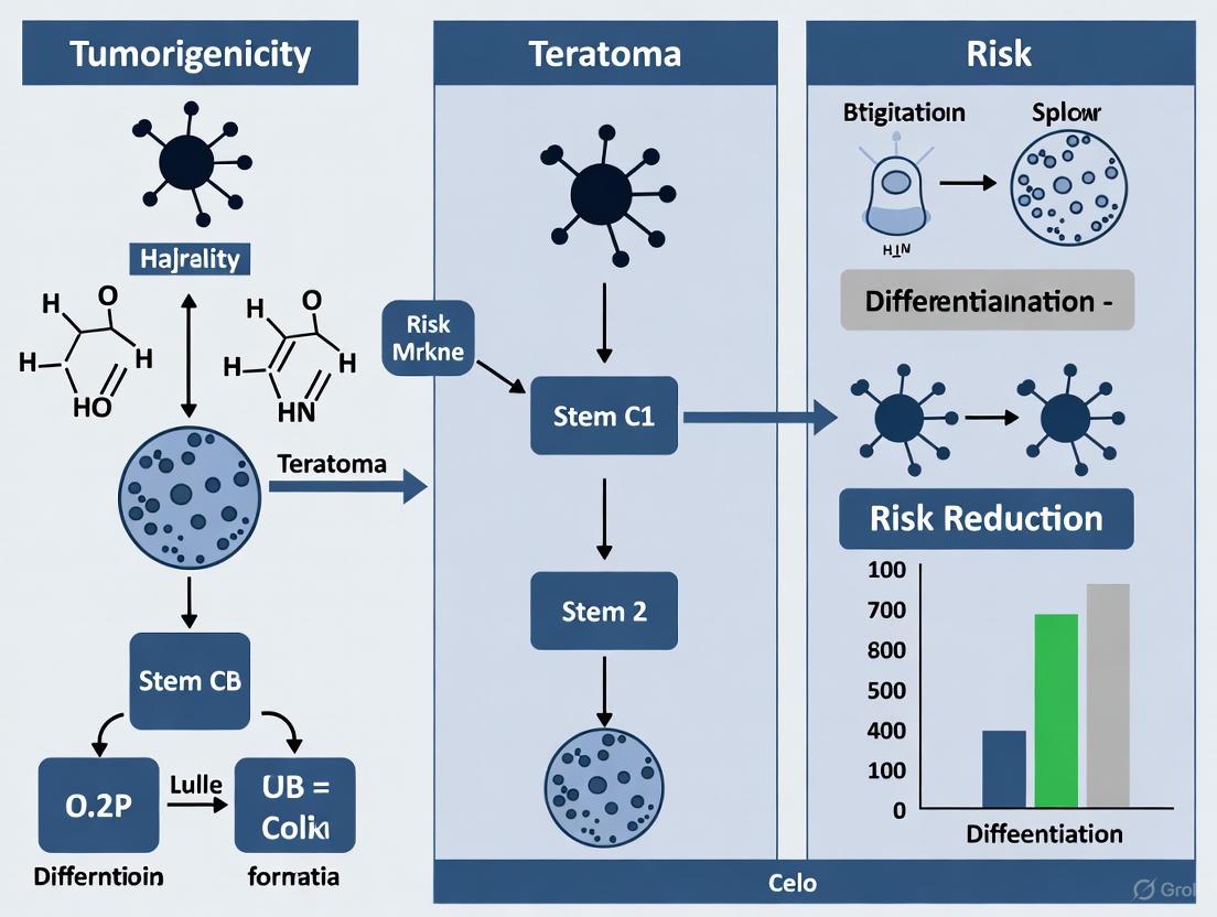
Visualizing the Link: From Pluripotency to Teratoma
The following diagram illustrates the core biological pathway that links the intrinsic property of pluripotency to the formation of a teratoma.
Experimental Protocols for Key Validation Assays
Protocol 1: Standard In Vivo Teratoma Formation Assay
This protocol is used to validate the pluripotency of hPSC lines or to test the tumorigenicity of their therapeutic derivatives [1] [3].
Stem Cell Preparation:
- Expand pluripotent stem cells under conditions that maintain pluripotency (e.g., feeder-free, xeno-free) [3].
- Verify cell quality via pluripotency markers (OCT4, NANOG, SSEA4) before harvesting [3].
- Harvest cells during the logarithmic growth phase. For injection, resuspend cells in an appropriate buffer, often mixed with an ECM like Matrigel to enhance engraftment [1] [3].
Animal Preparation and Injection:
- Use immunocompromised mice (e.g., NOD/SCID, NSG) aged 6-8 weeks, maintained in sterile, pathogen-free housing [1] [3].
- Anesthetize the mouse according to institutional guidelines. Shave and disinfect the injection site (common sites include subcutaneous, intramuscular, or under the testis capsule) [1].
- Load a fine-gauge syringe (e.g., 28.5 gauge insulin syringe) with the cell suspension. The typical cell number ranges from 1x10⁵ to 1x10⁷ per injection site [1] [3].
- Slowly inject the cells into the chosen site. Gently withdraw the needle and apply slight pressure to prevent leakage. Monitor the animal until it recovers from anesthesia [3].
Teratoma Monitoring and Analysis:
- Observe mice weekly. Palpate injection sites for nodules, which are typically visible within 6-12 weeks [3].
- Monitor tumor size with calipers. Pre-define humane endpoints (e.g., tumor diameter of 1.0–1.5 cm) to prevent animal distress [3].
- When endpoints are reached, sacrifice the animal and carefully excise the teratoma.
- Fix the teratoma in 10% formalin for 24-48 hours, then embed in paraffin and section at 5-10 µm thickness [3].
- Stain sections with Hematoxylin and Eosin (H&E) and evaluate under a microscope for the presence of tissues derived from all three germ layers (e.g., neural rosettes for ectoderm; cartilage or muscle for mesoderm; gut-like epithelium for endoderm) [2] [3]. Immunohistochemistry with lineage-specific markers can provide further confirmation.
Protocol 2: Selective Elimination of Undifferentiated hPSCs Using Small Molecules
This protocol is used to pre-treat a differentiated cell population to remove residual pluripotent cells prior to transplantation [5].
Procedure:
- Generate your differentiated cell product from hPSCs using your standard protocol.
- Treat the mixed cell population with a small-molecule inhibitor. For example, a single treatment with Quercetin (which targets the antiapoptotic factor Survivin) can be used [5].
- The treatment selectively induces mitochondrial accumulation of p53 and apoptotic cell death in undifferentiated hPSCs, while sparing differentiated cell types like dopamine neurons and smooth-muscle cells [5].
- After treatment, the differentiated cell product can be washed and prepared for transplantation. This pre-treatment has been shown to be sufficient to prevent teratoma formation in subsequent transplantation experiments [5].
FAQs on Teratoma Composition and Analysis
FAQ 1: What defines a teratoma in the context of pluripotent stem cell research?
A teratoma is a benign tumor composed of tissues representing all three embryonic germ layers—ectoderm, mesoderm, and endoderm—when pluripotent stem cells are xenografted into immunodeficient mice. The ability to form teratomas is a defining characteristic (sine qua non) of pluripotent stem cells and is considered the "gold-standard" assay to confirm pluripotency. These tumors contain complex, disorganized structures with differentiated tissues such as neural tissue (ectoderm), cartilage or muscle (mesoderm), and epithelial structures (endoderm) [9] [10] [11].
FAQ 2: How does the transplantation site affect teratoma formation efficiency and composition?
The site of implantation significantly influences the efficiency of teratoma formation, but histological composition across sites is generally consistent. The table below summarizes key findings from site-dependent studies:
Table: Teratoma Formation Efficiency by Injection Site
| Injection Site | Formation Efficiency | Key Observations | Reference |
|---|---|---|---|
| Kidney Capsule | 100% | Often used for its high efficiency. | [12] |
| Subcutaneous (with Matrigel) | 80-100% | Easy to monitor and remove; larger proportion of solid tissues. | [9] [11] |
| Intratesticular | 60-94% | High efficiency, but slightly longer latency. | [9] [11] |
| Intramuscular | 12.5% | Reported as most convenient and reproducible in one study. | [9] [12] |
While the efficiency and growth latency vary, site-specific differences in the histological composition of the resulting teratomas are generally not observed [9] [11]. Subcutaneous teratomas are often distinct, easier to remove, and cause minimal discomfort to the animal model [9].
FAQ 3: What factors can lead to failed or inconsistent teratoma formation?
Failed teratoma formation can often be traced back to issues with the stem cells or the experimental procedure. Here is a troubleshooting guide based on common problems:
Table: Troubleshooting Teratoma Formation Experiments
| Problem | Potential Cause | Recommended Solution |
|---|---|---|
| No teratoma formation | Low cell viability or insufficient cell number. | Ensure high cell viability (>90%) and use at least 1x10^6 cells per injection [10] [11]. |
| Suboptimal injection site. | Switch to a higher-efficiency site like subcutaneous with Matrigel or kidney capsule [9] [12]. | |
| Excessive differentiation in starting culture | Stem cell culture is not fully undifferentiated. | Maintain cultures by removing differentiated areas before passaging and avoid overgrowth [7]. |
| High variability in teratoma growth | Inconsistent cell handling. | Minimize time culture plates are out of the incubator; ensure cell aggregates for injection are evenly sized [7]. |
| Low cell attachment post-passaging | Over-dissociation of cells. | Reduce pipetting and incubation time with dissociation reagents to avoid generating overly small aggregates [7]. |
FAQ 4: What are the primary strategies to eliminate the tumorigenic risk of residual pluripotent stem cells in cell therapy products?
The primary challenge for clinical applications is that even a few contaminating undifferentiated hPSCs within differentiated derivatives can form teratomas after transplantation [13] [14]. Strategies focus on targeting unique properties of pluripotent cells:
- Genetic Modification with Suicide Genes: A highly specific strategy involves inserting a suicide gene, like the herpes simplex virus thymidine kinase (HSV-TK), into a pluripotency-specific genetic locus such as NANOG via homologous recombination. When exposed to a prodrug like ganciclovir (GCV), only the undifferentiated cells expressing the TK gene are eliminated, leaving the differentiated therapeutic cell population unharmed [13].
- Antibody-Based Cell Depletion: This method uses antibodies against hPSC-specific surface markers (e.g., SSEA-5) to identify and remove undifferentiated cells from a mixed population through immunodepletion or cytotoxic killing [13].
- Small Molecule Inhibitors: Certain small molecules can selectively induce cell death in pluripotent stem cells without harming differentiated progeny, offering a non-genetic alternative for risk reduction [14].
Experimental Protocols for Key Analyses
Protocol 1: Standardized Teratoma Formation Assay
This protocol is adapted from established methods for assessing the pluripotency and tumorigenic potential of human pluripotent stem cells (hPSCs) [9] [11].
- Cell Preparation:
- Culture hPSCs under standard, feeder-free conditions (e.g., on Matrigel-coated plates in mTesR1 medium) to maintain undifferentiated state [13] [7].
- At ~80% confluence, harvest cells using a gentle dissociation reagent like TrypLE or collagenase. Ensure cells are in a single-cell suspension or small, uniform clumps.
- Wash cells twice with PBS and resuspend in an appropriate injection solution, typically PBS mixed with 30% Matrigel, to enhance teratoma formation efficiency [9] [11]. Keep the cell suspension on ice until injection.
- Animal Model and Injection:
- Use immunodeficient mice such as NOD/SCID IL2Rγ⁻⁄⁻ or SCID/beige mice aged 6-8 weeks.
- For subcutaneous injection, slowly inject 1-5 million cells in a 200 µL volume into the hind leg or flank region using a 27-gauge needle [13] [11].
- Monitor animals regularly for teratoma formation. Growth latency typically ranges from 6 to 12 weeks.
- Tissue Harvesting and Analysis:
- Surgically remove teratomas once they reach a predetermined size (e.g., 1.5 cm diameter).
- Fix teratomas in 10% neutral buffered formalin for 24-48 hours.
- Process fixed tissues through a standard ethanol dehydration series, embed in paraffin, and section at 5-10 µm thickness.
- Stain sections with Hematoxylin and Eosin (H&E) for general histology.
- Analyze multiple sections from different parts of the teratoma to identify representative tissues from all three germ layers (e.g., neural rosettes for ectoderm, cartilage for mesoderm, gut-like epithelium for endoderm) [9] [15].
Protocol 2: Validating Pluripotent Cell Elimination via Suicide Gene Strategy
This protocol describes how to test the effectiveness of a genetic safety switch, such as the TK/GCV system, both in vitro and in vivo [13].
- In Vitro Validation:
- Culture genetically modified hPSCs (e.g., NANOG-TK knock-in) alongside wild-type controls.
- Differentiate a portion of the TK-modified cells into the desired cell type (e.g., neural progenitor cells or cardiomyocytes).
- Treat undifferentiated TK-hPSCs, differentiated TK-cell derivatives, and wild-type controls with Ganciclovir (GCV) at varying concentrations (e.g., 1-10 µM) for several days.
- Assess cell viability using assays like ATP-based luminescence or flow cytometry. The expected outcome is selective death of undifferentiated TK-hPSCs, while their differentiated progeny and wild-type cells remain viable [13].
- In Vivo Validation (Preventing Teratoma Formation):
- Inject TK-modified hPSCs subcutaneously into immunodeficient mice as described in Protocol 1.
- One day post-injection, administer GCV to the mice via intraperitoneal injection (e.g., 10 mg/kg/day) for 1-2 weeks.
- Monitor the injection sites for teratoma growth over 12-16 weeks. Effective elimination should result in a significant reduction or complete absence of teratoma formation in the GCV-treated group compared to the untreated control group [13].
- In Vivo Validation (Aborting Established Teratomas):
- Allow teratomas to establish from TK-modified hPSCs for 6-8 weeks.
- Administer GCV for 1-2 weeks.
- Analyze harvested teratomas for extensive apoptosis (e.g., via TUNEL assay) and regression, confirming the functionality of the suicide gene in an in vivo setting [13].
Research Reagent Solutions
The following table details essential materials and reagents used in teratoma formation and analysis studies.
Table: Key Reagents for Teratoma Research
| Reagent / Material | Function in Experiment | Example Usage |
|---|---|---|
| Matrigel | Basement membrane extract; enhances teratoma formation efficiency and provides structural support for injected cells. | Mixed with cell suspension in PBS at 30% concentration for subcutaneous injection [9] [11]. |
| Immunodeficient Mice (e.g., NOD/SCID IL2Rγ⁻⁄⁻) | Host organism that does not reject transplanted human cells, allowing teratoma growth. | Used as the in vivo model for teratoma formation assays [13] [11]. |
| mTeSR1 / mTeSR Plus | Defined, feeder-free culture medium; maintains human PSCs in a pluripotent state. | Used for culturing hPSCs prior to harvesting for injection [13] [7]. |
| Ganciclovir (GCV) | Antiviral prodrug; selectively kills cells expressing the herpes simplex virus thymidine kinase (HSV-TK) suicide gene. | Administered via intraperitoneal injection to mice to eliminate TK-expressing undifferentiated stem cells [13]. |
| Anti-GFP Antibody | Immunohistochemical detection; identifies GFP-tagged cancer cells in co-culture or invasion studies within teratomas. | Used to track invasion of GFP-expressing tumor cells into human teratoma-derived tissues [15]. |
The Scientist's Toolkit: Diagrams and Workflows
Teratoma Assay Workflow
This diagram illustrates the standard experimental workflow for performing a teratoma formation assay.
Safety Strategy: Suicide Gene Mechanism
This diagram outlines the genetic strategy for eliminating residual pluripotent stem cells to enhance the safety of cell therapies.
Frequently Asked Questions (FAQs)
Q1: Is VEGF absolutely essential for all types of teratoma growth? No, the essentiality of VEGF depends on the cellular context. Research demonstrates that teratomas derived from pluripotent stem cells (PSCs), such as embryonic stem cells (ESCs), are highly dependent on VEGF for angiogenesis and growth. Genetically disrupting the VEGF gene in these cells completely abrogates their tumorigenic potential. In contrast, certain oncogene-driven tumors (e.g., from ras or neu-transformed adult fibroblasts) can form aggressive, angiogenic tumors even when they are VEGF-null, utilizing VEGF-independent pathways [16].
Q2: What are the primary mechanisms of VEGF/VEGFR2 signaling in teratoma angiogenesis? VEGF-A binding to VEGFR2 initiates a critical signaling cascade that promotes teratoma angiogenesis. The key steps include:
- Receptor Dimerization & Autophosphorylation: VEGF binding induces VEGFR2 dimerization and autophosphorylation of specific tyrosine residues within its intracellular domain [17] [18].
- Downstream Pathway Activation: This activates major downstream signaling pathways, including:
- RAS/RAF/MEK/ERK: Promotes endothelial cell proliferation and differentiation.
- PI3K/AKT/mTOR: Enhances endothelial cell survival.
- p38/MAPK: Regulates endothelial cell migration and tube formation [18]. These signals drive endothelial cell proliferation, migration, survival, and ultimately, the formation of new blood vessels to support teratoma growth [17] [18].
Q3: What are the best strategies to eliminate residual undifferentiated PSCs and reduce teratoma risk? Multiple strategies have been developed to purge residual teratoma-initiating cells:
- Antibody-Based Cell Removal: Using monoclonal antibodies against cell surface markers highly specific to undifferentiated PSCs (e.g., SSEA-5) for fluorescence-activated cell sorting (FACS) or magnetic-activated cell sorting (MACS) to deplete them prior to transplantation [19].
- Small Molecule Inhibitors: Treating cell populations with inhibitors like YM155, a survivin inhibitor, which selectively kills pluripotent stem cells while sparing differentiated progeny like CD34+ hematopoietic stem cells [20].
- Suicide Gene Strategies: Engineering PSCs to express a suicide gene (e.g., inducible caspase-9) under the control of a pluripotency-specific promoter, allowing for selective elimination of undifferentiated cells upon administration of a prodrug [20].
Q4: Why might anti-VEGF therapies fail, and what are potential combination strategies? Anti-VEGF monotherapy can fail due to compensatory angiogenic signaling. Tumors may upregulate alternative pro-angiogenic factors such as FGF-2 [21]. FGF-2 can recruit pericytes via a PDGFRβ-dependent mechanism, stabilizing vessels and conferring resistance to VEGF inhibition [21]. Combination therapy, such as dual inhibition of VEGF and PDGF signaling, has been shown to overcome this resistance and produce a superior antitumor effect in resistant models [21].
Troubleshooting Experimental Challenges
Challenge: Variable Teratoma Formation in Xenograft Models
- Potential Cause: Inconsistent numbers of residual undifferentiated PSCs in the graft.
- Solutions:
- Implement a rigorous pre-transplantation purification step using validated surface markers (e.g., SSEA-5, TRA-1-60) to remove undifferentiated cells [19].
- Use a highly sensitive assay, such as digital PCR, to quantify residual undifferentiated PSCs in your final cell product to better correlate cell dose with tumorigenic risk [22].
- Standardize the differentiation protocol and use highly efficient culture assays to ensure minimal pluripotent cell contamination [22].
Challenge: Inconsistent Response to VEGFR2 Inhibitor Treatment
- Potential Cause: Activation of alternative pro-angiogenic pathways (e.g., FGF-2, PDGF) bypassing VEGF blockade [16] [21].
- Solutions:
- Profile the tumor's expression of multiple angiogenic factors (VEGF, FGF-2, PDGF) post-treatment to identify potential resistance mechanisms.
- Consider combination therapy targeting multiple pathways simultaneously, such as using a VEGFR2 inhibitor alongside a PDGFRβ inhibitor [21].
Quantitative Data on VEGF and Teratoma Formation
Table 1: Tumorigenicity of VEGF-Deficient Cells in Different Contexts
| Cell Type | Genetic Alteration | VEGF Status | Tumorigenic Outcome | Key Finding |
|---|---|---|---|---|
| Embryonic Stem (ES) Cells | None (Teratoma model) | VEGF -/- | No tumor formation (for at least 50 days) | VEGF is indispensable for ES cell-derived teratoma angiogenesis [16]. |
| Adult Dermal Fibroblasts | Transformed with ras or neu oncogene | VEGF -/- | Aggressive tumor formation (100% take rate) | Oncogene-driven tumorigenesis can proceed via VEGF-independent angiogenesis [16]. |
Table 2: Strategies for Eliminating Teratoma-Forming Cells
| Strategy | Method | Mechanism of Action | Key Advantage |
|---|---|---|---|
| Surface Marker Depletion [19] | FACS/MACS with anti-SSEA-5 | Physical removal of undifferentiated cells | Non-invasive; uses intrinsic cell markers |
| Small Molecule Inhibition [20] | Treatment with YM155 | Inhibits survivin, killing pluripotent cells | More efficient than some suicide genes; low toxicity to HSCs |
| Suicide Gene [20] | iCaspase-9 + AP20187 prodrug | Induces apoptosis in undifferentiated cells | Can be used pre- or post-transplantation |
Experimental Protocols
Protocol: Validating VEGFR2 Signaling Inhibition In Vitro
Objective: To assess the efficacy and specificity of a VEGFR2 inhibitor on downstream signaling pathways in endothelial cells.
Materials:
- Human Umbilical Vein Endothelial Cells (HUVECs)
- VEGFR2 inhibitor (e.g., Sunitinib, Sorafenib)
- Recombinant Human VEGF-A
- Cell culture medium and supplements
- Lysis Buffer (containing protease and phosphatase inhibitors)
- Antibodies for Western Blot: anti-phospho-VEGFR2 (Tyr1175), anti-VEGFR2, anti-phospho-ERK, anti-ERK, anti-β-Actin.
Method:
- Cell Preparation: Seed HUVECs in culture plates and serum-starve them for 4-6 hours to quiesce the cells.
- Inhibitor Pre-treatment: Pre-treat cells with a range of concentrations of the VEGFR2 inhibitor (e.g., 0.1 µM, 1 µM, 10 µM) or a vehicle control (DMSO) for 1-2 hours.
- Stimulation: Stimulate the cells with VEGF-A (e.g., 50 ng/mL) for 10-15 minutes.
- Cell Lysis: Lyse the cells immediately on ice using ice-cold lysis buffer.
- Western Blot Analysis:
- Determine protein concentration of the lysates.
- Separate equal amounts of protein by SDS-PAGE and transfer to a PVDF membrane.
- Probe the membrane with the specified antibodies.
- Key Validation: Successful inhibition is confirmed by a dose-dependent decrease in phosphorylated VEGFR2 and its downstream effector, ERK, in response to VEGF stimulation, without a change in total protein levels.
Protocol: In Vivo Teratoma Assay with Anti-Angiogenic Treatment
Objective: To evaluate the effect of VEGFR2 blockade on the growth and vascularization of PSC-derived teratomas.
Materials:
- Immunodeficient mice (e.g., NOD-SCID or NSG)
- Undifferentiated PSCs or differentiated cell product.
- VEGFR2 neutralizing antibody (e.g., DC101 for mouse models) or small molecule inhibitor.
- Matrigel for cell suspension.
- In vivo imaging system (if using luciferase-expressing cells).
Method:
- Cell Transplantation: Harvest PSCs and resuspend in a 1:1 mixture of culture medium and Matrigel. Inject cells subcutaneously or under the testis capsule of anesthetized mice.
- Treatment Regimen: Randomize mice into treatment and control groups.
- Treatment Group: Administer VEGFR2 blocking agent (e.g., DC101 antibody at 20-40 mg/kg, i.p., twice weekly).
- Control Group: Administer isotype control antibody or vehicle.
- Tumor Monitoring: Monitor tumor formation weekly by palpation and/or bioluminescent imaging. Record tumor volume.
- Endpoint Analysis: At the experimental endpoint, harvest tumors.
- Weigh and measure tumors.
- Process for histology: Fix tumors, embed in paraffin, and section. Perform immunohistochemistry for CD31 (vascular endothelial marker) to quantify microvessel density and assess vascular morphology [16].
Signaling Pathway and Experimental Workflow Diagrams
VEGFR2 Signaling Pathway in Angiogenesis
Experimental Workflow for Validating Anti-Angiogenic Therapy
The Scientist's Toolkit: Key Research Reagents
Table 3: Essential Reagents for Studying VEGF/VEGFR2 in Teratomas
| Reagent / Tool | Function / Target | Example Product(s) | Application in Research |
|---|---|---|---|
| VEGFR2 Neutralizing Antibody | Blocks ligand binding to VEGFR2 | DC101 (anti-mouse VEGFR2) | In vivo inhibition of angiogenesis in mouse teratoma models [16]. |
| VEGFR2 Tyrosine Kinase Inhibitor | Small molecule inhibiting VEGFR2 kinase activity | Sunitinib (Type I), Sorafenib (Type II) | Pharmacological blockade of VEGFR2 signaling; can be used in vitro and in vivo [18]. |
| Anti-Human Pluripotency Marker Antibodies | Binds surface markers on undifferentiated PSCs | Anti-SSEA-5, Anti-TRA-1-60 | Identification and removal of teratoma-initiating cells via FACS/MACS [19]. |
| Anti-CD31 (PECAM-1) Antibody | Labels vascular endothelial cells | Anti-CD31 for IHC/IF | Histological quantification of tumor microvessel density (angiogenesis) [16]. |
| Survivin Inhibitor | Selectively induces apoptosis in PSCs | YM155 | Purging residual undifferentiated cells from differentiated cell populations pre-transplantation [20]. |
| Recombinant VEGF Protein | Activates VEGFR2 signaling | Recombinant Human VEGF-A165 | Positive control for stimulating angiogenesis in in vitro assays. |
FAQ: Ethical Considerations in Stem Cell Research
Q1: What are the primary ethical advantages of using iPSCs over ESCs in research? The primary ethical advantage of induced pluripotent stem cells (iPSCs) over embryonic stem cells (ESCs) is that their generation does not require the destruction of human embryos, which is a central point of ethical debate for ESCs [23] [24]. Furthermore, iPSC technology avoids the ethical concerns related to egg donation, including the health risks to women from invasive procedures and issues regarding appropriate compensation for donors [24]. This removes a significant burden from women and circumvents the ethical debate about the commodification of human body parts [24].
Q2: Do iPSCs fully resolve all ethical issues associated with stem cell research? No, the generation of iPSCs does not resolve all ethical issues. While they circumvent concerns about embryo destruction, they introduce other ethical considerations [24] [25]. These include:
- Potential for Human Cloning: If iPSCs were reprogrammed to full embryonic potential, the technology could be used for reproductive cloning [24].
- Genetic Manipulation: The use of genetically altered cells in therapies raises concerns about unintended consequences of modifying the human genome [24] [25].
- Chimera Formation: The use of iPSCs to create chimeras (organisms with cells from different species) raises ethical questions about the boundaries of scientific intervention in nature [24].
- Justice and Access: There are concerns about the equitable distribution of and access to potentially expensive iPSC-derived therapies [24].
Q3: What is the significance of the NANOG locus in improving the safety of stem cell therapies? The NANOG locus is highly specific to pluripotent stem cells and is rapidly downregulated when these cells differentiate [13] [4]. This makes it an ideal genetic "safe harbor" for introducing suicide genes. By placing an inducible suicide gene, such as herpes simplex virus thymidine kinase (TK) or inducible Caspase 9 (iCasp9), into this locus via homologous recombination, researchers can create stem cell lines where undifferentiated cells—and only undifferentiated cells—can be selectively eliminated before or after transplantation, thereby preventing teratoma formation [13] [4].
Troubleshooting Guide: Teratoma Risk Mitigation
Problem: Teratoma Formation from Residual Undifferentiated PSCs in Therapeutic Cell Products
Background: Even a small number of residual undifferentiated pluripotent stem cells (PSCs) within a differentiated cell product can lead to teratoma formation after transplantation. This is a major safety hurdle for clinical applications [26] [4].
Solution 1: Genetic Modification with a Suicide Gene This strategy involves engineering a "safety switch" into the PSCs that allows for the selective elimination of undifferentiated cells.
- Experimental Protocol:
- Targeting Vector Design: Construct a targeting vector containing a suicide gene cassette (e.g., HSV-TK or iCasp9) flanked by long homology arms specific to the 3'-untranslated region (UTR) of the endogenous NANOG gene [13] [4].
- Stem Cell Transfection: Introduce the targeting vector into your pluripotent stem cell line (e.g., via electroporation).
- Selection and Screening: Select for successfully transfected cells (e.g., using puromycin resistance if included in the cassette). Screen clones for correct homologous recombination at the NANOG locus using techniques like Southern blotting or PCR [13].
- Validation: Validate that the engineered cell line maintains normal pluripotency, karyotype, and differentiation potential [4].
- Application:
- In vitro: Differentiate the engineered PSCs into your desired therapeutic cell type. Before transplantation, treat the cell population with the pro-drug ganciclovir (if using TK) or the small molecule AP20187 (if using iCasp9) to activate the suicide gene and eliminate any remaining NANOG-positive, undifferentiated PSCs [13] [4].
- In vivo: If a teratoma is detected post-transplantation, administering the pro-drug can eliminate the contaminating pluripotent cells [13].
Solution 2: Small Molecule-Based Depletion This approach uses small molecules that are selectively toxic to undifferentiated PSCs.
- Experimental Protocol:
- Cell Culture: Establish your differentiated cell population from PSCs.
- Compound Administration: Treat the mixed cell culture with a small molecule that targets undifferentiated cells. Examples include the survivin inhibitor YM155, though its specificity should be verified for your specific cell types [4].
- Dosage and Timing Optimization: Determine the optimal concentration and duration of treatment that maximizes the killing of undifferentiated PSCs while minimizing damage to the differentiated therapeutic cells. This requires careful dose-response assays [4].
- Validation: After treatment, assess the viability of both the differentiated target cells and the undifferentiated PSCs. Use flow cytometry for pluripotency markers (e.g., TRA-1-60, SSEA4) to confirm the depletion of undifferentiated cells [26].
Diagram: Suicide Gene Strategy for Teratoma Prevention
This diagram illustrates the genetic strategy for eliminating undifferentiated pluripotent stem cells to prevent teratoma formation.
Problem: Assessing Pluripotency and Tumorigenicity of Novel Cell Lines
Background: Researchers developing new PSC lines or differentiation protocols need robust methods to validate pluripotency and rule out malignancy.
Solution: Standardized Teratoma Assay The teratoma assay is the gold standard for testing pluripotency and assessing malignancy potential in vivo [27] [28].
- Experimental Protocol:
- Cell Preparation: Harvest undifferentiated PSCs. A typical injection uses 5-10 million cells resuspended in Matrigel/PBS [27].
- Animal Model: Use immunodeficient mice (e.g., NOD/SCID, Rag2-/-;γc-/-). The injection site (subcutaneous, intramuscular, under the kidney capsule) should be reported as it can influence assay outcomes [27] [28].
- Monitoring and Endpoint: Monitor mice for teratoma formation. The assay typically runs for 8-12 weeks, or until tumors reach a predefined size limit. Record the time to tumor appearance and growth kinetics [27].
- Histological Analysis: Extract teratomas, fix, section, and stain with Hematoxylin and Eosin (H&E). A qualified pathologist should examine the sections for the presence of differentiated tissues from all three germ layers (ectoderm, mesoderm, endoderm) to confirm pluripotency. The teratoma should also be scrutinized for "embryonal carcinoma elements" or undifferentiated tissues, which indicate malignancy [28].
Troubleshooting Notes:
- Lack of Standardization: The teratoma assay suffers from a lack of standardization across labs. It is critical to report detailed methods, including the animal strain, sex, age, number of cells injected, injection site, and assay duration to ensure reproducibility [28].
- In Vitro Alternatives: In vitro assays like ScoreCard and PluriTest can assess pluripotency but are generally not accepted by regulatory authorities as standalone tests for the safety of clinical-grade cell products. They are useful for preliminary screening [28].
Research Reagent Solutions
Table: Key Reagents for Teratoma Risk Reduction Strategies
| Reagent / Tool | Function / Application | Key Consideration |
|---|---|---|
| NANOG-targeting Vector [13] [4] | Knocks in suicide gene into the NANOG locus for specific targeting of undifferentiated PSCs. | High specificity for pluripotent state; use homologous recombination for precise genomic integration. |
| Inducible Caspase 9 (iCasp9) [4] | Safety switch; dimerizing drug AP20187 induces apoptosis in cells expressing the construct. | Highly potent and sensitive; allows for >1 million-fold depletion of undifferentiated hPSCs. |
| Herpes Simplex Virus Thymidine Kinase (HSV-TK) [13] | Safety switch; converts prodrug ganciclovir into a toxic compound, killing dividing cells. | Well-established system; FDA-approved drugs available for activation. |
| Small Molecule Inhibitors (e.g., YM155) [4] | Selectively toxic to undifferentiated PSCs in a mixed culture. | Must be rigorously validated for specificity to avoid harming differentiated cell product. |
| Immunodeficient Mice (e.g., NOD/SCID) [27] [28] | In vivo host for teratoma assay to validate pluripotency and assess tumorigenicity. | Strain, cell number, and injection site can affect results and require standardization. |
| Pluripotency Markers (TRA-1-60, SSEA-4) [28] | Flow cytometry or immunocytochemistry to identify and quantify residual undifferentiated PSCs. | Essential for quality control before and after safety procedures. |
FAQ: What is the fundamental clinical threat posed by residual undifferentiated hPSCs?
Residual undifferentiated human pluripotent stem cells (hPSCs) present a direct risk of iatrogenic tumorigenesis in cell therapy recipients. The core of the threat is their capacity to form teratomas—benign tumors containing tissues from all three embryonic germ layers. These tumors can arise from even very small numbers of undifferentiated cells that contaminate a differentiated cell product intended for therapy. Studies have demonstrated that the systemic injection of hPSCs can produce multisite teratomas in immune-deficient animal models in as little as 5 weeks [20]. This risk is a major limitation hindering the widespread clinical application of hPSC-derived therapies across various medical fields, including hematology, neurology, and diabetes treatment [20] [14] [29].
FAQ: How can a small number of residual cells cause a tumor?
hPSCs are defined by their abilities of self-renewal and pluripotency. Unlike therapeutic differentiated cells, which have a limited lifespan and a defined function, undifferentiated hPSCs retain the capacity for unlimited division and can generate any cell type in the body. When transplanted into a permissive in vivo environment, a single residual hPSC can clonally expand and spontaneously differentiate in a disorganized fashion, leading to the formation of a teratoma. The minimum number of cells required to form a teratoma can be as low as 10,000 cells, and since clinical doses can be in the range of billions of cells, even a contamination level of 0.001% could be therapeutically unacceptable [4] [29].
FAQ: What are the primary strategies to eliminate residual hPSCs?
Researchers have developed multiple strategies to purge undifferentiated hPSCs from final cell products. The table below summarizes the main approaches:
Table 1: Strategies for Eliminating Residual Undifferentiated hPSCs
| Strategy | Mechanism | Key Example(s) | Advantages & Limitations |
|---|---|---|---|
| Physical Separation | Sorting cells based on pluripotency-specific surface antigens (e.g., SSEA-4, TRA-1-60). | Flow cytometry or magnetic-activated cell sorting (MACS) [20]. | Limitation: Can affect cell viability; results affected by gating; not all pluripotent cells may express the marker [20]. |
| Suicide Gene Therapy | Genetically engineering hPSCs with a "kill-switch" activated by a small molecule. | iCaspase-9/AP20187: Induces apoptosis in cells expressing the transgene [20] [4]. | Advantage: Can be very specific and efficient.Limitation: Requires genetic modification; potential toxicity of prodrugs to therapeutic cells (e.g., CD34+ HSCs) [20]. |
| Small Molecule Inhibition | Using chemicals that selectively induce apoptosis in pluripotent cells. | YM155 (Survivin inhibitor): Targets survivin, a protein hPSCs rely on for survival [20]. | Advantage: High efficiency; no genetic modification needed; shown to be non-toxic to some therapeutic cells like CD34+ HSCs [20]. |
| Genome-Edited Safeguards | Inserting drug-inducible safety switches into endogenous pluripotency genes. | NANOG-iCaspase9: Knocks-in iCaspase9 into the NANOG locus, which is highly specific to the pluripotent state [4]. | Advantage: Highly specific (>1 million-fold depletion); cannot be silenced without loss of pluripotency [4]. |
FAQ: How do we detect and quantify residual undifferentiated cells for quality control?
Sensitive and specific assays are critical for quantifying residual hPSCs in a final cell product to assess teratoma risk. The required sensitivity is extremely high, needing to detect as few as 1 undifferentiated cell in 1,000,000 to 100,000,000 differentiated cells [29]. The table below compares key detection methodologies.
Table 2: Methods for Detecting Residual Undifferentiated hPSCs
| Method | Principle | Sensitivity | Throughput & Notes |
|---|---|---|---|
| In Vivo Teratoma Assay | Injecting cells into immunodeficient mice and monitoring for tumor formation [29]. | High (biological readout) | Time-consuming (weeks to months), costly, and low-throughput. Used as a gold-standard functional test [29]. |
| Flow Cytometry | Detecting cell surface markers of pluripotency (e.g., TRA-1-60) via antibodies [29]. | ~0.01% | Rapid but limited sensitivity. Affected by gating strategies and marker specificity [29] [30]. |
| RT-qPCR | Measuring mRNA levels of pluripotency-associated genes. | ~0.01% [30] | Moderately sensitive and high-throughput. Sensitivity can be insufficient for large cell doses; relies on good marker genes [29] [30]. |
| Droplet Digital PCR (ddPCR) | Absolutely quantifying nucleic acid copies by partitioning a sample into thousands of droplets. | ~0.001% (1 in 105 cells) or better [29] [30] | Highly sensitive, precise, and reproducible. Ideal for quality control. Targets can include LIN28A or specific long non-coding RNAs (lncRNAs) like LNCPRESS2 [29] [30]. |
| High-Efficiency Culture (HEC) | Culturing the cell product under conditions that favor hPSC growth over differentiated cells. | Can reach 0.00002% with MACS pre-enrichment [29] | Very sensitive but labor-intensive and time-consuming (weeks in culture) [29]. |
Experimental Protocol: Detection of Residual hiPSCs using LIN28/ddPCR
This protocol allows for the highly sensitive quantification of residual undifferentiated hiPSCs in a background of differentiated cells, such as cardiomyocytes [30].
- Sample Preparation: Prepare a mixture of your hPSC-derived cell product. As a control, create a standard curve by spiking known numbers of undifferentiated hiPSCs (e.g., 10, 100, 1000 cells) into a fixed number of the target differentiated cell type (e.g., 1 x 10^6 cells).
- RNA Extraction: Isolate total RNA from the test sample and standard curve samples using a standard phenol-chloroform extraction method (e.g., TRIzol) or a commercial kit. Treat samples with DNase to remove genomic DNA contamination.
- Reverse Transcription: Convert equal amounts of total RNA (e.g., 500 ng) into cDNA using a reverse transcriptase enzyme and oligo(dT) or random hexamer primers.
- Droplet Digital PCR (ddPCR):
- Prepare a 20 µL reaction mixture containing the cDNA template, ddPCR supermix, and a TaqMan probe and primer set specific for LIN28A (or another highly specific marker like an lncRNA).
- Generate droplets from the reaction mixture using a droplet generator (~20,000 droplets per sample).
- Perform PCR amplification on the droplet emulsion using the following cycling conditions (optimized for the LIN28-1 probe/primer set [30]):
- 95°C for 10 minutes (enzyme activation)
- 40 cycles of:
- 94°C for 30 seconds (denaturation)
- 64°C for 60 seconds (annealing/extension)
- 98°C for 10 minutes (enzyme deactivation)
- 4°C hold
- Droplet Reading and Analysis: Read the droplets using a droplet reader that measures fluorescence in each droplet. The software will count the number of fluorescence-positive (containing the target) and negative droplets.
- Quantification: The concentration of the target LIN28A mRNA in the original sample is calculated automatically by the software using Poisson statistics. The number of residual undifferentiated cells can be extrapolated from the standard curve.
FAQ: Are markers like OCT4 and NANOG sufficient to define pluripotency and risk?
No. While markers like OCT4 (POU5F1) and NANOG are strongly associated with the undifferentiated state, their expression alone does not demonstrate functional pluripotency. It is critical to understand that:
- They are markers of the "undifferentiated state," not "pluripotency." Nullipotent cells (which cannot differentiate) from certain tumors can also express these markers [31].
- They can be expressed in some differentiated cell types, leading to potential false positives if used for purging [4].
- Functional pluripotency must be demonstrated through rigorous differentiation assays that show the ability to generate cells of all three germ layers [31]. For safety purposes, the most reliable systems for eliminating hPSCs use genes with expression highly restricted to the pluripotent state, such as NANOG, for driving suicide genes [4].
The Scientist's Toolkit: Key Research Reagents
Table 3: Essential Reagents for Studying hPSC Tumorigenicity Risk
| Reagent / Tool | Function in Research | Example & Key Detail |
|---|---|---|
| Survivin Inhibitor | Selective small molecule for eliminating hPSCs by inducing apoptosis. | YM155: A chemical survivin inhibitor; shown to be more efficient than suicide gene/prodrug systems and non-toxic to human CD34+ hematopoietic stem cells [20]. |
| Inducible Caspase-9 (iCaspase-9) System | Genetically encoded suicide gene for targeted cell ablation. | AP20187: The small molecule dimerizer drug that activates iCaspase-9, triggering rapid apoptosis in cells expressing the transgene [20] [4]. |
| hPSC-Specific Marker Antibodies | Identification and depletion of undifferentiated cells via flow cytometry. | Anti-TRA-1-60 & Anti-SSEA-4: Antibodies against common surface markers used for fluorescence-activated cell sorting (FACS) or magnetic-activated cell sorting (MACS) [20] [29]. |
| Digital PCR Reagents | Ultra-sensitive detection and absolute quantification of residual hPSCs. | LIN28A TaqMan Assay: Probe and primer set for quantifying the pluripotency-associated gene LIN28A via ddPCR. Can detect 1 hiPSC in 100,000 cardiomyocytes [30]. |
| Long-Term Culture Media | Enrichment and expansion of potential residual hPSCs from a cell product. | Laminin-521 & Essential 8 Medium: Components of a high-efficiency culture (HEC) system used to detect rare residual hPSCs by providing an optimal environment for their growth [29]. |
Visual Guide: The Pathway from Residual Cell to Clinical Risk
The following diagram illustrates the core safety problem and the strategic points for intervention.
Visual Guide: Mechanism of a Genome-Edited Safety Switch
This diagram details the structure and function of a sophisticated genetic safeguard against teratoma formation.
Advanced Assays for Teratoma Risk Assessment: From In Vivo to In Vitro Platforms
The SCID mouse teratoma assay remains a cornerstone for evaluating the pluripotency and tumorigenic risk of human Pluripotent Stem Cells (hPSCs) and their derivatives in regenerative medicine. By transplanting hPSCs into immunodeficient mice, this assay tests their ability to form teratomas—benign tumors containing tissues from all three embryonic germ layers (ectoderm, mesoderm, and endoderm) [32] [33]. For cell therapy products, it is a critical biosafety test to ensure that no residual undifferentiated cells with tumorigenic potential remain in a differentiated cell population destined for clinical use [22] [34]. Despite its status as a historical "gold standard," the assay faces significant challenges regarding standardization, sensitivity, and ethical use of animals, driving the development of robust in vitro alternatives [33] [35] [36].
Frequently Asked Questions (FAQs) & Troubleshooting
FAQ 1: What is the minimum number of cells required to form a teratoma, and how can I improve the sensitivity of my assay?
The minimum number of cells needed can vary significantly based on the assay protocol. A standardized subcutaneous assay co-injecting hESCs with mitotically inactivated feeders and Matrigel has demonstrated high sensitivity [34].
Table 1: Teratoma Formation Sensitivity Based on Cell Number
| Number of hESCs Injected | Teratoma Formation Efficiency | Time to Teratoma Formation |
|---|---|---|
| 5 x 10⁵ and 1 x 10⁵ | 100% efficiency | 6-8 weeks [34] |
| 1 x 10³ | Variable | Longer follow-up required [34] |
| 1 x 10² | Possible, but inconsistent | Requires larger number of animals and extended follow-up (>12 weeks) [34] |
Troubleshooting Low Engraftment:
- Problem: Low teratoma formation rates, especially with low cell numbers.
- Solution:
- Use a Support Matrix: Co-inject cells with Matrigel or similar basement membrane extracts. This provides a scaffold that enhances cell survival and engraftment [32] [34].
- Co-transplant with Feeders: Mitotically inactivated feeder cells (e.g., Mouse Embryonic Fibroblasts - MEFs) provide crucial supportive signals for undifferentiated hPSCs. This has been shown to significantly improve the assay's sensitivity [34].
- Add ROCK Inhibitor: Supplement the cell suspension with a Rho-associated coiled-coil kinase (ROCK) inhibitor (e.g., Y-27632) prior to transplantation. This increases hPSC survival after single-cell dissociation [34].
- Optimize Injection Site: The intra-muscular route (e.g., gastrocnemius muscle) is highly vascularized and easy to access, leading to high teratoma formation efficiency (95-100%) [32].
FAQ 2: My teratoma assay results are inconsistent between experiments. How can I standardize my protocol?
Lack of standardization is a widely recognized issue that compromises data comparability [35] [36]. Key variables to control are listed in the table below.
Table 2: Key Parameters for Teratoma Assay Standardization
| Parameter | Recommended Standardization | Impact on Variability |
|---|---|---|
| Cell Preparation | Use a consistent dissociation method. Define cell viability thresholds. Pre-treat with ROCK inhibitor [34]. | Cell health and aggregation affect engraftment. |
| Injection Site | Choose one site (e.g., subcutaneous, intramuscular) and stick to it across all experiments [32] [34]. | Different sites have varying vascularization and microenvironmental cues, affecting teratoma growth and composition [33]. |
| Cell Number | Use a defined, quantified cell number for injection. For sensitivity assays, a titration series is recommended [34]. | The primary variable for determining tumorigenic potential. |
| Support Matrix | Always use the same lot and concentration of Matrigel or equivalent [32] [34]. | Provides a consistent extracellular environment for cell growth. |
| Mouse Strain | Use immunodeficient mice with a consistent genetic background (e.g., NOD/SCID, NSG). Report the strain used [32] [36]. | Different strains have varying levels of immune leakage, affecting teratoma acceptance and growth. |
| Assay Duration | Monitor until teratoma reaches ~1 cm³ or for a pre-defined period (e.g., 12-20 weeks). Do not allow overgrowth [32] [33]. | Under-grown teratomas may not show all germ layers; over-growth causes animal distress. |
FAQ 3: What are the definitive criteria for a successful pluripotency assay, and how do I distinguish a teratoma from a malignant tumor?
A successful assay for pluripotency requires definitive histological evidence of tissues derived from all three embryonic germ layers [32] [34].
- Ectoderm: Look for neural tissues (e.g., rosettes, pigmented cells, neural epithelium) or stratified squamous epithelium [32].
- Mesoderm: Look for cartilage, bone, muscle (smooth or striated), or adipose tissue [32].
- Endoderm: Look for respiratory epithelium (ciliated), intestinal epithelium (with goblet cells), or glandular structures [32].
Troubleshooting Malignancy Concerns:
- Problem: Distinguishing a benign teratoma from a malignant teratocarcinoma.
- Solution: A true teratoma is a benign, multi-layered tumor. The presence of embryonal carcinoma (EC) elements—poorly differentiated, rapidly dividing cells that resemble embryonic carcinoma cells—indicates malignancy and classifies the tumor as a teratocarcinoma [33] [36]. These EC elements are highly undifferentiated and can be metastatic. Their presence in a cell therapy product would lead to its exclusion from clinical use [33]. Histopathological analysis must be performed by a trained pathologist to make this critical distinction.
FAQ 4: Are there animal-free alternatives to the teratoma assay that are accepted by regulators?
While the teratoma assay is still required by many regulatory authorities for final safety assessment of clinical products, several in vitro alternatives are gaining traction for research and characterization [22] [36].
Troubleshooting the Need for an Alternative:
- Problem: The teratoma assay is time-consuming, costly, raises ethical concerns, and is poorly standardized.
- Solution: Implement orthogonal in vitro assays to complement or, in some contexts, replace the in vivo assay.
- Highly Efficient Culture Assays (HEC): These sensitive in vitro assays can detect very low numbers of residual undifferentiated hPSCs within a differentiated population, often with superior sensitivity compared to in vivo assays [22].
- Molecular Assays: Digital PCR (dPCR) can be used to detect and quantify RNA transcripts specific to undifferentiated hPSCs, offering a highly sensitive and quantitative readout for residual pluripotent cells [22].
- Bioinformatic Assays: Tools like PluriTest use transcriptomic data from the test cell line to compute a "pluripotency score" and a "novelty score" by comparing it to a large reference database of validated pluripotent cell lines [35] [36]. ScoreCard is another assay that quantitatively evaluates the differentiation potential by analyzing germ layer-specific gene expression patterns after spontaneous or directed differentiation [35] [36].
Experimental Workflow & Decision Pathway
The following diagram illustrates the key steps in performing a standardized teratoma assay and the critical decision points for data interpretation.
Research Reagent Solutions
Table 3: Essential Materials for a Standardized Teratoma Assay
| Reagent / Material | Function | Key Considerations |
|---|---|---|
| Immunodeficient Mice (e.g., NOD/SCID, NSG) | In vivo host for teratoma formation. Prevents immune rejection of human cells [32] [34]. | Maintain in specific pathogen-free (SPF) conditions. Strain choice impacts engraftment efficiency. |
| Basement Membrane Matrix (e.g., Matrigel) | Extracellular matrix scaffold. Enhances cell survival, engraftment, and teratoma formation [32] [34]. | Keep on ice to prevent polymerization. Use consistent lots for reproducibility. |
| ROCK Inhibitor (e.g., Y-27632) | Small molecule that inhibits Rho-associated kinase. Dramatically improves survival of dissociated hPSCs [34]. | Add to cell suspension medium prior to injection. |
| Mitotically Inactivated Feeders (e.g., MEFs) | Provide essential supportive signals for the survival and growth of undifferentiated hPSCs post-transplantation [34]. | Co-injection significantly increases assay sensitivity. |
| Defined hPSC Culture Media | Maintains cells in a pluripotent state prior to harvest and injection. | Use consistent, high-quality media to ensure cell health and genetic stability. |
| Histology Reagents (Paraformaldehyde, Paraffin, H&E Stain) | For tissue preservation, sectioning, and staining to visualize teratoma morphology and germ layer differentiation [32]. | Essential for final analysis. H&E is the standard stain for initial germ layer identification. |
The SCID mouse teratoma assay remains a critical, though imperfect, tool for assessing the functional pluripotency and tumorigenic risk of hPSCs. Its successful implementation hinges on careful standardization of protocols, particularly in cell preparation, injection method, and histological analysis. While it continues to be a requirement for the safety assessment of clinical-grade cell products, the field is actively developing and validating sophisticated in vitro molecular and bioinformatic assays [22] [35]. These alternatives promise to reduce animal use, increase throughput, and improve quantitative accuracy, ultimately supporting the safer clinical translation of stem cell-based therapies.
This technical support resource addresses key challenges in detecting residual human pluripotent stem cells (hPSCs) in therapeutic products. The content focuses on applying digital PCR (dPCR) for sensitive identification of hPSC-specific RNA biomarkers to mitigate teratoma formation risk, a major safety concern in regenerative medicine. The guidance is framed within the context of stem cell tumorigenicity and teratoma formation risk reduction research.
Frequently Asked Questions
FAQ 1: What are the key advantages of digital PCR over qPCR for residual hPSC detection?
Digital PCR (dPCR) provides several critical advantages for detecting trace levels of undifferentiated hPSCs in differentiated cell products.
- Absolute Quantification Without Standards: dPCR does not require a standard calibration curve, enabling direct nucleic acid concentration measurement. This eliminates variability introduced by external calibrators, which can be unstable and cause day-to-day variability in qPCR [37] [38].
- Superior Sensitivity and Tolerance: dPCR is more robust against PCR inhibitors present in complex biological samples and offers high sensitivity, which is crucial for detecting extremely rare events like residual hPSCs [37] [39].
- Precision for Rare Targets: By partitioning a sample into thousands of individual reactions, dPCR allows for the detection of a single molecule of the target RNA, making it ideal for identifying minute quantities of pluripotency markers amidst a background of differentiated cells [38] [40].
FAQ 2: Which RNA biomarkers are most effective for sensitive hPSC detection?
The most effective biomarkers are those with high expression in hPSCs and near-undetectable expression in the differentiated cell therapy product. The optimal choice can depend on the specific hPSC line and its differentiated progeny.
- Traditional Protein-Coding Genes: LIN28 is a highly sensitive marker. A ddPCR assay using LIN28 was able to detect 0.001% undifferentiated hPSCs spiked into cardiomyocytes [40].
- Long Non-Coding RNAs (lncRNAs): Emerging research identifies lncRNAs as excellent biomarkers due to their often higher specificity. A 2023 study identified LNCPRESS2, LINC00678, and LOC105370482 as highly specific markers for human chemically induced PSCs (hCiPSCs). A ddPCR assay using these could detect 1 to 3 residual hPSCs in a background of 1 million islet cells [29].
- Marker Selection Strategy: A 2025 review emphasizes that biomarker expression must be rigorously validated across multiple hPSC lines and the final differentiated cell product to ensure specificity. RNA-seq is a powerful tool for discovering differentially expressed transcripts [22] [29].
Table 1: Comparison of Key Biomarkers for Residual hPSC Detection via dPCR
| Biomarker | Type | Reported Sensitivity | Key Advantage |
|---|---|---|---|
| LIN28 | mRNA | 0.001% (1 in 10⁵ cells) [40] | Well-characterized, high expression in hPSCs |
| LNCPRESS2 | lncRNA | 0.0001% (1 in 10⁶ cells) [29] | High specificity, low expression in differentiated cells |
| LINC00678 | lncRNA | 0.0001% (1 in 10⁶ cells) [29] | High specificity, low expression in differentiated cells |
| NANOG | mRNA | Varies with assay | Core pluripotency factor; requires careful validation for specificity [4] |
FAQ 3: What is a detailed protocol for setting up a ddPCR assay for hPSC-specific lncRNAs?
The following protocol is adapted from a 2023 study that successfully detected residual hCiPSCs in derived islet cells [29].
Workflow: Detection of Residual hPSCs using ddPCR
Step-by-Step Methodology:
Sample Preparation and RNA Extraction:
cDNA Synthesis:
- Reverse-transcribe the extracted RNA into complementary DNA (cDNA) using a High-Capacity cDNA Reverse Transcription Kit.
- Use random hexamer primers to ensure comprehensive conversion of all RNA species, including lncRNAs [29].
ddPCR Reaction Setup:
- Prepare the ddPCR reaction mix containing:
- ddPCR Supermix for Probes (no dUTP).
- Target-specific FAM-labeled TaqMan assay (primers and probe) for the lncRNA (e.g., LNCPRESS2).
- A HEX-labeled assay for a reference gene for normalization (if required).
- The synthesized cDNA template.
- A sample volume of 20 µL is standard for systems like the QX200 Droplet Digital PCR system [29] [40].
- Prepare the ddPCR reaction mix containing:
Droplet Generation and PCR Amplification:
Droplet Reading and Data Analysis:
- Place the plate in a droplet reader, which counts the fluorescent-positive and negative droplets for each sample.
- The concentration of the target lncRNA (in copies/µL) is calculated automatically from the fraction of positive droplets using Poisson statistics [37] [38].
- The result can be used to back-calculate the number of residual hPSCs in the original sample based on the measured lncRNA copies per cell.
FAQ 4: How can I troubleshoot high background noise or false positives in my dPCR assay?
High background can arise from several sources. The table below outlines common issues and solutions.
Table 2: Troubleshooting Guide for dPCR Assays in hPSC Detection
| Problem | Potential Cause | Solution |
|---|---|---|
| High false-positive droplet count | Non-specific amplification or probe degradation [39]. | Redesign primers/probe to improve specificity; aliquot and store probes correctly. |
| Poor partition resolution (rain) | Suboptimal PCR efficiency or inhibitor carryover [37]. | Re-optimize annealing temperature; ensure high-quality, inhibitor-free RNA/cDNA. |
| Inconsistent results between replicates | Inaccurate droplet generation or pipetting errors [38]. | Check droplet generator for faults; use calibrated pipettes and master mixes. |
| Low or no positive signal | cDNA synthesis failure or incorrect assay design for the specific hPSC line. | Check cDNA synthesis with a positive control; validate biomarker expression in your cells [22] [29]. |
FAQ 5: What are the critical validation steps for ensuring an accurate and sensitive dPCR assay?
Robust assay validation is required for quality control. Key steps include:
- Limit of Detection (LOD) and Sensitivity: Determine the lowest number of hPSCs that can be reliably detected. Perform spike-in experiments where known numbers of hPSCs (e.g., 1, 10, 100) are mixed with a large number of differentiated cells (e.g., 10^6 or 10^7) and processed through the entire workflow. This confirms the assay's ability to detect contaminants at the 0.0001% level [22] [29].
- Specificity Testing: Verify that the signal from your biomarker is specific to undifferentiated hPSCs. Test the assay on RNA extracted from the final differentiated cell product (with no residual hPSCs) to ensure no background signal. Also, test on various other cell types that might be present [4] [29].
- Linearity and Dynamic Range: Create a standard curve by testing serial dilutions of hPSC RNA in differentiated cell RNA. The measured concentration should correlate linearly with the expected input [22].
The Scientist's Toolkit
Table 3: Essential Research Reagent Solutions for dPCR-based hPSC Detection
| Item | Function/Description | Example Product/Catalog |
|---|---|---|
| Droplet Digital PCR System | Platform for partitioning samples, amplifying targets, and reading results. | Bio-Rad QX200 Droplet Digital PCR System [37] |
| ddPCR Supermix for Probes | Optimized reaction mix for probe-based assays in droplet formats. | Bio-Rad ddPCR Supermix for Probes (no dUTP) |
| TaqMan Assay for lncRNA | Primer and probe set designed specifically for the target hPSC biomarker. | Custom-designed assays for LNCPRESS2, LINC00678, etc. [29] |
| RNA Extraction Kit | For high-quality, contaminant-free total RNA isolation from cell pellets. | PureLink RNA Mini Kit [29] [39] |
| cDNA Synthesis Kit | For efficient reverse transcription of RNA to cDNA, including lncRNAs. | High-Capacity cDNA Reverse Transcription Kit [29] |
Technology Comparison and Decision Framework
The following diagram illustrates the position of dPCR among other key methodologies for detecting residual hPSCs, highlighting its role in balancing sensitivity, specificity, and practicality.
The Highly Efficient Culture (HEC) assay represents a significant advancement in the safety assessment of human pluripotent stem cell (hPSC)-derived therapies. As hPSCs hold immense promise for regenerative medicine due to their ability to differentiate into any cell type, they simultaneously pose a substantial safety risk through potential teratoma formation if residual undifferentiated cells remain in cell therapy products (CTPs) [22] [41]. The HEC assay has been developed as a robust in vitro method to detect these residual hPSCs with superior sensitivity and reproducibility compared to traditional in vivo teratoma assays [42] [6].
International multi-site studies coordinated by the Health and Environmental Sciences Institute's Cell Therapy-TRAcking, Circulation & Safety (CGT-TRACS) Committee have validated the HEC assay as a sensitive tool for tumorigenicity evaluation of hPSC-derived products [42] [43]. This technical guide provides comprehensive troubleshooting and procedural support to ensure optimal implementation of HEC assays in quality control frameworks for cell therapy development.
Key Concepts and Definitions
Pluripotent Stem Cells (PSCs): Cells with the capacity to differentiate into all derivatives of the three primary germ layers (ectoderm, endoderm, and mesoderm). Includes both embryonic stem cells (ESCs) and induced pluripotent stem cells (iPSCs) [44] [45].
Teratoma: A type of tumor consisting of multiple tissue types that can form when pluripotent stem cells are transplanted into immunodeficient mice. While often benign, their formation in patients receiving cell therapies is unacceptable [44] [28].
Tumorigenicity: The ability of cells to cause tumor formation, a key safety concern for hPSC-derived products [43] [41].
Residual Undifferentiated Cells: Pluripotent stem cells that remain in a differentiated cell therapy product, posing a potential tumorigenicity risk [42] [41].
Limit of Detection (LOD): The lowest number of PSCs that can be reliably detected in a background of differentiated cells [42].
Experimental Protocol: HEC Assay Implementation
Principle
The HEC assay functions by creating optimal culture conditions that selectively promote the expansion of potentially contaminating residual undifferentiated pluripotent stem cells within a differentiated cell product. These conditions enable even trace numbers of PSCs to form detectable colonies, providing a highly sensitive in vitro method for tumorigenicity risk assessment [42].
Step-by-Step Workflow
Step 1: Sample Preparation and Spike-in Controls
- Prepare differentiated cell product (e.g., iPSC-derived cardiomyocytes) as the background matrix [42]
- Establish spike-in controls by adding known numbers of human induced pluripotent stem cells (hiPSCs) into the differentiated cell population
- Recommended spike-in levels: 5-100 hiPSCs per 1 million differentiated cells to validate assay sensitivity [42]
Step 2: HEC Culture Setup
- Plate cells in culture conditions optimized for pluripotent stem cell growth
- Include Rho kinase inhibitor (ROCKi) to enhance PSC survival and clonogenicity [42]
- Use appropriate feeder cells or extracellular matrix components (e.g., Matrigel)
- Maintain cultures in PSC-specific medium supporting undifferentiated growth
Step 3: Culture Monitoring and Maintenance
- Incubate cultures for 14-21 days to allow colony formation [42]
- Monitor regularly for emergence of characteristic PSC colonies
- Perform medium changes every 48 hours to maintain optimal nutrient supply
- Document colony appearance and growth patterns
Step 4: Colony Identification and Analysis
- Identify undifferentiated stem cell colonies by morphological characteristics (compact, dome-shaped colonies with defined borders)
- Confirm pluripotency through immunocytochemistry for markers (OCT4, NANOG, SOX2, TRA-1-60) [42] [28]
- Count colonies to determine detection sensitivity
Step 5: Data Interpretation
- Calculate True Positive Rate (TPR) across multiple replicates [42]
- Determine Limit of Detection (LOD) based on lowest spike-in level consistently detected
- Establish assay reproducibility through statistical analysis of inter-laboratory results
Performance Specifications
Table 1: HEC Assay Performance Metrics from International Validation Studies
| Parameter | Performance Value | Experimental Context |
|---|---|---|
| Detection Sensitivity | 5 hiPSCs in 1 million cardiomyocytes | All participating facilities detected colonies at this spike-in level [42] |
| Assay Duration | 14-21 days | Time required for colony formation from trace PSCs [42] |
| True Positive Rate (TPR) | High (exact values in publication) | Rate of correct detection of spiked hiPSCs [42] |
| Reproducibility | High between facilities | Majority of variance attributed to repeatability rather than inter-site differences [42] |
| Comparison to in vivo | Superior sensitivity | More sensitive than traditional teratoma assays in mice [6] |
Troubleshooting Guide
Common Issues and Solutions
Table 2: HEC Assay Troubleshooting Guide
| Problem | Potential Causes | Solutions | Prevention Tips |
|---|---|---|---|
| Low colony formation efficiency | Suboptimal culture conditions | Include ROCKi in initial plating; Verify matrix quality; Confirm medium freshness | Quality test all reagents with known PSC lines before use |
| High background differentiation | Inadequate PSC-supportive conditions | Optimize seeding density; Use freshly prepared cytokines; Validate feeder cell activity | Pre-qualify lots of critical reagents; Maintain consistent culture handling |
| Variable results between replicates | Inconsistent cell handling | Standardize cell counting methods; Use single-cell suspensions; Ensure even distribution | Train multiple operators on standardized protocols; Use automated cell counters |
| Failure to detect low spike-in levels | Inhibitors in differentiated cell population | Incorporate additional washing steps; Adjust initial cell plating density | Characterize matrix effects from specific differentiated cell types |
| Contamination in long cultures | Aseptic technique issues | Implement antibiotic/antimycotic regimen; Regular mycoplasma testing | Use dedicated incubators for long-term cultures; Regular cleaning schedules |
Advanced Technical Challenges
Matrix Effects from Differentiated Cell Products:
- Different target cell types may secrete factors that inhibit or promote PSC growth
- Solution: Conduct matrix-specific validation for each CTP type
- Include additional controls with conditioned media from target cells
Assay Transferability Between Laboratories:
- Standardize critical reagents and cell lines across sites
- Establish shared training protocols for colony identification
- Implement cross-validation studies before technology transfer
Research Reagent Solutions
Table 3: Essential Reagents for HEC Assay Implementation
| Reagent Category | Specific Examples | Function in HEC Assay | Key Considerations |
|---|---|---|---|
| Inhibitors | Rho kinase inhibitor (ROCKi) | Enhances single-cell survival and clonogenicity of PSCs [42] | Use at optimal concentration; Prepare fresh stock solutions |
| Extracellular Matrices | Matrigel, Laminin-521 | Provides structural support mimicking stem cell niche | Batch variability requires pre-qualification for PSC support |
| Culture Media | mTeSR, Essential 8 | Maintains pluripotent state; Supports undifferentiated growth | Quality check each lot for consistent performance |
| Detection Antibodies | Anti-OCT4, Anti-SOX2, Anti-NANOG, Anti-TRA-1-60 | Confirms pluripotent identity of colonies [28] | Validate specificity and appropriate dilutions |
| Cell Viability Reagents | Trypan blue, Calcein-AM | Assesses initial cell quality and viability | Standardize counting methods across operators |
| Feeder Cells | Mouse embryonic fibroblasts (MEFs) | Secretes supportive factors for PSC growth | Irradiate properly to prevent overgrowth; Quality test support capability |
Frequently Asked Questions
Q1: How does the sensitivity of the HEC assay compare to traditional in vivo teratoma assays?
The HEC assay demonstrates superior sensitivity compared to traditional in vivo teratoma assays. International validation studies have shown that the HEC assay can detect as few as 5 residual hiPSCs in a background of 1 million differentiated cells, a detection level that exceeds the sensitivity of in vivo models [42] [6]. Additionally, the HEC assay provides quantitative results with greater reproducibility across different laboratories.
Q2: What is the appropriate duration for HEC assays, and how is the endpoint determined?
The standard HEC assay runs for 14-21 days [42]. This timeframe allows trace PSCs to proliferate into detectable colonies while minimizing spontaneous differentiation that can occur in prolonged cultures. The endpoint is determined by the emergence of characteristic pluripotent stem cell colonies, which are subsequently confirmed through morphological assessment and pluripotency marker staining.
Q3: Can the HEC assay be validated for regulatory submissions?
Yes, the HEC assay is gaining recognition for regulatory applications. The international multi-site evaluation conducted through HESI's CGT-TRACS Committee specifically aimed to generate data supporting the adoption of the HEC assay for regulatory decision-making [6] [43]. The demonstrated reproducibility and sensitivity across multiple independent laboratories provide a strong foundation for regulatory validation.
Q4: How does the HEC assay compare to molecular methods like ddPCR for residual PSC detection?
While droplet digital PCR (ddPCR) offers high sensitivity for detecting PSC-specific transcripts, the HEC assay provides functional information about the tumorigenic potential of residual cells. The two methods are complementary: ddPCR detects molecular markers, while the HEC assay confirms the functional capacity of PSCs to proliferate and form colonies [43]. Many developers implement both methods for comprehensive safety assessment.
Q5: What controls should be included in each HEC assay run?
Each HEC assay should include:
- Positive controls: Known numbers of PSCs spiked into differentiated cells (e.g., 5, 10, 50, 100 PSCs per million) [42]
- Negative controls: Differentiated cell product alone (no spike-in)
- Process controls: Reference PSC lines plated at clonal density to confirm culture support capability
- Background controls: Differentiated cells cultured in parallel to assess any spontaneous colony formation
Q6: How can inter-laboratory variability be minimized when implementing HEC assays?
The international validation study found that the majority of variability came from repeatability rather than reproducibility between facilities [42]. To minimize variability:
- Standardize critical reagents across sites
- Establish clear colony identification criteria with reference images
- Implement cross-training of personnel
- Use standardized data collection and analysis templates
- Participate in proficiency testing programs
Decision Pathway for HEC Assay Implementation
The Highly Efficient Culture assay represents a paradigm shift in safety assessment for hPSC-derived cell therapies. With its superior sensitivity, reproducibility, and ethical advantages over in vivo teratoma assays, the HEC method is positioned to become a standard tool for quality control in regenerative medicine. By implementing the troubleshooting guides, standardized protocols, and reagent solutions outlined in this technical support document, researchers can confidently integrate HEC assays into their safety assessment frameworks, advancing the development of safer stem cell therapies while addressing the critical challenge of tumorigenicity risk.
FAQs and Troubleshooting Guides for Teratoma Risk Reduction Research
This technical support center addresses common experimental challenges in stem cell tumorigenicity research, focusing on the application of Flow Cytometry, Next-Generation Sequencing (NGS), and Surface-Enhanced Raman Scattering (SERS) for detecting and eliminating residual undifferentiated human pluripotent stem cells (hPSCs) to mitigate teratoma formation risk.
Flow Cytometry Section
FAQ: How can flow cytometry be used to improve the safety of hPSC-derived cell therapies?
Flow cytometry is vital for immunophenotyping, which identifies cell surface and intracellular proteins to classify cells. In hPSC-derived therapies, it can detect and quantify residual undifferentiated hPSCs in a differentiated cell population, a critical step for quality control. Its advantages include [46]:
- High-throughput analysis: It can rapidly analyze thousands of cells per second.
- Multiparametric analysis: It can simultaneously measure multiple parameters on individual cells for a comprehensive profile.
- Quantitative assessment: It provides semi-quantitative data on marker expression levels, useful for monitoring the purity of a cell product.
FAQ: What are the limitations of flow cytometry in this context, and how can they be addressed?
A key limitation is the difficulty in achieving high-marker analysis (>30 protein markers) due to spectral spillover between fluorophores, which requires lengthy optimization [47]. An emerging solution is multi-pass high-dimensional flow cytometry. This method uses cellular barcoding with near-infrared laser particles to track and repeatedly measure the same single cell across multiple cycles, simplifying panel design and reducing spectral spillover [47].
Troubleshooting Guide: Common Flow Cytometry Issues
| Issue | Potential Source | Recommended Solution |
|---|---|---|
| Weak or No Signal | Low antigen expression or accessibility. | Use brighter fluorochromes for rare proteins. For intracellular targets, ensure correct fixation and permeabilization. Use secretion inhibitors (e.g., Brefeldin A) for cytokines [48]. |
| Antibody concentration is too dilute. | Titrate the antibody concentration for your specific cell type and experimental conditions [48]. | |
| High Background Fluorescence | Non-specific binding via Fc receptors. | Use an Fc receptor blocking reagent prior to staining [48]. |
| High autofluorescence from dead cells or poor compensation. | Use a viability dye. Ensure compensation controls are bright and properly set. Increase wash steps [48]. | |
| Poor Separation Between Populations | Spectral spillover spreading. | Use tools like a spectra viewer to design panels with minimal emission spectrum overlap. Assign bright fluorochromes to low-abundance antigens [46] [48]. |
Next-Generation Sequencing (NGS) Section
FAQ: What role does NGS play in functional genomics for teratoma risk research?
NGS is a powerful tool for functional genomics. One advanced application is Saturation Genome Editing (SGE), which combines CRISPR-Cas9 with NGS to comprehensively analyze the functional impact of thousands of genetic variants in their native genomic context [49]. This protocol can be used to study genes critical for pluripotency and tumorigenicity, helping to identify variants that may increase the risk of teratoma formation.
Experimental Protocol: Saturation Genome Editing (SGE) Workflow
The diagram below outlines the key steps in an SGE experiment to functionally evaluate genetic variants [49].
Troubleshooting Guide: Common NGS-related Issues in SGE
| Issue | Potential Source | Recommended Solution |
|---|---|---|
| Low HDR Efficiency | Low efficiency of homology-directed repair. | Optimize the ratio of CRISPR-Cas9 to donor DNA. Use synchronized cell cycles and HDR-enhancing molecules. |
| Inadequate Library Diversity | Insufficient representation of all designed variants in the final library. | Ensure high-quality, complex starting library. Use sufficient cells during transfection to maintain diversity. |
| Poor NGS Data Quality | Inadequate sequencing depth or poor library preparation. | Follow meticulous protocol for NGS library prep. Ensure sufficient sequencing depth to cover all variants. |
Surface-Enhanced Raman Scattering (SERS) Section
FAQ: What are the analytical advantages of SERS for detecting residual hPSCs?
SERS provides a powerful method for chemical-specific analysis with a much larger signal from fewer molecules compared to spontaneous Raman scattering [50]. This high sensitivity makes it promising for detecting low-abundance biomarkers. Furthermore, SERS nanoparticles produce very sharp spectral lines, enabling multiplexed detection of several targets simultaneously, which is crucial for addressing the molecular heterogeneity of residual hPSCs [51].
FAQ: What are key considerations for a newcomer trying to use SERS?
- Not All Molecules Are Enhanced Equally: Molecules with electronic resonance in the visible region (e.g., rhodamine) or those that form charge-transfer complexes on plasmonic surfaces give the strongest signals [50].
- The Enhancement is Short-Range: The SERS effect occurs within a few nanometers of the nanostructured metal surface. Molecules must be adsorbed on or very near the surface to be detected [50].
- Signal Intensities Depend on Nanostructures: Most of the signal originates from "hotspots"—nanoscale gaps with extremely high electric fields. Reproducibly creating these hotspots is a key challenge for quantitative SERS [50].
- The Molecule Detected May Change: Molecules can undergo photoreactions on the metal surface, altering their vibrational frequencies. Using low laser powers (<1 mW) can minimize this effect [50].
Experimental Protocol: SERS for Multiplexed Tissue Classification
The diagram below illustrates a protocol for using SERS nanoparticles to classify tissue, for example, in detecting cancer, a concept applicable to identifying teratoma-forming cells [51].
Troubleshooting Guide: Common SERS Issues
| Issue | Potential Source | Recommended Solution |
|---|---|---|
| Weak or No SERS Signal | Analyte does not adsorb to the metal surface. | Functionalize nanoparticles with a capture agent (e.g., boronic acid for glucose) to pull the target analyte to the surface [50]. |
| Low electric field enhancement. | Use nanostructures with high-density hotspots, such as sculptured thin films or nanoparticle dimers [52]. | |
| Irreproducible / Variable Signal | Inhomogeneous distribution of hotspots. | Use engineered substrates (e.g., via Glancing Angle Deposition) for better uniformity. Measure multiple spots (>100) to average out heterogeneity [50] [52]. |
| Inaccurate Quantification | Non-uniform analyte adsorption and hotspot distribution. | Use an internal standard, such as a stable isotope variant of the target molecule or a co-adsorbed reference molecule, to correct for variance [50]. |
Research Reagent Solutions for Teratoma Risk Reduction
The table below details key reagents used in featured experiments for detecting and eliminating residual hPSCs.
| Research Reagent | Function in Teratoma Risk Research | Application Note |
|---|---|---|
| Survivin Inhibitor (YM155) | Selective chemical purge of residual hiPSCs; induces apoptosis in pluripotent cells that rely on survivin for survival. | More efficient at killing hiPSCs than suicide gene/prodrug systems and shows no toxicity on hematopoietic CD34+ cells, preserving therapeutic cell function [20]. |
| Inducible Caspase-9 (iCaspase-9) | Genetically encoded "safety switch"; activation with AP20187 drug triggers apoptosis in cells expressing the construct. | Can be knocked into specific genes (e.g., NANOG) for selective pluripotent cell elimination, or ACTB for killing all differentiated progeny if needed [4]. |
| Laser Particles (LPs) | Optical barcodes for multi-pass flow cytometry; allow tracking and repeated measurement of the same single cell. | Enable high-dimensional immunophenotyping with reduced spectral spillover by splitting marker panels across multiple measurement cycles [47]. |
| Anti-CD47 / Anti-CA9 SERS NPs | Actively targeted nanoparticles for multiplexed molecular imaging of cell surface biomarkers. | Allows simultaneous detection of multiple tumor-specific proteins on cell surfaces, improving classification accuracy of abnormal cells [51]. |
| Passively Targeted SERS NPs | Non-specific tissue permeability-based nanoparticles for detecting structural tissue abnormalities. | Binds at higher concentrations and penetrates deeper in cancerous/abnormal tissues due to an enhanced permeability and retention effect [51]. |
The table below summarizes quantitative data from key studies on strategies to eliminate residual undifferentiated hPSCs.
| Strategy / Reagent | Target Cell | Key Efficacy Metric | Toxicity on Therapeutic Cells (e.g., CD34+) | Citation |
|---|---|---|---|---|
| Survivin Inhibitor (YM155) | hiPSCs | Efficiently kills hiPSCs; eradicates teratoma formation in vivo. | No toxicity observed in vitro or in adoptive transfers. | [20] |
| iCaspase-9 (NANOG locus) | Undifferentiated hPSCs | >10⁶-fold depletion of hPSCs; prevents teratoma formation. | Spares differentiated bone, liver, or forebrain progenitors (>95% viable). | [4] |
| iCaspase-9 / AP20187 | hiPSCs | Dose-dependent hiPSC death; not full eradication in vitro. | Toxic effect observed; strongly impaired CD34+-derived human hematopoiesis. | [20] |
| Thymidine Kinase / Ganciclovir | Undifferentiated hPSCs | Less efficient and rapid than iCaspase-9/AP20187. | Not specified in results, but bystander effect can be a concern. | [20] |
In stem cell tumorigenicity and teratoma formation risk reduction research, functional in vitro models provide critical tools for assessing the malignant potential of residual undifferentiated pluripotent stem cells (hPSCs) in cell therapy products. The soft agar colony formation assay specifically tests anchorage-independent growth, a hallmark of cell transformation, while growth in low attachment assays evaluates survival and proliferation without surface adhesion. These assays offer valuable alternatives to in vivo teratoma assays, with some studies suggesting that in vitro assays such as digital PCR detection of hPSC-specific RNA and the highly efficient culture assay have superior detection sensitivity for residual undifferentiated cells [22] [53]. This technical support center provides comprehensive troubleshooting and methodological guidance for researchers implementing these critical safety assays.
Core Concepts and Research Applications
Understanding the Assay Principles
Soft Agar Colony Formation Assay: This well-established method assesses the anchorage-independent growth ability of cells, specifically detecting the tumorigenic potential of malignant tumor cells [54]. Unlike plate colony formation assays that only measure anchorage-dependent growth, the soft agar assay characterizes the capability of cells to proliferate without attachment to a substrate—a key characteristic of transformed cells [54]. The assay works by suspending cells in a semi-solid agar matrix, preventing attachment-dependent growth, thus allowing only transformed cells to form colonies.
Growth in Low Attachment Assays: These assays evaluate cell survival, proliferation, and colony formation under low-adhesion conditions, often using specialized plates with ultra-low attachment surfaces or hydrogel-based systems. While not explicitly detailed in the search results, these assays share conceptual similarities with scaffold-free culture approaches used in other applications such as卵泡培养 [55].
Relevance to Stem Cell Tumorigenicity Research
In hPSC-derived cell therapy development, these functional assays address a critical safety concern: even a small number of residual undifferentiated hPSCs (10,000 or even fewer) can form a teratoma in vivo [4]. As cell therapies often involve transplanting billions of cells, even 0.001% remaining hPSCs might be therapeutically unacceptable, necessitating sensitive detection methods [4]. The soft agar assay specifically helps researchers:
- Quantify the transformation potential of cells after experimental manipulations [56]
- Assess the oncogenic potential of bacterial infections in cellular models [56]
- Evaluate the efficacy of safety strategies designed to eliminate residual undifferentiated cells [13] [4]
Troubleshooting Guide: Soft Agar Colony Formation Assay
Common Technical Challenges and Solutions
Table 1: Frequently Encountered Issues and Recommended Solutions
| Problem | Possible Causes | Recommended Solutions |
|---|---|---|
| Agar solidifies prematurely | Temperature too low during preparation | Pre-heat a beaker of distilled water and place agar-containing flask in it; keep media warm before mixing [57] |
| Poor colony formation | High heat killing cells; improper agar setting | Ensure top-agar is not too hot before use; place bottom agar in CO2 incubator at 37°C once made [57] |
| Too many colonies to count | Incorrect cell density | Perform cell titration; generally 5,000-10,000 cancer cells on a 6-well plate provides appropriate colony numbers [57] |
| Cells forming monolayer on well bottom | Base layer integrity compromised | Add cell agar layer immediately after base layer solidifies; add cell layer gently to avoid disrupting base layer [58] |
| Base layer contraction creating spaces | Extended incubation at 4°C | Avoid storing base layer at 4°C for extended periods as gel contracts, creating spaces for cells to fall into [58] |
| Media won't mix with agar | Temperature mismatch between components | Bring media to a warm temperature before mixing with agar; keep required media in water bath before use [57] |
Optimization Strategies for Reliable Results
Cell Density Considerations: The appropriate cell concentration varies by cell type and experimental goals. For standard applications, 5,000-10,000 cancer cells on a 6-well plate typically yields a manageable number of colonies [57]. More sensitive assays can detect as few as 500 cells [58]. Always perform preliminary titration experiments to determine the optimal cell density for your specific cell type.
Timing and Incubation: Incubation periods depend on the tumorigenicity of the cell line. Monitor colony formation for 2-3 weeks before counting, adjusting according to your specific cell line [54]. Most cancer cell lines require approximately 7 days, though aggressive cells (e.g., skin melanoma) may need only 5 days, while slow-growing cells (e.g., brain tumor cells) may require up to 10 days [58].
Quality Control Measures:
- Maintain sterile conditions throughout the procedure
- Minimize repeated heating of agar solutions as this alters concentration [58]
- Monitor media color during incubation (6-8 days) and replace if turning yellow [58]
- Use gentle techniques when adding cell layers to avoid disrupting the base layer [58]
Frequently Asked Questions (FAQs)
Experimental Design and Setup
Q: Can antibiotics be used in soft agar assays? A: Yes, antibiotics can be included in the media and will diffuse through the cell agar layer. Any regularly used media is acceptable as long as it is prepared at 2X concentration [58].
Q: Should media be replaced during the incubation period? A: Cells should be monitored during the 6-8 day incubation, and media should be replaced if it begins to turn yellow. Replacement media should be at 1X concentration [58].
Q: Can prepared agar solutions be stored for future use? A: Prepared 1.2% agar solution and 2X DMEM/20% FBS can be stored at 4°C as long as they remain sterile. However, minimize the number of times the agar solution is reheated, as this alters its concentration [58].
Technical Considerations
Q: How can compounds be added for drug testing? A: When adding compounds during the media overlay step, consider volume dilution in your calculations. For a typical setup with 50 μL base layer and 75 μL cell layer, the total volume will be 225 μL. Add compounds at appropriate concentrations to achieve 1X final concentration in the total volume [58].
Q: Can cells be transfected while in soft agar? A: No, transfection is not possible when cells are in the soft agar matrix because the semi-solid environment prevents DNA delivery to cells. Instead, transfert cells in a standard dish, harvest after 24 hours, then add to soft agar to begin the incubation [58].
Q: What staining methods are available for colony visualization? A: Colonies can be stained with INT (p-iodonitro tetrazolium violet) for visualization:
- Prepare 0.1% INT solution in 1X PBS
- Carefully aspirate culture medium and add INT staining solution (1 mL/well for 24-well plate)
- Incubate 16 hours at 37°C
- Replace INT solution with PBS before examination Note: INT-stained colonies cannot be quantified with CyQuant dye [58].
Detection and Analysis
Q: How does fluorescence-based detection work? A: Some assays use CyQuant dye, a green fluorescent dye that shows strong fluorescence enhancement when bound to nucleic acids. The dye is added after cell lysis and nucleic acid release, detecting as few as 500 cells and colonies before they are microscopically visible [58].
Q: Does CyQuant dye differentiate between living and dead cells? A: No, CyQuant does not differentiate between living and dead cells. The assay starts with a consistent cell number between wells as a background reference. Signal above this background indicates cell growth. Run a cell dose curve in parallel to identify cell numbers or compare samples for relative values [58].
Q: How is the IC50 value determined using this assay? A: IC50 can be calculated by plotting relative fluorescence units (RFU) against drug concentration, similar to standard dose-response curve analysis [58].
Research Reagent Solutions and Materials
Table 2: Essential Materials for Soft Agar Colony Formation Assays
| Reagent/Material | Function/Purpose | Specifications & Considerations |
|---|---|---|
| Agar | Forms semi-solid matrix for anchorage-independent growth | Prepare 5% solution; autoclave at 121°C for 15 min; can be stored at 4°C [54] |
| Cell Culture Medium | Provides nutrients for cell growth and colony formation | Use complete medium; for assays with antibiotics, prepare as 2X concentration [58] |
| Cell Culture Dishes/Plates | Platform for agar layers and cell growth | Various sizes available; choose based on scale needs [54] |
| Fetal Bovine Serum (FBS) | Supplements medium with growth factors | Use 20% FBS in 2X DMEM for certain applications [58] |
| CyQuant Dye | Fluorescent detection of colonies | Binds nucleic acids; detects as few as 500 cells [58] |
| INT Staining Solution | Visualizes colonies for manual counting | 0.1% p-iodonitro tetrazolium violet in PBS [58] |
| Trypsin-EDTA | Detaches cells for counting and seeding | Use 0.25% solution; standard cell culture grade [54] |
| Phosphate Buffered Saline (PBS) | Washing and dilution | Standard formulation, sterile [54] |
Table 3: Recommended Volumes and Cell Densities for Different Culture Formats
| Culture Vessel | Base & Top Agar Volume | Cells/Well | Media Volume for Feeding |
|---|---|---|---|
| 96-well plate | 0.1 mL/well | 500 | 0.05 mL/well |
| 48-well plate | 0.2 mL/well | 1,000 | 0.1 mL/well |
| 12-well plate | 0.8 mL/well | 2,500 | 0.5 mL/well |
| 6-well plate | 1.0 mL/well | 2,500 | 0.5 mL/well |
| 35 mm dish | 1.5 mL/dish | 5,000 | 0.75 mL/dish |
| 60 mm dish | 3.0 mL/dish | 7,500 | 1.5 mL/dish |
| 100 mm dish | 5.0 mL/dish | 12,500 | 2.5 mL/dish |
Detailed Experimental Protocols
Standardized Soft Agar Colony Formation Assay Protocol
Materials Preparation:
- Prepare 5% agar solution: Dissolve 5g agar powder in 100mL deionized water, autoclave at 121°C for 15 minutes. Store at 4°C; reheat until completely dissolved when needed [54].
- Production of base agar layer:
Cell Preparation and Seeding:
- Prepare cell suspension:
- Remove medium from culture dish and wash cells with PBS
- Add 0.5mL 0.25% trypsin-EDTA (37°C) for 3-5 minutes, collect detached cells with complete medium
- Centrifuge at 600 × g for 5 minutes, resuspend in complete medium
- Count cells and adjust concentration appropriately (e.g., 1 × 10^3 cells/mL) [54]
- Production of cell-containing top agar layer:
- Add 9.4mL cell suspension (37°C) to 0.6mL 5% agar solution (50°C), mix homogeneously
- Important: Maintain agar at 50°C and complete medium at 37°C; mix quickly to avoid inhomogeneous agglomeration [54]
- Pipette appropriate volume onto solidified base layer after confirmation of complete solidification
- Allow to solidify for 30 minutes at room temperature
- Add appropriate volume of complete medium on top to prevent agar drying
- Maintain in 37°C humidified incubator with 5% CO2 [54]
Colony Detection and Analysis:
- Monitoring and incubation: Monitor colony formation for 2-3 weeks before counting, adjusting based on cell line characteristics [54]
- Colony counting options:
- Data analysis: Determine average colony numbers across replicates; compare experimental conditions to controls
Advanced Technique: Recovering Colonies from Soft Agar
Recovering soft agar colonies for further analysis presents technical challenges. An effective method includes:
- Use the back of a 200μL tip to lightly punch the colony, collecting as little agar as possible
- Add PBS to the tube containing the colony and centrifuge at 500 × g (slow speeds preferred)
- This protocol has proven effective with NIH3T3 cell colonies and should work for other cell types [57]
Visualizing Experimental Workflows and Research Context
Soft Agar Assay Research Context
Soft Agar Assay Procedural Workflow
Soft agar colony formation and growth in low attachment assays represent crucial functional models for assessing tumorigenicity risk in stem cell research and therapy development. By implementing the troubleshooting guidance, optimized protocols, and technical solutions provided in this document, researchers can enhance the reliability and reproducibility of their safety assessment data. These in vitro models continue to evolve as important tools for ensuring the safety of hPSC-derived cell therapies while contributing to the reduction of teratoma formation risks in clinical applications.
Optimizing Product Safety: Strategies for Process Improvement and Risk Mitigation
Refining Differentiation Protocols to Minimize Residual Undifferentiated hPSCs
This technical support center provides targeted guidance to help researchers mitigate the critical safety risk of teratoma formation in human pluripotent stem cell (hPSC)-derived therapies. The content is framed within the critical mission of reducing stem cell tumorigenicity.
Understanding the Risk: Teratoma Formation from Residual hPSCs
Q1: Why is eliminating residual undifferentiated hPSCs critical for clinical applications?
Human pluripotent stem cells (hPSCs), including both embryonic and induced pluripotent stem cells, hold immense promise for regenerative medicine due to their unique capacity for unlimited self-renewal and differentiation into any cell type in the body [26]. However, this same pluripotency makes them intrinsically tumorigenic. Teratoma formation—a type of benign tumor containing tissues from all three germ layers—poses a formidable clinical obstacle [22] [26].
Even a small number of undifferentiated hPSCs contaminating a therapeutic cell population can lead to teratoma formation after transplantation. Studies in mouse models have shown that the presence of only 20 to 100 undifferentiated embryonic stem cells within a population of differentiated cells was sufficient to eventually form teratomas [26]. A recent clinical case report underscores this real-world risk, describing a patient who developed an immature, metastatic teratoma at the injection site two months after receiving autologous iPSC-derived pancreatic beta cells for diabetes treatment [26]. Ensuring the complete elimination of these residual undifferentiated cells is therefore a critical safety step in the manufacturing of any hPSC-derived cell therapy product (CTP) [22].
FAQs: Detection and Risk Assessment
Q2: What are the most sensitive methods to detect residual undifferentiated hPSCs in a final cell product?
To ensure patient safety, regulatory science requires highly sensitive methods to detect and quantify any residual undifferentiated hPSCs. The table below summarizes the key analytical methods for assessing this risk.
Table 1: Methods for Detecting Residual Undifferentiated hPSCs
| Method Type | Method Name | Key Principle | Reported Sensitivity | Key Advantage |
|---|---|---|---|---|
| In Vitro Assay | Digital PCR (dPCR) | Detection of hPSC-specific RNA molecules [22] | Superior sensitivity [22] | High sensitivity, quantitative, avoids animal use |
| In Vitro Assay | Highly Efficient Culture Assay (HEC) | Enriches for and amplifies rare undifferentiated hPSCs [22] | Superior sensitivity [22] | High sensitivity, functional readout |
| In Vivo Assay | In Vivo Teratoma Assay | Injection of cells into immunodeficient mice (e.g., NSG, NOG) to monitor for tumor formation [22] [26] | Varies; one study found ( 2 \times 10^5 ) iPSCs sufficient [26] | Traditional "gold standard," provides in vivo context |
| In Vitro Assay | Flow Cytometry | Antibody-based detection of pluripotency surface markers (e.g., TRA-1-60) [59] | Moderate (e.g., confirms < 10% differentiation [59]) | Fast, can process large cell numbers |
Recent consensus recommends that in vitro assays like digital PCR and highly efficient culture assays offer superior detection sensitivity compared to conventional in vivo teratoma assays [22]. These modern methods are being validated to guide internationally harmonized safety procedures for hPSC-derived products [22].
Q3: Beyond residual hPSCs, what other tumorigenicity risks should be considered?
The risk of tumor formation is not limited to teratomas from undifferentiated cells. Other critical risks include:
- Tumors from Differentiated Precursors: Tumors can sometimes develop from the intended differentiated cell population if precursor cells undergo oncogenic transformation [26].
- Genetic Instability: hPSCs can acquire genetic abnormalities (e.g., trisomy of chromosome 20 or 12) during prolonged in vitro culture, which can elevate their tumorigenic potential. Karyotypic analysis is recommended to monitor this [59] [26].
Troubleshooting Guide: Common Differentiation Protocol Issues
Problem 1: Poor Differentiation Efficiency Leading to High Residual Pluripotency
Potential Causes and Recommended Actions:
- Starting Cell Quality: The most critical factor. Starting hPSCs must be of high quality.
- Action: Before differentiation, assess pluripotency markers (e.g., OCT3/4, TRA-1-60) via flow cytometry or immunostaining. Ensure they are highly expressed (>90%). Routinely remove any spontaneously differentiated areas from the culture [59].
- Action: Monitor genetic stability using karyotyping or the hPSC Genetic Analysis Kit and use lower-passage cells [59].
- Incorrect Seeding Confluence: Initiating differentiation at low cell density is a common failure point.
- Action: It is critical that cultures reach >95% confluency within 48 hours before starting differentiation. If not, do not proceed. Optimize seeding density for your specific hPSC line, typically within a range of ( 3.5 - 8.0 \times 10^5 ) cells/well of a 12-well plate [59].
- Suboptimal Dissociation: Inconsistent single-cell suspension during passaging or seeding leads to uneven differentiation.
- Action: Use a gentle dissociation reagent (e.g., Gentle Cell Dissociation Reagent) and incubate at 37°C for 8-10 minutes to achieve a uniform single-cell suspension. Avoid using suboptimal reagents [59].
Problem 2: Cell Death or Detachment During Differentiation
Potential Causes and Recommended Actions:
- Inappropriate Culture Matrix: The extracellular matrix coating is essential for cell survival.
- Action: Ensure cultureware is coated with a qualified matrix like Corning Matrigel. Note that Vitronectin has been found to perform poorly for some cardiomyocyte differentiation protocols. Transition cells to Matrigel for a couple of passages before differentiation if necessary [59].
- Harsh Handling: Mechanical stress during media changes can dislodge cells.
- Action: Always use a pipette for media changes and DO NOT aspirate directly onto the cell layer. Be gentle when adding or removing media [59].
Problem 3: Failed or Inconsistent Functional Output (e.g., No Beating in Cardiomyocytes)
Potential Causes and Recommended Actions:
- Timing and Patience: For some lineages like cardiomyocytes, functional maturation takes time.
- Action: Small beating areas may appear around Day 8, but a full lawn of beating cells may not be visible until around Day 15. Continue the maintenance protocol and do not discard cultures prematurely [59].
- Underlying Low Efficiency: If no beating is observed by Day 15, the root cause is likely poor differentiation efficiency.
- Action: Troubleshoot from the beginning: re-check starting cell quality, confluency, and dissociation methods. Confirm cardiomyocyte identity using immunocytochemistry for markers like cardiac troponin T (cTNT) or functional assays like microelectrode array (MEA) [59].
The Scientist's Toolkit: Essential Research Reagents
The following table lists key reagents and tools used in the experiments and troubleshooting guides cited above.
Table 2: Key Research Reagent Solutions for hPSC Differentiation and Quality Control
| Reagent / Tool Name | Function / Application | Example Use Case |
|---|---|---|
| Gentle Cell Dissociation Reagent | Generates uniform single-cell suspensions for reproducible seeding [59] | Preparing hPSCs for differentiation initiation at precise densities [59] |
| Y-27632 (ROCKi) | Rho kinase inhibitor; improves survival of single hPSCs [59] | Added to plating media to enhance cell attachment and viability after passaging [59] |
| Corning Matrigel hESC-Qualified Matrix | Defined extracellular matrix coating for cell culture vessels [59] | Providing the optimal substrate for cell attachment and survival during differentiation protocols [59] |
| STEMdiff Trilineage Differentiation Kit | Validated system to assess hPSC differentiation potential into all three germ layers [59] | Quality control check of starting hPSC line pluripotency before beginning a directed differentiation protocol [59] |
| hPSC Genetic Analysis Kit | Detects common karyotypic abnormalities in hPSCs [59] | Monitoring the genetic stability of hPSC lines in culture to mitigate tumorigenicity risks [59] |
| Digital PCR (dPCR) | Ultra-sensitive nucleic acid detection technology [22] | Quantifying trace levels of pluripotency-associated RNA in a final cell therapy product to assess contamination [22] |
Methodologies: Detailed Experimental Protocols
Protocol 1: Validating Starting hPSC Quality for Differentiation
This pre-differentiation quality control check is essential for successful outcomes [59].
- Visual Morphology Check: Observe cultures daily. Colonies should be large, compact, and have dense centers with defined edges. Routinely remove any areas of spontaneous differentiation prior to passaging.
- Pluripotency Marker Staining: Confirm high expression (>90%) of key markers.
- Fixation: Use 4% paraformaldehyde.
- Staining: Incubate with antibodies against intracellular markers (e.g., OCT3/4) and surface markers (e.g., TRA-1-60).
- Analysis: Quantify via flow cytometry or immunofluorescence imaging.
- Genetic Analysis: Perform routine karyotyping (e.g., using hPSC Genetic Analysis Kit) to ensure genetic integrity.
- Passaging: Passage cells when they are 70-80% confluent using a gentle dissociation reagent to maintain a healthy, undifferentiated state.
Protocol 2: Highly Efficient Culture (HEC) Assay for Residual hPSC Detection
This in vitro assay is recommended for its high sensitivity in detecting rare, residual undifferentiated hPSCs [22].
- Sample Preparation: Prepare a single-cell suspension from your final differentiated cell product.
- Culture under Pluripotency-Permissive Conditions: Plate the cells in a culture system optimized for hPSC growth (e.g., using mTeSR Plus medium on Matrigel-coated plates). These conditions selectively allow any residual undifferentiated cells to proliferate.
- Amplification and Enrichment: Culture for a defined period to amplify the number of any residual hPSCs.
- Endpoint Analysis: After the culture period, detect the presence of amplified hPSCs using a highly sensitive method like digital PCR for pluripotency genes or staining for alkaline phosphatase (ALP) or pluripotency markers. The limit of detection (LOD) of the entire process must be validated.
Visual Workflows for Risk Mitigation
The following diagrams outline the core strategies and workflows for minimizing tumorigenicity risk.
Strategic Framework for Tumorigenicity Risk Reduction
Residual hPSC Detection Pathways
The primary safety challenge for human pluripotent stem cell (hPSC)-derived therapies is the risk of tumor formation, specifically teratomas, from residual undifferentiated cells. Research confirms that even a small number of undifferentiated hPSCs within a differentiated cell population can lead to teratoma formation post-transplantation [26]. A recent clinical case highlighted this risk, reporting an immature teratoma at the injection site of a patient who received autologous induced pluripotent stem cell (iPSC)-derived pancreatic beta cells [26]. Therefore, robust purification strategies are not merely a quality improvement but an essential safety requirement. Cell sorting technologies, particularly Magnetic-Activated Cell Sorting (MACS), are critical for removing tumorigenic undifferentiated cells and ensuring the safety of hPSC-derived cell therapy products (CTPs) [22] [26].
Frequently Asked Questions (FAQs) on Cell Sorting and MACS
Q1: How does cell sorting contribute to reducing teratoma risk in hPSC-based therapies? Cell sorting directly addresses teratoma risk by physically separating and removing residual undifferentiated pluripotent stem cells from the desired, differentiated cell population. Techniques like MACS use antibodies targeting specific surface markers (e.g., TRA-1-60, SSEA-4) highly expressed on hPSCs. By depleting these marked cells, the final cell product has a significantly lower concentration of tumorigenic cells, thereby mitigating the risk of teratoma formation upon transplantation [26].
Q2: What are the main advantages of using MACS for purifying therapeutic cell populations? MACS is valued for its practicality in a research and clinical setting:
- Relatively Inexpensive and Fast: It is more cost-effective and faster than other high-end technologies like Flow Cytometry [60].
- High Versatility and Scalability: The process can be scaled up or down depending on the sample size and need [60].
- Clinical Compatibility: The technology is approved by the FDA for certain clinical applications, making it a relevant choice for therapeutic development [61].
- Gentle on Cells: When compared to FACS, which uses high-pressure fluidics, MACS is generally considered a gentler process, promoting better cell viability [60].
Q3: What are the key limitations of MACS that researchers should consider? Despite its advantages, MACS has several limitations:
- Effect on Cell Populations: The attached magnetic beads and the magnetic field can potentially activate or damage delicate cells, such as certain stem cell populations, affecting their physiology [60].
- Bead Removal: For some downstream applications, the magnetic beads may need to be separated from the cells after sorting, adding an extra step to the workflow [62].
- Equipment and Throughput: The technique requires specific columns and magnets. Running multiple samples simultaneously is challenging, which can limit throughput [60].
- Resolution: MACS typically offers lower resolution for distinguishing between cell populations with similar marker expression levels compared to fluorescence-activated cell sorting (FACS) [62].
Q4: When should I consider using FACS instead of, or in combination with, MACS? FACS is often the preferred method when your experimental needs require:
- High-Resolution Multiparametric Sorting: When you need to sort based on multiple surface or intracellular markers simultaneously or distinguish cells based on the level (intensity) of marker expression [63].
- Highest Purity Requirements: FACS can achieve very high purity levels, often exceeding what is routinely possible with MACS alone [63].
- Complex Gating Strategies: When the target population is best defined by a combination of light-scatter properties and multiple fluorescence parameters [63]. Many protocols use both technologies in tandem: MACS for a rapid, initial enrichment of a target population, followed by FACS for a high-purity, high-resolution final sort [61]. This hybrid approach can save time and cost while achieving superior results.
Q5: My cells are clumping during the sort. How can I resolve this? Cell clumping is a common issue that can clog instruments and reduce sort purity and yield. To mitigate clumping:
- Reduce Cell-to-Cell Adhesion: Increase the concentration of EDTA (e.g., to 5mM) in your sample buffer to chelate cations that facilitate adhesion [63].
- Use Specialized Dissociation Agents: For adherent cells, use enzymes like Accutase instead of trypsin to create a healthier single-cell suspension [63].
- Address Cell Debris: If the sample has many dead cells, the released DNA can make cells "sticky." Adding DNAse (e.g., 10 U/mL) to the buffer can help digest this free DNA [63].
- Filter the Sample: Always pass your final cell suspension through a nylon mesh (e.g., 40 µm) immediately before sorting to remove pre-existing aggregates [63] [64].
Troubleshooting Guides for Common Experimental Issues
Table 1: Troubleshooting MACS and Cell Sorting
| Problem | Potential Cause | Solution |
|---|---|---|
| Low Purity after MACS | Column overloading; insufficient washing; non-specific antibody binding. | Use fewer total cells per column; increase wash volumes; include a Fc receptor blocking step; use a second column for re-purification [64] [61]. |
| Low Cell Yield/Recovery | Excessive magnetic force; overly vigorous washing; cells adhering to tube walls. | Ensure the magnetic force is appropriate for the bead size; gentle pipetting during washes; use low-bind tubes and buffers with higher protein content (e.g., 2% BSA) [63] [60]. |
| Poor Cell Viability Post-Sort | Cells are stressed prior to sort; excessive pressure during FACS; prolonged sort time. | Ensure cells are healthy and in log-phase growth before sorting; use the largest nozzle size and lowest pressure compatible with the cell type; keep cells on ice and process quickly [63]. |
| Instrument Clogging | High degree of cell clumping; high dead cell percentage; sample debris. | Implement aggressive clump-reduction strategies (see FAQ A5); pre-filter sample through a cell strainer; remove dead cells with a dead cell removal kit prior to sorting [63]. |
| Inconsistent Results Between Sorts | Variation in sample preparation; antibody incubation time/temperature; instrument settings. | Standardize every step of the protocol from cell detachment to antibody staining; use consistent incubation times (e.g., 15 min at 4°C); document instrument settings and laser powers for FACS [63] [64]. |
Detailed Protocol: CD90-Based MACS for Mesenchymal Stem Cells (MSCs)
This protocol, adapted from a study on rabbit synovial fluid-derived MSCs, exemplifies a robust positive-selection strategy [64] [61].
1. Sample Preparation:
- Harvest cells and create a single-cell suspension using 0.25% Trypsin-EDTA.
- Neutralize trypsin with complete culture medium.
- Pass the cell suspension through a 40 µm cell strainer to remove aggregates.
- Centrifuge at 300-600 × g for 10 minutes and resuspend the pellet in MACS running buffer (PBS, pH 7.2, 0.5% BSA, 2 mM EDTA).
- Count cells using a hemocytometer.
2. Magnetic Labeling:
- For every 10^7 total cells, centrifuge and completely aspirate the supernatant.
- Resuspend the pellet in 80 µL of cold MACS running buffer.
- Add 20 µL of microbeads conjugated with an anti-CD90 antibody (or another cell-specific antibody) per 10^7 cells.
- Mix thoroughly and incubate for 15 minutes in the dark at 4°C.
- Add 1-2 mL of buffer per 10^7 cells to wash, then centrifuge at 300 × g for 10 minutes. Aspirate the supernatant completely.
- Resuspend the cells in 500 µL of buffer per 10^7 cells.
3. Magnetic Separation:
- Place a compatible column in the magnetic separator.
- Rinse the column with 500 µL of buffer (per 10^7 cells).
- Apply the cell suspension to the column.
- Collect the flow-through containing unlabeled (negative) cells.
- Wash the column 2-3 times with 500 µL of buffer, collecting all washings with the flow-through. This is the negatively selected fraction.
- Remove the column from the magnet and place it over a clean collection tube.
- Apply 1 mL of buffer to the column and immediately flush out the magnetically labeled (positive) cells using the plunger. This is the CD90-positive, MSC-enriched fraction.
4. Post-Sort Culture:
- Centrifuge the sorted cell fraction at 300 × g for 10 minutes.
- Aspirate the supernatant and resuspend the cell pellet in complete culture medium.
- Plate the cells in a culture dish and incubate at 37°C in a 5% CO2 humidified atmosphere [64] [61].
Workflow Diagram: MACS Positive Selection
Quantitative Data and Performance Metrics
Table 2: Comparison of Key Cell Sorting Technologies
| Parameter | Magnetic-Activated Cell Sorting (MACS) | Fluorescence-Activated Cell Sorting (FACS) | Buoyancy-Activated Cell Sorting (BACS) |
|---|---|---|---|
| Principle | Magnetic bead labeling & separation [60] | Fluorescent labeling & droplet deflection [63] | Antibody-coated microbubbles float target cells [60] |
| Throughput | High (bulk sorting) [62] | Low to Medium (single-cell) [63] | High [60] |
| Purity | High (>95% with optimization) [61] | Very High (>98%) [63] | Reported as High [62] |
| Cell Viability | Generally High [60] | Can be lower due to pressure & shear stress [63] | Reported as High (gentle process) [60] |
| Cost | Relatively Low [60] | High (equipment & maintenance) [63] | Low (no columns/magnets) [60] |
| Multiplexing | Limited (typically 1-2 populations per run) | High (multiple populations simultaneously) [63] | Limited |
| Best For | Rapid enrichment/depletion, large cell numbers, clinical settings [64] [61] | High-purity, complex multi-parameter sorts [63] | Rapid, gentle isolation without specialized equipment [60] |
Table 3: Teratoma Risk and Detection Sensitivity of hPSC Assays
| Assay Type | Detection Limit for hPSCs | Key Advantage | Key Disadvantage |
|---|---|---|---|
| In Vivo Teratoma Assay (in immunodeficient mice) | Highly sensitive, but qualitative [22] | Gold standard, provides biological context | Time-consuming (8-24 weeks), costly, low throughput [22] |
| Digital PCR (dPCR) | High sensitivity for rare residual hPSCs [22] | Highly sensitive, quantitative, rapid | Only detects known pluripotency markers; does not confirm functional tumorigenicity [22] |
| Highly Efficient Culture Assay (HEC) | Superior sensitivity [22] | Highly sensitive, can detect functional hPSCs | Still a surrogate measure; requires in vitro culture [22] |
| Flow Cytometry | Moderate (typically 0.1-1%) | Quantitative, multi-parameter, fast | Lower sensitivity compared to molecular methods, requires specific surface markers [26] |
The Scientist's Toolkit: Essential Reagents and Materials
Table 4: Key Research Reagent Solutions for Cell Sorting
| Item | Function/Application | Example |
|---|---|---|
| Anti-CD90 Magnetic Microbeads | Positive selection of mesenchymal stem cells (MSCs) via MACS [64]. | Monoclonal anti-rabbit CD90 antibody conjugated to microbeads for isolating synovial fluid MSCs [64]. |
| MACS Running Buffer | Provides an optimal ionic and protein environment for antibody binding and cell viability during MACS separation [64]. | PBS, pH 7.2, supplemented with 0.5% Bovine Serum Albumin (BSA) and 2 mM EDTA [64]. |
| Annexin V-Conjugated Microbeads | Negative selection of apoptotic sperm cells by binding to phosphatidylserine; the principle can be applied to other apoptotic cell types [65]. | MACS ART Annexin V System for selecting non-apoptotic sperm in fertility treatments [65]. |
| DNAse I | Reduces cell clumping by digesting free DNA released from dead cells, crucial for maintaining a single-cell suspension during sorting [63]. | Addition to cell suspension buffer (e.g., 10 U/mL) to improve flow and prevent nozzle clogs in FACS [63]. |
| Accutase | A gentle enzyme blend for detaching adherent cells to create a healthy single-cell suspension with better viability than trypsin [63]. | Used for detaching delicate cells like stem cells prior to sorting to minimize clumping and damage [63]. |
| rBC2LCN Lectin | Binds to specific glycan structures on the surface of hPSCs; used in novel detection and removal strategies for tumorigenic cells [22]. | Recombinant lectin used in highly sensitive assays to detect residual undifferentiated hPSCs [22]. |
Decision Framework for Purification Strategy Selection
This diagram outlines a logical pathway for selecting the most appropriate purification strategy based on key experimental requirements.
Implementing In-Process Controls and Setting Scientifically Justified Acceptance Criteria
For researchers and drug development professionals working with human pluripotent stem cells (hPSCs), the tumorigenic risk of residual undifferentiated cells is a major obstacle to clinical translation. Residual hPSCs can form teratomas, a type of tumor containing multiple tissue types, in recipients of cell therapy products (CTPs) [22] [41]. Implementing robust in-process controls and scientifically justified acceptance criteria throughout the manufacturing process is critical to mitigate this risk and ensure patient safety. This guide provides practical troubleshooting advice and methodologies for establishing a rigorous control strategy.
Frequently Asked Questions (FAQs) and Troubleshooting Guides
Q1: What are the key limitations of traditional in vivo tumorigenicity assays, and what modern in vitro methods are recommended for setting acceptance criteria?
A: Traditional in vivo assays, which involve injecting the cell product into immunodeficient mice (e.g., NOD-SCID, NSG), have several limitations for setting modern acceptance criteria [22] [66].
- Low Sensitivity: They require a large number of cells (often the maximum feasible dose) to form a tumor and have poor detection limits for rare residual hPSCs.
- Low Translational Relevance: The behavior of human cells in an animal host may not accurately predict their behavior in a human patient.
- Long Duration and High Cost: These assays can take months to complete and are expensive.
Modern, highly sensitive in vitro assays are now recommended for setting acceptance criteria due to their superior sensitivity and reproducibility [22] [66]. The following table compares the key methods.
| Assay Type | Methodology | Key Advantages | Reported Sensitivity | Suitability for In-Process Controls |
|---|---|---|---|---|
| In Vivo Tumorigenicity | Injection of CTP into immunodeficient mice (e.g., NSG) and monitoring for tumor formation. | Conventional regulatory requirement; tests biological potency. | Low (e.g., requires ~10,000-100,000 hPSCs for teratoma formation) | Poor due to long duration and cost. |
| Digital PCR (dPCR) | Absolute quantification of hPSC-specific RNA transcripts (e.g., pluripotency markers) without a standard curve. | High sensitivity, quantitative, reproducible, faster than in vivo assays. | High (Superior to in vivo assays) [22] | Excellent for lot-release testing. |
| Highly Efficient Culture (HEC) Assay | Culture of CTP under conditions that highly favor the survival and proliferation of any residual hPSCs. | High sensitivity, can detect biologically viable pluripotent cells. | High (Superior to in vivo assays) [22] | Excellent for process validation. |
Troubleshooting Tip: If your in-process controls show highly variable results, consider that the limit of detection (LOD) for each assay must be properly validated for your specific product and manufacturing process. Multi-site validation studies have shown that in vitro assays like dPCR and HEC offer significantly greater sensitivity and reproducibility [66].
Q2: What strategies can be used as in-process controls to proactively eliminate tumorigenic hPSCs during manufacturing?
A: Implementing proactive elimination strategies during the differentiation and manufacturing process is crucial. These strategies often target unique characteristics of hPSCs. The workflow below illustrates a comprehensive strategy integrating multiple in-process controls.
Troubleshooting Guide for Elimination Strategies:
Problem: Low Purity After Cell Sorting.
- Potential Cause: Antibody/lectin specificity is not optimal for your specific cell type, causing differentiation-associated markers to be co-targeted.
- Solution: Validate the specificity of your chosen marker (e.g., recombinant lectin rBC2LCN) using flow cytometry on both your hPSC bank and the final differentiated CTP. Re-titrate antibody concentrations [41].
Problem: Reduced Viability of Differentiated Product.
- Potential Cause: Small molecule inhibitors are cytotoxic to the desired differentiated cell types at the concentrations used.
- Solution: Perform a dose-response curve for the inhibitor to find a concentration window that is selectively toxic to hPSCs but spares the differentiated CTP. Extend the treatment duration at a lower concentration if needed [41].
Q3: How do I define "scientifically justified" acceptance criteria for my product?
A: "Scientifically justified" means your criteria are based on robust, well-controlled experimental data and a logical rationale, rather than arbitrary limits. This is a core principle of rigorous scientific and ethical oversight [67] [68] [69]. The following diagram outlines the key pillars for justifying acceptance criteria.
Troubleshooting Tip: A common deficiency during regulatory review is a lack of linkage between the acceptance criterion and the clinical dose. Justify your criterion by calculating the total number of residual hPSCs a patient would receive at the maximum clinical dose and providing a safety margin based on in vivo data.
The Scientist's Toolkit: Key Research Reagent Solutions
The following table details essential reagents and their functions in developing controls and assays for teratoma risk reduction.
| Reagent / Tool | Function / Target | Brief Explanation of Application |
|---|---|---|
| rBC2LCN Lectin | Cell Surface Glycans | Recombinant lectin that specifically binds to hPSC-specific glycoproteins; used for sensitive detection or removal of residual hPSCs via FACS or MACS [22]. |
| Pluripotency Marker Antibodies | Intracellular/ Surface Proteins (e.g., OCT4, TRA-1-60) | Antibodies against classic pluripotency transcription factors and surface markers; used in immunostaining, flow cytometry, or to create antibody-based elimination tools [41]. |
| Rho Kinase Inhibitor (ROCKi) | ROCK Signaling Pathway | Small molecule used to enhance the survival of single hPSCs, critical for achieving high sensitivity in the Highly Efficient Culture (HEC) assay [22]. |
| Digital PCR (dPCR) Assays | Pluripotency-Specific RNA/DNA | Provides absolute quantification of hPSC-specific transcripts (e.g., from RNA or cDNA) with high sensitivity; used for lot-release testing due to superior LOD compared to qPCR [22] [66]. |
| Immunodeficient Mice (e.g., NSG) | In Vivo Environment | Used in traditional in vivo tumorigenicity assays to assess the biological potency of residual hPSCs to form teratomas; required by some regulators despite limitations [22]. |
FAQs on Assay Variability in Teratoma Risk Assessment
1. Why is assay robustness especially critical in teratoma formation risk research? Assay robustness is non-negotiable in teratoma risk assessment because the biological stakes are extremely high. The presence of even a tiny number of residual undifferentiated human pluripotent stem cells (hPSCs)—potentially as few as 10—can lead to teratoma formation in vivo [4]. Your assays need to reliably detect these rare cells amidst a large population of differentiated therapeutic cells. Inconsistent or poorly reproducible assays can lead to false negatives, allowing contaminated cell therapy products (CTPs) to advance, or false positives, causing you to reject safe and potentially life-saving treatments [22] [70]. Robust assays are your primary defense against these risks, ensuring that safety data is reliable and reproducible from experiment to experiment and across different laboratories.
2. What are the most common sources of variability when testing for residual hPSCs? Several factors can introduce damaging variability into your teratoma risk assessments:
- Cell Culture Practices: Inconsistent cell handling, passaging, and differentiation protocols can dramatically affect the composition and quality of your cell samples, directly impacting assay results [71]. The use of misidentified or contaminated cell lines is a catastrophic source of irreproducible data [70].
- Sample Preparation: The method used to prepare cells for analysis (e.g., creating single-cell suspensions for flow cytometry or extracting RNA for PCR) can influence the detection of hPSC-specific markers [22].
- Assay Methodology and Sensitivity: Different technologies have vastly different sensitivities. While in vivo teratoma formation assays in immunodeficient mice are considered the traditional standard, they are low-throughput, lengthy, and can have variable results. In contrast, modern in vitro assays like digital PCR (dPCR) and Highly Efficient Culture (HEC) assays offer superior sensitivity and reproducibility for detecting residual hPSCs [22].
- Data Analysis: Using inappropriate statistical methods for data that does not follow a normal distribution can lead to incorrect conclusions about the level of hPSC contamination [71].
3. My hPSC depletion strategy works perfectly in vitro, but I still observe teratomas in vivo. What could be wrong? This is a classic sign that your in vitro safety assay lacks the necessary sensitivity or predictive power. The assay you used to confirm hPSC depletion (e.g., flow cytometry for surface markers) may not be capable of detecting the very small number of residual pluripotent cells that are sufficient to initiate a tumor in vivo [22]. You should transition to a more sensitive orthogonal method. Furthermore, some depletion strategies, such as certain suicide gene/prodrug systems, can have off-target toxic effects that compromise the fitness of your therapeutic cells (like CD34+ hematopoietic stem cells), giving residual hPSCs a chance to proliferate after transplantation [20]. It is crucial to validate your depletion method with an assay that has a limit of detection (LOD) low enough to provide a sufficient safety margin.
Troubleshooting Guide: Improving Your Assays
| Problem Area | Potential Cause | Solution and Best Practices |
|---|---|---|
| Irreproducible hPSC Detection | • Inconsistent cell culture.• Low-sensitivity assay (e.g., poor antibody, suboptimal PCR primers).• Assay not validated for your specific hPSC line or differentiated product. | • Implement standardized cell culture protocols and authenticate cell lines regularly [71].• Adopt highly sensitive digital PCR (dPCR) or Highly Efficient Culture (HEC) assays, which can detect rare residual hPSCs more reliably than traditional methods [22].• Fully validate the assay's Limit of Detection (LOD) for each new cell therapy product [22]. |
| High Background Noise in Specificity Assays | • The "pluripotent" marker being used (e.g., SURVIVIN) is also expressed in your differentiated cell product. | • Use a marker with higher specificity for the pluripotent state, such as NANOG [4].• Employ a genetically engineered safeguard where a "kill-switch" like inducible Caspase-9 (iCasp9) is under the control of the highly specific NANOG promoter, enabling selective ablation of only undifferentiated cells [4]. |
| Variable Results in Teratoma Formation Assays | • Variability in immunodeficient mouse models (e.g., NSG, NOG).• Inconsistent cell injection techniques or site.• The assay is inherently long, costly, and low-throughput. | • Standardize animal protocols and use defined mouse strains [22].• Where possible, replace or supplement the in vivo assay with a more robust and reproducible orthogonal in vitro assay with a validated correlation to in vivo outcomes [22]. |
Detailed Experimental Protocols
Protocol 1: Highly Sensitive In-Vitro Detection of Residual hPSCs using Digital PCR
This protocol is adapted from consensus recommendations for evaluating teratoma formation risk [22].
Sample Preparation (RNA Extraction):
- Collect a representative sample of your final hPSC-derived cell therapy product (CTP). The sample size should be justified based on desired LOD.
- Lyse cells and extract total RNA using a column-based or magnetic bead-based method. Include a DNase treatment step to remove genomic DNA contamination.
- Precisely quantify the RNA using a spectrophotometer.
cDNA Synthesis:
- Convert equal amounts of total RNA (e.g., 1 µg) from each sample into complementary DNA (cDNA) using a reverse transcription kit with random hexamers and/or oligo-dT primers.
- Include a no-reverse transcriptase control (-RT) for each sample to confirm the absence of genomic DNA amplification.
dPCR Assay Setup and Run:
- Design TaqMan probe-based assays for hPSC-specific genes (e.g., NANOG, POUSF1/OCT4) and a reference gene (e.g., GAPDH, HPRT1).
- Prepare the dPCR reaction mix according to manufacturer's instructions (e.g., Bio-Rad QX200 or Thermo Fisher QuantStudio systems). A typical 20 µL reaction contains dPCR supermix, forward and reverse primers, FAM-labeled probe for the target gene, VIC-labeled probe for the reference gene, and the cDNA template.
- Partition the reaction mixture into thousands of nanodroplets or wells.
- Run the PCR amplification on a thermal cycler with standard cycling conditions.
Data Analysis:
- Use the manufacturer's software to analyze the raw data. The software will count the number of positive and negative partitions for each target.
- Calculate the absolute copy number of the target and reference genes in your sample using Poisson statistics.
- Express the result as copies of the hPSC-specific gene per µg of RNA or per number of cells (determined by the reference gene). The LOD and LOQ of the assay must be pre-defined during validation.
Protocol 2: Selective Depletion of Residual hPSCs using a Survivin Inhibitor
This protocol is based on a study comparing purge strategies for hematopoietic applications [20].
Preparation of Differentiated Cell Product:
- Differentiate your hPSCs toward your desired lineage (e.g., hematopoietic progenitors, cardiomyocytes) using your established protocol.
- Harvest the cells and create a single-cell suspension. Determine the exact cell count and viability.
In-Vitro Pharmacological Purging:
- Prepare a working solution of the survivin inhibitor YM155 (e.g., from a 10 mM stock in DMSO).
- Resuspend the cell pellet at a defined concentration (e.g., 1-5 x 10^5 cells/mL) in culture medium containing the appropriate cytokines for your differentiated cells.
- Treat the cell culture with a defined concentration of YM155. The study by [20] used this method successfully.
- Include a vehicle control (DMSO only) treated under identical conditions.
- Incubate the cells for 24-48 hours under standard culture conditions (37°C, 5% CO2).
Assessment of Purging Efficiency:
- After treatment, analyze the cells by flow cytometry for pluripotency markers (e.g., TRA-1-60, SSEA4) to quantify the depletion of residual hPSCs.
- Perform a functional assay to confirm the absence of teratoma-forming cells. This can be done by plating a large number of the purified cells (e.g., 1-10 million) onto a feeder layer in hPSC culture conditions and monitoring for the outgrowth of hPSC colonies over 1-2 weeks [22].
- Crucially, assess the viability and functionality of your differentiated therapeutic product (e.g., colony-forming unit assay for hematopoietic cells, beating for cardiomyocytes) to ensure the YM155 treatment was not toxic to them [20].
Research Reagent Solutions
| Reagent / Tool | Function in Teratoma Risk Reduction | Key Consideration |
|---|---|---|
| Survivin Inhibitor (YM155) | Small molecule that induces apoptosis in undifferentiated hPSCs by inhibiting the survivin protein, which is crucial for hPSC survival [20]. | Must be validated for lack of toxicity on the specific therapeutic differentiated cell type (e.g., CD34+ cells) [20]. |
| Inducible Caspase-9 (iCasp9) System | A genetically encoded "safety switch." A modified Caspase-9 protein is activated by a small molecule drug (AP20187), triggering apoptosis in cells that express it [4]. | Can be placed under control of a pluripotency-specific promoter (e.g., NANOG) for selective hPSC killing, or a universal promoter for ablating the entire graft [4]. |
| Digital PCR (dPCR) | A highly sensitive and absolute nucleic acid quantification method used to detect trace levels of hPSC-specific RNA transcripts in a differentiated cell product [22]. | Superior sensitivity and reproducibility for residual hPSC detection compared to qPCR, making it ideal for robust quality control [22]. |
| Highly Efficient Culture (HEC) Assay | An in vitro functional assay that maximizes the opportunity for any residual hPSC in a sample to proliferate and form colonies, detecting them with high sensitivity [22]. | Serves as a powerful functional orthogonal method to molecular assays like dPCR, bridging the gap between in vitro detection and in vivo tumorigenicity [22]. |
Visualized Workflows and Relationships
Diagram 1: Teratoma Risk Assessment Strategy
Diagram 2: NANOG-iCasp9 Safety Switch Mechanism
Stem cell research, particularly using organoids and other three-dimensional (3D) models, has revolutionized our understanding of human development and disease. However, the same pluripotent characteristics that make human pluripotent stem cells (hPSCs) powerful tools also present significant safety challenges, primarily the risk of teratoma formation. A teratoma is a benign tumor containing a haphazard mixture of tissues from all three germ layers, which can form when even small numbers of undifferentiated hPSCs remain in a transplanted cell population [1]. This technical support center provides essential guidance for researchers and drug development professionals working to mitigate these risks while advancing the field of stem cell-based therapies.
FAQs: Understanding Teratoma Risk in Organoid Systems
What exactly is a teratoma and why does it form in stem cell research?
A teratoma is a benign tumor characterized by rapid growth in vivo and its disorganized mixture of tissues, which often contain semi-semblances of organs, teeth, hair, muscle, cartilage, and bone. These tumors typically contain remnants of all three germ layers, making them a "gold standard" for assessing pluripotency in stem cell research [1]. Teratomas form when pluripotent stem cells undergo uncontrolled differentiation and self-organization into various somatic tissues in an in vivo environment, unlike organoids which involve gradually controlled differentiation in vitro [72].
How do teratoma risks differ between organoid cultures and in vivo applications?
In organoid cultures, the risk is primarily theoretical as the systems remain in vitro. However, for clinical applications where hPSC-derived cell populations are transplanted into patients, the risk becomes significant. Studies have shown that as few as 10,000 undifferentiated hPSCs can form a teratoma in vivo, meaning that even 0.001% remaining hPSCs in a billion-cell transplant could be therapeutically unacceptable [4]. Transplantation of certain hPSC-derived liver and pancreatic populations has yielded teratomas in animal models [4].
What factors influence teratoma formation propensity?
Teratoma formation is affected by three main factors: (1) PSC type - teratoma incidence varies depending on the specific stem cell line used; (2) Cell number - there is a "critical threshold" that needs to be achieved, with approximately 1×10⁵ cells needed for intramyocardial teratomas compared to 1×10⁴ for skeletal muscles; and (3) Delivery route - the site of cell implantation can affect teratoma formation efficiency [1].
Troubleshooting Guides: Preventing and Addressing Teratoma Formation
Problem: Residual Undifferentiated Cells in Differentiation Cultures
Issue: Even after extensive differentiation protocols, your organoid cultures or cell products contain residual undifferentiated hPSCs, creating teratoma risk.
Solution: Implement a selective ablation system targeting undifferentiated cells.
Genetic Safeguard Approach: Engineer hPSC lines with an inducible Caspase9 (iCaspase9) cassette knocked into the NANOG locus, creating a NANOGiCasp9 system. NANOG is highly specific to the pluripotent state and sharply downregulated during differentiation [4].
- Protocol: Use Cas9 RNP/AAV6-based genome editing to knock-in an iCaspase9 cassette and fluorescent reporter (YFP) immediately downstream of the NANOG coding sequence, separated by T2A self-cleaving peptides. Target both NANOG alleles to prevent escape [4].
- Application: Treat cells with 1 nM AP20187 (AP20) for 24 hours to activate iCaspase9 specifically in NANOG-positive undifferentiated cells, achieving >10⁶-fold depletion while sparing >95% of differentiated progenitors [4].
Alternative Method: Fluorescence-activated cell sorting (FACS) using pluripotency surface markers (SSEA-3, SSEA-4, TRA-1-60, TRA-1-81), though with lower specificity as these markers may also be expressed on some differentiated cells [4].
Problem: Need for a "Kill Switch" for Transplanted Cells
Issue: For in vivo applications, you need a safeguard to eliminate the entire hPSC-derived cell product if adverse events arise, such as tumor formation from unexpected differentiation or genetic abnormalities.
Solution: Implement an orthogonal safety switch system to eliminate all transplanted cells upon demand.
- Pan-Lineage Ablation System: Engineer hPSC lines with a second safety switch under the control of a ubiquitous promoter, such as ACTB (beta-actin), driving expression of iCaspase9 (ACTBiCaspase9) or herpes simplex virus thymidine kinase (ACTBTK) [4].
- ACTBiCaspase9 Protocol: Knock-in the iCaspase9 cassette into the ACTB locus. Upon administration of AP20187, all cells expressing the construct undergo apoptosis regardless of differentiation status.
- ACTBTK Protocol: Knock-in the HSVtk cassette into the ACTB locus. Upon administration of ganciclovir, the thymidine kinase converts it to a toxic compound that kills dividing cells and their neighbors via bystander effect.
- Application: These systems provide a fail-safe mechanism to eliminate the entire cell product if adverse events are detected post-transplantation [4].
Problem: Variability in Teratoma Formation Assays
Issue: Inconsistent results when using teratoma formation as a pluripotency or safety assay.
Solution: Standardize teratoma formation protocols and monitoring methods.
Standardized Teratoma Formation Protocol: [1]
- Cell Preparation: Culture hPSCs on feeder layers or in defined mTESR-1 medium. Harvest using collagenase Type IV.
- Cell Delivery: Resuspend cells in Matrigel for enhanced engraftment. Use 28.5-gauge insulin syringes for injection.
- Animal Models: Use immunodeficient strains (Nu/Nu nude, SCID) to prevent cell rejection. The minimum cell number varies by injection site: ~1×10⁵ for intramyocardial, ~1×10⁴ for skeletal muscle [1].
- Monitoring: Implement non-invasive longitudinal imaging with reporter genes (e.g., firefly luciferase for bioluminescence imaging) to track cell survival and teratoma formation [1].
Troubleshooting Tips:
- If teratomas form too slowly, verify cell viability pre-injection and consider increasing cell numbers within standardized ranges.
- For high variability between injections, ensure consistent Matrigel concentration and cell distribution in the injection mixture.
- If no teratomas form, verify pluripotency of the starting population through other assays and check immunodeficient status of host animals.
Table 1: Comparison of Teratoma Risk Mitigation Strategies
| Strategy | Mechanism | Efficacy | Time Frame | Advantages | Limitations |
|---|---|---|---|---|---|
| NANOGiCasp9 System [4] | Inducible apoptosis of NANOG+ cells | >10⁶-fold depletion of hPSCs | 24 hours | High specificity to pluripotent state; Rapid action | Requires genetic modification |
| Surface Marker-Based Depletion [4] | FACS sorting using pluripotency markers | Variable (1-log or less) | 2-4 hours | No genetic modification required | Lower specificity and efficacy |
| Pharmacological (YM155) [4] | SURVIVIN inhibition | Limited efficacy | 24-48 hours | Simple application | Non-specific; toxic to differentiated cells |
| ACTBiCasp9 System [4] | Inducible apoptosis of all engineered cells | Near-complete ablation | 24-48 hours | Pan-lineage coverage; Fail-safe mechanism | Requires genetic modification; Eliminates entire graft |
Table 2: Teratoma Formation Parameters by Injection Site
| Injection Site | Minimum Cell Number | Time to Formation | Engraftment Efficiency | Notes |
|---|---|---|---|---|
| Intramyocardial [1] | ~1×10⁵ cells | 8-12 weeks | Variable | Higher technical challenge |
| Skeletal Muscle [1] | ~1×10⁴ cells | 6-10 weeks | Moderate | Easier accessibility |
| Subcutaneous Dorsal [1] | 0.5×10³-1×10³ cells | 4-8 weeks | High | Simple monitoring |
Essential Experimental Workflows
Teratoma Formation Assay Workflow
Dual Safeguard Engineering Workflow
The Scientist's Toolkit: Essential Research Reagents
Table 3: Key Reagents for Teratoma Risk Management
| Reagent/Cell Line | Function | Application Notes |
|---|---|---|
| NANOGiCasp9-YFP hPSCs [4] | Pluripotent cell-specific safeguard | Enables >10⁶-fold depletion of undifferentiated hPSCs with 1 nM AP20187 |
| ACTBiCasp9 hPSCs [4] | Pan-lineage ablation safeguard | Provides fail-safe elimination of all transplanted cells upon demand |
| AP20187 (AP20) [4] | iCaspase9 dimerizer drug | Optimal concentration: 1 nM; Higher concentrations may downregulate NANOG |
| Matrigel [1] | ECM for 3D culture and injection | Enhances cell engraftment in teratoma assays; composition variability can affect results |
| Immunodeficient Mice (Nu/Nu, SCID) [1] | In vivo teratoma assay hosts | Prevent cell rejection; teratoma formation efficiency varies by strain |
| Firefly Luciferase Reporter [1] | Non-invasive cell tracking | Enables longitudinal monitoring of cell survival and teratoma formation via BLI |
Regulatory and Ethical Considerations
When implementing these safety strategies, researchers must adhere to international guidelines for stem cell research. The International Society for Stem Cell Research (ISSCR) provides updated guidelines that emphasize rigorous oversight and ethical conduct [69]. Key considerations include:
- All stem cell research must undergo appropriate ethical review and oversight [69]
- Human stem cell-based embryo models (SCBEMs) must not be transplanted to the uterus of a living animal or human host [69]
- Research should maintain transparency and prioritize patient welfare in clinical translation [69]
By integrating these safety principles, troubleshooting guides, and experimental protocols, researchers can advance organoid technology and stem cell applications while systematically addressing the critical challenge of teratoma risk.
Validation and Harmonization: Benchmarking Assays for Regulatory Compliance
FAQ: Addressing Key Challenges in Multi-Site Stem Cell Research
Q1: What is the primary safety concern addressed by the HESI International Cell Therapy Committee for hPSC-derived therapies? The primary safety concern is the risk of teratoma formation from residual undifferentiated human pluripotent stem cells (hPSCs) present in cell therapy products (CTPs). Even a small number of contaminating undifferentiated cells can lead to tumor formation after transplantation, posing a significant clinical hurdle [22] [26].
Q2: How does the HESI framework ensure consistency in tumorigenicity risk assessment across different research sites? The framework promotes internationally harmonized procedures by providing consensus recommendations on the most sensitive methods for detecting residual undifferentiated hPSCs. It emphasizes the use of highly sensitive in vitro assays, such as digital PCR and highly efficient culture assays, which often show superior detection sensitivity compared to conventional in vivo assays [22].
Q3: What are the critical strategies for eliminating tumorigenic hPSCs from therapeutic cell populations? Consensus recommendations highlight several clinically viable strategies for removing residual undifferentiated cells, most of which target hPSC-specific markers. These include [26]:
- Pharmacological small-molecule compounds
- Antibody-based approaches for immunological targeting
- Utilization of microRNAs (miRNAs)
Q4: Why is a "single opinion" or central IRB model recommended for multi-site studies? The distributed system of multiple local Institutional Review Board (IRB) reviews can significantly delay the commencement of multicenter research. Adopting a single IRB of record (central IRB) reduces duplication of effort, streamlines the ethical review process, and helps accelerate study start-up, ensuring patients have timely access to potentially beneficial treatments [73] [74].
Troubleshooting Common Experimental Issues
Table: Troubleshooting Residual hPSC Detection
| Problem | Potential Cause | Recommended Solution |
|---|---|---|
| Low detection sensitivity in residual hPSC assay | Assay limit of detection (LOD) is not optimized for your specific cell therapy product (CTP) | Validate the in vitro assay (e.g., digital PCR) for your specific CTP; use spike-in experiments with known numbers of hPSCs to determine actual LOD for your matrix [22]. |
| Inconsistent teratoma formation in animal models | Variable differentiation efficiency of hPSC batch; low number of residual hPSCs below the detection threshold of in vivo assays. | Implement a highly sensitive in vitro assay like the highly efficient culture (HEC) assay for routine quality control, as it can be more sensitive than in vivo models [22]. |
| Genetic instability in the master hPSC line | Accumulation of genetic alterations during prolonged in vitro culture. | Rigorously karyotype hPSC lines at regular intervals; use genetically intact, integration-free induced pluripotent stem cells (iPSCs) to avoid transgene-induced tumorigenesis [26]. |
Standardized Experimental Protocols for Tumorigenicity Assessment
Protocol 1: Highly Efficient Culture (HEC) Assay for Detecting Residual Undifferentiated hPSCs
Principle: This in vitro method is designed to maximize the opportunity for any residual undifferentiated hPSCs in a differentiated cell product to proliferate and form colonies, providing a highly sensitive readout for tumorigenic potential [22].
Methodology:
- Sample Preparation: The final hPSC-derived cell therapy product is dissociated into a single-cell suspension.
- Culture Conditions: Cells are plated at multiple densities on culture surfaces coated with a substrate that supports hPSC attachment (e.g., Matrigel). The culture medium is a defined, enriched medium optimized for the survival and proliferation of undifferentiated hPSCs (e.g., containing Rho kinase inhibitor/ROCKi to enhance single-cell survival).
- Culture Duration: Cultures are maintained for a predetermined period (e.g., 2-3 weeks), with medium changes performed regularly.
- Colony Detection and Analysis: Plates are fixed and stained for markers specific to undifferentiated hPSCs, such as alkaline phosphatase (ALP) or pluripotency transcription factors (e.g., OCT4, NANOG). The number of positive colonies is counted.
- Calculation: The frequency of residual undifferentiated hPSCs is calculated based on the number of positive colonies and the total number of cells plated. The assay's limit of detection (LOD) must be rigorously established [22].
Protocol 2:In VivoTeratoma Assay in Immunodeficient Mice
Principle: This is the conventional in vivo bioassay for assessing the tumor-forming potential of hPSCs and their derivatives by transplanting them into immunocompromised mice and monitoring for tumor growth [22] [26].
Methodology:
- Cell Preparation: The test article (hPSCs or differentiated CTP) is prepared in an appropriate injection vehicle.
- Animal Model: Immunodeficient mouse strains such as NOD/SCID (NSG) or NOG are used. A range of cell doses is typically tested.
- Transplantation: Cells are administered via a relevant route, such as intramuscular, subcutaneous, or under the kidney capsule. The site of injection influences the assay's sensitivity.
- Monitoring: Mice are monitored regularly over an extended period (e.g., 12-36 weeks) for the formation of palpable tumors.
- Histopathological Analysis: Upon termination, any resulting tumors are excised, fixed, sectioned, and stained (e.g., with H&E). A confirmed teratoma will contain disorganized tissues derived from all three embryonic germ layers (ectoderm, mesoderm, and endoderm) [26].
Research Reagent Solutions for Teratoma Risk Reduction
Table: Essential Reagents for hPSC Safety Assessment
| Reagent / Tool | Primary Function in Research | Key Considerations for Use |
|---|---|---|
| Digital PCR (dPCR) | Absolute quantification of RNA/DNA from residual hPSCs; highly sensitive detection of pluripotency markers (e.g., OCT4, NANOG) [22]. | Superior for detecting very low abundance targets compared to qPCR; requires validation of primers and probe specificity for the specific hPSC line. |
| Pluripotency Surface Markers (e.g., rBC2LCN, SSEA-4) | Antibody-based identification and sorting of undifferentiated hPSCs via FACS or MACS for removal from differentiated populations [22] [26]. | Efficiency of removal must be validated with a functional assay (e.g., HEC); some markers may be expressed on other cell types. |
| Rho Kinase Inhibitor (ROCKi) | Enhances survival of single hPSCs in culture, increasing the sensitivity of the Highly Efficient Culture (HEC) assay [22]. | Critical for maximizing colony formation from single residual hPSCs in detection assays. |
| Immunodeficient Mouse Models (NSG, NOG) | In vivo assessment of teratoma-forming potential of hPSCs and their derivatives [22] [26]. | The tumor-producing dose at the 50% end-point (TPD50) can be calculated; assays are lengthy and low-throughput. |
| Small Molecule Inhibitors/Cytotoxic Agents | Selective elimination of undifferentiated hPSCs from a mixed culture based on their unique metabolic or signaling dependencies [26]. | Must be validated to ensure no detrimental effects on the differentiated therapeutic cell population. |
Workflow for Standardizing Multi-Site Teratoma Risk Assessment
The following diagram illustrates the consensus-based workflow for standardizing risk assessment across multiple research sites, from initial cell preparation to final product release.
FAQs on Assay Selection and Sensitivity
What is the core difference in sensitivity between in vitro and in vivo assays for detecting residual pluripotent stem cells?
The core difference lies in the biological context and what each assay measures. In vitro assays often provide superior detection sensitivity, meaning they can identify a very small number of target cells (like residual undifferentiated human pluripotent stem cells, or hPSCs) within a sample. Recent consensus recommends that in vitro assays, such as digital PCR for hPSC-specific RNA and highly efficient culture assays, have superior detection sensitivity compared to conventional in vivo models for evaluating the teratoma formation risk of hPSC-derived cell therapy products [22].
Conversely, in vivo assays (like those testing for teratoma formation in immune-deficient mice) offer superior physiological relevance. They test the biological consequence—whether the residual cells can actually form a tumor in a living organism—which integrates complex factors like the host's immune system and cellular microenvironment that cannot be replicated in a dish [75]. A study comparing assays for detecting antiviral cytotoxic T-cell activity found that after in vitro restimulation, the in vitro method was more sensitive than all five of the in vivo assays tested [76].
My in vitro assay shows no residual pluripotent cells, but my in vivo assay still results in teratoma formation. Why does this happen?
This discrepancy can occur for several key reasons:
- Difference in What is Measured: Your in vitro assay is likely detecting a specific marker (e.g., a surface antigen or RNA transcript). However, a negative result means the marker is below the assay's detection limit. The in vivo assay, on the other hand, measures functional tumorigenic potential. It is possible that a small population of residual hPSCs that escaped in vitro detection retained the ability to proliferate and form a teratoma in the permissive in vivo environment [22].
- Assay Limit of Detection (LOD): The sensitivity of your in vitro assay might be insufficient. For example, if your assay has an LOD of 0.001%, injecting 10 million cells could still mean introducing 100 potentially tumorigenic cells into the mouse, which may be enough to initiate teratoma formation [20] [4].
- Loss of Marker Expression: The residual pluripotent cells might have downregulated the specific marker your in vitro assay detects but retained their pluripotency and tumorigenic capacity.
How can I improve the sensitivity of my in vitro safety assays?
Improving in vitro sensitivity involves a multi-pronged approach:
- Use Orthogonal Methods: Combine different assay types. For example, use a highly sensitive nucleic acid-based method (like digital PCR) alongside a functional cell culture-based method (like a highly efficient culture assay) to cross-validate results [22].
- Increase the Sample Size: Analyzing a larger number of cells from your final product increases the probability of detecting a rare event.
- Employ Enrichment Strategies: Before analysis, use techniques like magnetic-activated cell sorting (MACS) to deplete differentiated cells, thereby effectively enriching for any residual undifferentiated cells and improving the chance of detection [22].
- Validate Assay Performance: Rigorously validate the LOD for your specific assay and cell product according to regulatory guidelines to understand its true capabilities [77] [22].
Troubleshooting Guide: Resolving Discrepancies Between Assay Results
| Problem | Potential Causes | Recommended Solutions |
|---|---|---|
| Negative in vitro assay, positive in vivo teratoma formation | In vitro assay LOD is too high; residual cells are present but undetected [22]. | Lower the assay's LOD; use a more sensitive technique (e.g., switch from qPCR to dPCR); include a sample enrichment step [22]. |
| The in vitro assay target is not expressed by all tumorigenic cells [4]. | Use a multi-analyte in vitro approach targeting several pluripotency markers to increase detection coverage. | |
| Positive in vitro assay, negative in vivo teratoma formation | The detected cells (e.g., expressing a marker) have lost their tumorigenic potential in vivo [22]. | Use an in vitro functional assay (e.g., colony-forming assay) to confirm tumorigenic potential. |
| The mouse model's immune system or the implantation site is not sufficiently permissive [75]. | Ensure use of highly immunodeficient mouse strains (e.g., NSG) and confirm model suitability with positive controls. | |
| High variability in assay results | Lack of assay standardization and validation [77]. | Implement strict Standard Operating Procedures (SOPs) and perform pre-study validation to determine the assay's precision and robustness [77]. |
| Inconsistent cell sampling or preparation. | Ensure homogeneous cell sampling and standardized protocols for cell handling and differentiation. |
Experimental Protocols for Key Assays
Protocol: Highly Efficient Culture (HEC) Assay for Detecting Residual Pluripotent Stem Cells
Principle: This in vitro method involves culturing the final cell product under conditions highly favorable for the survival and proliferation of undifferentiated pluripotent stem cells, thereby enriching and detecting them.
- Sample Preparation: Prepare a single-cell suspension of your hPSC-derived cell therapy product. It is critical to know the total number of cells being assayed.
- Plating:
- Plate a portion of the cells onto a layer of irradiated mouse embryonic fibroblasts (iMEFs) or in a defined, feeder-free culture medium that supports pluripotent stem cell growth.
- Positive Control: Plate a known, low number of undifferentiated hPSCs (e.g., 100 cells) spiked into a sample of your differentiated product.
- Negative Control: Plate a sample of your differentiated product only.
- Culture: Culture the cells for 2-3 weeks, changing the medium every other day. Use a medium that selectively supports pluripotent cell growth while discouraging the growth of differentiated cells (e.g, containing ROCK inhibitor for the first 24 hours to enhance survival).
- Analysis: After the culture period, score the plates for the presence of hPSC colonies. Colonies are typically identified by their distinct morphology (compact, with defined borders) and can be confirmed by alkaline phosphatase (ALP) staining or immunocytochemistry for pluripotency markers (e.g., OCT4, NANOG).
- Calculation: The LOD can be calculated based on the number of colonies observed and the total number of cells plated. For example, if no colonies are observed after plating 10 million cells, the assay sensitivity can be reported as <0.00001% [22].
Protocol: In Vivo Tumorigenicity Assay in Immunodeficient Mice
Principle: This assay tests the functional ability of residual cells in your product to form a teratoma in a living organism.
- Animal Model Selection: Use a highly immunodeficient mouse strain such as NOD/SCID IL2Rγnull (NSG) to prevent immune rejection of the human cells [20].
- Cell Preparation and Injection:
- Prepare your hPSC-derived cell product for injection. The maximum feasible dose (MFD) is typically used.
- Test Group: Inject the cell product subcutaneously or under the kidney capsule of mice. The latter site is often more permissive for teratoma formation.
- Positive Control: Inject a known number of undifferentiated hPSCs (e.g., 10,000-100,000 cells) to confirm the model's permissiveness.
- Negative Control: Inject a fully differentiated, non-tumorigenic cell type.
- Observation Period: Monitor the mice for a sufficient duration, typically up to 6 months, as teratomas can have a long latency.
- Endpoint Analysis:
- Palpate injection sites regularly for tumor formation.
- Use in vivo bioimaging (e.g., bioluminescence if cells are engineered to express luciferase) to track cell growth over time [20].
- At the study endpoint, perform a necropsy, excise and weigh any tumors.
- Histologically analyze tumors to confirm they are teratomas (containing tissues from all three germ layers) [22].
Experimental Workflow and Safety Strategies
The following diagram illustrates a recommended workflow for assessing tumorigenicity risk, integrating both in vitro and in vivo approaches.
Sensitivity Testing Workflow
This diagram contrasts two primary strategies for eliminating residual pluripotent stem cells to enhance product safety.
Residual Cell Purging Strategies
The Scientist's Toolkit: Key Research Reagent Solutions
| Research Reagent | Function/Brief Explanation | Example Application |
|---|---|---|
| Survivin Inhibitor (YM155) | A small molecule that disrupts the survivin pathway, which is crucial for pluripotent stem cell survival. It can selectively kill undifferentiated hPSCs [20]. | Purging residual hPSCs from a differentiated cell product before transplantation [20]. |
| Inducible Caspase 9 (iCasp9) System | A genetically encoded "safety switch." A modified Caspase 9 is introduced into hPSCs. A small molecule (AP20187) induces dimerization, triggering apoptosis specifically in the engineered cells [4]. | Can be placed under a pluripotency-specific promoter (e.g., NANOG) to kill only undifferentiated cells, or a universal promoter to eliminate the entire graft if needed [4]. |
| Digital PCR (dPCR) | A highly sensitive nucleic acid detection technique that provides absolute quantification of target molecules without needing a standard curve. | Detecting extremely low levels of pluripotency-associated RNA transcripts (e.g., from NANOG) in a final cell product, offering superior sensitivity over qPCR [22]. |
| Flow Cytometry Antibodies (e.g., anti-SSEA-4, TRA-1-60) | Antibodies conjugated to fluorophores that bind to specific cell surface markers highly expressed on pluripotent stem cells. | Quantifying the percentage of residual undifferentiated cells in a sample. However, sensitivity may be lower than nucleic acid-based methods [20] [22]. |
| Rho Kinase (ROCK) Inhibitor | A small molecule (e.g., Y-27632) that inhibits ROCK signaling, significantly reducing apoptosis in dissociated hPSCs. | Used in highly efficient culture assays to improve the plating efficiency and survival of single residual hPSCs, increasing the assay's sensitivity [22]. |
| Immunodeficient Mice (e.g., NSG) | Mouse models with severely compromised immune systems, allowing for the engraftment and growth of human cells without rejection. | The gold-standard in vivo model for assessing the functional tumorigenicity of hPSC-derived products via teratoma formation [20] [22]. |
A technical guide for stem cell researchers
LOD and LOQ Fundamentals: Core Concepts for Risk Assessment
1. What are the Limit of Detection (LOD) and Limit of Quantitation (LOQ), and why are they critical in stem cell tumorigenicity research?
The Limit of Detection (LOD) is the lowest amount of analyte in a sample that can be detected—but not necessarily quantified as an exact value—by your analytical procedure. The Limit of Quantitation (LOQ) is the lowest concentration that can be quantitatively measured with stated precision and accuracy under stated experimental conditions [78] [79]. In the context of stem cell tumorigenicity research, these parameters are vital for developing sensitive assays to detect and quantify minimal residual undifferentiated cells in cell therapy products. Establishing robust LODs and LOQs helps in setting detection thresholds for teratoma-initiating cells, directly contributing to risk reduction strategies [80] [81].
2. How do LOD and LOQ relate to the teratoma assay and other safety tests?
The teratoma assay, often considered a "gold standard" for assessing the pluripotent differentiation capacity of human pluripotent stem cells (PSCs), also provides insight into their malignant potential [81]. Analytical methods with defined LOD/LOQ are used to characterize the cell products before transplantation. Furthermore, sensitive methods are required for in vivo monitoring. For example, one study demonstrated that MRI could detect teratomas with a volume >8 mm³, while a combination of serum biomarkers (CEA, AFP, HCG) could detect teratomas >17 mm³ with high sensitivity [80]. Your analytical methods should be sufficiently sensitive (have a low enough LOD/LOQ) to support the sensitivity of your biological safety assays.
Determining LOD and LOQ: Methodologies and Protocols
There are multiple accepted approaches for determining LOD and LOQ. The International Council for Harmonisation (ICH) guideline Q2(R1) describes several, and the choice of method should be matched to your analytical technique [78].
1. Based on the Standard Deviation of the Blank and the Calibration Curve Slope
This method is widely applicable for quantitative assays, particularly those utilizing a calibration curve (e.g., HPLC, LC-MS). It does not require a visually noisy baseline [78] [82].
Protocol:
- Prepare and Analyze Blanks: Analyze a statistically significant number (e.g., n=10-20) of independent blank samples. The blank should contain all matrix components except the analyte of interest [79] [83].
- Generate a Calibration Curve: Prepare and analyze a series of standard solutions at low concentrations in the expected range of the LOD/LOQ. The slope (S) should be estimated from this curve [82].
- Calculate the Standard Deviation: Calculate the standard deviation (σ) of the responses from the blank samples.
- Compute LOD and LOQ:
Troubleshooting Tip: The standard deviation (σ) can also be estimated as the standard error of the regression from the calibration curve analysis, which is often readily available in statistical software outputs like Microsoft Excel [82].
2. Based on the Signal-to-Noise Ratio (S/N)
This approach is typically used for analytical methods that exhibit continuous background noise, such as chromatography [78] [83].
Protocol:
- Prepare a Test Solution: Prepare a sample with the analyte at a concentration near the expected LOD or LOQ.
- Compare Signals: Compare the measured signals from the analyte to the background noise level.
- Establish Ratios:
Troubleshooting Tip: The European Pharmacopoeia defines the noise (h) as the maximum amplitude of the background noise in a chromatogram obtained from a blank injection, observed over a distance equal to 20 times the width at half-height of the peak of interest [83].
3. Based on Visual Evaluation
This method can be applied for non-instrumental methods or for assays where the response is assessed visually (e.g., some gel electrophoresis techniques) [78].
- Protocol:
- Prepare Samples: Analyze samples with known concentrations of the analyte.
- Determine Minimum Level: Establish the minimum level at which the analyte can be reliably detected by the analyst or instrument. ICH Q2 states that this detection is typically assessed through a series of concentrations of decreasing value [78].
- Statistical Analysis: For a more rigorous approach, nominal logistics regression can be used to analyze the probability of detection across different concentrations. The LOD may be set at a high probability of detection (e.g., 99%) [78].
The table below summarizes these key methodologies.
| Method | Best Suited For | Key Inputs Needed | Typical Calculations |
|---|---|---|---|
| Standard Deviation & Slope [78] [82] | Quantitative assays with calibration curves (e.g., HPLC, ELISA). | Standard deviation of blank (σ) or regression, slope of calibration curve (S). | LOD = 3.3 σ / SLOQ = 10 σ / S |
| Signal-to-Noise (S/N) [78] [83] | Instrumental methods with baseline noise (e.g., chromatography). | Signal from low-concentration sample, noise from blank. | LOD: S/N ≥ 3LOQ: S/N ≥ 10 |
| Visual Evaluation [78] | Non-instrumental methods or qualitative/semi-quantitative assays. | Known analyte concentrations, analyst/instrument detection capability. | Lowest level reliably detected. |
Experimental Workflow for LOD/LOQ Determination
The following diagram outlines a general decision and experimental workflow for determining LOD and LOQ, incorporating best practices from the search results.
Application in Stem Cell Research: Key Reagent Solutions
The table below lists essential materials and reagents used in experiments focused on detecting tumorigenic events in stem cell research, linking analytical chemistry concepts to practical biology.
| Research Reagent / Material | Function in Teratoma Risk Assessment |
|---|---|
| Pluripotency Markers (e.g., OCT3/4, SOX2, NANOG) [80] | Target analytes in assays to detect residual undifferentiated cells in a differentiated cell product. Establishing a LOD for their detection is crucial. |
| Serum Biomarkers (e.g., CEA, AFP, HCG) [80] | Used as non-invasive in vivo biomarkers to monitor for teratoma formation. Their individual and combined LODs determine the sensitivity of the monitoring strategy. |
| Cell Line with Known Teratoma Potential [80] | Serves as a positive control for tumorigenicity assays. Used to validate the LOD of detection methods (e.g., imaging, biomarker tests). |
| Low-Concentration Calibrators [79] [82] | Essential for generating the calibration curve in the "Standard Deviation & Slope" method for quantifying specific analytes (e.g., secreted factors, genomic markers). |
| Appropriate Blank Matrix [78] [84] | A critical reagent for LOD determination. For a cell-based assay, this could be conditioned media from a non-tumorigenic cell type or a sample confirmed to be analyte-free. |
Frequently Asked Questions
1. We have calculated our LOD/LOQ. Are we done?
No. Regulatory guidelines like ICH Q2(R1) require that the proposed LOD and LOQ values be confirmed by experimental validation [82]. You should prepare and analyze a suitable number of samples (e.g., n=6) known to be near or at the proposed LOD and LOQ concentrations. For the LOQ, you should also demonstrate that the precision (Relative Standard Deviation) and accuracy (bias) meet predefined goals at that concentration level [79].
2. What is the 'Limit of Blank' (LOB) and how is it different from LOD?
The Limit of Blank (LOB) is the highest apparent analyte concentration expected to be found when replicates of a blank sample (containing no analyte) are tested. It is calculated as LOB = meanblank + 1.645 * SDblank (assuming a one-sided 95% interval) [78] [79]. The LOD is higher than the LOB, as it accounts for the distribution of both the blank and a low-concentration sample. The relationship is often expressed as LOD = LOB + 1.645 * SDlowconcentration_sample [79].
3. Our method has a complex sample matrix. How does this affect LOD?
The sample matrix can dramatically influence the LOD/LOQ. A complex matrix can increase the background signal or the standard deviation of the blank, leading to a higher (worse) LOD [84]. It is crucial to use a blank that accurately reflects your sample matrix and to prepare calibration standards in the same matrix to account for these effects.
FAQ: Tumorigenicity Testing for Stem Cell-Based Products
What are the core ICH safety guidelines relevant for tumorigenicity assessment?
The International Council for Harmonisation (ICH) provides several foundational safety guidelines. While a dedicated ICH guideline specifically for tumorigenicity testing of stem cell-based products is not yet established, the following are critically relevant:
- ICH S2(R1): "Genotoxicity Testing and Data Interpretation for Pharmaceuticals Intended for Human Use" provides guidance on standard genetic toxicology batteries for predicting potential human risks, which forms part of the foundational safety assessment [85].
- ICH S6(R1): "Preclinical Safety Evaluation of Biotechnology-Derived Pharmaceuticals" is a key guideline for the non-clinical testing of biologics, which includes products derived from cell lines [86].
- ICH Q5A: "Viral Safety Evaluation of Biotechnology Products Derived from Cell Lines of Human or Animal Origin" outlines principles for ensuring the viral safety of biotechnology products, a related critical quality concern [86].
How do EMA guidelines address the use of tumorigenic cells?
The European Medicines Agency (EMA) provides more direct guidance on the use of cells with tumorigenic potential. A specific scientific guideline exists: "Use of tumorigenic cells of human origin for the production of biological/biotechnological medicinal products" [87]. This document outlines the EU's current regulatory thinking on the risk assessment of purified products derived from tumourigenic cells. Furthermore, for Advanced Therapy Medicinal Products (ATMPs) like cell therapies, the overarching "Guideline on human cell-based medicinal products" is applicable [86]. These guidelines emphasize the need for a thorough risk-based approach to manage the potential tumorigenic risk.
What is the regulatory significance of a reported teratoma case in a clinical trial?
A reported case of an immature teratoma in a patient who received autologous iPSC-derived pancreatic beta cells underscores the critical importance of robust tumorigenicity risk mitigation strategies [26]. This tumor, detected just two months post-transplantation, contained pluripotency markers (OCT4- and SOX2-positive cells) and had metastasized. This real-world event highlights several key regulatory expectations:
- Justification for stringent controls: It validates regulatory concerns and reinforces the requirement for highly efficient differentiation protocols and purification processes.
- Need for sensitive detection methods: It stresses the importance of assays capable of detecting very low levels of residual undifferentiated pluripotent stem cells (PSCs) in final products.
- Comprehensive risk assessment: It shows that risk assessments must consider not only teratoma formation from residual PSCs but also the potential for transgene reactivation or oncogene activation in differentiated cells.
What key experiments are required to address tumorigenicity concerns?
A comprehensive testing strategy is required to satisfy regulatory requirements for tumorigenicity risk assessment. The table below summarizes the core experimental approaches.
Table 1: Key Experimental Approaches for Tumorigenicity Risk Assessment
| Experiment Type | Key Objective | Critical Parameters & Methods |
|---|---|---|
| In Vitro Pluripotency Marker Analysis | Quantify residual undifferentiated PSCs in the final product. | Methods: Flow cytometry, immunocytochemistry, RT-qPCR.Markers: OCT4, SOX2, NANOG, SSEA-4, TRA-1-60.Sensitivity: Must be validated to detect low abundance cells (e.g., <0.1%) [26]. |
| In Vivo Tumorigenicity Assay | Assess the potential for tumor formation in a live animal model. | Model: Immunodeficient mice (e.g., NOD/SCID, NSG).Test Article: Final cell product, positive control (undifferentiated PSCs).Route: Relevant to clinical use (e.g., intramuscular, subcutaneous).Duration: Long-term observation (often up to 6 months) for delayed tumor formation [26]. |
| Genetic Stability Monitoring | Identify karyotypic abnormalities that increase oncogenic potential. | Methods: Karyotyping (G-banding), SNP arrays, Whole Genome Sequencing.Focus: Detect common hPSC aberrations (e.g., trisomy 12, 20, X) [26]. |
How can I design a protocol to eliminate tumorigenic stem cells from a final product?
Several strategies have been developed to purge residual undifferentiated human Pluripotent Stem Cells (hPSCs) from differentiated cell populations. The choice of method depends on the cell type, scale, and clinical applicability.
Table 2: Strategies for Elimination of Tumorigenic hPSCs from Differentiated Cell Products
| Strategy | Mechanism of Action | Key Advantages | Key Limitations |
|---|---|---|---|
| Small Molecule Inhibitors | Targets signaling pathways essential for hPSC survival (e.g., HIPPO, PI3K). | Clinically translatable; easily scalable; no genetic modification. | Potential off-target toxicity on differentiated cells; requires careful optimization and wash-out [26]. |
| Antibody-Based Cell Sorting | Binds to hPSC-specific surface markers (e.g., SSEA-4, TRA-1-60) for negative selection. | High specificity; can be GMP-compliant. | Efficiency depends on antibody specificity and marker expression; may not remove all hPSCs if marker expression is heterogeneous [26]. |
| MicroRNA (miRNA) Targeting | Introduces miRNAs that induce apoptosis specifically in hPSCs. | High specificity for pluripotent state; can be potent. | Delivery method (e.g., vectors) can raise safety concerns; requires extensive safety profiling [26]. |
The following workflow diagram illustrates a generalized protocol integrating one of these elimination strategies into the cell production process.
What are the essential reagents for testing the efficiency of PSC elimination?
A robust toolkit of research reagents is required to develop and validate your tumorigenicity risk reduction strategy.
Table 3: Research Reagent Solutions for Tumorigenicity Testing
| Reagent / Material | Function | Example Application |
|---|---|---|
| Pluripotency Marker Antibodies | Detect and quantify residual undifferentiated PSCs. | Flow cytometry (SSEA-4, TRA-1-60); Immunocytochemistry (OCT4, SOX2, NANOG) for characterization [26]. |
| hPSC-Specific Small Molecules | Selectively eliminate hPSCs from a mixed culture. | Addition to culture media post-differentiation to induce cell death in residual undifferentiated cells [26]. |
| Magnetic Cell Sorting (MACS) Kits | Physically separate cells based on surface marker expression. | Depletion of SSEA-5 positive cells from a final cell product via negative selection [26]. |
| qPCR Assays for Pluripotency Genes | Sensitive molecular detection of low levels of PSCs. | Quantifying expression of OCT4, NANOG in final cell product relative to a standard curve of spiked-in PSCs [26]. |
| Immunodeficient Mouse Models | Serve as the in vivo bioassay for tumor-forming potential. | NOD/SCID or NSG mice are injected with the cell product and monitored long-term for teratoma/tumor formation [26]. |
Where can I find the most recent updates to these guidelines?
Regulatory guidelines are constantly evolving. For the most current information, you should regularly check the official websites of regulatory agencies.
- EMA: The "Guidelines relevant for advanced therapy medicinal products" page is a central hub for all ATMP-related guidance [86].
- FDA: Search the "FDA Guidance Documents" database for relevant topics [85].
- ICH: Monitor the ICH website for the development of new guidelines and revisions to existing ones. Recent updates in 2025 have also covered areas like GMP and GCP, indicating a dynamic regulatory landscape [88] [89].
This technical support content was framed within the context of a broader thesis on stem cell tumorigenicity teratoma formation risk reduction research.
Frequently Asked Questions (FAQs)
FAQ 1: What is the primary safety concern associated with human pluripotent stem cell (hPSC)-derived therapies? The primary concern is tumorigenicity, specifically the risk of teratoma formation from residual undifferentiated hPSCs present in the cell therapy product (CTP). Teratomas are benign tumors that can contain tissues from all three germ layers, and their potential formation must be rigorously assessed before clinical use [53] [90].
FAQ 2: Are in vitro assays sufficient for assessing teratoma risk, or are in vivo studies still required? International consensus is shifting towards recognizing that advanced in vitro assays can offer superior sensitivity for detecting residual undifferentiated hPSCs compared to traditional in vivo teratoma assays. In vitro methods like digital PCR and highly efficient culture (HEC) assays provide highly sensitive, quantitative, and reproducible data. These methods are being validated for use in quality control and can support internationally harmonized safety evaluation procedures [53] [43].
FAQ 3: What are the key elements of a comprehensive biosafety framework for cell therapies? A practice-oriented biosafety framework should operationalize the assessment of several key risks:
- Tumorigenicity/Oncogenicity/Teratogenicity: Risk of tumor formation.
- Toxicity: Harmful effects on the recipient.
- Immunogenicity: Undesired immune responses.
- Biodistribution: Movement and persistence of cells in the body.
- Cell Product Quality: Sterility, identity, potency, viability, and genetic stability [90] [91] [92].
FAQ 4: How is the international community working to harmonize safety assessments? Organizations like the Health and Environmental Sciences Institute (HESI) are driving harmonization through its CGT-TRACS Committee. This international network of experts from industry, academia, and regulatory bodies works on collaborative projects to develop and validate tools and methods. Key initiatives include international multi-site studies to evaluate the reproducibility of assays like droplet digital PCR (ddPCR) for residual hPSC detection and efforts to create formal technical standards [43].
Troubleshooting Guides
Guide 1: Excessive Differentiation in hPSC Starting Cultures
Problem: Your human pluripotent stem cell (hPSC) cultures show excessive differentiation (>20%) before initiating differentiation protocols for therapy. This can introduce variability and increase tumorigenic risk [7].
Solutions:
- Check Reagent Age: Ensure complete cell culture medium is less than 2 weeks old when stored at 2-8°C.
- Remove Differentiated Areas: Manually remove areas of differentiation from cultures prior to passaging.
- Minimize Environmental Stress: Avoid having culture plates outside the incubator for extended periods (>15 minutes).
- Optimize Passaging:
- Passage cultures when colonies are large, compact, and dense in the center, but before they overgrow.
- Ensure cell aggregates after passaging are evenly sized.
- If using ReLeSR, decrease incubation time if your cell line is sensitive.
- Adjust Seeding Density: Plate fewer cell aggregates during passaging to decrease colony density [7].
Guide 2: Interpreting a Positive Signal in a Residual hPSC Detection Assay
Problem: Your quality control assay (e.g., ddPCR) has detected a low-level signal for a pluripotency marker in your final cell therapy product.
Actions and Considerations:
- Confirm Assay Sensitivity: Verify that the assay was validated with a known sensitivity level (e.g., capable of detecting 1 residual hPSC in 1 million differentiated cells) [53] [43].
- Correlate with Other Assays: Cross-check the result with an alternative method. For instance, if ddPCR for OCT4 mRNA is positive, consider running a highly efficient culture (HEC) assay to test for functional, undifferentiated cells [53].
- Quantify the Risk: Use the quantitative data from the assay to estimate the total number of residual hPSCs in the final product dose. Compare this number to your risk assessment and established safety thresholds.
- Investigate the Source: Review your differentiation protocol. The issue may originate from an inefficient differentiation step, inadequate purification, or selective expansion of a subpopulation of cells during the process.
The tables below summarize key quantitative information from recent studies and recommendations on teratoma risk assessment.
Table 1: Comparison of Methods for Detecting Residual Undifferentiated hPSCs
| Method | Principle | Key Performance Metrics | Advantages | Limitations |
|---|---|---|---|---|
| In Vivo Teratoma Assay [53] | Injection of CTP into immunodeficient mice to assess tumor formation. | Gold standard for functional pluripotency; can take ~37-70 days for tumor growth [27]. | Assesses biological function in a complex in vivo environment. | Low throughput, time-consuming, expensive, ethically burdensome, lower sensitivity than some in vitro assays. |
| Digital PCR (ddPCR) [43] | Absolute quantification of PSC-specific RNA/DNA targets without a standard curve. | High sensitivity; reproducible detection in international multi-site studies [43]. | High sensitivity and reproducibility; quantitative; amenable to standardization. | Detects nucleic acids, not necessarily live, tumorigenic cells. |
| Highly Efficient Culture (HEC) Assay [53] | Culture of CTP under conditions highly permissive for PSC growth. | High sensitivity for detecting functional, viable residual PSCs. | Detects functional, clonogenic PSCs; very high sensitivity. | Longer duration than PCR-based methods (several days to weeks). |
Table 2: Key Considerations for Validating Residual hPSC Detection Assays
| Validation Parameter | Description | Importance for Harmonization |
|---|---|---|
| Sensitivity/Limit of Detection | The lowest number of hPSCs that can be reliably detected in a background of differentiated cells. | Critical for setting safety thresholds and comparing data across labs. International studies are establishing baselines [43]. |
| Specificity | The assay's ability to exclusively detect undifferentiated hPSCs and not differentiated cell types in the CTP. | Ensures risk is not over- or under-estimated due to cross-reactivity. |
| Accuracy & Precision | Accuracy: Closeness to the true value. Precision: Reproducibility of the measurement. | Fundamental for generating reliable and comparable data for regulatory submissions. |
| Robustness/Ruggedness | The assay's reliability when performed under small, deliberate variations in protocol or by different analysts/labs. | Essential for transferring methods between sites and for global standardization [53]. |
Experimental Protocols
Protocol 1: Droplet Digital PCR (ddPCR) for Residual hPSC Detection
This protocol is based on international multi-site evaluation studies aimed at standardizing this method [43].
1. Sample Preparation (RNA Extraction):
- Lyse a known number of cells from your Cell Therapy Product (CTP).
- Extract total RNA using a column-based or magnetic bead-based method.
- Quantify RNA concentration and assess purity (A260/A280 ratio).
- Convert RNA to cDNA using a reverse transcription kit with random hexamers and/or oligo(dT) primers.
2. Assay Setup:
- Target Selection: Choose at least two validated, PSC-specific genetic markers (e.g., OCT4/POU5F1, NANOG). Include a reference gene (e.g., for a housekeeping gene) for normalization.
- Reaction Preparation: Prepare the ddPCR reaction mix containing:
- cDNA template
- ddPCR Supermix
- Fluorescently labeled probe-based assays for the target and reference genes.
- Droplet Generation: Load the reaction mix into a droplet generator to create thousands of nanoliter-sized water-in-oil droplets, effectively partitioning the sample.
3. PCR Amplification:
- Transfer the droplets to a 96-well PCR plate.
- Perform endpoint PCR on a thermal cycler using optimized cycling conditions for the chosen assays.
4. Droplet Reading and Analysis:
- Place the plate in a droplet reader, which counts each droplet and measures the fluorescence intensity in each channel.
- Analyze the data using the manufacturer's software. The software will classify droplets as positive or negative for the target and reference genes.
- Quantification: The concentration of the target (copies/μL) is calculated using Poisson statistics based on the fraction of positive droplets. Calculate the number of residual hPSCs relative to the total cell number analyzed.
Protocol 2: Highly Efficient Culture (HEC) Assay for Functional Residual hPSCs
This protocol tests for the presence of viable, clonogenic undifferentiated cells [53].
1. Sample Preparation and Plating:
- Dissociate the CTP into a single-cell suspension and perform an accurate cell count.
- Plate the cells at a high density (e.g., 1-5 million cells per well) in a 6-well plate coated with Matrigel or similar matrix. Use multiple plates/dilutions to ensure accurate quantification.
- Positive Control: Spike a known, low number of hPSCs (e.g., 10-100 cells) into a sample of differentiated CTP and plate separately.
- Negative Control: Plate the differentiated CTP alone.
2. Culture Conditions:
- Culture the cells in a medium optimized for hPSC growth (e.g., mTeSR Plus or mTeSR1) [7].
- Change the medium daily.
- Culture for 2-4 weeks, allowing any residual hPSCs to form colonies.
3. Analysis and Quantification:
- After the culture period, fix the cells and immunostain for pluripotency markers (e.g., OCT4, TRA-1-60).
- Count the number of stained colonies.
- Calculate the frequency of residual undifferentiated hPSCs in the original CTP based on the number of colonies formed and the total number of cells plated.
Experimental Workflow Visualization
Diagram Title: Safety Assessment Workflow for hPSC-Based Therapies
Diagram Title: Path to International Standards via Collaborative Bodies
The Scientist's Toolkit: Research Reagent Solutions
Table 3: Essential Materials for hPSC Culture and Safety Assessment
| Item | Function/Brief Explanation | Example(s) |
|---|---|---|
| Defined Culture Medium | Supports the growth and maintenance of undifferentiated hPSCs. Critical for maintaining consistent starting material. | mTeSR Plus, mTeSR1 [7] |
| Cell Dissociation Reagent | Used for passaging hPSCs. Non-enzymatic reagents help maintain cell viability and control aggregate size. | ReLeSR, Gentle Cell Dissociation Reagent [7] |
| Extracellular Matrix (ECM) | Coats culture surfaces to provide a substrate for hPSC attachment and growth in feeder-free conditions. | Matrigel, Vitronectin XF [7] |
| Probe-based Assays for ddPCR | Sequence-specific, fluorescently labeled assays that allow absolute quantification of PSC-specific markers in residual testing. | Commercially available ddPCR assays for OCT4/POU5F1, NANOG [43] |
| Immunostaining Antibodies | Used to detect and visualize proteins specific to undifferentiated cells (e.g., in the HEC assay) or specific lineages in teratomas. | Antibodies against OCT4, NANOG, TRA-1-60, SSEA-4 [53] |
| Immunodeficient Mice | In vivo model used for the traditional teratoma assay, as they do not reject injected human cells. | Rag2−/−;γc−/− mice, other strains [27] |
Conclusion
The field of stem cell therapy is undergoing a critical transition in teratoma risk assessment, moving from traditional in vivo models toward more sensitive, reproducible, and human-relevant in vitro methods. Consensus, as outlined in the latest 2025 recommendations, strongly supports technologies like digital PCR and Highly Efficient Culture assays for their superior detection capabilities. A multi-faceted strategy—combining optimized differentiation processes, rigorous in-process testing, and validated, highly sensitive assays for final product release—is paramount for ensuring patient safety. Future directions must focus on the global harmonization of these safety protocols, the continuous discovery of novel PSC-specific biomarkers, and the development of integrated risk-assessment platforms that can accelerate the delivery of safe and effective stem cell therapies to patients.
References
- 1. Teratoma formation: A tool for monitoring pluripotency in stem ... [https://www.ncbi.nlm.nih.gov/books/NBK133287/]
- 2. Teratomas produced from human pluripotent stem cells ... [https://pmc.ncbi.nlm.nih.gov/articles/PMC5338735/]
- 3. In Vivo Teratoma Formation Assay Protocol [https://www.creative-biolabs.com/stem-cell-therapy/in-vivo-teratoma-formation-assays.htm]
- 4. Improving the safety of human pluripotent stem cell ... [https://www.nature.com/articles/s41467-020-16455-7]
- 5. Inhibition of pluripotent -derived stem by... cell teratoma formation [https://pubmed.ncbi.nlm.nih.gov/23918355/]
- 6. Evaluating teratoma formation risk of pluripotent stem cell- ... [https://hesiglobal.org/publication/evaluating-teratoma-formation-risk-of-pluripotent-stem-cell-derived-cell-therapy-products-a-consensus-recommendation-from-the-health-and-environmental-sciences-institutes-international-cell/]
- 7. Troubleshooting Guide for hPSC Culture [https://www.stemcell.com/troubleshooting-tips-for-hpsc-culture.html]
- 8. Basic pluripotent stem cell culture protocol [https://www.stembook.org/protocols/basic-pluripotent-stem-cell-culture-protocol/]
- 9. Teratoma formation by human embryonic stem cells is site ... [https://pubmed.ncbi.nlm.nih.gov/18393673/]
- 10. Teratoma Formation and Analysis Service [https://altogenlabs.com/biology-laboratory-cro-services/teratoma-formation-and-analysis-service/]
- 11. Human Induced Pluripotent Stem Cells Develop Teratoma ... [https://pmc.ncbi.nlm.nih.gov/articles/PMC2996086/]
- 12. Teratoma formation by human embryonic stem cells [https://www.sciencedirect.com/science/article/pii/S187350610900018X]
- 13. A Scalable Approach to Prevent Teratoma Formation of ... [https://pmc.ncbi.nlm.nih.gov/articles/PMC3463330/]
- 14. Elimination of tumorigenic pluripotent stem cells from their ... [https://pubmed.ncbi.nlm.nih.gov/40541178/]
- 15. An experimental platform for studying growth and ... [https://pmc.ncbi.nlm.nih.gov/articles/PMC263844/]
- 16. Contrasting effects of VEGF gene disruption in embryonic ... [https://pmc.ncbi.nlm.nih.gov/articles/PMC176189/]
- 17. Targeting vascular endothelial growth receptor-2 (VEGFR-2) [https://pmc.ncbi.nlm.nih.gov/articles/PMC12106596/]
- 18. Recent progress on vascular endothelial growth factor ... [https://jhoonline.biomedcentral.com/articles/10.1186/s13045-022-01310-7]
- 19. Monoclonal Antibodies to Identify and Remove Teratoma ... [https://techfinder.stanford.edu/technology/monoclonal-antibodies-identify-and-remove-teratoma-initiating-stem-cells]
- 20. Preventing Pluripotent Cell Teratoma in Regenerative ... [https://pmc.ncbi.nlm.nih.gov/articles/PMC5442801/]
- 21. Therapeutic paradigm of dual targeting VEGF and PDGF ... [https://www.nature.com/articles/s41467-020-17525-6]
- 22. Evaluating teratoma formation risk of pluripotent stem cell- ... [https://www.sciencedirect.com/science/article/pii/S146532492500684X]
- 23. The Science and Ethics of Induced Pluripotency: What Will ... [https://pmc.ncbi.nlm.nih.gov/articles/PMC3127559/]
- 24. Ethics and Induced Pluripotent Stem Cells [https://embryo.asu.edu/pages/ethics-and-induced-pluripotent-stem-cells]
- 25. Research and therapy with induced pluripotent stem cells ... [https://stemcellres.biomedcentral.com/articles/10.1186/s13287-019-1455-y]
- 26. Elimination of tumorigenic pluripotent stem cells from their ... [https://pmc.ncbi.nlm.nih.gov/articles/PMC12277835/]
- 27. Defining the Teratoma as a Model for Multi-Lineage Human ... [https://pmc.ncbi.nlm.nih.gov/articles/PMC7704916/]
- 28. Teratoma Assay for Testing Pluripotency and Malignancy ... [https://pmc.ncbi.nlm.nih.gov/articles/PMC9967860/]
- 29. Estimating residual undifferentiated cells in human ... [https://www.nature.com/articles/s41598-023-43798-0]
- 30. Highly sensitive droplet digital PCR method for detection of ... [https://pmc.ncbi.nlm.nih.gov/articles/PMC6581767/]
- 31. Section 2: Pluripotency and the Undifferentiated State [https://www.isscr.org/basic-research-standards/pluripotency]
- 32. Teratoma Formation: A Tool for Monitoring Pluripotency in ... [https://pmc.ncbi.nlm.nih.gov/articles/PMC4402211/]
- 33. Reconsidering pluripotency tests: Do we still need teratoma ... [https://pmc.ncbi.nlm.nih.gov/articles/PMC7615844/]
- 34. Standardization of the Teratoma Assay for Analysis of ... [https://journals.plos.org/plosone/article?id=10.1371/journal.pone.0045532]
- 35. Evaluating Strategies to Assess the Differentiation Potential ... [https://link.springer.com/article/10.1007/s12015-024-10793-5]
- 36. Teratoma Assay for Testing Pluripotency and Malignancy ... [https://www.mdpi.com/1422-0067/24/4/3879]
- 37. Droplet digital PCR of viral DNA/RNA, current progress ... [https://pmc.ncbi.nlm.nih.gov/articles/PMC8013307/]
- 38. Digital PCR: from early developments to its future application ... [https://pubs.rsc.org/en/content/articlehtml/2025/lc/d5lc00055f]
- 39. Highly Sensitive Detection of Human Pluripotent Stem ... [https://link.springer.com/article/10.1007/s12015-022-10402-3]
- 40. Highly sensitive droplet digital PCR method for detection of ... [https://www.sciencedirect.com/science/article/pii/S2352320415000498]
- 41. Elimination of tumorigenic pluripotent stem cells from their ... [https://www.sciencedirect.com/science/article/pii/S221367112500147X]
- 42. International evaluation study of a highly efficient culture ... [https://pubmed.ncbi.nlm.nih.gov/36852420/]
- 43. TRAcking, Circulation, & Safety (CGT-TRACS) - HESI [https://hesiglobal.org/cell-therapy-tracking-circulation-safety-ct-tracs/]
- 44. Deconstructing Stem Cell Tumorigenicity: A Roadmap to ... [https://pmc.ncbi.nlm.nih.gov/articles/PMC2733374/]
- 45. Possible Strategies to Reduce the Tumorigenic Risk of ... [https://www.mdpi.com/1422-0067/25/10/5177]
- 46. An Update on Flow Cytometry Analysis of Hematological ... [https://pmc.ncbi.nlm.nih.gov/articles/PMC12190783/]
- 47. High-dimensional multi-pass flow cytometry via spectrally ... [https://www.nature.com/articles/s41551-023-01144-9]
- 48. Flow Cytometry Troubleshooting Guide [https://www.novusbio.com/support/support-by-application/flow-cytometry/troubleshooting?srsltid=AfmBOorqtNDvicgZUSurzYyS2HcdzHdXmMXM6eStErIRw-iOWYKz3RCD]
- 49. Protocol for the functional evaluation of genetic variants ... [https://www.sciencedirect.com/science/article/pii/S2666166725001169]
- 50. A Newcomer's Guide to Using Surface Enhanced Raman ... [https://www.spectroscopyonline.com/view/demystifying-sers-newcomer-s-guide-using-surface-enhanced-raman-scattering]
- 51. Surface-Enhanced Raman Scattering Nanoparticles for ... [https://pmc.ncbi.nlm.nih.gov/articles/PMC6202635/]
- 52. Sculptured thin films: Overcoming the limitations of surface ... [https://www.sciencedirect.com/science/article/pii/S266652392200112X]
- 53. Evaluating teratoma formation risk of pluripotent stem cell ... [https://pubmed.ncbi.nlm.nih.gov/40392167/]
- 54. Soft Agar Colony Formation Assay [https://www.creative-bioarray.com/soft-agar-colony-formation-assay.htm]
- 55. 用于无支架小鼠卵巢卵泡培养的定制琼脂糖模具的生产和使用 [https://www.jove.com/cn/t/68871/production-use-customizable-agarose-molds-for-scaffold-free-mouse]
- 56. Soft agar colony formation assay to quantify mouse ... [https://www.sciencedirect.com/science/article/pii/S2666166723003465]
- 57. How to Overcome Minor Issues in Anchorage-Independent ... [https://bitesizebio.com/31172/issues-anchorage-independent-assays/]
- 58. FAQ: Cell Transformation Assay, Standard Soft Agar [https://www.cellbiolabs.com/faq/cell-based-assays-faq/cell-transformation/standard-soft-agar]
- 59. Troubleshooting Guide for STEMdiff™ Cardiomyocyte Kits [https://www.stemcell.com/troubleshooting-guide-for-human-pluripotent-stem-cell-derived-cardiomyocytes.html]
- 60. Magnetic Activated Cell Sorting: Principle of MACS... [https://www.akadeum.com/technology/macs-magnetic-activated-cell-sorting/?srsltid=AfmBOopac5W-9PsMab0TrZnbosi4aDsmHDQTxNQHKaQxne9uD9IBfhM5]
- 61. Magnetic-Activated Cell Sorting Strategies to Isolate and ... [https://www.jove.com/v/57466/magnetic-activated-cell-sorting-strategies-to-isolate-purify-synovial]
- 62. Cell Purification: Improve Sample Purity in Cell Separation [https://www.akadeum.com/types-of-cell-separation/cell-purification/?srsltid=AfmBOoqU4f1KjTFCDWQmkpmB2AXhifjOg64HPO0UgjmZ9BEDhQbvwUmY]
- 63. FAQs for Cell Sorting - Flow Cytometry Core Facility [https://med.virginia.edu/flow-cytometry-facility/equipment/faqs-for-cell-sorting/]
- 64. Magnetic-Activated Cell Sorting Strategies to Isolate and ... [https://pmc.ncbi.nlm.nih.gov/articles/PMC6126689/]
- 65. Magnetic-Activated Cell Sorting (MACS) Sperm [https://www.mitrone.ie/blogs/news/magnetic-activated-cell-sorting-macs-sperm-what-you-need-to-know?srsltid=AfmBOorXd9DbBFHMdciYz-5gktf8btY1jn4fvqnKlyBVPx1kMmsJQUyM]
- 66. HESI Insights - May 2025 - HESI [https://hesiglobal.org/hesi-insights-may-2025/]
- 67. What does “appropriate scientific justification” mean for the ... [https://pmc.ncbi.nlm.nih.gov/articles/PMC12143132/]
- 68. What Constitutes Appropriate Scientific Justification in Stem ... [https://iscrm.uw.edu/what-constitutes-appropriate-scientific-justification-in-stem-cell-research/]
- 69. Guidelines — International Society for Stem Cell Research [https://www.isscr.org/guidelines]
- 70. Assay Guidance Manual for Drug Discovery: Robust or Go ... [https://pmc.ncbi.nlm.nih.gov/articles/PMC9590373/]
- 71. Achieve Robust and Reproducible Assays to Ride the Drug ... [https://www.slas.org/publications/electronic-laboratory-neighborhood/2021-eln-articles/achieve-robust-and-reproducible-assays-to-ride-the-drug-discovery-pipeline/]
- 72. To Better Generate Organoids, What Can We Learn From ... [https://pmc.ncbi.nlm.nih.gov/articles/PMC8324104/]
- 73. Consensus guidelines for central IRBs in the multicenter ... [https://elsihub.org/grant-abstract/consensus-guidelines-central-irbs-multicenter-study-setting]
- 74. Recommendations for promoting international multi‐site ... [https://pmc.ncbi.nlm.nih.gov/articles/PMC7078944/]
- 75. In Vitro vs In Vivo: Complete Comparison + Selection Guide [https://www.assaygenie.com/in-vitro-vs-in-vivo-complete-comparison-selection-guide-research-methods/?srsltid=AfmBOooQsigCbEb5APRhvK2VeqUPALMF22VNiP-gdmT3H6y2lEaUk3PT]
- 76. Comparison of the sensitivity of in vivo and in vitro assays ... [https://pubmed.ncbi.nlm.nih.gov/8402949/]
- 77. In Vivo Assay Guidelines - Assay Guidance Manual - NCBI [https://www.ncbi.nlm.nih.gov/books/NBK92013/]
- 78. Method Validation Essentials, Limit of Blank ... [https://www.biopharminternational.com/view/method-validation-essentials-limit-blank-limit-detection-and-limit-quantitation]
- 79. Limit of Blank, Limit of Detection and Limit of Quantitation [https://pmc.ncbi.nlm.nih.gov/articles/PMC2556583/]
- 80. Comparison of Magnetic Resonance Imaging and Serum ... [https://pmc.ncbi.nlm.nih.gov/articles/PMC4750097/]
- 81. Assessment of established techniques to determine ... [https://www.nature.com/articles/s41467-018-04011-3]
- 82. Determining LOD and LOQ Based on the Calibration Curve [https://www.sepscience.com/hplc-solutions-126-chromatographic-measurements-part-5-determining-lod-and-loq-based-on-the-calibration-curve-6959]
- 83. The Limit of Detection | LCGC International [https://www.chromatographyonline.com/view/limit-detection]
- 84. A tutorial for computing limits of detection and ... [https://www.sciencedirect.com/science/article/abs/pii/S0003267021011685]
- 85. S2(R1) Genotoxicity Testing and Data Interpretation for ... [https://www.fda.gov/regulatory-information/search-fda-guidance-documents/s2r1-genotoxicity-testing-and-data-interpretation-pharmaceuticals-intended-human-use]
- 86. Guidelines relevant for advanced therapy medicinal products [https://www.ema.europa.eu/en/human-regulatory-overview/advanced-therapy-medicinal-products-overview/guidelines-relevant-advanced-therapy-medicinal-products]
- 87. Use of tumorigenic cells of human origin for the production of ... [https://www.ema.europa.eu/en/use-tumorigenic-cells-human-origin-production-biological-biotechnological-medicinal-products-scientific-guideline]
- 88. "Pharma Guidelines : Updates from 2025 , ICH , FDA ..." | LinkedIn EMA [https://www.linkedin.com/posts/emad-al-shorman-4b0545134_2025-a-year-of-major-updates-in-pharmaceutical-activity-7379565305632559104-Mrn_]
- 89. E6(R3) Explained: A ICH to the Guide GCP Update | IntuitionLabs 2025 [https://intuitionlabs.ai/articles/ich-e6-r3-gcp-guidelines-2025]
- 90. Safety Assessment of Stem Cell-Based Therapies [https://pmc.ncbi.nlm.nih.gov/articles/PMC12609812/]
- 91. Safety Assessment of Stem Cell-Based Therapies [https://pubmed.ncbi.nlm.nih.gov/41227306/]
- 92. Safety Assessment of Stem Cell-Based Therapies [https://www.mdpi.com/2073-4409/14/21/1660]