Strategic Approaches to Enhance Stem Cell Engraftment for Personalized Therapies
This article synthesizes current research and emerging strategies to overcome the central challenge in regenerative medicine: the poor survival and engraftment of transplanted stem cells.
Strategic Approaches to Enhance Stem Cell Engraftment for Personalized Therapies
Abstract
This article synthesizes current research and emerging strategies to overcome the central challenge in regenerative medicine: the poor survival and engraftment of transplanted stem cells. Aimed at researchers and drug development professionals, it provides a comprehensive analysis spanning foundational biology, methodological innovations in cell preparation and delivery, optimization techniques for the host microenvironment, and the latest validation data from preclinical and clinical studies. By integrating insights from recent advancements in metabolic preconditioning, biomaterial engineering, and multi-omics, this review serves as a strategic guide for developing more effective and reliable personalized stem cell therapies.
The Engraftment Barrier: Understanding the Hostile Post-Transplantation Microenvironment
Quantitative Data on Transplanted Cell Death
The table below summarizes key quantitative findings on the survival rates of various cell types after transplantation, illustrating the significant scale of cell death that occurs post-engraftment [1].
| Cell Type | Host Model / Tissue | Time Post-Transplantation | Reported Survival Rate |
|---|---|---|---|
| Neonatal Cardiomyocytes | Normal rat heart | 1 week | 28% [1] |
| Skeletal Myoblasts | Infarcted mouse heart | 3 days | 7% [1] |
| Smooth Muscle Cells | Infarcted rat heart | 1 week | 15% [1] |
| Unfractionated Bone Marrow Cells | Infarcted rat heart | 3 days | 6% [1] |
| Mesenchymal Stem Cells | Infarcted porcine heart | Not specified | ~5% [1] |
| Cardiomyocytes (Various) | Acutely necrotic myocardium | 24 hours | 32% (TUNEL-positive) [1] |
| Cardiomyocytes (Various) | Acutely necrotic myocardium | 4 days | 10% (TUNEL-positive) [1] |
Key Observations from Quantitative Data
- Most cell death occurs early: The majority of graft cell death happens within the first few days to a week after transplantation [1].
- Predictive power of early survival: Pro-survival effects measured biochemically at three days are predictive of long-term engraftment benefits [1].
- Influence of host tissue environment: Cell survival is directly proportional to tissue perfusion, with the lowest survival rates occurring in acutely necrotic and ischemic myocardium [1].
Core Experimental Protocols for Quantification
Protocol 1: Biochemical Assessment of Graft Viability via LacZ Enzymatic Activity
This method provides a high-throughput alternative to histology for tracking genetically modified cell survival [1].
Detailed Methodology:
- Cell Preparation: Genetically modify donor cells to express the LacZ gene, which encodes the enzyme β-galactosidase.
- Transplantation: Inject the modified cells into the target host tissue (e.g., mouse heart).
- Tissue Harvesting: At the desired time point (e.g., 3 days post-transplantation), harvest the recipient tissue.
- Homogenization: Lyse the tissue to release the cellular contents, including the β-galactosidase enzyme from surviving graft cells.
- Enzymatic Reaction: Incubate the tissue lysate with a colorimetric or fluorometric substrate for β-galactosidase (e.g., ONPG or MUG).
- Signal Detection: Measure the resulting color or fluorescence intensity, which is directly proportional to the number of surviving LacZ-expressing cells.
- Quantification: Compare the signal to a standard curve generated from a known number of LacZ-positive cells to determine the absolute number of surviving graft cells.
Protocol 2: Quantitative PCR-Based Assessment of Human Genomic DNA in Mouse Tissue
This method quantifies human cell survival in xenotransplantation models by targeting repetitive genomic sequences [1].
Detailed Methodology:
- Cell Transplantation: Inject human cells (e.g., human embryonic stem cell-derived cardiomyocytes) into the host mouse tissue.
- DNA Extraction: At the chosen time point, extract total genomic DNA from the transplanted tissue.
- Primer Design: Design PCR primers that specifically amplify repetitive DNA sequences unique to the human genome, such as the Alu elements.
- qPCR Amplification: Perform quantitative real-time PCR (qPCR) using the extracted DNA and the Alu-specific primers.
- Standard Curve: Run a parallel qPCR reaction with a standard curve created from mixtures containing known quantities of human genomic DNA diluted into mouse genomic DNA.
- Quantification: The cycle threshold (Ct) values from the experimental samples are interpolated from the standard curve to determine the amount of human DNA present, which correlates with the number of surviving human cells.
Critical Consideration: Both biochemical methods require correction for the time lag between actual cell death and the subsequent loss of the biochemical signal (enzyme or DNA) [1]. When optimized, these methods can detect as few as 1 graft cell amid 40,000 host cells [1].
Troubleshooting Guide: Quantification Experiments
Problem: Excessive differentiation in human pluripotent stem cell (hPSC) cultures before transplantation.
- Cause: Overgrown colonies, old culture medium, or prolonged exposure of cultures outside the incubator [2].
- Solution:
Problem: Low cell attachment after plating following passaging.
- Cause: Insufficient initial cell density, over-digestion during passaging, or using incorrectly coated culture plates [2].
- Solution:
- Plate a higher number of cell aggregates (2-3 times higher) to maintain a densely confluent culture [2].
- Reduce incubation time with passaging reagents if the cell line is particularly sensitive [2].
- Ensure the use of non-tissue culture-treated plates when coating with Vitronectin XF and tissue culture-treated plates when using Corning Matrigel [2].
Problem: Inconsistent results in biochemical quantification assays (LacZ or Alu-qPCR).
- Cause: Signal decay over time or suboptimal assay conditions.
- Solution:
- Always include and consistently use a standard curve from a known number of cells for accurate quantification [1].
- Optimize and validate the time window for signal detection, accounting for the lag between cell death and signal loss [1].
- Ensure tissue is homogenized thoroughly and consistently across samples.
Frequently Asked Questions (FAQs)
Q1: What are the primary pathways that lead to transplanted cell death? The three predominant pathways are [1]:
- Anoikis: Programmed cell death due to loss of matrix attachment during cell preparation and before new attachments form in the host tissue.
- Ischemia: Nutrient and oxygen deprivation because grafted cell clumps are initially avascular, pushing cells beyond the diffusion limit.
- Inflammation: Exposure to reactive oxygen species and inflammatory cytokines from the host's innate immune response in the injured tissue.
Q2: Why is it important to quantify cell survival so early (within the first week) after transplantation? Most transplanted cell death occurs within the first few days. An assessment of graft size at three days post-transplantation has been shown to predict long-term histological engraftment, allowing for more rapid screening of pro-survival interventions [1].
Q3: What are the advantages of biochemical quantification methods over histology? Biochemical approaches, such as the LacZ and Alu-qPCR assays, offer higher throughput, are less labor-intensive than histomorphometric analysis, and provide a quantitative rather than observational measure. This system can drastically reduce the time required to test combinatorial approaches to enhance graft viability [1].
Q4: Can machine learning be applied to predict transplantation outcomes? Yes. Recent studies have developed AI models that incorporate both pre-transplant factors and post-transplant changes (e.g., platelet engraftment, creatinine levels, GvHD) to predict survival outcomes in patients with high accuracy (over 90%) [3] [4]. These models help in risk stratification and identifying deviations from expected survival.
The Scientist's Toolkit: Key Reagents & Materials
| Item | Function / Application |
|---|---|
| LacZ Reporter System | Genetic modification of donor cells for tracking via β-galactosidase activity in biochemical survival assays [1]. |
| Alu-specific PCR Primers | Amplification of human-specific repetitive DNA sequences for quantifying human cell survival in mouse tissue via qPCR [1]. |
| Vitronectin XF / Matrigel | Defined extracellular matrix substrates for coating culture vessels to support the attachment and growth of human pluripotent stem cells in a feeder-free system [2]. |
| Gentle Cell Dissociation Reagent | Non-enzymatic solution for passaging adherent stem cell cultures while preserving cell viability and surface proteins [2]. |
| Carbamylated EPO (CEPO) | A cytoprotective cytokine that has been identified as a pro-survival factor for human embryonic stem cell-derived cardiomyocyte grafts, acting via pathways additive to heat shock [1]. |
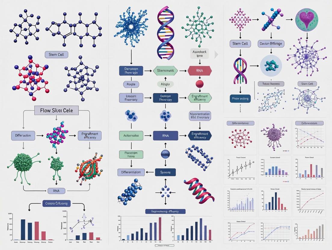
Experimental Workflow and Cell Death Pathways
The following diagrams outline the core experimental workflow for quantifying cell death and the primary signaling pathways that lead to graft cell loss.
Experimental Workflow for Cell Survival Quantification
Signaling Pathways in Transplanted Cell Death
Troubleshooting Guides
Troubleshooting Poor Cell Survival in Ischemic Niches
Q: My stem cells show poor survival and engraftment after transplantation into ischemic tissue models. What are the key factors to investigate?
A: Low cell survival often results from the harsh ischemic microenvironment. Focus on preconditioning strategies and precise characterization of the metabolic conditions.
- Primary Issue: The ischemic core is characterized by severe hypoxia, nutrient deprivation, and toxic metabolite accumulation, leading to rapid cell death post-transplantation.
- Investigation Checklist:
- Verify Oxygen & Glucose Levels: Measure dissolved oxygen and glucose concentration in your in vitro ischemic model to ensure it accurately reflects in vivo conditions. The metabolic shift in ischemia involves increased glucose uptake but can lead to uncoupled glycolysis and accumulation of toxic intermediates [5].
- Assess Metabolic Preconditioning: Pre-treat your stem cells (e.g., MSCs) in vitro with sub-lethal hypoxia or molecules that enhance their oxidative metabolism. Research indicates that stem cells can improve cardiac metabolism in ischemic heart disease by modulating pathways like AMPK/PGC-1α, which regulate fatty acid oxidation and mitochondrial function [5].
- Check for Inflammatory Mediators: The ischemic region has a robust inflammatory response. Analyze the culture medium or in vivo site for high levels of pro-inflammatory cytokines (TNF-α, IL-1β) which can directly damage cells [6].
- Recommended Protocol: Metabolic Preconditioning of MSCs
- Culture Control: Maintain stem cells in standard growth medium.
- Preconditioning Phase: For the experimental group, culture cells for 24-48 hours in a specialized medium. This medium should be:
- Hypoxic: Maintained at 1-3% O₂.
- Metabolically Primed: Supplemented with compounds like Desferrioxamine (a hypoxia mimetic) or low doses of Rotenone (to enhance mitochondrial resilience).
- Harvest and Transplant: Harvest cells post-preconditioning and proceed with transplantation.
- Validation: Confirm efficacy by measuring upregulation of hypoxia-inducible factors (HIF-1α) and survival genes (e.g., Bcl-2, PGC-1α) pre- and post-transplantation [5] [6].
Supporting Data: Key Metabolic Parameters in Ischemic Heart Disease Table: Metabolic Alterations in Ischemic Tissue to Inform Preconditioning Strategies [5]
| Metabolic Pathway | Change in Ischemia | Potential Therapeutic Target |
|---|---|---|
| Glucose Metabolism | ↑ Glycolytic flux, potential uncoupling from oxidation | Activate HIF-1α; enhance pyruvate dehydrogenase activity |
| Fatty Acid Oxidation | Initially increases, then decreases in severe failure | Modulate CPT1 and ACC via AMPK/PGC-1α pathway |
| Branched-Chain Amino Acid (BCAA) Metabolism | Impaired catabolism, leading to BCAA accumulation | Activate rate-limiting enzyme BCKDH |
Diagram: Ischemic Stressors Impact on Stem Cell Survival. The core stressors of ischemia trigger interconnected pathways that converge to compromise stem cell viability.
Troubleshooting Host Immune Rejection
Q: The administered allogeneic stem cells are being cleared by the host immune system, negating potential therapeutic benefits. How can I modulate this response?
A: Immune rejection is a major barrier for allogeneic cell therapies. Utilize the inherent immunomodulatory properties of MSCs or employ genome editing for immune evasion.
- Primary Issue: The host's immune system, particularly T cells and Natural Killer (NK) cells, recognizes transplanted allogeneic cells as foreign and mounts a cytotoxic response.
- Investigation Checklist:
- Confirm Cell Type Purity: Ensure your stem cell population is free of contaminating immune-activating cells.
- Profile Secretome: Analyze the secretome of your stem cells in vitro. Effective immunomodulatory MSCs should secrete factors like prostaglandin E2 (PGE2), indoleamine 2,3-dioxygenase (IDO), and transforming growth factor-beta (TGF-β) [7] [8].
- Monitor Host Immune Response: In vivo, track the infiltration of CD8+ cytotoxic T cells and NK cells into the engraftment site over time.
- Recommended Protocol: Enhancing Immunomodulation via MSC Priming
- Priming Stimulus: Treat MSCs for 24 hours with a cocktail of pro-inflammatory cytokines (e.g., IFN-γ at 50 ng/mL and TNF-α at 20 ng/mL). This "licensing" step potently upregulates their immunosuppressive mechanisms without inducing apoptosis [8].
- Functional Assay (In Vitro): Co-culture primed MSCs with activated peripheral blood mononuclear cells (PBMCs). A successful priming should lead to a significant reduction in PBMC proliferation (measured by CFSE dilution or BrdU assay) and a shift from a Th1 (pro-inflammatory) to a Th2 (anti-inflammatory) cytokine profile.
- Validation (In Vivo): After transplanting primed MSCs, use flow cytometry on engraftment site tissue to show a decreased ratio of CD8+/CD4+ T cells and an increase in regulatory T cells (Tregs) [7] [8].
Troubleshooting Metabolic Crisis and Inefficient Engraftment
Q: The stem cells integrate into the host tissue but fail to restore function, potentially due to a metabolic mismatch. How can I ensure my cells are metabolically compatible?
A: A metabolic crisis can occur if the energy metabolism of the transplanted cells is not optimized for the recipient's tissue environment.
- Primary Issue: Transplanted cells may rely on a primary metabolic pathway (e.g., glycolysis) that is inefficient or detrimental in the target tissue, which might require energy from other sources like fatty acid oxidation.
- Investigation Checklist:
- Analyze Metabolic Phenotype: Use a Seahorse Analyzer or similar tool to profile the oxidative phosphorylation and glycolytic rates of your stem cells before transplantation.
- Check for Lipotoxicity: In tissues like the heart, assess for lipid accumulation, which can be toxic. This indicates a mismatch between fatty acid uptake and oxidation capacity [5].
- Evaluate BCAA Levels: Accumulation of branched-chain amino acids (BCAAs) and their metabolites can inhibit key metabolic enzymes like PDH, worsening the energy crisis [5].
- Recommended Protocol: Driving Metabolic Maturation for Cardiac Repair
- Differentiation & Maturation: When generating cardiomyocytes from iPSCs, extend the maturation phase.
- Metabolic Switching: During the final week of culture, switch the medium from high-glucose to one containing a physiological mix of energy substrates, including fatty acids (e.g., palmitate conjugated to BSA) and ketone bodies (e.g., β-hydroxybutyrate). This forces the cells to shift their metabolism from glycolysis to oxidative phosphorylation, mimicking the adult heart [5] [9].
- Functional Validation: Confirm the metabolic shift by measuring an increase in the oxygen consumption rate (OCR) linked to fatty acid oxidation and a decrease in the extracellular acidification rate (ECAR). In vivo, these metabolically matured cells should show superior contractile function and engraftment in infarcted hearts [5].
Frequently Asked Questions (FAQs)
Q1: What are the most promising types of stem cells for overcoming these stressors in personalized therapy?
A: Induced Pluripotent Stem Cells (iPSCs) and Mesenchymal Stem Cells (MSCs) are at the forefront. iPSCs offer a perfect genetic match for autologous therapies and can be differentiated into any desired cell type. Their use in clinical trials is rapidly expanding, with over 115 global trials involving 83 distinct PSC-derived products as of 2024 [9]. MSCs, particularly from allogeneic sources like umbilical cord tissue, are prized for their potent immunomodulatory and anti-inflammatory properties, which allow them to sidestep immune rejection and actively suppress damaging inflammation at the engraftment site [10] [8]. The emergence of iPSC-derived MSCs (iMSCs) promises a combination of scalability and consistency for future therapies [9].
Q2: Beyond preconditioning, how can I genetically engineer stem cells to be more resilient?
A: Gene editing tools like CRISPR-Cas9 are revolutionizing the creation of resilient stem cells. Key strategies include:
- Enhancing Hypoxia Tolerance: Knock in genes for HIF-1α or other pro-survival factors under the control of a hypoxia-responsive promoter.
- Boosting Paracrine Signaling: Engineer cells to overexpress and secrete higher levels of therapeutic growth factors (e.g., VEGF for angiogenesis, GDNF for neuroprotection) [7] [6].
- Immune Evasion: Knock out major histocompatibility complex (MHC) class I and II genes to create "universal" donor cells, while simultaneously expressing non-classical HLA molecules (e.g., HLA-G) to inhibit NK cell attack [7] [11].
Q3: Are there any FDA-approved stem cell therapies that have successfully navigated these challenges?
A: Yes, recent approvals highlight the clinical translation of stem cell-based therapies. Key examples include:
- Ryoncil (remestemcel-L): Approved in 2024, it is the first MSC therapy for pediatric steroid-refractory acute graft-versus-host disease. It works by modulating the aggressive immune response, directly addressing the stressor of immune attack [9].
- Lyfgenia (lovo-cel): An autologous cell-based gene therapy approved in 2023 for sickle cell disease. It involves genetically modifying the patient's own hematopoietic stem cells to produce functional hemoglobin, correcting the underlying metabolic/functional deficit [9]. These approvals confirm that strategies targeting immune and metabolic stressors can lead to successful clinical outcomes.
The Scientist's Toolkit: Key Research Reagent Solutions
Table: Essential Reagents for Investigating Stem Cell Engraftment Stressors
| Reagent / Material | Function / Application | Key Consideration |
|---|---|---|
| CRISPR-Cas9 Systems | Gene editing for knockout (e.g., MHC) or knock-in of protective genes (e.g., HIF-1α). | Use non-integrating delivery methods (e.g., ribonucleoprotein complexes) for safety [7]. |
| Seahorse XF Analyzer | Real-time profiling of cellular metabolic phenotypes (glycolysis vs. oxidative phosphorylation). | Essential for validating metabolic preconditioning and maturation protocols [5]. |
| Cytokine Priming Cocktails (e.g., IFN-γ, TNF-α) | "Licensing" MSCs to enhance their immunomodulatory secretome prior to transplantation. | Optimize concentration and exposure time to avoid inducing senescence [8]. |
| Hypoxia Chambers / Workstations | Preconditioning stem cells in physiologically relevant low oxygen (1-3% O₂). | Prefer chambers over chemical mimetics for more physiologically accurate modeling [6]. |
| iPSC-Derived Differentiated Cells | Patient-specific cells for autologous therapy, reducing immune rejection risk. | Ensure rigorous characterization and purification to eliminate undifferentiated cells that could form tumors [7] [9]. |
| Allogeneic Umbilical Cord MSCs | Readily available, immunomodulatory cell source with high proliferation potential. | Source from reputable banks that provide full characterization and pathogen screening [10]. |
Diagram: Stressor-Driven Strategy Selection. A logical workflow for selecting the correct troubleshooting strategy based on the primary stressor identified.
FAQ: Core Concepts and Definitions
What is engraftment in the context of stem cell therapy? Engraftment is the multi-stage process by which transplanted stem cells navigate to the target tissue (homing), survive the hostile post-transplantation microenvironment, integrate into the host tissue, and ultimately establish long-term, functional activity to restore normal physiology [12] [13]. It is the cornerstone of a successful stem cell transplant, as the therapeutic benefits depend on these cells taking up residence and working properly.
How does engraftment differ from simple homing? Homing is a specific, initial phase of engraftment. It refers to the journey of infused stem cells from the bloodstream to the bone marrow niche, a process guided by molecular signals [13] [14]. Engraftment is a broader term that encompasses homing, followed by survival, proliferation, and the functional integration of these cells into the host tissue [12].
What are the primary mechanisms through which engrafted cells exert their therapeutic effects? Engrafted cells can function via multiple mechanisms, which are often context-dependent. The key mechanisms, with examples of their primary functions, are summarized in the table below.
Table 1: Key Therapeutic Mechanisms of Engrafted Stem Cells
| Mechanism | Primary Function | Example Applications |
|---|---|---|
| Differentiation | Replaces lost or damaged cells | Parkinson's disease, spinal cord injury, osteoarthritis [12] |
| Paracrine Signaling | Promotes healing via secreted factors | Heart failure, wound healing [12] [15] |
| Immunomodulation | Controls autoimmune & inflammatory responses | Multiple sclerosis, Crohn's disease [12] |
| Homing & Migration | Travel to the site of injury | Stroke, rheumatoid arthritis [12] |
| Engraftment & Integration | Functional incorporation into tissue | Retinal diseases, diabetes [12] |
| Anti-apoptotic & Anti-fibrotic | Reduces cell death and scarring | Liver disease, pulmonary fibrosis [12] |
FAQ: Technical Challenges and Troubleshooting
What is the single greatest challenge to successful engraftment? The most significant immediate challenge is the massive loss of cell viability post-transplantation. Studies indicate that up to 90% of transplanted stem cells can undergo apoptosis within the first few days due to a hostile microenvironment characterized by ischemia, nutrient deprivation, metabolic stress, and inflammatory signals [15].
Why might my transplanted cells fail to home properly to the bone marrow niche? Improper homing is often due to disruptions in the multi-step adhesion and signaling cascade. Key factors to investigate include:
- Donor Cell Receptors: Check the expression and functionality of key homing receptors on your stem cells, such as CXCR4 (for SDF-1/CXCL12), VLA-4 (for VCAM-1), and CD44 [13] [14].
- Host Environment: Ensure the recipient's bone marrow endothelium is properly conditioned (e.g., via irradiation) to upregulate essential adhesion molecules like E-selectin and VCAM-1 [14].
- Cell Quality: The homing capacity of stem cells can be compromised by the donor's health status, age, and the protocols used for cell expansion [12].
My cells seem to home and survive initially but fail to achieve long-term functional integration. What could be wrong? This points to a failure in the final stages of engraftment. Potential causes include:
- Inhospitable Niche: The stem cell niche may be damaged by previous treatments, disease (e.g., leukemic cells creating a hostile environment), or aging, preventing long-term support [13].
- Lack of Vascular Support: Long-term survival requires integration with or formation of a vascular network for ongoing oxygen and nutrient delivery [15].
- Immune Rejection: Even with immunosuppression, host immune responses may eventually target the transplanted cells, especially in allogeneic settings [12].
- Insufficient Cell Fitness: The transplanted cells may lack the intrinsic self-renewal capacity or differentiation potential needed for sustained repopulation.
Experimental Protocols for Engraftment Analysis
Protocol 1: Assessing Homing Efficiency in a Pre-Clinical Model
This protocol outlines the steps to quantify the number of donor cells that successfully reach the bone marrow shortly after transplantation.
Methodology:
- Cell Preparation and Labeling:
- Isolate donor hematopoietic or mesenchymal stem cells (HSCs/MSCs).
- Label cells with a fluorescent dye (e.g., CFSE) or express a reporter gene like GFP/Luciferase for tracking.
- Transplantation:
- Use an immunodeficient mouse model (e.g., NSG or NRG mice) to allow human cell engraftment [13].
- Infuse the labeled cells via the tail vein.
- Tissue Collection and Analysis:
- At a critical early time point (e.g., 18-24 hours post-transplant), euthanize the mice and harvest the femurs and tibias.
- Flush the bone marrow to create a single-cell suspension.
- Quantification:
- Flow Cytometry: Analyze the bone marrow cells to determine the percentage of cells that are positive for the donor label. For human cells in mice, use antibodies against human CD45 or CD34 [16].
- Calculation: Homing efficiency is typically calculated as: (Number of donor cells in BM / Number of donor cells infused) x 100%.
Protocol 2: Evaluating In Vivo Cell Survival Post-Transplantation
This protocol uses bioluminescence imaging (BLI) to non-invasively monitor the survival and proliferation of transplanted cells over time.
Methodology:
- Engineer Reporter Cells:
- Stably transduce your stem cells with a luciferase gene.
- Transplantation and Imaging:
- Transplant luciferase-expressing cells into the target organ or systemically in an animal model.
- At regular intervals (e.g., days 1, 3, 7, 14, etc.), inject the animal with the luciferin substrate.
- Use an in vivo imaging system (IVIS) to capture the bioluminescent signal, which is proportional to the number of live cells.
- Data Analysis:
- Quantify the total flux (photons/second) in a defined region of interest.
- Plot the signal intensity over time. A sustained or increasing signal indicates successful survival and engraftment, while a rapid decline indicates cell death.
Research Reagent Solutions Toolkit
This table lists essential reagents and their applications for studying and enhancing engraftment.
Table 2: Essential Reagents for Engraftment Research
| Reagent / Tool | Primary Function in Engraftment Research | Key Examples & Applications |
|---|---|---|
| CXCR4 Antagonists/Agonists | Modulate SDF-1/CXCR4 axis to study or enhance homing. AMD3100 mobilizes cells; Agonists can promote retention. | Homing assays, stem cell mobilization [13] |
| Functional Blocking Antibodies | Inhibit specific molecular interactions to determine their role in homing and adhesion. | Antibodies against VLA-4, LFA-1, E-selectin ligands [14] |
| Flow Cytometry Antibody Panels | Identify, characterize, and isolate pure populations of stem cells and their progeny. | Human: CD34+, CD45+, CD90+; Mouse: Lineage-, Sca-1+, c-Kit+ [17] [16] |
| Cytokines & Growth Factors | Precondition cells to enhance survival, proliferation, and homing potential post-transplant. | SCF, TPO, FGF2; Hypoxic preconditioning upregulates pro-survival genes [15] |
| 3D Scaffolds & Hydrogels | Provide a physical and biochemical support structure to improve cell retention, survival, and integration at the transplant site. | Synthetic or natural polymers (e.g., PEG, collagen); can be laden with oxygen carriers or growth factors [15] |
| Lentiviral Vectors | Genetically engineer stem cells to express reporter genes (GFP, Luciferase) for tracking or therapeutic genes (e.g., anti-apoptotic). | Creating stable cell lines for fate mapping and in vivo imaging studies [18] |
Signaling Pathways in Engraftment
The following diagram illustrates the key signaling pathways involved in the homing and engraftment of hematopoietic stem cells, from circulation to niche lodgment.
Diagram 1: Key Molecular Interactions in Hematopoietic Stem Cell Homing. This figure outlines the multi-step cascade from initial rolling via selectins, firm adhesion via activated integrins, to final transmigration and lodgment in the niche, driven by key ligand-receptor pairs like SDF-1/CXCR4 and VCAM-1/VLA-4 [13] [14].
Strategies to Enhance Engraftment Efficiency
Research has identified several strategies to overcome the barriers to engraftment. Key approaches with their mechanisms and experimental support are summarized below.
Table 3: Strategies to Overcome Engraftment Challenges
| Challenge | Strategy | Mechanism of Action | Evidence & Protocols |
|---|---|---|---|
| Poor Cell Survival | Metabolic Preconditioning | Culture cells in mild hypoxia (1-5% O₂) or transient serum deprivation to upregulate pro-survival genes (e.g., VEGF, HSP70) and induce autophagy [15]. | Hypoxic preconditioning for 48h doubled MSC survival under stress [15]. |
| Poor Cell Survival | Oxygen & Nutrient Delivery | Use oxygen-generating biomaterials (e.g., Perfluorocarbons (PFCs), CaO₂ nanoparticles) to provide sustained local oxygen release post-transplant [15]. | PFC-laden scaffolds increased bone formation by 2.5-fold in defect models [15]. |
| Inefficient Homing | Modulating the SDF-1/CXCR4 Axis | Pre-treat cells with small molecules or cytokines to increase CXCR4 receptor surface expression. | Protocols involve incubating cells with SCF, FGF2, or hypoxia before transplant [13]. |
| Hostile Niche | Niche Priming | Use conditioning regimens (e.g., low-dose irradiation) or co-administer supportive cells (e.g., MSCs) to make the recipient niche more receptive [13]. | Improves engraftment in myeloablated and aged mouse models. |
| Low Functional Output | 3D Culture & Organoids | Transplant cells as 3D spheroids or within biomaterial scaffolds to preserve native cell-cell signaling and enhance tissue integration [15] [18]. | 3D spheroids show enhanced paracrine signaling and in vivo survival vs. 2D cells [15]. |
Engineering Cell Resilience and Smart Delivery Systems
For researchers and drug development professionals working in personalized regenerative medicine, a significant challenge lies in the poor survival and engraftment of transplanted stem cells. The harsh ischemic microenvironment of damaged host tissues leads to catastrophic cell death, with studies showing over 90% of intravenously infused mesenchymal stem cells (MSCs) die within one week of transplantation [19]. Preconditioning strategies—particularly metabolic and hypoxic priming—have emerged as essential techniques to enhance cellular fitness before transplantation. These approaches activate endogenous defense mechanisms, significantly improving stem cell survival, retention, and ultimate therapeutic efficacy [20] [21]. This technical resource provides detailed protocols, troubleshooting guidance, and mechanistic insights to support the implementation of these critical techniques within your research programs aimed at improving engraftment efficiency.
Frequently Asked Questions (FAQs)
Q1: What is the fundamental rationale behind preconditioning stem cells before transplantation?
Preconditioning addresses the critical issue of poor cell survival post-transplantation. When stem cells are injected into damaged tissues, they encounter a harsh microenvironment characterized by hypoxia, nutrient deprivation, inflammation, and oxidative stress [22]. Most transplanted cells die before they can contribute to repair processes. Preconditioning with sublethal hypoxic or metabolic stress activates cellular survival pathways in a controlled ex vivo setting, preparing the cells to better withstand the challenging in vivo conditions they will face after transplantation [20] [21].
Q2: How does hypoxic preconditioning specifically improve the therapeutic efficacy of mesenchymal stem cells (MSCs)?
Hypoxic preconditioning enhances MSC function through multiple interconnected mechanisms:
- Improved Survival: Activates anti-apoptotic pathways (Akt, Bcl-2) and increases resistance to oxidative stress [20] [21].
- Enhanced Paracrine Function: Upregulates secretion of pro-angiogenic factors (VEGF, HGF, bFGF) and immunomodulatory molecules (IL-6, IL-10, IDO) [21] [23].
- Metabolic Adaptation: Shifts energy production towards glycolysis, promoting better survival in low-oxygen environments [24].
- Increased Motility and Homing: Boosts expression of homing receptors (CXCR4) and migration-related proteins [21] [24].
Q3: What are the key differences between physiological and pathological microenvironment simulation in pre-activation strategies?
- Physiological Microenvironment Simulation aims to maintain stem cells in a state that mirrors their natural niche (e.g., using 1-5% O₂ for hypoxia, 3D culture). This helps preserve "youthfulness," stemness, and inherent biological properties during ex vivo expansion [21].
- Pathological Microenvironment Simulation pre-adapts cells to specific disease conditions they will encounter after transplantation (e.g., inflammatory cytokine exposure, severe metabolic stress). This generates cells with enhanced capacity to function within the specific pathological context of the recipient [21].
Q4: What is the typical time frame for observing functional benefits from preconditioned stem cells in vivo?
Functional improvements begin to manifest within days post-transplantation. Studies report:
- Enhanced cell retention detectable within 24 hours [22]
- Increased paracrine factor secretion measurable within 1-3 days [20]
- Histological and functional improvements (reduced infarct size, improved graft function) evident within 1-2 weeks [20]
- Significant functional recovery in disease models observed within 2-4 weeks [20]
Experimental Protocols for Preconditioning Strategies
Protocol 1: Standard Hypoxic Preconditioning for Mesenchymal Stem Cells
Principle: Mimics the physiological oxygen tension of stem cell niches (1-5% O₂) to enhance cellular fitness before transplantation [25] [21].
Materials:
- Mesenchymal stem cells (bone marrow, adipose, or other sources)
- Hypoxia chamber or incubator (capable of maintaining 1-5% O₂, 5% CO₂, balance N₂)
- Standard cell culture media and supplements
- Serum-free media for starvation conditions (optional)
- Trypsin-EDTA for cell detachment
- Equipment for viability assessment (flow cytometer with Annexin V/PI staining)
Procedure:
- Cell Preparation: Culture MSCs under standard conditions (37°C, 21% O₂, 5% CO₂) until 70-80% confluence.
- Hypoxic Exposure: Transfer cells to hypoxia chamber pre-set to 1-2% O₂, 5% CO₂, and balanced N₂.
- Incubation Duration: Maintain cells in hypoxic conditions for 24-72 hours. Studies indicate 48 hours provides optimal benefits for most applications [21] [24].
- Media Considerations: Use standard culture media. For enhanced metabolic preconditioning, consider using serum-free media or media with reduced glucose during the final 12-24 hours.
- Cell Harvest: After preconditioning, detach cells using standard procedures and wash with PBS.
- Quality Assessment: Evaluate cell viability using trypan blue exclusion or Annexin V/PI staining. Expect >85% viability for successful preconditioning.
- Immediate Use: Transplant preconditioned cells immediately after harvesting for optimal results.
Key Parameters for Optimization:
- Oxygen Concentration: 1-2% for strong preconditioning effect; 2-5% for milder effects
- Duration: 24-72 hours, with 48 hours often optimal
- Cell Density: 60-80% confluence at start of preconditioning
- Passage Number: Use early passage cells (
Protocol 2: Pharmacological Preconditioning with Deferoxamine (DFO)
Principle: Chemical stabilization of HIF-1α using iron chelators to mimic hypoxic adaptation without requiring specialized equipment [20] [23].
Materials:
- Mesenchymal stem cells
- Deferoxamine mesylate (DFO)
- Dimethyl sulfoxide (DMSO) for stock solution
- Phosphate buffered saline (PBS)
- Cell culture media and standard lab equipment
Procedure:
- DFO Solution Preparation: Dissolve DFO in DMSO to create a 100 mM stock solution. Aliquot and store at -20°C.
- Working Solution: Dilute stock DFO in culture media to achieve final concentrations of 100-200 μM. Ensure DMSO concentration does not exceed 0.1%.
- Cell Treatment: Replace standard culture media with DFO-containing media.
- Incubation: Treat cells for 12-24 hours under standard normoxic conditions (37°C, 21% O₂, 5% CO₂).
- Cell Harvest: Remove DFO-containing media, wash cells with PBS, and harvest using standard procedures.
- Validation: Verify HIF-1α stabilization via Western blot or increased expression of downstream targets (VEGF, GLUT-1).
Protocol 3: Cytokine Priming with Interferon-Gamma (IFN-γ)
Principle: Activates immunomodulatory pathways in MSCs to enhance their therapeutic potency, particularly for inflammatory conditions [23].
Materials:
- Recombinant human IFN-γ
- MSC culture media
- Sterile PBS
Procedure:
- IFN-γ Solution: Prepare IFN-γ stock solution according to manufacturer instructions.
- Working Concentration: Add IFN-γ to culture media at 10-50 ng/mL final concentration.
- Cell Treatment: Incubate MSCs with IFN-γ-containing media for 24-48 hours under standard culture conditions.
- Validation: Assess immunomodulatory marker expression (IDO, PGE2, HLA-G) via PCR or Western blot.
Table 1: Effects of Hypoxic Preconditioning on MSC Properties and Therapeutic Outcomes
| Parameter | Change with Preconditioning | Magnitude of Effect | Reference |
|---|---|---|---|
| Cell Survival Post-Transplantation | Increased | 2-3 fold improvement at 24 hours | [22] [24] |
| VEGF Secretion | Upregulated | 2-4 fold increase | [21] |
| Glucose Consumption Rate | Reduced | 25-40% decrease | [24] |
| Migration Capacity | Enhanced | 2-3 fold increase in transwell assays | [21] [24] |
| In Vivo Retention | Improved | >50% higher at 24 hours post-implantation | [24] |
| Anti-inflammatory Effects | Strengthened | Significant increase in IL-10, IDO activity | [21] [23] |
Table 2: Comparison of Preconditioning Methods and Applications
| Method | Key Mechanisms | Optimal Conditions | Best Applications |
|---|---|---|---|
| Hypoxic Preconditioning | HIF-1α stabilization, metabolic shift to glycolysis | 1-2% O₂ for 48 hours | Myocardial infarction, stroke, limb ischemia |
| Pharmacological (DFO) | HIF-1α stabilization via PHD inhibition | 100-200 μM for 24 hours | When hypoxia equipment unavailable |
| Cytokine Priming (IFN-γ) | IDO upregulation, enhanced immunomodulation | 10-50 ng/mL for 24-48 hours | GvHD, autoimmune diseases, inflammatory conditions |
| Metabolic Priming | Glycogen storage, enhanced energy reserves | Glucose-free media for 12-24 hours | Highly ischemic tissues |
Molecular Mechanisms of Hypoxic Preconditioning
HIF-1α Signaling Pathway in Preconditioned Stem Cells
The diagram below illustrates the core molecular pathway activated during hypoxic preconditioning, which confers enhanced survival and function to stem cells.
Figure 1: HIF-1α Signaling Pathway in Hypoxic Preconditioning
This pathway demonstrates how hypoxic stress triggers HIF-1α stabilization, leading to transcriptional activation of genes responsible for enhanced cell survival, metabolic adaptation, and tissue repair capabilities [25] [26] [21].
Troubleshooting Common Experimental Issues
Problem 1: Poor Cell Viability After Hypoxic Preconditioning
- Potential Cause: Oxygen concentration too low or exposure duration too long
- Solution: Optimize oxygen levels (test 1%, 2%, and 5% O₂) and time course (24, 48, 72 hours). Perform pilot studies with Annexin V/PI staining to identify sublethal conditions [21]
- Prevention: Ensure proper calibration of hypoxia chamber and monitor cell density (maintain 60-80% confluence)
Problem 2: Inconsistent Therapeutic Benefits Between Batches
- Potential Cause: Donor variation or cellular senescence
- Solution: Standardize donor selection criteria and use early passage cells (P3-P6). Implement quality control checks including senescence-associated β-galactosidase staining [25]
- Prevention: Characterize MSC markers and differentiation potential for each batch. Use preconditioned cells immediately after processing
Problem 3: Insufficient HIF-1α Pathway Activation
- Potential Cause: Inadequate hypoxia or insufficient exposure time
- Solution: Validate HIF-1α stabilization via Western blot. Consider combining hypoxia with pharmacological preconditioning (DFO 100μM) [20] [23]
- Prevention: Include positive controls in experiments and verify hypoxia chamber function with oxygen indicators
Problem 4: Low Cell Retention Despite Preconditioning
- Potential Cause: Suboptimal transplantation technique or cell preparation
- Solution: Use biomaterial scaffolds to enhance retention [22]. Ensure cells are in single-cell suspension without clumps
- Prevention: Optimize injection volume and rate. Consider incorporating homing factors (SDF-1) in transplantation protocol
The Scientist's Toolkit: Essential Research Reagents
Table 3: Key Reagents for Preconditioning Experiments
| Reagent/Category | Specific Examples | Function/Application | Considerations |
|---|---|---|---|
| Hypoxia Inducers | Hypoxia chambers/modular incubators, AnaeroPacks | Create low-oxygen environment (1-5% O₂) | Calibrate regularly; verify O₂ levels with sensors |
| Pharmacological Agents | Deferoxamine (DFO), Dimethyloxalylglycine (DMOG) | HIF-1α stabilizers; mimic hypoxia | Dose optimization critical; potential cytotoxicity at high doses |
| Cytokines for Priming | IFN-γ, TNF-α, IL-1β | Enhance immunomodulatory capacity | Batch-to-batch variability; use carrier proteins for stability |
| Metabolic Modulators | 2-Deoxy-D-glucose, Low glucose media | Induce metabolic preconditioning | Monitor viability closely; optimize duration |
| Analysis Tools | HIF-1α antibodies, VEGF ELISA kits, PCR primers for hypoxia genes | Validate preconditioning effects | Include appropriate controls (normoxic cells) |
| Viability Assays | Annexin V/PI staining, MTT assay, ATP assays | Assess cell health post-preconditioning | Use multiple complementary assays |
Preconditioning strategies represent essential tools for advancing stem cell-based personalized therapies. By implementing these protocols and troubleshooting guides, researchers can significantly enhance the engraftment efficiency and therapeutic potential of stem cells. The metabolic and hypoxic priming approaches detailed here enable the creation of cellular products better equipped to survive and function within the challenging microenvironments of diseased tissues. As personalized medicine advances, these preconditioning strategies will play an increasingly important role in developing effective, patient-specific regenerative therapies.
Advanced Biomaterial Scaffolds and 3D Culture for Enhanced Viability
Frequently Asked Questions (FAQs)
FAQ 1: What are the most critical factors to control for maintaining high viability in 3D bioprinted cultures? The most critical factors can be categorized into general 3D culture variables and bioprinting-specific parameters. General variables include cell concentration (avoiding apoptosis from high density or low proliferation from low density), the crosslinking process (which can expose cells to harsh chemicals), and sample thickness (with structures thicker than 0.2 mm risking core necrosis). Bioprinting-specific variables are needle type and size (smaller diameters increase shear stress), print pressure (increased pressure increases shear stress), and print time, as longer sessions can negatively impact viability depending on the bioink and cell type [27].
FAQ 2: How does scaffold surface topography influence stem cell behavior in tissue engineering? Scaffold surface topography provides physical cues that directly regulate key cellular behaviors. Specific nanoscale and microscale features, such as grooves and micropatterns, can guide cell orientation, promote axon guidance in nerve regeneration, and enhance cell proliferation and differentiation. For instance, stripe patterns with a groove width of 10-20 µm have been shown to optimally regulate the orientation growth of Schwann cells, while a protein pattern width of 40 µm can improve the growth rate and orientation of dorsal root ganglion (DRG) axons [28]. The topography essentially mimics the natural extracellular matrix, providing a physical microenvironment conducive to tissue regeneration.
FAQ 3: Why is oxygenation a particular challenge in 3D cultures, and how can it be improved? Oxygenation is a major challenge because oxygen diffusion is limited to about 0.5–1 mm³ in volume. In standard gas-impermeable cultureware, steep oxygen gradients form, leading to core hypoxia or anoxia in larger 3D aggregates. This is exacerbated by factors like high cell seeding density, increased media height, and a high cellular oxygen consumption rate (OCR) [29]. Improvement strategies include using gas-permeable cultureware, implementing rotational or dynamic culture systems to improve oxygen transfer, and engineering scaffolds with integrated microchannels to facilitate nutrient and oxygen transport [27] [29].
FAQ 4: How can I quickly pinpoint the cause of viability loss in my bioprinting experiment? A systematic approach using the correct controls is the most efficient method. It is essential to include a 2D control (to rule out issues with your base cell culture), a 3D pipetted control (a non-printed, encapsulated thin film to isolate problems with your bioink or crosslinking method), and a 3D printed control (a simple printed thin film to identify issues related to the printing process itself, such as pressure or needle type) [27]. Comparing viability across these controls will quickly narrow down the source of the problem.
Troubleshooting Guides
Guide 1: Troubleshooting Low Post-Printing Viability
| Problem Area | Specific Issue | Recommended Solution |
|---|---|---|
| Bioprinting Parameters | High shear stress | Use larger diameter or tapered needle tips. Conduct a 24-hour viability study testing different pressures and needle types [27]. |
| Excessive print pressure | Test and use the minimum pressure required for consistent extrusion. High pressure increases cell shear stress [27]. | |
| Prolonged print time | Optimize bioink for faster printing and establish maximum print time for your cell-bioink combination [27]. | |
| Scaffold Environment | Poor cell adhesion | Select or modify scaffold composition to enhance protein adsorption. A balanced blend of natural (e.g., silk fibroin) and synthetic (e.g., thermoplastic polyurethane) polymers can improve fibronectin and laminin adhesion [30]. |
| Inimical surface properties | Engineer surface topography with microgrooves or nanofibers to provide physical cues that promote cell attachment, proliferation, and spatial distribution [28]. | |
| Culture Conditions | Core hypoxia in thick constructs | Redesign construct geometry to include microchannels or reduce sample thickness. Utilize gas-permeable culture vessels to improve oxygen supply [27] [29]. |
Guide 2: Optimizing Biomaterial Scaffolds for Stem Cell Engraftment
| Scaffold Characteristic | Impact on Stem Cells | Optimization Strategy for Engraftment |
|---|---|---|
| Material Composition | Influences protein adsorption, cell adhesion, and viability. | Blend natural (e.g., Silk Fibroin) and synthetic (e.g., TPU) polymers. A 1:1 ratio has shown superior cell viability (~95%) compared to other blends [30]. |
| Surface Topography | Directs cell alignment, differentiation, and neurite outgrowth. | Implement microgrooves (10-40 µm width) or aligned nanofibers to guide cell growth and spatial organization, crucial for nerve and muscle tissue [28]. |
| Porosity & Architecture | Affects nutrient diffusion, waste removal, and 3D tissue formation. | Design constructs with interconnected pores and integrate microchannels to overcome diffusion limits, preventing core necrosis [27] [31]. |
| Mechanical Properties | Modulates stem cell differentiation via mechanotransduction. | Tune the stiffness and elasticity of the scaffold to match the target native tissue, working in coordination with topographical cues [28]. |
Experimental Protocols
Protocol 1: 24-Hour Bioprinting Parameter Viability Study
Objective: To determine the optimal combination of print pressure and needle type that maximizes cell viability for a specific bioink formulation.
Materials:
- Bioink with encapsulated cells
- Bioprinter
- Multiple needle types (e.g., various gauges, tapered tips)
- Sterile culture plates
- Cell viability assay kit (e.g., Live/Dead)
- Confocal microscope or fluorescence imager
Methodology:
- Prepare Bioink: Encapsulate your target cell line at a standard concentration in the bioink.
- Set Up Parameters: Define a simple printing pattern (e.g., a grid or thin film).
- Print Test Constructs: Print the pattern using different combinations of needle types and a range of extrusion pressures. Keep the print time consistent.
- Culture: Transfer all constructs to culture media and incubate for 24 hours.
- Assess Viability: After 24 hours, perform a Live/Dead assay according to the manufacturer's instructions. Image multiple regions of each construct.
- Analyze: Quantify the percentage of live vs. dead cells for each parameter set. The condition with the highest live-cell percentage and uniform distribution is optimal [27].
Protocol 2: Scaffold Biocompatibility and Cell Adhesion Assessment
Objective: To evaluate the ability of a novel scaffold material to support cell adhesion and proliferation.
Materials:
- Scaffold samples (e.g., different polymer compositions)
- Human Umbilical Vein Endothelial Cells (HUVECs) or other relevant cell line
- Cell culture media and reagents
- MTT assay kit
- Scanning Electron Microscope (SEM)
- Live/Dead staining kit
Methodology:
- Seed Cells: Seed a standardized number of cells (e.g., third-passage HUVECs) onto each scaffold sample and a control surface.
- Cell Adhesion (SEM): After a set period (e.g., 24 hours), fix the cell-scaffold constructs, process for SEM, and image to observe cell morphology and attachment qualitatively [30].
- Cell Viability (MTT): At various time points (e.g., 24, 48, 72 hours), perform an MTT assay. This colorimetric assay measures metabolic activity, providing a quantitative estimate of the number of viable cells adhered to the scaffold [30].
- Live/Dead Staining: At the endpoint, perform Live/Dead staining to visualize cell distribution and viability across the scaffold structure qualitatively [30].
- Validation: Correlate the experimental results with computational models, such as molecular dynamics simulations predicting protein (e.g., fibronectin) adhesion energy to the scaffold surface [30].
Data Presentation
Table 1: Quantitative Impact of Scaffold Composition on Cell Viability
This table summarizes experimental data on how blending different polymers affects cell health, a key consideration for scaffold design.
| Scaffold Composition (Silk Fibroin : TPU) | Cell Viability (%) (via MTT Assay) | Key Observation |
|---|---|---|
| SF:TPU-1/1 (50% TPU) | 94.7% | Balanced composition; strongest protein adsorption and highest cell viability [30]. |
| SF:TPU-7/3 (30% TPU) | 85.5% | Higher natural polymer content; moderate viability [30]. |
| SF:TPU-3/7 (70% TPU) | 78.9% | Higher synthetic polymer content; lowest viability in this series [30]. |
Table 2: Oxygen Diffusion Limits in Standard Culture Systems
This table highlights the critical role of media height in creating oxygen gradients, a common pitfall in 3D culture.
| Culture System | Typical Media Height (mm) | Implication for Oxygen Diffusion |
|---|---|---|
| 96-well plate | 3.12 – 6.25 | Significant gradients can form, especially in dense 3D aggregates [29]. |
| 24-well plate | 2.63 – 5.26 | Steep oxygen gradients are likely, risking anoxia in the core of constructs [29]. |
| 6-well plate | 1.04 – 3.13 | Gradients are still present and must be considered in experimental design [29]. |
| T-75 Flask | 1.07 – 2.00 | Lower media height reduces gradient severity, but monitoring is still advised [29]. |
Pathway and Workflow Visualization
Title: Oxygen Limitation Pathway in 3D Culture
Title: Bioprinting Parameter Optimization Workflow
The Scientist's Toolkit: Research Reagent Solutions
| Item | Function & Rationale |
|---|---|
| Bacterial Cellulose (BC) | A highly pure and crystalline biopolymer that forms a nanofibril network. It is used to create scaffolds with high porosity, excellent liquid retention, and transparency, ideal for wound healing and soft tissue engineering [31]. |
| Alginate-Based Bioinks | Naturally derived polysaccharides that form gentle gels via ion exchange (e.g., with calcium ions). They are widely used for cell encapsulation and bioprinting due to their biocompatibility and ability to mimic aspects of the extracellular environment [27] [31]. |
| Hyaluronic Acid (HA) | A natural glycosaminoglycan found in skin. HA-based hydrogels promote fibroblast proliferation and keratinocyte migration, making them excellent for scaffolds aimed at wound healing and re-epithelialization [31]. |
| Chitosan (CS) | A cationic polysaccharide with inherent antibacterial and hemostatic properties. Its derivatives, like Carboxymethyl-CS, are water-soluble and foster successful cell growth and tissue regeneration [31]. |
| Silk Fibroin (B. mori SF) | A natural polymer known for exceptional biocompatibility, high tensile strength, and controlled biodegradability. It is often blended with synthetic polymers to enhance the structural integrity and bioactivity of scaffolds [30]. |
| Thermoplastic Polyurethane (TPU) | A synthetic polymer valued for its superior elasticity, durability, and blood compatibility. It provides the robust mechanical properties required for scaffolds in dynamic environments, such as blood vessels [30]. |
Innovative Oxygen and Nutrient Delivery to Sustain Cells Pre-Engraftment
Troubleshooting Guides and FAQs
Frequently Asked Questions (FAQs)
Q1: What is the primary cause of poor cell survival in tissue-engineered constructs prior to engraftment? The primary cause is hypoxia and nutrient deprivation due to the lack of a functional vascular network. Oxygen diffusion is limited to approximately 100-200 μm from a blood source. Cells located beyond this diffusion limit experience a hypoxic environment, leading to anaerobic respiration, lactic acid build-up, low pH, and ultimately, apoptosis. This is especially critical in the core of large scaffolds [32] [33].
Q2: Why is simply increasing ambient oxygen concentration not a viable solution? Exposing cells to high oxygen tensions (hyperoxia) can be as detrimental as hypoxia. Excessive oxygen leads to the production of reactive oxygen species (ROS), which cause oxidative stress, lipid peroxidation, protein carboxylation, and DNA damage, ultimately slowing proliferation and inducing apoptosis. A controlled, sustained supply is essential to avoid both extremes [32] [34].
Q3: What are the key differences between oxygen "carriers" and oxygen "generators"?
- Oxygen Carriers (e.g., Perfluorocarbons, Hemoglobin-based carriers) work by dissolving and releasing oxygen passively. They have a high oxygen solubility and exhibit a linear relationship between oxygen partial pressure and concentration [33].
- Oxygen Generators (e.g., Calcium Peroxide, Magnesium Peroxide) undergo a chemical reaction with water to produce oxygen over time. This provides a more active and sustained release but requires careful control of byproducts like ROS [15] [33].
Q4: How can I monitor oxygen levels within my 3D construct in vitro? While this guide focuses on delivery methods, standard techniques for monitoring include using oxygen-sensitive fluorescent probes (e.g., Ru(II)-polypyridyl complexes) or microsensors that can be inserted into the scaffold. For real-time, non-invasive monitoring, commercially available optical sensor patches integrated into bioreactor systems can be used.
Troubleshooting Common Problems
Problem: Rapid Cell Death in the Core of a 3D Scaffold
- Core Issue: Critical hypoxia and nutrient waste build-up due to diffusion limitations [32].
- Troubleshooting Steps:
- Verify Scaffold Thickness: Measure the thickness of your construct. If it exceeds 200 μm, incorporate internal oxygen delivery strategies.
- Assess Oxygen Release Kinetics: If using an oxygen-releasing material, characterize its release profile. A rapid burst release may not support long-term survival. Switch to a material with more sustained release, such as calcium peroxide (CaO₂) in a polymer matrix [33].
- Check Cell Seeding Density: A very high cell density can consume oxygen and nutrients faster than they can diffuse. Optimize the cell seeding density for your specific scaffold volume and porosity [32].
- Preventive Strategy: Integrate oxygen-generating biomaterials (OGBs) like CaO₂ or oxygen-carrying perfluorocarbons (PFCs) directly into the scaffold fabrication process to ensure continuous local supply [34] [33].
Problem: Inconsistent Engraftment and Poor Cell Retention In Vivo
- Core Issue: Hostile transplantation microenvironment (ischemia, immune response) and insufficient integration with host vasculature [15].
- Troubleshooting Steps:
- Precondition Cells: Prior to transplantation, precondition stem cells under hypoxic conditions (1-5% O₂). This activates hypoxia-inducible factor (HIF-1α), upregulating pro-survival genes (VEGF, GLUT-1) and enhancing resilience to the ischemic in vivo environment [35] [15].
- Employ Protective Biomaterials: Encapsulate cells in hydrogels or scaffolds that provide both oxygen and ROS-scavenging capabilities. This protects cells from the initial inflammatory burst and oxidative stress post-implantation [15] [33].
- Promote Rapid Vascularization: Co-deliver angiogenic growth factors (e.g., VEGF) with the cell construct to accelerate host blood vessel ingrowth and integration [32].
- Preventive Strategy: Use a combination of strategies: 1) Hypoxic preconditioning of cells, 2) Embedding in an oxygen-releasing hydrogel, and 3) Functionalization with angiogenic cues [35] [15].
Problem: Cytotoxicity Observed with Oxygen-Generating Materials
- Core Issue: Toxicity is likely due to a rapid release of high concentrations of oxygen and/or reactive oxygen species (ROS) from the material, or an undesirable shift in local pH [33].
- Troubleshooting Steps:
- Measure Local pH: The decomposition of solid peroxides can produce hydroxide ions, increasing pH. Incorporate buffering agents into your biomaterial (e.g., within a hydrogel) to maintain physiological pH [33].
- Modulate Release Kinetics: Slow down the oxygen release by changing the material encapsulation method. Using a different polymer matrix (e.g., PLGA, PEGDA) or adjusting the particle size of the oxygen source (e.g., CaO₂) can provide more controlled release [15] [33].
- Co-deliver Antioxidants: Incorporate ROS-scavenging molecules (e.g., catalase, ascorbic acid) into the delivery system to neutralize harmful ROS produced during oxygen generation [15].
- Preventive Strategy: Thoroughly characterize the oxygen release profile and byproducts of your OGB in vitro before proceeding to cell studies. Start with lower concentrations of the oxygen source and gradually increase to find the optimal, non-toxic dose.
Quantitative Data and Experimental Protocols
The following table summarizes key quantitative data on different oxygen-supplying materials to aid in selection and comparison.
Table 1: Comparison of Oxygen-Supplying Materials for Cell Sustenance
| Material | Mechanism of Action | Oxygen Release Duration | Key Advantages | Key Challenges |
|---|---|---|---|---|
| Calcium Peroxide (CaO₂) | Hydrolysis to H₂O₂, then decomposition to O₂ and H₂O [33] | >10 days in vitro [33] | High oxygen yield; sustained release [15] | Can increase pH; potential ROS generation [33] |
| Perfluorocarbons (PFCs) | Physical dissolution and release of O₂ [34] [33] | Varies with formulation & encapsulation [33] | High O₂ solubility; chemically inert [34] [33] | Rapid clearance in vivo; can be difficult to functionalize [15] |
| Hemoglobin-Based Carriers | Reversible binding of O₂, similar to red blood cells [34] | Hours to days, depending on cross-linking [34] | Physiological, sigmoidal O₂ release profile [34] | Risk of renal toxicity from dimers; ROS generation [34] |
| Magnesium Peroxide (MgO₂) | Similar to CaO₂ [33] | Sustained release (data specific to formulation) | Slower reaction with water than CaO₂ [33] | Potential ROS generation; slower initial O₂ release [33] |
Detailed Experimental Protocols
Protocol 1: Fabrication of a CaO₂-Incorporated PLGA Scaffold for Enhanced Cell Survival
This protocol is adapted from methods used to sustain fibroblast viability under hypoxia [33].
Objective: To create a 3D porous scaffold that provides a continuous oxygen supply for up to 10 days.
Materials:
- Poly(D,L-lactide-co-glycolide) (PLGA)
- Calcium Peroxide (CaO₂) powder
- Solvent (e.g., Dichloromethane, DCM)
- Porogen (e.g., Sodium Chloride, NaCl crystals)
- Cell culture medium
Method:
- Solution Preparation: Dissolve PLGA pellets in DCM to create a 10% (w/v) solution.
- Particle Incorporation: Uniformly disperse fine CaO₂ powder (e.g., 5% w/w of polymer) into the PLGA solution using a sonicator or high-speed homogenizer.
- Porogen Addition: Add sieved NaCl particles (e.g., 150-250 μm) to the mixture as a porogen. The ratio of PLGA/NaCl is typically 1:9 to create a highly porous structure.
- Casting and Drying: Pour the mixture into a mold and allow the solvent to evaporate completely over 24 hours.
- Porogen Leaching: Immerse the solidified scaffold in deionized water for 48 hours, changing the water frequently, to leach out the NaCl, creating interconnected pores.
- Sterilization: Sterilize the scaffold via exposure to UV light for 1-2 hours per side or ethylene oxide gas. Avoid autoclaving as it may degrade the polymer or trigger premature oxygen release.
- Pre-hydration: Before cell seeding, soak the scaffold in culture medium to initiate a controlled oxygen release.
Validation: Measure oxygen tension within the scaffold using micro-sensors over 10 days. Compare cell viability (e.g., via Live/Dead assay) in CaO₂-containing scaffolds versus controls under hypoxic conditions (1-5% O₂) [33].
Protocol 2: Hypoxic Preconditioning of Mesenchymal Stem Cells (MSCs)
This protocol enhances the innate resilience of cells before transplantation, improving their survival in the hostile engraftment site [35] [15].
Objective: To activate cellular stress-response pathways and improve MSC survival post-transplantation.
Materials:
- MSCs at 70-80% confluence
- Standard MSC culture medium
- Hypoxic chamber or multi-gas CO₂ incubator
Method:
- Cell Preparation: Culture MSCs in standard normoxic conditions (20% O₂, 5% CO₂) until they reach 70-80% confluence.
- Hypoxic Exposure: Replace the culture medium and place the cells in a hypoxic environment (1-5% O₂, 5% CO₂, balanced N₂) for 24-48 hours [15].
- Harvesting: After the preconditioning period, harvest the cells using standard techniques (e.g., trypsinization) for immediate transplantation or encapsulation.
- Validation (Optional): Confirm preconditioning efficacy by analyzing the upregulation of markers like HIF-1α, VEGF, or GLUT-1 via PCR or western blot.
Key Consideration: The optimal duration and oxygen concentration for preconditioning may vary depending on the cell source and application. A dose-response test is recommended.
Signaling Pathways and Experimental Workflows
Oxygen Stress Response and Preconditioning Mechanism
This diagram illustrates the cellular response to low oxygen and the rationale behind hypoxic preconditioning.
Title: Cellular Oxygen Stress and Preconditioning
Integrated Experimental Workflow for Pre-Engraftment Sustenance
This workflow outlines a comprehensive strategy from material preparation to in vivo assessment.
Title: Integrated Strategy for Cell Sustenance
The Scientist's Toolkit: Research Reagent Solutions
Table 2: Essential Materials for Oxygen and Nutrient Delivery Research
| Reagent/Material | Function/Application | Key Considerations |
|---|---|---|
| Calcium Peroxide (CaO₂) | Solid peroxide-based oxygen generator for sustained O₂ release in scaffolds [33]. | Particle size affects release kinetics; can increase pH; requires buffering strategies [33]. |
| Perfluorocarbons (PFCs) | Synthetic oxygen carriers with high O₂ solubility for dissolving and releasing oxygen in biomaterials [34] [33]. | Look for emulsions or modified PFCs conjugated to hydrogels for longer in vivo retention [15]. |
| Poly(Lactide-co-Glycolide) (PLGA) | A biodegradable polymer used to fabricate microspheres and 3D scaffolds for controlled release of oxygen and drugs [33]. | Ester-terminated vs. carboxyl-terminated types degrade at different rates; molecular weight affects mechanical properties. |
| Polyethylene Glycol Diacrylate (PEGDA) | A hydrogel precursor used to create biocompatible, tunable networks for cell encapsulation and O₂ delivery [15]. | UV photo-polymerization required; mesh size and stiffness can be adjusted by molecular weight and crosslink density. |
| Hypoxia-Inducible Factor (HIF) Prolyl Hydroxylase Inhibitors | Small molecules that stabilize HIF-1α, mimicking hypoxic preconditioning and upregulating pro-survival genes [34]. | Concentration and exposure time are critical to avoid off-target effects. Useful for clinical translation of preconditioning. |
| Reactive Oxygen Species (ROS) Scavengers (e.g., Catalase) | Enzymes co-encapsulated with oxygen generators to decompose harmful H₂O₂ byproduct into water and oxygen [15]. | Essential for mitigating oxidative stress caused by high O₂ flux from peroxide-based systems. |
Refining the Host Niche and Managing the Immune Response
Modulating the Host Immune System to Prevent Rejection
For researchers in personalized therapies, achieving successful stem cell engraftment requires navigating a complex landscape of immune-mediated rejection. Your work involves balancing the need for effective engraftment with the risk of provoking undesirable immune responses, from alloreactive T-cells and antibody-forming cells to innate immune mechanisms like natural killer (NK) cell activation [36]. This technical support center provides targeted troubleshooting guides and experimental protocols to help you identify and overcome the specific immune challenges compromising your engraftment efficiency.
★ Core Rejection Mechanisms & Research FAQs
FAQ 1: What are the primary immune mechanisms causing stem cell rejection in immunocompetent hosts?
Rejection can be initiated by both adaptive and innate immune cells. The key mechanisms and the cell types involved are summarized in the table below.
Table 1: Primary Immune Mechanisms in Stem Cell Rejection
| Immune Mechanism | Effector Cells | Key Recognition Pathway | Result on Graft |
|---|---|---|---|
| T-cell Mediated Rejection | Alloreactive CD4+ & CD8+ T cells | Direct/Indirect allorecognition of mismatched HLA | Graft infiltration and apoptosis [36] |
| Antibody-Mediated Rejection (AMR) | B cells & Plasma Cells | Donor-specific anti-HLA antibodies [37] | Complement activation, endothelial injury [36] |
| NK cell 'Missing Self' | Natural Killer (NK) cells | Lack of inhibitory KIR-HLA class I interaction [36] | Cytotoxicity and microvascular inflammation [36] |
| Innate Myeloid Activation | Monocytes / Macrophages | Signal regulatory protein α-CD47 pathway [36] | Phagocytosis and inflammation [36] |
FAQ 2: A significant number of our experimental models show microvascular inflammation (MVI) despite no detectable donor-specific antibodies (DSA). What could be the trigger?
This is a classic presentation of 'Missing Self' recognition by NK cells [36]. In this scenario, the donor's cells lack the specific HLA class I allotypes (e.g., C1, C2, Bw4) needed to engage the inhibitory receptors (KIRs) on the recipient's NK cells. Without this inhibitory signal, the NK cells become activated and can cause endothelial damage, manifesting as MVI, independent of DSAs [36].
Troubleshooting Guide:
- Genotype: Perform high-resolution HLA class I genotyping for both donor and recipient.
- Profile KIRs: Evaluate the recipient's inhibitory KIR genotype.
- Cross-reference: Identify "missing" donor HLA ligands corresponding to the recipient's inhibitory KIRs (e.g., donor is C1/C1 and recipient has a KIR2DL1 genotype) [36].
★ Troubleshooting Guides & Experimental Protocols
Troubleshooting Guide 1: Mitigating NK Cell 'Missing Self' Rejection
Problem: Poor engraftment or graft injury linked to NK cell activity in the absence of DSA.
Solution: Implement a pre-transplant 'Missing Self' risk assessment.
Table 2: Workflow for Assessing 'Missing Self' Risk
| Step | Action | Technical Detail | Research Tool |
|---|---|---|---|
| 1 | Donor/Recipient HLA Class I Typing | High-resolution typing for HLA-A, -B, -C. | PCR-based Sequence-Specific Oligonucleotide (SSO) or Next-Generation Sequencing (NGS). |
| 2 | Recipient KIR Genotyping | Identify genes for inhibitory KIRs (2DL1, 2DL2/3, 3DL1). | PCR-Sequence Specific Primers (PCR-SSP) or KIR NGS. |
| 3 | Ligand-Ligand Analysis | Map donor HLA to recipient KIRs. | Use software (e.g., PING) for KIR-HLA binding prediction. |
| 4 | In Vitro Validation (Optional) | Co-culture recipient NK cells with donor-derived cells/targets. | Measure NK cell activation (CD107a, IFN-γ) and cytotoxicity. |
Experimental Protocol 1: In Vitro Model of 'Missing Self' Induced Endothelial Damage
Aim: To demonstrate the causal role of missing self in triggering endothelial damage, independent of the adaptive immune system [36].
Methodology:
- NK Cell Isolation: Isolate NK cells from recipient peripheral blood using a negative selection magnetic-activated cell sorting (MACS) kit.
- Target Cell Culture: Culture human glomerular endothelial cells (or other relevant tissue-specific endothelial cells) from a donor with known HLA class I mismatch.
- Co-culture Setup: Co-culture purified NK cells with endothelial cells at various effector-to-target (E:T) ratios.
- Control Groups: Include controls where NK cell inhibition is restored (e.g., by blocking activating receptors like NKG2D).
- Outcome Measurement:
- Endpoint: Measure endothelial cell lysis using a standard ⁵¹Cr-release assay or real-time cytotoxicity assay (e.g., xCelligence).
- Activation: Assess NK cell activation by flow cytometry for CD107a degranulation and IFN-γ production.
Troubleshooting Guide 2: Managing Pre-sensitized Hosts with High Anti-HLA Antibody Titers
Problem: Hosts with pre-existing anti-HLA antibodies (e.g., against HLA-A2) rapidly reject HLA-mismatched grafts.
Solution: Develop and test engineered regulatory T-cells (Tregs) with chimeric antigen receptors (CARs) for targeted immunosuppression.
Experimental Protocol 2: Suppression of Alloantibody Production Using CHAR-Tregs
Aim: To generate and validate the function of Chimeric anti-HLA Antibody Receptor (CHAR) Tregs in suppressing alloantigen-specific B cells from pre-sensitized recipients [37].
Methodology:
- CHAR-Treg Engineering:
- Isolate CD4+CD25+CD127lo Tregs from patient PBMCs by FACS.
- Transduce Tregs with a lentiviral vector encoding a CHAR. The CHAR's extracellular domain is a single-chain variable fragment (scFv) derived from an anti-HLA-A2 antibody, fused to intracellular T-cell signaling domains (e.g., CD28/CD3ζ) modified for a suppressive phenotype [37].
- B Cell Co-culture:
- Isolate B cells from a pre-sensitized patient with high anti-HLA-A2 antibody titers.
- Suppression Assay:
- Co-culture CHAR-Tregs with the patient's B cells in the presence of HLA-A2-positive antigen-presenting cells.
- Control Groups: Use untransduced Tregs or non-specific CAR-Tregs as controls.
- Outcome Measurement:
- Primary: Quantify anti-HLA-A2 antibody levels in supernatant over 7-10 days using Luminex-based single antigen bead assay.
- Secondary: Assess B cell proliferation via CFSE dilution and apoptosis via Annexin V staining by flow cytometry.
Diagram: CHAR-Treg Engineering Workflow for Targeted B Cell Suppression
★ The Scientist's Toolkit: Research Reagent Solutions
Table 3: Essential Research Reagents for Rejection Studies
| Research Reagent | Specific Example / Clone | Function in Experiment |
|---|---|---|
| MACS Cell Isolation Kits | Human CD4+CD25+ Regulatory T Cell Isolation Kit; Human NK Cell Isolation Kit | Negative selection for high-purity immune cell isolation. |
| Flow Cytometry Antibodies | Anti-human CD107a (e.g., H4A3), IFN-γ (e.g., 4S.B3), CD3 (e.g., UCHT1), CD56 (e.g., HCD56) | Phenotyping and functional analysis of immune cells. |
| Luminex Assay Kits | Single Antigen Bead Assay for HLA Antibody Detection | Precise quantification of donor-specific anti-HLA antibodies. |
| Lentiviral Vectors | psPAX2, pMD2.G (packaging plasmids) | Engineering of CAR-/CHAR-expressing cells (T cells, Tregs). |
| Cytokines & Supplements | Recombinant IL-2; TGF-β | Ex vivo expansion and stability maintenance of Treg cultures. |
★ Advanced Research Considerations
FAQ 3: How does the timing of immunomodulatory treatments, like checkpoint inhibitors, impact transplantation outcomes?
Administrating immune checkpoint inhibitors (ICIs) prior to stem cell transplantation significantly increases the risk of post-transplant inflammatory adverse events [38]. One study found 82% of patients receiving pre-transplant ICI experienced an inflammatory AE, compared to 50% in the post-transplant group. These events also occurred earlier (median 57 vs. 195 days) and were more likely to require corticosteroid treatment [38].
Experimental Recommendation: In models combining ICI therapy with transplantation, carefully time the ICI administration and monitor for gastrointestinal inflammation, a common manifestation.
FAQ 4: Beyond HLA, what other pre-transplant host factors should we monitor?
A high pre-transplant Immune Dysregulation and Disease Activity (IDDA) score is a significant predictor of poor outcomes, including lower event-free survival, after allogeneic hematopoietic stem cell transplantation [39]. This highlights the need to assess the recipient's global immune status, not just allo-sensitization.
★ Key Takeaways for Your Research
- Profile Beyond HLA: Incorporate KIR genotyping and 'Missing Self' analysis into your pre-clinical risk assessment to account for innate NK cell-mediated rejection [36].
- Target B Cells Precisely: The CHAR-Treg technology represents a promising platform for achieving antigen-specific immunosuppression, effectively silencing alloantibody-producing B cells without broad immunosuppression [37].
- Time Your Interventions: The timing of immunomodulatory drugs relative to transplantation is critical. Pre-transplant ICIs can predispose hosts to earlier and more frequent inflammatory complications [38].
- Quantify Immune Status: Utilize scoring systems like the IDDA score to stratify recipient risk based on their baseline immune dysregulation, which can powerfully predict post-engraftment events [39].
Promoting Vascularization for Long-Term Cell Survival
Technical Support Center
Frequently Asked Questions (FAQs)
FAQ 1: What are the primary mechanical strategies to enhance pre-vascularization in engineered tissues? Several scaffold-based and external stimulus-based strategies are effective. Scaffold-based approaches include optimizing physical properties like stiffness (aim for a range that supports endothelial cell function), increasing surface roughness, creating anisotropic structures, and designing interconnected, hierarchical pore structures. Incorporating hollow channels into scaffolds is particularly effective for promoting patterned vascular networks. External mechanical strategies include applying dynamic cyclic stretch and using perfusion bioreactors, which facilitate the formation and maturation of preformed vascular networks in vitro [40].
FAQ 2: Why is my engineered tissue failing to integrate with the host's blood vessels after implantation? Failed integration can stem from several issues. First, the preformed vascular network may lack proper alignment with the host vessels at the implantation site. Strategies like introducing stiffness gradients or anisotropic structures in your scaffold can guide alignment. Second, the host's biological environment is critical; the same engineered tissue can show vastly different vascularization and cell survival outcomes in different animal models (e.g., nude mice vs. nude rats). Carefully select your in vivo model to match your experimental goals. Lastly, ensure the preformed network is sufficiently mature and stable to prevent leakage upon perfusion [40] [41].
FAQ 3: How can I improve the survival and function of stem cell-derived islets (SC-islets) after transplantation? Research indicates that SC-islets can exhibit superior engraftment compared to primary human islets. Focus on promoting rapid host vascular ingrowth and reinnervation. Studies show that while vascular density might be similar, the functionality of the vasculature in SC-islet grafts, indicated by higher oxygenation, can be significantly better. This enhanced vascular function contributes to lower apoptosis rates in the grafted cells [42].
FAQ 4: What is the critical diffusion limit for oxygen in avascular tissues, and how does it guide scaffold design? Cells typically cannot survive beyond 100–200 μm from a blood vessel due to oxygen diffusion limitations. This fundamentally guides scaffold design; to prevent central necrosis, your scaffold architecture must ensure that no cell is farther than this distance from a capillary or a pre-formed vascular channel. This principle necessitates the creation of dense, full-coverage vascular networks within the tissue construct [43].
Troubleshooting Guides
Problem: Poor In Vitro Formation of Capillary-like Networks
- Potential Cause 1: Suboptimal scaffold stiffness.
- Solution: Test a range of substrate stiffnesses. Very soft or excessively stiff substrates can disrupt endothelial cell adhesion, proliferation, and tube formation. Find a stiffness that promotes collective cell migration and stable tube formation [40].
- Potential Cause 2: Lack of supporting cells.
- Potential Cause 3: Insufficient mechanical stimulation.
- Solution: Implement a dynamic culture system. Utilize bioreactors that provide perfusion flow to generate physiological shear stress, which encourages endothelial cell alignment and network maturation [40].
Problem: Inadequate Anastomosis with Host Vasculature Post-Implantation
- Potential Cause 1: Mismatch in alignment between the preformed network and host vessels.
- Solution: Use guiding cues within the scaffold. Anisotropic substrates with micro-grooves or aligned fibers can direct the orientation of the preformed vascular network, facilitating morphological matching and integration with host vessels at the implant site [40].
- Potential Cause 2: Host-mediated inflammatory response.
- Solution: The host model significantly impacts outcomes. If inflammation is degrading your graft or disrupting vascular patterning, consider switching animal models (e.g., from mouse to rat or vice versa) or the anatomical implantation site, as the host's immune and wound-healing response is a major variable [41].
Problem: Central Necrosis in a Thick Engineered Tissue Construct
- Potential Cause: The construct exceeds the oxygen diffusion limit, and the internal vascular network is underdeveloped.
- Solution: Integrate patterned hollow channels into your scaffold design prior to seeding cells. These channels act as "railroad tracks" for guided vascularization, allowing for rapid host blood vessel ingrowth and perfusion post-implantation, thereby supplying oxygen and nutrients to the core of the construct [40] [41].
Table 1: Mechanical Scaffold Properties for Optimizing Vascularization
| Property | Target/ Optimal Range | Effect on Vascularization |
|---|---|---|
| Stiffness | Moderate range (cell-type specific) | Promotes endothelial cell adhesion, proliferation, and capillary-like tube formation. Excessively high stiffness can disrupt cell-cell junctions [40]. |
| Pore Structure | Interconnected & hierarchical | Facilitates cell migration, nutrient waste exchange, and ingrowth of host blood vessels [40]. |
| Architectural Cue | Anisotropic fibers/grooves | Guides endothelial cell and vascular network alignment, promoting better integration with host vasculature [40]. |
| Vascular Patterning | Incorporation of hollow channels (~50-100µm spacing) | Enables the formation of patterned, perfusable vascular networks that anastomose with host circulation [41]. |
Table 2: Host Model-Dependent Engraftment Outcomes
| Host Model | Vascular Patterning | Cardiomyocyte Graft Survival | Key Characteristics |
|---|---|---|---|
| Athymic Nude Mouse | Robust guided vascularization; retention of patterned huCD31+ vessels [41]. | Sparse [41]. | Supports robust formation of chimeric (host-graft) vessels carrying host blood [41]. |
| Athymic Nude Rat | Disrupted vascular patterning; robust inflammation [41]. | >3-fold larger than in mice [41]. | Inflammatory changes can degrade grafts or disrupt vascular architecture, but superior for certain cell survival [41]. |
Experimental Protocols
Protocol 1: Generating a Pre-vascularized Tissue Construct using Endothelial Cords
This protocol details a method for creating an engineered tissue with patterned endothelial cords to guide vascularization in vivo [41].
- Mold Preparation: Use a PDMS mold featuring parallel channels (e.g., 50-200 µm in width).
- Endothelial Cord Formation:
- Prepare a suspension of human endothelial cells (e.g., HUVECs) and stromal cells (e.g., human fibroblasts) in neutralized collagen solution.
- Pipette the cell-collagen suspension into the channels of the PDMS mold.
- Incubate (37°C, 5% CO₂) to allow collagen polymerization, forming solid "endothelial cords."
- Tissue Encapsulation:
- Carefully encapsulate the entire structure of endothelial cords within a fibrin hydrogel. The fibrinogen solution can be mixed with other cell types relevant to your tissue (e.g., stem cell-derived cardiomyocytes).
- Add thrombin to initiate fibrin clotting, embedding the cords in a 3D tissue matrix.
- In Vitro Culture (Optional): Culture the constructs in a bioreactor with perfusion to precondition the network.
- Implantation: Surgically suture the engineered tissue onto the target organ (e.g., epicardial surface of the heart) or into a site like the intraperitoneal gonadal fat pad of an immunodeficient host.
Protocol 2: Perfusion Bioreactor Culture for Vascular Maturation
This protocol promotes the formation and maturation of vascular networks within 3D constructs through the application of physiological shear stress [40] [43].
- Construct Seeding: Seed your scaffold with endothelial and supporting cells as required.
- Bioreactor Setup: Place the cell-seeded construct into a perfusion bioreactor system. Ensure a tight seal to direct flow through the construct's pores or channels.
- Flow Regime:
- Initiate with a low, steady flow rate (e.g., 0.1 mL/min) to allow cell attachment.
- Gradually increase the flow rate over several days to apply a defined, physiological shear stress (e.g., 1-15 dyn/cm²).
- Culture Maintenance: Culture the constructs under perfusion for 1-4 weeks, providing continuous nutrient supply and mechanical stimulation.
- Endpoint Analysis: Analyze the constructs for vascular network morphology (e.g., immunostaining for CD31), maturity (e.g., α-SMA coverage), and permeability.
Signaling Pathways in Vascular Network Assembly
Figure 1: Key Signaling Pathways in Angiogenesis
The Scientist's Toolkit: Research Reagent Solutions
Table 3: Essential Reagents for Vascularization Experiments
| Reagent / Material | Function / Application | Examples / Notes |
|---|---|---|
| Human Umbilical Vein Endothelial Cells (HUVECs) | Primary endothelial cells for forming capillary-like tubes in vitro; widely used in co-culture systems [40]. | Readily available, but have limited proliferation capacity [40]. |
| Mesenchymal Stem Cells (MSCs) | Function as supporting pericytes; stabilize new vessels and secrete pro-angiogenic factors (VEGF, bFGF, SDF-1) [45] [46]. | Can be derived from bone marrow or other tissues; exhibit homing to injury sites [45]. |
| Fibrin Hydrogel | A natural, biodegradable scaffold for 3D cell encapsulation; allows for cell-mediated remodeling and vessel formation [41]. | Used in the endothelial cord protocol for tissue encapsulation [41]. |
| Type I Collagen | Major ECM component used for forming initial endothelial cord structures and 3D cultures [41]. | Provides a natural substrate for cell attachment and migration. |
| Vascular Endothelial Growth Factor (VEGF) | Key cytokine for initiating endothelial capillary formation; critical for tip-stalk cell selection during sprouting [43] [44]. | Short half-life requires controlled delivery strategies (e.g., heparin-binding) [43]. |
| Patterned PDMS Molds | To create microfluidic channels or templates for generating aligned vascular structures like endothelial cords [41]. | Enables precise spatial control over vascular network architecture. |
| Perfusion Bioreactor | Provides dynamic culture conditions with fluid shear stress to enhance vascular network maturation and function [40] [43]. | Essential for scaling up thick tissue constructs. |
Leveraging Multi-Omics Data for Patient-Specific Protocol Design
Troubleshooting Guides
FAQ 1: Why is there a discordance between mRNA and protein abundance in my multi-omics data from stem cell differentiation, and how does this impact engraftment potential?
Answer: Discordance between mRNA and protein levels is biologically common during dynamic processes like stem cell differentiation and can significantly impact the functional maturity of cells, a key determinant of engraftment efficiency. Simply relying on transcriptomic data is insufficient for predicting cellular phenotype.
- Primary Cause: Protein synthesis (ks) and degradation (kd) rates are gene-specific and not constant during differentiation, meaning cells are often not in a transcriptional-protein steady state [47].
- Impact on Engraftment: A differentiated cell might express the correct mRNA markers (suggesting lineage specificity) but lack the corresponding functional proteins, leading to poor survival or function upon transplantation.
- Solution:
- Employ Kinetic Modeling: Move beyond simple correlation. Use a birth-death kinetic model to understand protein dynamics [47]:
Ṗ(t) = k_s * R_cyto(t) - k_d * P(t)WhereṖis the rate of protein change,R_cytois cytoplasmic mRNA,k_sis synthesis rate, andk_dis degradation rate. - Measure Cytoplasmic mRNA: Use subcellular fractionation RNA-seq, as only cytoplasmic mRNA is available for translation. This provides a more accurate predictor of protein output than total mRNA [47].
- Investigate Post-Transcriptional Regulation: Integrate microRNA (miRNA) abundance data. Model miRNAs as potential repressors of protein synthesis rate (
k_s) to identify candidates that may be inhibiting the expression of critical proteins for engraftment [47].
- Employ Kinetic Modeling: Move beyond simple correlation. Use a birth-death kinetic model to understand protein dynamics [47]:
FAQ 2: How can I effectively integrate different omics data types (e.g., genomics, transcriptomics, proteomics) to identify robust biomarkers for predicting engraftment efficiency?
Answer: Successful integration requires a structured approach to move from disjointed datasets to a unified biological insight that can predict clinical outcomes like engraftment.
- Challenge: Data from different omics layers (genome, transcriptome, proteome) have different scales, distributions, and technical noise.
- Solution - A Sequential Integration Workflow:
- Data Generation & Collection: Ensure sample overlap. Leverage public repositories like The Cancer Genome Atlas (TCGA) or Clinical Proteomic Tumor Analysis Consortium (CPTAC) for model system data [48].
- Preprocessing & QC: Perform rigorous quality control for each data type individually (e.g., using FASTQC for sequencing data, filter low-quality proteins in MS data) [49].
- Normalization & Batch Correction: Normalize data (e.g., library size scaling for RNA-seq, total intensity for proteomics) and apply batch correction algorithms (e.g., Harmony, Seurat's integration methods) to remove technical variation [49].
- Simultaneous Data Integration: Use tools designed for parallel integration of multiple datatypes. These can identify multimodal molecular signatures that are more robust biomarkers than any single-omics marker [48].
- Validation: Candidate biomarkers (e.g., a protein:RNA ratio or a multi-omics cluster) must be validated in a separate cohort of cells followed by an in vivo engraftment assay.
FAQ 3: My stem cell cultures show high differentiation heterogeneity before transplantation. How can multi-omics help ensure a pure, therapeutic-grade cell population?
Answer: Multi-omics can identify subtle pre-transplantation heterogeneity and pinpoint the molecular drivers of off-target differentiation, enabling protocol refinement.
- Cause: Culture conditions (e.g., passaging techniques, medium age) can induce spontaneous, uncontrolled differentiation [2].
- Multi-Omics Application:
- Single-Cell Multi-Omics: Use technologies like CITE-seq (simultaneous single-cell transcriptomics and surface protein quantification) to characterize heterogeneity in your pre-engraftment cell product. This can identify subpopulations committing to undesired lineages.
- Identify Regulatory Networks: Integrated analysis can reveal key transcription factors (from ATAC-seq/RNA-seq) and surface proteins (from proteomics) that define the optimal therapeutic cell state.
- Inform Sorting Strategies: The identified surface markers (e.g., from proteomics) can be used to Fluorescence-Activated Cell Sorting (FACS) to purify the optimal cell population immediately before transplantation, directly improving engraftment efficiency.
Experimental Protocols
Protocol 1: Kinetic Analysis of mRNA-Protein Discordance During Differentiation
Objective: To quantify the dynamics of protein synthesis and degradation for key engraftment-related genes during stem cell differentiation.
Materials:
- Human iPSCs undergoing directed differentiation.
- RNA extraction kit with cytoplasmic fractionation capability.
- Mass Spectrometer (e.g., LC-MS/MS) with TMT labeling.
- Bulk RNA-sequencing and small RNA-sequencing services.
Methodology:
- Time-Course Sampling: Collect triplicate samples at multiple time points (e.g., 0, 24, 48, 72, 96 hours) during differentiation [47].
- Multi-Omics Quantification:
- Cytoplasmic RNA: Fractionate cell lysates to isolate cytoplasmic RNA for bulk RNA-seq.
- Protein: Extract total protein for TMT-based quantitative mass spectrometry.
- microRNA: Perform small RNA-seq on total RNA.
- Data Preprocessing: Align RNA-seq reads (e.g., using STAR), quantify gene expression, and process MS data for protein abundance [49].
- Model Fitting: For each gene of interest, fit the cytoplasmic mRNA and protein abundance time-series data to the birth-death model using non-linear least squares to estimate synthesis (
k_s) and degradation (k_d) rates [47]. - Identify Candidates: Genes with poor model fit (low R²) are candidates for post-transcriptional regulation. Correlate their
k_sestimates with miRNA dynamics to nominate regulatory miRNA-mRNA pairs.
Protocol 2: Integrated Multi-Omics Clustering for Cell Product Stratification
Objective: To stratify patient-specific stem cell-derived products based on multi-omics profiles and correlate these strata with in vivo engraftment outcomes.
Materials:
- Cell products from multiple patient-derived iPSC lines.
- DNA/RNA/Protein extraction kits.
- Access to multi-omics integration software (e.g., based on MOFA or similar).
- Access to a suitable animal model for engraftment testing.
Methodology:
- Data Generation: From each cell product, generate:
- Genomics: Whole Genome Sequencing (WGS) to identify patient-specific variants [50].
- Transcriptomics: Bulk RNA-seq to profile global gene expression.
- Proteomics: Mass spectrometry to quantify protein levels.
- Data Integration: Use a multi-omics integration tool to simultaneously analyze all three data types. This will reduce the dimensions of the data and group samples (cell products) into clusters based on shared molecular patterns across all omics layers [48].
- Biomarker Extraction: For each cluster, identify the driving features (e.g., specific genetic variants, mRNA, and proteins) that define that group.
- Correlation with Engraftment: Transplant cell products from each molecular cluster into your animal model and measure engraftment efficiency (e.g., by bioluminescent imaging or histology). Statistically link high-engraftment clusters to their defining multi-omics features.
Data Presentation
Table 1: Public Multi-Omics Data Repositories for Stem Cell Research
| Repository | Primary Focus | Available Data Types | Web Link |
|---|---|---|---|
| The Cancer Genome Atlas (TCGA) | Cancer | RNA-Seq, DNA-Seq, miRNA-Seq, SNV, CNV, DNA methylation, RPPA [48] | https://cancergenome.nih.gov/ |
| Clinical Proteomic Tumor Analysis Consortium (CPTAC) | Cancer (proteomics) | Proteomics data corresponding to TCGA cohorts [48] | https://cptac-data-portal.georgetown.edu/ |
| International Cancer Genomics Consortium (ICGC) | Cancer | Whole genome sequencing, somatic and germline mutations [48] | https://icgc.org/ |
| Target ALS | Amyotrophic Lateral Sclerosis | Postmortem tissue, longitudinal biofluids, iPSCs, WGS, RNA-seq, clinical data [50] | https://www.targetals.org/ |
| Omics Discovery Index (OmicsDI) | Consolidated datasets | Genomics, transcriptomics, proteomics, metabolomics from 11 repositories [48] | https://www.omicsdi.org/ |
Table 2: Key Tools for Single-Cell and Multi-Omics Data Analysis
| Analysis Step | Recommended Tools | Brief Function Description |
|---|---|---|
| Read Alignment | STAR | Splice-aware alignment of RNA-seq reads to the genome [49]. |
| Quality Control | FASTQC, MultiQC | Generate QC metrics for raw sequencing files and aggregated reports [49]. |
| Normalization & Batch Correction | Seurat, Scanpy, Harmony | Normalize data, integrate datasets, and correct for batch effects [49]. |
| Dimensionality Reduction & Visualization | PCA, t-SNE, UMAP | Project high-dimensional data into 2D/3D for visualization and exploration [49]. |
| Clustering & Cell Type ID | K-means, Graph-based | Identify distinct cell populations and annotate cell types using marker genes [49]. |
| Trajectory Inference | Monocle3, Slingshot | Reconstruct cellular differentiation paths and pseudotemporal ordering [49]. |
Visualizations
Multi-Omics Engraftment Workflow
mRNA-Protein Kinetic Model
The Scientist's Toolkit
Table 3: Research Reagent Solutions for Multi-Omics Studies
| Reagent / Material | Function in Multi-Omics Protocol |
|---|---|
| Induced Pluripotent Stem Cells (iPSCs) | Patient-specific starting material for differentiation; enables study of genetic background on engraftment potential [18]. |
| Tandem Mass Tag (TMT) Reagents | Multiplexing reagents for mass spectrometry that allow simultaneous quantification of proteins from multiple samples (e.g., different time points) in a single run, reducing technical variability [47]. |
| Cytoplasmic Fractionation Kit | Separates nuclear and cytoplasmic RNA, allowing for specific quantification of the mRNA pool available for translation, which is critical for accurate kinetic modeling [47]. |
| CRISPR-Cas9 System | Gene editing tool used to validate candidate genes or miRNAs identified from multi-omics analysis by creating knock-outs or introducing mutations in iPSCs [18]. |
| Vitronectin XF / Defined Matrices | Feeder-free, defined extracellular matrix coatings for consistent maintenance and differentiation of iPSCs, reducing uncontrolled differentiation and background noise in omics data [2]. |
Bench to Bedside: Assessing Efficacy in Models and Clinical Trials
The field of preclinical research is undergoing a significant transformation, moving from a heavy reliance on traditional animal models toward more human-relevant systems. While animal models have provided foundational insights into biological processes and disease mechanisms, they often fail to fully recapitulate human-specific pathophysiology, leading to high attrition rates in clinical trials [51] [52]. This challenge has accelerated the adoption of advanced human organoid technologies that offer enhanced physiological relevance for disease modeling, drug screening, and personalized therapeutic development [53] [54].
Within this paradigm shift, a critical focus has emerged on improving the engraftment efficiency of stem cells, a fundamental requirement for advancing regenerative medicine and cell-based therapies. This technical support center addresses the key methodological challenges and troubleshooting strategies researchers encounter when working across this spectrum of preclinical models, with particular emphasis on optimizing stem cell integration and functionality.
Technical FAQs and Troubleshooting Guides
Animal Model Section
Q1: What are the primary limitations of using standard mouse models for studying the human immune system?
Standard mouse models exhibit significant differences from humans in their Major Histocompatibility Complex (MHC), which is functionally distinct from the human Human Leukocyte Antigen (HLA) system. Mouse immune systems are optimized for ground-level pathogens, whereas human immune systems are more adapted to airborne invaders. These differences can limit the translatability of preclinical findings [55].
Q2: How can I create a "humanized" mouse model to better study human immune responses?
Humanized mice are created by engrafting human hematopoietic stem cells (HSCs) or peripheral blood mononuclear cells (PBMCs) into immunodeficient mice. The standard protocol involves:
- Preconditioning: Mice undergo irradiation or myeloablative chemotherapy to create space for engraftment.
- Cell Injection: An intravenous injection of human stem cells is administered to restart the immune system.
- Cell Source Selection: CD34+ HSCs from cord blood are often preferred as they support comprehensive reconstitution of both lymphoid and myeloid lineages and reduce the risk of graft-versus-host disease compared to PBMCs [55].
Q3: Why did my humanized mouse experiment fail, and how can I improve success rates?
Failed engraftment is often linked to issues with the starting cell material. The most common reasons for failure and their solutions are outlined below [55]:
| Problem Area | Specific Issue | Recommended Solution |
|---|---|---|
| Starting Material | Low cell count or viability | Source cells from a vendor that provides high-quality, accurately quantified cell products. |
| Small lot size from a single donor | Ensure the entire sample size for an experiment comes from the same donor to minimize batch effects. | |
| Technical Process | Inadequate cell infusion dose | Optimize and verify the cell count before infusion; inadequate doses may lead to animal death. |
| In-house cell isolation failures | Consider purchasing pre-isolated cells to de-risk the workflow and ensure consistency. |
Organoid and Stem Cell Section
Q4: My patient-derived organoids (PDOs) have low formation efficiency. What critical steps might I be missing?
Low organoid formation efficiency often stems from suboptimal tissue collection and initial processing. Adherence to the following protocol is critical [56]:
- Sample Collection: Transfer tissue samples immediately into cold Advanced DMEM/F12 medium supplemented with antibiotics.
- Processing Time: Process samples promptly. Delays reduce cell viability and formation efficiency.
- Preservation Strategy: If immediate processing is not possible, use one of two validated methods:
- Short-term storage (≤6-10 hour delay): Wash tissue with antibiotic solution and store at 4°C in DMEM/F12 with antibiotics.
- Long-term storage (>14 hour delay): Cryopreserve tissue in a freezing medium (e.g., 10% FBS, 10% DMSO in 50% L-WRN conditioned medium). Note: A 20-30% variability in live-cell viability can be observed between these two preservation methods [56].
Q5: My organoid cultures show high batch-to-batch variability. How can I standardize my protocols?
Variability arises from differences in cell sources, growth factor combinations, and culture conditions. Standardization efforts should focus on:
- Matrix Gels: Traditional Matrigel has undefined composition. Consider switching to defined synthetic hydrogels or decellularized tissue-derived matrices for better consistency [53].
- Growth Media: Use precise, quantified concentrations of essential components. The table below summarizes key factors for different organoid types [53].
- Automation: Implement automated systems for high-throughput screening to enhance reproducibility and translational relevance [51].
Essential Research Reagent Solutions for Organoid Culture
| Reagent Category | Specific Examples | Function in Culture |
|---|---|---|
| Core Growth Factors | EGF, Noggin, R-Spondin 1, Wnt-3a | Maintain stemness and promote self-renewal in epithelial organoids (e.g., intestinal). |
| Specialized Cytokines | FGF-10, HGF, BMP | Direct lineage-specific differentiation (e.g., in liver, lung, kidney organoids). |
| Small Molecule Inhibitors/Activators | Y-27632 (ROCK inhibitor), A 83-01 (TGF-β inhibitor), CHIR-99021 (Wnt activator) | Enhance cell survival, control differentiation pathways, and modulate key signaling. |
| Scaffolding Matrices | Matrigel, Synthetic PEG-based hydrogels, Decellularized tissue hydrogels | Provide a 3D structural support that mimics the native extracellular matrix (ECM). |
Q6: What are the key mechanisms by which transplanted stem cells exert their therapeutic effects?
Stem cells function as "living drugs" through multiple interconnected mechanisms, not just differentiation. The primary mechanisms and their applications are [12]:
- Differentiation: Direct replacement of lost or damaged cells (e.g., dopaminergic neurons for Parkinson's disease).
- Paracrine Signaling: Secretion of factors that promote healing and repair in host tissues (e.g., in heart failure or wound healing).
- Immunomodulation: Controlling autoimmune and inflammatory responses (e.g., in multiple sclerosis or Crohn's disease).
- Homing & Migration: Traveling to the site of injury following chemotactic signals.
- Anti-apoptotic & Anti-fibrotic Actions: Reducing cell death and scarring in diseased tissues (e.g., in liver disease or pulmonary fibrosis).
General Technical Troubleshooting
Q7: My immunoassays are producing inconsistent results. What are the common culprits?
Inconsistent results in assays like ELISA can often be traced to a few key areas. A logical troubleshooting path is recommended [57]:
- Reagents: Compare to quality control (QC) release data. Ensure all reagents and antibodies are well-mixed and have not expired.
- Equipment: Check the physical condition of hardware, pipette calibration, and programming of automated steps (incubation, washing, reading).
- Technique: Avoid stacking plates during incubation, which creates uneven temperature. Ensure consistent pipetting and prevent wells from drying out after washing [57].
Q8: How can I integrate organoids into more complex, physiologically relevant systems?
To overcome the limitation of organoids lacking a full tissue microenvironment, consider these advanced integrations:
- Organoid-on-Chip: Combine organoids with microfluidic devices to control flow, shear stress, and gradient formation, better mimicking in vivo conditions [51] [56].
- Co-culture Systems: Integrate organoids with immune cells or other stromal elements to study epithelial-immune crosstalk and therapy responses [51] [56].
- Apical-Out Polarity: Generate organoids with reversed polarity to provide direct access to the luminal surface for drug permeability and host interaction studies [56].
Essential Experimental Workflows and Signaling Pathways
Workflow: Establishing Patient-Derived Colorectal Organoids
The following diagram outlines the key steps for generating and validating patient-derived colorectal organoids, highlighting critical decision points to maximize success [56].
Pathway: Core Signaling for Stem Cell Maintenance
The self-renewal and differentiation of stem cells within organoids are governed by a few highly conserved signaling pathways. Manipulating these pathways is fundamental to successful organoid culture [51] [53] [51].
For researchers focused on improving the engraftment efficiency of stem cells in personalized therapies, the current clinical trial landscape presents both unprecedented opportunities and complex challenges. The fundamental goal of stem cell therapy—to achieve functional integration of transplanted cells into host tissues—hinges on successful engraftment. This process of cell homing, survival, proliferation, and integration is the critical bridge between cell administration and therapeutic efficacy [12].
Recent FDA approvals and trial designs reflect a growing sophistication in addressing these cellular dynamics. With the first FDA-approved mesenchymal stem cell (MSC) therapy emerging in late 2024 and multiple induced pluripotent stem cell (iPSC)-based therapies entering advanced clinical trials in 2025, the field is transitioning from exploratory research to targeted clinical application [9]. This technical support center provides troubleshooting guidance and analytical frameworks to help researchers navigate this evolving landscape while optimizing engraftment parameters in their experimental designs.
Recent FDA Approvals: Analyzing Trends in Stem Cell and Advanced Therapy Trials
Recently Approved Stem Cell and Gene Therapy Products
The FDA's Approved Cellular and Gene Therapy Products list remains highly selective, with each approval establishing important precedents for future stem cell-based therapies [9].
Table 1: Recently FDA-Approved Stem Cell and Gene Therapy Products (2023-2025)
| Product Name | Approval Date | Cell Type/Technology | Indication | Key Engraftment Consideration |
|---|---|---|---|---|
| Ryoncil (remestemcel-L) | December 2024 | Allogeneic Bone Marrow-Derived MSCs | Pediatric steroid-refractory acute graft-versus-host disease | Immunomodulation without long-term engraftment; cells exert paracrine effects [9] |
| Lyfgenia (lovotibeglogene autotemcel) | December 2023 | Autologous CD34+ hematopoietic stem cells (gene-modified) | Sickle cell disease | Myeloablative conditioning required for hematopoietic stem cell engraftment [9] |
| Omisirge (omidubicel-onlv) | April 2023 | Cord Blood-Derived Hematopoietic Progenitor Cells (nicotinamide-modified) | Hematologic malignancies (post-transplant) | Enhanced homing and engraftment through nicotinamide modification [9] |
Notable FDA-Authorized Clinical Trials for Stem Cell Therapies (2025)
Beyond fully approved products, the FDA has authorized numerous clinical trials investigating novel stem cell approaches, many of which directly address engraftment optimization.
Table 2: Selected FDA-Authorized Stem Cell Clinical Trials Active in 2025
| Therapy Name | Cell Type | Indication | Trial Phase | Engraftment-Related Innovation |
|---|---|---|---|---|
| OpCT-001 | iPSC-derived photoreceptor cells | Retinal degeneration (retinitis pigmentosa) | Phase I/IIa | Retinal integration; first iPSC therapy for photoreceptor diseases [9] |
| FT819 | iPSC-derived CAR T-cells | Systemic lupus erythematosus | Phase I (RMAT designated) | Off-the-shelf allogeneic approach with gene editing to prevent rejection [9] |
| CYP-001 (Cymerus iMSCs) | iPSC-derived MSCs | High-Risk Acute Graft-Versus-Host Disease | Phase I | Consistent, scalable cell source vs. donor-derived MSCs [9] |
| Autologous iPSC-derived Dopaminergic Neural Progenitor Cells | Autologous iPSC-derived neurons | Parkinson's Disease | Phase I | Autologous approach potentially avoiding immune rejection [9] |
| MyoPAXon | iPSC-derived CD54+ muscle progenitor cells | Duchenne Muscular Dystrophy | Phase I | Muscle tissue integration for long-term protein expression [9] |
Table 3: Essential Research Reagent Solutions for Engraftment Studies
| Reagent/Resource | Function in Engraftment Research | Example Application |
|---|---|---|
| REPROCELL StemRNA Clinical iPSC Seed Clones | GMP-compliant, quality-controlled starting material | Provides standardized, reproducible iPSC source for consistent differentiation [9] |
| Nicotinamide-based Expansion Supplements | Enhances stem cell self-renewal and homing capacity | Used in Omisirge to improve cord blood stem cell engraftment [9] |
| Genetic Modification Tools (CRISPR-Cas9, lentiviral vectors) | Enables genetic engineering to enhance cell survival, tracking, or function | Creating reporter lines for engraftment tracking; modifying cells for immune evasion [58] [18] |
| Defined Differentiation Kits | Produces specific, pure cell populations for transplantation | Generating dopaminergic neurons or retinal cells with high purity for better integration [9] |
| Cytokine and Chemokine Panels | Modulates homing and integration signals | Testing combinations to enhance migration to target tissues [12] |
Troubleshooting Guides and FAQs: Addressing Engraftment Challenges
FAQ 1: What are the primary factors causing poor engraftment efficiency in our stem cell trials, and how can we address them?
Issue: Low engraftment efficiency remains a major translational barrier, with multiple contributing factors.
Troubleshooting Guide:
- Host Microenvironment Mismatch: Ensure the differentiated state of your stem cells matches the target tissue niche. Use transcriptional profiling to verify cell maturity before transplantation [12] [18].
- Inflammatory Rejection: Even with immunosuppression, innate immune responses can clear transplanted cells. Consider alternative immunosuppression regimens or genetic modification of cells to evade immune detection (e.g., HLA silencing) [58] [18].
- Insufficient Cell Viability Post-Transplantation: Pre-condition cells with pro-survival factors and use anti-apoptotic agents during the transplantation process. Optimize delivery methods to minimize shear stress [12].
- Inadequate Homing Signals: Engineer cells to overexpress appropriate homing receptors (e.g., CXCR4 for bone marrow) that match chemokines secreted by target tissues [12].
FAQ 2: How can we better monitor and quantify long-term engraftment in preclinical models?
Issue: Traditional endpoint analyses provide limited information about engraftment dynamics.
Solution Strategies:
- Bioluminescent/Fluorescent Reporter Systems: Genetically engineer stem cells with luciferase or fluorescent proteins for non-invasive tracking. Use advanced imaging systems to monitor cell location, expansion, and survival over time [58].
- PCR-Based Engraftment Assays: Develop species-specific PCR assays (for xenograft models) to quantify the percentage of human cells in different tissues at multiple time points [58].
- Multi-Modal Approaches: Combine imaging with histological validation to correlate signal intensity with actual cell integration and function.
FAQ 3: Our team is preparing an IND application. What engraftment-related data does the FDA typically require?
Issue: Regulatory expectations for demonstrating potential engraftment efficacy can be unclear.
Key Evidence Requirements:
- Proof of Concept in Relevant Animal Models: Include data showing your cells can engraft, survive, and function appropriately in immunocompromised or disease-relevant models for a sufficient duration (typically 3-6 months minimum) [9].
- Biodistribution Studies: Comprehensive data showing where your cells localize after administration and whether they migrate to non-target tissues [9].
- Tumorigenicity Assessment: Rigorous testing demonstrating your cell product does not form teratomas or other inappropriate growths, especially for pluripotent stem cell-derived therapies [18] [9].
- Cell Tracking and Persistence Data: Evidence documenting how long your cells persist in the target tissue and whether they maintain their intended phenotype [9].
FAQ 4: What's the difference between an FDA-authorized trial and an FDA-approved product, and how does this affect our research strategy?
Clarification:
- FDA-Authorized Trial: The FDA has reviewed an Investigational New Drug (IND) application and allowed clinical studies to proceed after 30 days with no objections or earlier with FDA feedback. This does NOT mean the therapy is proven safe and effective for widespread use [9].
- FDA-Approved Product: The FDA has granted a Biologics License Application (BLA), determining the product is safe, pure, and potent for its intended use based on substantial evidence from controlled trials. This permits marketing with post-marketing obligations [9].
Research Implications: Design your preclinical engraftment studies with BLA requirements in mind, even during early development. This forward-thinking approach facilitates smoother regulatory transitions.
Experimental Protocols: Methodologies for Assessing Engraftment Efficiency
Protocol: Quantitative Assessment of Stem Cell Engraftment Using Flow Cytometry
Purpose: To precisely quantify the percentage of donor-derived cells in recipient tissues over multiple time points.
Materials:
- Single-cell suspensions from target tissue
- Species-specific antibodies (e.g., anti-human HLA for xenografts)
- Viability dyes (e.g., propidium iodide)
- Flow cytometer with appropriate lasers and filters
Methodology:
- Sample Preparation: Harvest target tissues at predetermined time points post-transplantation (e.g., 1, 2, 4, 8, 12 weeks). Create single-cell suspensions using appropriate enzymatic digestion.
- Staining Protocol: Incubate cells with species-specific surface markers for 30 minutes at 4°C. Include viability staining to exclude dead cells from analysis.
- Flow Cytometry Analysis: Acquire a minimum of 100,000 events per sample. Use fluorescence-minus-one (FMO) controls to establish gating boundaries.
- Quantification: Calculate engraftment percentage as (Number of donor-derived cells / Total live cells) × 100. Normalize across animals and time points.
Troubleshooting Note: If background signal is high, consider using additional negative selection markers to exclude resident cell populations.
Protocol: Functional Integration Assessment of iPSC-Derived Neurons
Purpose: To evaluate whether transplanted neuronal cells functionally integrate into existing neural circuits.
Materials:
- Electrophysiology setup (patch clamp or multi-electrode array)
- Neural activity reporters (e.g., GCaMP)
- Tissue sectioning equipment
Methodology:
- Preparation: Allow 4-12 weeks for graft maturation post-transplantation of iPSC-derived neural precursors.
- Electrophysiological Recording: Perform whole-cell patch clamp recordings on graft-derived neurons in acute brain slices. Assess passive membrane properties, action potential generation, and synaptic connectivity.
- Circuit Integration: Use anterograde and retrograde tracing techniques to determine whether grafted cells receive input from and send projections to appropriate host brain regions.
- Functional Validation: Combine with behavioral tests relevant to the disease model (e.g., motor function tests for Parkinson's disease models).
Technical Consideration: Always include untransplanted control animals to establish baseline network activity and rule on non-specific effects of the transplantation procedure itself.
Visualizing Engraftment Pathways and Workflows
Stem Cell Engraftment Cascade
FDA Clinical Trial Pathway for Stem Cell Products
The evolving clinical trial landscape for stem cell therapies demands increasingly sophisticated approaches to engraftment optimization. Recent FDA approvals and trial authorizations demonstrate several strategic directions:
First, product engineering is becoming central to overcoming engraftment barriers. Technologies such as nicotinamide modification of cord blood cells (Omisirge) and genetic engineering of iPSCs to evade immune recognition represent the next frontier in ensuring cell persistence [9].
Second, the field is moving toward standardized, scalable cell sources. The submission of Drug Master Files for clinical-grade iPSC lines (e.g., REPROCELL's StemRNA) addresses batch-to-batch variability that can significantly impact engraftment consistency [9].
Finally, advanced monitoring methodologies are becoming essential components of trial design. Incorporating multimodal engraftment assessment—combining imaging, molecular, and functional readouts—provides the comprehensive data package needed for both scientific optimization and regulatory approval.
For researchers focused on personalized therapies, these developments underscore the importance of designing engraftment studies that not only demonstrate scientific principle but also meet the practical requirements of regulatory pathways. By aligning experimental approaches with the evolving clinical landscape, the research community can accelerate the development of stem cell therapies that achieve reliable, functional engraftment and deliver on the promise of regenerative medicine.
Comparative Analysis of Engraftment Success Across Different Stem Cell Types
Achieving robust and timely engraftment is a critical determinant of success in stem cell-based regenerative medicine and personalized therapies. Engraftment—the process by which transplanted stem cells establish themselves in the host tissue and begin producing functional progeny—varies significantly across different stem cell sources and transplantation contexts. Within the framework of personalized medicine, which aims to tailor treatments to individual patient characteristics, understanding these variables is paramount for optimizing clinical outcomes. This technical support document provides a comparative analysis of engraftment success across major stem cell types, offering evidence-based troubleshooting guidance to help researchers overcome common experimental and clinical challenges.
FAQs: Troubleshooting Common Engraftment Challenges
Q1: What are the primary factors causing delayed platelet engraftment following hematopoietic stem cell transplantation (HSCT), and how can it be mitigated?
Delayed platelet engraftment prolongs thrombocytopenia, increasing bleeding risk and transfusion dependence. Evidence indicates that co-infusion of Mesenchymal Stem Cells (MSCs) can significantly accelerate platelet recovery. A systematic review of 47 clinical studies concluded that MSC co-infusion is a safe and effective strategy, with platelet engraftment times averaging 21.61 days in MSC recipients compared to often longer periods in controls [59]. The proposed mechanism involves MSC secretion of supportive cytokines like thrombopoietin (TPO) and modulation of the bone marrow niche [59].
Q2: Which stem cell source typically demonstrates superior engraftment kinetics in hematologic reconstitution?
Umbilical Cord Blood (UCB) is a valuable source, but its use is often associated with a higher incidence of graft failure and delayed engraftment due to a limited cell dose [60]. In contrast, transplants using haploidentical donors or matched unrelated donors (MUD) with peripheral blood stem cells (PBSC) can achieve robust engraftment, with neutrophil recovery often occurring around day +12 to +13 post-transplant [60]. The choice involves a trade-off between cell dose, HLA matching, and immune reconstitution speed.
Q3: What is a major clinical challenge associated with graft failure, and what is a potential salvage strategy?
Graft failure (GF) is a life-threatening complication. A documented case of severe aplastic anemia highlights that after two consecutive graft failures from MUD transplants, a successful third HSCT from a haploidentical donor achieved durable engraftment and stable immune reconstitution [60]. This demonstrates that haploidentical HSCT can be a viable salvage therapy, even in complex scenarios with multiple prior failures.
Q4: How can the risk of somatic mutation accumulation in expanding stem cell populations be minimized from a tissue organization perspective?
Theoretical models suggest that hierarchical tissue organization is a key mechanism for limiting the accumulation of somatic mutations, which is a critical consideration for the long-term safety of stem cell therapies. Hierarchies where a small number of slowly dividing stem cells produce more rapidly dividing progenitor cells can closely approach the theoretical minimum number of cell divisions needed to produce a large number of differentiated cells, thereby reducing the "divisional load" and associated cancer risk [61].
Comparative Engraftment Data: Quantitative Analysis
Table 1: Clinical Engraftment Timelines and Success Rates Across Stem Cell Applications
| Stem Cell Type / Application | Average Neutrophil Engraftment (Days) | Average Platelet Engraftment (Days) | Reported Success / Efficacy Rate | Key Contextual Factors |
|---|---|---|---|---|
| HSCT with MSC Co-infusion [59] | 13.96 | 21.61 | ~79% of studies reported enhanced engraftment | Derived from bone marrow or umbilical cord; benefits most consistent for platelet recovery. |
| Matched Unrelated Donor (MUD) HSCT [60] | 12 - 13 | 13 - 15 | Case-specific; graft failure is a risk. | Used for severe aplastic anemia; graft failure can occur despite initial engraftment. |
| Haploidentical HSCT (as salvage) [60] | Achieved post-Day 12 | Achieved post-Day 15 | Successful as a third transplant after two prior graft failures. | Can achieve stable, multi-lineage donor chimerism >95% after previous failures. |
| General Stem Cell Therapy (Various Conditions) [62] | Not Specified | Not Specified | ~80% for joint repair/anti-inflammatory; 60-70% for blood cancers. | Success is measured via clinical observation, lab tests (e.g., inflammatory markers), and patient-reported outcomes. |
Table 2: Key Signaling Molecules and Their Roles in Engraftment and Hematopoietic Recovery
| Molecule / Factor | Primary Function in Engraftment | Associated Stem Cell Type / Context |
|---|---|---|
| Thrombopoietin (TPO) [59] | Megakaryopoiesis and platelet production. | MSC secretome; hematopoietic recovery. |
| Interleukin-6 (IL-6) [59] | Hematopoietic progenitor proliferation and differentiation. | MSC secretome; niche support. |
| Stem Cell Factor (SCF) [59] | Promotion of HSC survival and proliferation. | MSC secretome; niche support. |
| Transforming Growth Factor-beta (TGF-β) [63] | Fate regulation of muscle stem/progenitor cells. | Myogenic stem/progenitor cells; paracrine signaling. |
| Fibroblast Growth Factor 2 (FGF2) [63] | Regulation of myogenic cell proliferation. | Muscle stem/progenitor cells (Syndecan-dependent). |
| R-spondin 3 (RSPO3) [63] | Regulation of myogenic cell proliferation. | Muscle stem/progenitor cells (Syndecan-dependent). |
Detailed Experimental Protocols
Protocol: MSC Co-infusion to Accelerate Hematopoietic Engraftment
Objective: To assess the impact of MSC co-transplantation on the rate of neutrophil and platelet recovery post-HSCT.
Methodology Overview: This protocol is based on a systematic review of clinical studies involving 1777 patients [59].
MSC Source and Preparation:
- Sources: Bone Marrow (BM) or Umbilical Cord (UC) are common.
- Expansion: Culture and expand MSCs in vitro according to GMP standards.
- Characterization: Confirm MSC phenotype (e.g., CD73+, CD90+, CD105+, CD34-, CD45-) and viability prior to infusion.
Patient Conditioning and HSCT:
- Administer standard conditioning regimen (e.g., fludarabine/cyclophosphamide-based) for the underlying disease.
- Infuse the primary hematopoietic stem cell graft (e.g., from BM, PBSC, or UCB).
MSC Administration:
- Timing: Co-infuse MSCs intravenously either on the same day as the HSC graft or shortly after.
- Dosing: A common dose range is 1-10 x 10^6 MSCs per kilogram of patient body weight.
Post-Transplant Monitoring and Endpoints:
- Neutrophil Engraftment: Defined as the first of three consecutive days with an absolute neutrophil count (ANC) > 0.5 x 10^9/L.
- Platelet Engraftment: Defined as the first of three consecutive days with an untransfused platelet count > 20 x 10^9/L.
- Safety: Monitor for infusion-related adverse events and potential complications.
Protocol: Analysis of Donor-Derived Chimerism Post-Engraftment
Objective: To quantitatively evaluate the success of engraftment and monitor for graft failure by assessing the proportion of donor-derived cells in the recipient's blood and immune subsets.
Methodology Overview: Adapted from a case report utilizing a third haploidentical HSCT [60].
Sample Collection: Collect peripheral blood (PB) from the recipient at regular intervals post-transplant (e.g., days +30, +60, +90, +180).
Cell Separation:
- Isolate specific immune cell subsets (B cells, T cells, Natural Killer (NK) cells) from PB using magnetic bead-based cell sorting (e.g., FACS or MACS).
Chimerism Analysis:
- Perform Short Tandem Repeat (STR) PCR or similar DNA-based profiling on:
- Total peripheral blood mononuclear cells (PBMCs).
- Isolated pure populations of B cells, T cells, and NK cells.
- Quantify the percentage of donor-derived DNA in each sample.
- Perform Short Tandem Repeat (STR) PCR or similar DNA-based profiling on:
Interpretation:
- Successful Engraftment: Stable, high levels (>95%) of donor chimerism across all cell lineages over time.
- Graft Failure: A progressive decline in donor chimerism in total PB and across all immune cell subsets.
Diagram 1: MSC co-infusion engraftment workflow.
The Scientist's Toolkit: Key Research Reagent Solutions
Table 3: Essential Reagents for Engraftment and Stem Cell Niche Research
| Reagent / Material | Primary Function | Example Application |
|---|---|---|
| Anti-thymocyte Globulin (r-ATG) [60] | In vivo T-cell depletion; prevents graft rejection. | Component of conditioning regimens for HSCT. |
| Granulocyte Colony-Stimulating Factor (G-CSF) [60] | Mobilizes HSCs from bone marrow; accelerates neutrophil recovery. | Administered post-transplant to hasten engraftment. |
| Magnetic Cell Sorting Kits (B/T/NK cells) [60] | Isolation of pure immune cell populations from blood. | Used for detailed lineage-specific chimerism analysis. |
| Cyclosporine A (CsA) & Mycophenolate Mofetil (MMF) [60] | Immunosuppressants for Graft-versus-Host Disease (GvHD) prophylaxis. | Critical for preventing alloreactive attacks in allogeneic HSCT. |
| Defined MSC Culture Media [59] | Expands and maintains MSCs in vitro without differentiation. | Preparation of clinical-grade MSC doses for co-infusion. |
| Ligands (FGF2, TGF-β1, RSPO3) [63] | Investigate paracrine signaling in stem cell fate. | In vitro assays to study niche signals affecting progenitor proliferation. |
Diagram 2: MSC mechanism in hematopoietic support.
Conclusion
Enhancing stem cell engraftment is not a single-threaded challenge but requires a multi-faceted strategy that integrates cellular engineering, niche modulation, and personalized patient profiling. The convergence of advanced biomaterials, metabolic preconditioning, and data-driven insights from multi-omics is paving the way for a new generation of regenerative therapies. Future progress hinges on interdisciplinary collaboration to translate these optimized protocols from robust preclinical models into clinical practice, ultimately fulfilling the promise of reliable and effective personalized stem cell medicine.
References
- 1. Systems Approaches to Preventing Transplanted Cell Death ... [https://pmc.ncbi.nlm.nih.gov/articles/PMC2587485/]
- 2. Troubleshooting Guide for hPSC Culture [https://www.stemcell.com/troubleshooting-tips-for-hpsc-culture.html]
- 3. Survival Prediction in Allogeneic Haematopoietic Stem Cell ... [https://pmc.ncbi.nlm.nih.gov/articles/PMC12381535/]
- 4. Predicting Patient Death after Allogeneic Stem Cell ... [https://www.sciencedirect.com/science/article/pii/S2666636723015464]
- 5. Metabolic regulation for the treatment of ischemic heart ... [https://www.nature.com/articles/s44325-025-00089-z]
- 6. Stem Cell Therapy Approaches for Ischemia - PubMed Central [https://pmc.ncbi.nlm.nih.gov/articles/PMC12250106/]
- 7. Stem cell therapy: a revolutionary cure or a pandora's box - PMC [https://pmc.ncbi.nlm.nih.gov/articles/PMC12096755/]
- 8. Review Article Potential Mechanism and Perspectives of ... [https://www.sciencedirect.com/science/article/pii/S269994042500027X]
- 9. Current Landscape of FDA Stem Cell Approvals and Trials ... [https://www.reprocell.com/blog/current-landscape-of-fda-stem-cell-approvals-and-trials-2023-2025]
- 10. Stem Cell Therapy: Overview, Benefits & Risks (2025) [https://www.dvcstem.com/post/stem-cell-therapy]
- 11. Predictions for Cell Therapy in 2025 [https://www.mantellassociates.com/predictions-for-cell-therapy-in-2025/]
- 12. Stem cells as biological drugs for incurable diseases - PMC [https://pmc.ncbi.nlm.nih.gov/articles/PMC12505543/]
- 13. Homing and Engraftment of Hematopoietic Stem Cells ... [https://www.mdpi.com/1718-7729/31/2/44]
- 14. Hematopoietic stem cell homing: The long, winding and ... [https://www.elsevier.es/en-revista-inmunologia-322-articulo-hematopoietic-stem-cell-homing-the-S0213962608700465]
- 15. Modulation of Stem Cell Survival and Engraftment [https://pmc.ncbi.nlm.nih.gov/articles/PMC12439257/]
- 16. Flow cytometry and cell sorting [https://www.frontiersin.org/journals/medicine/articles/10.3389/fmed.2023.1287884/full]
- 17. Cytometry in Stem Cell Research and Therapy - PMC [https://pmc.ncbi.nlm.nih.gov/articles/PMC4293101/]
- 18. Stem cell therapy: a revolutionary cure or a pandora's box [https://stemcellres.biomedcentral.com/articles/10.1186/s13287-025-04334-1]
- 19. Preconditioning is an effective strategy for improving the ... [https://pmc.ncbi.nlm.nih.gov/articles/PMC7245776/]
- 20. Preconditioning Strategy in Stem Cell Transplantation Therapy [https://pmc.ncbi.nlm.nih.gov/articles/PMC3728897/]
- 21. Potential pre-activation strategies for improving therapeutic ... [https://stemcellres.biomedcentral.com/articles/10.1186/s13287-022-02822-2]
- 22. Insight on stem cell preconditioning and instructive ... [https://www.sciencedirect.com/science/article/abs/pii/S0142961216300412]
- 23. approaches to improve the efficacy of mesenchymal stromal... Priming [https://link.springer.com/article/10.1186/s13287-019-1224-y]
- 24. (PDF) Hypoxic of Mesenchymal Stromal Preconditioning ... Cells [https://www.academia.edu/129600893/Hypoxic_Preconditioning_of_Mesenchymal_Stromal_Cells_Induces_Metabolic_Changes_Enhances_Survival_and_Promotes_Cell_Retention_In_Vivo]
- 25. as a Hypoxic to Maintain the... | IntechOpen Preconditioning Strategy [https://www.intechopen.com/chapters/72836]
- 26. adult Priming by stem pretreatments for applications in... cells hypoxic [https://jbiomedsci.biomedcentral.com/articles/10.1186/1423-0127-20-63]
- 27. Troubleshooting Guide: 3D Bioprinted Culture Viability - Allevi [https://www.allevi3d.com/troubleshooting-issues-with-3d-viability/]
- 28. The Influence of the Surface Topographical Cues of ... [https://pmc.ncbi.nlm.nih.gov/articles/PMC8328695/]
- 29. The Importance of Proper Oxygenation in 3D Culture - PMC [https://pmc.ncbi.nlm.nih.gov/articles/PMC8042214/]
- 30. A study on the cellular adhesion properties of a hybrid ... [https://www.nature.com/articles/s41598-025-01545-7]
- 31. Biological scaffold as potential platforms for stem cells [https://pmc.ncbi.nlm.nih.gov/articles/PMC11056631/]
- 32. Oxygen and nutrient delivery in tissue engineering [https://pmc.ncbi.nlm.nih.gov/articles/PMC6852121/]
- 33. Insight on Oxygen-Supplying Biomaterials Used to ... [https://www.mdpi.com/2227-9059/11/6/1592]
- 34. Controlled oxygen delivery to power tissue regeneration [https://www.nature.com/articles/s41467-024-48719-x]
- 35. Engineering strategies to enhance the research progress of ... [https://stemcellres.biomedcentral.com/articles/10.1186/s13287-025-04471-7]
- 36. Sensitization in Transplantation Assessment of Risk 2025 ... [https://pmc.ncbi.nlm.nih.gov/articles/PMC12331380/]
- 37. Tackling Organ Rejection | MUSC [https://www.musc.edu/content-hub/News/2025/08/15/tackling-organ-rejection]
- 38. Checkpoint Inhibition Prior to Stem Cell Transplantation ... [https://pubmed.ncbi.nlm.nih.gov/39904838/]
- 39. Pre-Transplant Immune Dysregulation Predicts for Poor ... [https://pubmed.ncbi.nlm.nih.gov/39760904/]
- 40. Mechanical strategies to promote vascularization for tissue ... [https://pmc.ncbi.nlm.nih.gov/articles/PMC11441985/]
- 41. Engineered tissue vascularization and engraftment ... [https://www.nature.com/articles/s41598-022-23895-2]
- 42. Efficient Vascular and Neural Engraftment of Stem Cell ... [https://pmc.ncbi.nlm.nih.gov/articles/PMC11189832/]
- 43. Vascularization Strategies for Tissue Engineering - PMC [https://pmc.ncbi.nlm.nih.gov/articles/PMC2817665/]
- 44. Rebuilding the Vascular Network: In vivo and in vitro ... [https://www.frontiersin.org/journals/cell-and-developmental-biology/articles/10.3389/fcell.2021.639299/full]
- 45. Stem Cell Therapy for Vascular Conditions [https://celltexbank.com/vascular-conditions/]
- 46. Application of stem cells in engineered vascular graft and ... [https://www.sciencedirect.com/science/article/abs/pii/S1084952122002907]
- 47. Integration of a multi-omics stem cell differentiation dataset ... [https://pmc.ncbi.nlm.nih.gov/articles/PMC10204997/]
- 48. Multi-omics Data Integration, Interpretation, and Its Application [https://pmc.ncbi.nlm.nih.gov/articles/PMC7003173/]
- 49. A Guide to Single-Cell Multiomics Data Analysis [https://www.bdbiosciences.com/en-us/learn/science-thought-leadership/blogs/single-cell-multiomics-data-analysis-guide]
- 50. Frequently Asked Questions (FAQ) [https://www.targetals.org/faqs-for-researchers/]
- 51. Stem Cells and Organoids: A Paradigm Shift in Preclinical ... [https://pmc.ncbi.nlm.nih.gov/articles/PMC12299584/]
- 52. Challenges and Solutions of Using Animal Models [https://pubmed.ncbi.nlm.nih.gov/40325588/]
- 53. Organoids as preclinical models of human disease: progress ... [https://pmc.ncbi.nlm.nih.gov/articles/PMC11046574/]
- 54. Human organoids: New strategies and methods for ... [https://www.sciencedirect.com/science/article/pii/S0092867422008443]
- 55. Overcoming Challenges with Animal Models in Research [https://cgt.global/overcoming-challenges-with-animal-models-in-research/]
- 56. A Practical Guide to Developing and Troubleshooting Patient ... [https://pmc.ncbi.nlm.nih.gov/articles/PMC12566426/]
- 57. Troubleshooting Immunoassays [https://www.anshlabs.com/resources/immunoassay-troubleshooting/]
- 58. The role of stem cells in precision medicine: next-generation ... [https://jenci.springeropen.com/articles/10.1186/s43046-025-00328-5]
- 59. Mesenchymal stem cell infusion to accelerate ... [https://www.sciencedirect.com/science/article/abs/pii/S104084282500263X]
- 60. Successful third haploidentical hematopoietic stem cell ... [https://pmc.ncbi.nlm.nih.gov/articles/PMC12284796/]
- 61. Hierarchical tissue organization as a general mechanism ... [https://www.nature.com/articles/ncomms14545]
- 62. Stem Cell Success Rate: Evaluated (2025) [https://www.dvcstem.com/post/stem-cell-success-rate]
- 63. Single-Cell Analysis of the Muscle Stem Cell Hierarchy ... [https://www.sciencedirect.com/science/article/pii/S2211124720302357]