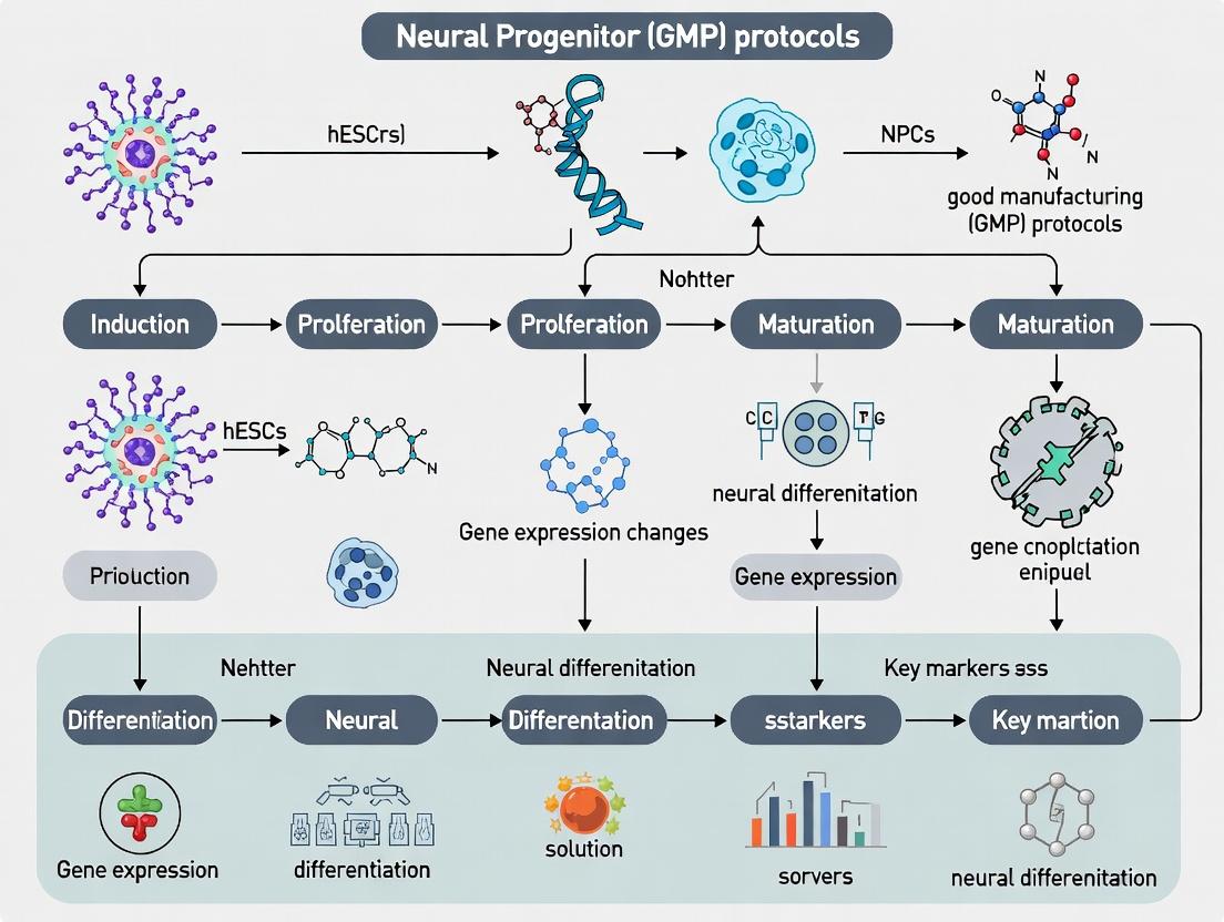
A Comprehensive Guide to GMP-Compliant Neural Progenitor Differentiation from Human Embryonic Stem Cells
This guide provides a detailed roadmap for researchers and industry professionals aiming to differentiate human embryonic stem cells (hESCs) into neural progenitors under Good Manufacturing Practice (GMP) conditions.
A Comprehensive Guide to GMP-Compliant Neural Progenitor Differentiation from Human Embryonic Stem Cells
Abstract
This guide provides a detailed roadmap for researchers and industry professionals aiming to differentiate human embryonic stem cells (hESCs) into neural progenitors under Good Manufacturing Practice (GMP) conditions. It covers the fundamental biology of neural induction, presents step-by-step, scalable methodologies, addresses common troubleshooting and optimization challenges, and discusses critical validation and comparative analyses essential for therapeutic and drug discovery applications. The content is designed to bridge the gap between research-scale protocols and the stringent requirements of clinical translation and pharmaceutical development.
The Science of Neural Fate: Understanding GMP hESC Differentiation to Neural Progenitors
Definition and Context
Neural progenitor cells (NPCs) are multipotent, self-renewing cells of the central nervous system (CNS) that are committed to the neural lineage and can differentiate into neurons, astrocytes, and oligodendrocytes. Within the thesis context of current Good Manufacturing Practice (GMP)-compliant differentiation from human embryonic stem cells (hESCs), NPCs represent a critical, scalable intermediate cell population for regenerative therapies and disease modeling.
Key Molecular Markers
NPC identity is defined by a core set of transcription factors and cell surface proteins. Markers differ between rostral (forebrain) and caudal (hindbrain/spinal cord) patterning.
Table 1: Core Neural Progenitor Cell Markers
| Marker Type | Marker Name | Expression & Function |
|---|---|---|
| Transcription Factors | SOX1, SOX2, SOX3 (SRY-box) | Maintain progenitor state, pluripotency links. |
| PAX6 (Paired box 6) | Rostral NPC identity, neuroectodermal fate. | |
| NESTIN (Intermediate Filament) | Cytoskeletal protein, hallmark of NPCs. | |
| Cell Surface Proteins | CD133 (Prominin-1) | Cell membrane protrusions, enrichment marker. |
| SSEA-1 (Stage-Specific Embryonic Antigen-1) | Lewis X carbohydrate, marks rodent/human NPCs. | |
| FORSE-1 (Forebrain Surface Embryonic Antigen-1) | Forebrain-specific glycolipid antigen. |
Table 2: Markers for Regional Patterning in hESC-Derived NPCs
| Region | Key Transcription Factors | Typical Morphogen Cues in Differentiation |
|---|---|---|
| Forebrain | PAX6, FOXG1, SIX3, OTX2 | Dual SMAD inhibition + WNT inhibition (e.g., IWR-1-endo). |
| Midbrain | LMX1A, FOXA2, OTX2 | FGF8 + SHH (floor plate patterning). |
| Hindbrain/Spinal Cord | HOX gene family (HOXB4, etc.), OLIG2 | Retinoic Acid (RA) + SHH (ventral patterning). |
Therapeutic Potential
NPCs offer a dual therapeutic mechanism: cell replacement and trophic support. Their application in GMP-directed research is pivotal for treating neurodegenerative diseases, stroke, and spinal cord injury.
Table 3: Therapeutic Applications of hESC-Derived NPCs
| Disease Target | Proposed Mechanism | Clinical Trial Phase (as of 2024) |
|---|---|---|
| Parkinson's Disease | Dopaminergic neuron replacement. | Multiple Phase I/II trials (e.g., STEM-PD, BlueRock). |
| Spinal Cord Injury | Myelination, bridge formation, trophic support. | Phase I/II (e.g., Asterias/Lineage Cell Therapeutics). |
| Age-related Macular Degeneration | Retinal pigment epithelium (RPE) support. | Preclinical & Phase I studies. |
| Stroke | Trophic factor secretion, modulation of inflammation. | Several Phase I trials completed. |
Detailed Protocols
Protocol 1: GMP-Compliant hESC to Rostral Neural Prospector Differentiation
Objective: Generate PAX6+ neural rosettes under defined, xeno-free conditions suitable for scale-up.
Materials:
- GMP-grade hESC line.
- Defined, xeno-free hESC maintenance medium (e.g., TeSR-E8 or equivalent).
- Neural Induction Medium (NIM): DMEM/F-12, 1% N-2 Supplement, 1% Non-Essential Amino Acids, 1% GlutaMAX.
- GMP-grade small molecules: SB431542 (TGF-β/Activin/NODAL inhibitor), LDN193189 (BMP inhibitor), XAV939 (WNT inhibitor).
- Recombinant human albumin, GMP-grade.
- Matrix: Recombinant human laminin-521.
Method:
- Culture hESCs: Maintain hESCs on laminin-521 in E8 medium to ~70% confluence.
- Initiate Differentiation (Day 0): Switch to Neural Induction Medium (NIM) supplemented with 10 μM SB431542, 100 nM LDN193189 (Dual SMAD inhibition), and 2 μM XAV939.
- Medium Change: Feed cells daily with fresh NIM + inhibitors for 7 days.
- Rosette Formation (Day 7-10): Neural rosette structures should appear. Manually or enzymatically (using dispase) isolate rosettes for passage.
- NPC Expansion: Plate rosette fragments on fresh laminin-521 in NPC expansion medium (NIM + 20 ng/mL bFGF). Cells can be passaged as aggregates or dissociated with Accutase.
- Characterization: Analyze by flow cytometry for SOX1/2, PAX6, and NESTIN. Purity of >85% PAX6+ cells is typical for rostral NPCs.
Protocol 2: Characterization of NPCs via Flow Cytometry
Objective: Quantify the percentage of NPC marker-positive cells.
Materials:
- Single-cell NPC suspension.
- Fixation/Permeabilization buffer kit.
- PBS with 2% fetal bovine serum (FBS) or BSA.
- Primary antibodies: Mouse anti-PAX6, Rabbit anti-SOX2.
- Isotype control antibodies.
- Fluorochrome-conjugated secondary antibodies (e.g., Alexa Fluor 488, 647).
- Flow cytometer.
Method:
- Harvest and wash NPCs in PBS.
- Fix and permeabilize cells according to kit instructions.
- Incubate cell aliquots (~1x10^6 cells/tube) with primary antibody or isotype control (30 min, 4°C).
- Wash twice with permeabilization buffer.
- Incubate with appropriate secondary antibody (20 min, 4°C, in the dark).
- Wash, resuspend in PBS, and analyze on flow cytometer. Use unstained and single-stained controls for compensation.
Visualizations
Title: hESC to Neural Progenitor Differentiation Workflow
Title: Key Signaling Pathways in Neural Induction
The Scientist's Toolkit: Research Reagent Solutions
Table 4: Essential Materials for GMP hESC-NPC Research
| Item | Function & Rationale | Example Product/Catalog |
|---|---|---|
| GMP-hESC Line | Starting cell source. Must be ethically derived, karyotypically normal, and banked under GMP. | WA09 (H9), Master Cell Bank. |
| Xeno-Free Basal Medium | Eliminates animal-derived components, reducing immunogenicity and variability. | TeSR-E8, StemFlex. |
| Recombinant Laminin-521 | Defined, human-derived extracellular matrix for adhesion and signaling. | Biolamina LN-521. |
| Dual SMAD Inhibitors | Drives neural induction by blocking mesendodermal differentiation. | SB431542, LDN193189. |
| WNT Pathway Inhibitor | Promotes anterior/rostral neural fate. | XAV939, IWR-1-endo. |
| GMP-Grade bFGF (FGF-2) | Expands and maintains NPC population in culture. | Recombinant human FGF-basic. |
| Cell Dissociation Reagent | Gentle enzymatic passaging of NPCs. | Accutase, recombinant trypsin. |
| Flow Cytometry Antibodies | Characterization and QC of NPC marker expression. | Anti-PAX6, SOX2, NESTIN. |
| Mycoplasma Detection Kit | Essential for routine testing of cell bank and culture sterility. | PCR-based detection kit. |
Why GMP? The Critical Importance of Manufacturing Standards for Clinical Translation
Application Note: GMP-Compliant Neural Progenitor Cell (NPC) Differentiation from Human Embryonic Stem Cells (hESCs)
The clinical translation of human embryonic stem cell (hESC)-derived neural progenitor cells (NPCs) for neurodegenerative disorders hinges on reproducible, safe, and well-characterized manufacturing processes. Good Manufacturing Practice (GMP) provides the essential framework to achieve this, transitioning research-grade protocols into therapeutic products suitable for human trials.
Table 1: Impact of Non-GMP vs. GMP Processes on Critical Quality Attributes (CQAs) of hESC-NPCs
| Critical Quality Attribute (CQA) | Research-Grade (Non-GMP) Process Outcome | GMP-Compliant Process Outcome |
|---|---|---|
| Genetic Stability | Potential for undetected karyotypic abnormalities due to limited testing. | Regular, validated karyotyping/CNV analysis; strict passaging limits. |
| Purity & Identity | Variable NPC marker expression (e.g., PAX6, SOX1: 60-85%). | Defined acceptance criteria (e.g., >90% PAX6+); validated flow cytometry assays. |
| Potency | Inconsistent differentiation efficiency into target neurons (e.g., dopaminergic neurons: 30-70%). | Standardized, quantitative functional assays with defined potency units. |
| Sterility | Risk of Mycoplasma, bacterial, fungal contamination. | Aseptic processing, final product sterility testing per pharmacopoeia. |
| Documentation & Traceability | Incomplete records of reagents, cell history. | Full traceability from donor tissue to final vial (Device Master Record, Batch Records). |
The core challenge is replacing research reagents (e.g., animal-sourced components, undefined matrices) with GMP-grade equivalents without altering the product's biological function.
Protocols
Protocol 1: GMP-Compliant Feeder-Free Maintenance of hESCs
Objective: To maintain pluripotent hESCs under defined, xeno-free conditions suitable for initiating differentiation.
- Culture Vessel Coating: Coat cell culture vessels with GMP-grade, human recombinant laminin-521 (e.g., 0.5 µg/cm²) in DPBS. Incubate ≥ 2 hours at 37°C.
- Medium Preparation: Use a commercially available, fully defined, xeno-free hESC maintenance medium. Pre-warm to 37°C.
- Passaging: At ~70% confluence, aspirate medium. Wash with DPBS. Add GMP-grade recombinant enzyme (e.g., TrypLE Select) and incubate at 37°C for 5-7 minutes.
- Neutralization & Seeding: Neutralize enzyme with complete medium. Centrifuge at 200 x g for 4 minutes. Resuspend in fresh medium and seed at a defined seeding density (e.g., 15,000 cells/cm²) in coated vessels.
- Quality Control: Daily morphology assessment. Regular testing for pluripotency markers (OCT4, NANOG) and karyotype stability.
Protocol 2: Directed Differentiation of hESCs to Neural Progenitor Cells (NPCs) Using Dual SMAD Inhibition
Objective: To generate a homogeneous, expandable population of PAX6/SOX1-positive neural rosettes under defined conditions.
Table 2: GMP-Grade Reagent Substitution for Neural Induction
| Research Reagent | GMP-Grade Equivalent | Critical Function |
|---|---|---|
| Matrigel (Mouse Sarcoma) | Recombinant Laminin-521 or Synthemax II-S | Substrate for cell adhesion |
| KnockOut Serum Replacement (KOSR) | Defined, albumin-free, protein-free medium | Provides base nutrients |
| Recombinant Noggin (Research Grade) | GMP-produced Recombinant Noggin | BMP pathway inhibition |
| Recombinant SB431542 (Research Grade) | GMP-produced Small Molecule (ALK5 inhibitor) | TGF-β pathway inhibition |
| N2 Supplement (Research Grade) | GMP-produced, fully defined N2 formulation | Neural specification supplement |
Procedure:
- Day -1: Seed high-quality hESCs as single cells onto GMP-grade laminin-coated plates at an optimized density (e.g., 50,000 cells/cm²) in maintenance medium. Target ~90% confluence for initiation.
- Day 0 (Induction Start): Aspirate maintenance medium. Replace with Neural Induction Medium: Defined basal medium (e.g., DMEM/F-12 with GlutaMAX) supplemented with GMP-grade N2 supplement, 100 ng/mL GMP-grade recombinant Noggin (or LDN-193189), and 10 µM GMP-grade SB431542.
- Days 1-6: Perform 100% medium change daily with fresh Neural Induction Medium. Monitor formation of compact cell colonies with emerging rosette structures.
- Days 7-10 (Rosette Selection): By day 7, distinct rosettes should be visible. Mechanically or enzymatically (using GMP-grade dispase) isolate rosette structures. Re-plate isolated rosettes on laminin-coated plates in NPC Expansion Medium: Neurobasal medium supplemented with GMP-grade B27 (without vitamin A), 20 ng/mL GMP-grade recombinant bFGF, and 20 ng/mL GMP-grade recombinant EGF.
- NPC Expansion: Passage cells as aggregates using gentle cell dissociation reagent. Expand cells for 3-5 passages, characterizing at each stage. Cryopreserve in defined, animal component-free cryopreservation medium.
Diagrams
Title: GMP-Compliant hESC to NPC Differentiation Workflow
Title: GMP Quality Systems Pyramid for NPC Manufacturing
The Scientist's Toolkit: Essential GMP-Ready Research Reagents
Table 3: Key Reagent Solutions for Transitioning to GMP NPC Differentiation
| Reagent Category | Specific Example (GMP-Grade) | Function in Protocol | Critical Consideration |
|---|---|---|---|
| Basal Media | DMEM/F-12, GlutaMAX, HEPES | Provides essential nutrients and buffer for neural induction. | Must be sourced with Drug Master File (DMF) or equivalent regulatory backing. |
| Defined Supplement | N2 Supplement, B-27 Supplement (Xeno-free) | Provides hormones, antioxidants, and proteins for neural cell survival and specification. | Avoids animal-derived components; ensures lot-to-lot consistency. |
| Extracellular Matrix | Recombinant Human Laminin-521 | Defined substrate for cell attachment, replacing Matrigel. | Eliminates tumor-derived variability and immunogenicity risks. |
| Growth Factors | Recombinant Human Noggin, bFGF, EGF | Directs cell fate (Noggin) and supports proliferation (bFGF/EGF). | Required with Certificate of Analysis (CoA) detailing purity, sterility, endotoxin levels. |
| Small Molecules | SB431542 (ALK5 inhibitor), LDN-193189 (BMP inhibitor) | Chemically defined pathway inhibitors for robust neural induction. | Preferred over protein factors for stability and cost; must be sourced as GMP starting materials. |
| Dissociation Agents | Recombinant Trypsin (TrypLE Select) | Enzymatic passaging of cells. | Animal-free, defined protease activity; reduces cleavage variability. |
| Cryopreservation Medium | Defined, protein-free freezing medium | Long-term storage of Master Cell Banks. | Contains DMSO and defined cryoprotectants; supports high post-thaw viability. |
Within a GMP-compliant research program aimed at generating neural progenitor cells (NPCs) from human embryonic stem cells (hESCs), the initial selection of the hESC line is a critical, foundational decision. The chosen line's inherent characteristics profoundly influence the efficiency, reproducibility, and safety of the subsequent differentiation process and the final cellular product. This application note details the essential characteristics to evaluate and provides a structured framework for selecting an appropriate hESC line for GMP neural progenitor differentiation.
Key Characteristics for Evaluation
Genomic Stability and Karyotype
A normal, stable karyotype is non-negotiable for therapeutic applications. Recurrent abnormalities, particularly gains on chromosomes 1, 12, 17, and 20, are common in cultured hESCs and can confer a growth advantage, potentially affecting differentiation propensity and tumorigenicity.
Table 1: Genomic Stability Assessment Criteria
| Characteristic | Acceptance Criterion | Assessment Method |
|---|---|---|
| Karyotype | Normal (46, XX or XY) by G-banding at passage ≥P30. | G-banding karyotype analysis (≥20 metaphase spreads). |
| Submicroscopic Variants | No known pathogenic copy number variations (CNVs). | High-resolution array CGH or SNP array. |
| Short Tandem Repeat (STR) Profile | Unique, consistent profile matching cell bank. | DNA fingerprinting (10-16 loci). |
Pluripotency and Differentiation Capacity
The line must demonstrate robust, verifiable pluripotency and a proven capacity for neural lineage commitment. This is assessed through marker expression and functional assays.
Table 2: Pluripotency and Neural Propensity Metrics
| Marker/Assay | Expected Result (Pluripotency) | Expected Result (Neural Propensity) |
|---|---|---|
| Surface Markers | >90% SSEA-4+, TRA-1-60+ by flow cytometry. | N/A |
| Transcription Factors | High expression of OCT4, NANOG, SOX2 (qPCR/ICC). | N/A |
| In Vitro Differentiation | Positive for markers of all three germ layers (ectoderm: PAX6, mesoderm: Brachyury, endoderm: SOX17). | High yield of PAX6+/SOX1+ NPCs upon directed differentiation (≥70%). |
| In Vivo Teratoma Assay | Formation of tissues from all three germ layers in immunocompromised mice. | N/A |
Microbial Safety and GMP Derivation History
For clinical translation, the cell line's derivation and banking history must align with regulatory standards to minimize risk of adventitious agent transmission.
Table 3: Safety and GMP Compliance Criteria
| Aspect | Ideal Status | Documentation Required |
|---|---|---|
| Derivation Conditions | Under xeno-free conditions, without animal-derived components. | Master Cell Bank (MCB) and Working Cell Bank (WCB) records. |
| Pathogen Testing | Full panel negative (HIV, HBV, HCV, Mycoplasma, etc.) on MCB. | Certificates of Analysis (CoA) from qualified lab. |
| Cell Line Provenance | Ethical approval and informed consent for derivation are documented. | Institutional Review Board (IRB) statements. |
Growth and Cloning Characteristics
Practical considerations for scaling and single-cell passaging under GMP conditions are vital.
Table 4: Practical Growth Characteristics
| Parameter | Optimal Range | Protocol Impact |
|---|---|---|
| Population Doubling Time | 20-30 hours in log phase. | Determines production timeline. |
| Single-Cell Survival Rate | >30% after enzymatic dissociation in ROCK inhibitor. | Enables clonal expansion and single-cell seeding for differentiation. |
| Saturation Density | Consistent, high-density colonies without excessive differentiation. | Informs seeding density for differentiation initiation. |
Selection Protocol: A Stepwise Decision Framework
Protocol Title: Systematic Evaluation and Selection of hESC Lines for GMP NPC Differentiation
Objective: To empirically compare and select the most suitable hESC line based on predefined criteria for robustness, stability, and neural differentiation efficiency.
Materials:
- Candidate hESC lines (e.g., WA09/H9, RC-17, MAN-1, etc.).
- Essential 8 or similar xeno-free, chemically defined medium.
- Recombinant laminin-521 or vitronectin-coated plates.
- ROCK inhibitor (Y-27632).
- Neural induction medium (e.g., dual SMAD inhibition-based: DMEM/F12, N2 supplement, Noggin, SB431542).
- Fixatives, antibodies for flow cytometry/ICC (OCT4, NANOG, PAX6, SOX1).
- RNA extraction kit, cDNA synthesis kit, qPCR reagents.
- Karyotyping and Mycoplasma detection services.
Procedure:
Phase 1: Preliminary Screening & Expansion
- Revival & Expansion: Thaw vials from the WCB of each candidate line onto coated plates in Essential 8 medium supplemented with 10µM ROCK inhibitor. Maintain cultures in a 37°C, 5% CO2 incubator, passaging as clumps using EDTA every 5-7 days.
- Mycoplasma Testing: Confirm cultures are mycoplasma-free using a PCR-based assay.
- Bank Preparation: At passage 3 post-thaw, prepare a research cell bank for each line. Cryopreserve in aliquots using controlled-rate freezing.
Phase 2: Core Characterization Assays
- Pluripotency Verification (Day 7):
- Harvest cells from one well of a 6-well plate. Perform flow cytometry for SSEA-4 and TRA-1-60. Fix parallel wells for immunocytochemistry for OCT4 and NANOG.
- Extract RNA and perform qPCR for POUSF1 (OCT4), NANOG, and SOX2. Normalize to housekeeping genes (e.g., GAPDH).
Karyotype Analysis (Day 10):
- Submit cells from one T25 flask per line at passage ≥30 (from the original WCB or after extended culture) for standard G-banding karyotype analysis (minimum 20 metaphases).
Neural Differentiation Trial (Days 1-10):
- Seed a defined number of single hESCs (e.g., 50,000 cells/cm²) on laminin-511-coated plates in Essential 8 + ROCKi.
- Day 0: Upon confluence (24h later), switch to neural induction medium.
- Day 7-10: Harvest cells. Analyze by flow cytometry for co-expression of PAX6 and SOX1. Calculate the percentage of PAX6+/SOX1+ double-positive NPCs.
Phase 3: Data Integration & Line Selection
- Scorecard Analysis: Create a weighted scoring matrix based on Tables 1-4. Assign scores (e.g., 1-5) for each criterion.
- Decision: Select the line that achieves:
- Non-negotiable: Normal karyotype, negative pathogen tests, >90% pluripotency markers.
- Highest Score: In weighted categories of neural differentiation efficiency (>70% PAX6+/SOX1+), growth stability, and GMP-compliant history.
The Scientist's Toolkit: Essential Research Reagents
Table 5: Key Reagent Solutions for hESC Line Characterization
| Reagent/Category | Example Product | Primary Function |
|---|---|---|
| Xeno-Free Culture Medium | Essential 8, mTeSR Plus | Maintains hESCs in a defined, feeder-free pluripotent state. |
| Recombinant Attachment Matrix | Laminin-521, Vitronectin (VTN-N) | Provides a defined substrate for hESC adhesion and survival, critical for GMP. |
| ROCK Inhibitor | Y-27632 dihydrochloride | Enhances single-cell survival after dissociation by inhibiting apoptosis. |
| Neural Induction Cocktail | Dual SMAD Inhibitors (Noggin + SB431542) | Drives efficient neural conversion by inhibiting BMP and TGF-β pathways. |
| Pluripotency Antibody Panel | Anti-OCT4, SSEA-4, TRA-1-60 | Validates undifferentiated status via flow cytometry and immunocytochemistry. |
| Neural Progenitor Antibody Panel | Anti-PAX6, SOX1, Nestin | Identifies and quantifies early neural progenitor cells. |
| qPCR Assay Kit | TaqMan hPSC Scorecard Panel | Profiles lineage-specific gene expression to assess pluripotency and differentiation bias. |
Visualizations
Diagram 1 Title: hESC Line Selection Workflow for GMP NPC Programs
Diagram 2 Title: Dual SMAD Inhibition Drives Neural Specification
1. Introduction & Thesis Context Within the broader thesis on achieving robust, scalable, and GMP-compliant differentiation of human embryonic stem cells (hESCs) into neural progenitor cells (NPCs), the foundational step of recapitulating embryonic neural induction is paramount. This process involves the conversion of naive epiblast cells to neuroectoderm, a fate defaultly suppressed by TGF-β/Activin/Nodal and BMP signaling. In vitro, this is achieved by dual-SMAD inhibition, a cornerstone protocol that must be optimized for reproducibility and clinical translation. This application note details current protocols and reagent solutions for this critical phase.
2. Key Signaling Pathways & Quantitative Data Summary
Table 1: Core Signaling Pathways in Early Neural Induction
| Pathway | Key Ligands | Role in Early Development | Effect of Inhibition | Key Target Genes |
|---|---|---|---|---|
| BMP | BMP4, BMP2 | Promotes epidermal/trophoblast fate; suppresses neural fate | Induces neuroectoderm | ID1, ID2, MSX1 ↓; SOX1, PAX6 ↑ |
| TGF-β/Activin/Nodal | Nodal, Activin A | Maintains pluripotency; directs mesendodermal fate | Synergizes with BMP inhibition to enhance neural induction | NANOG ↓; SOX2 ↑ |
| FGF | FGF2, FGF4 | Supports epiblast survival; primes for neural differentiation | Required for efficient neural conversion | FGFR1; |
Table 2: Comparative Efficiency of Neural Induction Protocols (Representative Data)
| Protocol Name | Key Inhibitors/Drugs | Duration (Days) | % PAX6+ NPCs (by Flow Cytometry) | Key Marker Expression (qPCR) | Reference/Scale |
|---|---|---|---|---|---|
| Classic Dual-SMAD | SB431542 (TGF-βi) + LDN-193189 (BMPi) | 10-12 | 85-95% | High SOX1, PAX6, FOXG1 | Chambers et al., 2009 (Lab) |
| GMP-Adapted Dual-SMAD | A 83-01 (TGF-βi) + LDN-193189 (BMPi) in defined media | 10-12 | 88-93% | Consistent NES, SOX2 | Kirpatrick et al., 2021 (Pilot Scale) |
| BMP Inhibition Only | LDN-193189 or Noggin | 10-12 | 60-75% | Moderate PAX6, higher MIXL1 (mixed fate) | Zhang et al., 2010 |
3. Detailed Experimental Protocols
Protocol 3.1: GMP-Compliant Neural Induction via Dual-SMAD Inhibition Objective: To differentiate hESCs (maintained in a defined, xeno-free matrix) into a highly pure population of neuroepithelial cells.
Materials & Pre-Culture:
- hESCs: Master cell bank-derived, pluripotent (OCT4+ >90%).
- Matrix: Recombinant human laminin-521 (5 µg/cm²) or GMP-qualified synthetic substrate.
- Basal Medium: DMEM/F-12 + GlutaMAX, supplemented with 1% N-2 supplement.
- Small Molecules: A 83-01 (TGF-β receptor inhibitor, 5 µM), LDN-193189 (BMP receptor inhibitor, 100 nM). Use GMP-grade if available.
- Rock Inhibitor (Y-27632): 10 µM, for plating only.
- Equipment: 37°C, 5% CO₂ incubator; certified biosafety cabinet.
Procedure: Day -1: Seed hESCs as single cells using TrypLE Select onto laminin-521-coated plates in Essential 8 Flex Medium + 10 µM Y-27632. Target 50-70% confluence for Day 0. Day 0 (Induction Start): Aspirate medium. Rinse once with DMEM/F-12. Add neural induction medium: Basal Medium + 5 µM A 83-01 + 100 nM LDN-193189. Days 2 & 4: Perform a full medium change with fresh neural induction medium. Days 6-12: Monitor morphology. Colonies should thicken, form raised, columnar epithelial structures (neural rosettes). Change medium every other day. Endpoint Analysis (Day 10-12): Harvest cells for analysis. A successful induction yields >85% PAX6+/SOX1+ cells by immunocytochemistry. For passaging, use gentle cell dissociation reagent to maintain rosette structures.
Protocol 3.2: Quality Control Assessment via Flow Cytometry Objective: Quantify the percentage of neural progenitor cells post-induction.
- Cell Harvest: Wash cells with DPBS, dissociate using Accutase for 5-7 min at 37°C. Neutralize with basal medium + 10% FBS (or BSA). Pellet at 300g for 5 min.
- Fixation & Permeabilization: Resuspend pellet in 4% PFA for 15 min at RT. Wash with DPBS. Permeabilize with 90% ice-cold methanol for 30 min on ice. Wash with FACS buffer (DPBS + 2% BSA).
- Staining: Aliquot 1x10⁶ cells per tube. Incubate with primary antibody (anti-PAX6, anti-SOX1) diluted in FACS buffer for 1 hr at RT. Use isotype controls. Wash.
- Detection: Incubate with appropriate fluorophore-conjugated secondary antibody for 45 min at RT, protected from light. Wash.
- Analysis: Resuspend in FACS buffer + DAPI (for live/dead discrimination). Analyze on a flow cytometer. Gate on single, live cells and calculate % positive for PAX6 and/or SOX1.
4. The Scientist's Toolkit: Research Reagent Solutions
Table 3: Essential Materials for Neural Induction
| Item | Function | Example (Vendor-Neutral) |
|---|---|---|
| Defined, Xeno-Free Basal Medium | Provides consistent nutrient base without animal-derived components. | DMEM/F-12 with HEPES |
| N-2 Supplement | Serum-free supplement essential for survival and differentiation of neural cells. | 100X formulation (e.g., recombinant human) |
| TGF-β Pathway Inhibitor | Blocks Activin/Nodal signaling, synergizing with BMP inhibition. | A 83-01 or SB431542 (GMP-grade available) |
| BMP Pathway Inhibitor | Antagonizes BMP signaling, releasing default neural differentiation. | LDN-193189 or recombinant Noggin |
| GMP-Qualified Extracellular Matrix | Provides adhesion substrate supporting polarized neuroepithelium formation. | Recombinant human laminin-521 or synthetic peptide hydrogel |
| Gentle Dissociation Reagent | Enzymatically detaches cells while preserving surface markers and viability. | TrypLE Select or Accutase |
| Neural Lineage Antibodies | For quality control via immunostaining and flow cytometry. | Anti-PAX6, anti-SOX1, anti-Nestin, anti-SOX2 |
5. Pathway & Workflow Visualizations
Diagram 1: Logic of Neural Induction via Dual-SMAD Inhibition (Max 760px)
Diagram 2: GMP Neural Induction Workflow (Max 760px)
Diagram 3: Dual-SMAD Inhibition Molecular Targets (Max 760px)
Within the broader thesis investigating robust, scalable, and standardized differentiation of human embryonic stem cells (hESCs) into neural progenitors (NPs) under Good Manufacturing Practice (GMP) conditions, establishing rigorous identity benchmarks is paramount. This application note details the critical quality attributes (CQAs) of purity, potency, and identity that define a successful GMP-compliant neural progenitor product for research and clinical applications.
Critical Quality Attributes (CQAs) and Quantitative Benchmarks
To ensure batch-to-batch consistency and therapeutic predictability, NPs must be characterized against the following benchmarks.
Table 1: Identity and Purity Benchmarks for GMP Neural Progenitors
| CQA Category | Specific Marker/Trait | Target Benchmark (Quantitative) | Assay Method | Rationale |
|---|---|---|---|---|
| Purity | Co-expression of PAX6 & SOX1 | ≥ 90% of total cells | Flow Cytometry | Definitive neuroectodermal identity. |
| Purity | Presence of OCT4 (Pluripotency) | ≤ 2% of total cells | Flow Cytometry | Ensures complete exit from pluripotent state. |
| Purity | Presence of SOX17 (Endoderm) / Brachyury (Mesoderm) | ≤ 1% each | Flow Cytometry | Confirms absence of non-neural lineage contaminants. |
| Identity | Nestin (Intermediate Filament) | ≥ 95% of total cells | Immunocytochemistry | Progenitor cell state marker. |
| Identity | Forebrain Identity (FOXG1) | ≥ 85% of PAX6+ cells | qRT-PCR / Imaging | Regional specification benchmark for cortical fates. |
| Potency | In Vitro Differentiation Capacity (βIII-tubulin+ neurons) | ≥ 70% neuronal yield after 14-day differentiation | Immunocytochemistry | Functional capacity to generate post-mitotic neurons. |
| Potency | In Vivo Teratoma Formation | 0% incidence (in permissive model) | In Vivo Assay | Safety benchmark; confirms lack of pluripotent residue. |
| Genomic Stability | Karyotype (e.g., G-banding) | Normal (46, XY/XX) through P10 | Karyotyping | Ensures genetic integrity over passages. |
Table 2: Key Secretory Profile for Potency Assessment
| Analytic | Method | Target Range (pg/mL/10^6 cells/24h) | Implication |
|---|---|---|---|
| Vascular Endothelial Growth Factor (VEGF) | ELISA | 500 - 2000 | Pro-angiogenic/trophic factor secretion. |
| Brain-Derived Neurotrophic Factor (BDNF) | ELISA | 100 - 500 | Neurotrophic support capacity. |
| Hepatocyte Growth Factor (HGF) | ELISA | 50 - 300 | Paracrine signaling activity. |
Detailed Experimental Protocols
Protocol 3.1: Dual-Label Flow Cytometry for PAX6/SOX1 Co-expression
Objective: Quantify the percentage of cells co-expressing definitive neuroectodermal transcription factors PAX6 and SOX1. Materials: See "Scientist's Toolkit," Table 3. Procedure:
- Harvesting: Dissociate NP monolayer using Accutase for 5-7 min at 37°C. Quench with DPBS/2% BSA. Pellet at 300g for 5 min.
- Fixation & Permeabilization: Resuspend cell pellet in 100 µL of BD Cytofix/Cytoperm solution. Incubate for 20 min at 4°C. Wash with 1x BD Perm/Wash buffer.
- Staining: Resuspend cells in Perm/Wash buffer. Add directly conjugated antibodies: Anti-PAX6-Alexa Fluor 488 (1:100) and Anti-SOX1-PE (1:50). Include isotype controls. Incubate for 45 min at 4°C in the dark.
- Analysis: Wash cells twice, resuspend in flow buffer. Analyze on a calibrated flow cytometer. Use FSC-A/SSC-A to gate on single cells, then assess fluorescence in FL1 (PAX6) vs. FL2 (SOX1) channels. Calculate percentage in dual-positive quadrant.
Protocol 3.2: In Vitro Potency Assay (Neuronal Differentiation)
Objective: Assess the functional capacity of NPs to differentiate into βIII-tubulin (TUBB3) positive neurons. Materials: Neuronal differentiation medium (DMEM/F-12, N2 supplement, B27 supplement without vitamin A, 1 µM cAMP, 10 ng/mL BDNF). Procedure:
- Plating: Seed dissociated NPs onto poly-L-ornithine/laminin-coated 24-well plates at 50,000 cells/cm² in NP expansion medium.
- Induction: After 24h, switch to neuronal differentiation medium.
- Maintenance: Feed cells by replacing 50% of the medium every other day for 14 days.
- Fixation & Staining: On day 14, fix with 4% PFA for 15 min. Permeabilize with 0.1% Triton X-100. Block with 5% normal goat serum. Incubate with primary anti-βIII-tubulin antibody (1:500) overnight at 4°C. Incubate with Alexa Fluor 555 secondary (1:1000) for 1h at RT. Counterstain nuclei with DAPI.
- Quantification: Image 10 random fields per well using a fluorescence microscope. Calculate the percentage of TUBB3+ cells relative to total DAPI+ nuclei.
Visualizations
Diagram 1: Key Signaling Pathways in hESC to NP Commitment
Diagram 2: GMP NP Characterization Workflow
The Scientist's Toolkit: Essential Research Reagent Solutions
Table 3: Key Reagents for GMP NP Characterization
| Reagent / Material | Function / Target | Example Product (Research-Grade) | Critical Note for GMP |
|---|---|---|---|
| Dual-SMAD Inhibitors | Induces neuroectoderm via TGF-β/BMP inhibition. | SB431542 (TGF-βi), LDN193189 (BMPi) | Transition to GMP-grade small molecules is essential. |
| PAX6 & SOX1 Antibodies | Definitive neuroectodermal identity confirmation. | Anti-PAX6 Alexa Fluor 488, Anti-SOX1 PE | Validate clones for specificity; seek GMP-compliant alternatives. |
| Accutase | Gentle, xeno-free cell dissociation. | Recombinant Accutase solution | Preferred over trypsin for NP passaging; use GMP version. |
| hESC-Qualified Basement Membrane Matrix | Provides adhesion substrate for polarised NP growth. | Geltrex, Cultrex BME | Define lot-to-lot consistency; final product requires animal-free matrix. |
| Neuronal Differentiation Kit | Standardized medium for potency assay. | STEMdiff Neuronal Differentiation Kit | Useful for research benchmarking; final process may require defined, xeno-free media. |
| Flow Cytometry Validation Beads | Instrument calibration and assay standardization. | CS&T Beads, Rainbow Calibration Particles | Mandatory for quantitative, reproducible flow data across batches. |
| Mycoplasma Detection Kit | Sterility and safety testing. | PCR-based detection kit | Routine testing required; use highly sensitive, validated kits. |
Step-by-Step Protocol: Scalable GMP Differentiation of hESCs to Neural Progenitors
Within a GMP-compliant thesis focused on generating neural progenitor cells (NPCs) from human embryonic stem cells (hESCs), the initial culture phase is critical. Consistent, high-quality NPC differentiation mandates a starting population of hESCs that is uniformly pluripotent, genetically stable, and free of spontaneous differentiation. This application note details a robust, defined culture system for maintaining hESC pluripotency, serving as the essential pre-differentiation foundation for downstream neural lineage specification.
Defined Culture System Components and Rationale
A defined system eliminates serum and undefined feeders, using a basal medium supplemented with specific recombinant growth factors and matrix proteins to precisely control pluripotency signaling pathways.
Table 1: Core Components of a Defined hESC Pluripotency Maintenance System
| Component Category | Specific Example(s) | Function & Rationale |
|---|---|---|
| Basal Medium | mTeSR Plus, StemFlex, E8 medium | Chemically defined, nutrient-optimized formulations lacking serum or albumin. Provide consistent base for growth factor activity. |
| Essential Growth Factors | Recombinant human FGF-β (bFGF) at 100 ng/mL; Recombinant human TGF-β1/Activin/Nodal agonists | bFGF: Activates MAPK/ERK and PI3K pathways, promoting self-renewal. TGF-β1/Activin/Nodal: Activates SMAD2/3 signaling, sustaining core pluripotency transcription factor network (OCT4, NANOG, SOX2). |
| Defined Matrix | Recombinant human Vitronectin; Synthemax II-SC | Provides integrin-mediated adhesion (e.g., via αVβ5) in a defined, xeno-free format, replacing Matrigel. |
| Rho-associated kinase (ROCK) inhibitor | Y-27632 (10 µM) | Used during passaging to inhibit apoptosis (anoikis), improving single-cell survival and clonal recovery. |
| Cell Dissociation Agent | Gentle Cell Dissociation Reagent (GCDR); ReLeSR | Enzyme-free or mild enzymatic agents for clump or single-cell passaging, minimizing membrane damage. |
Key Signaling Pathways for Pluripotency Maintenance
The defined system co-activates two primary signaling axes to sustain the pluripotent ground state.
Diagram Title: Defined Culture Signaling for hESC Pluripotency
Detailed Protocol: Routine Maintenance of hESCs in Defined Conditions
Materials:
- Cells: Undifferentiated hESC line (e.g., H9, HUES9).
- Coating Solution: Recombinant human Vitronectin (VTN-N) at 5 µg/mL in DPBS+/+.
- Complete Culture Medium: mTeSR Plus or equivalent.
- Passaging Reagents: DPBS-/-, Gentle Cell Dissociation Reagent (GCDR), mTeSR Plus supplemented with 10 µM Y-27632 (ROCKi medium).
- Equipment: 37°C incubator (5% CO2, >95% humidity), biological safety cabinet, centrifuge, inverted microscope, water bath.
Procedure:
- Matrix Coating: Coat tissue culture plates with VTN-N solution (0.5 mL/well for 12-well). Incubate at room temperature for 1 hour. Aspirate immediately before use. Do not let wells dry.
- Daily Maintenance: Aspirate spent medium. Add fresh, pre-warmed complete medium daily. Monitor colonies daily for morphology (tight, high-refractivity borders) and check for spontaneous differentiation (flattened, spread cells).
- Passaging (Every 5-7 days at ~80% confluence): a. Aspirate medium and rinse cells with DPBS-/-. b. Add GCDR (0.5 mL/well of 12-well) and incubate at 37°C for 5-8 minutes. c. Once cell colonies begin to detach at edges, aspirate GCDR. Gently add 1 mL of ROCKi medium per well and dislodge cells by pipetting. d. Transfer cell suspension to a conical tube. Rinse well with ROCKi medium to collect remaining cells. e. Optional: Centrifuge at 300 x g for 5 minutes. Aspirate supernatant and resuspend in ROCKi medium. Alternatively, plate directly without centrifugation to minimize stress. f. Perform a viable cell count. Plate cells at a density of 1.0-1.5 x 10^4 cells/cm² onto VTN-N coated plates in ROCKi medium. g. After 24 hours, replace ROCKi medium with standard complete medium. Continue daily feeding.
- Quality Control: At each passage, assess viability via trypan blue exclusion (target >90%). Regularly confirm pluripotency via immunocytochemistry (OCT4, NANOG, SSEA-4) and karyotype analysis (every 10-15 passages).
Table 2: Critical Quantitative Parameters for Routine Culture
| Parameter | Optimal Range | Purpose & Notes |
|---|---|---|
| Seeding Density | 1.0 - 1.5 x 10^4 cells/cm² | Prevents over-confluence and spontaneous differentiation. Ensures consistent colony formation. |
| Passage Frequency | Every 5 - 7 days | Maintains cells in log-phase growth, prevents over-confluence. |
| Medium Change Frequency | Daily | Ensures nutrient replenishment and stable factor concentration. |
| ROCK Inhibitor (Y-27632) | 10 µM | Used for 24 hours post-passage only. Prolonged use can alter biology. |
| Confluence at Passaging | 70 - 85% | Ideal for maintaining undifferentiated state. |
Pre-Differentiation Readiness Assessment
Before initiating neural induction, assess hESC quality.
Protocol: Immunocytochemistry for Pluripotency Markers
- Culture hESCs on VTN-N-coated glass coverslips in a 24-well plate.
- At ~70% confluence, aspirate medium, rinse with DPBS, and fix with 4% paraformaldehyde for 15 minutes.
- Permeabilize with 0.1% Triton X-100 for 10 minutes. Block with 3% BSA for 1 hour.
- Incubate with primary antibodies (e.g., anti-OCT4, anti-NANOG) diluted in blocking buffer overnight at 4°C.
- Rinse 3x with PBS. Incubate with appropriate fluorescent secondary antibodies and DAPI (1 µg/mL) for 1 hour at RT in the dark.
- Rinse and mount. Image using a fluorescence microscope. Target: >95% nuclear co-expression of OCT4/NANOG.
The Scientist's Toolkit: Essential Research Reagent Solutions
Table 3: Key Reagents for Defined hESC Culture
| Reagent Name | Supplier Examples | Function in Protocol |
|---|---|---|
| mTeSR Plus Medium | STEMCELL Technologies | Defined, feeder-free culture medium. Contains necessary FGF and TGF-β factors. |
| Recombinant Human Vitronectin (VTN-N) | Thermo Fisher Scientific | Defined, xeno-free adhesion substrate for cell attachment and spreading. |
| Gentle Cell Dissociation Reagent (GCDR) | STEMCELL Technologies | Enzyme-free solution for gentle passaging, preserving surface markers. |
| Y-27632 (ROCK Inhibitor) | Tocris, Selleckchem | Small molecule inhibitor used to significantly improve survival of dissociated hESCs. |
| Anti-OCT4 / NANOG Antibodies | Cell Signaling Technology, Abcam | Primary antibodies for quality control via immunostaining to confirm pluripotency. |
| StemCell Karyostat Kit | Thermo Fisher Scientific | GMP-compatible qPCR-based assay for rapid detection of common culture-acquired karyotypic abnormalities. |
Introduction Within a GMP-compliant framework for generating neural progenitor cells (NPCs) from human embryonic stem cells (hESCs), efficient and robust neural induction is the critical first step. This protocol application note details established methods, focusing on the widely adopted Dual-SMAD inhibition protocol and its alternatives, providing detailed methodologies for implementation in a regulated research environment.
1. Core Signaling Pathways and Established Protocols Neural induction involves diverting pluripotent cell fate from default epidermal differentiation toward neuroectoderm. Key pathways targeted include BMP/TGFβ and Wnt.
Diagram 1: Neural Induction Signaling Pathways
2. Quantitative Comparison of Neural Induction Protocols The following table summarizes key parameters and outcomes for established protocols.
Table 1: Comparison of Primary Neural Induction Protocols
| Protocol | Core Components | Duration | Reported PAX6+ NPC Efficiency | Key Advantages | Considerations for GMP |
|---|---|---|---|---|---|
| Dual-SMAD Inhibition | LDN-193189 (BMP inh.) + SB431542 (TGFβ/Activin/Nodal inh.) in basal media (e.g., N2B27) | 10-12 days | 80-95% | Highly efficient, rapid, chemically defined. | Requires xeno-free inhibitor sources; cost optimization. |
| SDIA (Stromal Cell-Induced) | Co-culture with PA6 or MS5 stromal cells. | 14-21 days | 60-80% | Can pattern cells; minimal supplements. | Xenogenic, variable, less defined, complex quality control. |
| Aggregation (EB-Based) | Formation of embryoid bodies in neural induction media, often with SMAD inhibitors. | 10-14 days | 70-90% | Scalable, mimics development. | EB size variability, more complex process control. |
| Monolayer (Minimal Media) | Default differentiation in low-attachment plates with minimal growth factors. | 14+ days | 50-70% | Simple, low-cost, no inhibitors. | Lower efficiency, longer timeline, less consistent. |
3. Detailed Experimental Protocols
Protocol 3.1: GMP-Adherent Dual-SMAD Inhibition for NPC Generation Objective: Generate a homogeneous population of neuroepithelial progenitors from hESCs. Materials: See "The Scientist's Toolkit" below. Procedure:
- Pre-differentiation hESC Culture: Maintain hESCs on a GMP-grade substrate (e.g., recombinant laminin-521) in defined, xeno-free mTeSR or E8 medium. Ensure >90% confluence and typical pluripotency morphology.
- Neural Induction Initiation (Day 0): Aspirate pluripotency medium. Rinse once with DMEM/F-12. Add neural induction medium A: N2B27 base supplemented with 100 nM LDN-193189 and 10 µM SB431542.
- Medium Exchange (Days 2, 4, 6, 8): Aspirate spent medium completely. Refresh with neural induction medium A.
- Neuroepithelium Expansion (Day 10+): By day 10, dense rosette structures should be visible. Replace medium with neural induction medium B: N2B27 base supplemented with 20 ng/mL bFGF (FGF2). Culture for an additional 2-4 days, passaging as needed using gentle enzymatic (e.g., Dispase) or manual dissection to expand primitive NPCs.
- Characterization (Day 12-14): Analyze by flow cytometry for PAX6 and SOX1 expression (expect >85% positive). Perform immunocytochemistry for N-CADHERIN and NESTIN. Perform qPCR for downregulation of OCT4 (POU5F1) and upregulation of PAX6, SOX1.
Protocol 3.2: Aggregate-Based Neural Induction with SMAD Inhibition Objective: Generate NPCs via embryoid body formation, suitable for scalable suspension culture. Procedure:
- EB Formation (Day 0): Dissociate hESCs to single cells using a GMP-grade enzyme (e.g., TrypLE). Resuspend in neural induction medium A (see 3.1) containing 10 µM Y-27632 (ROCKi). Seed at 1-3x10^5 cells/mL in low-attachment plates or flasks to form aggregates.
- Medium Exchange (Days 2, 4): Gently transfer aggregates to a conical tube. Allow to settle for 5-10 minutes. Aspirate supernatant and resuspend in fresh neural induction medium A without Y-27632.
- Neural Commitment (Days 6-10): On day 6, exchange medium to neural induction medium B with bFGF. Continue feeding every other day. By day 10, EBs should display a translucent, neural-like periphery.
- NPC Harvest (Day 10-12): Collect EBs, dissociate with papain or TrypLE, and plate on laminin-coated vessels in NPC maintenance medium (N2B27 + bFGF) to obtain a monolayer culture for expansion and characterization.
Diagram 2: Dual-SMAD Protocol Workflow
4. The Scientist's Toolkit: Research Reagent Solutions
Table 2: Essential Materials for Neural Induction Protocols
| Reagent / Material | Function / Role | Example GMP-Compatible Sources/Considerations |
|---|---|---|
| LDN-193189 (HCl) | Selective BMP type I receptor (ALK2/3) inhibitor. Blocks BMP-SMAD1/5/8 signaling. | Source from certified GMP vendors. Prepare aliquots in DMSO per stability data. |
| SB431542 (or A83-01) | TGF-β/Activin/Nodal type I receptor (ALK4/5/7) inhibitor. Blocks TGFβ-SMAD2/3 signaling. | Source from certified GMP vendors. A83-01 has a longer half-life. |
| N2 & B27 Supplements | Chemically defined serum replacements essential for neural cell survival and growth. | Use xeno-free, GMP-manufactured formulations (e.g., Thermo Fisher). |
| DMEM/F-12 & Neurobasal | Base media components for N2B27 formulation. | Use GMP-grade, endotoxin-tested. |
| Recombinant Laminin-521 | GMP-grade cell culture substrate supporting pluripotent and neural stem cell adhesion. | Essential for xeno-free, defined adherent protocols. |
| Recombinant bFGF (FGF2) | Expands and maintains primitive neural progenitor cells post-SMAD inhibition. | Use GMP-grade, carrier-free, defined activity. |
| ROCK Inhibitor (Y-27632) | Enhances survival of single pluripotent cells and aggregates during initiation. | Use in initial dissociation/EB formation step only. |
| GMP-Grade Dissociation Enzyme | For gentle passaging of neuroepithelium or EB dissociation (e.g., TrypLE, Dispase). | Essential for maintaining cell health and GMP traceability. |
This document serves as an application note and protocol guide within the broader thesis on achieving robust, reproducible, and scalable Good Manufacturing Practice (GMP)-compliant differentiation of human embryonic stem cells (hESCs) into neural progenitor cells (NPCs). The cornerstone of this process is the formulation of chemically defined, xeno-free media using GMP-grade supplements. Sourcing appropriate GMP-grade B27, N2, and recombinant growth factors is critical for ensuring product consistency, safety, and regulatory compliance for downstream clinical applications.
Sourcing GMP-Grade Components: Key Considerations & Data
The transition from research-grade to GMP-grade supplements involves stringent verification of source, documentation, and quality controls.
Table 1: Comparison of Research-Grade vs. GMP-Grade Supplement Attributes
| Attribute | Research-Grade (e.g., RUO) | GMP-Grade (e.g., cGMP, MHRA/EMA compliant) |
|---|---|---|
| Intended Use | Research, non-clinical | Manufacture of therapeutics for human use |
| Quality System | ISO 9001, QMS (variable) | Full cGMP (21 CFR Part 210/211, ICH Q7) |
| Traceability & TSE/BSE | Certificate of Analysis (CoA) may be provided | Full traceability from raw materials. TSE/BSE statement (EU TSE 999/2001). |
| Documentation | Limited DMF (Drug Master File) access | Regulatory Support File (RSF), Type II/III DMF available for cross-reference in IND/IMPD. |
| Manufacturing | Non-classified or ISO 7/8 environment | Certified, audited facilities; often ISO 14644 Class 7/8. |
| Change Control | Notification may not be provided | Strict, validated change control process with customer notification. |
| Price | Baseline (1x) | Typically 5x to 20x higher |
Table 2: Example GMP-Grade Suppliers and Product Specifications (Current Market)
| Component | Example Supplier(s) | GMP Designation | Key Format & Documentation |
|---|---|---|---|
| B27 Supplement | Thermo Fisher (Gibco), STEMCELL Technologies | cGMP, for cell therapy manufacturing | Xeno-free, liquid (50x), serum-free. Full RSF, DMF. |
| N2 Supplement | Thermo Fisher (Gibco), PeproTech | Manufactured under cGMP | Xeno-free, liquid (100x). CoA, TSE statement, DMF. |
| Recombinant Human FGF-basic (bFGF) | PeproTech, R&D Systems (Bio-Techne) | cGMP, EU GMP Annex 1 | Lyophilized, carrier protein-free. >98% purity, endotoxin <0.1 EU/μg. |
| Recombinant Human EGF | CellGenix, PeproTech | GMP Grade | Lyophilized. Full characterization, viral safety testing. |
| Recombinant Human Noggin | R&D Systems (Bio-Techne) | GMP-Like / Carrier-Free | High specific activity. CoA includes sterility, mycoplasma, endotoxin. |
Experimental Protocols
Protocol 1: Qualification of a New Lot of GMP-Grade B27/N2 for hESC Maintenance
Objective: To validate the performance of a new GMP-grade supplement lot in supporting undifferentiated hESC growth prior to differentiation studies.
- Baseline Culture: Maintain H9 or equivalent hESC line in feeder-free conditions (e.g., on Vitronectin) with a control (validated) media batch.
- Test Media Preparation: Prepare test media using identical basal medium (e.g., DMEM/F-12 + GlutaMAX) and the new lot of GMP-grade B27 (1:50) or N2 (1:100). Include necessary GMP-grade TGF-β1 and FGF2.
- Experimental Design: Seed cells at 15,000 cells/cm² in triplicate for control and test groups.
- Assessment (Day 5):
- Viability/Proliferation: Perform live/dead staining and calculate population doubling time.
- Pluripotency: Analyze via flow cytometry for OCT4 (>95% positive) and NANOG.
- Morphology: Microscopic evaluation for typical undifferentiated colony morphology.
- Acceptance Criteria: Test lot must support viability, doubling time, and pluripotency marker expression within 15% of the control lot.
Protocol 2: Neural Induction Using Defined GMP-Grade Components
Objective: To direct hESCs to neural progenitor cells (NPCs) using a dual SMAD inhibition protocol with fully sourced GMP-grade components.
- Day -1: Prepare a fully GMP-grade neural induction basal medium (NIBM): DMEM/F-12, Neurobasal medium (1:1), 1x GlutaMAX.
- Day 0: Dissociate hESC colonies to single cells using GMP-grade enzyme. Seed at 50,000 cells/cm² on GMP-qualified recombinant Laminin-511-coated plates in NIBM supplemented with:
- 10 µM GMP-grade Y-27632 (ROCKi) - (24h only, for survival)
- 1x GMP-grade N2 Supplement
- 1x GMP-grade B27 Supplement (without Vitamin A)
- 100 ng/mL GMP-grade Noggin/GDF
- 10 µM SB431542 (small molecule, sourced with GMP certificate)
- Media Change (Day 1-10): Change media daily with NIBM + N2 + B27(-A) + Noggin + SB431542.
- Monitoring: Observe rosette formation by day 7.
- NPC Harvest (Day 10-12): Manually or enzymatically harvest rosette structures. Passage to form expandable NPCs in GMP-grade NPC maintenance media (NIBM + B27 + 20 ng/mL GMP-grade FGF2 & EGF).
The Scientist's Toolkit: Research Reagent Solutions
| Item | Function in GMP Neural Differentiation |
|---|---|
| GMP-grade Basal Media (DMEM/F-12, Neurobasal) | Chemically defined, animal origin-free foundation for all media formulations. |
| GMP-grade B27 Supplement (Xeno-free, without Vitamin A) | Provides hormones, antioxidants, and fatty acids crucial for neural cell survival and maturation. The "without Vitamin A" version prevents premature differentiation during early patterning. |
| GMP-grade N2 Supplement | Provides essential proteins and hormones (e.g., insulin, transferrin) for neural precursor proliferation. |
| GMP-grade Recombinant Laminin-511/521 | Defined, xeno-free extracellular matrix for robust attachment and polarization of neural rosettes. |
| GMP-grade Small Molecule Inhibitors (SB431542, DMH1) | Chemically defined alternatives to recombinant proteins (e.g., Noggin) for TGF-β and BMP pathway inhibition, enhancing lot-to-lot consistency. |
| GMP-grade Recombinant Growth Factors (FGF2, EGF, Noggin) | Precisely control proliferation and lineage specification. GMP-grade ensures low endotoxin, high purity, and full traceability. |
| cGMP-Compliant Cell Dissociation Enzyme | Animal-free, recombinant enzyme (e.g., TrypLE Select) for consistent and gentle passaging of sensitive neural cells. |
Visualizations
Diagram 1: GMP Media Sourcing Workflow
Diagram 2: Dual-SMAD Inhibition Mechanism
Within a GMP-compliant research thesis focusing on the differentiation of human embryonic stem cells (hESCs) into neural progenitors (NPs), the choice of culture platform is paramount. The transition from traditional 2D monolayers to scalable 3D suspension bioreactors represents a critical step towards generating the large, consistent, and high-quality cell populations required for therapeutic applications and drug screening. This application note details protocols and considerations for this scale-up process.
Comparative Platform Analysis: 2D vs. 3D
Table 1: Quantitative Comparison of Culture Platforms for hESC-Derived Neural Progenitors
| Parameter | 2D Monolayer Culture | 3D Suspension Bioreactor (Aggregate/Microcarrier) |
|---|---|---|
| Scalability | Limited by surface area; requires multiple stacks/hyperflasks. | Highly scalable from bench-top (<100 mL) to industrial (>1000 L) bioreactors. |
| Typical Cell Yield | ~0.5-1.0 x 10^5 cells/cm² per harvest. | >1-5 x 10^6 cells/mL of culture medium. |
| Cell Microenvironment | Heterogeneous (gradients of nutrients, gases, signals). | More homogeneous with controlled parameters (pH, pO2, nutrients). |
| Cell-Cell Contacts | Primarily 2D, planar. | 3D, mimicking developmental niches. |
| Process Monitoring & Control | Low; manual sampling, inconsistent. | High; integrated probes for pH, DO, temperature, and metabolite analysis. |
| Medium Consumption | Higher per cell due to gradients and static conditions. | Lower per cell due to efficient mixing and perfusion options. |
| Labor Intensity | High (manual feeding, passaging). | Low (automated feeding, sampling, and control). |
| GMP Adaptation | Challenging for large-scale production. | Designed for closed, automated, and validated GMP processes. |
| Differentiation Efficiency | Can be high but variable between vessels. | Can be highly consistent and optimized at scale. |
Detailed Protocols
Protocol 1: 2D Monolayer Expansion and Neural Induction of hESCs
This protocol establishes a baseline for GMP-grade hESC culture and initial neural commitment.
Objective: To expand GMP-grade hESCs and initiate neural progenitor differentiation in a 2D, adherent format.
Materials:
- GMP-grade hESC line (e.g., RC-17, ESI-017)
- Recombinant laminin-521 or GMP-grade Matrigel
- Defined, xeno-free hESC medium (e.g., TeSR-E8 or equivalent)
- Neural induction medium (NIM): DMEM/F-12, 1% N-2 supplement, 1% non-essential amino acids, 1% GlutaMAX
- ROCK inhibitor (Y-27632)
- Cell dissociation reagent (e.g., Gentle Cell Dissociation Reagent)
Procedure:
- Coating: Coat culture vessels with recombinant laminin-521 (0.5 µg/cm²) in PBS for 2 hours at 37°C.
- hESC Thawing & Expansion: Thaw hESCs in pre-warmed medium containing 10 µM ROCK inhibitor. Seed onto coated vessels at 15,000-20,000 cells/cm². Culture in defined hESC medium, changing daily. Passage at ~70% confluence using gentle dissociation reagent.
- Neural Induction: When hESC cultures reach ~80% confluence, switch to Neural Induction Medium (NIM).
- Medium Change: Change 90% of NIM daily for 10-12 days. Observe morphological change to columnar neuroepithelial cells forming rosettes.
- NP Passaging: At day 10-12, dissociate rosette areas with dissociation reagent. Re-plate aggregates onto fresh laminin-coated plates in NPM (Neural Progenitor Maintenance medium: NIM + 20 ng/mL bFGF). Cells can now be expanded or transitioned to 3D.
Protocol 2: Transitioning 2D NPs to 3D Suspension Bioreactors
This protocol details the adaptation of 2D-derived neural progenitors to stirred-tank suspension culture.
Objective: To generate and expand 3D neural progenitor aggregates (neurospheres) in a controlled bioreactor system.
Materials:
- 2D-derived hESC-NPs (from Protocol 1)
- Aggregation plates (low-attachment U-bottom 96-well) or bioreactor
- Neural Progenitor Expansion Medium (NPM) + 10 µM ROCK inhibitor (for initial aggregation)
- Controlled, stirred-tank bioreactor (e.g., DASbox Mini Bioreactor System)
- Perfusion or fed-batch setup with cell retention
- Online sensors for pH and Dissolved Oxygen (DO)
Procedure: Part A: Initial 3D Aggregate Formation (Static)
- Harvest 2D NPs as single cells or small clusters using dissociation reagent.
- Resuspend cells in NPM + ROCK inhibitor. Filter through a 40 µm cell strainer.
- Option 1 (Aggregation Plates): Seed 5,000-10,000 cells/well in low-attachment U-bottom plates. Centrifuge at 100 x g for 3 min to aggregate. Incubate.
- Option 2 (Bioreactor): Seed cells directly into the bioreactor vessel pre-filled with medium + ROCK inhibitor. Use an initial agitation strategy (e.g., 40-60 rpm intermittent stirring) to encourage aggregation.
Part B: Bioreactor Expansion
- Transfer pre-formed aggregates or continue culture in the bioreactor.
- Set Points: Maintain pH at 7.2-7.4 (controlled via CO₂ or base), DO at 40-60% air saturation (controlled via gas mixing), temperature at 37°C.
- Agitation: Optimize impeller speed (typically 60-100 rpm) to prevent settling and avoid shear-induced damage (evidenced by numerous small fragments).
- Feeding Strategy: Implement a fed-batch (daily medium exchange) or perfusion system (continuous medium renewal) starting at day 3. Aim for a glucose concentration >2 mM.
- Monitoring: Sample daily for cell count & viability (via nuclei staining), aggregate size analysis (target 150-300 µm diameter), and metabolite analysis (glucose, lactate, ammonium).
- Passaging: When aggregates reach optimal size/confluence (typically every 5-7 days), dissociate with a gentle enzyme (e.g., Accutase) for 15-20 min, then triturate. Reseed a portion of the cells as single cells into fresh medium + ROCK inhibitor to form new aggregates at the desired seeding density (e.g., 0.5-1.0 x 10^6 cells/mL).
The Scientist's Toolkit
Table 2: Key Research Reagent Solutions for Scalable hESC-NP Culture
| Item | Function & Importance |
|---|---|
| GMP-grade hESC Line | Starting cell material with documented provenance, karyotype, and free of adventitious agents. Essential for clinical translation. |
| Recombinant Laminin-521 | Xeno-free, defined substrate for robust attachment and maintenance of hESC pluripotency and early neural rosette formation. |
| Defined, Xeno-Free Media | Eliminates batch variability and safety concerns associated with serum or animal-derived components. Critical for GMP. |
| Small Molecule ROCK Inhibitor (Y-27632) | Enhances single-cell survival during passaging and critical transitions (e.g., 2D to 3D), improving yield and viability. |
| Gentle Cell Dissociation Reagent | Enzyme-free, defined solution for detaching cells as small clusters, minimizing damage to cell surface proteins. |
| N-2 & B-27 Supplements | Defined hormone, vitamin, and protein mixes essential for neural induction, survival, and long-term progenitor maintenance. |
| Basic Fibroblast Growth Factor (bFGF) | Critical mitogen for maintaining neural progenitor cells in a proliferative, undifferentiated state during expansion phases. |
| Controlled Bioreactor System | Provides environmental control (pH, DO, temp, nutrients), scalability, and process data logging for consistent, high-yield 3D culture. |
| Online Metabolite Analyzer (e.g., Nova) | Enables real-time monitoring of glucose, lactate, glutamine, etc., allowing predictive feeding and health assessment. |
Signaling and Workflow Diagrams
Title: Workflow for Scaling hESC to Neural Progenitors
Title: Key Signaling in hESC to Neural Progenitor Differentiation
Within a broader thesis on GMP-compliant differentiation of neural progenitor cells (NPCs) from human embryonic stem cells (hESCs), the establishment of master and working cell banks is a critical foundation. Reproducible differentiation protocols require a starting population of hESCs or early NPCs with defined characteristics, viability, and genetic stability. This application note details GMP-compliant procedures for harvesting and cryopreserving cell banks to ensure long-term supply, traceability, and regulatory compliance for downstream therapeutic development.
Key Principles of GMP Cell Banking
The principle of the cell banking system is to create a characterized Master Cell Bank (MCB) from a single progenitor cell pool, from which Working Cell Banks (WCBs) are derived. This minimizes population doublings and phenotypic drift. Key GMP principles applied include:
- Traceability: Complete documentation of cell lineage, reagents, and processes.
- Standardization: Use of qualified, endotoxin-tested reagents and closed systems where possible.
- Quality Control (QC): Rigorous testing for viability, identity, sterility (mycoplasma, bacteria/fungi), and genetic stability.
- Contamination Control: Aseptic technique in Grade A/B cleanrooms.
Table 1: Comparative Analysis of Cryopreservation Solutions for hESC-Derived NPCs
| Cryopreservation Medium Component | Concentration Range | Post-Thaw Viability (%) | Recovery Efficiency (Relative to Pre-freeze) | Key Functional Role | Reference (Typical) |
|---|---|---|---|---|---|
| DMSO (Standard) | 10% | 70 - 85% | 60 - 75% | Permeable cryoprotectant, prevents ice crystal formation. | de Soure et al., 2016 |
| DMSO (with gradual reduction) | 10% -> 2% (step-down) | 85 - 92% | 78 - 85% | Reduces cytotoxic stress post-thaw. | Liu et al., 2020 |
| Trehalose | 0.2 - 0.4 M | 65 - 80% | 55 - 70% | Non-permeating stabilizer, protects membrane integrity. | Matsumura et al., 2022 |
| Human Serum Albumin (HSA) | 1 - 5% | N/A (additive) | Improves 5-15% over base | Provides extracellular protein matrix, reduces apoptosis. | GMP Standard |
| Y-27632 (ROCKi) | 10 µM | N/A (additive) | Improves 20-30% over base | Inhibits rho-associated kinase, enhances single-cell survival. | Watanabe et al., 2007 |
| Commercial GMP Cryomedium | Proprietary | 88 - 95% | 80 - 90% | Xeno-free, optimized [DMSO] with stabilizers; pre-qualified. | Supplier Data |
Table 2: GMP Cell Bank QC Release Criteria (Example for an NPC Bank)
| Test Category | Specific Assay | Acceptance Criteria | Method (Example) | |
|---|---|---|---|---|
| Viability & Potency | Post-Thaw Viability | ≥ 80% | Trypan Blue Exclusion / Flow Cytometry (7-AAD) | |
| Recovery & Growth | Confluence in standard time ± 20% | Bright-field microscopy / Incucyte | ||
| Identity | NPC Marker Expression | ≥ 90% PAX6+/NESTIN+; ≤ 5% OCT4+ | Flow Cytometry / Immunocytochemistry | |
| Pluripotency Marker Absence | ≤ 1% OCT4/TRA-1-60+ | Flow Cytometry | ||
| Sterility | Bacteriology/Fungistry | No growth (14 days) | USP <71> / Ph. Eur. 2.6.27 | |
| Mycoplasma | Negative | PCR-based assay (e.g., MycoAlert) | ||
| Safety | Endotoxin | ≤ 0.5 EU/mL | LAL Assay | |
| Genetic Stability | Karyotype | Normal diploid (46, XX or XY) | G-banding at passage of bank creation | |
| Optional/Stability Monitoring | Genetic Integrity | No major CNVs vs. MCB | SNP Array / qPCR for known oncogenes |
Detailed Protocols
Protocol 4.1: Harvesting hESC-Derived Neural Progenitor Cells for Banking
Objective: To gently dissociate and collect a homogeneous, viable single-cell suspension from an adherent NPC culture.
Materials:
- GMP-grade Neural Progenitor Cells (differentiated from hESCs).
- GMP-grade DPBS, without Ca2+/Mg2+.
- GMP-grade enzyme (e.g., recombinant Trypsin/EDTA, TrypLE Select, or Accutase).
- GMP-grade Trypsin Inhibitor or complete medium with serum/HSA.
- Qualified ROCK inhibitor (Y-27632, 10mM stock).
- Centrifuge with certified containers.
- Cell counter and viability analyzer.
Procedure:
- Pre-harvest: Confirm NPC morphology (rosettes, columnar cells). Add ROCK inhibitor (final 10µM) to culture medium 1 hour pre-harvest.
- Wash: Aspirate medium. Gently rinse cell layer with 5-10 mL room temperature DPBS to remove residual serum/enzymes.
- Dissociation: Add pre-warmed (37°C) GMP-grade dissociation enzyme (e.g., 3 mL Accutase per T225 flask). Incubate at 37°C for 5-7 minutes. Monitor under microscope for single-cell detachment.
- Neutralization: Gently tap flask. Add an equal volume of cold (2-8°C) complete medium containing Trypsin Inhibitor or 5% HSA to neutralize the enzyme.
- Collection: Transfer cell suspension to a pre-cooled centrifuge tube. Rinse flask with cold DPBS to collect residual cells.
- Centrifugation: Spin at 300 x g for 5 minutes at 4°C.
- Resuspension: Carefully decant supernatant. Resuspend cell pellet in a small volume of cold, serum-free, albumin-containing basal medium (e.g., DMEM/F-12 with 1% HSA). Keep on wet ice.
- Counting & Viability Assessment: Perform cell count and viability check using an automated cell counter (e.g., Via1-Cassette or Trypan Blue). Adjust concentration for cryopreservation.
Protocol 4.2: GMP-Compliant Controlled-Rate Cryopreservation
Objective: To preserve NPCs in a viable state with maximum recovery, using a controlled freezing process.
Materials:
- Harvested NPC single-cell suspension (Protocol 4.1).
- Pre-formulated GMP Cryopreservation Medium (e.g., CryoStor CS10) OR prepared medium: Basal medium + 10% DMSO + 10% HSA.
- Pre-labeled, sterile cryogenic vials (2 mL, internally threaded).
- Controlled-rate freezer (CRF) or validated freezing container (e.g., Mr. Frosty filled with isopropanol).
- -80°C freezer, liquid nitrogen storage vapor phase tank.
Procedure:
- Preparation: Pre-cool cryogenic vials and cryopreservation medium on wet ice. Label vials with unique identifier (Cell Line, Passage, Bank, Vial #, Date).
- Mixing: Dilute the harvested cell suspension to the target final concentration (e.g., 1-5 x 10^6 cells/mL) using the ice-cold cryopreservation medium. Mix gently by inversion. Critical: Keep suspension on ice at all times before freezing.
- Aliquoting: Aseptically aliquot 1.0 - 1.5 mL of cell suspension into each pre-cooled cryovial. Tighten caps securely.
- Controlled-Rate Freezing:
- Option A (Controlled-Rate Freezer - Gold Standard): Place vials in CRF. Run program: 1) Cool from 4°C to -5°C at -1°C/min. 2) Hold at -5°C for 5-10 min (seeding optional). 3) Cool to -40°C at -1°C/min. 4) Cool to -100°C at -10°C/min. 5) Transfer to liquid nitrogen vapor phase (-150°C to -196°C).
- Option B (Freezing Container): Place vials in freezing container. Place immediately in a -80°C freezer for 18-24 hours (ensures approx. -1°C/min cooling). Note: Validate this method for your specific cell type and container.
- Long-Term Storage: After 24 hours at -80°C (for Option B), promptly transfer vials to the vapor phase of a liquid nitrogen storage system. Record exact storage location.
Protocol 4.3: Thawing and Recovery of Cryopreserved NPCs
Objective: To rapidly thaw NPCs while minimizing osmotic stress and DMSO toxicity.
Materials:
- Pre-warmed complete NPC expansion medium (e.g., Neurobasal/DMEM-F12 with growth factors).
- Pre-warmed DPBS.
- Water bath at 37°C.
- Centrifuge.
- ROCK inhibitor (Y-27632).
Procedure:
- Rapid Thaw: Retrieve vial from LN2. Immediately place in a 37°C water bath with gentle agitation until only a small ice crystal remains (~1-2 minutes). Do not submerge vial cap.
- Decontamination: Wipe vial thoroughly with 70% ethanol and move to biosafety cabinet.
- Dilution: Gently transfer thawed cell suspension to a conical tube containing 9 mL of pre-warmed complete medium supplemented with ROCK inhibitor (10µM). Add dropwise while gently swirling the tube to gradually dilute DMSO.
- Centrifugation: Spin at 300 x g for 5 minutes at room temperature.
- Reseeding: Aspirate supernatant, ensuring DMSO is removed. Gently resuspend pellet in fresh, warm complete medium + ROCK inhibitor. Seed cells at high density (e.g., 5-10 x 10^4 cells/cm²) onto GMP-quality, pre-coated culture vessels.
- Medium Change: After 18-24 hours, replace medium with standard NPC expansion medium without ROCK inhibitor to encourage proliferation and normal morphology.
Visualizations
Diagram 1: GMP NPC Cell Banking System Workflow
Diagram 2: Post-Thaw Cell Stress & Protection Pathways
The Scientist's Toolkit: Essential Reagents & Materials
Table 3: Key GMP-Compliant Research Reagent Solutions for Cell Banking
| Item | Function in Protocol | Key Considerations for GMP |
|---|---|---|
| GMP-Grade Dissociation Reagent (e.g., Accutase, TrypLE Select) | Gentle enzymatic detachment to generate single-cell suspension for uniform freezing. | Animal-origin free, recombinant, endotoxin-tested, with Certificate of Analysis (CoA). |
| Defined, Xeno-Free Cryopreservation Medium (e.g., CryoStor CS10) | Optimized, ready-to-use formulation providing cryoprotection and post-thaw recovery support. | Pre-qualified, serum-free, defined composition, supports regulatory filing. |
| Human Serum Albumin (HSA) | Carrier protein in custom cryomedium; reduces cell stress and adsorptive losses. | Pharmaceutical-grade, purified from human plasma, tested for viruses and prions. |
| ROCK Inhibitor (Y-27632 dihydrochloride) | Small molecule added pre-harvest and post-thaw to inhibit rho-kinase, drastically improving single-cell survival. | High-purity (>98%), sourced from qualified vendor, prepared in sterile, endotoxin-free conditions. |
| Controlled-Rate Freezer | Provides a reproducible, documented cooling profile critical for consistent cell recovery. | Validated temperature profile, alarm systems, data logging for audit trail (21 CFR Part 11 compliant if digital). |
| Internally Threaded Cryogenic Vials | Secure, leak-resistant containment for cells under liquid nitrogen storage. | Sterile, DNAse/RNAse free, validated for liquid nitrogen exposure, with unique labeling area. |
| Liquid Nitrogen Storage System | Long-term storage of cell banks in vapor phase (-150°C to -196°C). | Monitored (temperature, LN2 level), alarmed, with robust inventory management system. |
| Automated Cell Counter (e.g., NucleoCounter) | Accurate and reproducible cell count and viability assessment pre-freeze and post-thaw. | Calibrated, validated method, reduces operator-dependent variability. |
Troubleshooting GMP Neural Differentiation: Solving Common Problems and Enhancing Yield
Within the framework of current Good Manufacturing Practice (GMP)-compliant differentiation of human embryonic stem cells (hESCs) into neural progenitors, low neural induction efficiency presents a critical bottleneck. This compromises yield, purity, and economic viability for downstream applications in disease modeling, drug screening, and cell therapy. This Application Note systematically analyzes prevalent causes and provides detailed corrective protocols to optimize this essential process.
Key factors contributing to suboptimal neural induction are summarized below.
Table 1: Major Causes of Low Neural Induction Efficiency and Associated Impact Ranges
| Cause Category | Specific Factor | Typical Impact on Efficiency (vs. Optimized) | Key Evidence/Mechanism |
|---|---|---|---|
| Starting Cell State | hESC Line Variability | 20-60% Reduction | Genetic background, epigenetic state affecting differentiation propensity. |
| Poor Pre-Induction Pluripotency | 40-80% Reduction | Spontaneous differentiation, low expression of OCT4, NANOG. | |
| High Passage Number | 30-70% Reduction | Karyotypic instability, accumulated epigenetic drift. | |
| Induction Protocol | Inconsistent BMP Inhibition | 50-90% Reduction | Incomplete or transient SMAD1/5/8 inhibition leads to non-neural fate. |
| Suboptimal FGF2 Concentration/Timing | 30-70% Reduction | Inadequate ERK/MAPK signaling for neural precursor survival/proliferation. | |
| Inadequate Cell Density | 40-75% Reduction | Disrupted autocrine/paracrine signaling (e.g., Noggin, FGFs). | |
| Culture Environment | Batch Variability of Matrices (e.g., Matrigel) | 20-50% Reduction | Inconsistent presentation of adhesion and signaling molecules. |
| Media Component Degradation (e.g., FGF2) | 25-60% Reduction | Loss of active signaling ligand potency. | |
| Suboptimal Oxygen Tension (>20% O₂) | 15-40% Reduction | Increased oxidative stress, non-physiological conditions. |
Detailed Corrective Action Protocols
Protocol 3.1: Pre-Induction hESC Quality Control & Preparation
Objective: Ensure a homogeneous, pluripotent, and healthy starting population.
- Culture Assessment: 72 hours pre-induction, image colonies. Discard cultures with >10% spontaneous differentiation (morphologically distinct, opaque centers).
- Flow Cytometry: Harvest a sample cell aliquot. Fix, permeabilize, and stain for OCT4 (PE conjugate, #sc-5279) and NANOG (Alexa Fluor 488 conjugate, #SC-293121). Analyze; accept only cultures with >95% dual-positive cells.
- Passaging: Using a gentle cell dissociation reagent (e.g., ReLeSR), passage cells at a ratio of 1:6 to 1:8 onto GMP-grade, recombinant laminin-521 (5 µg/cm²)-coated plates. Use mTeSR Plus medium.
- Final Density: Seed for induction at a critical density of 8.5 x 10⁴ cells/cm² (± 10%). Allow attachment for 18-24 hours before initiating neural induction.
Protocol 3.2: Optimized Dual-SMAD Inhibition Neural Induction
Objective: Achieve robust, synchronous neural conversion via definitive BMP and TGFβ/Activin/Nodal pathway inhibition. Materials: See Scientist's Toolkit. Workflow:
- Day 0: Replace mTeSR Plus with Neural Induction Medium - Dual SMADi.
- Days 1-3: Perform 100% daily medium change with Neural Induction Medium - Dual SMADi.
- Day 4: Transition to Neural Maintenance Medium with FGF2 only.
- Days 5-7: Perform 50% medium change daily with Neural Maintenance Medium + FGF2.
- Day 7 Analysis: Fix cells and immunostain for PAX6 (ectodermal/neural progenitor marker) and SOX1 (early neural plate marker). Calculate induction efficiency as (PAX6+ cells / total DAPI+ nuclei) x 100%. Target efficiency: >85%.
Protocol 3.3: Monitoring & Troubleshooting During Induction
Objective: Identify early failure and intervene.
- Day 3 Morphology Check: Under phase contrast, >70% of cells should exhibit columnar, rosette-like arrangements. Persistent flat, epithelial sheets suggest inadequate BMP inhibition.
- Corrective Action: Increase concentration of LDN-193189 by 25 nM (e.g., from 100 nM to 125 nM) for the next 24-48 hours.
- Excessive Cell Death (>25% by Day 4): Indicates potential FGF2 deprivation or toxic metabolite buildup.
- Corrective Action: Increase frequency of medium changes to twice daily (every 12 hours) and confirm fresh FGF2 aliquot is used.
Visualization of Signaling Pathways and Workflows
Title: Signaling Pathways in Dual-SMAD Inhibition Neural Induction
Title: Optimized Neural Induction and QC Workflow
The Scientist's Toolkit: Key Research Reagent Solutions
Table 2: Essential Materials for GMP-Compliant Neural Induction
| Item | Function & Rationale | Example (GMP-grade if possible) |
|---|---|---|
| Recombinant Laminin-521 | Defined, xeno-free cell culture substrate promoting pluripotency and uniform differentiation. Eliminates batch variability of Matrigel. | Biolamina LN521 |
| LDN-193189 (HCl) | Potent and selective small molecule inhibitor of BMP type I receptors (ALK2/3). Critical for initiating neural fate via SMAD1/5/8 inhibition. | STEMCELL Technologies #72147 |
| SB-431542 (Hydrate) | Selective inhibitor of TGF-β, Activin, and Nodal type I receptors (ALK4/5/7). Blocks SMAD2/3 signaling, synergizes with LDN. | STEMCELL Technologies #72234 |
| Recombinant Human FGF-2 (bFGF) | Sustains neural precursor cell survival and proliferation via MAPK/ERK pathway signaling during and after induction. | PeproTech #100-18B (GMP) |
| Neural Induction Medium Base | Chemically defined, serum-free basal medium (e.g., DMEM/F-12 + N2 supplement) providing essential nutrients for neuroectoderm. | Gibco PSC Neural Induction Medium |
| PAX6 / SOX1 Antibodies | Key markers for quantifying neural induction efficiency via immunocytochemistry or flow cytometry. | R&D Systems IC5786P (PAX6) & IC3368P (SOX1) |
1. Introduction Within the rigorous framework of developing a clinically applicable, Good Manufacturing Practice (GMP)-compliant process for deriving neural progenitor cells (NPCs) from human embryonic stem cells (hESCs), the emergence of non-neural lineage contaminants (mesodermal and endodermal cells) presents a critical bottleneck. This contamination compromises population purity, introduces unpredictable variability in downstream applications, and poses a significant safety risk for cell therapy. This document outlines the sources of contamination, quantitative assessment methods, and targeted purification protocols to ensure robust neural lineage commitment.
2. Quantitative Assessment of Contamination The following table summarizes key molecular markers and typical contamination levels observed in suboptimal differentiation protocols, based on recent flow cytometry and qPCR data.
Table 1: Markers for Identifying Non-Neural Contaminants in NPC Cultures
| Lineage | Key Specific Markers | Typical Contaminant % in Suboptimal Protocols* | Acceptable Threshold for GMP-NPCs* |
|---|---|---|---|
| Mesoderm | BRA (T), TBXT (T), HAND1 (T), CD44 (S) | 5-25% | < 2% |
| Endoderm | SOX17 (T), FOXA2 (T), AFP (S), CXCR4 (S) | 3-15% | < 1% |
| Target NPCs | PAX6, SOX1, NESTIN (S), FOXG1 (F) | 60-85% | > 95% |
Abbreviations: T=Transcription factor (intracellular), S=Surface protein, F=Forebrain-specific. Percentages are illustrative aggregates from recent literature.
3. Experimental Protocols
Protocol 3.1: Dual SMAD Inhibition with Optimized Timing for Neural Induction Objective: To robustly direct hESCs toward neuroectoderm while suppressing mesendodermal fates. Materials: GMP-grade hESCs, vitronectin-coated plates, GMP-commercial E8 medium, Neural Induction Medium (NIM: DMEM/F12, NEAA, GlutaMAX, N2 supplement). Reagents: GMP-grade SB431542 (TGF-β/Activin/Nodal inhibitor), LDN193189 (BMP inhibitor), CHIR99021 (GSK-3β inhibitor, used with precise timing).
- Culture hESCs to ~80% confluence in E8 medium.
- Day 0: Dissociate to single cells using gentle dissociation reagent. Seed at 1.5 x 10^5 cells/cm² on vitronectin in E8 + 10µM Y-27632 (ROCKi).
- Day 1: Replace medium with fresh E8.
- Day 2: Initiate neural induction. Switch to NIM supplemented with 10µM SB431542 and 100nM LDN193189.
- Days 2-4 (Critical Window): Do NOT add WNT agonists. Monitor colony morphology daily for uniform rosette emergence.
- Day 5-7: Continue dual SMAD inhibition. By day 7, >90% of cells should express PAX6. Passage rosettes mechanically or using gentle enzymatic dissociation for further expansion as NPCs. Note: Early, inadvertent activation of WNT signaling via GSK-3β inhibitors (e.g., CHIR99021) prior to day 5 is a major source of mesendodermal contamination.
Protocol 3.2: Flow Cytometry-Based Quantification of Contaminants Objective: To accurately quantify the percentage of mesodermal and endodermal cells within an NPC population. Materials: Single-cell NPC suspension, fixation/permeabilization buffer, flow cytometry buffer (PBS + 2% FBS). Antibody Panel: * Live/Dead discriminator (e.g., Fixable Viability Dye). * APC-conjugated anti-PAX6 (for NPCs). * PE-conjugated anti-CD44 (for mesoderm). * FITC-conjugated anti-CXCR4 (for endoderm).
- Harvest and dissociate NPCs to a single-cell suspension. Count cells.
- Stain with viability dye following manufacturer's protocol.
- Fix and permeabilize cells for 20 minutes at 4°C.
- For intracellular staining (PAX6), incubate cells with antibody diluted in permeabilization buffer for 45 minutes at 4°C.
- Wash twice. For surface staining (CD44, CXCR4), resuspend cell pellet in flow buffer with antibodies for 30 minutes at 4°C.
- Wash twice, resuspend in flow buffer, and analyze on a flow cytometer. Use isotype and fluorescence-minus-one (FMO) controls for gating.
Protocol 3.3: Metabolic Selection/Purification of NPCs Objective: To exploit metabolic differences to selectively eliminate contaminating mesendodermal progenitors. Principle: Early neuroectoderm cells have lower glycolytic activity and higher oxidative phosphorylation dependence compared to mesendodermal cells. Materials: Glucose-free DMEM, Galactose, L-Lactate, Sodium Pyruvate, N2 supplement.
- Following initial neural induction (Protocol 3.1, Day 7), dissociate forming NPC rosettes.
- Plate cells at a moderate density (5 x 10^4 cells/cm²) on poly-ornithine/laminin-coated plates.
- Apply Metabolic Selection Medium: Glucose-free DMEM supplemented with 5mM Galactose, 1mM Pyruvate, 4mM L-Lactate, and N2 supplement.
- Culture for 3-5 days, with daily medium changes. Contaminating mesendodermal cells, reliant on glycolysis, will progressively die off.
- Return cells to standard NPC culture medium. Assess purity via Protocol 3.2.
4. The Scientist's Toolkit: Research Reagent Solutions
Table 2: Essential Reagents for Controlling Lineage Contamination
| Reagent | Function in Context | Key Consideration for GMP |
|---|---|---|
| LDN193189 (BMP Inhibitor) | Suppresses default BMP-driven mesoderm/trophoblast differentiation. Critical for neural fate initiation. | Source GMP-manufactured small molecule or qualify research-grade via extensive testing. |
| SB431542 (TGF-β Inhibitor) | Blocks Activin/Nodal signaling, pivotal for preventing endoderm and mesoderm specification. | Ensure batch-to-batch consistency in activity. |
| GMP-grade hESC-qualified Vitronectin | Provides defined, xeno-free substrate for consistent cell attachment and signaling, reducing variability. | Essential for moving away from Matrigel. |
| Chemically Defined N2/B27 Supplements | Provides essential hormones, lipids, and proteins for neural survival/growth without undefined factors. | Use xeno-free, GMP-formulated versions. |
| Lineage-Specific Surface Marker Antibodies (CD44, CXCR4) | Enables flow-based quantification and potential FACS-based depletion of contaminants. | Validation for in-process testing is critical; therapeutic use requires clinical-grade conjugates. |
| Galactose (for Metabolic Selection) | Non-fermentable carbon source that forces cells to rely on mitochondrial respiration, negatively selecting glycolytic contaminants. | Requires high purity. Effectiveness must be validated per cell line. |
5. Visualization of Key Concepts
Title: Signaling Pathways Driving Contamination vs. Neural Fate
Title: Workflow for GMP-NPC Derivation and Contamination Control
Optimizing Growth Factor Concentrations and Timing for Cost-Effective GMP Production
This application note is framed within a broader thesis on developing a robust, scalable, and Good Manufacturing Practice (GMP)-compliant process for differentiating human embryonic stem cells (hESCs) into neural progenitor cells (NPCs) for therapeutic applications. A critical cost driver in such processes is the use of recombinant growth factors and small molecules. This document provides detailed protocols and data for optimizing the concentration, timing, and combination of these reagents to achieve high-purity NPC differentiation while significantly reducing production costs.
The following table summarizes quantitative data from recent studies and internal optimization experiments on key signaling pathways for hESC to NPC differentiation. Concentrations are compared between research-grade (standard protocol) and cost-optimized GMP-targeted conditions.
Table 1: Comparative Analysis of Neural Induction Factors
| Signaling Pathway | Key Factor / Inhibitor | Research-Grade Standard Concentration | Cost-Optimized GMP Concentration | Critical Time Window (Days from hESC) | Primary Function in Differentiation |
|---|---|---|---|---|---|
| TGF-β/Activin-Nodal Inhibition | SB431542 (Small Molecule) | 10 µM | 5 µM | Day 0 - Day 10 | Promotes neural ectoderm fate by inhibiting SMAD2/3. |
| BMP Inhibition | Dorsomorphin / LDN-193189 | 0.2-1 µM (LDN) | 0.1 µM (LDN) | Day 0 - Day 7 | Blocks BMP-SMAD1/5/8, prevents epidermal differentiation. |
| Wnt/β-catenin Modulation | CHIR99021 (GSK-3β Inhibitor) | 3 µM | 1-2 µM (Pulsed) | Day 3 - Day 5 (Pulse) | Anterior-posterior patterning; concentration/timing critical. |
| FGF Signaling | bFGF (FGF2) | 20 ng/mL | 8-12 ng/mL | Day 0 - Day 14 | Supports pluripotency exit and neural progenitor proliferation. |
| Sonic Hedgehog (Shh) | Purmorphamine / Recombinant Shh | 1 µM / 100 ng/mL | 0.5 µM / 50 ng/mL | Day 7 onward (for patterning) | Patterns NPCs to ventral fates (e.g., midbrain). |
Table 2: Cost-Benefit Analysis of Optimization (Per 1-Liter Medium Batch)
| Reagent | Standard Protocol Cost | Optimized Protocol Cost | Cost Reduction (%) | Impact on NPC Yield/Purity |
|---|---|---|---|---|
| SB431542 | $420 | $210 | 50% | No significant change in PAX6+ purity (>85%). |
| LDN-193189 | $850 | $85 | 90% | Maintained >90% SOX1+ neural ectoderm. |
| CHIR99021 (Pulsed) | $300 | $100 | 67% | Improved anterior NPC consistency. |
| GMP-grade bFGF | $1200 | $720 | 40% | Maintained proliferation rate (Doubling time ~36h). |
| Total | $2770 | $1115 | ~60% | Purity: >88% PAX6+/NESTIN+ NPCs. |
Detailed Experimental Protocols
Protocol 3.1: Base Neural Induction from hESCs with Optimized Factors
Objective: Generate a homogeneous population of rosette-forming neuroepithelial cells from GMP-hESCs.
Materials: See "Scientist's Toolkit" below. Pre-culture: Maintain GMP-hESCs in defined, feeder-free conditions (e.g., on Vitronectin in E8 medium). Day 0: Initiation of Differentiation
- Dissociate hESC colonies into single cells using gentle, GMP-compliant dissociation reagent.
- Seed cells at a density of 1.5 x 10^5 cells/cm² on GMP-grade, laminin-511-coated plates in Neural Induction Medium - Base (NIM-B).
- NIM-B Formulation: DMEM/F-12 with GlutaMAX, 1% N-2 Supplement, 1% Non-Essential Amino Acids, 0.1 mM β-mercaptoethanol.
- Add Optimized Factors on Day 0: SB431542 (5 µM), LDN-193189 (0.1 µM), and bFGF (10 ng/mL).
- Place cells in a standard 37°C, 5% CO2 incubator. Day 3: Medium Change & Wnt Modulation Pulse
- Aspirate spent medium completely.
- Replace with fresh NIM-B containing SB431542 (5 µM), LDN-193189 (0.1 µM), and bFGF (10 ng/mL).
- Add CHIR99021 to a final concentration of 2 µM (initiates anterior neural bias). Day 5: End CHIR Pulse
- Aspirate medium.
- Replace with NIM-B containing only SB431542 (5 µM) and bFGF (10 ng/mL). LDN-193189 can be omitted after Day 7 if neural rosettes are evident. Day 7-10: Rosette Formation and NPC Expansion
- Perform full medium changes every other day with NIM-B + SB431542 (5 µM) + bFGF (10 ng/mL).
- By Day 10, distinct neural rosette structures should be visible under phase-contrast microscopy. Day 10-14: NPC Harvest
- Mechanically or enzymatically (using low-dose dispase) harvest rosette structures.
- Replate as aggregates or dissociated cells on poly-ornithine/laminin-coated vessels in NPC Maintenance Medium (NIM-B + bFGF (10 ng/mL) only; SB431542 is removed).
Protocol 3.2: Quality Control Assessment for Optimized NPCs
Objective: Quantify the efficiency and purity of NPC generation via immunocytochemistry and flow cytometry.
Materials: 4% PFA, Permeabilization buffer (0.3% Triton X-100), Blocking buffer (5% normal serum), Primary/secondary antibodies, Flow cytometry staining buffer. Immunocytochemistry for Rosettes (Day 10):
- Fix cells with 4% PFA for 15 minutes at room temperature (RT).
- Permeabilize and block with blocking buffer for 1 hour at RT.
- Incubate with primary antibodies (e.g., anti-PAX6, anti-N-Cadherin) overnight at 4°C.
- Wash and incubate with appropriate fluorescent secondary antibodies for 1 hour at RT.
- Image using a fluorescence microscope. Calculate rosette purity as (PAX6+ rosette area / total area) x 100%. Flow Cytometry for Dissociated NPCs (Day 14):
- Dissociate NPC cultures to single cells.
- Fix and permeabilize using a commercial intracellular staining kit.
- Stain with conjugated antibodies against NPC markers (e.g., Alexa Fluor 647-anti-PAX6, PE-anti-SOX1).
- Run samples on a flow cytometer. Analyze a minimum of 10,000 events. Purity should exceed 85% for dual-positive population.
Signaling Pathway & Workflow Diagrams
Title: Growth Factor Timing in Neural Differentiation
Title: Optimized NPC Differentiation Workflow
The Scientist's Toolkit: Research Reagent Solutions
Table 3: Essential Materials for GMP-NPC Differentiation Optimization
| Item | GMP-Compatible Example / Vendor Consideration | Primary Function in Protocol |
|---|---|---|
| GMP-hESC Line | Master cell bank derived under xeno-free conditions. | Starting material; ensures traceability and safety for clinical application. |
| Basal Medium | DMEM/F-12, GlutaMAX, USP/EP grade. | Base nutrient support for differentiation. |
| Cell Culture Coating | Recombinant Human Laminin-511 (GMP grade). | Provides defined adhesion substrate for hESCs and NPCs. |
| Dual-SMAD Inhibitors | SB431542 & LDN-193189 (sourced from GMP-manufacturing compliant vendors). | Chemically defines neural induction by inhibiting competing pathways. |
| Recombinant Human bFGF | Albumin-free, carrier-free, GMP-grade FGF2. | Supports neural progenitor survival and proliferation. |
| GSK-3β Inhibitor | CHIR99021 (high-purity, for research use leading to GMP synthesis). | Controls Wnt signaling for anterior-posterior patterning. |
| Dissociation Reagent | Gentle, enzyme-free, defined dissociation buffer (GMP intent). | Enables serial passaging and harvesting as single cells/aggregates. |
| Characterization Antibodies | Conjugated anti-PAX6, SOX1, NESTIN (validated for flow/ICC). | Critical for in-process quality control and lot release testing. |
| Serum-Free Freezing Medium | Defined, animal component-free cryopreservation medium. | Ensures high viability recovery of NPCs from master banks. |
Within a GMP-compliant research thesis focused on differentiating human embryonic stem cells (hESCs) into clinical-grade neural progenitor cells (NPCs), maintaining precise control over cell density and passaging protocols is paramount. Optimal proliferative expansion and consistent differentiation potential are density-dependent. This document outlines the critical parameters and standardized protocols for managing NPC cultures to ensure reproducibility, genomic stability, and suitability for downstream therapeutic applications.
Table 1: Impact of Seeding Density on NPC Proliferation and Fate
| Parameter | Low Density (<15,000 cells/cm²) | Optimal Density (40,000 - 70,000 cells/cm²) | High Density (>90,000 cells/cm²) |
|---|---|---|---|
| Doubling Time | Increased (>48 hrs) | Optimal (24-36 hrs) | Increased (>48 hrs) |
| Spontaneous Differentiation | High (>25%) | Low (<10%) | Moderate (15-20%) |
| Viability Post-Passage | Reduced (<80%) | High (>95%) | Reduced (80-85%) |
| Key Marker Expression | ↓PAX6, ↓SOX1, ↑βIII-Tubulin | ↑↑PAX6, ↑↑SOX1, ↓βIII-Tubulin | ↓PAX6, ↓SOX1, ↑GFAP |
| Recommended Action | Replate at higher density or discard | Maintain for expansion | Immediate passaging required |
Table 2: Passaging Reagent Comparison for GMP NPCs
| Reagent | Mechanism | Typical Incubation | Pros (GMP Context) | Cons |
|---|---|---|---|---|
| Accutase | Proteolytic & collagenolytic | 5-7 min, 37°C | Defined, xeno-free; gentle on surface markers | Lot variability requires testing |
| StemPro Accutase | Defined enzyme mixture | 5-10 min, 37°C | Fully defined, GMP-manufactured, consistent | Higher cost |
| Gentle Cell Dissociation Reagent | Enzyme-free, chelation-based | 15-20 min, 37°C | Minimal clumping, preserves cell integrity | Slower; may leave small clusters |
| Trypsin/EDTA | Proteolytic | 3-5 min, 37°C | Fast, effective | Harsh; can damage epitopes; animal-sourced |
Detailed Protocols
Protocol 3.1: Routine Monitoring and Density Assessment
Objective: To quantitatively assess NPC confluence and determine the optimal time for passaging.
- Daily Imaging: Capture phase-contrast images at 10x magnification from 3 predefined fields.
- Confluence Analysis: Use image analysis software (e.g., ImageJ) to calculate confluence percentage. Passage Trigger: 80-85% confluence.
- Morphology Check: Visually confirm a homogeneous monolayer of small, tightly packed cells with sharp, refractive borders. Note any flattened, elongated, or vacuolated cells indicative of stress or differentiation.
- Viability Sampling (Optional, 2x/week): Harvest a small representative sample for trypan blue exclusion counting. Maintain viability >95%.
Protocol 3.2: Enzymatic Passaging of NPCs using GMP-grade Accutase
Objective: To subculture proliferative NPCs while maintaining pluripotency markers and high viability. Materials: See "The Scientist's Toolkit" below. Procedure:
- Preparation: Pre-warm NPC expansion medium, Accutase, and DPBS in a 37°C water bath. Coat culture vessels with GMP-grade laminin (5 µg/cm²) for 1 hour at 37°C.
- Wash: Aspirate spent medium from the NPC culture (at 80-85% confluence). Gently rinse with 5 mL of DPBS per 75 cm² flask to remove residual factors.
- Dissociation: Add pre-warmed Accutase (3 mL per 75 cm²). Incubate at 37°C for 5-7 minutes. Monitor under microscope until >90% of cells are detached and appear as small clusters (5-20 cells).
- Neutralization: Add an equal volume of pre-warmed NPC expansion medium containing 10 µM ROCK inhibitor (Y-27632) to neutralize the enzyme.
- Trituration & Centrifugation: Gently pipette the cell suspension 5-7 times to break up large clusters. Transfer to a conical tube. Centrifuge at 300 x g for 5 minutes.
- Reseeding: Aspirate supernatant. Resuspend pellet in fresh, pre-warmed NPC expansion medium + ROCK inhibitor. Perform a cell count using a hemocytometer or automated counter.
- Plating: Seed cells at the optimal density of 40,000 – 70,000 viable cells per cm² onto the laminin-coated vessel. Distribute cells evenly.
- Post-Passage Care: After 24 hours, perform a full medium change with standard NPC expansion medium (without ROCK inhibitor). Resume daily feeding.
Visualizations
NPC Passaging Decision Workflow
Signaling in High-Density NPC Culture
The Scientist's Toolkit
Table 3: Essential Research Reagents for GMP NPC Culture & Passaging
| Item | Function & GMP Relevance | Example Product (GMP-grade) |
|---|---|---|
| GMP Laminin-511/521 | Defined, xeno-free substrate critical for NPC attachment, survival, and maintenance of stemness. Essential for reproducible coating. | iMatrix-511 Silk (Nippi), Recombinant Human Laminin-521 (Biolamina) |
| Defined Neural Expansion Medium | Serum-free, chemically defined medium supporting robust NPC proliferation while suppressing spontaneous differentiation. | STEMdiff SMADi Neural Induction Medium, PSC Neural Induction Medium (Thermo Fisher) |
| GMP Enzymatic Dissociation Reagent | For gentle, consistent cell detachment with minimal impact on surface markers and viability. A key variable to control. | StemPro Accutase (Thermo Fisher), Recombinant Trypsin (TrypLE Select) |
| ROCK Inhibitor (Y-27632) | Enhances single-cell and clump survival post-passage by inhibiting apoptosis. Used for 24h after seeding. | Y-27632 dihydrochloride (Tocris) - sourced for GMP workflows |
| Cell Count and Viability Reagent | For accurate quantification of live/dead cells to determine precise seeding densities. Essential for process control. | Trypan Blue Solution (0.4%), or automated systems (Nexcelom) with disposable slides |
| GMP-Grade Basic FGF (bFGF/FGF-2) | Key mitogen in NPC expansion medium to maintain proliferative state and multipotency. | Recombinant Human FGF-basic (PeproTech) |
In the context of Good Manufacturing Practice (GMP) differentiation of human embryonic stem cells (hESCs) into neural progenitor cells (NPCs) for therapeutic applications, ensuring genetic stability during process scale-up is paramount. The transition from research-scale cultures to clinically relevant bioreactor volumes introduces selective pressures and potential mutagenic stresses that can induce chromosomal abnormalities and genomic mutations. Such aberrations can compromise the safety and efficacy of the final cell product, potentially leading to tumorigenicity or loss of function. This document provides detailed application notes and protocols for monitoring karyotype and genomic integrity, forming a critical component of a comprehensive quality control strategy for GMP-compliant neural differentiation protocols.
Table 1: Comparison of Genomic Stability Monitoring Techniques
| Assay | Target Aberration | Sensitivity | Throughput | Time to Result | Typical Scale-Up Application Point |
|---|---|---|---|---|---|
| Karyotyping (G-banding) | Numerical & large structural chromosomal changes (>5-10 Mb) | ~5-10% mosaicism | Low | 7-14 days | Master Cell Bank (MCB), Working Cell Bank (WCB), End-of-Production Cells |
| Fluorescence In Situ Hybridization (FISH) | Targeted aneuploidies (e.g., chr12, 17, 20) & specific translocations | ~1-5% mosaicism | Medium | 2-3 days | In-process testing, Post-thaw recovery, Pre-differentiation check |
| SNP Microarray | Copy Number Variations (CNVs), Loss of Heterozygosity (LOH), Uniparental Disomy (UPD) | 20-50 kb for CNVs | High | 3-5 days | MCB/WCB characterization, Stability assessment at critical passages |
| Next-Generation Sequencing (NGS) - Targeted Gene Panel | Mutations in known oncogenes/tumor suppressors (e.g., TP53, PIK3CA) | ~1-5% allele frequency | High | 5-7 days | Safety assessment of final NPC product |
| Quantitative PCR (qPCR) | Targeted amplification of specific loci (e.g., TERT promoter) | Varies by assay | Very High | 1 day | Rapid screening for common aberrations during serial passaging |
Table 2: Common Karyotypic Aberrations Observed in hESCs During Scale-Up
| Chromosomal Abnormality | Reported Frequency in Long-Term Culture | Potential Impact on NPC Differentiation & Safety |
|---|---|---|
| Trisomy 12 | 1-2% of lines by passage 30 | Enhanced self-renewal, risk of teratoma formation |
| Trisomy 17 | <1% of lines | Associated with uncontrolled proliferation |
| Trisomy 20 | <1% of lines | Observed in some culture-adapted lines |
| Isochromosome 20q | ~1-2% of lines | Contains anti-apoptotic gene BCL2L1, confers survival advantage |
| 1q Duplication | Rare | Potential driver of genomic instability |
Detailed Experimental Protocols
Protocol 3.1: G-Banded Karyotyping for hESC-Derived Neural Progenitors
Purpose: To assess numerical and gross structural chromosomal integrity at defined scale-up stages. Materials: Colcemid (KaryoMAX), hypotonic solution (0.075M KCl), fixative (3:1 methanol:acetic acid), Giemsa stain, chromosome mounting medium, pre-cleaned microscope slides. Procedure:
- Cell Harvesting: Culture NPCs to 70-80% confluence in a T-25 flask. Add colcemid (final conc. 0.1 µg/mL) and incubate for 45-60 minutes at 37°C.
- Mitotic Arrest & Hypotonic Treatment: Gently dissociate cells, pellet, and resuspend in pre-warmed 0.075M KCl. Incubate for 20 minutes at 37°C.
- Fixation: Add freshly prepared cold fixative dropwise to the cell suspension. Pellet cells, discard supernatant, and resuspend in fresh fixative. Repeat twice. Store pellet at -20°C or proceed to dropping.
- Slide Preparation: Drop cell suspension onto clean, wet, chilled slides. Age slides overnight at 60°C.
- G-Banding (Trypsin-Giemsa): a. Treat slides with 0.025% trypsin in Hank's Balanced Salt Solution for 20-45 seconds. b. Rinse in phosphate buffer. c. Stain in 5% Giemsa solution in phosphate buffer for 5-7 minutes. d. Rinse gently in deionized water and air dry.
- Analysis: Examine under 100x oil immersion. Capture and analyze a minimum of 20 metaphase spreads using an automated karyotyping system. Report according to ISCN (International System for Human Cytogenomic Nomenclature).
Protocol 3.2: High-Resolution SNP Microarray for Copy Number Variant Detection
Purpose: To detect submicroscopic copy number changes and LOH with high resolution. Materials: Genomic DNA extraction kit (e.g., Qiagen DNeasy), HumanCytoSNP-12 or comparable SNP array (Illumina), Scanner (iScan), analysis software (e.g., BlueFuse Multi, Nexus Copy Number). Procedure:
- DNA Extraction: Isolate high-quality genomic DNA from ≥1x10^6 NPCs. Confirm concentration (≥50 ng/µL) and purity (A260/280 ~1.8) via spectrophotometry.
- Amplification & Fragmentation: Perform whole-genome amplification of 200 ng DNA per manufacturer's protocol. Fragment amplified DNA.
- Hybridization: Denature fragmented DNA, hybridize to the SNP array beadchip at 48°C for 16-20 hours.
- Washing, Staining, & Imaging: Perform the recommended series of washes and single-base extension staining with fluorescently labeled nucleotides. Dry chips and scan using the iScan system.
- Data Analysis: Import intensity data into analysis software. Use matched HapMap normal references. Apply aberration detection algorithms (ADM-2) with a threshold of 6.0. Filter findings (minimum probe count: 5-10; minimum size: 50-100 kb). Visually inspect all called regions.
Visualizations
Genomic Stability Monitoring Workflow in GMP NPC Production
Hierarchy of Genomic Stability Assays
The Scientist's Toolkit: Key Research Reagent Solutions
Table 3: Essential Materials for Genetic Stability Monitoring
| Item Name & Vendor Example | Function in Protocol | Critical Notes for GMP |
|---|---|---|
| KaryoMAX Colcemid Solution (Thermo Fisher) | Arrests cells in metaphase for chromosome spreading. | Use GMP-grade if available. Precisely control concentration and exposure time. |
| HumanCytoSNP-12 v2.1 BeadChip (Illumina) | Genome-wide SNP genotyping for CNV and LOH detection. | Ensure lot-to-lot consistency. Validate assay for sensitivity/specificity in your cell type. |
| Qiagen DNeasy Blood & Tissue Kit | Isolation of high-quality genomic DNA for downstream assays. | Use traceable, nuclease-free reagents. Include RNase A treatment step. |
| CytoCell FISH Probe Panel (e.g., 12/17/X/Y) (Oxford Gene Tech) | Targeted interphase/metaphase FISH for common aneuploidies. | Validate probe efficiency and specificity on fixed NPCs. Include positive/negative controls. |
| Ion AmpliSeq Cancer Hotspot Panel v2 (Thermo Fisher) | Targeted NGS for mutations in 50 oncogenes/tumor suppressors. | Requires sequencer access. Establish clear variant allele frequency (VAF) reporting threshold (e.g., 5%). |
| CellCountess Automated Cell Counter (Invitrogen) | Accurate cell counting for seeding consistency during scale-up. | Regular calibration is essential. High passage number itself is a risk factor for instability. |
| GMP-qualified hESC Basal Medium & Neural Induction Supplements | Provides consistent, defined culture environment to minimize selective pressure. | Vendor audit and raw material qualification are mandatory. Document all media changes. |
Validating GMP Neural Progenitors: Functional Assays and Comparative Analysis for R&D
Within the framework of a thesis focused on developing a robust, GMP-compliant protocol for differentiating human embryonic stem cells (hESCs) into definitive neural progenitor cells (NPCs), phenotypic validation is a critical quality control checkpoint. The transition from pluripotency to a committed neural lineage must be rigorously quantified to ensure batch-to-batch consistency, a prerequisite for downstream applications in disease modeling, drug screening, and cell therapy. Flow cytometry provides the essential quantitative, single-cell resolution data needed to validate the efficiency and purity of the differentiation process. This document details application notes and protocols for the flow cytometric analysis of key early neural markers: the transcription factors PAX6 and SOX1, and the intermediate filament protein NESTIN.
Key Markers & Their Significance in GMP Neural Differentiation
The coordinated expression of specific markers delineates the progression from hESCs to neuroectoderm and primitive NPCs.
- PAX6: A paired-box transcription factor critical for eye and central nervous system development. It is one of the earliest markers of neuroectodermal commitment. High, homogeneous PAX6 expression indicates efficient specification toward a rostral neural fate.
- SOX1: An HMG-box transcription factor expressed in the early neural plate and definitive neuroectoderm. Co-expression with PAX6 is a hallmark of specified, proliferative neuroepithelial cells.
- NESTIN: A Class VI intermediate filament protein expressed in neural stem and progenitor cells. Its expression confirms the acquisition of a progenitor cell structural phenotype but is less specific than PAX6/SOX1.
- Pluripotency Marker (e.g., OCT4): Must be significantly downregulated to confirm exit from the pluripotent state.
- Non-Neural Contaminant Markers (e.g., SOX17, Brachyury): Analysis of mesendodermal markers is crucial to confirm the absence of off-target differentiation.
Table 1: Key Markers for Phenotypic Validation of Early Neural Differentiation
| Marker | Type | Expression in hESCs | Expression in Target NPCs | Primary Function/Indicator |
|---|---|---|---|---|
| PAX6 | Nuclear Transcription Factor | Negative/Low | High (>70% target) | Early neuroectoderm, rostral neural identity |
| SOX1 | Nuclear Transcription Factor | Negative | High (>60% target) | Definitive neuroectoderm |
| NESTIN | Cytoplasmic Intermediate Filament | Negative | High (>85% target) | Neural progenitor cell structural marker |
| OCT4 (POU5F1) | Nuclear Transcription Factor | High | Low/Negative (<5% target) | Pluripotency exit |
| SOX17 | Nuclear Transcription Factor | Negative | Negative (<2% target) | Absence of definitive endoderm contamination |
| SSEA-4 | Surface Glycolipid | High | Low/Negative | Loss of pluripotent surface antigen |
Detailed Protocols
Protocol A: Intracellular Staining for Transcription Factors (PAX6, SOX1, OCT4)
This protocol is optimized for the fixation and permeabilization required to detect nuclear antigens.
Materials:
- Single-cell suspension of differentiated NPCs and undifferentiated hESC control.
- Flow cytometry buffer (e.g., PBS + 2% FBS or BSA).
- Fixation/Permeabilization Kit (e.g., Foxp3/Transcription Factor Staining Buffer Set).
- Primary antibodies: Anti-PAX6, Anti-SOX1, Anti-OCT4 (recommended conjugated for direct staining).
- Fluorophore-conjugated secondary antibodies (if using indirect staining).
- Viability dye (e.g., 7-AAD or DAPI for fixed cells).
- Flow cytometer with appropriate laser/filter configuration.
Method:
- Harvesting: Dissociate 3D neural rosettes or 2D cultures using gentle enzymatic (e.g., Accutase) or non-enzymatic cell dissociation reagents to generate a single-cell suspension.
- Washing: Wash cells twice in cold flow buffer. Count and aliquot ~0.5-1 x 10^6 cells per test condition into FACS tubes.
- Viability Staining (Optional Live-Dead Discriminant): Resuspend cells in PBS with a viability dye (e.g., Fixable Viability Dye) for 20-30 min on ice. Wash twice with flow buffer.
- Fixation & Permeabilization: Resuspend cell pellet thoroughly in 1 mL of freshly prepared Fixation/Permeabilization working solution. Incubate for 30-60 minutes at 4°C in the dark.
- Washing: Add 2 mL of 1X Permeabilization Buffer, centrifuge (300-400 x g, 5 min), and decant supernatant.
- Antibody Staining: Resuspend cell pellet in 100 µL of Permeabilization Buffer containing pre-titrated amounts of fluorochrome-conjugated antibodies (e.g., anti-PAX6-AF488, anti-SOX1-PE, anti-OCT4-APC). Include appropriate isotype and fluorescence-minus-one (FMO) controls.
- Incubation: Incubate for 30-45 minutes at 4°C in the dark.
- Final Wash: Wash cells twice with 2 mL of Permeabilization Buffer, then once with flow buffer.
- Resuspension & Acquisition: Resuspend cells in 300-500 µL of flow buffer. Analyze immediately on a flow cytometer. For delayed acquisition, resuspend in 1% PFA in PBS and store at 4°C in the dark for up to 24 hours.
Protocol B: Staining for Cytoplasmic/Intermediate Filament Protein (NESTIN) and Surface Markers
This protocol uses milder permeabilization suitable for cytoplasmic antigens and can be combined with surface marker staining.
Materials:
- As in Protocol A, plus antibodies for NESTIN and optional surface markers (e.g., anti-TRA-1-60 for pluripotency control).
- Permeabilization buffer (e.g., PBS with 0.1-0.5% Triton X-100 or saponin).
Method:
- Steps 1-3: Follow Protocol A steps 1-3 for harvesting, washing, and optional live-dead staining.
- Surface Staining (if applicable): Incubate cells with antibodies against surface markers diluted in flow buffer for 20-30 min on ice. Wash twice with flow buffer.
- Fixation: Fix cells with 4% PFA (in PBS) for 15 min at room temperature. Wash twice with flow buffer.
- Permeabilization: Resuspend cells in 0.5 mL of ice-cold permeabilization buffer (e.g., 0.1% Triton X-100) for 15 min on ice.
- Intracellular Staining: Add fluorochrome-conjugated anti-NESTIN antibody directly to the permeabilization buffer. Incubate for 30 min on ice.
- Final Wash & Acquisition: Wash cells twice with permeabilization buffer, then once with flow buffer. Resuspend and acquire as in Step 9 of Protocol A.
Data Analysis & GMP Considerations
For GMP-relevant research, establishing stringent acceptance criteria is paramount.
- Gating Strategy: Exclude debris and doublets using FSC-A/SSC-A and FSC-H/FSC-A plots. Gate on viable cells (viability dye negative). Analyze marker expression on this population.
- Quantification: Report the percentage of positive cells for each marker relative to isotype/FMO controls. Mean Fluorescence Intensity (MFI) can provide information on expression levels.
- Acceptance Criteria (Example): A successful GMP-directed differentiation batch may require: PAX6+ >70%, SOX1+ >60%, NESTIN+ >85%, OCT4+ <5%. These thresholds must be empirically defined and validated for each specific protocol.
The Scientist's Toolkit: Research Reagent Solutions
Table 2: Essential Reagents for Flow Cytometric Validation of Neural Progenitors
| Reagent/Category | Example Product/Type | Critical Function in the Protocol |
|---|---|---|
| Cell Dissociation Reagent | Accutase, Gentle Cell Dissociation Reagent | Generates single-cell suspension from adherent cultures or 3D structures while preserving antigen integrity. |
| Fixation/Perm Kit | Foxp3/Transcription Factor Staining Buffer Set | Ensures optimal preservation and access to nuclear transcription factors (PAX6, SOX1, OCT4) for antibody binding. |
| Flow Cytometer Antibodies | Conjugated anti-human PAX6, SOX1, NESTIN, OCT4 | Primary detection tools. Directly conjugated antibodies reduce steps and variability. Must be validated for specificity. |
| Viability Dye | Fixable Viability Stain (FVS), 7-AAD | Distinguishes live from dead cells during analysis, preventing false-positive signals from compromised cells. |
| Flow Cytometry Buffer | PBS + 2% FBS + 0.09% Azide | Maintains cell health during staining, blocks non-specific Fc receptor binding, and prevents bacterial growth. |
| Control Reagents | Isotype Controls, FMO Controls, Compensation Beads | Essential for setting accurate positive/negative gates and correcting for spectral overlap (compensation). |
| Analysis Software | FlowJo, FCS Express | Enables complex data analysis, including sequential gating, statistical quantification, and visualization of co-expression. |
Visualizations
Title: Workflow from hESCs to Neural Progenitor Validation
Title: SMAD Inhibition Drives Neural Fate Commitment
Title: Sequential Gating Strategy for Flow Cytometry
Within a GMP-compliant thesis investigating neural progenitor (NP) differentiation from human embryonic stem cells (hESCs), functional potency assays are critical quality attributes. These assays confirm the biological functionality and multipotency of derived NPs by demonstrating their capacity to terminally differentiate into neuronal and glial lineages. This document provides application notes and detailed protocols for executing these essential assays, supporting product characterization for clinical and drug development applications.
Table 1: Benchmark Differentiation Efficiencies for GMP-Grade Neural Progenitors
| Differentiation Target | Typical Efficiency Range (GMP-Grade NPs) | Key Markers Assessed | Common Assessment Timepoint (Days post-induction) |
|---|---|---|---|
| Neurons (Generic) | 60-85% βIII-tubulin+ | βIII-tubulin, MAP2, NeuN | 14-21 |
| Dopaminergic Neurons | 25-50% TH+ | Tyrosine Hydroxylase (TH), FOXA2, Nurr1 | 28-35 |
| Glutamatergic Neurons | 40-70% vGLUT1+ | vGLUT1, Tbr1 | 21-28 |
| GABAergic Neurons | 30-60% GABA+ | GABA, GAD65/67 | 21-28 |
| Astrocytes | 70-90% GFAP+ | GFAP, S100β | 35-42 |
| Oligodendrocytes | 20-45% O4+ / MBP+ | O4, MBP, Olig2 | 42-56 |
Table 2: Functional Maturity Metrics for Derived Neurons
| Functional Assay | Readout Method | Typical Result (Mature Cultures) |
|---|---|---|
| Spontaneous Activity | Calcium Imaging (Peaks/min/cell) | 0.5 - 2.5 |
| Evoked Response | Patch Clamp (Peak Na+ Current, nA) | -0.5 to -2.0 |
| Synaptic Presence | Immunocytochemistry (Synapsin+ puncta/neuron) | 15 - 40 |
| Dopamine Release (DA Neurons) | HPLC (pg/mL/10^6 cells/day) | 50 - 200 |
Detailed Protocols
Protocol 3.1: Multipotent Differentiation to Mixed Neuronal and Glial Fates
Purpose: To assess the intrinsic multipotency of NP batches by generating a mixed culture of neurons and glia. Materials:
- GMP-grade Neural Progenitor Cells (NPCs)
- Neural Maintenance Medium (NMM): DMEM/F-12 + GlutaMAX, 1x N2 Supplement, 1x Non-Essential Amino Acids (NEAA)
- Complete Differentiation Medium (CDM): NMM + 1x B27 Supplement (serum-free), 20 ng/mL BDNF, 20 ng/mL GDNF, 0.5 mM dibutyryl-cAMP
- Poly-L-ornithine (PLO) and Laminin-coated plates
- Recombinant Human LIF (10 ng/mL, optional for glial bias)
Procedure:
- Seeding: Harvest NPCs and seed at 25,000 - 50,000 cells/cm² on PLO/Laminin-coated plates in NMM.
- Induction: After 24 hours, replace medium entirely with Complete Differentiation Medium (CDM).
- Maintenance: Perform a 50% medium change with fresh CDM every other day for the duration of the differentiation.
- Maturation: Culture for 28-35 days to allow for the emergence of both neuronal (14-21 days) and glial (≥28 days) phenotypes.
- Analysis: Fix cells at predetermined timepoints (e.g., day 21, day 35) for immunocytochemistry (ICC) against βIII-tubulin (neurons), GFAP (astrocytes), and O4 (oligodendrocytes). Quantify percentages via flow cytometry or high-content imaging.
Protocol 3.2: Directed Differentiation to Midbrain Dopaminergic Neurons
Purpose: To specifically assess the potential for dopaminergic neuron differentiation, relevant for Parkinson’s disease research/therapy. Materials:
- GMP-grade NPs patterned to a midbrain fate (FOXA2+/LMX1A+)
- DA Neuron Differentiation Medium: NMM + 1x B27, 20 ng/mL BDNF, 20 ng/mL GDNF, 20 ng/mL TGF-β3, 200 µM ascorbic acid, 1 µM Purmorphamine (SHH agonist), 0.5 mM cAMP.
- PLO/Laminin-coated plates.
Procedure:
- Seeding: Seed patterned NPs at 30,000 cells/cm².
- Initiation: Culture in DA Neuron Differentiation Medium for 7 days, with full medium changes every day.
- Maturation: Continue culture with medium changes every other day for an additional 21-28 days.
- Functional Validation: At day 28-35, fix for ICC (TH, FOXA2, Nurr1). Perform HPLC on conditioned medium for dopamine/secretion analysis. For electrophysiology, transfer to recording chambers.
Protocol 3.3: Astrocyte Enrichment Protocol
Purpose: To evaluate glial differentiation capacity and generate functional astrocytes. Materials:
- NPs or early glial progenitors.
- Astrocyte Progenitor Medium (APM): DMEM/F-12 + GlutaMAX, 1x N2, 1x NEAA, 20 ng/mL CNTF, 10 ng/mL BMP-4.
- Astrocyte Maturation Medium (AMM): APM + 1% FBS (characterized, GMP-grade if applicable).
Procedure:
- Specification: Culture NPs in APM for 10-14 days with medium changes every other day.
- Expansion & Maturation: Switch to Astrocyte Maturation Medium (AMM). Cells may be passaged once confluent using standard dissociation reagents.
- Analysis: After 35-42 total days, assess morphology and purity via GFAP/S100β ICC. Functional assays can include glutamate uptake assays or response to inflammatory stimuli (e.g., IL-1β-induced GFAP upregulation).
Signaling Pathways and Workflow Diagrams
Diagram Title: Signaling Pathways Guiding Neural and Glial Differentiation from NPs
Diagram Title: Potency Assay Workflow in GMP NP Batch Release
The Scientist's Toolkit: Research Reagent Solutions
Table 3: Essential Materials for Functional Potency Assays
| Item & Example | Function in Assay | Critical Quality Consideration for GMP Context |
|---|---|---|
| GMP-hESC Derived Neural Progenitors | Starting cellular material for all differentiation assays. Must be well-characterized. | Master/Working Cell Bank qualification, sterility, mycoplasma-free, karyotypically normal, identity/purity confirmed. |
| Defined Neural Media (e.g., DMEM/F-12 + GlutaMAX) | Base nutrient support for differentiation. | Use of GMP-grade, endotoxin-tested raw materials. Consistent formulation across batches. |
| Cell Culture Supplements (N2, B27, NEAA) | Provide hormones, vitamins, and proteins essential for neural survival and differentiation. | Xeno-free or human-derived versions preferred. Rigorous vendor qualification and CoA review for each lot. |
| Growth Factors (BDNF, GDNF, CNTF, BMP-4) | Direct lineage specification, survival, and maturation. | Recombinant human, carrier-free, GMP-grade where available. Aliquot to preserve activity; validate dose-response. |
| Small Molecules (Dorsomorphin, CHIR99021, cAMP) | Modulate key signaling pathways (BMP, WNT, etc.) with high reproducibility. | >98% purity, validated stability in solution. Concentration optimization required for each NP line. |
| Extracellular Matrix (Poly-L-ornithine, Laminin) | Provides adherent substrate mimicking the neural microenvironment. | GMP-sourced, consistent coating protocols to ensure batch-to-batch reproducibility of differentiation. |
| Characterization Antibodies (βIII-tubulin, GFAP, TH, O4) | Target-specific detection for ICC/flow cytometry to quantify differentiation efficiency. | Validated for specificity and sensitivity. Lot-to-lot consistency is critical for quantitative comparison. |
| Functional Assay Kits (Calcium Dyes, ELISA/HPLC for DA) | Measure physiological activity (e.g., excitability, neurotransmitter release). | Assay must be validated for linearity, precision, and sensitivity in the specific cell type. |
Application Notes
In the context of a thesis on GMP (Good Manufacturing Practice)-compliant neural progenitor (NP) differentiation from human embryonic stem cells (hESCs), transcriptomic and epigenetic profiling is critical for defining identity, potency, and safety. These analyses validate the fidelity of differentiation protocols by confirming that the molecular signatures of derived NPs match bona fide developmental benchmarks and lack aberrant oncogenic or off-target gene programs.
Key Applications:
- Batch-to-Batch Consistency: RNA-Seq and ATAC-Seq provide quantitative, genome-wide metrics to ensure reproducible differentiation across cell lots, a cornerstone of GMP manufacturing.
- Lineage Purity Assessment: Single-cell RNA sequencing (scRNA-seq) identifies contamination from undifferentiated hESCs or non-neural lineages (e.g., mesoderm), which is crucial for therapeutic safety.
- Epigenetic Stability: Mapping histone modifications (H3K27ac, H3K4me3) and DNA methylation reveals the stability of neural enhancers and promoters, predicting long-term functional stability of NPs.
- Identifying Critical Checkpoints: Profiling across a time-course differentiation pinpoints key transcriptional and chromatin accessibility changes, enabling protocol optimization.
Table 1: Key Transcriptomic Markers for hESC to Neural Progenitor Differentiation
| Cell Stage | Positive Markers (Expected FPKM/CPM Range) | Negative Markers (Expected FPKM/CPM Range < 5) | Assay |
|---|---|---|---|
| Pluripotent hESCs | OCT4 (50-200), NANOG (30-150), SOX2 (40-120) | PAX6, SOX1 | Bulk RNA-Seq |
| Early Neural Rosette | SOX1 (20-80), PAX6 (15-60), FOXG1 (10-50) | OCT4, BRACHYURY (T) | Bulk RNA-Seq |
| Definitive Neural Progenitor | NES (Nestin, 40-100), SOX2 (neural, 30-90), MSI1 (20-70) | SOX10 (neural crest), TWIST1 (mesoderm) | scRNA-seq |
Table 2: Epigenetic Landscape Changes During Neural Differentiation
| Genomic Feature | hESC State | Neural Progenitor State | Assay | Functional Implication |
|---|---|---|---|---|
| SOX1 promoter | Closed (Low ATAC signal) | Open (High ATAC signal) | ATAC-Seq | Activation of neural program |
| OCT4 enhancer | Open, H3K27ac+ | Closed, H3K4me1 only | ChIP-Seq | Silencing of pluripotency |
| PAX6 neural enhancer | Primed (H3K4me1 only) | Active (H3K27ac+, H3K4me1+) | ChIP-Seq | Commitment to forebrain fate |
| Genome-wide DNA Methylation | Low (~70-80% CpG methylation) | Dynamic increase (~85-90%) | WGBS/EPIC array | Lineage restriction |
Experimental Protocols
Protocol 1: Bulk RNA Sequencing for Differentiation Time-Course Analysis
Objective: To obtain global gene expression profiles at critical stages of NP differentiation. Materials: TRIzol Reagent, DNase I, magnetic bead-based RNA clean-up kit, Qubit fluorometer, Bioanalyzer, Stranded mRNA library prep kit, Illumina sequencer.
Procedure:
- Sampling: Harvest cells at D0 (hESC), D5 (neural induction), D10 (rosette), and D15 (expanded NP). Use biological triplicates.
- RNA Extraction: Lyse cells in TRIzol, separate phases with chloroform, and precipitate RNA with isopropanol. Treat with DNase I.
- QC: Quantify RNA with Qubit. Assess integrity (RIN > 9.0) on a Bioanalyzer.
- Library Prep: Using 500ng total RNA, perform poly-A selection, fragmentation, first/second strand cDNA synthesis, adapter ligation, and PCR amplification per kit instructions.
- Sequencing: Pool libraries and sequence on an Illumina NovaSeq platform for 2x150 bp reads, aiming for 30-40 million reads per sample.
- Bioinformatic Analysis: Align reads to the human reference genome (GRCh38) using STAR. Generate gene counts with featureCounts. Perform differential expression analysis (DESeq2) and GSEA.
Protocol 2: ATAC-Seq for Chromatin Accessibility Mapping in Neural Progenitors
Objective: To identify open chromatin regions and infer transcription factor binding activity in purified NPs. Materials: NP cell suspension, Nextera Tn5 Transposase (Tagmentase), MinElute PCR Purification Kit, Qubit dsDNA HS Assay Kit, Real-Time PCR system.
Procedure:
- Nuclei Preparation: Wash 50,000 viable NPs in cold PBS. Lyse cells in cold lysis buffer (10mM Tris-HCl pH7.4, 10mM NaCl, 3mM MgCl2, 0.1% IGEPAL CA-630) for 3 minutes on ice. Pellet nuclei.
- Tagmentation: Resuspend nuclei in transposase reaction mix (25 μL 2x TD Buffer, 2.5 μL TDE1, 0.1% Tween-20, nuclease-free water). Incubate at 37°C for 30 minutes. Immediately purify DNA with MinElute columns.
- Library Amplification: Amplify tagmented DNA with 1-12 cycles of PCR using indexed primers. Use qPCR to determine the optimal cycle number to avoid saturation.
- Clean-up & QC: Purify the final library using double-sided SPRI bead selection (0.5x and 1.5x ratios). Check fragment distribution on a Bioanalyzer (characteristic nucleosomal ladder).
- Sequencing & Analysis: Sequence on Illumina platform. Process reads: trim adapters, align to GRCh38 (Bowtie2), remove mitochondrial reads and duplicates. Call peaks (MACS2). Compare to hESC ATAC-Seq data.
Protocol 3: scRNA-seq for Assessing Population Heterogeneity
Objective: To deconvolute the NP population and identify rare off-target cells. Materials: Single cell suspension, Chromium Controller & Chip B (10x Genomics), Chromium Next GEM Single Cell 3' Reagent Kits v3.1, DynaBeads MyOne SILANE, Thermal cycler.
Procedure:
- Cell Preparation: Accutase-dissociated NPs are filtered through a 40μm strainer, counted, and viability assessed (>90%). Adjust concentration to 700-1200 cells/μL.
- GEM Generation & Barcoding: Load cells, gel beads, and partitioning oil onto a Chromium Chip B. Within each droplet (GEM), reverse transcription occurs, adding a cell-specific barcode to cDNA.
- Library Construction: Break droplets, recover barcoded cDNA. Perform cleanup with DynaBeads. Amplify cDNA via PCR. Fragment, end-repair, A-tail, and index via a second PCR to add sample indexes and P5/P7 adapters.
- Sequencing: Pool libraries and sequence on an Illumina NovaSeq (28-8-91 cycle configuration).
- Analysis: Use Cell Ranger pipeline for demultiplexing, alignment (to GRCh38), and UMI counting. Downstream analysis in R (Seurat package): QC filtering, normalization, PCA, clustering (Louvain), and marker identification. Project data onto reference developmental atlases.
Diagrams
Title: hESC to GMP Neural Progenitor Differentiation and QC Workflow
Title: Signaling and Gene Regulatory Network in Neural Differentiation
The Scientist's Toolkit: Research Reagent Solutions
Table 3: Essential Materials for Transcriptomic & Epigenetic Profiling in NP Research
| Item | Function/Application | Example Product/Catalog # |
|---|---|---|
| Dual SMAD Inhibitors | Drives efficient neural induction by inhibiting TGFβ/Nodal (SB431542) and BMP (LDN193189) pathways. | Tocris: SB431542 (1614), LDN193189 (6053) |
| Nucleic Acid Extraction Kits | High-purity RNA/DNA isolation for downstream sequencing; critical for GMP traceability. | Qiagen RNeasy Plus Mini Kit (74134), Zymo Quick-DNA/RNA Miniprep Kit (D7001) |
| Stranded mRNA Library Prep Kit | Converts RNA to sequencer-ready libraries while preserving strand information. | Illumina Stranded mRNA Prep (20040534) |
| Chromium Controller & Kit | Automated platform for generating single-cell barcoded libraries (10x Genomics). | 10x Genomics Chromium Next GEM Single Cell 3' Kit v3.1 (1000121) |
| Nextera Tn5 Transposase | Engineered transposase that simultaneously fragments and tags genomic DNA for ATAC-Seq. | Illumina Tagment DNA TDE1 Enzyme (20034197) |
| Magnetic Beads (SPRI) | Size-selective nucleic acid clean-up and purification for library preparation. | Beckman Coulter AMPure XP (A63881) |
| High-Fidelity DNA Polymerase | Accurate amplification of sequencing libraries with minimal errors. | NEB Q5 High-Fidelity DNA Polymerase (M0491L) |
| Bioanalyzer High Sensitivity Kits | Microfluidic capillary electrophoresis for precise quantification and quality control of DNA/RNA libraries. | Agilent High Sensitivity DNA Kit (5067-4626) |
| Validated Antibodies for ChIP | Antibodies validated for chromatin immunoprecipitation to map histone modifications. | Cell Signaling Technology: H3K27ac (8173S), H3K4me3 (9751S) |
| Reference RNA/DNA Standards | Standardized controls for assay calibration and cross-experiment normalization. | Lexogen SIRV-Set E0 (SIRV 100 002), Zymo Human Methylated & Non-methylated DNA Standards (D5011) |
Within the broader thesis on developing a robust, GMP-compliant process for generating neural progenitor cells (NPCs) from human embryonic stem cells (hESCs), this comparative analysis is critical. Variability in starting hESC lines and differentiation protocol parameters significantly impacts the yield, purity, safety, and functional reproducibility of the resulting NPCs. This document provides detailed application notes and protocols to systematically evaluate these critical variables.
Research Reagent Solutions & Essential Materials
| Item | Function & Rationale |
|---|---|
| GMP-grade hESC Lines | Starting biological material. Lines like H1, H9, and clinical-grade equivalents (e.g., RCe007-A) must be compared for inherent differentiation bias, genetic stability, and regulatory compliance. |
| Defined Neural Induction Media | Basal media (e.g., DMEM/F12 with N2 supplement) formulated for dual-SMAD inhibition. Must be serum-free and xeno-free for clinical translation. |
| Small Molecule Inhibitors | SB431542 (TGF-β/Activin/Nodal inhibitor) and LDN-193189 (BMP inhibitor) for dual-SMAD inhibition. Concentrations and timing are key protocol variants. |
| Recombinant Human Growth Factors | FGF2 (for pluripotency/early neural priming) and EGF/FGF2 (for NPC expansion). Source, carrier protein, and specific activity must be standardized. |
| Extracellular Matrix (ECM) | GMP-compatible, defined substrates (e.g., recombinant human laminin-521 or synthemax) for cell adhesion. Different coatings can affect neural rosette formation. |
| Flow Cytometry Antibodies | Conjugated antibodies against PAX6, SOX1, NESTIN, and OCT4 for quantifying NPC purity and pluripotent remnant depletion. |
| qPCR Assays | TaqMan assays for pluripotency (OCT4, NANOG) and neural lineage (PAX6, SOX1, FOXG1, OTX2) gene expression profiling. |
| Karyotyping Kits | G-band analysis or aCGH kits to monitor genomic integrity post-differentiation across lines and protocols. |
Experimental Protocol: Comparative Neural Induction
Objective: To compare the efficiency and reproducibility of NPC generation from three distinct hESC lines using two variants of the dual-SMAD inhibition protocol.
Protocol Variants
- Variant A (Standard 10-Day Inhibition): Continuous exposure to 10 µM SB431542 + 100 nM LDN-193189 for 10 days, with media change daily.
- Variant B (Sequential 7-Day Inhibition): Exposure to same inhibitors for 7 days, followed by 3 days in N2/B27 media supplemented with 20 ng/mL FGF2 only.
Detailed Methodology
- hESC Culture: Maintain three hESC lines (e.g., H1, H9, RCe007-A) on GMP-grade laminin-521 in E8 flex medium. Passage as clumps using 0.5 mM EDTA at ~80% confluence.
- Neural Induction Setup:
- Seed cells at a standardized density of 15,000 cells/cm² on laminin-521-coated plates in Essential 8 medium.
- At 24h post-seeding (Day 0), replace medium with defined neural induction medium (N2 supplement, DMEM/F12).
- Apply Variant A or B inhibitor regimens as described above.
- Include technical triplicates for each line/protocol combination.
- NPC Expansion (Day 10+):
- Manually pick or enzymatically dissociate (using Accutase) neural rosette structures.
- Re-plate in NPC expansion medium: DMEM/F12, N2 supplement, B27 without Vitamin A, 20 ng/mL EGF, 20 ng/mL FGF2.
- Culture for two passages to establish stable NPC cultures.
- Endpoint Analysis (Day 20/P2):
- Flow Cytometry: Dissociate to single cells, fix, permeabilize, and stain for PAX6 and NESTIN. Count OCT4+ cells to assess residual pluripotency.
- qPCR: Extract RNA, synthesize cDNA, and run TaqMan assays for target genes. Use GAPDH for normalization. Calculate ΔΔCt relative to undifferentiated hESCs.
- Karyotype Analysis: Harvest cells at metaphase for G-band analysis following kit instructions.
Table 1: NPC Yield and Purity by hESC Line and Protocol Variant
| hESC Line | Protocol Variant | PAX6+/NESTIN+ Purity (%) (Mean ± SD) | OCT4+ Cells (%) (Mean ± SD) | Fold Expansion (from seeding) |
|---|---|---|---|---|
| H1 | A: 10-Day Inhibition | 92.3 ± 3.1 | 0.5 ± 0.2 | 45.2 ± 5.7 |
| H1 | B: 7-Day Sequential | 88.7 ± 4.5 | 0.8 ± 0.3 | 52.1 ± 6.3 |
| H9 | A: 10-Day Inhibition | 95.1 ± 2.2 | 0.3 ± 0.1 | 48.9 ± 4.8 |
| H9 | B: 7-Day Sequential | 96.4 ± 1.8 | 0.2 ± 0.1 | 55.3 ± 5.1 |
| RCe007-A | A: 10-Day Inhibition | 89.5 ± 5.6 | 0.9 ± 0.4 | 41.7 ± 7.2 |
| RCe007-A | B: 7-Day Sequential | 85.2 ± 6.1 | 1.1 ± 0.5 | 47.5 ± 8.0 |
Table 2: Gene Expression Analysis (ΔΔCt Relative to hESCs)
| Gene | H1 (Variant A) | H9 (Variant A) | RCe007-A (Variant A) |
|---|---|---|---|
| PAX6 | -12.5 ± 0.8 | -13.2 ± 0.5 | -11.8 ± 1.1 |
| SOX1 | -10.1 ± 0.7 | -11.0 ± 0.6 | -9.5 ± 1.3 |
| FOXG1 | -8.3 ± 0.9 | -8.9 ± 0.7 | -7.8 ± 1.0 |
| OCT4 | 0.05 ± 0.02 | 0.02 ± 0.01 | 0.10 ± 0.05 |
Diagrams and Visualizations
Diagram Title: hESC to NPC Differentiation Workflow
Diagram Title: Dual-SMAD Inhibition Signaling Pathway
Within the broader research thesis on GMP-compliant neural progenitor (NP) differentiation from human embryonic stem cells (hESCs), benchmarking application suitability is critical. The capacity to generate well-characterized, scalable, and functionally consistent hESC-derived NPs under stringent quality standards directly enables three transformative downstream applications: disease modeling, high-throughput screening (HTS), and cell therapy. This document provides application notes and protocols to benchmark NPs for each specific use-case, ensuring that differentiation protocols yield fit-for-purpose cells.
Quantitative Application Benchmarks
The performance of hESC-derived NPs across key applications can be benchmarked against specific quantitative metrics. The following tables summarize target benchmarks for each application, derived from current literature and industry standards.
Table 1: Benchmark Metrics for Disease Modeling Applications
| Metric | Target Benchmark | Measurement Method | Relevance to Thesis |
|---|---|---|---|
| Phenotypic Reproducibility | >90% match to disease-specific morphological/cytokine profile | High-content imaging, ELISA | Ensures GMP-batch consistency models disease traits. |
| Genetic Stability | <5% karyotypic abnormality over 20 passages | Karyotyping (G-band), SNP array | Critical for long-term culture in chronic disease models. |
| Functional Maturation | Evoked electrophysiological activity by Day 35+ | Multi-electrode array (MEA), patch clamp | Validates utility for neuronal disease models (e.g., ALS, epilepsy). |
| Multi-lineage Potential | >80% Pax6+/Nestin+ NPs; differentiation to neurons, astrocytes, oligodendrocytes | Flow cytometry, immunocytochemistry | Confirms broad utility across CNS disease models. |
Table 2: Benchmark Metrics for High-Throughput Screening (HTS)
| Metric | Target Benchmark | Measurement Method | Relevance to Thesis |
|---|---|---|---|
| Batch-to-Batch Viability | >95% viability post-thaw for screening | Automated trypan blue exclusion | Enables reliable, large-scale compound library screening. |
| Assay Robustness (Z'-factor) | Z' > 0.5 in 384-well format | Fluorescent/ luminescent reporter assay | Indicates suitability for automated HTS platforms. |
| Scalability | Yield of >1x10^9 NPs per GMP-manufactured lot | Cell counting & expansion tracking | Supplies sufficient cells for entire screening campaigns. |
| Gene Editing Efficiency | >70% knock-in/knockout efficiency in NPs | T7E1 assay, NGS | Allows creation of isogenic disease lines for screening. |
Table 3: Benchmark Metrics for Cell Therapy Applications
| Metric | Target Benchmark | Measurement Method | Relevance to Thesis |
|---|---|---|---|
| Purity & Identity | >99% CD184+/CD271+/CD44- by flow cytometry | Flow cytometry (ISHAGE guidelines) | Meets regulatory requirements for product release. |
| Sterility & Safety | Negative for mycoplasma, endotoxin <0.5 EU/mL, no replication-competent viruses | Pharmacopoeial methods (USP<71>, <85>) | Non-negotiable for GMP and clinical dosing. |
| In Vivo Efficacy | >40% functional improvement in relevant rodent model (e.g., rotarod) | Behavioral testing, histology | Primary preclinical proof-of-concept. |
| Tumorigenicity Risk | 0% teratoma formation in NSG mouse bioassay (1x10^6 cells, 6 months) | Histopathology of injection site | Key safety benchmark for hESC-derived products. |
Detailed Experimental Protocols
Protocol 3.1: Benchmarking for Disease Modeling – Functional Maturation via Multi-Electrode Array (MEA)
Objective: To assess the electrophysiological maturation and network activity of hESC-derived NPs upon differentiation into neurons, a key benchmark for neurological disease modeling.
Materials:
- GMP-derived hESC-NPs (Passage 10-15)
- Neuronal differentiation media (e.g., Neurobasal, B27, BDNF, GDNF, cAMP)
- 48-well or 24-well MEA plates
- Multi-electrode array recording system (e.g., Axion Biosystems, Multi Channel Systems)
- Data acquisition and analysis software (e.g., AxIS, Neuroexplorer)
Procedure:
- Plate NPs for Differentiation: Dissociate NP aggregates to single cells using gentle enzymatic dissociation. Seed onto PLO/Laminin-coated MEA plates at a density of 5 x 10^4 cells per well in NP expansion medium.
- Neuronal Differentiation: After 24 hours, switch to neuronal differentiation medium. Perform a 50% medium change every other day for 35-40 days.
- MEA Recording: Place the MEA plate in the recording system inside a 37°C, 5% CO2 incubator. Equilibrate for 10 minutes.
- Data Acquisition: Record spontaneous electrical activity for 10 minutes per well. Use a sampling rate ≥10 kHz. Repeat recordings weekly from Day 28 onwards.
- Analysis: Calculate key metrics:
- Mean Firing Rate (MFR): Number of detected spikes per electrode per second. Target: >0.1 Hz by Day 35.
- Network Burst Detection: Synchronized firing across multiple electrodes. Target: Presence of organized bursts by Day 42.
- Burst Duration and Inter-Burst Interval.
Interpretation: NPs suitable for disease modeling (e.g., epilepsy, autism) should yield consistent, reproducibly active neuronal networks. Compare isogenic disease-vs-control lines for phenotypic differences.
Protocol 3.2: Benchmarking for HTS – 384-Well Viability and Robustness Assay
Objective: To determine the suitability of cryopreserved NP batches for automated HTS platforms by measuring viability, uniformity, and assay robustness.
Materials:
- Cryopreserved vial of GMP-hESC-NPs
- Pre-warmed NP expansion medium
- 384-well tissue culture-treated microplates
- Automated liquid handler
- Cell Titer-Glo 2.0 Assay kit
- Plate-reading luminometer
Procedure:
- Thaw and Plate Cells: Rapidly thaw a vial of NPs, dilute in pre-warmed medium, and count. Using an automated liquid handler, dispense a 40 μL cell suspension into each well of a 384-well plate. Target seeding density: 2,000 cells/well. Include control wells with medium only. Use at least 32 wells for positive controls (cells + DMSO) and 32 wells for negative controls (medium only).
- Incubation: Centrifuge plates briefly (100 x g, 1 min) to settle cells. Incubate at 37°C, 5% CO2 for 24 hours.
- Assay Execution: Equilibrate Cell Titer-Glo 2.0 reagent to room temperature. Add 20 μL of reagent to each well using the liquid handler. Shake orbically for 2 minutes, then incubate in the dark for 10 minutes.
- Luminescence Measurement: Read plate on a luminometer with 0.5-1 second integration time per well.
- Data Analysis: Calculate the Z'-factor, a measure of assay robustness suitable for HTS:
- Z' = 1 - [ (3 * SDpositive + 3 * SDnegative) / |Meanpositive - Meannegative| ]
- Where SD = standard deviation.
Interpretation: A Z'-factor > 0.5 indicates an excellent assay suitable for HTS. A value between 0 and 0.5 may be marginal. This benchmark confirms that NP health and plating uniformity are sufficient for automated screening campaigns.
Protocol 3.3: Benchmarking for Cell Therapy – In Vivo Tumorigenicity Bioassay
Objective: To evaluate the safety of hESC-derived NPs by assessing tumorigenic potential in an immunodeficient mouse model, a critical regulatory benchmark.
Materials:
- Final formulated GMP-hESC-NP cell product (1x10^6 cells)
- NOD.Cg-Prkdcscid Il2rgtm1Wjl/SzJ (NSG) mice, 8-10 weeks old
- Matrigel (optional, for orthotopic CNS injection)
- Stereotactic injection apparatus (for CNS models)
- Immunosuppressants (if using non-NSG mice)
- Materials for histopathology (perfusion, fixation, H&E staining)
Procedure:
- Cell Preparation: Prepare the NP cell product in the final formulation buffer on ice.
- Animal Injection: Anesthetize mice. For a subcutaneous model (standard for teratoma assay), inject 1x10^6 cells in 100 μL (50% medium, 50% Matrigel) into the hind limb. For a more disease-relevant model, perform stereotactic intracranial injection into the striatum or hippocampus.
- Monitoring: Monitor animals weekly for 6 months for weight loss, lethargy, or palpable mass formation.
- Termination and Analysis: Euthanize animals at 6 months or at the first sign of distress. Perform a full necropsy. Harvest the injection site and major organs (brain, liver, lungs, gonads). Fix in 4% PFA, process, embed in paraffin, section, and stain with Hematoxylin and Eosin (H&E).
- Histopathological Assessment: A board-certified veterinary pathologist should examine slides for the presence of teratomas (ectoderm, mesoderm, endoderm tissues) or other neoplasms.
Interpretation: A complete absence of teratoma or tumor formation at the 6-month endpoint is the target benchmark for clinical translation. Any evidence of tumorigenicity necessitates process re-optimization.
Visualization of Pathways and Workflows
Title: hESC-NP Application Benchmarking Workflow
Title: Key Signaling Pathways in GMP hESC to NP Differentiation
The Scientist's Toolkit: Research Reagent Solutions
Table 4: Essential Materials for hESC-NP Differentiation & Benchmarking
| Item | Function in Research | Example Product/Catalog # (for reference) |
|---|---|---|
| GMP-grade hESC Line | Starting cell source with documented lineage. Essential for clinical translation. | WAe009-A (H9), or similar from a stem cell bank. |
| SMAD Pathway Inhibitors | Dual inhibition (BMP & TGF-β) drives efficient neural induction from hESCs. | Dorsomorphin (BMPi), SB431542 (TGF-βi). |
| Recombinant Human FGF2 (bFGF) | Maintains NP proliferation and stemness in culture. Must be GMP-grade for therapy. | GMP Recombinant Human FGF-2. |
| Synthetic, Xeno-Free Basal Medium | Provides defined, consistent culture conditions; reduces variability and safety risks. | TeSR-E8, StemFlex, or equivalent. |
| LN-521 / Laminin-511 | Defined, human recombinant substrate for robust attachment and neural differentiation of hESCs/NPs. | Recombinant Human Laminin-521. |
| Neuronal Maturation Supplements | Critical for functional benchmarking in disease models (BDNF, GDNF, NT-3, cAMP). | Neurotrophin-3, db-cAMP. |
| Flow Cytometry Antibody Panel | Quantifies NP purity (CD184, CD271, CD44, CD24) and pluripotency clearance (TRA-1-60). | Anti-CD184 (CXCR4) APC, Anti-CD271 (LNGFR) PE. |
| Cell Viability/Cytotoxicity Assay | Measures cell health for HTS benchmark (e.g., Z'-factor). | CellTiter-Glo 2.0, RealTime-Glo MT. |
| Multi-Electrode Array (MEA) System | Records network electrophysiology for functional disease modeling benchmark. | Maestro Pro (Axion), Multiwell-MEA (Multichannel). |
| Mycoplasma Detection Kit | Critical quality control for cell banks and pre-therapy release testing. | MycoAlert PLUS (Lonza), PCR-based kits. |
Conclusion
Successfully generating GMP-compliant neural progenitors from hESCs requires a synthesis of developmental biology knowledge, robust and scalable process engineering, vigilant quality control, and comprehensive validation. This guide has outlined a pathway from foundational principles through practical execution, problem-solving, and final quality assessment. The ability to reliably produce these cells opens transformative avenues in regenerative medicine for neurological disorders, provides physiologically relevant models for drug discovery and toxicity testing, and serves as a critical starting point for generating specific neuronal subtypes. Future directions will focus on further process automation, enhancing functional maturation post-transplantation, and developing universal "off-the-shelf" allogeneic NPC products, ultimately accelerating the journey from bench to bedside.