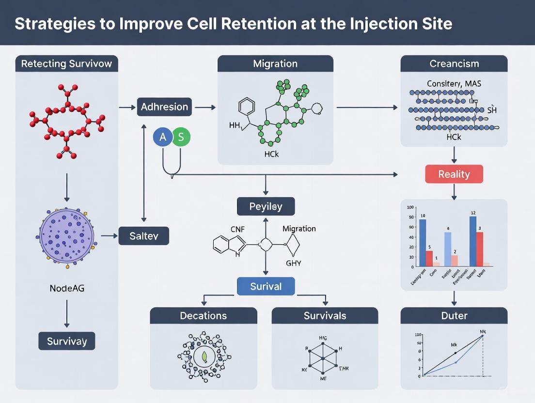
Advanced Strategies to Improve Cell Retention at the Injection Site: From Biomaterials to Engineering Solutions
This article provides a comprehensive overview of innovative strategies to overcome the critical challenge of low cell retention and viability at the injection site, a major hurdle in cell therapy...
Advanced Strategies to Improve Cell Retention at the Injection Site: From Biomaterials to Engineering Solutions
Abstract
This article provides a comprehensive overview of innovative strategies to overcome the critical challenge of low cell retention and viability at the injection site, a major hurdle in cell therapy and regenerative medicine. Tailored for researchers and drug development professionals, it synthesizes foundational principles, practical methodologies, optimization techniques, and comparative validation data. The scope spans the use of advanced biomaterial scaffolds like crosslinked hydrogels, cell preconditioning, and genetic engineering, offering a multi-faceted roadmap to enhance therapeutic outcomes by ensuring more delivered cells survive and function at the target location.
Understanding the Cell Retention Challenge: Why Injected Cells Don't Stay Put
The Critical Impact of Low Cell Retention on Therapeutic Efficacy
For researchers and drug development professionals, achieving high cell retention at the target site is a pivotal challenge that directly determines the success of advanced therapies. Low cell retention undermines therapeutic efficacy by drastically reducing the number of viable cells available to exert their intended biological effect, whether through direct tissue integration, paracrine signaling, or targeted drug delivery. This technical support center provides a comprehensive guide to diagnosing, troubleshooting, and resolving the multifaceted issues that compromise cell retention in experimental and therapeutic contexts.
Frequently Asked Questions (FAQs)
1. What are the primary biological consequences of low cell retention in cell therapy? Low cell retention directly diminishes the therapeutic dose at the target site, leading to suboptimal engraftment and reduced secretion of beneficial paracrine factors. This often results in inadequate tissue repair and compromised therapeutic outcomes.
2. How does the injection process itself affect cell retention? The mechanical shear forces experienced during injection can significantly impair cell viability and membrane integrity. Furthermore, the rapid washout of cells from the injection site into systemic circulation is a major cause of immediate cell loss.
3. What role does the host tissue microenvironment play in cell retention? The pathological microenvironment, characterized by elevated interstitial fluid pressure (IFP) and a dense extracellular matrix, can hinder cell penetration and integration. In tumors, although the Enhanced Permeability and Retention (EPR) effect can aid nanocarrier accumulation, it offers less than a 2-fold increase and is insufficient for many therapies [1].
4. Which technological solutions can improve cell retention? Strategies include using cell retention devices like Alternating Tangential Flow (ATF) filtration in bioreactors, embedding cells in biomaterial scaffolds such as hydrogels, and optimizing injection formulations to enhance viscosity and cell protection [2] [3].
5. How can I accurately quantify cell retention in my experiments? Imaging Flow Cytometry (IFC) is a powerful tool that combines high-throughput cell counting with morphological analysis, allowing for precise tracking and quantification of retained cells. It overcomes the limitations of conventional flow cytometry by providing visual confirmation [4].
Troubleshooting Guide: Low Cell Retention
| Problem Area | Specific Issue | Possible Root Cause | Recommended Solution |
|---|---|---|---|
| Injection Formulation | Rapid cell washout post-injection | Low viscosity formulation; lack of protective matrix | Embed cells in a thermosensitive hydrogel to enhance site-specific retention [3]. Optimize formulation osmolality and pH to be close to physiological conditions (∼300 mOsm/kg, pH 7.4) [5]. |
| Cell Viability | Poor cell survival during/after delivery | Shear stress during injection; hostile target microenvironment | Use a cell retention device like ATF to reduce shear during processing [2]. Pre-condition cells to the target environment (e.g., hypoxic conditions). |
| Host Microenvironment | High cell loss in specific tissues (e.g., solid tumors) | Elevated Interstitial Fluid Pressure (IFP); dense extracellular matrix | Utilize the Enhanced Permeability and Retention (EPR) effect with nano-sized carriers, and explore strategies to temporarily augment the EPR effect [1]. |
| Measurement & Analysis | Inaccurate quantification of retained cells | Reliance on methods lacking morphological validation; low sensitivity | Implement Imaging Flow Cytometry (IFC) for high-throughput, image-based cell counting and tracking [4] [6]. |
Experimental Protocols for Enhancing Retention
Protocol 1: Hydrogel-Embedded Cell Delivery for Enhanced Site Retention
This methodology details the use of a hydrogel matrix to significantly improve cell retention at the injection site, based on a strategy proven effective in glioblastoma models [3].
- Hydrogel-Nanoparticle (HG-NP) Preparation: Fabricate biodegradable polymeric nanoparticles (NPs) loaded with your therapeutic agent (e.g., cells, drugs).
- HG-NP Composite Formation: Mix the NPs with a thermosensitive hydrogel precursor (e.g., Poloxamer 407). The mixture should be liquid at room temperature to facilitate injection.
- Intratumoral/Intratissue Injection: Administer the HG-NP composite suspension into the target tissue using a standard syringe.
- In Situ Gelation: Upon exposure to body temperature (37°C), the hydrogel will undergo a sol-gel transition, forming a solid matrix that entraps the NPs/cells.
- Sustained Release: The hydrogel acts as a reservoir, allowing for the slow release of NPs/cells into the surrounding tissue, thereby enhancing long-term retention and uptake.
Protocol 2: Quantifying Retention Using Imaging Flow Cytometry
This protocol leverages IFC to accurately measure cell retention and proliferation, incorporating mitotic index analysis [4] [6].
- Cell Labeling: Label cells with a fluorescent cell tracer dye (e.g., CellTrace Violet) prior to injection.
- Sample Injection & Collection: Inject the labeled cells into your animal model or experimental system. After a predetermined period, harvest the target tissue.
- Single-Cell Suspension: Process the harvested tissue to create a single-cell suspension.
- Staining for IFC: Stain the cell suspension with a DNA dye (e.g., Propidium Iodide) to aid in cell cycle analysis and mitotic identification.
- Data Acquisition: Run samples on an Imaging Flow Cytometer (e.g., Amnis ImageStream).
- Data Analysis:
- Identify Retained Cells: Gate on the population positive for the CellTrace Violet label.
- Assess Proliferation: Use the Bright Detail Intensity feature within the IDEAS software to quantify the mitotic index within the retained cell population. The "Mean + xSD" algorithm can provide a more objective count than manual gating [6].
Data Presentation: Cell Retention Metrics
Comparative analysis of quantitative data is essential for evaluating the success of retention strategies. The table below summarizes key metrics from relevant studies.
Table 1: Quantitative Metrics in Cell Retention Research
| Metric | Value/Outcome | System/Model | Implication for Retention |
|---|---|---|---|
| Viable Cell Concentration | ≈2.9 · 10⁶ cells mL⁻¹ [2] | hMSC perfusion bioreactor | Competitive cell densities can be achieved in automated retention systems. |
| Microcarrier Aggregate Size (Median Diameter) | 470 μm (repeated-batch) vs. 250 μm (ATF) [2] | hMSC perfusion bioreactor | Cell retention devices can constrain aggregate size, potentially improving homogeneity and nutrient exchange. |
| EPR Effect Enhancement | <2-fold increase [1] | Nano-sized drug delivery to tumors | Passive targeting via EPR is modest, highlighting the need for active strategies. |
| Mitotic Index Quantification | High accuracy with "Mean + xSD" algorithm [6] | Caco2/HT-29 cell lines, IFC | Provides an objective, reproducible method to measure proliferation of retained cells. |
The Scientist's Toolkit: Essential Reagents & Materials
| Item | Function in Cell Retention Research |
|---|---|
| Thermosensitive Hydrogel (e.g., Poloxamer 407) | Serves as a scaffold for cells/nanoparticles, gelating at body temperature to create a protective depot that drastically reduces washout [3]. |
| Microcarriers (MCs) | Provides a high-surface-area substrate for the expansion of adherent cells (e.g., hMSCs) in bioreactors, facilitating scalable production before transplantation [2]. |
| Cell Retention Device (e.g., ATF, TFDF) | Used in perfusion bioreactor systems to retain cells and microcarriers while removing spent media, minimizing the high shear forces that can compromise cell health [2]. |
| Imaging Flow Cytometer (e.g., Amnis ImageStream) | Enables high-throughput, quantitative analysis of cell retention, viability, and proliferation by combining flow cytometry with high-resolution cellular imaging [4] [6]. |
| Fluorescent Tracers (e.g., CellTrace Violet, BODIPY) | Labels cells or therapeutic cargo, allowing for precise tracking, quantification, and localization post-injection using imaging techniques [7] [3]. |
Visualizing Workflows and Pathways
Cell Retention Enhancement Workflow
Signaling in the Host Microenvironment
Troubleshooting Guides and FAQs
Frequently Asked Questions
Q1: What are the primary components of a hostile microenvironment that lead to rapid cell clearance? A1: The primary components include:
- Immunosuppressive Cells: Tumor-associated macrophages (TAMs), regulatory T cells (Tregs), and myeloid-derived suppressor cells (MDSCs) that secrete factors to disable therapeutic cells [8].
- Metabolic Stressors: Nutrient deprivation (e.g., glucose, amino acids) and hypoxia that impair the survival and function of transplanted cells [9] [8].
- Inflammatory Mediators: High levels of pro-inflammatory cytokines (e.g., IL-6, MCP-1) and chemokines that can induce cell stress and death [10] [11].
- Physical Barriers: Abnormal, leaky vasculature and a dense, fibrotic extracellular matrix (ECM) that hinder cell infiltration and retention [8].
Q2: Beyond the general "hostile microenvironment," what specific mechanisms cause the rapid clearance of transfused or injected cells? A2: Rapid clearance is often an active immune process. Studies on red blood cells (RBCs) have shown that cells damaged by storage, chemical treatment (e.g., phenylhydrazine), or heat are cleared within hours by the reticuloendothelial system (RES). This rapid clearance is strongly associated with a pro-inflammatory "cytokine storm" (e.g., elevated KC/CXCL-1, MCP-1, IL-6) and can enhance immunogenicity, meaning the recipient's immune system is more likely to attack the transplanted cells [11].
Q3: What are the most promising engineering strategies to protect cells from these barriers? A3: The most promising strategies focus on creating a protective niche for the cells:
- Biomaterial Scaffolds and Hydrogels: Using crosslinked hyaluronic acid (HA) or other polymers to create a 3D scaffold that shields cells, improves retention, and provides a more favorable microenvironment [10] [12].
- Cell Preconditioning: Treating cells with cytokines (e.g., IL-1β, TGF-β1) or pharmacological agents (e.g., α-ketoglutarate) before transplantation to enhance their resistance to stress, improve their survival, and boost their migratory capabilities [10].
- Genetic Modifications: Engineering cells to overexpress pro-survival, anti-apoptotic, or homing genes to better withstand the hostile conditions [10] [13].
- Combining Multiple Strategies: Integrating preconditioning with biomaterial encapsulation to synergistically enhance cell resilience and retention [10].
Troubleshooting Common Experimental Issues
Problem: Poor Cell Survival and Retention at the Injection Site
| Symptom | Potential Cause | Solution / Strategy to Test |
|---|---|---|
| Low cell counts at site within 24 hours. | Rapid clearance by the immune system; anoikis (detachment-induced cell death). | Co-transplant cells with a crosslinked HA hydrogel. This provides a 3D ECM-mimetic structure to enhance retention and viability [12]. |
| High levels of apoptosis post-injection. | Hostile microenvironment: inflammatory cytokines, reactive oxygen species (ROS). | Precondition cells with IL-1β or TGF-β1 in vitro to upregulate pro-survival pathways and enhance stress resistance before transplantation [10]. |
| Lack of functional improvement despite some cell presence. | Metabolic suppression (hypoxia, nutrient starvation) in the microenvironment. | Pre-treat cells with α-ketoglutarate to upregulate VEGF and HIF-1α, promoting angiogenesis and improving adaptation to hypoxic stress [10]. |
| Cells fail to engraft or migrate from the injection site. | Dense physical barrier (e.g., desmoplastic reaction) and lack of homing signals. | Utilize a biomaterial scaffold that degrades over time, facilitating gradual cell egress. Alternatively, precondition with cytokines known to enhance migration, like IL-1β to upregulate MMP-3 [10] [8]. |
Problem: Inconsistent or Failed Therapy Outcomes
| Symptom | Potential Cause | Solution / Strategy to Test |
|---|---|---|
| High variability in therapeutic efficacy between animal subjects. | Inconsistent cell quality or damage prior to injection, leading to variable clearance. | Implement a rigorous quality control check pre-injection. Use flow cytometry to ensure consistent surface antigen expression and exclude preparations with high damage markers, as even minor alterations can drastically change clearance and immunogenicity [11]. |
| Therapy works in immunocompromised models but not in immunocompetent ones. | Robust host immune response clearing the therapeutic cells. | Consider short-term, localized immunosuppression. Alternatively, develop "stealth" cells by engineering them to downregulate immunogenic surface markers or overexpress immune-inhibitory ligands [8]. |
| Initial success followed by rapid failure. | The hostile microenvironment eventually overwhelms the protective strategies. | Employ a combination therapy. For example, in oncological settings, combine cell therapy with Tumor Microenvironment (TME)-reprogramming agents like STING agonists to convert "cold" immunosuppressive tumors into "hot" immunoreactive ones [9] [8]. |
Detailed Experimental Protocols
Protocol 1: Evaluating Cell Clearance Kinetics Using a Flow Cytometry-Based Recovery Assay
This protocol is adapted from methods used to study transfused red blood cells and can be modified for other cell types [11].
Objective: To quantitatively measure the rate at which injected cells are cleared from the recipient's circulation or tissue.
Materials:
- Donor cells (e.g., therapeutic cell line)
- Recipient animals (syngeneic if possible)
- Flow cytometer
- Antibodies for cell staining (e.g., against a unique surface antigen on donor cells)
- PBS for washing and dilution
Method:
- Label Donor Cells: Label donor cells with a fluorescent cell tracker (e.g., PKH26) or use transgenic donor cells that express a specific, detectable surface antigen (like the HOD antigen used in RBC studies) [11].
- Baseline Sample: Analyze a small aliquot of the donor cell suspension by flow cytometry to determine the baseline fluorescence or antigen expression profile.
- Cell Injection: Transfuse or inject a known number of cells (e.g., 100 µL of packed cells) into the recipient via a relevant route (e.g., tail vein, local injection).
- Time-Point Sampling: Collect peripheral blood or tissue samples from the recipient at multiple time points post-injection (e.g., 10 min, 30 min, 2 hours, 24 hours).
- Flow Cytometric Analysis: For each sample, use flow cytometry to quantify the percentage of donor-derived cells within the total cell population.
- Data Analysis: Calculate the post-transfusion recovery at each time point. The 24-hour recovery is often a key metric, defined as the percentage of donor cells remaining at 24 hours divided by the estimated percentage at time zero (extrapolated from early time points) [11].
Protocol 2: Hydrogel-Based Cell Delivery for Improved Retention
This protocol outlines the use of crosslinked hyaluronic acid (HA) hydrogel as a carrier for cell transplantation, based on methods for treating vitiligo [12].
Objective: To enhance the retention and viability of transplanted cells at the target site using a biomaterial scaffold.
Materials:
- Crosslinked HA hydrogel (e.g., BDDE-crosslinked, sterilized)
- Therapeutic cells (e.g., epidermal cells, mesenchymal stem cells)
- PBS
- Syringe and needle for injection
Method:
- Prepare Cell Suspension: Extract and prepare your therapeutic cells, ensuring high viability. The cited study achieved >85% viability using a 2-hour 37°C digestion method [12].
- Create Cell-Hydrogel Construct: Gently mix the cell pellet with the sterile, crosslinked HA hydrogel. The final concentration of HA should be optimized; the cited study used a 20 mg/mL hydrogel [12].
- Characterize Hydrogel (Optional but Recommended):
- Rheology: Test the viscoelastic properties of the hydrogel to ensure it has suitable mechanical strength for injection and retention.
- Swelling/Degradation: Measure the hydrogel's swelling ratio and degradation profile in vitro to predict its behavior in vivo [12].
- Transplantation: Load the cell-hydrogel mixture into a syringe and inject it into the target site (e.g., subcutaneously, into a wound bed, or a vitiligo lesion).
- Assessment: At desired endpoints, excise the tissue and analyze cell retention and viability using histology, immunofluorescence, or by retrieving cells for flow cytometric analysis.
Table 1: Impact of Cellular Insults on Clearance and Immunogenicity
This table summarizes data from a study on red blood cells, demonstrating how different insults lead to rapid clearance and adverse immune responses [11].
| Cell Treatment | 24-Hour Post-Transfusion Recovery | Pro-Inflammatory Cytokine Response | Enhancement in Alloimmunization (vs. Control) |
|---|---|---|---|
| Fresh RBCs (Control) | ~75% (C57BL/6 strain) | Baseline | 1-fold (Reference) |
| 14-Day Stored RBCs | ~30% (FVB strain) | Significantly Elevated (Storm) | Significant enhancement observed [11] |
| Phenylhydrazine-Treated RBCs | Near complete clearance by 24h | Elevated (KC/CXCL-1, MCP-1, IL-6) | 25.1-fold |
| Heat-Treated RBCs (50°C) | Near complete clearance by 24h | Elevated (KC/CXCL-1, MCP-1, IL-6) | 10.3-fold |
Table 2: Efficacy of Engineering Strategies in Preclinical Models
This table compiles data from various studies on strategies to overcome biological barriers, particularly in wound healing and cell therapy [10] [12].
| Engineering Strategy | Model | Key Outcome Metrics | Result |
|---|---|---|---|
| Cytokine Preconditioning (TGF-β1) | Murine wound model | Wound healing time; BMSCs survival and engraftment | Significant reduction in healing time; Enhanced cell survival [10] |
| Pharmacological Preconditioning (α-ketoglutarate) | Chemical-induced burn model | ADSCs survival; Expression of VEGF and HIF-1α; Wound closure | Improved cell survival; Increased angiogenic factors; Accelerated wound closure [10] |
| Crosslinked HA Hydrogel Delivery | Cell transplantation for vitiligo | Cell retention; Cell viability | Prolonged cell retention; Maintained high cell viability (~85%) [12] |
| Combined Preconditioning & Scaffolds | Various wound healing models | Acceleration of tissue repair; Reduction in scar formation | Synergistic effect, optimizing overall healing outcomes [10] |
The Scientist's Toolkit: Key Research Reagents & Materials
Table 3: Essential Materials for Investigating and Overcoming Cell Clearance
| Item | Function / Application | Example from Literature |
|---|---|---|
| Crosslinked Hyaluronic Acid (HA) Hydrogel | A 3D biomaterial scaffold used to encapsulate and deliver cells, providing mechanical support, enhancing retention, and improving the local microenvironment for cell survival [12]. | BDDE-crosslinked HA hydrogel used as a carrier for autologous epidermal cell transplantation in vitiligo treatment [12]. |
| Recombinant Cytokines (e.g., IL-1β, TGF-β1) | Used for in vitro preconditioning of cells to enhance their migratory capacity, stress resistance, and survival post-transplantation by modulating gene and protein expression profiles [10]. | IL-1β preconditioning to upregulate MMP-3 and enhance MSCs migration; TGF-β1 preconditioning to enhance BMSCs survival and engraftment [10]. |
| Pharmacological Preconditioning Agents (e.g., α-ketoglutarate) | Chemical agents used to prime cells, enhancing their biological functions such as paracrine activity and antioxidant responses, thereby improving therapeutic efficacy [10]. | α-ketoglutarate pretreatment of ADSCs to improve their survival in a burn model by increasing VEGF and HIF-1α expression [10]. |
| TAM Receptor Inhibitors (e.g., BMS-777607) | Used to inhibit the TYRO3/AXL/MERTK (TAM) family of phagocytic receptors. This allows researchers to probe the specific mechanisms of efferocytosis (dead cell clearance) in both professional and non-professional phagocytes [14]. | BMS-777607 used to inhibit TAM-receptor activity on hair follicle stem cells, thereby impairing their ability to engulf apoptotic corpses in vitro [14]. |
| Recombinant Annexin V | A protein that binds phosphatidylserine (PS). It is used to mask "eat-me" signals (PS) on the surface of apoptotic cells, allowing researchers to test the role of PS recognition in cell clearance pathways [14]. | Intradermal injection of annexin V to block PS on apoptotic corpses, delaying their clearance by hair follicle stem cells in vivo [14]. |
Signaling Pathways and Experimental Workflows
Diagram: Engineering Strategies to Overcome Hostile Microenvironment
This diagram illustrates the logical relationship between the key barriers, the engineering strategies used to overcome them, and the resulting functional improvements in cell therapy.
Diagram: Phagocytic Clearance Signaling Pathway
This diagram details the molecular signaling pathway by which both professional and non-professional phagocytes recognize and clear apoptotic cells, a key mechanism of rapid cell clearance.
FAQ: Biomaterials and Cell Retention
What is the core principle behind using biomaterials to mimic the Extracellular Matrix (ECM)?
The core principle is to create an artificial scaffold that replicates both the structural and biochemical properties of the native ECM found in tissues. The natural ECM is a dynamic, three-dimensional network of proteins (like collagen and laminin) and polysaccharides (like glycosaminoglycans) that provides structural support to cells and regulates crucial functions including cell survival, proliferation, and differentiation [15] [16]. Biomaterials engineered to mimic the ECM aim to supply a similar supportive and instructive microenvironment at a cell injection site. This helps to fill lesion cavities, favor transplanted cell engraftment, provide physical support, and tune the inflammatory response, thereby protecting cells and enhancing retention [15].
Why is cell retention at the injection site a major challenge, and how do ECM-mimicking biomaterials help?
A significant challenge is the rapid loss of transplanted cells from the injection site due to leakage back through the needle track, washout by body fluids, or the lack of a supportive structure in the host tissue [17]. This is particularly problematic in harsh microenvironments, such as those found in wounded or inflamed tissues, which can further compromise cell survival [18].
ECM-mimicking biomaterials, particularly hydrogels, address this by:
- Providing 3D Mechanical Support: They act as a temporary, hydrated matrix that physically entraps cells, reducing their washout. The viscoelastic properties of hydrogels can be tuned to match the target tissue, improving retention compared to low-viscosity saline solutions [19] [17].
- Biochemical Cues: They present native biochemical signals that promote cell adhesion, survival, and integration with the host tissue [15] [20].
- Creating a Protective Niche: The hydrogel matrix can shield cells from acute inflammatory attacks and provide a microenvironment that supports their function until they properly engraft [18].
What are the key properties of a biomaterial that enhance cell retention?
The following properties are critical for designing biomaterials that effectively improve cell retention:
| Property | Description | Impact on Cell Retention |
|---|---|---|
| Viscosity / Mechanical Properties | The resistance of a material to flow. Higher viscosity hydrogels are less prone to leakage [17]. | Minimizes backflow through the injection track and maintains a defined, localized cell depot at the target site. |
| Degradation Rate | The speed at which the biomaterial breaks down in the body [21]. | A controlled, slow degradation rate provides prolonged structural support, allowing more time for the transplanted cells to engraft and integrate with the host tissue [21]. |
| Biocompatibility | The ability of a material to perform with an appropriate host response without eliciting excessive inflammation [20]. | Prevents a severe foreign body response that could destroy transplanted cells and ensures harmonious integration with the host tissue. |
| Bioactivity | The presence of inherent biochemical signals (e.g., from native ECM components) or the ability to deliver pro-regenerative agents [15] [18]. | Promotes cell survival, proliferation, and direct communication with the host tissue, leading to more stable, long-term retention and functional engraftment. |
How does the choice of biomaterial (natural vs. synthetic) influence its function?
The source of the biomaterial dictates its inherent properties and its interaction with cells. The table below summarizes the main categories:
| Material Type | Examples | Key Characteristics | Considerations |
|---|---|---|---|
| Natural Polymers | Hyaluronic Acid (HA), Collagen, Chitosan, Gelatin, Decellularized ECM [21] [16] [20]. | Inherently bioactive, promote excellent cell adhesion and recognition, typically biodegradable. | Batch-to-batch variability, potential immunogenicity if not properly purified, mechanical strength can be low. |
| Synthetic Polymers | Polyethylene Glycol (PEG), Polylactic Acid (PLA) [16]. | Highly tunable physical properties (mechanical strength, degradation), reproducible manufacturing. | Lack inherent bioactivity (often requires functionalization with cell-adhesive peptides), degradation byproducts may cause inflammation. |
Troubleshooting Common Experimental Issues
Problem: Poor cell viability following suspension in the biomaterial.
- Potential Cause 1: Harsh cell dissociation from culture surfaces. Using aggressive enzymes like trypsin can damage cell surface proteins critical for survival and adhesion [22] [23].
- Potential Cause 2: Shear stress during mixing and injection. Physical forces can damage cells.
- Solution: Use wide-bore pipette tips when handling cell-biomaterial suspensions to minimize shear stress [24]. Mix the cell suspension gently by pipetting up and down slowly.
- Potential Cause 3: The biomaterial or buffer is toxic.
Problem: Rapid leakage of the cell-biomaterial suspension from the injection site.
- Potential Cause: The viscosity of the biomaterial is too low.
- Solution: Increase the polymer concentration of the hydrogel. For example, a study using hydrolyzed gelatin (HG) found that 20% HG provided significantly better retention in a beating rat heart model compared to 10% HG or a saline control [17]. Always optimize the concentration for your specific model and injection method.
Problem: Low cell retention and engraftment over time.
- Potential Cause 1: The host microenvironment is hostile (hypoxic, highly inflammatory).
- Solution: Precondition cells in vitro before transplantation. This can involve cytokine preconditioning (e.g., with IL-1β or TGF-β1) or hypoxic preconditioning to enhance their resilience and secretory profile, improving their chance of survival post-transplantation [18].
- Potential Cause 2: The biomaterial does not support cell adhesion.
Experimental Protocols
Protocol 1: Preparing a Crosslinked Hyaluronic Acid (HA) Hydrogel for Cell Delivery
This protocol is adapted from research using crosslinked HA to improve epidermal cell retention for vitiligo treatment [21].
Key Reagent Solutions:
- Hyaluronic Acid (HA): The main structural polymer.
- BDDE (Butylene glycol diglycidyl ether): A crosslinking agent that forms stable ether bonds with HA.
- Sodium Hydroxide (NaOH): Creates the alkaline conditions necessary for the crosslinking reaction.
Methodology:
- Dissolution: Dissolve 10g of HA powder in 100 mL of a 1% (w/v) NaOH solution at room temperature.
- Crosslinking: Add BDDE to the HA solution and mix thoroughly for 15 minutes.
- Reaction Incubation: Incubate the mixture in a 50°C water bath for 1 hour.
- Curing: Transfer the crosslinked gel and let it stand overnight at 4°C to complete the reaction.
- Purification: Transfer the gel into a dialysis membrane and dialyze against a large volume of phosphate-buffered saline (PBS) for 4 days at room temperature, changing the dialysate twice daily to remove unreacted chemicals and impurities.
- Homogenization and Sterilization: Qualify the purified gel to a final concentration of 20 mg/mL in PBS. Homogenize it at 4,000 rpm. Finally, sterilize the filled hydrogel by autoclaving at 121°C for 15 minutes [21].
Protocol 2: Evaluating Biomaterial Retention and DistributionIn Vivo
This protocol describes a method to visually assess the retention of an injectable formulation, as used in a study on triple-negative breast cancer [19] and cardiac cell therapy [17].
Key Reagent Solutions:
- Test Formulation: Your biomaterial (e.g., hydrogel) at the desired concentration.
- Tracer Molecule: A visually detectable agent such as Indian ink [17] or a fluorescent dye.
- Animal Model: An appropriate in vivo model (e.g., rodent).
Methodology:
- Preparation of Formulation: Mix the tracer molecule (e.g., Indian ink solution) with your biomaterial formulation. Ensure the tracer does not significantly alter the viscosity of the hydrogel [17].
- Injection: Inject the formulated tracer into the target tissue of the animal model (e.g., myocardium, tumor).
- Analysis:
- Macroscopic Imaging: Sacrifice the animal at a predetermined time point post-injection. Excise the target organ and capture images to document the distribution and leakage of the tracer.
- Quantification: Use image analysis software to quantify the stained area and its intensity, providing a quantitative measure of retention and spread [17].
- Histology: Process the tissue for histological sectioning. Observe the sections under a microscope to determine the precise localization of the tracer (e.g., interstitial space vs. blood vessels) and assess the interaction between the biomaterial and the host tissue [17].
Visualizing Key Concepts
ECM Mimicry for Cell Retention
Experimental Workflow for Testing a Biomaterial
The Scientist's Toolkit: Research Reagent Solutions
| Reagent / Material | Function in ECM Mimicry and Cell Retention |
|---|---|
| Hyaluronic Acid (HA) | A natural glycosaminoglycan (GAG) that is a major ECM component; can be crosslinked to form hydrogels that provide a hydrated, 3D environment for cells [21]. |
| Hydrolyzed Gelatin (HG) | A low-antigenicity polypeptide used to increase the viscosity of cell suspension, reducing leakage from the injection site in beating organs [17]. |
| Collagen | A primary fibrous protein in ECM; used in hydrogels and scaffolds to provide structural integrity and bioactive cell adhesion sites [18] [22]. |
| Chitosan | A natural polymer used to form injectable, thermosensitive hydrogels that can localize therapeutics and cells within a target tissue [19]. |
| Decellularized ECM (dECM) | The non-cellular component of tissue harvested and purified from allogeneic or xenogeneic sources; considered the "gold standard" bioactive material as it retains the complex native composition of the original tissue's ECM [20]. |
| RGD Peptide | A synthetic peptide (Arginine-Glycine-Aspartic acid) that mimics cell adhesion sites in fibronectin; used to functionalize synthetic biomaterials to make them bioactive [15] [16]. |
| TrypLE / Accutase | Gentle, animal-origin-free enzymatic dissociation reagents used to detach adherent cells while better preserving cell surface proteins and viability compared to trypsin [22] [23]. |
| Cell Dissociation Buffer | A non-enzymatic, chelating buffer used to detach light-adherent cell lines; ideal for applications where intact cell surface proteins are critical [22]. |
FAQs: Core Concepts and Material Selection
What are the primary functions of a cell carrier in transplantation? Cell carriers, often hydrogel-based scaffolds, serve multiple critical functions. Their primary role is to provide a three-dimensional (3D) microenvironment that enhances cell retention at the injection site and protects cells from the harsh in vivo environment, thereby maintaining high cell viability. They act as a temporary extracellular matrix (ECM), facilitating the delivery of nutrients and oxygen to the transplanted cells. Furthermore, their physical properties can be tuned to influence cell morphology, promote migration, and support key processes like angiogenesis and re-epithelialization at the wound site [21] [18].
How does the choice of biomaterial directly impact cell retention and viability? The biomaterial's physical and chemical properties directly dictate its performance. Key factors include:
- Rheological Behavior & Viscosity: Higher viscosity, achieved through crosslinking or increased concentration, reduces diffusion and cell leakage from the injection site. For example, 20% hydrolyzed gelatin (HG) demonstrated superior retention of cardiomyocytes in beating rat hearts compared to lower concentrations [17].
- Degradation Profile: A slower biodegradation profile, as seen with crosslinked hyaluronic acid (HA), allows for prolonged cell retention and support [21].
- Biocompatibility: Materials like HA, a natural component of skin ECM, and hydrolyzed gelatin offer low antigenicity, creating a favorable environment for cell survival [21] [17].
What are the key considerations when selecting a crosslinking agent for hydrogel carriers? The crosslinking agent determines the stability and biocompatibility of the hydrogel. Butanediol diglycidyl ether (BDDE) is one of the most mature and widely commercialized agents for crosslinking HA. Its epoxy groups react with primary alcohols on the HA backbone under alkaline conditions to form a stable, 3D ether-bonded network [21]. The choice of agent impacts the hydrogel's mechanical strength, swelling ratio, and degradation rate, all of which must be optimized for the specific application.
What common challenges arise from the host microenvironment after cell transplantation? The post-transplantation microenvironment is often hostile and can significantly compromise efficacy. Key challenges include:
- Hypoxia and Nutrient Deprivation: Limited blood supply can lead to oxidative stress and trigger anoikis (a form of cell death) [18].
- Excessive Inflammatory Mediators: A high level of inflammatory cytokines can damage transplanted cells [18].
- Immune Responses: For allogeneic transplants, immune rejection can destroy the transplanted cells.
Troubleshooting Guide: Common Experimental Problems
| Problem | Possible Causes | Recommended Solutions |
|---|---|---|
| Low Cell Retention at Injection Site | Carrier viscosity too low for target tissue [17].Rapid carrier degradation [21].Hostile microenvironment causing cell death [18]. | Optimize carrier concentration (e.g., use 20% HG for myocardial injection) [17].Utilize crosslinked hydrogels (e.g., BDDE-crosslinked HA) for slower degradation [21].Precondition cells (hypoxic/cytokine) to enhance stress resistance [18]. |
| Poor Cell Viability Post-Transplantation | Lack of oxygen and nutrients within the carrier [21].Shear stress during the injection process.Adverse immune reaction to the carrier or cells. | Ensure carrier porosity allows for nutrient/waste diffusion [18].Use a carrier with protective, lubricating properties.Select low-antigenicity materials (e.g., microbial-fermented HA, HG) [21] [17]. |
| Inconsistent Therapeutic Outcomes | High variability in donor cells [25].Inconsistent carrier fabrication or cell mixing process.Uncontrolled diffusion of cells from the target site [17]. | Implement rigorous quality control and characterization of starting materials [25].Standardize experimental protocols for carrier preparation and cell loading.Select a carrier that minimizes uncontrolled diffusion (e.g., 20% HG in beating hearts) [17]. |
| Difficulty in Scaling Manufacturing | Complex, resource-intensive legacy manufacturing processes [25].High variability of cell types and gene-editing techniques [25]. | Adopt automated manufacturing platforms and advanced culture media to normalize differences [25].Explore innovative delivery systems like hydrogel encapsulation to simplify logistics [25]. |
Detailed Experimental Protocols
This protocol describes the synthesis of a stable HA hydrogel for use as a cell delivery vehicle.
Key Materials:
- Hyaluronic acid powder (e.g., 2.5 million molecular mass)
- Butanediol diglycidyl ether (BDDE)
- Sodium hydroxide (NaOH) solution
- Phosphate-buffered saline (PBS)
Methodology:
- Dissolution: Dissolve 10 g of HA powder in 100 mL of a 1% (w/v) NaOH solution at room temperature.
- Crosslinking: Add BDDE dropwise to the HA solution and mix for 15 minutes to ensure homogeneity.
- Reaction Incubation: Incubate the mixture in a water bath at 50°C for 1 hour to facilitate the crosslinking reaction.
- Curing: Transfer the crosslinked gel and allow it to stand overnight at 4°C.
- Purification: Dialyze the resulting crosslinked HA hydrogel against PBS for 4 days (using an 8,000 Dalton molecular weight cutoff membrane) to remove unreacted reagents. Change the dialysate twice daily.
- Preparation for Use: Qualify the purified hydrogel to a concentration of 20 mg/mL in PBS and homogenize it at 4,000 rpm. Finally, sterilize the gels by autoclaving at 121°C for 15 minutes.
Characterization Tests:
- Rheology: Use a rotational rheometer to assess viscoelastic properties (e.g., at 10 Hz frequency).
- Swelling & Degradation: Use gravimetric analysis to measure the swelling ratio (
Es = [(Ws - W0)/W0] × 100%) and in vitro degradation profile in PBS over time [21]. - Morphology: Use scanning electron microscopy (SEM) on lyophilized samples to visualize the 3D porous structure.
This protocol outlines a method to test the effect of HG concentration on cell retention in a rodent myocardial infarction model.
Key Materials:
- Hydrolyzed Gelatin (HG) at various concentrations (0%, 10%, 20%)
- Human iPS cell-derived cardiomyocytes (hiPSC-CMs)
- Indian ink solution (for diffusion tracking)
- Rat myocardial infarction (MI) model
Methodology:
- Carrier Preparation: Prepare solutions of HG at 0% (control), 10%, and 20% concentrations in an appropriate buffer.
- Cell Suspension: Suspend the hiPSC-CMs in the different HG solutions.
- In Vivo Injection: Inject the cell-carrier suspensions directly into the myocardium of the rat MI model.
- Diffusion Assessment (Optional): In a separate group, inject Indian ink mixed with HG solutions to visually track the distribution and diffusion of the injectate within the beating heart.
- Analysis:
- Histological Evaluation: At the endpoint (e.g., 1 week post-transplantation), harvest hearts. Analyze cross-sections through the injection site via immunohistochemistry (e.g., for cardiac Troponin T) to quantify the area of retained cardiomyocytes.
- Functional Assessment: Monitor cardiac function over time (e.g., 2 and 4 weeks) using echocardiography and cardiac MRI to correlate cell retention with therapeutic outcome.
Signaling Pathways in Cell-Carrier Mediated Repair
The therapeutic effect of cells delivered via carriers is often mediated through paracrine signaling, influencing key pathways in wound healing.
Research Reagent Solutions
The table below lists key materials used in advanced cell-carrier systems, as featured in recent studies.
| Research Reagent | Function in Cell-Carrier Systems |
|---|---|
| Crosslinked Hyaluronic Acid (HA) | Serves as a biodegradable, biocompatible 3D scaffold that mimics the native extracellular matrix, prolonging cell retention and providing a supportive microenvironment [21]. |
| Hydrolyzed Gelatin (HG) | A low-antigenicity polypeptide used to fine-tune the viscosity of the cell-carrier solution. Optimizing its concentration is critical for minimizing cell leakage from dynamic injection sites (e.g., the beating heart) [17]. |
| BDDE (Butanediol Diglycidyl Ether) | A crosslinking agent that reacts with HA under alkaline conditions to form a stable hydrogel network with ether bonds, providing mechanical integrity and controlled degradation [21]. |
| α-Ketoglutarate | Used for pharmacological preconditioning of MSCs. It enhances cell survival and upregulates pro-angiogenic factors like VEGF and HIF-1α, improving outcomes in burn models [18]. |
| Caffeic Acid | A preconditioning agent that improves MSC viability and regenerative potential under hypoxic conditions by upregulating the secretion of VEGF and SDF-1, which enhances angiogenesis [18]. |
| Interferon-γ (IFN-γ) & TNF-α | Cytokines used for microenvironmental preconditioning. They modulate MSCs to promote macrophage polarization toward the healing-associated M2 phenotype, creating a favorable inflammatory microenvironment [18]. |
Practical Strategies for Enhanced Retention: Hydrogels, Scaffolds, and Engineering
Troubleshooting Guides
Common Experimental Challenges and Solutions
Table 1: Troubleshooting Common Hydrogel Formulation Issues
| Problem | Possible Causes | Recommended Solutions | Preventive Measures |
|---|---|---|---|
| Poor Gelation or Slow Gelation Kinetics | Incorrect polymer concentration, inappropriate crosslinker ratio, suboptimal pH or temperature, impure raw materials. | - Verify polymer degree of deacetylation (for Chitosan) or molecular weight (for HA) [26].- Optimize crosslinker concentration and type (e.g., genipin vs. glutaraldehyde) [26].- Ensure physiological temperature and pH conditions during preparation [26]. | - Characterize biomaterial properties before use.- Establish a standardized pre-gel solution preparation protocol. |
| Low Mechanical Strength or Fast Degradation | Insufficient crosslinking density, low molecular weight polymer, high degradation environment. | - Increase crosslinking density or use a different crosslinking strategy [26].- Blend with other polymers (e.g., collagen, synthetic polymers) to form composite hydrogels [27].- Incorporate nanoparticles to reinforce the matrix [28]. | - Perform rheological tests and degradation studies in vitro before in vivo use.- Pre-test hydrogel stability in simulated physiological conditions. |
| Inconsistent Drug/Cell Release Profile | Batch-to-batch variability in biomaterials, non-uniform hydrogel porosity, unreliable encapsulation process. | - Source materials from certified suppliers and document batch numbers [29].- Optimize mixing speed and time during drug/cell encapsulation to ensure homogeneity.- Use controlled freezing methods for cryo-processing. | - Implement strict quality control on raw materials.- Standardize the entire fabrication process from synthesis to encapsulation [29]. |
| Low Cell Viability or Poor Cell Retention | Cytotoxic crosslinking agents, inappropriate hydrogel stiffness, lack of cell-adhesive motifs. | - Use biocompatible crosslinkers like genipin instead of glutaraldehyde [26].- Tune the hydrogel's mechanical properties (elasticity, stiffness) to match the target tissue [27].- Functionalize with RGD peptides to enhance cell adhesion [27]. | - Perform cytocompatibility tests (e.g., live/dead assay) in vitro.- Characterize the mechanical properties of the hydrogel scaffold. |
| Premature Gelation in Syringe | Overly responsive to temperature or pH, incorrect handling or storage of pre-gel solution. | - Reformulate to adjust the sol-gel transition trigger point (e.g., change thermosensitive polymer ratio) [26].- Use a double-barrel syringe for hydrogels that gel upon mixing of components.- Cool the syringe and pre-gel solution before loading. | - Thoroughly map the gelation behavior of the formulation under different conditions before the experiment. |
Table 2: Addressing Specific Biomaterial Challenges
| Biomaterial | Unique Challenges | Specific Solutions |
|---|---|---|
| Hyaluronic Acid (HA) | - Overly rapid degradation by hyaluronidases [28].- Potential inflammatory response to low molecular weight fragments [28]. | - Use high molecular weight HA or crosslinked derivatives to slow degradation [28].- Modify HA with methacrylate groups to enhance stability and control mechanical properties. |
| Chitosan | - Low solubility at neutral pH, requiring acidic solvents [26].- Variable properties based on source and degree of deacetylation [26]. | - Use water-soluble chitosan derivatives like carboxymethyl-chitosan [26].- Source chitosan with a specified and consistent degree of deacetylation (e.g., >85%) [26]. |
| Collagen | - Low mechanical strength in pure form.- Potential for pathogen transmission (animal-derived). | - Blend with other polymers like HA or use chemical crosslinkers (e.g., EDC/NHS) to enhance strength [27].- Use recombinant human collagen or source from reputable, pathogen-tested suppliers. |
Systematic Troubleshooting Methodology
When encountering an experimental issue, follow a structured approach [30]:
- Identify the Problem: Clearly define what has gone wrong without assuming the cause (e.g., "No gel formation after 30 minutes at 37°C").
- List All Possible Explanations: Brainstorm potential causes for each component and step (e.g., polymer concentration, crosslinker activity, pH, temperature).
- Collect Data: Review your lab notebook, check control experiments, verify equipment calibration, and confirm storage conditions of reagents [30].
- Eliminate Explanations: Systematically rule out causes that the data shows are not relevant.
- Check with Experimentation: Design and conduct simple, controlled experiments to test the remaining hypotheses (e.g., testing a new batch of crosslinker).
- Identify the Cause: Based on the experimental results, pinpoint the root cause and implement a fix [30].
Frequently Asked Questions (FAQs)
FAQ 1: How can I improve the retention of cells at the injection site using hydrogel-based delivery systems?
The key is to optimize the hydrogel properties to act as a protective and supportive scaffold. Use biomaterials that mimic the native extracellular matrix (ECM), such as hyaluronic acid or chitosan, to enhance biocompatibility and cell interaction [28] [26]. Fine-tune the mechanical strength and gelation kinetics to ensure the hydrogel forms a stable matrix upon injection that withstands physiological forces. Furthermore, functionalize the hydrogel with cell-adhesive ligands (e.g., RGD peptides) and tune the degradation rate to match the pace of new tissue formation, thereby providing prolonged support for the encapsulated cells [27].
FAQ 2: What are the critical parameters to control when formulating an injectable chitosan-based hydrogel?
The most critical parameters are [26]:
- Degree of Deacetylation (DDA): Affects solubility, gelation, and biodegradability. A higher DDA often improves solubility in acidic conditions and biological activity.
- Molecular Weight: Influences viscosity, mechanical strength, and degradation rate.
- Crosslinking Method and Density: Determines gelation time, mechanical stability, and whether the process is cytocompatible (prefer physical or enzymatic crosslinking for cell encapsulation).
- pH and Ionic Strength: Crucially impact the sol-gel transition for chitosan-based systems.
FAQ 3: My hydrogel degrades too quickly in vivo. What strategies can I use to prolong its stability?
To prolong hydrogel stability:
- Increase Crosslinking Density: This creates a tighter network that is more resistant to degradation [26].
- Use Different Crosslinkers: Switch to a crosslinker that forms more stable bonds (e.g., genipin for chitosan instead of ionic crosslinks) [26].
- Create Composite Hydrogels: Incorporate more stable polymers or nanoparticles into the matrix to slow down degradation [27].
- Chemical Modification: Derivative the natural polymer (e.g., methacrylated HA) to create a network that is less susceptible to enzymatic breakdown [28].
FAQ 4: How can I ensure the controlled release of a therapeutic agent from my hydrogel system?
Controlled release is achieved by designing a "smart" system. You can exploit the disease microenvironment by using stimuli-responsive hydrogels that release their payload in response to specific triggers such as pH, enzymes (e.g., hyaluronidase in tumors), or reactive oxygen species (ROS) [28]. Alternatively, control the release through diffusion by adjusting the hydrogel's mesh size via crosslinking density. Incorporating the drug into nanoparticles first, and then dispersing these nanoparticles within the hydrogel matrix, can add another layer of release control [28].
FAQ 5: What are the most common sources of error in biomaterials experiments, and how can I avoid them?
Common errors include [31] [29]:
- Sample Contamination: Maintain strict aseptic techniques and use proper equipment.
- Biomaterial Variability: Source materials from reliable suppliers and fully characterize each batch (molecular weight, DDA, etc.) before use [29].
- Human Error in Repetitive Tasks: Automate processes like pipetting where possible [31].
- Equipment Calibration: Regularly calibrate instruments like pH meters and rheometers [31].
- Inconsistent Protocols: Use a central protocol hub to ensure all team members follow the same standardized procedures [31].
Experimental Protocols
Protocol 1: Evaluating Hydrogel Gelation Time via Tube Inversion Method
Purpose: To determine the sol-gel transition time of a thermosensitive injectable hydrogel.
Materials:
- Pre-gel polymer solution (e.g., Chitosan-based solution)
- Water bath or heating block set to 37°C
- Timer
- 2 ml vial tubes
Procedure:
- Prepare the hydrogel solution according to your formulated protocol and keep it on ice to prevent premature gelation.
- Pipette 1 ml of the solution into a 2 ml vial.
- Place the vial in a water bath or heating block set to 37°C and start the timer.
- At regular intervals (e.g., every 10-30 seconds), tilt the vial at a 90-degree angle.
- The gelation time is recorded as the point at which the solution no longer flows upon vial inversion.
- Repeat the experiment in triplicate (n=3) to ensure statistical significance.
Protocol 2: AssessingIn VitroCell Viability within a Hydrogel (Live/Dead Assay)
Purpose: To quantify the viability of cells encapsulated within a 3D hydrogel matrix.
Materials:
- Cell-laden hydrogel construct
- Phosphate Buffered Saline (PBS)
- Calcein-AM solution (2 µM in PBS, for live cells)
- Ethidium homodimer-1 solution (4 µM in PBS, for dead cells)
- Fluorescence microscope
Procedure:
- Culture cell-laden hydrogels in cell culture media for the desired duration.
- At the endpoint, carefully wash the hydrogels with PBS.
- Prepare the Live/Dead staining solution by combining Calcein-AM and Ethidium homodimer-1 in PBS.
- Incubate the hydrogels in the staining solution for 30-45 minutes at 37°C, protected from light.
- Gently wash the hydrogels with PBS to remove excess dye.
- Image the hydrogels using a fluorescence microscope with appropriate filters (green for live cells, red for dead cells).
- Quantify the number of live and dead cells from multiple images using image analysis software (e.g., ImageJ) and calculate the percentage of cell viability.
The Scientist's Toolkit: Research Reagent Solutions
Table 3: Essential Materials for Hydrogel-Based Cell Delivery Research
| Reagent / Material | Function in Research | Key Considerations |
|---|---|---|
| Hyaluronic Acid (HA) | Serves as a primary ECM-mimetic component; can be modified for cell adhesion and targeted delivery via CD44 receptors [28]. | Molecular weight is critical; HMW-HA is anti-inflammatory, while LMW-HA can be pro-inflammatory [28]. |
| Chitosan | A biocompatible and biodegradable polymer for forming injectable, stimuli-responsive (e.g., pH) hydrogels; possesses inherent antibacterial properties [26]. | Solubility requires acidic conditions; Degree of Deacetylation (DDA) must be specified and consistent [26]. |
| Collagen | Provides a natural, bioadhesive matrix that strongly supports cell attachment and proliferation [27]. | Source is critical for reproducibility and safety; mechanical strength is often low and requires reinforcement. |
| Genipin | A natural and much less cytotoxic crosslinker alternative to glutaraldehyde, used for crosslinking chitosan and collagen [26]. | Crosslinking is slower than with synthetic crosslinkers; may impart a blue color to the hydrogel. |
| Methacrylated Polymers (e.g., GelMA) | Allows for precise photopolymerization (light-crosslinking) to create hydrogels with tunable mechanical properties [27]. | Requires a photoinitiator (e.g., LAP) and a UV or visible light source for gelation. |
| RGD Peptide | A synthetic peptide sequence (Arginine-Glycine-Aspartic acid) that is grafted onto hydrogels to promote integrin-mediated cell adhesion [27]. | Coupling chemistry (e.g., using EDC/NHS) must be optimized to avoid affecting hydrogel properties. |
| Polyethylene Glycol (PEG) | A synthetic, bio-inert polymer often used as a spacer or to create hybrid hydrogels, improving stability and reducing non-specific protein adsorption [28]. | Can be di-acrylated (PEGDA) for crosslinking. Its non-adhesive nature may require functionalization with RGD. |
Supporting Diagrams
Diagram 1: Hydrogel Characterization and Troubleshooting Workflow
Diagram 2: Key Properties for Cell Retention at Injection Site
Stable vitiligo significantly impacts patients' quality of life and presents a considerable challenge to healthcare providers. In recent years, cell therapy has emerged as a promising treatment, with autologous epidermal cell transplantation regarded as a safe and cost-effective strategy [12]. However, the therapeutic outcome critically depends on the retention and viability of the transplanted cells at the target site. Without adequate support, injected cell suspensions experience significant loss due to mechanical forces during injection and the lack of a three-dimensional matrix to support cell viability post-injection [32].
Injectable hydrogels have been explored as a strategy to address both causes of low cell retention. Biomaterial-based cell delivery systems can provide mechanical protection that prevents cell membrane damage during injection while creating a supportive 3D microenvironment that maintains cell survival after delivery [32]. Crosslinked hyaluronic acid (HA) hydrogels represent a particularly promising candidate due to HA's status as a main component of skin extracellular matrix (ECM), offering inherent biocompatibility and biodegradability [12] [33].
This case study examines the development and optimization of BDDE-crosslinked HA hydrogels specifically for prolonging epidermal cell retention at injection sites, presenting key experimental data, methodological protocols, and troubleshooting guidance for researchers working in dermatological drug delivery and cell therapy.
Experimental Protocols: Methodology for Hydrogel Fabrication and Characterization
Hydrogel Preparation and Crosslinking
Table 1: Formulation Components for BDDE-Crosslinked HA Hydrogels
| Component | Concentration/Range | Function | Source |
|---|---|---|---|
| Hyaluronic acid (HA) | 5-12% (w/v) | Primary polymer matrix providing 3D structure | [12] [34] |
| 1,4-butanediol diglycidyl ether (BDDE) | 0.5-2% (w/v) | Crosslinking agent forming stable ether bonds | [12] [35] |
| Sodium hydroxide (NaOH) | 1% solution | Creates alkaline conditions for crosslinking | [12] [35] |
| Phosphate buffered saline (PBS) | - | Swelling and dialysis medium | [12] |
The fundamental protocol for creating BDDE-crosslinked HA hydrogels begins with dissolving HA powder (2.5 million Da molecular weight) in 1% NaOH solution at room temperature to form a homogeneous precursor gel [12]. BDDE crosslinker is then added dropwise to the HA solution and mixed thoroughly for 15 minutes. The mixture is incubated in a water bath at 50°C for 1 hour, followed by overnight storage at 4°C to complete the crosslinking reaction [12]. The resulting hydrogel is dialyzed against PBS for 4 days (8,000 Da molecular weight cutoff, changing dialysate twice daily) to remove unreacted BDDE and other byproducts. Finally, the crosslinked HA gel is qualified to 20 mg/mL in PBS, homogenized at 4,000 rpm, and sterilized at 121°C for 15 minutes [12].
Recent advancements have introduced salt treatment during fabrication to fine-tune mechanical and degradation properties. Incorporating sodium salts (citrate, sulfate, or chloride) at concentrations of 0-0.67 M into the HA solution prior to crosslinking influences HA chain conformation through Hofmeister effects and electrostatic interactions, resulting in modified swelling ratios, mechanical properties, and degradation rates after crosslinking and salt removal [34].
Cell Extraction and Encapsulation
For epidermal cell applications, researchers have developed a novel cell extraction method using a 2-hour incubation at 37°C that maintains high cell viability (~85%) [12]. After extraction, epidermal cells are mixed with the HA hydrogel to create a uniform cell-hydrogel suspension ready for injection. The optimal cell density should be determined based on specific cell types and applications, with typical densities ranging from 1×10^6 to 5×10^6 cells/mL for therapeutic applications.
Analytical Methods for Hydrogel Characterization
Table 2: Key Characterization Techniques for HA-BDDE Hydrogels
| Parameter | Test Method | Key Metrics | Significance |
|---|---|---|---|
| Rheological properties | Rotational rheometry | Storage modulus (G'), loss modulus (G"), viscosity | Determines mechanical strength and injectability |
| Morphology | Scanning electron microscopy (SEM) | Pore size, structure, connectivity | Influences cell migration and nutrient diffusion |
| Swelling behavior | Gravimetric method | Swelling ratio (%) | Indicates crosslinking density and water retention |
| Degradation profile | Gravimetric/enzymatic assay | Mass loss over time, degradation rate | Predicts in vivo longevity |
| Biocompatibility | Cell viability assays | Live/dead staining, metabolic activity | Ensures material safety for clinical use |
Rheological characterization should be performed using an advanced rotational rheometer (e.g., Anton Paar Physica MCR301) with a constant frequency of 10 Hz and amplitude range of 0.01%-100% to determine viscoelastic properties [12]. For morphological analysis, lyophilized hydrogels should be sputter-coated with conductive gold coating before SEM imaging to visualize the internal porous structure [12].
Experimental Data: Performance of Crosslinked HA Hydrogels
Mechanical and Physical Properties
Table 3: Mechanical Properties of BDDE-Crosslinked HA Hydrogels with Variations
| Formulation | BDDE Concentration | Storage Modulus (G') | Swelling Ratio | Degradation Time |
|---|---|---|---|---|
| Basic crosslinked HA | 1.0% | ~350 Pa | ~90% | 2-3 weeks |
| Salt-treated HA (citrate) | 1.0% | 420-580 Pa* | 75-85%* | 3-4 weeks* |
| HA-PLLA composite (3%) | 1.0% | 790 Pa | 70% | 4-5 weeks |
| High crosslink density | 2.0% | ~1400 Pa | 60% | 5-6 weeks |
*Range depends on salt type and concentration [34]
The data demonstrates that modifying crosslinking parameters and incorporating composite materials significantly enhances hydrogel performance. The optimized HA-PLLA composite formulation (3% PLLA, 1.0% BDDE, 48 hours crosslinking) achieved a storage modulus of 790 Pa, representing a 2.3-fold enhancement over conventional hydrogels [35]. This improved mechanical strength directly correlates with better retention at the injection site.
Biological Performance
In animal studies, the combination of the novel cell extraction method with HA-based hydrogel scaffold achieved prolonged cell retention without compromising cell viability [12]. The 3D scaffold structure provided mechanical protection during injection, reducing cell membrane damage, while creating a favorable microenvironment that supported long-term cell survival post-injection. The hydrogel system maintained its structural integrity long enough to facilitate cell engraftment and tissue integration, addressing a critical limitation of weak hydrogels that typically biodegrade within one to two weeks [32].
The Scientist's Toolkit: Essential Research Reagents
Table 4: Key Research Reagent Solutions for Hydrogel Cell Delivery Systems
| Reagent/Category | Specific Examples | Function/Application | Considerations |
|---|---|---|---|
| Hyaluronic acid variants | High MW (1.8-2.5 MDa), modified HA derivatives | Primary scaffold material | Molecular weight affects viscosity and degradation |
| Crosslinking agents | BDDE, divinyl sulfone (DVS), diacetyl hydrazide (ADH) | Forms stable 3D network | BDDE is most established with proven safety profile |
| Characterization kits | Rheometry, SEM preparation, viability assays | Material and biological analysis | Standardized kits improve reproducibility |
| Primary skin cells | Keratinocytes, melanocytes, dermal fibroblasts | Disease-relevant cell models | Retain native phenotype for clinically relevant data |
| Enzymatic degradation assays | Hyaluronidase-based testing | Predicts in vivo longevity | Concentration should mimic physiological conditions |
Troubleshooting Guide: Addressing Common Experimental Challenges
Hydrogel Formation Issues
Problem: Insufficient or inconsistent gelation
- Potential Cause: Low crosslinker concentration or suboptimal reaction conditions
- Solution: Ensure BDDE concentration of at least 0.5% (w/v) and maintain precise temperature control during crosslinking (50°C for 1 hour followed by 4°C overnight) [12]
- Prevention: Use fresh BDDE stock, verify HA molecular weight, and maintain alkaline conditions (pH >13) during crosslinking
Problem: Excessive gel stiffness leading to injection difficulties
- Potential Cause: High crosslink density from excessive BDDE or extended reaction time
- Solution: Reduce BDDE concentration to 0.5-1.0% or shorten crosslinking time [35]
- Alternative Approach: Incorporate salt treatment prior to crosslinking to modify chain interactions without increasing covalent crosslinks [34]
Cell Viability Problems
Problem: Low cell viability after encapsulation
- Potential Cause: Mechanical shear stress during mixing or injection
- Solution: Use gentle mixing techniques and consider protective shear-thinning hydrogel formulations [32]
- Alternative Approach: Optimize cell extraction method - a 2-hour incubation at 37°C has demonstrated ~85% viability [12]
Problem: Rapid decline in cell viability post-encapsulation
- Potential Cause: Inadequate nutrient diffusion or hypoxic conditions within hydrogel
- Solution: Modify hydrogel porosity by adjusting crosslinking density or incorporating porogens
- Prevention: Ensure hydrogel storage modulus between 100-500 Pa for optimal balance between mechanical protection and nutrient diffusion [32]
In Vivo Performance Limitations
Problem: Poor retention at injection site
- Potential Cause: Fast degradation or insufficient mechanical strength
- Solution: Develop composite hydrogels such as HA-PLLA systems that show 2.3-fold improvement in storage modulus [35]
- Alternative Approach: Implement double-network strategies with secondary crosslinking mechanisms for enhanced stability [32]
Problem: Excessive inflammatory response
- Potential Cause: Residual crosslinker or degradation products
- Solution: Extend dialysis time to 5-7 days with frequent buffer changes to ensure complete removal of unreacted BDDE [12] [35]
- Quality Control: Implement rigorous quantification of residual BDDE using HPLC or similar methods
Frequently Asked Questions (FAQ)
Q1: What is the advantage of using BDDE over other crosslinkers for HA hydrogels? BDDE (1,4-butanediol diglycidyl ether) represents the most mature and commercially established crosslinking technology for HA hydrogels. Its epoxy groups react with primary alcohols in the HA skeleton under alkaline conditions to form stable ether bonds, creating a 3D network with controlled degradation kinetics [12]. Compared to alternatives like divinyl sulfone (DVS), BDDE-crosslinked HA has demonstrated favorable safety profiles and regulatory acceptance.
Q2: How can I tune the degradation rate of HA-BDDE hydrogels for my specific application? Degradation kinetics can be modulated through several approaches: (1) varying BDDE concentration (0.5-2%) to adjust crosslinking density, (2) incorporating composite materials like PLLA microspheres to enhance enzymatic resistance, (3) pre-crosslinking salt treatments that influence chain arrangement and degradation profiles, and (4) controlling hydrogel porosity which affects enzyme penetration [34] [35].
Q3: What cell densities are appropriate for encapsulation in HA-BDDE hydrogels? Optimal cell density depends on the specific cell type and application purpose. For epidermal cell transplantation, densities of 1-5×10^6 cells/mL have shown success. It's critical to balance density with nutrient diffusion limitations - higher densities may require increased hydrogel porosity or modified crosslinking density to maintain viability.
Q4: How does the HA-BDDE hydrogel protect cells during injection? The hydrogel provides mechanical protection through its shear-thinning properties. Under high shear stress during injection, the hydrogel temporarily liquefies, reducing mechanical forces on encapsulated cells. Immediately after injection, the hydrogel rapidly recovers its solid structure, retaining cells at the target site [32]. This behavior is quantified by the storage modulus (G') and loss modulus (G"), with optimal protection observed in hydrogels with G' between 100-500 Pa.
Q5: What sterilization methods are appropriate for HA-BDDE hydrogels? Autoclaving (121°C for 15 minutes) has been successfully employed for HA-BDDE hydrogels [12]. Alternative methods include gamma irradiation or sterile filtration of precursor solutions before crosslinking. The chosen method should be validated to ensure it doesn't degrade HA chains or affect crosslinking efficiency.
Visualizing Processes and Workflows
Diagram 1: Experimental Workflow for HA-BDDE Hydrogel Preparation and Cell Encapsulation. This flowchart outlines the key steps in fabricating crosslinked HA hydrogels for epidermal cell delivery, from initial polymer solution preparation through final injection and retention assessment.
Diagram 2: Mechanisms of Enhanced Cell Retention and Viability. This diagram illustrates how HA-BDDE hydrogels improve therapeutic outcomes through dual mechanisms: providing mechanical protection during injection and establishing a supportive 3D microenvironment after delivery.
Crosslinked HA-BDDE hydrogels represent a promising platform for enhancing epidermal cell retention at injection sites. Through optimized fabrication parameters, including BDDE concentration, crosslinking time, and potential composite material incorporation, researchers can tailor hydrogel properties to specific therapeutic requirements. The experimental data demonstrates that properly formulated HA-BDDE systems can significantly improve both short-term cell retention during injection and long-term viability through the provision of a supportive 3D microenvironment.
Future research directions should focus on developing smarter responsive systems that react to specific biological stimuli in the microenvironment, such as pH, reactive oxygen species (ROS), or enzyme activity [33]. Additionally, advanced fabrication technologies including 3D bioprinting and organ-on-chip systems may enable more precise control over hydrogel architecture and cellular organization [36]. As these technologies mature, crosslinked HA hydrogels are poised to play an increasingly important role in advancing cell-based therapies for dermatological conditions and beyond.
Whole Cell and Cell Membrane-Coated Carriers for Improved Biocompatibility
Frequently Asked Questions (FAQs)
FAQ 1: What are the primary strategies to improve cell retention at the injection site? The main strategies involve embedding cells within a protective biomaterial scaffold or using cell membrane-coated nanoparticles (CM-NPs). For whole cells, biomaterials like hydrolyzed gelatin (HG) and crosslinked hyaluronic acid (HA) hydrogel create a 3D microenvironment that enhances retention and viability [17] [12]. For synthetic nanocarriers, coating them with natural cell membranes (e.g., from red blood cells, mesenchymal stem cells) confers properties like immune evasion and improved biocompatibility, which helps the carriers remain at the target site longer [37] [38] [39].
FAQ 2: How does hydrolyzed gelatin (HG) concentration affect cell retention in beating heart tissue? Optimizing HG concentration is critical. Research shows that in the dynamic environment of a beating heart, a higher concentration of HG (20%) provides superior retention of human iPS cell-derived cardiomyocytes (hiPSC-CMs) compared to lower concentrations or no HG [17]. This is because 20% HG minimizes diffusion within the myocardium, keeping the transplanted cells localized at the injection site [17]. The table below summarizes the key quantitative findings.
Table 1: Effect of HG Concentration on Cell Retention in a Rat MI Model
| Group | Cell Retention (% cTnT-positive area) | Ejection Fraction (4 weeks post-Tx) |
|---|---|---|
| CMs only | 2.00% ± 0.93% | 45.5% ± 3.0% |
| CMs + 10% HG | 3.39% ± 1.69% | Data not specified |
| CMs + 20% HG | 5.77% ± 2.90% | 50.5% ± 4.1% |
Data sourced from [17]. EF was measured via cardiac MRI; the sham control group EF was 33.8% ± 4.7%.
FAQ 3: My biomaterial-based cell delivery system is triggering an immune response. How can I improve its biocompatibility? A powerful strategy is to use cell membrane-coated nanoparticles. By cloaking synthetic nanoparticles in membranes derived from native cells like red blood cells (RBCs), platelets, or leukocytes, the resulting CM-NPs are recognized by the body as "self" rather than foreign. This biomimetic approach reduces opsonization and clearance by the immune system, leading to extended circulation time and decreased immunogenicity [38] [40] [39]. For example, RBC membrane coating provides a natural "stealth" effect, significantly reducing macrophage uptake [38].
FAQ 4: What are the key sources of cell membranes for coating carriers, and what are their advantages? Different cell membrane sources offer unique targeting and biocompatibility properties. The choice depends on the specific application, such as targeting tumors or inflamed tissues [37] [40] [39].
Table 2: Common Cell Membrane Sources and Their Functional Advantages
| Membrane Source | Key Functional Advantages |
|---|---|
| Red Blood Cell (RBC) | Superior immune evasion; significantly prolonged circulation half-life [38] [40] [39]. |
| Mesenchymal Stem Cell (MSC) | Innate tumor-homing capability; low immunogenicity [10] [40]. |
| Platelet | Natural targeting of inflamed endothelium and damaged vasculature; immune evasion [38] [40]. |
| Leukocyte (e.g., Neutrophil) | Targets inflammatory sites; can cross intact endothelial barriers [38] [40]. |
| Cancer Cell | Homotypic targeting (binding to similar tumor cells) for enhanced tumor accumulation [37] [39]. |
FAQ 5: How can I assess the biocompatibility of my new cell-carrier system? Biocompatibility testing should follow a risk-based approach as outlined in the ISO 10993 series. Key tests include [41] [42]:
- Cytotoxicity: Assesses if the material or its leachates are toxic to living cells (e.g., using MTT assays).
- Sensitization: Determines the potential to cause allergic reactions (e.g., Guinea Pig Maximization Test).
- Irritation: Evaluates local tissue reaction (e.g., Intracutaneous test).
- Systemic Toxicity: Checks for acute or subchronic adverse effects in vivo. A Biological Evaluation Plan (BEP) must be developed first to identify the necessary tests based on the nature and duration of patient contact [41].
Troubleshooting Guides
Problem: Poor Cell Retention and Viability After Transplantation
Potential Causes and Solutions:
Cause: Inadequate Carrier Viscosity or Mechanical Support.
- Solution: Optimize the concentration of the biomaterial carrier to increase viscosity, which reduces diffusion and washout. For instance, using 20% hydrolyzed gelatin (HG) instead of 10% demonstrated significantly higher retention of cardiomyocytes in a beating heart [17].
- Protocol: Optimizing Hydrolyzed Gelatin (HG) for Intramyocardial Injection
- Materials: Hydrolyzed Gelatin (HG), sterile PBS, cells for transplantation.
- Preparation: Prepare HG solutions at varying concentrations (e.g., 0%, 10%, 20% w/v) in sterile PBS.
- Mixing with Cells: Gently mix the cell pellet with the HG solution to create a uniform suspension. Avoid vigorous mixing to maintain cell viability.
- In Vivo Retention Test: Inject the cell-HG suspensions into your target tissue (e.g., rodent myocardial infarction model). After a predetermined period (e.g., 1 week), sacrifice the animals and process the tissue for histology.
- Analysis: Quantify cell retention using immunohistochemistry (e.g., staining for human-specific markers like cTnT for cardiomyocytes). Compare the retained cell area across different HG concentrations to determine the optimal formulation [17].
Cause: Lack of a Protective 3D Microenvironment.
- Solution: Use a crosslinked hydrogel as a carrier. Crosslinked Hyaluronic Acid (HA) hydrogel, for example, provides a 3D scaffold that protects cells from anoikis and the harsh in vivo environment, thereby improving viability and retention [10] [12].
- Protocol: Preparing Crosslinked HA Hydrogel for Cell Delivery
- Materials: Hyaluronic acid (HA), crosslinker (e.g., BDDE), NaOH, PBS.
- Crosslinking: Dissolve 10g of HA powder in 100 mL of 1% NaOH. Add BDDE crosslinker and mix thoroughly. Incubate the mixture in a 50°C water bath for 1 hour, then let it sit at 4°C overnight.
- Purification: Transfer the crosslinked HA into a dialysis tube and dialyze against PBS for 4 days, changing the dialysate twice daily, to remove residual chemicals.
- Sterilization and Use: Homogenize the purified hydrogel in PBS to achieve a uniform consistency (e.g., 20 mg/mL). Fill into syringes and sterilize via autoclaving (121°C for 15 min). Mix with the extracted cells prior to injection [12].
Problem: Rapid Clearance of Nanocarriers by the Immune System
Potential Causes and Solutions:
- Cause: Opsonization and Recognition as Foreign Material.
- Solution: Camouflage synthetic nanoparticles with natural cell membranes. This biomimetic coating presents "self-markers" to the immune system, effectively enabling immune escape [37] [39].
- Protocol: Preparation of Cell Membrane-Coated Nanoparticles (CM-NPs)
- Materials: Source cells (e.g., RBCs, platelets), desired nanoparticle core (e.g., PLGA), hypotonic lysing buffer, homogenizer, sonicator, extruder.
- Cell Membrane Isolation: Harvest and wash the source cells. Lyse the cells using a hypotonic buffer and centrifuge at high speed to collect the membrane fraction. Purify the membranes through a series of washing and centrifugation steps.
- Membrane Vesiculation: Sonicate the membrane pellet to form small vesicles.
- Coating: Co-incubate the pre-formed synthetic nanoparticles with the membrane vesicles. The final and critical step is to extrude the mixture through polycarbonate porous membranes (e.g., 100-400 nm) several times. This process forces the fusion of the cell membrane onto the nanoparticle surface, creating a core-shell structure [37] [38] [40].
Problem: Insufficient Targeting to Specific Tissues
Potential Causes and Solutions:
- Cause: Lack of Active Targeting Moieties.
- Solution: Select a cell membrane source with inherent homing capabilities. For example, Mesenchymal Stem Cell (MSC) membranes confer tumor-homing properties, while platelet membranes naturally adhere to inflamed endothelial cells in atherosclerotic plaques [38] [40]. Alternatively, surfaces can be functionally modified with targeting ligands like aptamers or antibodies for enhanced precision [40] [39].
Experimental Protocols & Data
Detailed Methodology: Coating Nanoparticles with MSC Membranes for Tumor Targeting
This protocol outlines the creation of biomimetic nanoparticles for targeted drug delivery to breast cancer tumors [40].
- MSC Membrane Isolation: Culture MSCs to confluence. Harvest and wash the cells. Resuspend the cell pellet in a hypotonic lysing buffer containing protease inhibitors. Incubate on ice to swell and lyse the cells. Homogenize the lysate using a Dounce homogenizer. Centrifuge the homogenate at high speed (e.g., 20,000 x g) to pellet cell membranes and debris. Wash and re-pellet the membranes several times to purify.
- Membrane Vesiculation: Resuspend the final membrane pellet in PBS. Sonicate the suspension on ice using a probe sonicator for short bursts to form small vesicles. Clarify by centrifugation to remove large aggregates.
- Preparation of Nanoparticle Core: Synthesize or obtain the desired nanoparticle core (e.g., PLGA nanoparticles loaded with a chemotherapeutic like Doxorubicin) using standard methods like nano-precipitation or emulsion.
- Fusion via Extrusion: Co-incubate the pre-formed nanoparticles with the MSC membrane vesicles at a predetermined mass ratio. Pass the mixture through a polycarbonate porous membrane (e.g., 200 nm) using a mini-extruder for 10-20 cycles. This physical extrusion promotes the fusion of the membrane onto the nanoparticle core.
- Characterization: Verify successful coating by measuring the particle size and zeta potential (which should shift towards that of native MSC membranes). Confirm using techniques like Transmission Electron Microscopy (TEM) to visualize the core-shell structure and Western blot to identify membrane protein retention.
Research Reagent Solutions
Table 3: Essential Materials for Cell and Cell Membrane-Based Carrier Research
| Reagent/Material | Function in Research |
|---|---|
| Hydrolyzed Gelatin (HG) | A low-antigenicity polypeptide used as an injectable additive to increase solution viscosity, thereby improving cell retention at the injection site [17]. |
| Crosslinked HA Hydrogel | A 3D scaffold biomaterial derived from hyaluronic acid. Serves as a protective, biocompatible, and degradable carrier for transplanted cells, enhancing their retention and viability [12]. |
| PLGA Nanoparticles | A synthetic, biodegradable polymer commonly used as the core nanoparticle for drug encapsulation. Serves as the "payload" base for cell membrane coating [40]. |
| Cell Membrane Vesicles | The foundational coating material, isolated from source cells (RBCs, MSCs, Platelets, etc.), which confers biological functions like immune evasion and targeted binding [37] [40]. |
| Mini-Extruder | A critical piece of lab equipment used to physically fuse cell membrane vesicles onto nanoparticle cores, creating the final cell membrane-coated nanoparticle (CM-NP) [40]. |
Biocompatibility Testing Pathways
Adhering to a structured testing plan is essential for regulatory approval and ensuring patient safety [41].
Frequently Asked Questions (FAQs)
FAQ 1: What is the primary goal of preconditioning MSCs for transplantation? The primary goal is to enhance the cells' survival, retention, and therapeutic efficacy after transplantation into the hostile in vivo environment. Preconditioning acts as a "training" stimulus, activating cytoprotective pathways and enhancing paracrine function, which collectively improve the cells' ability to withstand stressors like inflammation, hypoxia, and nutrient deprivation at the injection site [10] [43] [44].
FAQ 2: How does hypoxic preconditioning improve MSC retention and function? Hypoxic preconditioning typically involves culturing MSCs at low oxygen tension (e.g., 1% O2) for 24-72 hours. This strategy enhances MSC survival and function by upregulating the expression of pro-survival and pro-angiogenic factors like HIF-1α, VEGF, and HGF. It also improves the cells' homing ability and enhances the regenerative and immunomodulatory content of their secretome, including small extracellular vesicles [45] [46] [47].
FAQ 3: Can multiple preconditioning strategies be combined? Yes, combinatorial approaches are being actively explored to further optimize outcomes. For instance, pharmacological preconditioning can be effectively combined with encapsulation in biomaterial scaffolds. The scaffold provides structural support and improves cell retention at the target site, while pharmacological priming directly enhances cellular viability and paracrine function, leading to synergistic therapeutic benefits [10] [43].
FAQ 4: Does cytokine priming affect the immunogenicity of MSCs? No, studies indicate that priming with proinflammatory cytokines such as IFN-γ, TNF-α, and IL-1β does not adversely modify the typical immunophenotype of MSCs. Instead, it enhances their immunomodulatory capacity by increasing the secretion of anti-inflammatory factors like PGE2, IDO, and IL-6, without inducing the expression of co-stimulatory molecules that trigger immune rejection [48] [44].
FAQ 5: How long are the effects of preconditioning maintained? Research suggests that the effects of priming are not merely transient. For example, cytokine-primed MSCs have been shown to maintain their enhanced immunomodulatory profile over time and even remain responsive to a second inflammatory stimulus, which mimics what would occur after the cells are infused into a patient with an active disease [48].
Troubleshooting Guides
Table 1: Common Preconditioning Challenges and Solutions
| Challenge | Potential Causes | Recommended Solutions |
|---|---|---|
| Poor Post-Transplantation Cell Survival | Hostile microenvironment (hypoxia, inflammation), anoikis. | Apply hypoxic (1-3% O2, 24-72h) or pharmacological (e.g., Celastrol 1μM, 1h) preconditioning to activate pro-survival pathways [10] [43]. |
| Inadequate Immunomodulatory Effects | Weak inflammatory licensing, high inter-donor variability. | Prime with a cytokine cocktail (e.g., IFN-γ 20ng/ml + TNF-α 10ng/ml + IL-1β 20ng/ml for 24h) to enhance and standardize IDO and PGE2 production [48] [44]. |
| Low Cell Retention & Engraftment | Physical washout from injection site, poor homing signals. | Combine preconditioning with encapsulation in biomaterial scaffolds (e.g., chitosan-based hydrogels) to anchor cells locally [10] [43]. |
| Inconsistent Experimental Results | Unoptimized priming protocols, variations in oxygen levels, serum batches. | Standardize preconditioning parameters (duration, O2 concentration, cytokine/drug concentration) and use defined culture media to minimize variability [44] [47]. |
Troubleshooting Hypoxic Preconditioning
- Problem: Inconsistent HIF-1α Activation
- Solution: Regularly calibrate the hypoxic workstation or chamber. Use chemical hypoxia mimetics like cobalt chloride (CoCl₂) as a more standardized alternative, though physiological hypoxia is preferred.
- Problem: Increased Cell Death During Priming
- Solution: Titrate the oxygen concentration and duration. Sublethal hypoxia (1-5% O₂) for 24-48 hours is typically optimal. Extending beyond 72 hours or using severe hypoxia (<0.5% O₂) may induce apoptosis.
Troubleshooting Cytokine Preconditioning
- Problem: Inflammatory Priming Induces Pro-Fibrotic MSC Phenotype
- Solution: Optimize cytokine concentrations and exposure time. High concentrations over long periods can be detrimental. A cocktail of IFN-γ, TNF-α, and IL-1β for 24 hours is a common and effective protocol for immunomodulation [48].
- Solution: Characterize the secretome post-priming. Proteomic analysis can confirm a desirable profile, such as the upregulation of anti-inflammatory factors like TSG-6 and IL-6 [49].
Troubleshooting Pharmacological Preconditioning
- Problem: Cytotoxicity from Pharmacological Agents
- Solution: Conduct a dose-response and time-course assay for every new batch of the pharmacological agent. For celastrol, a short burst treatment (1 hour) at 1 μM is effective and minimizes toxicity [43].
- Problem: Lack of Translational Relevance
Detailed Experimental Protocols
Protocol: Cytokine Preconditioning of MSCs
This protocol is designed to enhance the immunomodulatory potency of MSCs for treating inflammatory conditions.
Key Reagent Solutions:
- Base Medium: Dulbecco's Modified Eagle Medium (DMEM) supplemented with 10% platelet lysate or 1% FBS for stimulation.
- Priming Cocktail: Recombinant human IFN-γ (20 ng/ml), TNF-α (10 ng/ml), and IL-1β (20 ng/ml) prepared in base medium [48].
- Trypsin/EDTA (0.25%): For cell detachment.
Methodology:
- Cell Culture: Culture MSCs (bone marrow or adipose-derived) in standard complete growth medium until 70-90% confluent. Use cells between passages 3-6.
- Seeding for Priming: Seed 5.0 × 10⁵ MSCs into a culture flask or plate and allow to adhere for 24 hours.
- Cytokine Priming: Replace the medium with the priming cocktail containing IFN-γ, TNF-α, and IL-1β.
- Incubation: Incubate the cells for 24 hours at 37°C in a 5% CO₂ incubator.
- Post-Priming Processing: After incubation, wash the cells three times with PBS to remove residual cytokines. Harvest the cells using trypsin/EDTA for transplantation or proceed to secretome analysis [48].
Protocol: Pharmacological Preconditioning with Celastrol
This protocol uses celastrol to boost MSC resistance to oxidative stress and improve paracrine function.
Key Reagent Solutions:
- Celastrol Stock: Prepare a 1 mM stock solution of celastrol in DMSO. Store at -20°C.
- Working Solution: Dilute the stock in low-serum medium (e.g., α-MEM with 1% FBS) to a final concentration of 1 μM. Ensure the final DMSO concentration is ≤0.1% (v/v) [43].
- Control Vehicle: Prepare a solution of 0.1% DMSO (v/v) in low-serum medium.
Methodology:
- Cell Culture: Culture MSCs to 90% confluence in complete growth medium.
- Stimulation: Replace the medium with the pre-warmed 1 μM celastrol working solution.
- Incubation: Incubate the cells for 1 hour at 37°C and 5% CO₂.
- Recovery: Remove the celastrol solution, rinse the cells three times with PBS, and add complete growth medium.
- Post-Recovery: Allow the cells to recover for 4 hours before harvesting for encapsulation or transplantation [43].
Table 2: Efficacy of Different Preconditioning Strategies on MSC Properties
| Preconditioning Strategy | Key Parameters & Improvement | Model System | Reference |
|---|---|---|---|
| Hypoxic Preconditioning | • Angiogenic Potential: ↑ VEGF, HGF secretion• sEV miRNA: ↑ miR-125b-5p packaging• Wound Closure: Significant acceleration | In vitro (HUVECs, fibroblasts); In vivo (rat excisional wound) | [46] [47] |
| Cytokine Preconditioning (IFN-γ, TNF-α, IL-1β) | • Immunomodulation: ↑ IDO, PGE2, TSG-6• Donor Variability: Significantly reduced• Macrophage Polarization: Enhanced M2 phenotype | In vitro (NK cells, dendritic cells, T-lymphocytes) | [48] [44] [49] |
| Pharmacological Preconditioning (Celastrol) | • Cell Viability: ↑ from 58% to 75% (hMSC, 7 days in hydrogel)• Paracrine Factors: ↑ VEGFa (3.0-fold), ↑ SDF-1α (1.8-fold)• Angiogenesis: ↑ Neovessel density in vivo | In vitro (hMSC/rMSC in hydrogel); In vivo (rat subcutaneous implant) | [43] |
| Low-Intensity Ultrasound (LIUS) | • Homing Ability: Improved• Anti-apoptotic Attributes: Reinforced• Scar Formation: Effectively diminished | In vivo (skin trauma model) | [50] |
Signaling Pathways and Workflows
Diagram Title: Cellular Signaling Pathways Activated by Preconditioning
The Scientist's Toolkit: Essential Research Reagents
Table 3: Key Reagents for Preconditioning Experiments
| Reagent | Function/Application in Preconditioning | Example from Literature |
|---|---|---|
| Recombinant Human IFN-γ | Licenses MSCs, upregulates IDO and PGE2, enhancing immunomodulation. | Used at 20 ng/ml in a cocktail with TNF-α and IL-1β for 24h [48]. |
| Celastrol | Natural antioxidant; activates Nrf2 pathway, improving cell survival and pro-angiogenic secretome under stress. | Preconditioning with 1 μM for 1 hour, followed by a 4h recovery [43]. |
| Chitosan-based Thermosensitive Hydrogel | Injectable scaffold for cell encapsulation; improves cell retention and provides a protective 3D microenvironment at the injection site. | Used to encapsulate MSCs post-preconditioning for in vivo delivery [43]. |
| Dimethyloxalylglycine (DMOG) | HIF-1α stabilizer (hypoxia mimetic); used to simulate hypoxic preconditioning in standard incubators. | A chemical alternative to physical hypoxia chambers [45]. |
| Antibody Arrays / ELISA Kits | Critical for validating priming efficacy by quantifying secreted factors (e.g., VEGF, SDF-1α, IDO, PGE2). | Used to measure increased VEGFa and SDF-1α in celastrol-preconditioned MSC secretome [43]. |
Genetic Modifications to Enhance Cell Survival and Resistance to Stress
Frequently Asked Questions (FAQs)
Q1: What are the primary genetic engineering strategies to enhance cell survival in harsh microenvironments?
A: Research focuses on two primary genetic strategies to enhance cell resilience. First, gene modification can be used to overexpress protective proteins, such as growth factors or anti-apoptotic genes, to improve stress resistance [10]. Second, leveraging cellular signaling pathways that naturally control stress responses is key. For instance, in breast cancer cells, deacetylation of the MED1 protein acts as a molecular switch, enabling cells to reprogram gene expression under stress (like hypoxia), enhancing survival and growth [51] [52]. These strategies aim to help therapeutic cells withstand the hostile conditions often found at transplantation sites.
Q2: Why do my genetically modified cells show poor survival after transplantation in vivo?
A: Poor survival post-transplantation is frequently linked to the hostile target microenvironment and insufficient cell retention. Even successfully modified cells can be compromised by factors like hypoxia, oxidative stress, and inflammatory mediators at the injection site [10]. Furthermore, a critical mechanical issue is cell leakage from the injection site. Studies show that using a carrier like 20% hydrolyzed gelatin (HG) can significantly improve the retention of human iPS cell-derived cardiomyocytes in the myocardium compared to cell suspensions alone [17]. Therefore, combining genetic modifications with an optimized delivery vehicle is often essential for success.
Q3: How can I minimize off-target effects in CRISPR-Cas9 editing to ensure accurate genetic modifications?
A: Minimizing off-target effects is crucial for safe and accurate edits. Key strategies include:
- Designing highly specific guide RNAs (gRNAs): Use online prediction tools to select gRNA sequences with minimal homology to non-target genomic sites [53].
- Using high-fidelity Cas9 variants: These engineered versions of the Cas9 enzyme have reduced off-target cleavage activity while maintaining on-target efficiency [53].
- Employing robust controls: Always include positive and negative controls in your experiments to accurately interpret results and identify background noise [53].
Q4: What are the key considerations for choosing a biomaterial carrier for cell delivery?
A: Selecting a biomaterial carrier is vital for enhancing cell retention and viability. Key considerations are summarized in the table below.
| Consideration | Description | Example |
|---|---|---|
| Material Composition | Biocompatible materials that mimic the extracellular matrix (ECM) provide a supportive 3D structure. | Hyaluronic acid (HA), collagen, chitosan [10] [12]. |
| Physical Properties | Viscosity and mechanical strength should be tunable to prevent leakage and support the cells. | Crosslinked HA hydrogels offer structural stability; 20% HG optimizes retention in beating hearts [12] [17]. |
| Degradation Profile | The carrier should degrade at a rate that matches tissue regeneration without releasing toxic byproducts. | Crosslinking HA with BDDE creates a stable, slowly degrading network [12]. |
| Impact on Viability | The carrier must maintain high cell viability during and after the delivery process. | A novel epidermal cell extraction method combined with an HA scaffold achieved ~85% cell viability [12]. |
Troubleshooting Guides
Problem: Low Cell Editing Efficiency
Low editing efficiency can stall experiments and reduce the yield of modified cells.
- Potential Causes and Solutions
- Cause: Suboptimal guide RNA (gRNA) design.
- Solution: Verify that your gRNA sequence is unique to the target site and of optimal length. Use design tools to predict and minimize off-target effects [53].
- Cause: Inefficient delivery of CRISPR components.
- Solution: Optimize your delivery method (e.g., electroporation, lipofection, viral vectors) for your specific cell type. Different cells have varying transfection efficiencies [53].
- Cause: Low expression of Cas9 or gRNA.
- Solution: Ensure you are using a strong, cell-type-specific promoter. Verify the quality and concentration of your plasmid DNA or mRNA to prevent degradation [53].
- Cause: Suboptimal guide RNA (gRNA) design.
Problem: Low Cell Retention at Injection Site
This is a common hurdle in cell therapy that limits therapeutic efficacy.
- Potential Causes and Solutions
- Cause: Lack of a supportive scaffold, leading to cell washout.
- Solution: Suspend cells in a biocompatible hydrogel. Research demonstrates that 20% hydrolyzed gelatin (HG) significantly enhances the retention of cardiomyocytes in beating rat hearts compared to lower concentrations or no carrier [17].
- Cause: Hostile transplantation microenvironment (hypoxia, inflammation).
- Solution: Precondition your cells before transplantation. Exposing mesenchymal stem cells (MSCs) to cytokines like TGF-β1 or to pharmacological agents like α-ketoglutarate can enhance their survival, engraftment, and angiogenic potential post-transplantation [10].
- Cause: Injection technique and carrier viscosity.
- Solution: The optimal viscosity of the delivery vehicle can differ between static and dynamic tissues. While 10% HG worked best in a static heart model, 20% HG was superior for retention in a pulsating, in vivo heart [17]. Always validate your carrier concentration in a physiologically relevant model.
- Cause: Lack of a supportive scaffold, leading to cell washout.
Experimental Protocols & Data
Detailed Protocol: Preconditioning MSCs with Cytokines
This protocol outlines how to precondition Mesenchymal Stem Cells (MSCs) with TGF-β1 to enhance their survival and engraftment after transplantation, based on successful murine model studies [10].
- Cell Culture: Expand human MSCs (e.g., bone marrow-derived) in standard culture flasks using complete growth medium.
- Preconditioning Medium Preparation: Supplement fresh growth medium with recombinant human TGF-β1. The effective concentration used in referenced studies is 10 ng/mL [10].
- Treatment: When MSCs reach 70-80% confluence, replace the standard medium with the preconditioning medium.
- Incubation: Incubate the cells for 24 hours at 37°C and 5% CO₂ [10].
- Harvesting for Transplantation: After incubation, wash the cells with PBS, trypsinize, and centrifuge to form a cell pellet. Resuspend the preconditioned MSCs in an appropriate buffer or biomaterial carrier (e.g., 20% HG) for transplantation.
Quantitative Data on Carrier Concentration and Cell Retention
The table below summarizes key quantitative findings from studies investigating biomaterial carriers for cell delivery.
| Biomaterial Carrier | Cell Type | Key Quantitative Finding | Experimental Model | Source |
|---|---|---|---|---|
| 20% Hydrolyzed Gelatin (HG) | Human iPS cell-derived cardiomyocytes | Highest cell retention (5.77%), significant increase in ejection fraction from 25.5% to 33.7% (2 weeks) and 50.5% (4 weeks). | Rat myocardial infarction model | [17] |
| 10% Hydrolyzed Gelatin (HG) | Human iPS cell-derived cardiomyocytes | Moderate cell retention (3.39%), not statistically significant vs. cells alone. | Rat myocardial infarction model | [17] |
| Crosslinked HA Hydrogel | Autologous epidermal cells | Achieved ~85% cell viability post-extraction and prolonged cell retention. | Vitiligo treatment model | [12] |
Signaling Pathway: MED1 Deacetylation in Stress Response
This diagram illustrates the molecular switch that enhances cancer cell survival under stress, a mechanism that inspires protective genetic strategies.
Experimental Workflow: Enhancing Cell Therapy Outcomes
This workflow outlines a combined strategy of genetic engineering and biomaterial-assisted delivery to improve cell survival and retention.
The Scientist's Toolkit: Research Reagent Solutions
| Reagent / Material | Function in Experiment |
|---|---|
| High-Fidelity Cas9 Variants | Engineered versions of the Cas9 enzyme that significantly reduce off-target editing effects, improving the safety and accuracy of genetic modifications [53]. |
| Cytokines (e.g., TGF-β1, IL-1β) | Used for preconditioning cells to enhance their resilience. They modulate gene expression to improve survival, migration, and paracrine activity post-transplantation [10]. |
| Hydrolyzed Gelatin (HG) | A soluble, low-antigenicity polypeptide used as a cell delivery vehicle. Its concentration can be finely tuned (e.g., to 20%) to optimize viscosity and dramatically improve cell retention in tissues [17]. |
| Crosslinked Hyaluronic Acid (HA) Hydrogel | A biocompatible, 3D scaffold that provides structural and biochemical support for transplanted cells, mimicking the native extracellular matrix to prolong retention and maintain cell viability [10] [12]. |
| SIRT1 Inhibitors/Activators | Chemical tools to manipulate the SIRT1 pathway. Inhibiting SIRT1 could potentially block the MED1 deacetylation switch, a mechanism cancer cells use for stress resistance [51] [52]. |
Optimizing Your Approach: Fine-Tuning Formulations and Overcoming Microenvironments
In the field of therapeutic cell delivery, a central challenge is the poor retention and survival of cells at the injection site. Hydrogels, as three-dimensional (3D) scaffolds, have emerged as a promising solution to this problem. Their effectiveness, however, hinges on the precise balancing of three interdependent physical properties: degradation rate, stiffness, and porosity. An optimal equilibrium supports cell survival, proliferation, and integration, while a mismatch can lead to rapid cell loss or failure to engraft. This technical support center provides troubleshooting guides and experimental protocols to help researchers navigate this complex design space, with the ultimate goal of improving outcomes in cell-based therapies.
Core Property Interactions and Troubleshooting
This section addresses the most common challenges researchers face when designing hydrogels for cell retention.
FAQ 1: How do I prevent my hydrogel from degrading too quickly before cells can engraft?
Rapid degradation compromises the structural support for encapsulated cells, leading to premature cell exposure to a hostile in vivo environment.
- Potential Causes & Solutions:
- Cause: Low crosslinking density. The network structure is too loose.
- Solution: Increase the degree of chemical crosslinking. For example, in methacrylated collagen, a higher degree of methacrylation substitution leads to a more densely crosslinked network upon UV exposure, significantly slowing degradation [54].
- Cause: Hydrogel is susceptible to specific enzymatic activity at the injection site.
- Solution: Tune the hydrogel's biochemical composition. Using synthetic polymers or modifying natural polymers (like hyaluronic acid or collagen) can reduce the density of cleavable sites for enzymes like matrix metalloproteinases (MMPs) [33].
- Cause: The degradation rate is inherently mismatched with the pace of new tissue formation.
- Solution: Employ a hybrid crosslinking strategy. Combining a slow-degrading covalent crosslink with a fast-degrading ionic or peptide crosslink can create a dual-stage degradation profile that offers initial support followed by active cell-mediated remodeling [55].
- Cause: Low crosslinking density. The network structure is too loose.
FAQ 2: My high-stiffness hydrogel supports cell differentiation but limits cell proliferation and migration. What can I do?
Excessively stiff matrices can mechanically confine cells, inhibiting their ability to spread, proliferate, and migrate through the matrix, which is crucial for tissue integration.
- Potential Causes & Solutions:
- Cause: Static, high elastic modulus.
- Solution: Design hydrogels with stress-relaxing properties. Use dynamic or reversible crosslinks (e.g., ionic bonds, supramolecular interactions) that allow the network to reorganize in response to cell-generated forces. This maintains overall structural integrity while permitting local remodeling and cell spreading [55] [56].
- Cause: Stiffness and degradation are coupled.
- Solution: Decouple stiffness from degradation kinetics. The salt-treatment method for hyaluronic acid hydrogels is an excellent example, where adding salts like sodium citrate before chemical crosslinking allows for tuning of the mechanical properties (stiffness) and swelling ratio independently of the degree of crosslinking, which influences degradation [34].
- Cause: Porosity is too low.
- Solution: Introduce controlled porosity. A stiff but highly porous network can facilitate nutrient/waste diffusion and provide physical space for cell migration, counteracting the confinement effect. Techniques like in-situ bubble formation can create interconnected pores without drastically reducing stiffness [57].
- Cause: Static, high elastic modulus.
FAQ 3: How can I increase hydrogel porosity without causing a significant loss of mechanical strength?
Porosity is vital for nutrient diffusion and cell migration, but large pores can weaken the scaffold.
- Potential Causes & Solutions:
- Cause: Porogen leaching creates large, irregular voids that act as mechanical defects.
- Solution: Use a porogen that contributes to mechanical integrity or creates a more uniform pore structure. The in-situ bubble formation method creates a porous network that is simultaneously formed with and entrapped by the rapid gelation of the polymer, leading to a more integrated structure [57].
- Cause: The polymer concentration is too low to form a robust network around the pores.
- Solution: Optimize the polymer molecular weight and concentration. Using a higher molecular weight polymer at a moderate concentration can create a strong fibrous network that defines the pore walls, maintaining strength even with high porosity. For instance, a study showed that a 2.5 wt.% high molecular weight (200,000 Da) HA hydrogel could form a desirable porous structure [57].
- Cause: The crosslinking is insufficient to stabilize the porous architecture.
- Solution: Apply a secondary, milder crosslinking step after pore formation to reinforce the pore walls without collapsing the structure.
- Cause: Porogen leaching creates large, irregular voids that act as mechanical defects.
Quantitative Data for Hydrogel Design
The following tables summarize key quantitative relationships from recent literature to guide initial experimental design.
Table 1: Tuning Hyaluronic Acid Hydrogel Properties via Salt Treatment (Pre-Crosslinking) [34]
| Salt Type (Hofmeister Series) | Concentration Range | Effect on Storage Modulus (Stiffness) | Effect on Swelling Ratio | Effect on Degradation Rate |
|---|---|---|---|---|
| Sodium Citrate (Chaotropic) | 0 - 0.67 M | Significant Increase | Decrease | Slower |
| Sodium Sulfate (Kosmotropic) | 0 - 0.67 M | Moderate Increase | Decrease | Slower |
| Sodium Chloride (Neutral) | 0 - 0.67 M | Mild Increase | Mild Decrease | Mildly Slower |
Note: This method allows for independent tuning of mechanical properties from the degree of crosslinking.
Table 2: Impact of Collagen Hydrogel Degradation Rate on Chondrogenesis [54]
| Methacrylation Degree | Degradation Profile | Mesenchymal Stem Cell (MSC) Proliferation | Chondrogenic Differentiation Outcome |
|---|---|---|---|
| Low (MC10) | Fast | High | Efficient in vitro and in vivo |
| Medium (MC30) | Moderate | Moderate | Optimal for in situ cartilage regeneration |
| High (MC80) | Slow | Low | Reduced differentiation |
Note: A degradation rate that matches the tempo of new tissue formation yields the best therapeutic outcome.
Essential Experimental Protocols
This protocol allows for independent tuning of HA hydrogel stiffness and degradation.
- Solution Preparation: Dissolve HA (5% w/v) in distilled water.
- Salt Addition: Add a sodium salt (e.g., sodium citrate, sulfate, or chloride) to the HA solution at the desired concentration (e.g., 0.1 M to 0.67 M). Mix thoroughly.
- Crosslinking: Add the crosslinker 1,4-butanediol diglycidyl ether (BDDE) to the HA-salt solution and allow the reaction to proceed.
- Salt Removal: After crosslinking, wash the hydrogels extensively in deionized water to remove the salt.
- Characterization: Characterize the hydrogel's storage modulus (rheology), swelling ratio (gravimetric analysis), and degradation rate (enzymatic or hydrolytic).
This method creates interconnected pores without the need for post-processing lyophilization.
- Reaction Mixture: Dissolve HA (e.g., 10 wt.% for 100,000 Da MW) in distilled water. Add cystamine dihydrochloride as a crosslinker at a 2:1 molar ratio of carboxyl (HA) to amino groups (crosslinker).
- Catalyst Addition: Add EDC and NHS as catalysts in a 1.5:1 molar ratio with the carboxyl group of HA.
- Gelation and Pore Formation: Immediately transfer the reaction mixture to a mold and incubate at 37°C. The reaction between EDC and carboxyl groups generates CO₂ bubbles, while rapid gelation entraps them, forming an interconnected porous structure.
- Characterization: Assess pore structure using Cryo-SEM on hydrated samples. Measure elastic modulus via compression testing or rheology.
Protocol 3: Characterizing Hydrogel Stiffness and Degradation
These are standard methods critical for evaluating hydrogel design.
- Rheological Analysis for Stiffness:
- Load the pre-gel solution onto the rheometer plate.
- Perform a time sweep at 37°C and 1 rad/s to monitor the storage (G') and loss (G") moduli during gelation.
- Once gelation is complete, perform a frequency sweep (e.g., 0.1-10 rad/s) at a fixed strain (e.g., 1%) to measure the plateau storage modulus, which represents the hydrogel's stiffness [57] [56].
- In Vitro Degradation Test:
- Weigh the hydrogel to obtain the initial mass (W₀).
- Incubate the hydrogel in a relevant buffer (e.g., PBS) or a solution containing a specific enzyme (e.g., collagenase for collagen-based gels, hyaluronidase for HA gels) at 37°C.
- At predetermined time points, remove the hydrogel, blot dry, and record the wet mass (Wₜ).
- Calculate the remaining mass percentage: (Wₜ / W₀) × 100% [54].
Visualizing the Design and Analysis Workflow
The following diagram illustrates the logical workflow for designing and optimizing a hydrogel for cell retention.
The Scientist's Toolkit: Research Reagent Solutions
Table 3: Essential Materials for Hydrogel-Based Cell Retention Studies
| Material / Reagent | Function in Research | Key Considerations |
|---|---|---|
| Hyaluronic Acid (HA) | Base polymer for hydrogel; mimics native ECM; injectable. | Molecular weight and concentration directly affect viscosity, gelation, and final mesh size [57]. |
| Methacrylated Collagen | Chemically modified collagen enabling photo-crosslinking for controlled stiffness & degradation. | Degree of substitution determines crosslinking density and degradation rate [54]. |
| Nanofibrillar Cellulose (NFC) | Chemically defined, synthetic hydrogel scaffold; avoids variability of animal-derived matrices. | Preserves T-cell and CAR-T cell function better than Matrigel/BME in 3D cultures [58]. |
| BDDE (1,4-butanediol diglycidyl ether) | A common covalent crosslinker for polysaccharides like HA. | Concentration critically determines the degree of crosslinking and thus hydrogel stability [34]. |
| EDC/NHS Chemistry | Catalyst system for forming amide bonds; used in crosslinking and in-situ pore formation. | Enables fabrication without UV light. EDC reaction generates CO₂ for porous networks [57]. |
| Irgacure 2959 | A photoinitiator for UV-induced free radical polymerization of modified polymers (e.g., methacrylated gels). | Cytocompatibility must be verified; concentration and UV exposure time impact cell viability [54]. |
| Sodium Salts (Citrate, Sulfate) | Used in pre-crosslinking treatment to fine-tune chain conformation and final hydrogel properties via the Hofmeister effect. | Allows decoupling of mechanical properties from the chemical crosslinking density [34]. |
Modulating the Host Microenvironment to Support Transplanted Cells
For researchers in regenerative medicine and drug development, a central challenge is the poor retention and engraftment of transplanted cells. A significant factor in this challenge is the failure of the host microenvironment at the injection site to support the incoming cells. Current methods, such as injecting cell suspensions, often result in massive cell loss, with long-term engraftment rates frequently reported at less than 1% [59]. This technical brief outlines strategies for engineering the host microenvironment to overcome these hurdles, enhance cell retention, and ultimately improve therapeutic outcomes.
Troubleshooting Guide: Common Challenges in Supporting Transplanted Cells
FAQ 1: Why are my transplanted cells dying or being washed away from the injection site?
Answer: The most common reason is the lack of a supportive, protective carrier that integrates with the host tissue.
- Underlying Cause: Injecting cells in a simple saline suspension fails to provide a temporary, biomimetic extracellular matrix (ECM). This leaves cells vulnerable to anoikis (detachment-induced cell death), immune attack, and mechanical clearance through body fluids [59].
- Solution: Utilize a biomaterial carrier or hydrogel. An ideal carrier acts as a synthetic ECM, providing mechanical support, enhancing retention, and shielding cells during the critical post-transplantation period. The carrier should facilitate integration with host tissue, promote vascularization, and be replaced by healthy tissue over time [59].
FAQ 2: How do I choose the right biomaterial for my specific cell type and application?
Answer: The choice of material depends on the required mechanical properties, degradation profile, and bioactivity. The table below summarizes key material classes.
Table 1: Comparison of Biomaterial Classes for Cell Delivery
| Material Class | Examples | Key Advantages | Key Limitations | Ideal Application Contexts |
|---|---|---|---|---|
| Natural Materials | Collagen, Hyaluronic Acid, Alginate, Chitosan [59] [19] | Biologically active, promote cell adhesion and growth, enzymatically degradable. | Lot-to-lot variability, risk of immunogenicity, structural complexity [59]. | General cell encapsulation; immunoisolation (e.g., alginate for pancreatic islets) [59]. |
| Synthetic Polymers | Polyethylene Glycol (PEG) [59] | Highly reproducible, tunable degradation rate, controllable mechanical properties, definable chemical composition [59]. | Lacks innate cell adhesion sites; requires chemical modification for biofunctionality [59]. | Applications requiring precise control over the cellular niche and presentation of bioactive signals. |
| Ceramics | Tricalcium Phosphate, Hydroxyapatite [59] | Enhance osteoblast adhesion and differentiation; similar to the inorganic phase of bone. | Brittle, difficult to shape for implantation [59]. | Bone tissue engineering, often used in composite materials with polymers [59]. |
| Hydrogels | Alginate, Chitosan, PEG-based [59] [19] | High water content, biocompatible, injectable for minimally invasive delivery, form a biomimetic 3D niche [59]. | Properties are highly dependent on cross-linking density and polymer composition. | A versatile choice for most applications; allows independent tuning of mechanics, bioactivity, and degradation [59]. |
FAQ 3: My cells are not migrating or functioning properly within the host tissue. What microenvironmental factors should I optimize?
Answer: Beyond simple retention, the biochemical and mechanical cues of the carrier are critical for cell function. Key factors to optimize include:
- Matrix Stiffness and Ligand Presentation: Bulk material stiffness can drive stem cell fate, but in 3D cultures, cells may interpret changes in stiffness as changes in adhesive ligand presentation. Tuning the ligand density (e.g., RGD peptides) independently from stiffness is crucial for directing cell morphology and differentiation [59].
- Matrix Degradation: The carrier must degrade at a rate that allows transplanted cells to proliferate, migrate, and make space for infiltrating host cells and new tissue formation. This can be controlled via hydrolytic or enzymatically cleavable cross-linkers in synthetic hydrogels [59].
- Permeability and Diffusivity: The polymer mesh structure, determined by cross-linking density and polymer weight percent, controls the diffusion of nutrients, oxygen, and biological factors. Increasing density can decrease diffusivity, inhibiting processes like capillary formation independent of matrix elasticity [59].
The Scientist's Toolkit: Key Reagent Solutions
Table 2: Essential Research Reagents for Microenvironment Engineering
| Research Reagent | Function in Modulating the Microenvironment |
|---|---|
| Polyethylene Glycol (PEG) | A synthetic polymer backbone for creating hydrogels with highly tunable mechanical properties and degradation kinetics [59]. |
| RGD Peptide | A common cell-adhesive peptide that can be grafted onto synthetic materials (e.g., PEG) to promote integrin-mediated cell adhesion and survival [59]. |
| Matrix Metalloproteinase (MMP)-Sensitive Peptides | Cross-linkers that allow cell-mediated degradation of the hydrogel, facilitating cell migration and tissue remodeling [59]. |
| Vascular Endothelial Growth Factor (VEGF) | A pro-angiogenic factor that can be incorporated into the carrier to promote the formation of new blood vessels and support transplanted cell viability [59] [60]. |
| Chitosan/Xanthan Gum (XCSgel) | An example of a natural polymer-based injectable hydrogel shown to enhance the retention and sustained release of co-formulated therapeutics, improving local efficacy [19]. |
Experimental Protocol: Evaluating a Synthetic Hydrogel for Cell Retention
This protocol provides a methodology for assessing the performance of a PEG-based hydrogel in a subcutaneous transplantation model.
Objective: To quantify the retention, viability, and engraftment of transplanted cells delivered in a tunable PEG hydrogel versus a standard saline suspension.
Materials:
- PEG macromer with proteolytically degradable and cell-adhesive sequences
- Photoinitiator (e.g., Irgacure 2959)
- Cells for transplantation (e.g., mesenchymal stem cells)
- Phosphate Buffered Saline (PBS)
- Fluorescent cell tracker dye (e.g., CM-Dil)
- Athymic nude mice or immunocompetent syngeneic models
- In vivo imaging system (IVIS)
- Histology supplies (fixative, paraffin, antibodies for immunohistochemistry)
Workflow Diagram: Hydrogel-Based Cell Transplantation Workflow
Methodology:
- Hydrogel Formation: Prepare a sterile solution of 4-8% (w/v) PEG macromer containing RGD adhesion peptides and MMP-sensitive cross-linkers. Mix the polymer solution with the photoinitiator and your target cells to form a pre-gel solution.
- Cell Encapsulation: Expose the pre-gel solution to low-intensity UV light (365 nm) for 2-5 minutes to form a stable, cell-laden hydrogel in vitro.
- Transplantation:
- Experimental Group: For in vivo delivery, the liquid pre-gel solution can be injected subcutaneously into the rodent model and cross-linked in situ via transdermal UV exposure.
- Control Group: Inject an equivalent number of cells suspended in PBS at the same site.
- Cell Tracking: Label cells with a fluorescent dye prior to transplantation. Use an IVIS system at 24 hours, 7 days, and 14 days post-transplantation to quantify the fluorescent signal and compare retention between groups.
- Endpoint Analysis:
- Histology: Harvest the injection site and process for H&E staining. Perform immunohistochemistry for markers of vascularization (CD31), proliferation (Ki67), and specific cell lineage markers to assess engraftment and integration with host tissue.
- Flow Cytometry: Digest the hydrogel and surrounding tissue to analyze the number and viability of recovered transplanted cells and characterize the composition of host immune cells that have infiltrated the area.
Key Signaling Pathways in Host-Transplant Cell Crosstalk
Understanding the molecular crosstalk is vital for designing advanced microenvironments. The following pathways are critical for orchestrating the host's response to the transplanted cells and the biomaterial carrier.
Pathway Diagram: Key Signaling Pathways in the Host Microenvironment
The host response is coordinated by key signaling axes [60] [61]:
- The CCL2/CCR2 Axis: Attracts CCR2+ myeloid-derived suppressor cells (MDSCs) and regulatory T cells (Tregs), which can create an immunosuppressive local environment [60].
- The CXCL12/CXCR4/CXCR7 Axis: This axis is critical for stem cell homing but in the context of a tumor-like or inflamed microenvironment, it can also recruit suppressive immune cells and exclude CXCR4+ effector T cells from the area. Furthermore, signaling through CXCR7 promotes endothelial cell differentiation and vessel formation [60].
- VEGF/VEGFR Signaling: A master regulator of angiogenesis, triggering the "angiogenic switch" necessary for building a nutrient supply for the new tissue [60].
- Matrix Metalloproteinase (MMP) Activity: Secreted by both transplanted and host cells, MMPs degrade the ECM and the biomaterial carrier, facilitating cell migration, tissue ingrowth, and the release of sequestered growth factors [60].
FAQs: Core Concepts for Practitioners
Q1: What is the fundamental synergy between scaffold design and cell preconditioning? The synergy is a division of labor. Cell preconditioning (pharmacological, cytokine, or hypoxic treatment) enhances the intrinsic resilience and paracrine activity of cells before transplantation. It "primes" them to withstand the hostile in vivo microenvironment. Conversely, the scaffold provides immediate external, physical support post-injection, enhancing cell retention, shielding cells from inflammatory attacks, and facilitating the diffusion of nutrients and the therapeutic factors secreted by the preconditioned cells. Together, they address both the cellular and environmental challenges of transplantation simultaneously [10] [18].
Q2: In a beating heart model, why did a 20% hydrolyzed gelatin (HG) solution outperform 10% HG for cell retention? This result underscores the critical difference between static (ex vivo) and dynamic (in vivo) testing environments. In static heart tissue, 10% HG minimized leakage. However, in the dynamic, pulsating environment of a beating heart, a higher viscosity is required. The 20% HG solution demonstrated superior retention because its increased viscosity better resisted the wash-out effects from blood flow and tissue motion, leading to significantly higher retention of human iPS cell-derived cardiomyocytes and greater improvement in cardiac function in a rat myocardial infarction model [17].
Q3: How does scaffold pore size influence osteogenic differentiation under dynamic culture conditions? Under dynamic perfusion culture, larger pore sizes significantly enhance early osteogenic commitment. Research using 3D-printed β-TCP scaffolds showed that 1000 µm pores supported significantly higher expression of key osteogenic markers (Runx2, BMP-2, ALP) compared to 500 µm pores. The larger pores improve nutrient transport, oxygen availability, and waste removal throughout the scaffold under fluid flow, creating a more favorable microenvironment for homogeneous cell distribution and accelerated differentiation of mesenchymal stem cells [62].
Q4: What is the advantage of using a microporous microgel scaffold for neural progenitor cell (NPC) delivery? Traditional bulk hydrogels or nanoporous microgels often limit cell survival and proliferation due to confined space and poor nutrient diffusion. A phase-separated microporous microgel (PSMM) scaffold, fabricated using an aqueous two-phase system (ATPS), creates microscale voids within the gel. This internal microporosity provides NPCs with more space to survive, proliferate, and spread, resulting in significantly higher cell viability and metabolic activity over 7 days in culture compared to conventional microgels. This enhanced microenvironment supports better outcomes after transplantation into stroke cavities [63].
Troubleshooting Guides
Table 1: Troubleshooting Cell Retention and Engraftment
| Problem | Possible Cause | Solution | Key Reference |
|---|---|---|---|
| Poor cell retention after injection | Low-viscosity carrier solution washing out in dynamic tissue (e.g., heart). | Optimize viscosity of the delivery vehicle (e.g., use 20% hydrolyzed gelatin over 10% for myocardial injection). | [17] |
| Low cell survival post-transplantation | Hostile microenvironment (hypoxia, inflammation). | Precondition cells with molecules like baicalin or caffeic acid to enhance their antioxidant and anti-inflammatory capacity. | [64] [18] |
| Lack of physical protection and supportive niche. | Use a protective, biocompatible scaffold (e.g., microporous alginate microgel) to shield cells and support 3D growth. | [63] | |
| Inadequate osteogenic differentiation in bioreactors | Suboptimal scaffold architecture limiting nutrient flow in dynamic culture. | Use scaffolds with larger pore sizes (e.g., 1000 µm) to enhance perfusion and shear stress, promoting osteogenic lineage commitment. | [62] |
| Inefficient vascularization of implanted construct | Lack of pro-angiogenic cues. | Use a composite scaffold system (e.g., microgels in a Matrigel/collagen matrix) and/or pre-condition cells to upregulate VEGF secretion. | [63] [18] |
Table 2: Troubleshooting Scaffold and Preconditioning Strategies
| Problem | Possible Cause | Solution | Key Reference |
|---|---|---|---|
| Preconditioning yields inconsistent results | Unoptimized concentration of preconditioning agent. | Perform a dose-response study to identify the optimal concentration that enhances function without causing toxicity. | [18] |
| Transplanted cells fail to modulate inflammation | Cells are not sufficiently "licensed" to exert immunomodulatory effects. | Precondition MSCs with cytokines like IFN-γ and TNF-α to enhance their ability to polarize macrophages to the anti-inflammatory M2 phenotype. | [18] |
| Scaffold degradation does not match tissue growth | Standard scaffold degradation kinetics are not patient-specific. | Develop patient-specific scaffolds using 3D-bioprinting and computational modeling to tailor biodegradation rates. | [65] |
| Encapsulated cells show poor viability and proliferation | Scaffold internal structure is too dense, restricting cell growth. | Implement a pore-forming strategy (e.g., ATPS) to create microporous scaffolds that support cell spreading and proliferation. | [63] |
Detailed Experimental Protocols
Protocol 1: Intramyocardial Injection of Cells Using Hydrolyzed Gelatin for Enhanced Retention
This protocol is adapted from a study demonstrating that 20% hydrolyzed gelatin (HG) maximizes the retention and therapeutic efficacy of human iPS cell-derived cardiomyocytes (hiPSC-CMs) in a rat model [17].
Key Materials:
- Hydrolyzed Gelatin (HG)
- hiPSC-CMs or other therapeutic cells
- Animal model (e.g., rat myocardial infarction model)
- Imaging system (e.g., echocardiogram, cardiac MRI)
Step-by-Step Method:
- Preparation of HG-Cell Suspension: Dissolve HG in the appropriate cell culture medium to prepare 20% (w/v) stock solution. Gently mix the hiPSC-CMs with the 20% HG solution to create a uniform cell suspension. Note: The final cell concentration should be determined based on the experimental design.
- Animal Model Preparation: Establish the myocardial infarction (MI) model in rats following standard surgical procedures. Allow the model to stabilize before cell transplantation.
- Intramyocardial Injection: Load the HG-cell suspension into a syringe. Carefully perform multiple intramyocardial injections around the infarct border zone. A typical injection volume is 10-20 µL per site.
- Functional Assessment:
- Perform echocardiography before transplantation (PreTx) and at regular intervals post-transplantation (e.g., 2 weeks).
- Conduct cardiac Magnetic Resonance Imaging (MRI) at a later endpoint (e.g., 4 weeks) to quantitatively assess cardiac function and parameters like Left Ventricular Ejection Fraction (LVEF).
- Histological Analysis: At the study endpoint, harvest the hearts. Process the tissue for histology (e.g., cryosectioning). Stain sections for human-specific cardiac troponin T (cTnT) to identify and quantify the retained human cardiomyocytes.
Visualization of Workflow:
Diagram 1: Experimental workflow for intramyocardial cell delivery using hydrolyzed gelatin.
Protocol 2: Fabrication of a Phase-Separated Microporous Microgel (PSMM) Scaffold for NPC Delivery
This protocol details the creation of an injectable microgel scaffold with internal microporosity to support the survival and delivery of neural progenitor cells (NPCs) for stroke treatment [63].
Key Materials:
- Alginate (e.g., 0.75 wt.%)
- Polyvinyl Alcohol (PVA, e.g., 5 wt.%)
- Primary Rat Neural Progenitor Cells (NPCs)
- Calcium chloride (CaCl₂) solution for crosslinking
- Gas-shearing device (central needle for alginate/PVA, outer needle for nitrogen gas)
Step-by-Step Method:
- Preparation of Pore-Forming Solution: Prepare an alginate/PVA solution by mixing 0.75 wt.% alginate with 5 wt.% PVA. Ensure a homogeneous mixture.
- NPC Encapsulation: Gently mix the isolated primary rat NPCs with the alginate/PVA solution.
- Gas-Shearing Microgel Fabrication:
- Load the cell-laden alginate/PVA solution into a syringe connected to the central needle of the gas-shearing device.
- Use nitrogen gas flowing through the outer needle to generate shear forces that break the solution into microdroplets.
- Collect the microdroplets in a CaCl₂ solution to ionically crosslink the alginate, forming solid microgels.
- PVA Leaching to Create Microporosity: Culture the resulting microgels in a standard cell culture medium. The PVA will gradually leach out over approximately 24 hours, leaving behind a network of microscale voids within the alginate microgels.
- Scaffold Formation and Injection: Pack the PSMMs within a supportive interstitial matrix (e.g., Matrigel/collagen I) to form the final composite scaffold. This scaffold can then be injected into the target site, such as the ischemic cavity of a stroke model.
- Validation: Assess NPC viability and proliferation within the PSMM over 7 days using live/dead staining and metabolic activity assays (e.g., MTS assay).
Visualization of Fabrication Process:
Diagram 2: Fabrication process for phase-separated microporous microgels (PSMM).
Signaling Pathways in Preconditioning
Preconditioning enhances MSC function through specific molecular pathways. The following diagram summarizes the key pathways activated by different preconditioning stimuli, leading to improved therapeutic outcomes [10] [18].
Visualization of Signaling Pathways:
Diagram 3: Key signaling pathways in MSC preconditioning.
The Scientist's Toolkit: Research Reagent Solutions
Table 3: Essential Materials for Scaffold-Based Cell Delivery Research
| Reagent / Material | Function / Application | Key Considerations |
|---|---|---|
| Hydrolyzed Gelatin (HG) | A low-antigenicity, viscosity-modifying agent for injectable cell delivery. Enhances cell retention in dynamic tissues like the heart. | Concentration is critical (e.g., 20% for heart). Less prone to gelation than standard gelatin, allowing finer viscosity control [17]. |
| β-Tricalcium Phosphate (β-TCP) | A synthetic, osteoconductive, and biodegradable ceramic for bone tissue engineering scaffolds. | Often used in 3D-printed scaffolds. Pore size (e.g., 500µm vs. 1000µm) must be optimized for the culture system (static vs. dynamic) [62]. |
| Alginate | A natural polysaccharide used for forming hydrogels via ionic crosslinking (e.g., with Ca²⁺). | Biocompatible and versatile. Can be combined with pore-forming agents like PVA to create microporous structures for enhanced cell survival [63]. |
| Baicalin | A flavonoid compound used for pharmacological preconditioning of MSCs. | Preconditioning enhances the anti-inflammatory efficacy of MSC-derived exosomes, potentially via the PRRX2 pathway, improving bone defect repair [64]. |
| Caffeic Acid / α-Ketoglutarate | Pharmacological preconditioning agents for MSCs. | Enhances MSC survival and paracrine activity (e.g., VEGF secretion) under hostile conditions like hypoxia, improving angiogenesis in wound healing [18]. |
| Polyvinyl Alcohol (PVA) | A sacrificial polymer used in aqueous two-phase systems (ATPS) to create micropores within hydrogels. | Leaches out after scaffold formation, creating void spaces that support cell proliferation and spreading within the scaffold [63]. |
Addressing Scalability and Manufacturing Challenges for Clinical Translation
Troubleshooting Guides and FAQs
Frequently Asked Questions (FAQs)
Q1: What are the primary economic bottlenecks in scaling autologous cell therapies? The primary bottleneck is high labor costs, which account for nearly 50% of the Cost of Goods Sold (COGS) [66]. Each autologous therapy batch serves only one patient, requiring individual manufacturing and rigorous quality control processes. Every single dose undergoes extensive release testing for sterility, potency, and identity, making the "QC Tax" a significant cost driver compared to traditional pharmaceuticals where one test can cover a million doses [66].
Q2: How does the supply chain differ between autologous and allogeneic cell therapies? The supply chain models are fundamentally different, impacting scalability and logistics [66]:
- Autologous (Patient-Specific): Follows a complex "Vein-to-Vein" logistic. This is a circular, time-critical chain requiring zero-error custody from cell collection from the patient, through transport and manufacturing, to reinfusion into the exact same patient [66].
- Allogeneic (Off-the-Shelf): Fits the traditional pharmaceutical "Linear Distribution" model. A single large batch from a healthy donor is manufactured, tested, and can be distributed to treat many patients, allowing for advance production and hospital stocking [66].
Q3: What are key regulatory considerations for cell therapy clinical trials? The FDA emphasizes that developers must establish a robust clinical trial quality management system [67]. Key challenges include:
- Adverse Event Management: Complex biological effects require research teams with substantial knowledge to handle severe events like Cytokine Release Syndrome (CRS) and Immune effector Cell-Associated Neurotoxicity Syndrome (ICANS) [67].
- Cell Source and Collection: Protocols must clearly define patient health status and collection conditions. For allogeneic cells, donor informed consent and storage qualifications are critical [67].
- Long-Term Follow-Up: Especially for gene-modified products, long-term follow-up is necessary to confirm safety and monitor for potential delayed toxicity [67].
Q4: What strategies can improve consistency in cell therapy manufacturing? Automation and intentional process design are central to improving consistency [68].
- Automation: Applying fit-for-purpose technologies to labor-intensive or variable steps reduces human error and improves throughput. This extends to analytical workflows for real-time monitoring [68].
- Translational Research: A "start-with-the-end-in-mind" approach uses scaled-down processes that mirror cGMP early in development. This establishes standardized methods and assays, reducing risks during later-stage tech transfer [68].
Q5: How can clinical trials for cell therapies address patient diversity and enrollment? Despite new guidance from agencies, challenges remain due to historical trust issues [69]. Effective strategies include:
- Community Collaboration: Partnering with community organizations, faith-based groups, and HBCUs to foster trust and reach underrepresented populations [69].
- Patient-Centric Design: Designing trials for participant convenience, reducing burden, and ensuring the trial provides a tangible benefit to the patient [69].
Troubleshooting Common Scalability Challenges
| Challenge | Symptom | Potential Root Cause | Recommended Solution |
|---|---|---|---|
| High Process Variability | Inconsistent cell product quality or yield between batches. | Manual, open processing steps; variable starting material (especially in autologous therapies) [68] [66]. | Implement closed, automated systems ("factories in a box"); use standardized process frameworks to reduce human intervention [68] [66]. |
| Viral Vector Bottleneck | Delays in manufacturing due to lack of critical raw materials. | Limited GMP-grade lentiviral vector manufacturing capacity; long wait times (up to 18 months) [66]. | Investigate non-viral delivery methods (e.g., electroporation, lipid nanoparticles) to break this dependency [66] [70]. |
| Prohibitive Cost of Goods | Therapy cost is too high for widespread adoption. | Labor-intensive processes; "QC Tax" of testing every single dose (autologous); expensive raw materials [66]. | Prioritize automation to cut labor costs; pursue allogeneic models to amortize QC costs across thousands of doses [68] [66]. |
| Translational Failure | Process that worked in research fails in a GMP environment. | Research-driven processes not designed for compliant manufacturing; gaps in scalability assessment [68]. | Engage in translational research services early to bridge the gap, using scaled-down models that mirror cGMP conditions [68]. |
The table below summarizes key comparative data between autologous and allogeneic cell therapy models, crucial for making strategic scalability decisions [66].
| Feature | Autologous (Patient-Specific) | Allogeneic (Off-the-Shelf) |
|---|---|---|
| Economic Model | Service (High marginal cost) | Product (Low marginal cost) |
| Scale Strategy | Scale-Out (More machines/people) | Scale-Up (Bigger bioreactors) |
| Primary Cost Driver | Labor & QC (Testing every batch) | Raw Materials (Media & Vectors) |
| Release Testing | 1 test per 1 dose | 1 test per 1,000+ doses |
| Therapeutic Persistence | High (Cells are "self") | Limited (Risk of immune rejection) |
Detailed Experimental Protocols for Scalability and Retention
Protocol 1: Assessing Scalability of a Biomanufacturing Process
This protocol outlines the steps for transitioning from a manual, research-scale process to a scalable, automated one, directly impacting the consistency and viability of cells available for injection [68].
1. Objective: To evaluate and adapt a research cell therapy process for scalable GMP manufacturing, ensuring consistent cell yield, viability, and potency.
2. Materials:
- Research Reagent Solutions:
- GMP-Grade Cell Culture Media: Provides nutrients and growth factors under defined, compliant conditions.
- Lentiviral Vector (or alternative): For cell engineering; a primary scalability bottleneck [66].
- Cell Dissociation Reagents: For harvesting cells from bioreactors without damaging surface markers.
- QC Assay Kits (Flow Cytometry, ddPCR, Potency): For in-process testing and final product release [68].
3. Methodology: * Process Mapping: Deconstruct the research workflow into individual unit operations (e.g., activation, transduction, expansion, harvest). * Risk Assessment: Identify steps that are labor-intensive, prone to variability, or create bottlenecks (e.g., manual media changes, open-transduction). * Scale-Down Model Development: Create a small-scale model that accurately mirrors the proposed GMP process. * Parallel Processing: Run multiple small-scale batches using the new, scalable methods (e.g., automated bioreactors, closed-system fluid transfer) alongside the old manual method as a control [68]. * Comparative Analytics: Measure critical quality attributes (CQAs) like cell count, viability, transduction efficiency, and identity/function for both the new and control batches. Use standardized, qualifiable assays [68]. * Data Integration: Implement real-time monitoring systems to track process parameters and cell growth, enabling data-driven decision-making [68].
Protocol 2: In Vitro Functional Potency Assay
A robust potency assay is critical for QC and correlates with in vivo efficacy and cell retention.
1. Objective: To measure the specific biological function of the cell therapy product that is linked to its intended therapeutic mechanism.
2. Materials:
- Research Reagent Solutions:
- Target Cells: Cell lines or primary cells expressing the target antigen (for CAR-T) or specific differentiation markers.
- Cytokine Detection Kits (e.g., ELISA/Luminex): To quantify secretory functions (e.g., IFN-γ release).
- Flow Cytometry Antibodies: For detecting surface activation markers or target cell killing via specific staining.
- Bioreactor for Co-culture: To allow controlled interaction between therapeutic cells and target cells.
3. Methodology: * Co-culture Setup: Seed therapeutic cells and target cells at a pre-determined effector-to-target ratio in a controlled bioreactor system. * Stimulation: Allow for a defined period of interaction (e.g., 4-24 hours). * Response Measurement: * Cytotoxic Activity: Measure specific target cell lysis using flow cytometry-based assays (e.g., membrane integrity dyes). * Cytokine Secretion: Collect supernatant and quantify key functional cytokines (e.g., IFN-γ, IL-2) using ELISA or multiplex assays. * Cell Proliferation: Track the division of therapeutic cells using dye dilution assays. * Data Analysis: Compare the functional response to a reference standard to ensure batch-to-batch consistency and confirm product potency before release.
Signaling Pathways and Workflow Visualizations
Cell Therapy Scalability Strategy Diagram
Clinical Trial Roadmap for Cell Therapies
The Scientist's Toolkit: Research Reagent Solutions
| Reagent / Material | Function in Scalability & Retention Research |
|---|---|
| GMP-Grade Culture Media | Provides defined, consistent nutrients for cell growth and maintenance under compliant manufacturing standards, reducing batch-to-b variability [68]. |
| Lentiviral Vectors | Engineered viruses used to genetically modify cells (e.g., create CAR-T cells). A major bottleneck due to complex and costly GMP manufacturing [66]. |
| Lipid Nanoparticles (LNPs) | A non-viral delivery platform as an alternative to viral vectors. Used to encapsulate and deliver mRNA or other genetic material, offering a more scalable production path [70]. |
| Cell Dissociation Reagents | Enzymatic or non-enzymatic solutions used to detach adherent cells from bioreactor surfaces without damaging cell surface proteins crucial for function. |
| Flow Cytometry Antibodies & Kits | Critical for Quality Control (QC). Used to characterize cell products, measure identity (surface markers), purity, and potentially potency, ensuring batch consistency [68] [67]. |
| qPCR/ddPCR Assay Kits | Provide highly sensitive and quantitative measurement of vector copy number, transgene expression, and other critical quality attributes for final product release [68]. |
| Cryopreservation Media | Specialized solutions containing cryoprotectants (e.g., DMSO) to freeze and preserve cell products for storage and transport, which is vital for autologous therapy logistics [67]. |
Evaluating Success: Analytical Methods, Model Systems, and Comparative Data
In Vivo Imaging and Tracking Methodologies for Quantifying Cell Retention
Within the broader thesis on strategies to improve cell retention at the injection site, the non-invasive monitoring of transplanted cells is a critical technological pillar. In vivo imaging is considered of pivotal importance for designing optimal treatment strategies, as it allows researchers to serially monitor transplanted cell fate, including their localization, number, and viability [71]. This is essential because a major hurdle blocking the widespread clinical acceptance of cellular therapy is that the mechanisms underlying its success or failure are still poorly understood [71]. Optimization of these complex processes benefits greatly from dedicated metrics that can provide longitudinal or kinetic data, facilitating early treatment modification [71].
The fundamental challenge in quantifying cell retention lies in the fact that after transplantation, cells are subject to dynamic processes. The ideal imaging modality would have excellent spatial resolution and molecular sensitivity, be able to guide the delivery of cells, and serially monitor their fate [72]. Currently, no single modality perfectly fulfills all these criteria. The choice of technique often involves a trade-off between factors such as spatial resolution, sensitivity, cost, and the ability to perform long-term monitoring [72] [71]. The subsequent sections will detail the specific methodologies, their applications, and troubleshooting guidelines to empower researchers in selecting and implementing the most appropriate tracking strategy for their specific research context.
Imaging Modalities for Cell Tracking
Various imaging modalities are available for tracking cells, each with distinct strengths and limitations. The choice of modality depends heavily on the specific experimental question, whether it is short-term homing or long-term viability and function.
Table 1: Comparison of Major In Vivo Cell Tracking Modalities
| Modality | Key Strengths | Key Limitations | Best Suited For | Common Labels/Probes |
|---|---|---|---|---|
| MRI [72] [71] | High spatial resolution; near real-time image guidance for cell delivery; no ionizing radiation. | Lower molecular sensitivity; potential effects of magnetic fields/labels on stem cell biology; quantification challenges. | Imaging delivery and short-term homing; assessing initial cell localization with high anatomical context. | Superparamagnetic iron oxide (SPIO); 19F-based agents. |
| PET [72] [71] | High molecular sensitivity; quantitative; allows tomographic localization. | Requires radiotracers; lower spatial resolution; limited anatomical detail without CT/MRI fusion. | Long-term monitoring of cell viability (with reporter genes); sensitive whole-body tracking. | 18F-FDG; 64Cu-based probes; Reporter genes (e.g., HSV1-tk). |
| SPECT [71] | Sensitive; allows for dual-isotope imaging; more widely available than PET. | Requires radiotracers; lower resolution and sensitivity than PET. | Determining immediate success of cell delivery; tracking homing to specific organs. | 111In; 99mTc. |
| Bioluminescence Imaging (BLI) [72] | High sensitivity; relatively low cost; no background autofluorescence. | Requires genetic modification of cells; low spatial resolution; limited tissue penetration. | Longitudinal monitoring of cell survival and proliferation in small animal models. | Luciferase enzymes. |
| Fluorescence Imaging [73] [74] | Can be used for both in vivo and in vitro; wide variety of probes. | Limited tissue penetration; autofluorescence background. | Superficial tissue imaging; live imaging of excised tissue; high-content screening. | Qtracker; CellTracker; Lipophilic dyes (DiI, DiO). |
| Ultrasound [72] | Real-time imaging; low cost; widely available; no ionizing radiation. | Lower sensitivity for cell tracking; requires gas-filled microbubbles as contrast agents. | Monitoring cells engineered with specific surface reporter genes detectable by targeted microbubbles. | Microbubbles targeting surface reporter genes (e.g., H-2Kk). |
The decision-making process for selecting an appropriate imaging modality and labeling strategy is complex. The following workflow diagram outlines the key questions a researcher must answer to arrive at a suitable methodology for their specific experiment.
Frequently Asked Questions (FAQs) and Troubleshooting
This section addresses common experimental challenges and questions researchers face when designing and executing cell tracking studies.
Labeling and Probe Selection
Q: My cell tracking signal is lost quickly after transplantation. What could be the cause? A: Rapid signal loss can occur due to several factors:
- Probe Efflux: Calcein and similar dyes that do not covalently bind to cellular components can be actively pumped out of cells by efflux transporters, especially in cell types exhibiting drug resistance [73]. Solution: Consider using probenecid to inhibit efflux or switch to dyes with reactive groups (e.g., CellTracker dyes) that form covalent bonds with intracellular components for longer retention [73].
- Cell Proliferation: With each cell division, the fluorescent signal is diluted among daughter cells. Solution: For long-term tracking through multiple divisions, use very bright and stable labels like Qtracker reagents, which are quantum dots sequestered in endosomes and can be detected for weeks [73].
- Label Leakage and Transfer: Lipophilic dyes (e.g., DiI, DiO) insert into membranes and can transfer to other cells upon membrane fusion, leading to false-positive signals [73]. Solution: For co-culture or in vivo studies where cell-to-cell transfer is a concern, use non-transferable labels like Qtracker or genetic labels [73].
Q: How do I choose a label for tracking cells that will later be fixed and immunostained? A: Many standard tracking dyes are lost during the permeabilization steps required for intracellular antibody staining. Dyes that insert into lipid membranes (e.g., DiI) will be removed by detergents, and non-covalent cytoplasmic dyes (e.g., Calcein) will leak out [75].
- Solution: Use a reactive dye like CFDA SE or CellTracker dyes that covalently bind to cellular components, providing better retention upon fixation and permeabilization [75]. Alternatively, Qtracker reagents are robust enough to survive fixation, permeabilization, and even paraffin processing [73].
Q: My labeled cells show a punctate, vesicular staining pattern. Is this normal? A: Yes, for certain reagents. Qtracker cell labeling reagents are taken up by the cell through endocytosis and sequestered in endosomes, which gives the label a punctate appearance. This is normal and cannot be changed to a uniform cytoplasmic stain [75].
Imaging and Quantification
Q: Why is there a discrepancy between the number of cells transplanted and the number I can detect via MRI? A: This is a common issue, particularly with superparamagnetic labels like SPIO used in MRI. The number of hypointense (dark) spots on MRI does not always correspond one-to-one with the number of transplanted cells, nor is it always associated with clinical outcome [71]. This can be due to label dilution, cell death, or the clustering of labeled cells which appears as a single hypointense region. Solution: Use MRI tracking qualitatively for homing and engraftment, and consider complementary methods (e.g., PET reporter genes, histology) for quantifying viable cell numbers.
Q: How can I improve the specificity of my in vivo optical imaging? A:
- Minimize Spectral Bleed-Through: When using multiple fluorescent labels, check single-color controls for bleed-through into other channels. If bleed-through occurs, reduce the dye concentration or choose dyes with more well-separated emission spectra [75].
- Adequate Washing: Ensure cells are thoroughly washed after staining to remove any unincorporated dye that can cause background signal and label non-target cells after introduction in vivo [75].
- Use Appropriate Controls: Hairless mice (e.g., athymic nude) are recommended for in vivo imaging to reduce light scattering caused by fur. If not possible, carefully remove hair with clippers or a depilatory [73].
Biological and Functional Assessment
Q: Does the labeling procedure itself affect my cells' biology or therapeutic function? A: Potentially, yes. It is critical to evaluate not only the imaging aspect but also whether the chosen technique impacts the therapeutic strategy [72].
- SPIO and Magnetic Fields: SPIO labeling combined with exposure to magnetic fields has been shown to affect the colony-forming ability and differentiation potential of human mesenchymal stem cells (MSCs) [72].
- Nuclear Stains: DNA-binding stains like DAPI or Hoechst disrupt transcription and proliferation and are not recommended for functional cell tracking [73].
- Solution: Always include functional assays (e.g., migration, proliferation, differentiation, secretion) to confirm that labeling does not adversely alter your cells' therapeutic properties.
Detailed Experimental Protocols
Protocol: Direct Labeling of Cells with Fluorescent Dyes for Short-Term Tracking
This protocol is adapted for tracking cell localization and initial retention over hours to a few days.
Materials:
- Cells for transplantation (e.g., MSCs, immune cells)
- CellTracker Green CMFDA Dye (or similar reactive dye) [73]
- Dulbecco's Phosphate-Buffered Saline (DPBS), serum-free
- Complete culture medium (with serum)
- Centrifuge
Method:
- Prepare Dye Solution: Create a working solution of the CellTracker dye (typical range 1-10 µM) in pre-warmed, serum-free DPBS. Note: Staining must not be performed in the presence of serum, as serum esterases can cleave the dye prematurely, preventing cellular uptake [75].
- Harvest and Wash Cells: Harvest cells using a standard method (e.g., trypsinization for adherent cells) and wash them with DPBS to remove any residual serum.
- Incubate with Dye: Resuspend the cell pellet in the dye working solution at a density of 1-5 x 10^6 cells/mL. Incubate for 30-45 minutes in a 37°C incubator protected from light.
- Remove Unincorporated Dye: Centrifuge the cells and carefully remove the supernatant containing the unincorporated dye.
- Post-Incubation: Resuspend the cells in pre-warmed complete culture medium and incubate for an additional 30 minutes in the 37°C incubator. This step allows for complete modification and covalent binding of the dye intracellularly.
- Wash and Resuspend: Wash the labeled cells 2-3 times with DPBS or an appropriate injection buffer. Count cells and resuspend at the desired concentration for transplantation.
- Transplant and Image: Transplant cells into your animal model and image using an appropriate in vivo imaging system. The covalent nature of the dye allows for tracking for several days, though signal will dilute with cell division [73].
Protocol: In Vivo Tracking of Systemically Administered Cells using Qtracker Labels
This protocol is designed for longer-term tracking of cells, particularly when using quantum dot-based labels that are highly photostable.
Materials:
- Qtracker 655 Cell Labeling Kit (or similar) [73]
- Cells for transplantation
- PBS or normal saline
- 28-32 gauge tuberculin syringe
Method:
- Label Cells In Vitro: Follow the manufacturer's instructions to label your cells with the Qtracker reagent. This involves mixing the component solutions and incubating with the cells to facilitate endocytosis.
- Prepare Cells for Injection: After labeling and washing, resuspend the cells in PBS or normal saline.
- Intravenous Injection (Example): For a 25-gram mouse, a recommended starting dosage is 25–50 µL of the Qtracker reagent (as used in the labeling step) diluted to an injection volume of 50–125 µL with PBS or saline. Note: Diluted reagent should be used immediately and not stored [73].
- Imaging: Anesthetize the animal and image using a fluorescence imager capable of detecting the far-red/near-infrared spectrum. Qtracker 655 can be excited with wavelengths from 250–615 nm, with lower wavelengths providing more efficient excitation [73]. Signal can be detected for weeks, allowing for longitudinal assessment of cell retention and distribution.
Table 2: Research Reagent Solutions for Cell Tracking
| Reagent Category | Example Products | Mechanism of Action | Key Applications | Considerations |
|---|---|---|---|---|
| Covalent Cytoplasmic Dyes | CellTracker Green CMFDA, CFDA SE | Cell-permeable dye converted to cell-impermanent reactive form by esterases; binds covalently to thiol groups. | Longer-term cell tracking (days); cells that will be fixed/permeabilized. | Signal retention depends on cell division rate and protein turnover. |
| Lipophilic Membrane Dyes | DiI, DiO, DiD | Inserts into lipid bilayers of the plasma membrane. | Short-term tracking; cell fusion assays. | Can transfer to adjacent cells upon contact; lost upon permeabilization. |
| Quantum Dots | Qtracker Reagents | Inorganic nanocrystals taken up by endocytosis and sequestered in endosomes. | Very long-term tracking (weeks); robust to fixation and processing. | Punctate staining pattern; potential cytotoxicity concerns require validation. |
| Superparamagnetic Labels | SPIO (Superparamagnetic Iron Oxide) | Particles create local magnetic field inhomogeneities, detectable as signal loss (hypointensity) on MRI. | Tracking cell delivery and short-term homing with high anatomical resolution. | Potential effects on stem cell biology; difficult to quantify; signal can be confounded by clots or bleeding. |
| Reporter Genes | Luciferase, HSV1-tk, GFP | Genetically encoded protein produces light (luciferase) or binds radiotracers (HSV1-tk) or fluoresces (GFP). | Monitoring long-term cell viability, proliferation, and location; only viable cells are imaged. | Requires genetic modification of cells; potential immunogenicity. |
Advanced Methodologies and Future Directions
Reporter Gene Imaging for Long-Term Viability
For studies focusing on long-term cell retention and viability, reporter gene imaging is a superior option. Unlike direct labeling, where the signal is diluted and lost over time and does not indicate cell health, reporter genes are only expressed in living, functionally active cells [72] [71]. A common approach is the use of the herpes simplex virus type 1 thymidine kinase (HSV1-tk) reporter gene with PET imaging. When cells expressing this gene are exposed to a radiolabeled substrate (e.g., 18F-FHBG), the substrate is phosphorylated and trapped within the cell, allowing for highly sensitive, tomographic quantification of the location and number of viable cells [72]. This method is crucial for distinguishing between cells that have remained at the injection site and are alive versus those that are dead or have released their label.
Novel Culture and Live Imaging of Human Tissue
Recent advancements, such as the development of a microneedle-based culture technique, offer new ex vivo models for studying cell behavior in a near-native tissue environment. This method involves using microneedles to infuse culture medium directly into excised human skin tissue, preventing central necrosis and maintaining tissue integrity for over 11 days [74]. This system enables time-lapse imaging of various skin cells, extracellular matrices, and appendage structures. Researchers can adapt this model to track the retention and integration of transplanted cells within a complex human tissue microenvironment, providing high-resolution, single-cell level data over extended periods that is difficult to achieve in live animals [74].
Integrating Single-Cell Omics and Imaging
The integration of single-cell imaging with single-cell omics technologies (e.g., scRNA-seq) represents the future of comprehensive cell characterization. This combined approach allows for a more effective characterization of the underlying mechanisms that drive a phenotype. For instance, a population of cells showing poor retention at an injection site could be isolated and subjected to scRNA-seq to identify unique gene expression signatures associated with poor engraftment [76]. Machine learning algorithms can then be used to identify complex patterns in these large multidimensional datasets, potentially revealing novel biomarkers for predicting and improving cell retention and therapeutic efficacy [76].
Comparative Analysis of Retention and Viability Across Different Scaffold Types
This technical support guide addresses common experimental challenges in scaffold-based research, framed within a broader thesis on strategies to improve cell retention at injection sites. The FAQs below are designed to help researchers troubleshoot specific issues during their experiments.
FAQ 1: Why is cell retention at the target site so low in my in vivo experiments? Low cell retention is frequently due to a harsh microenvironment at the injection site, particularly from inflammatory reactions that generate reactive oxygen species (ROS). This oxidative stress can cause significant deterioration and death of transplanted cells. Using a scaffold with ROS-scavenging properties, such as chitosan/acellular dermal matrix (CHS/ADM), can protect cells and significantly enhance retention [77].
FAQ 2: My 3D-cultured cells show poor viability and organization. What am I doing wrong? This is often a problem of the scaffold's architecture. If the porosity is too low or the pores are poorly interconnected, cells cannot effectively penetrate, exchange nutrients and waste, or form natural 3D cell-to-cell interactions. Ensure your scaffold has high porosity (>90%) and interconnected pores of an appropriate size (e.g., ~42 µm for some cell types) to facilitate a more in vivo-like environment and improve cell survival and organization [78].
FAQ 3: How do I choose between scaffold-based and scaffold-free 3D culture for a therapy aimed at injection? The choice depends on the trade-off between structural support and invasiveness.
- Scaffold-based systems provide a physical framework that enhances cell viability, retention, and provides protection during and after injection. They are superior for delivering cells to harsh microenvironments [79] [80].
- Scaffold-free systems (e.g., spheroids) offer high cell density and are less complex. However, they may lack mechanical support and can be challenging to standardize in size and function, which can lead to variable experimental results [81]. For injectable therapies where cell retention is critical, scaffold-based approaches currently offer more reliable performance.
FAQ 4: What is a key advantage of using graded scaffold architectures over uniform ones? Graded or biomimetic architectures, such as Triply Periodic Minimal Surfaces (TPMS) in graded configurations (e.g., Split-P, Neovius), have been shown to demonstrate superior compressive strength and specific energy absorption compared to their uniform counterparts. This makes them particularly suitable for load-bearing applications in bone tissue engineering, as they better balance mechanical integrity with biological compatibility [82].
Quantitative Data Comparison of Scaffold Types
The following tables summarize key performance metrics for different scaffold types, based on recent research findings.
Table 1: Performance Metrics of Biomaterial-Based Scaffolds
| Scaffold Type | Key Feature | Reported Cell Viability/Retention | Primary Application/Test Model |
|---|---|---|---|
| CHS/ADM [77] | ROS-scavenging activity | Significantly enhanced in vivo stem cell retention; Reduced ROS-induced cellular death. | Cutaneous wound healing (Mouse model) |
| PLGA Fibrous Micro-Scaffolds [79] | Injectable, highly porous fibrous carrier | Enhanced cell retention at injected site compared to free cells. | Injectable therapy for deep tissue wounds (In vitro & in vivo assessment) |
| Alvetex Scaffold [78] | >90% porosity, inert polystyrene | Maintains natural 3D cell morphology; No nutrient limitation. | General 3D cell culture, Liver models, Tissue bioengineering |
| TPMS-based Scaffolds (Gyroid) [82] | Biomimetic, highly interconnected pore geometry | Supported highest early-stage proliferation (A549 cell line). | Bone tissue engineering |
Table 2: Comparison of Scaffold-Based and Scaffold-Free Approaches
| Feature | Scaffold-Based 3D Culture | Scaffold-Free 3D Culture (Spheroids) |
|---|---|---|
| Mechanical Support | Provides physical framework and shape retention [80] | Relies solely on cell-cell interactions; limited structural support [81] |
| Cell Microenvironment | Tailorable cell-matrix interactions; mimics ECM [83] [80] | Excellent for cell-cell interactions; limited ECM mimicry [83] |
| Standardization | More consistent structure (e.g., Alvetex) [78] | Challenging to standardize size and viability [81] |
| Best Use Case | Enhancing cell retention in harsh microenvironments; load-bearing applications [82] [77] | Oncology drug screening; studying tumor heterogeneity [83] [81] |
Detailed Experimental Protocols
Protocol 1: Fabrication and Cell Seeding of Fibrous PLGA Micro-Scaffolds via Dewetting
This protocol is adapted for creating injectable, fibrous cell carriers [79].
Materials & Reagents:
- Polymer: Poly(lactic-co-glycolic acid) (PLGA), Mw = 35–60 kg/mol.
- Solvent: Acetone.
- Substrate: Glass slides (1 cm x 2 cm), cleaned with piranha solution.
- Stamp: Polydimethylsiloxane (PDMS) stamp with parallel strip pattern.
Methodology:
- Film Preparation: Place a drop of 2.5% (w/w) PLGA solution in acetone on a glass slide. Press a PDMS stamp against the drop until the solvent completely evaporates, then remove the stamp to reveal an imprinted polymer film.
- Dewetting: Incubate the imprinted PLGA film on the glass slide in deionized water at 50°C for approximately 2 hours. This process causes the thin PLGA strips to dewet and form fibrous scaffolds.
- Scaffold Transfer: Float the entire film off the glass slide and carefully pick it up using a new slide, a PEG-silane modified slide, or an agarose-coated slide. Secure the edges of the film using a small amount of 10% (w/w) PLGA solution as an adhesive.
- Sterilization: Sterilize the assembled scaffold by exposure to UV light for 15 minutes inside a biological safety cabinet.
- Cell Seeding: Seed cells (e.g., at a density of 1 x 10^5 cells/mL) onto the fibrous scaffolds placed in a multi-well plate. Incubate for 1-3 days to form cell/polymer constructs.
- Preparation for Injection: Section the resulting cell/polymer constructs into lengths of 0.5–1 mm using surgical scissors and disperse them in culture medium for injection.
Protocol 2: Evaluating the ROS-Protective Effect of CHS/ADM Scaffolds
This protocol outlines a method to test a scaffold's ability to protect cells from oxidative stress [77].
Materials & Reagents:
- Scaffolds: CHS/ADM scaffolds and control ADM scaffolds.
- Cells: Mesenchymal Stem Cells (MSCs).
- ROS Inducer: Hydrogen peroxide (H₂O₂) or a system to simulate an inflammatory environment.
- Assessment Kits: Cell Counting Kit-8 (CCK-8) for viability, assay kits for intracellular ROS detection (e.g., using DCFH-DA probe).
Methodology:
- Cell Seeding: Seed MSCs onto both CHS/ADM and plain ADM scaffolds.
- ROS Challenge: Expose the cell-scaffold constructs to a controlled ROS-rich environment (e.g., by adding H₂O₂ to the culture medium).
- Viability Assessment: After exposure, use the CCK-8 assay according to manufacturer instructions to quantify and compare cell viability between the two scaffold groups.
- Intracellular ROS Measurement: Lyse the cells from the scaffolds after ROS exposure. Use an intracellular ROS detection assay to measure and compare the levels of oxidative stress in MSCs from the CHS/ADM group versus the control ADM group.
- In Vivo Validation: Implant the MSC-seeded scaffolds into an animal wound model (e.g., a mouse excisional wound repair model). After a set period, analyze the wound tissue to compare the viable retention of implanted stem cells and the rate of wound healing between the experimental and control groups.
Signaling Pathways & Experimental Workflows
Diagram 1: ROS-Scavenging Scaffold Mechanism in Wound Healing
Diagram 2: Workflow for Injectable Micro-Scaffold Fabrication
The Scientist's Toolkit: Research Reagent Solutions
This table details key materials used in the featured experiments and their functions.
Table 3: Essential Research Reagents and Materials
| Item | Function/Application | Specific Example/Note |
|---|---|---|
| PLGA (Poly(lactic-co-glycolic acid)) [79] | Biodegradable polymer for creating injectable fibrous micro-scaffolds. | Mw = 35–60 kg/mol; dissolved in acetone for film fabrication. |
| Chitosan [77] | Natural polymer providing ROS-scavenging functionality in composite scaffolds. | ≥ 80% degree of deacetylation; confers protective barrier against oxidative stress. |
| Acellular Dermal Matrix (ADM) [77] | Natural collagen-based scaffold; provides a biocompatible 3D structure for cell delivery. | Derived from decellularized skin; serves as the base for CHS/ADM composites. |
| Alvetex Scaffold [78] | A ready-to-use, highly porous polystyrene scaffold for routine 3D cell culture. | >90% porosity; inert and reproducible; enables genuine 3D cell growth. |
| Mesenchymal Stem Cells (MSCs) [77] [80] | A commonly used cell type in regenerative therapy and scaffold testing. | Valued for paracrine function; often tested for retention and viability in scaffolds. |
| Cell Counting Kit-8 (CCK-8) [77] | A colorimetric assay for convenient and sensitive quantification of cell viability. | Used to assess cell proliferation and cytotoxicity in scaffold experiments. |
However, I can provide guidance on how to find the specialized information needed for your research.
How to Find Specialized Scientific Information
To gather the high-quality, specific data required for your thesis, I recommend the following approaches:
- Use Academic Databases: Search specialized databases like PubMed, Google Scholar, and Scopus. Using precise keywords is crucial for effective searches.
- Refine Your Search Terms: The following targeted keyword combinations may help you locate relevant primary research articles:
"cell retention" AND "injection site" AND biomaterials"therapeutic cell delivery" AND hydrogels"in vivo cell tracking" AND "functional outcome""injection site retention" AND quantification methods
- Review Literature in Key Journals: Focus on high-impact journals in the fields of biomaterials, drug delivery, and regenerative medicine, such as Biomaterials, Journal of Controlled Release, Nature Biomedical Engineering, and Advanced Drug Delivery Reviews.
I hope these strategies are helpful for your research. If you are able to find specific papers or data points and would like help interpreting or summarizing them, please feel free to ask!
Lessons from Clinical and Preclinical Applications in Wound Healing and Dermatology
Frequently Asked Questions (FAQs) on Cell Retention
FAQ 1: Why is cell retention at the target site considered a major hurdle in cell-based therapies? Low cell retention is a common challenge across all delivery routes. Studies frequently show that fewer than 5% of injected cells remain at the site of injection within days of transplantation [84]. This massive cell loss is attributed to several factors, including the mechanical "washout" of cells from the injection site, significant cell death due to inflammatory microenvironments, and physical damage sustained during the injection process itself [85] [84].
FAQ 2: What are the primary mechanical forces that damage cells during injection, and how can their impact be minimized? As cells pass through a narrow needle, they experience substantial mechanical forces, primarily shear stress. The magnitude of this shear stress (τ) can be calculated by Poiseuille’s equation: τ = 4Qη/πR³, where Q is the flow rate, η is the dynamic viscosity of the medium, and R is the needle radius [84]. This stress is maximal at the needle walls. To minimize cell damage:
- Use larger diameter needles where possible, as shear stress is inversely proportional to the cube of the needle radius.
- Reduce injection flow rates (Q), as shear stress increases linearly with faster flow.
- Utilize laminar flow conditions by calculating the Reynold's number to avoid turbulent flow, which can be more damaging [84].
FAQ 3: Beyond the injection process itself, what other factors influence final cell retention and engraftment? Cell retention is influenced by a sequence of stages, and optimizing only one is insufficient. Key considerations include:
- Pre-delivery factors: The cell suspension vehicle (e.g., saline vs. specialized buffers) can significantly affect pre-injection viability. High cell density suspensions can lead to nutrient and oxygen deprivation, while low densities are prone to sedimentation [84].
- Post-delivery factors: Even cells that are initially retained face risks from the host's inflammatory response, immune destruction, and apoptosis (anoikis) due to a lack of proper attachment [84]. The choice of delivery route (e.g., intramyocardial, intracoronary, intravenous) also critically determines the initial retention rate [85].
FAQ 4: What are the most promising strategies currently being developed to improve cell retention? Research is focused on multi-faceted "cell engineering" strategies that go beyond simple injection. The most promising approaches include:
- Biomaterial Encapsulation: Housing cells within protective matrices like hydrogels that shield them from the environment and provide structural support [86] [87].
- Biomimetic Hydrogels: Using hydrogels designed for sustained release of therapeutic cargo (e.g., growth factors, statins) that can modulate the wound microenvironment to be more receptive to healing [88].
- Genetic Engineering and Preconditioning: Modifying cells to enhance their survival, homing, and reparative functions [87].
- Cell Surface Modification: Altering the external properties of cells to improve their targeting and attachment to the injury site [87].
Troubleshooting Guide: Low Cell Retention
This guide addresses common problems and offers solutions to improve cell retention in your experiments.
| Problem | Potential Causes | Recommended Solutions |
|---|---|---|
| Low immediate cell retention post-injection | - High "washout" from the injection site.- Incorrect delivery route for the target tissue.- Excessive injection velocity causing backflow. | - Strategy: Utilize a bioengineered scaffold or hydrogel to anchor cells in place [86] [87].- Protocol: Select the delivery route with the highest documented retention for your application (e.g., intramyocardial injection has shown retention rates of ~14-19% in some cardiac studies) [85].- Technique: Use a slower, controlled injection rate and pause briefly before withdrawing the needle. |
| High cell death during/after injection | - High shear stress during passage through a small-gauge needle.- Hostile (inflammatory) microenvironment at the target site.- Anoikis (detachment-induced cell death). | - Strategy: Precondition cells with protective factors or use genetic engineering to enhance their resilience [87].- Protocol: Characterize the injection process by calculating shear stress. Optimize by using the largest feasible needle diameter and lowest acceptable flow rate [84].- Reagent: Suspend cells in a vehicle that promotes viability. Avoid standard parenteral solutions if they reduce cell health [84]. |
| Poor long-term engraftment | - Immune rejection of transplanted cells.- Lack of integration with host tissue.- Insufficient pro-survival signaling. | - Strategy: Employ biomaterial encapsulation to create a protected niche and facilitate paracrine signaling [84] [87].- Protocol: Co-deliver growth factors (e.g., VEGF, PDGF) or use hydrogels that release them to modulate the local microenvironment and promote angiogenesis [86] [88]. |
| Inconsistent retention between experiments | - Variable cell preparation (viability, concentration).- Inconsistent injection technique or equipment.- Sedimentation of cells in the syringe leading to uneven dosing. | - Protocol: Standardize cell preparation protocols. Express cell concentration as a volume fraction to account for different cell sizes [84].- Tool: Use injection devices designed for reproducibility. Ensure cells are maintained in a homogeneous suspension during loading and injection [84]. |
Quantitative Data on Cell Delivery and Retention
The table below summarizes quantitative data on cell retention rates from preclinical and clinical studies, highlighting the impact of the delivery route.
Table: Cell Retention Rates Across Different Delivery Routes
| Delivery Route | Species / Setting | Cell Type | Cell Retention Rate (%) | Time Point Post-Injection | Functional Recovery (Y/N) | Citation |
|---|---|---|---|---|---|---|
| Intracoronary Injection | Pig MI Model | Mesenchymal Stem Cells | 13.7% | 4 hours | No data | [85] |
| Intracoronary Injection | Patients with MI | Bone Marrow Mononuclear Cells | 17.3% ± 6.2 | 4 hours | Yes (↑ LVEF) | [85] |
| Systemic Intravenous | Pig I/R Model | Endothelial Progenitor Cells | 0.5% | 1 hour | Not Significant | [85] |
| Intramyocardial (Transendocardial) | Patients with Cardiomyopathy | CD34+ Stem Cells | 14% ± 5 | 18 hours | Yes (↑ LVEF) | [85] |
| Intramyocardial (Transepicardial) | Rat MI Model | Cardiac-derived Stem Cells | 17.6% ± 11.5 | 1 hour | Yes (↑ Fractional Area Change) | [85] |
| General Injectable Therapy | Preclinical/Clinical Aggregate | Various Cell Types | < 5% | Within days | Variable | [84] |
Detailed Experimental Protocols
Protocol 1: Preparing a Layered Hydrogel for Sustained Release and Improved Cell Retention
This protocol is adapted from studies showing enhanced wound healing and tissue repair using hydrogel-based delivery systems [86] [88].
Objective: To create a gelatin-hyaluronic acid (GA-HA) layered hydrogel containing cyclodextrin (CD)-statin inclusion complexes for sustained therapeutic release and support of cellular retention.
Key Reagent Solutions:
- Gelatin-Hyaluronic Acid (GA-HA) Polymers: Form the scaffold's backbone, providing structural integrity and biocompatibility.
- Crosslinker (e.g., Genipin or EDC/NHS): Controls the hydrogel's mechanical strength and degradation rate. The concentration is adjusted to achieve either burst or sustained release.
- Cyclodextrin (CD) Inclusion Complexes: Acts as a carrier for hydrophobic drugs (e.g., statins like mevastatin/simvastatin), improving their solubility and enabling controlled release from the hydrogel matrix [88].
- Therapeutic Cargo (e.g., Statins, Cells, Growth Factors): The active agent. Statins, for instance, modulate cholesterol synthesis and have been shown to promote re-epithelialization, angiogenesis, and dampen inflammation in wound models [88].
Methodology:
- Hydrogel Fabrication:
- Prepare separate solutions of gelatin and hyaluronic acid in a suitable buffer.
- Synthesize the cyclodextrin-statin inclusion complex by mixing statin and cyclodextrin in solution under controlled stirring.
- Incorporate the CD-statin complex into the gelatin or HA solution.
- Layer the GA and HA solutions in a mold and induce crosslinking by adding the crosslinker. The degree of crosslinking is controlled by concentration and reaction time.
- Characterize the hydrogel by performing swelling and degradation assays to confirm its stability and release profile [88].
- In Vitro Biocompatibility Testing:
- Seed primary human keratinocytes or fibroblasts on the surface of the hydrogel or within its matrix if 3D culture is intended.
- Assess cytotoxicity using assays like MTT or Live/Dead staining.
- Perform migration assays (e.g., scratch assay) to demonstrate the hydrogel's ability to enhance cell movement, a key factor in healing [88].
- In Vivo Application in Pre-clinical Models:
- Apply the statin-CD hydrogel topically to wounds in animal models (e.g., splinted db/db mice for chronic wounds).
- Assess effectiveness by measuring re-epithelialization (H&E staining), collagen deposition (Masson's Trichrome), angiogenesis (CD31 staining), and inflammation (CD45 staining) [88].
Protocol 2: Evaluating and Optimizing Injection Parameters for Cell Viability
This protocol provides a systematic approach to minimizing cell damage during the injection process [84].
Objective: To characterize and optimize injection parameters (needle gauge, flow rate) to maximize cell viability and retention.
Key Reagent Solutions:
- Cell Suspension Vehicle: A biocompatible buffer proven to maintain cell viability during storage and injection, not standard saline or parenteral solutions which can be detrimental [84].
- Viability Stains: e.g., Trypan Blue for immediate live/dead counts, or Calcein-AM/Ethidium homodimer-1 for more advanced fluorescence-based viability assays post-injection.
Methodology:
- System Characterization:
- Calculate the Reynold's Number (Re) for your injection setup (syringe + needle) at different flow rates to confirm flow is laminar and not turbulent.
- Use Poiseuille’s equation to estimate the maximum shear stress (τ) cells will experience at different flow rates and needle diameters.
- Experimental Setup:
- Prepare a standardized, homogeneous cell suspension at the desired concentration. Note that high cell concentrations increase viscosity and shear forces [84].
- Use a syringe pump to ensure a consistent and controllable flow rate (Q).
- Viability Assessment:
- Pre-injection: Take a sample of the cell suspension and perform a cell count and viability measurement.
- Post-injection: Collect the ejected cell suspension. Perform a second cell count and viability measurement.
- Calculate the percent viability loss:
[(Pre-injection Viability - Post-injection Viability) / Pre-injection Viability] * 100.
- Optimization:
- Repeat the process with different needle gauges (larger diameters) and slower flow rates.
- Plot viability loss against the calculated shear stress to identify the maximum safe parameters for your specific cell type.
Visualizations
Diagram: Multi-Factorial Strategy to Enhance Cell Retention
Diagram Title: Strategic Framework for Cell Retention
Diagram: Experimental Workflow for Testing Retention
Diagram Title: Cell Retention Experiment Workflow
The Scientist's Toolkit: Key Research Reagent Solutions
The following table details essential materials and their functions for research aimed at improving cell retention.
Table: Essential Reagents for Cell Retention Research
| Reagent Category | Specific Examples | Function in Cell Retention Research |
|---|---|---|
| Biomaterial Scaffolds | Gelatin-Hyaluronic Acid (GA-HA) Hydrogels, Cyclodextrin-complexed Hydrogels | Provides a 3D structural matrix to physically anchor transplanted cells, prevents washout, and can be designed for sustained release of therapeutic agents [86] [88]. |
| Cell Suspension Vehicles | Specialized Biocompatible Buffers (not standard saline) | Maintains high cell viability and homogeneous suspension during the pre-injection and injection phases, critical for accurate dosing and survival [84]. |
| Therapeutic Cargos | Statins (Mevastatin, Simvastatin), Growth Factors (VEGF, PDGF, EGF) | Modulates the target microenvironment to reduce inflammation, promote angiogenesis, and enhance the survival and integration of retained cells [86] [88]. |
| Cell Engineering Tools | Genetic Modification Vectors, Preconditioning Agents (e.g., cytokines, hypoxia) | Enhances the intrinsic resilience of cells to stress (e.g., shear, inflammatory response) and improves their homing and engraftment capabilities [87]. |
Conclusion
Enhancing cell retention at the injection site is not a single-threaded challenge but requires an integrated approach combining advanced biomaterials, cell engineering, and microenvironment modulation. The convergence of crosslinked hydrogels, which provide a protective 3D scaffold, with strategies like cell preconditioning presents a powerful path forward. As evidenced by research in vitiligo and wound healing, success hinges on creating a supportive niche that ensures delivered cells remain viable and functional. Future directions will likely involve smarter, stimuli-responsive biomaterials and more sophisticated genetic engineering, moving the field toward highly reliable and efficacious cell therapies for a broad range of diseases. The continued translation of these engineering strategies from bench to bedside promises to unlock the full potential of regenerative medicine.
References
- 1. Nano- drug delivery: Is the enhanced permeability and retention ... [https://pmc.ncbi.nlm.nih.gov/articles/PMC7397928/]
- 2. Cell retention in scalable, perfusion-based mesenchymal ... [https://www.frontiersin.org/journals/bioengineering-and-biotechnology/articles/10.3389/fbioe.2025.1611703/full]
- 3. Intratumoral injection of hydrogel-embedded nanoparticles enhances... [https://pubs.rsc.org/en/content/articlelanding/2020/nr/d0nr05053a]
- 4. Imaging flow cytometry: from high - resolution ... [https://www.frontiersin.org/journals/bioengineering-and-biotechnology/articles/10.3389/fbioe.2025.1580749/full]
- 5. reactions of biologics and mitigation Injection site strategies [https://aapsopen.springeropen.com/articles/10.1186/s41120-025-00108-4]
- 6. Optimization of Mitotic Index Quantification Using the ... [https://link.springer.com/article/10.1007/s10517-025-06525-5]
- 7. From proliferation to mitochondrial membrane potential ... [https://www.nature.com/articles/s41420-025-02391-2]
- 8. Reprogramming the tumor microenvironment to boost ... [https://www.frontiersin.org/journals/immunology/articles/10.3389/fimmu.2025.1677548/full]
- 9. Editorial: Advancing cancer therapy: innovative strategies ... [https://pmc.ncbi.nlm.nih.gov/articles/PMC12425710/]
- 10. Engineering strategies to enhance the research progress ... [https://pmc.ncbi.nlm.nih.gov/articles/PMC12220656/]
- 11. Rapid clearance of transfused murine RBCs is associated ... [https://pmc.ncbi.nlm.nih.gov/articles/PMC3175302/]
- 12. Injectable crosslinked HA hydrogel: a promising carrier for ... [https://www.frontiersin.org/journals/medicine/articles/10.3389/fmed.2025.1583271/full]
- 13. Innovative approaches to boost mesenchymal stem cells ... [https://www.sciencedirect.com/science/article/pii/S2590006425000341]
- 14. Stem cells tightly regulate dead cell clearance to maintain ... [https://www.nature.com/articles/s41586-024-07855-6]
- 15. Mimicking Extracellular Matrix via Engineered Nanostructured ... [https://pmc.ncbi.nlm.nih.gov/articles/PMC9185766/]
- 16. Biofabrication methods for reconstructing extracellular ... [https://www.sciencedirect.com/science/article/pii/S2452199X23002669]
- 17. Retention of locally injected human iPS cell-derived ... [https://www.nature.com/articles/s41598-025-87885-w]
- 18. Engineering strategies to enhance the research progress of ... [https://stemcellres.biomedcentral.com/articles/10.1186/s13287-025-04471-7]
- 19. An injectable hydrogel enhances intratumoral retention ... [https://www.sciencedirect.com/science/article/pii/S0168365925003815]
- 20. Extracellular Matrix-Based Biomaterials and Their Influence ... [https://pmc.ncbi.nlm.nih.gov/articles/PMC7231673/]
- 21. Injectable crosslinked HA hydrogel: a promising carrier for cell ... [https://pmc.ncbi.nlm.nih.gov/articles/PMC12104057/]
- 22. Cell Culture Troubleshooting Guide [https://www.thermofisher.com/us/en/home/references/gibco-cell-culture-basics/cell-culture-troubleshooting/cell-culture-troubleshooting-guide]
- 23. A Beginner's Guide to Cell Culture: Practical Advice for ... [https://pmc.ncbi.nlm.nih.gov/articles/PMC10000895/]
- 24. Cell Preparation Considerations and Troubleshooting [https://www.aatbio.com/resources/application-notes/cell-preparation-considerations-and-troubleshooting]
- 25. 2025 cell and gene challenges: Scalability, supply chain ... [https://www.cellgenetherapyreview.com/3975-Featured-Articles/617533-2025-cell-and-gene-challenges-Scalability-supply-chain-and-manufacturing/]
- 26. Chitosan based smart injectable hydrogels for biomedical ... [https://pmc.ncbi.nlm.nih.gov/articles/PMC12595387/]
- 27. Advancements in Hydrogels: A Comprehensive Review of ... [https://www.mdpi.com/2073-4360/17/15/2026]
- 28. Advances in hyaluronic acid-based biomaterials: applications ... [https://pmc.ncbi.nlm.nih.gov/articles/PMC12534349/]
- 29. How can you avoid common biomaterials mistakes in ... [https://www.linkedin.com/advice/0/how-can-you-avoid-common-biomaterials-mistakes-bioengineering]
- 30. How to be a Better Troubleshooter in Your Laboratory [https://www.goldbio.com/blogs/articles/how-to-be-a-better-troubleshooter?srsltid=AfmBOoodZeVmS4r3FbWRRGJMkappYr-UiPlIViUC642wIQhDw5WpdqW1]
- 31. Sources Of Error In Biology Experiments And How Lab ... [https://genemod.net/blog/main-sources-of-error-in-biology-experiments-and-how-lab-automation-can-help]
- 32. Injectable Hydrogels with In Situ Double Network ... [https://pmc.ncbi.nlm.nih.gov/articles/PMC4529129/]
- 33. Harnessing the potential of hydrogels for advanced ... [https://www.nature.com/articles/s41392-024-01852-x]
- 34. Fine-tuning the mechanical and degradation properties of ... [https://www.sciencedirect.com/science/article/abs/pii/S0144861725014663]
- 35. Orthogonal design-driven in situ encapsulation of hyaluronic ... [https://pubs.rsc.org/en/content/articlehtml/2025/ra/d5ra04259c]
- 36. How Primary Skin Cells are Advancing Dermatology Drug ... [https://app.scientist.com/blog/2025/10/02/how-primary-skin-cells-are-advancing-dermatology-drug-discovery]
- 37. Cell membrane biomimetic nanoplatforms: a new strategy ... [https://www.sciencedirect.com/science/article/pii/S2590006425009147]
- 38. Cell membrane-coated nanoparticles: A new frontier in ... [https://www.sciencedirect.com/science/article/abs/pii/S1385894725100260]
- 39. Advances in cell membrane-coated nanoparticles [https://pubmed.ncbi.nlm.nih.gov/40709463/]
- 40. Advancements in Cell Membrane-Derived Biomimetic ... [https://pmc.ncbi.nlm.nih.gov/articles/PMC12083498/]
- 41. Biocompatibility Testing for Medical Devices 101 [https://www.greenlight.guru/blog/medical-device-biocompatibility]
- 42. Guide to Biocompatibility and Design for Medical Industry [https://www.sharrettsplating.com/blog/biocompatibility-design-considerations/]
- 43. Pharmacological Preconditioning Improves the Viability ... [https://pmc.ncbi.nlm.nih.gov/articles/PMC8342149/]
- 44. Priming approaches to improve the efficacy of ... [https://stemcellres.biomedcentral.com/articles/10.1186/s13287-019-1224-y]
- 45. Priming of the Cells: Hypoxic Preconditioning for Stem Cell ... [https://pmc.ncbi.nlm.nih.gov/articles/PMC5634089/]
- 46. HYPOXIA PRIMING AS A MINIMAL MANIPULATION ... [https://www.sciencedirect.com/science/article/abs/pii/S1465324924002536]
- 47. Use of priming strategies to advance the clinical application of ... [https://pmc.ncbi.nlm.nih.gov/articles/PMC10824041/]
- 48. Improving the therapeutic profile of MSCs: Cytokine ... [https://pmc.ncbi.nlm.nih.gov/articles/PMC11872697/]
- 49. Inflammatory priming of mesenchymal stromal cells ... [https://www.sciencedirect.com/science/article/pii/S0006291X25011064]
- 50. Exploration of the optimal retention method in vivo for stem ... [https://www.sciencedirect.com/science/article/pii/S2352320425000884]
- 51. Scientists Find Molecular Switch That Helps Cancer Cells ... [https://scitechdaily.com/scientists-find-molecular-switch-that-helps-cancer-cells-defy-death/]
- 52. Breast Cancer Cells Achieve Stress Resistance by ... [https://www.genengnews.com/topics/cancer/breast-cancer-cells-achieve-stress-resistance-by-transcription-molecular-switch/]
- 53. Troubleshooting Guide for Common CRISPR-Cas9 Editing ... [https://synapse.patsnap.com/article/troubleshooting-guide-for-common-crispr-cas9-editing-problems]
- 54. The degradation rate of collagen-based hydrogels regulates ... [https://jlse.springeropen.com/articles/10.1186/s42825-025-00221-w]
- 55. Engineered self-assembling hydrogel systems for ... [https://pmc.ncbi.nlm.nih.gov/articles/PMC12628861/]
- 56. A Practical Guide to Hydrogels for Cell Culture - PMC [https://pmc.ncbi.nlm.nih.gov/articles/PMC5800304/]
- 57. Fabrication of Injectable, Porous Hyaluronic Acid Hydrogel ... [https://pmc.ncbi.nlm.nih.gov/articles/PMC7284757/]
- 58. Impact of 3D cell culture hydrogels derived from basement ... [https://pmc.ncbi.nlm.nih.gov/articles/PMC12362413/]
- 59. Engineering the matrix microenvironment for cell delivery ... [https://pmc.ncbi.nlm.nih.gov/articles/PMC3762938/]
- 60. The prospect of tumor microenvironment-modulating ... [https://pmc.ncbi.nlm.nih.gov/articles/PMC9772844/]
- 61. Modulating the Tumour Microenvironment by Intratumoural ... [https://www.mdpi.com/2072-6694/12/12/3824]
- 62. Advancing Scaffold Architecture for Bone Tissue Engineering [https://pmc.ncbi.nlm.nih.gov/articles/PMC12470323/]
- 63. Injectable micropore-forming microgel scaffold for neural ... [https://www.nature.com/articles/s41467-025-64240-1]
- 64. Article 3D-printed scaffold loaded baicalin-preconditioned ... [https://www.sciencedirect.com/science/article/pii/S2589004225018267]
- 65. Acellular Extracellular Matrix Scaffolds in Regenerative ... [https://pmc.ncbi.nlm.nih.gov/articles/PMC12565388/]
- 66. The manufacturing cliff: Scaling cell therapies [https://www.drugdiscoverynews.com/the-manufacturing-cliff-scaling-cell-therapies-16837]
- 67. Overview of Cellular Therapeutics Clinical Trials - PMC [https://pmc.ncbi.nlm.nih.gov/articles/PMC12193569/]
- 68. Smarter Ways to Scale in Cell and Gene Therapy ... [https://www.patheon.com/us/en/insights-resources/blog/smarter-pathways-to-scale-in-cell-and-gene-therapy-manufacturing.html]
- 69. The biggest challenges clinical trials will face in 2025 [https://www.clinicaltrialsarena.com/features/clinical-trials-challenges-expect-2025/]
- 70. mRNA-LNP vaccines: rational design, delivery optimization ... [https://pubs.rsc.org/en/content/articlehtml/2025/tb/d5tb01972a?page=search]
- 71. In Vivo Tracking Techniques for Cellular Regeneration ... [https://pmc.ncbi.nlm.nih.gov/articles/PMC3579215/]
- 72. In vivo imaging for stem cell therapy: new developments and ... [https://pmc.ncbi.nlm.nih.gov/articles/PMC3072214/]
- 73. Cell Tracing and Tracking Support—Getting Started [https://www.thermofisher.com/us/en/home/technical-resources/technical-reference-library/cell-analysis-support-center/cell-tracing-tracking-support/cell-tracing-tracking-support-getting-started.html]
- 74. Development of a novel technology for long-term culture ... [https://www.nature.com/articles/s41598-025-94022-0]
- 75. Cell Tracing and Tracking Support—Troubleshooting [https://www.thermofisher.com/us/en/home/technical-resources/technical-reference-library/cell-analysis-support-center/cell-tracing-tracking-support/cell-tracing-tracking-support-troubleshooting.html]
- 76. Computational Methods for Single-Cell Imaging and Omics ... [https://pmc.ncbi.nlm.nih.gov/articles/PMC8801747/]
- 77. A barrier against reactive oxygen species: chitosan/acellular dermal... [https://stemcellres.biomedcentral.com/articles/10.1186/s13287-020-01901-6]
- 78. The Science of Alvetex [https://www.reprocell.com/alvetex/the-science-of-alvetex]
- 79. Dewetting Based Fabrication of Fibrous Micro- Scaffolds as Potential... [https://pmc.ncbi.nlm.nih.gov/articles/PMC4292840/]
- 80. Frontiers | Hydrogel Scaffolds to Deliver Cell Therapies for Wound... [https://www.frontiersin.org/journals/bioengineering-and-biotechnology/articles/10.3389/fbioe.2021.660145/full]
- 81. Scaffold Free 3D Cell Culture Market Size - Growth Report ... [https://www.researchnester.com/reports/scaffold-free-3d-cell-culture-market/3934]
- 82. Mechanical and Cell attachment Evaluation of Additively ... [https://www.sciencedirect.com/science/article/pii/S1751616125003534]
- 83. Advancement in Scaffold-Based 3D Cell Culture Models for ... [https://pmc.ncbi.nlm.nih.gov/articles/PMC12606569/]
- 84. Translational considerations in injectable cell-based ... [https://pmc.ncbi.nlm.nih.gov/articles/PMC5677964/]
- 85. All Roads Lead to Rome (the Heart): Cell Retention and ... [https://pmc.ncbi.nlm.nih.gov/articles/PMC8174178/]
- 86. Emerging Therapies in Chronic Wound Healing [https://pmc.ncbi.nlm.nih.gov/articles/PMC12619873/]
- 87. Cell engineering techniques improve pharmacology of ... [https://www.sciencedirect.com/science/article/pii/S266653442100009X]
- 88. STATIN-CYCLODEXTRIN HYDROGELS ACCELERATE ... [https://woundheal.org/meeting/program/2025/I.01.cgi]