Benchmarking Automated vs Manual Stem Cell Culture: A Comprehensive Guide for Scalable and Reproducible Therapies
This article provides a systematic comparison of automated and manual stem cell culture systems for researchers, scientists, and drug development professionals.
Benchmarking Automated vs Manual Stem Cell Culture: A Comprehensive Guide for Scalable and Reproducible Therapies
Abstract
This article provides a systematic comparison of automated and manual stem cell culture systems for researchers, scientists, and drug development professionals. It explores the foundational principles of both approaches, examines practical methodologies and applications in therapeutic manufacturing, addresses key troubleshooting and optimization strategies using AI and advanced monitoring, and delivers a critical validation of system performance based on recent studies. The analysis synthesizes evidence to guide the selection and implementation of culture systems, highlighting how automation, integrated with quality engineering and machine learning, enhances scalability, reproducibility, and compliance in advanced therapy medicinal products (ATMPs).
Understanding Stem Cell Culture Systems: From Manual Foundations to Automated Platforms
Stem cell culture represents a cornerstone of modern regenerative medicine, drug discovery, and developmental biology research. Manual cell culture techniques, despite the advent of automation, remain the fundamental backbone of laboratory practice, providing the essential principles upon which all advanced systems are built. These hands-on methods require a deep understanding of cell biology, aseptic technique, and precise environmental control to maintain cell viability, pluripotency, and genetic stability. The historical context of stem cell culture begins with the isolation of embryonic stem cells (ESCs) in 1981 and the groundbreaking discovery of induced pluripotent stem cells (iPSCs) in 2007, which collectively established the foundational techniques researchers still use today [1].
This guide objectively compares manual techniques against emerging automated alternatives, providing a scientific benchmark for researchers, scientists, and drug development professionals. We present core principles through detailed experimental protocols and quantitative data comparisons, framing the analysis within the broader thesis of evaluating manual versus automated systems for stem cell research. The persistence of manual methods in an era of increasing automation underscores their critical role in establishing basic scientific understanding, protocol development, and as a reference standard for validating automated platforms.
Core Principles and Historical Development
The practice of manual stem cell culture is governed by several non-negotiable principles that ensure experimental validity and reproducibility. These principles have evolved through decades of research but remain consistent in their application across diverse laboratory settings.
Fundamental Ethical Principles
Stem cell research operates within a robust ethical framework guided by international standards. According to the International Society for Stem Cell Research (ISSCR), the primary mission is to alleviate human suffering caused by illness and injury through rigorous, transparent scientific inquiry [2]. Key principles include:
- Integrity of the Research Enterprise: Research must ensure information is trustworthy, reliable, and responsive to scientific uncertainties through independent peer review and oversight [2].
- Primacy of Patient/Participant Welfare: The welfare of research subjects must never be compromised, and interventions should only be applied after rigorous independent review of safety and efficacy [2].
- Respect for Research Subjects: Requires valid informed consent and accurate information about risks and benefits, with surrogate consent for those lacking decision-making capacity [2].
- Transparency: Timely exchange of scientific information through publication of both positive and negative results [2].
- Social and Distributive Justice: Benefits of research should be distributed justly with particular emphasis on addressing unmet medical needs [2].
Historical Context and Technique Evolution
The historical journey of stem cell research began with foundational contributions in the late 19th and early 20th centuries, with pivotal milestones including the isolation of embryonic stem cells in 1981 and the discovery of induced pluripotent stem cells (iPSCs) in 2007 [1]. These breakthroughs established the fundamental techniques for stem cell culture that researchers still use and refine today.
Manual techniques developed through this history emphasize direct researcher involvement in every aspect of culture maintenance, from routine passaging to morphological assessment under microscopy. This hands-on approach allows for nuanced observations that can lead to new discoveries, as researchers develop an intuitive understanding of cell behavior through direct interaction with cultures. The historical dominance of manual methods has established them as the reference standard against which all new automated platforms must be validated [1].
Manual Versus Automated Culture: Experimental Comparisons
Mesenchymal Stem Cell Isolation Efficiency
A 2025 study directly compared manual and automated methods for isolating mononuclear cells (MNCs) and mesenchymal stem cells (MSCs) from bone marrow using density gradient centrifugation with Ficoll [3] [4]. Seventeen bone marrow samples were processed using both methods, with subsequent analysis of MNC counts, colony-forming unit (CFU) counts, MSC differentiation potential, and phenotypic characterization.
Table 1: Comparison of Manual vs. Automated MSC Isolation from Bone Marrow
| Parameter Assessed | Manual Method | Automated Sepax System | Statistical Significance |
|---|---|---|---|
| MNC Yield | Baseline | Slightly higher | Not specified |
| CFU Formation | Normal | Normal | No significant difference |
| MSC Characteristics | Standard | Standard | No significant difference |
| Differentiation Potential | Maintained | Maintained | No significant difference |
| Phenotypic Characterization | Normal | Normal | No significant difference |
| Process Environment | Cleanroom/GMP | Cleanroom/GMP | Identical conditions |
The study concluded that while the automated Sepax system demonstrated slightly higher MNC yields, no significant differences were observed in the critical metrics of CFU formation or MSC characteristics compared to manual isolation [3]. This finding underscores that manual methods remain capable of producing equivalent cell quality, with automation primarily offering advantages in yield consistency and potential reduction in operator-dependent variability.
T Cell Expansion for Autologous Therapy
Recent investigations into T cell therapy manufacturing reveal important comparisons between manual and automated processes. The transition from manual to automated processes often requires significant effort, time, and cost, which can hinder clinical access [5].
Table 2: T Cell Culture: Manual vs. Automated Process Comparison
| Process Characteristic | Manual Process (BECA-S) | Automated Process (BECA-Auto) |
|---|---|---|
| Culture Vessel | Single-chamber, open vessel | Modified closed vessel with tubing network |
| Operation Mode | Manual handling in BSC | Standalone automated system |
| Environmental Control | Traditional CO² incubator | Integrated climate control (37°C, 90% RH, 5% CO²) |
| Culture Area | Expandable (19-102.4 cm²) | Expandable (19-102.4 cm²) |
| Fluid Transfer | Manual pipetting | Automated peristaltic pumps |
| Sterility Maintenance | BSC dependent | Functionally closed system |
| Culture Outcome | Baseline expansion | No significant differences reported |
A key finding was that using the same culture vessel design (BECA-S) for both manual and automated systems enabled a seamless transition between methods while maintaining equivalent culture outcomes [5]. This suggests that the core principles of manual culture—appropriate surface area, gas exchange, and medium formulation—when directly translated to automated platforms, can yield comparable results.
Detailed Experimental Protocols for Manual Culture
Manual Isolation of Mononuclear Cells from Bone Marrow
This protocol, derived from Moñivas et al. (2025), details the standard manual method for isolating MNCs from bone marrow using density gradient centrifugation [3] [4]:
Materials:
- 100 mL undiluted bone marrow aspirate
- Sodium heparin (250 U/mL)
- Ficoll-Paque PLUS (Cytiva)
- Minimal essential medium (α-MEM) supplemented with 20% FBS, 10 mmol glutamine, and 1% antibiotic-antimycotic solution
- Five 50 mL centrifuge tubes
- Centrifuge with temperature control
Procedure:
- Sample Preparation: Collect bone marrow aspirate using syringes containing sodium heparin to prevent coagulation.
- Ficoll Distribution: Aliquot Ficoll-Paque PLUS evenly across five 50 mL tubes (approximately 20 mL per tube).
- Layer Sample: Carefully layer 20 mL of undiluted bone marrow over the Ficoll in each tube, maintaining a clear interface between the two phases.
- Centrifugation: Centrifuge tubes at 300 × g for 30 minutes at 21°C with the brake disengaged to prevent disruption of the gradient.
- Collect MNC Layer: After centrifugation, carefully collect the mononuclear cell layer at the sample-Ficoll interface using a sterile pipette.
- Wash Cells: Transfer collected MNCs to a new tube and wash with supplemented α-MEM medium by centrifuging at 1,250 rpm for 10 minutes at 21°C.
- Resuspend Pellet: Unify pellets from all tubes and resuspend in 50 mL of wash medium for subsequent culture or analysis.
Critical Steps:
- Maintain sterility throughout the procedure
- Avoid mixing the bone marrow with Ficoll during layering
- Do not engage the centrifuge brake to preserve the gradient integrity
- Work efficiently to minimize total processing time
Manual Mesenchymal Stem Cell Culture and Differentiation
This protocol establishes the standard procedure for obtaining and characterizing MSCs from isolated MNCs [3] [4]:
Materials:
- Isolated MNCs from bone marrow
- Culture flasks (175 cm², Thermo Fisher Scientific)
- Mesenchymal stem cell medium: α-MEM supplemented with 20% FBS, 10 mmol glutamine, and 1% antibiotic-antimycotic solution
- Trypsin/EDTA solution (BioWhittaker-Lonza)
- CO² incubator maintained at 37°C, 5% CO², and 100% humidity
Procedure for MSC Expansion:
- Seed MNCs: Plate MNCs at a density of 160,000 cells/cm² in 175 cm² culture flasks with MSC medium.
- Initial Culture: Incubate cells for 24 hours under standard conditions (37°C, 5% CO², 100% humidity).
- Remove Non-Adherent Cells: After 24 hours, carefully remove medium containing non-adherent cells and refresh with new medium.
- Continue Culture: Culture adherent cells (MSCs) with medium changes every 3-4 days until 70-80% confluence is reached.
- Passage Cells: Wash with PBS, detach using trypsin/EDTA solution for 15 minutes at 37°C, neutralize with serum-containing medium, and reseed at appropriate density.
Colony-Forming Unit (CFU) Assay:
- Seed MSCs: Plate MSCs at low density (215 cells per 60 cm² Petri dish).
- Culture: Maintain cells for 14 days under standard conditions without disturbance.
- Fix and Stain: Remove medium, fix cells with 4% paraformaldehyde for 30 minutes, and stain with 0.5% cresyl violet for 10 minutes.
- Count Colonies: Visually count colonies containing >50 cells to determine CFU capacity.
Adipogenic Differentiation Protocol:
- Seed MSCs: Plate MSCs at high density (2.1 × 10⁴ cells per cm²) in 60 cm² culture dishes.
- Induce Differentiation: Use commercially available adipogenic differentiation kits (e.g., MSC Differentiation BulletKit Adipogenic from Lonza) following manufacturer's instructions.
- Maintain Differentiation: Alternate between induction and maintenance media as specified.
- Detect Differentiation: Confirm adipogenic differentiation through lipid droplet accumulation visible under microscopy, typically after 14-21 days.
Diagram Title: Manual MSC Culture Workflow
The Scientist's Toolkit: Essential Research Reagents
Successful manual stem cell culture requires specific reagents and materials, each serving critical functions in maintaining cell health, pluripotency, and differentiation capacity.
Table 3: Essential Reagents for Manual Stem Cell Culture
| Reagent/Material | Function | Specific Example |
|---|---|---|
| Ficoll-Paque PLUS | Density gradient medium for isolating mononuclear cells from whole bone marrow | Cytiva [3] [4] |
| Heparin | Anticoagulant to prevent blood and bone marrow coagulation during collection | Sodium Heparin (250 U/mL) [3] [4] |
| α-MEM Medium | Basal nutrient medium for MSC culture providing essential amino acids, vitamins, and salts | Bio-Whittaker [3] [4] |
| Fetal Bovine Serum (FBS) | Critical supplement providing growth factors, hormones, and attachment factors for cell proliferation | 20% supplementation in α-MEM [3] [4] |
| Glutamine | Essential amino acid for protein synthesis and energy production in rapidly dividing cells | 10 mmol supplementation [3] [4] |
| Antibiotic-Antimycotic | Prevents bacterial and fungal contamination in culture | 1% solution in medium [3] [4] |
| Trypsin/EDTA | Enzyme solution for detaching adherent cells from culture surfaces during passaging | BioWhittaker-Lonza [3] [4] |
| Culture Flasks | Surface-treated polystyrene vessels providing appropriate attachment surface for cell growth | 175 cm² flasks (Thermo Fisher Scientific) [3] [4] |
Diagram Title: Culture Reagent Functional Relationships
Manual stem cell culture methods maintain their fundamental importance in stem cell research despite rapid advancements in automation technology. The core principles of aseptic technique, precise environmental control, and meticulous observation established through manual protocols form the essential foundation upon which all automated systems are built. While automation offers advantages in standardization, scalability, and contamination reduction, manual techniques provide researchers with an intimate understanding of cell behavior and the flexibility to adapt protocols for novel applications.
The experimental data presented demonstrates that manual methods continue to produce equivalent cell quality to automated systems in key parameters like differentiation potential and phenotypic characteristics. For the scientific community, manual culture remains an indispensable tool for basic research, protocol development, and as a reference standard for validating new technologies. As the field progresses toward increased automation, the core principles of manual stem cell culture will continue to inform best practices and ensure the rigorous scientific standards required for meaningful advancements in regenerative medicine and therapeutic development.
The field of stem cell research is undergoing a significant transformation, driven by the transition from traditional manual culture methods to advanced automated systems. Manual cell culture, while foundational to biological research, is inherently limited by its labor-intensive nature, proneness to human error, and challenges in achieving reproducibility at scale. These limitations are particularly critical in stem cell culture, where maintaining precise environmental conditions and cellular homogeneity is essential for experimental validity and therapeutic applications.
Automated cell biology systems address these challenges by integrating robotics, environmental control, and sophisticated software to standardize the entire culture process. The global market for these systems is projected to grow at a Compound Annual Growth Rate (CAGR) of 12%, reaching approximately $1.5 billion by 2025, underscoring their rapid adoption across research and pharmaceutical sectors [6]. This growth is primarily fueled by the escalating demands of cell therapy development, drug discovery pipelines, and the need for reproducible, high-quality cell production.
This guide provides a comprehensive comparison of automated and manual stem cell culture systems, presenting objective performance data, detailed experimental protocols, and essential technological frameworks to inform researchers and drug development professionals in their platform evaluation and selection processes.
Performance Benchmarking: Quantitative Comparison of Culture Systems
The performance differential between automated and manual cell culture systems can be quantified across multiple critical parameters. The following tables consolidate empirical data from comparative studies and market analyses to provide a clear, evidence-based comparison.
Table 1: Operational and Performance Metrics for Stem Cell Culture Systems
| Performance Parameter | Manual Culture Systems | Automated Cell Biology Systems | Data Source / Context |
|---|---|---|---|
| Throughput & Efficiency | |||
| Process Scalability | Limited by manual labor; challenging beyond laboratory scale | Highly scalable; systems like the Cellares Cell Shuttle can produce ~40,000 therapy batches/year [7] | Commercial-scale cell therapy manufacturing [7] |
| Hands-on Time | High; requires constant technician involvement | Up to 90% reduction in operator touchpoints [7] | Automated and closed cell therapy processing systems [7] |
| Quality & Output | |||
| Cell Yield | Often inadequate for commercial therapy; requires repeated passages leading to senescence [8] | High-yield expansion; 3D bioreactor systems overcome 2D limitations [8] | Mesenchymal stem cell expansion for regenerative medicine [8] |
| Genetic Stability & Phenotype | Increased senescence risk with passaging [8] | Enhanced genetic stability and reduced senescence demonstrated in 3D-cultured hUCMSCs [8] | Comparative study of 2D vs. 3D automated microcarrier-bioreactor system [8] |
| Differentiation Potential & Therapeutic Efficacy | Standard therapeutic properties | Enhanced capabilities in migration, angiogenesis, and anti-inflammatory responses [8] | hUCMSCs for diabetic wound repair [8] |
| Consistency & Control | |||
| Reproducibility | Prone to batch-to-batch variability due to human error | High reproducibility via predefined protocols and minimal human intervention [9] [10] | Cell therapy manufacturing and testing paradigms [9] [10] |
| Contamination Risk | Higher risk of microbial contamination [9] | Significantly reduced risk through closed systems [9] [7] | Good Manufacturing Practice (GMP) requirements [9] [7] |
| Process Standardization | Difficult to standardize across operators and labs [11] | Inherent standardization; enables adherence to strict regulatory guidelines [11] [7] | Market analysis and regulatory trends [11] [7] |
Table 2: Economic and Implementation Considerations
| Consideration | Manual Culture Systems | Automated Cell Biology Systems | Data Source / Context |
|---|---|---|---|
| Initial Investment | Lower capital expenditure | High capital expenditure ($2-5 million for turnkey suites) [7] | Market financial analysis [7] |
| Operational Cost Drivers | High, recurring labor costs | Reduced labor costs, but requires expensive consumables and skilled engineers [7] | Operational cost analysis [7] |
| Personnel Requirements | Trained technicians; less specialized skill set | Cross-disciplinary talent (biology, software, automation); shortage of skilled engineers [7] | Labor market analysis [7] |
| Return on Investment (ROI) | N/A (Baseline) | Businesses strategically implementing automation see a 537% ROI over five years [12] | General automation benchmarking data [12] |
| Typical Payback Period | N/A (Baseline) | Two-to-three-year payback from lower labor expenses [7] | Financial assessment of automated systems [7] |
Experimental Protocols for System Benchmarking
To generate the comparative data presented in the previous section, researchers employ standardized experimental protocols. Below is a detailed methodology for a direct comparison between automated 3D bioreactor and manual 2D culture systems, based on a published study investigating human umbilical cord mesenchymal stem cells (hUCMSCs) for diabetic wound repair [8].
Protocol: Comparative Expansion of hUCMSCs
Objective: To quantitatively compare the growth, quality, and therapeutic efficacy of hUCMSCs expanded in an automated 3D microcarrier-bioreactor system versus conventional manual 2D culture flasks.
Materials and Reagents:
- Primary Cells: hUCMSCs (P0) isolated from human umbilical cord tissue.
- Culture Media: Serum-free UltraMedia (e.g., RGM0051).
- 2D System: T175 culture flasks.
- 3D Automated System: DASEA Regenbio bioreactor system.
- Microcarriers: Pharmaceutical excipient-grade, animal-origin-free recombinant humanized collagen type I microcarriers (125-250 μm size range).
- Dissociation Reagent: UltraTryple (e.g., RGM0061) for 2D culture.
Methodology:
- Cell Seeding:
- 2D Manual Control: Inoculate ( 1.4 \times 10^6 ) primary hUCMSCs into a T175 flask containing serum-free UltraMedia. Change the medium every other day [8].
- 3D Automated System: Harvest approximately ( 2.0 \times 10^6 ) P1/P2 hUCMSCs from 2D culture and mix with 100 mg of microcarriers within the automated bioreactor [8].
Culture Maintenance:
- 2D System: Monitor cells until 90% confluence is reached. Dissociate with UltraTryple for passaging and harvesting [8].
- 3D Automated System: The bioreactor maintains the culture, using its high-precision peristaltic pumps and precise fluid control to ensure uniform nutrient distribution and minimal shear stress. The system offers flexible operational modes for monitoring and control [8].
Harvesting:
- 2D System: Standard enzymatic dissociation.
- 3D System: The dissolvable, microporous microcarriers allow for high cell recovery rates (up to 98%) while maintaining cell viability and multipotency [8].
Assessment and Analysis: Post-harvest, cells from both systems are subjected to a rigorous comparative analysis:
- Cell Yield and Viability: Total nucleated cell count and viability assays (e.g., trypan blue exclusion).
- Phenotypic Characterization: Flow cytometry for standard MSC surface markers (e.g., CD73, CD90, CD105).
- Functional Potency:
- Trilineage Differentiation Potential: Induce and quantify adipogenic, osteogenic, and chondrogenic differentiation.
- Gene Expression Analysis: RNA-sequencing and qRT-PCR to analyze expression levels of genes related to angiogenesis (e.g., VEGF) and anti-inflammatory pathways.
- In Vivo Therapeutic Efficacy: Evaluate the capability to accelerate diabetic wound repair in a mouse model, assessing wound closure rate, angiogenesis, and anti-inflammatory effects [8].
Workflow Visualization
The logical flow of the above benchmarking protocol is represented in the following diagram:
Core Technologies and Architectures of Automated Systems
Automated cell biology systems are not monolithic but are composed of integrated technological modules that work in concert to create a controlled, scalable environment for cell growth. Understanding these core components is essential for selecting the appropriate platform.
Technology Integration Framework
The architecture of an advanced automated cell culture system and its interaction with the biological workflow can be visualized as follows:
Key Research Reagent Solutions
The successful operation of automated stem cell culture systems depends on a suite of specialized reagents and consumables designed for reproducibility and scalability.
Table 3: Essential Reagents and Consumables for Automated Stem Cell Culture
| Reagent/Consumable | Function | Key Characteristics for Automation |
|---|---|---|
| Chemically Defined/Xeno-Free Media | Provides essential nutrients for cell growth and maintenance. | Formulated without animal components to reduce batch variability and regulatory risk; essential for GMP compliance [11] [13]. |
| Pharmaceutical-Grade Microcarriers | Provides a high-surface-area scaffold for 3D cell expansion in bioreactors. | Composed of recombinant humanized collagen; animal-origin-free (AOF); biodegradable with high porosity (e.g., ≥90%) [8]. |
| GMP-Grade Growth Factors & Cytokines | Directs stem cell self-renewal and differentiation. | High purity and consistency; rigorously tested for stability in bioreactor conditions; available in liquid forms for closed-system integration [14]. |
| Single-Use Bioreactor Vessels & Kits | Serves as a sterile, closed environment for cell culture. | Pre-sterilized and disposable to eliminate cleaning validation and cross-contamination; designed for integration with specific automated platforms [7]. |
The comprehensive benchmarking of automated versus manual stem cell culture systems reveals a clear paradigm shift toward automation for applications demanding scalability, reproducibility, and clinical-grade quality. Automated systems demonstrably enhance cell yield, maintain genetic stability, and can improve the therapeutic potency of stem cells, as evidenced by functional assays. While the initial capital investment and need for specialized expertise remain significant barriers, the long-term operational efficiency, contamination control, and robust return on investment present a compelling value proposition.
The choice between manual and automated systems is not absolute but must be aligned with the project's goals. Manual methods retain value for exploratory research, small-scale projects, and protocol development. However, for the advancement of stem cell therapies into commercialized medicines and high-throughput drug discovery, automated cell biology systems are an indispensable cornerstone of modern regenerative medicine and biopharmaceutical manufacturing.
Market Landscape and Growth Drivers for Automation in Bioprocessing
The global bioprocess automation market is experiencing significant growth, driven by the increasing demand for biologics, the need for production consistency, and advancements in digital technologies like artificial intelligence (AI) and machine learning (ML) [15] [16]. The market is expanding as automation proves vital for improving quality, reducing operational errors, and addressing capacity constraints and staff shortages in biomanufacturing [16].
Table 1: Global Bioprocess Automation Market Size and Projections
| Metric | 2024 Value | 2025 Value | 2030 Value | 2032 Value | 2034 Value | CAGR |
|---|---|---|---|---|---|---|
| Market Size | USD 4.96 Billion [17] / USD 5.4 Billion [15] | USD 6.05 Billion [15] / USD 6.5 Billion [18] | USD 10.6 Billion [18] | USD 13.59 Billion [17] | USD 16.88 Billion [15] | 10.4% - 13.7% [18] [17] |
The market is segmented by the type of controllers, scale of operation, and mode of operation [15]. Key trends include the integration of AI for real-time monitoring and predictive analytics, the adoption of single-use systems for flexibility, and a shift towards continuous processing methods like perfusion to improve yields [15] [17].
Table 2: Market Segments and Leading Categories
| Segmentation | Dominant Segment | Fastest-Growing Segment |
|---|---|---|
| Type of Controllers | Upstream Controllers [15] | Bioprocess Controllers [15] |
| Scale of Operation | Preclinical [15] | Commercial [15] |
| Mode of Operation | Batch [15] | Perfusion [15] |
| Compatibility | Single-Use Systems [15] | - |
Experimental Comparisons: Automated vs. Manual Cell Culture
A critical application of bioprocess automation is in stem cell and cell culture processes, which are fundamental to advanced therapies and drug development. The following experiments provide comparative data on the efficacy of automated versus manual methods.
Experiment 1: Isolation of Mononuclear Cells (MNCs) from Bone Marrow
- Experimental Objective: To compare the efficacy and reproducibility of isolating MNCs from bone marrow using manual and automated methods, and to investigate the impact on subsequent Mesenchymal Stem Cell (MSC) yield and characteristics [3].
- Protocol Details:
- Sample Source: Seventeen bone marrow aspirates from patients [3].
- Manual Method: MNCs were isolated using a density gradient centrifugation with Ficoll-Paque PLUS. The bone marrow sample was carefully layered over Ficoll in 50 mL tubes and centrifuged. The MNC layer was collected, washed, and resuspended [3].
- Automated Method: The same density gradient separation was performed using the Sepax S-100 automated cell processing system and its single-use kit (DGBS/Ficoll CS-900) [3].
- Downstream Analysis: MNCs from both methods were cultured to obtain MSCs. Analysis included MNC count, MSC count, colony-forming unit (CFU) assay, and phenotypic characterization of MSCs [3].
- Key Findings:
- The automated Sepax system demonstrated a slightly higher yield of MNCs compared to the manual method [3].
- No significant differences were observed in the number of colony-forming units (CFUs) or the differentiation potential and phenotypic characteristics of the MSCs derived from the two methods [3].
- This indicates that automated isolation is as effective as manual processing for producing high-quality MSCs for therapeutic applications [3].
Experiment 2: Production and Cultivation of 3D Alginate Cell Cultures
- Experimental Objective: To integrate a fully automated system for the production, cultivation, and screening of 3D alginate beads and compare the results with regular manual processes [19].
- Protocol Details:
- Cell Line: HeLa (cervix carcinoma cells) [19].
- Manual Production: Cells were suspended, mixed with alginate, and manually dispensed into a cross-linking solution to form beads [19].
- Automated Production: The same encapsulation process was performed using the Biomek Cell Workstation, a flexible system for automated cell cultivation [19].
- Cultivation and Screening: The proliferation and toxicity of cells within the alginate beads were evaluated at day 14 and 35 of cultivation, using both manual and automated high-throughput screening methods [19].
- Key Findings:
- The results for proliferation and toxicity were similar between manually and automatically produced alginate beads [19].
- The study concluded that the manual production process could be effectively replaced by automation, enabling industrial-scale production of 3D cell cultures for drug screening and development [19].
Experiment 3: Automated Culture of Human Mesenchymal Stem Cells (hMSCs)
- Experimental Objective: To compare the effects of manual and automated cell culture process steps on the growth, characterization, and stability of hMSCs [20].
- Protocol Details:
- Process Steps: The study compared a manual centrifugation step against an automated non-centrifugation process step performed using the TAP Biosystems' CompacT SelecT automated cell culture platform [20].
- Cell Analysis: Researchers analyzed hMSC morphology, number, viability, surface marker expression, and paracrine function [20].
- Key Findings:
- No significant difference was observed in hMSC growth and characteristics between the manual and automated process steps [20].
- A trend for greater variability in hMSC yield was noted after the automated process step, and some variability in paracrine activity was also detected [20].
- The study affirmed that automation can be successfully integrated into hMSC culture processes while maintaining cell quality [20].
Visualizing Automated Bioprocessing Workflows
The following diagrams illustrate the logical workflow of an automated bioprocessing system and a specific experimental protocol for cell isolation.
Diagram 1: Intelligent Automation Control Loop
Diagram 2: Automated vs. Manual MNC Isolation Protocol
The Scientist's Toolkit: Key Research Reagent Solutions
Table 3: Essential Materials for Automated Bioprocessing and Cell Culture
| Item | Function / Application | Example Use-Case |
|---|---|---|
| Ficoll-Paque PLUS | Density gradient medium for isolating mononuclear cells (MNCs) from bone marrow or blood [3]. | Separation of MNCs from bone marrow aspirates prior to MSC culture [3]. |
| Alginate | Natural polysaccharide for encapsulating cells to form 3D constructs for advanced cell culture models [19]. | Production of 3D alginate beads for cancer research and drug screening [19]. |
| Single-Use Bioreactors | Disposable bags or chambers for cell cultivation, reducing cross-contamination and cleaning needs [15]. | Upstream processing in scalable, flexible biomanufacturing of biologics [15]. |
| Bioprocess Control Software | Centralized software for monitoring, controlling, and analyzing bioprocess data in real-time [17]. | Integrated control of bioreactor parameters (pH, temp, DO) and data management [17]. |
| AI / Digital Twin Platform | Machine learning software that creates a virtual model of a bioprocess to predict outcomes and optimize conditions [21]. | Real-time prediction and adaptive control of bioreactor processes to maximize yield [21]. |
Defining Critical Quality Attributes (CQAs) in Stem Cell Manufacturing
Stem cell manufacturing stands at the forefront of regenerative medicine, but its clinical translation hinges on the consistent production of high-quality cells. This requires a rigorous framework of Critical Quality Attributes (CQAs)—physical, chemical, biological, or microbiological properties that must be within predefined limits to ensure product safety, identity, purity, potency, and efficacy [22] [23]. This guide objectively compares the performance of automated and manual stem cell culture systems in controlling these essential CQAs, providing a benchmark for researchers and developers in the field.
The Critical Quality Attributes (CQAs) Framework in Stem Cell Manufacturing
Defining and monitoring CQAs is fundamental for the development of any stem cell-based therapy. These attributes are the benchmarks against which product quality is measured throughout the manufacturing process.
For Mesenchymal Stem Cells (MSCs), the International Society for Cell & Gene Therapy (ISCT) has established minimal criteria for defining these cells, which have been widely adopted as core CQAs [22] [23]:
- Plastic Adherence: The cells must adhere to plastic culture surfaces under standard culture conditions.
- Specific Surface Marker Expression: MSCs must be positive for CD73, CD90, and CD105 (≥95%) and negative for CD45, CD34, CD14 or CD11b, CD79α or CD19, and HLA-DR (≤2% positive) [22].
- Trilineage Differentiation Potential: The cells must possess the in vitro capacity to differentiate into osteoblasts (bone), adipocytes (fat), and chondroblasts (cartilage) [24] [23].
However, the CQA landscape extends beyond these minimal criteria. A comprehensive set of attributes is vital for ensuring therapeutic efficacy and safety, particularly as processes are scaled.
Table 1: Key Critical Quality Attributes in Stem Cell Manufacturing
| CQA Category | Specific Attributes | Importance in Manufacturing |
|---|---|---|
| Cellular Characteristics | Cell morphology, viability, proliferation rate, confluency [25] | Primary indicators of culture health; deviations can signal underlying process issues [24]. |
| Identity & Purity | Surface marker profile, absence of undesired cell types (e.g., fibroblasts) [22] | Ensures the product consists of the intended cell type and is not contaminated with others. |
| Potency & Function | Differentiation potential, immunomodulatory activity, secretome profile (e.g., cytokine secretion), mitochondrial function [25] [24] [23] | Directly linked to the biological function and therapeutic mechanism of action [23]. |
| Safety | Sterility (bacteria, fungi), mycoplasma, endotoxin levels, genetic stability, karyotype [25] [22] | Ensures the product is free from contaminants and has no tumorigenic potential. |
| Environmental Conditions | Metabolic profile (nutrient consumption, waste production), redox system balance [25] [24] | Reflects the physiological state of the cells and can predict long-term culture stability. |
A pivotal concept in MSC manufacturing is the identification of a cell population with "homeostatic replication potential," which retains fundamental stem cell features like a consistent morphology, sustainable growth rate, balanced redox system, and stable mitochondrial function [24]. Studies show that cells harvested after losing this homeostasis—often indicated by increased pseudopod area, senescence markers, and reduced growth—exhibit diminished therapeutic efficacy in vivo, underscoring the importance of these attributes as CQAs [24].
Benchmarking Culture Systems: Quantitative Comparison of CQA Control
The transition from manual to automated culture systems represents a paradigm shift in stem cell biomanufacturing. The table below summarizes experimental data comparing the performance of different culture systems in maintaining critical CQAs.
Table 2: Quantitative Comparison of Culture Systems' Impact on Key CQAs
| Critical Quality Attribute (CQA) | Manual 2D Culture (Traditional) | Novel 3D Culture (Bio-Blocks) | Automated AI-Driven 2D/3D System |
|---|---|---|---|
| Proliferation / Growth Rate | Sustainable growth until a passage threshold, then decline [24]. | ~2-fold higher proliferation than other 3D systems (spheroids, Matrigel) over 4 weeks [26]. | Predictive modeling enables dynamic intervention to maintain optimal growth rates [25]. |
| Senescence & Apoptosis | Marked increase in late passages (e.g., X-Gal staining: 33.8% at P3 to 67.4% at P8) [24]. | Senescence reduced by 30–37%; apoptosis decreased 2–3-fold compared to other 3D systems [26]. | Real-time tracking allows for harvest before senescence onset, though system-specific data is needed. |
| Morphological Consistency | Consistent morphology in early passages; pseudopod area increases markedly after a threshold passage (e.g., from 3.2-3.8% to 5.7-10.7%) [24]. | Better retention of in vivo-like cell properties [27]. | AI-based image analysis (CNNs) enables continuous, non-invasive tracking of morphological changes with >90% accuracy [25]. |
| Secretome & EV Production | Secretome protein declined by 35% over 4 weeks [26]. | Secretome protein was preserved; EV production increased ~44% while other systems declined 30–70% [26]. | Sensor-based monitoring can dynamically adjust conditions to influence secretome, but direct data is limited. |
| Differentiation Potential & Stemness | Trilineage potential can be lost with prolonged passaging [24]. | Trilineage differentiation and stem-like markers (e.g., LIF, OCT4) were significantly higher [26]. | AI classifiers can forecast differentiation outcomes with high accuracy (e.g., 88%) from time-series imaging [25]. |
| Process Consistency & Contamination Risk | High variability between operators; contamination risk from manual handling [28]. | More controlled environment than manual 2D, but still requires manual operation. | Automated, closed systems drastically reduce human intervention, minimizing contamination risk and operator-induced variability [25]. |
Experimental Protocols for Key CQAs
The data presented in the comparison table are derived from standardized experimental protocols. Below are the methodologies for two critical assays: assessing morphology and secretome potency.
Protocol 1: AI-Based Morphology Analysis for Homeostasis [24]
- Objective: To quantitatively assess changes in cell morphology as an indicator of lost homeostatic replication potential.
- Procedure:
- Image Acquisition: Capture high-resolution images of cells (e.g., MSC-1) at sequential passages during routine subculturing.
- AI Analysis: Process images using an artificial intelligence-based morphology recognition system (e.g., Cell Pocket).
- Feature Identification: Train the system to identify and measure specific morphological features, such as the length and area of pseudopods.
- Quantification: Calculate the percentage of the total cell area occupied by pseudopod structures.
- Threshold Determination: Establish a passage number threshold where the pseudopod area increases markedly, indicating loss of homeostasis.
- Key Data Output: The percentage of pseudopod area per cell population at each passage.
Protocol 2: Functional Potency Assay of MSC-Derived Extracellular Vesicles (EVs) [26]
- Objective: To evaluate the angiogenic potency of EVs secreted by MSCs from different culture systems.
- Procedure:
- EV Collection and Isolation: Collect conditioned media from MSCs cultured in 2D, spheroids, Matrigel, and Bio-Blocks. Isolate EVs using standard ultracentrifugation or filtration methods.
- Endothelial Cell (EC) Dosing: Treat human umbilical vein endothelial cells (ECs) with the isolated EVs, using a standardized dose.
- Functional Assays:
- Proliferation: Measure EC proliferation after EV treatment.
- Migration: Perform a migration (e.g., scratch) assay to assess EC movement.
- Tube Formation: Assess the ability of ECs to form capillary-like tubes on Matrigel.
- Phenotypic Analysis: Evaluate expression of endothelial-specific markers like VE-cadherin via immunostaining or flow cytometry.
- Key Data Output: Quantitative metrics of EC proliferation, migration, tube formation, and marker expression.
The Scientist's Toolkit: Essential Reagents and Materials
Successful stem cell culture and CQA monitoring rely on a suite of specialized reagents and instruments.
Table 3: Key Research Reagent Solutions for Stem Cell Manufacturing
| Reagent / Material | Function / Application | Specific Examples / Notes |
|---|---|---|
| Serum-Free Culture Media | Supports cell growth and maintenance without animal-derived components, reducing variability and ethical concerns [22]. | RoosterNourish MSC-XF [26]; Serum-free T-cell expansion media [22]. |
| Characterized Cell Lines | Provides a consistent and well-defined starting material for process development and optimization. | Human adipose-derived MSCs (ASCs) from Lonza [26]. |
| Tri-lineage Differentiation Kits | Functional assay to confirm the differentiation potential of MSCs, a core CQA [23]. | Kits typically include induction media for osteogenic, adipogenic, and chondrogenic lineages. |
| Flow Cytometry Antibody Panels | Identity testing for positive (CD73, CD90, CD105) and negative (CD45, CD34) surface markers [22]. | Antibody titration is required for optimization [22]. |
| Extracellular Vesicle (EV) Production Media | Serum-free, low-particulate media used during conditioned media collection for EV isolation and secretome analysis [26]. | RoosterCollect EV-Pro [26]. |
| AI-Based Morphology Software | Quantitative analysis of cell morphology changes as a non-invasive indicator of cell state and homeostasis [24]. | Cell Pocket system (Shimadzu) [24]. |
| 3D Culture Platforms | Biomimetic scaffolds that better maintain stem cell phenotype and secretome over long-term culture compared to 2D [26]. | Bio-Blocks hydrogel platform [26]; Spheroids; Matrigel. |
The rigorous definition and consistent monitoring of Critical Quality Attributes are non-negotiable for the advancement of robust stem cell manufacturing. As the experimental data demonstrates, the choice of culture system profoundly impacts a product's CQA profile. While manual 2D cultures are foundational, they introduce significant variability and are prone to losing critical attributes like homeostasis. Advanced 3D systems excel at preserving the native stem cell phenotype and secretome, and automated, AI-driven platforms represent the future for scalable, reproducible, and clinically compliant manufacturing by enabling real-time CQA monitoring and control. The future of the field lies in adopting a Quality by Design (QbD) approach, where CQAs, not just a fixed process, definitively characterize the final product [23].
The Imperative for Scalability in Advanced Therapy Medicinal Products (ATMPs)
The field of Advanced Therapy Medicinal Products (ATMPs) represents a revolutionary approach to treating degenerative diseases, organ failure, and tissue damage. Among these therapies, cell-based treatments, particularly those utilizing mesenchymal stem cells (MSCs), have demonstrated immense therapeutic potential due to their multipotent nature, immunomodulatory properties, and anti-inflammatory effects [4]. However, a critical challenge impedes their widespread clinical application and commercial viability: the inability to efficiently scale manufacturing processes from laboratory research to industrial production.
The inherent complexity of stem cell biology, combined with stringent regulatory requirements for therapeutic applications, creates substantial bottlenecks in producing sufficient quantities of consistent, high-quality cells [29]. Traditional manual cell culture methods, while suitable for research settings, face significant limitations in reproducibility, labor intensiveness, and contamination risk when applied to larger-scale production [9]. This manufacturing challenge threatens to undermine the clinical potential of ATMPs, as therapeutic efficacy is directly dependent on both the quantity and quality of the administered cells [4].
This article provides a comprehensive comparison between automated and manual stem cell culture systems, examining their relative efficacies, limitations, and suitability for scalable ATMP manufacturing. Through analysis of experimental data and technical capabilities, we aim to establish a clear benchmarking framework to guide researchers, scientists, and drug development professionals in optimizing their manufacturing approaches for clinical translation.
Technical Comparison: Manual vs. Automated Culture Systems
Fundamental Operational Differences
Manual and automated cell culture systems differ fundamentally in their approach to cell processing. Manual methods rely on technician-performed operations in biological safety cabinets using open-system tools like pipettes and centrifuge tubes [4]. These processes demand highly trained operators executing standardized protocols, yet remain susceptible to individual technique variations. In contrast, automated systems like the Sepax S-100 employ closed-system processing with predefined protocols that minimize human intervention [4] [9]. These systems integrate fluid handling, centrifugation, and cell separation into a single controlled process, enhancing reproducibility and reducing contamination risks.
The environmental control aspects also differ significantly. While both approaches can be implemented in cleanroom environments following Good Manufacturing Practice (GMP) regulations, automated systems provide superior process monitoring and documentation capabilities essential for regulatory compliance [4] [9]. Automated platforms typically incorporate continuous parameter tracking (temperature, volume, timing) and data logging features that support the rigorous documentation requirements for therapeutic product manufacturing.
Quantitative Performance Comparison
The following table summarizes key performance metrics derived from comparative studies evaluating manual and automated methods for MSC manufacturing:
Table 1: Performance Comparison of Manual vs. Automated Cell Culture Systems
| Performance Metric | Manual Method | Automated Method (Sepax) | Research Findings |
|---|---|---|---|
| MNC Yield | Baseline | Slightly higher | Automated system demonstrated marginally improved MNC recovery [4] |
| Cell Viability | No significant difference | No significant difference | Both methods maintained comparable cell viability post-isolation [4] |
| CFU Formation | No significant difference | No significant difference | Similar colony-forming unit capacity observed [4] |
| MSC Characteristics | No significant difference | No significant difference | Phenotypic characterization and differentiation potential were equivalent [4] |
| Process Consistency | Technician-dependent | Highly reproducible | Automation reduces inter-operator variability [9] |
| Contamination Risk | Higher (open system) | Lower (closed system) | Automated closed systems minimize contamination opportunities [9] |
| Scalability Potential | Limited (labor-intensive) | High (automated processing) | Automation enables more efficient scale-up [9] [30] |
| Labor Requirement | High (hands-on time) | Reduced (minimal intervention) | Automation decreases personnel time and training requirements [9] |
| Documentation | Manual record-keeping | Automated data logging | Integrated monitoring supports regulatory compliance [9] |
The comparative data reveals a crucial finding: while automated systems offer operational advantages, they do not compromise the fundamental biological properties of the resulting cells [4]. This suggests that transitioning from manual to automated processing can be achieved without sacrificing product quality.
Experimental Data: Direct Comparison of Isolation Methods
Study Design and Methodologies
A rigorous comparative study examined manual and automated isolation methods for mononuclear cells (MNCs) from bone marrow aspirates, with subsequent evaluation of MSC yield and characteristics [4]. The study utilized seventeen bone marrow samples from patients aged 18-65 with chronic traumatic spinal cord injury, processed under GMP-compliant cleanroom conditions [4].
Manual Isolation Protocol:
- A 100 mL bone marrow sample was processed using five 50 mL tubes
- Density gradient centrifugation was performed with Ficoll-Paque PLUS for 30 minutes at 300g and 21°C
- The MNC phase was collected and washed with supplemented minimal essential medium
- Subsequent centrifugation at 1,250 rpm for 10 minutes yielded the final MNC pellet [4]
Automated Isolation Protocol:
- The same 100 mL bone marrow sample was processed using the Sepax S-100 system with the DGBS/Ficoll CS-900 kit
- The system automatically performed density gradient separation using identical Ficoll medium
- MNCs were recovered in a 150 mL transfer bag with 50 mL of wash medium [4]
For both methods, subsequent MSC culture utilized identical conditions: MNCs were seeded at 160,000 cells/cm² in 175 cm² flasks with supplemented medium and maintained at 37°C with 5% CO₂ [4].
Comparative Outcomes and Analysis
Table 2: Experimental Results from Comparative Isolation Study
| Experimental Measure | Manual Method Results | Automated Method Results | Statistical Significance |
|---|---|---|---|
| MNC Yield | Baseline reference | Slightly higher | Not statistically significant |
| MSC Expansion | Equivalent growth pattern | Equivalent growth pattern | No significant differences observed |
| CFU Assay | Standard colony formation | Standard colony formation | Comparable colony numbers and morphology |
| Adipogenic Differentiation | Normal differentiation capacity | Normal differentiation capacity | Equivalent lipid accumulation |
| Osteogenic Differentiation | Normal differentiation capacity | Normal differentiation capacity | Equivalent mineralization |
| Phenotypic Markers | Typical MSC marker expression | Typical MSC marker expression | Consistent CD105, CD166, STRO-1 profiles |
The experimental outcomes demonstrated that the isolation method did not significantly impact the fundamental biological properties of the resulting MSCs [4]. Both methods yielded cells with equivalent differentiation potential and phenotypic characteristics, suggesting that the choice between manual and automated approaches can be based on practical manufacturing considerations rather than concerns about product quality.
Visualization of Experimental Workflows
The following diagram illustrates the key methodological differences and comparative outcomes between manual and automated cell processing:
Comparative Workflow: Manual vs Automated Cell Processing
This visualization highlights the parallel processing approaches and their comparative outcomes, illustrating that while automated methods may offer slight advantages in MNC yield, both approaches produce cells with equivalent functional characteristics.
The Scientist's Toolkit: Essential Research Reagents and Materials
Successful implementation of either manual or automated cell culture systems requires specific reagents and materials optimized for stem cell processing. The following table details essential components used in the referenced studies:
Table 3: Essential Research Reagents for Stem Cell Culture Processing
| Reagent/Material | Function/Purpose | Example Products | Application Notes |
|---|---|---|---|
| Ficoll-Paque PLUS | Density gradient medium for MNC separation | Cytiva Ficoll-Paque PLUS | Critical for isolating mononuclear cells from bone marrow aspirates [4] |
| α-MEM Medium | Basal culture medium for MSC expansion | Bio-Whittaker α-MEM | Supplements with serum and growth factors for optimal growth [4] |
| Fetal Bovine Serum | Essential growth factors and nutrients | FBS MSC-qualified | Requires qualification for MSC culture; potential xeno-free alternatives [4] |
| Trypsin/EDTA | Cell detachment solution | BioWhittaker Trypsin/EDTA | Standardized concentration and exposure time critical for cell viability [4] |
| Sepax CS-900 Kit | Automated processing consumables | Biosafe DGBS/Ficoll CS-900 | Single-use kit designed for Sepax S-100 system [4] |
| Antibiotic-Antimycotic | Contamination prevention | GibcoBRL Antibiotic-Antimycotic | Standard supplement for primary cell culture [4] |
| Cell Counting System | Quantitative cell analysis | Sysmex XN-20 | Automated hematology analyzer for consistent cell counting [4] |
| Differentiation Kits | Lineage-specific differentiation | Lonza MSC Differentiation BulletKit | Pre-optimized media for adipogenic, osteogenic, chondrogenic differentiation [4] |
Emerging Technologies and Future Directions
Advanced Automation Platforms
Next-generation automation systems are evolving beyond simple process replication to incorporate intelligent monitoring and adaptive control capabilities. These systems integrate process analytical technologies (PATs) that enable real-time quality assessment and process adjustment [9]. Modern automated platforms can monitor critical quality attributes (CQAs) including cell morphology, proliferation rates, and environmental conditions, allowing for dynamic process optimization during manufacturing [25].
The implementation of closed-system automation significantly enhances manufacturing scalability while reducing contamination risks [9]. These systems minimize manual interventions through integrated component management, including automated media exchange, cell passage, and harvest operations. For clinical-scale production, such platforms offer the additional advantage of comprehensive data logging, providing the extensive documentation required for regulatory submissions [9].
Microfluidic and AI Technologies
Microfluidic systems represent a promising technological approach for addressing specific bottlenecks in stem cell manufacturing, particularly through precise fluid control at microscales [31]. These systems enable high-precision cell manipulation with minimal reagent consumption, making them particularly valuable for process optimization and media development [31]. Microfluidic devices show special promise for cell separation, purification, and analysis applications where traditional methods face limitations in resolution or efficiency.
Artificial intelligence and machine learning approaches are increasingly being applied to stem cell manufacturing challenges [25]. AI-driven systems can perform real-time quality monitoring by analyzing high-resolution imaging data to track critical quality attributes including cell morphology, confluency, and potential differentiation status [25]. These technologies enable predictive modeling of culture outcomes based on environmental parameters and processing conditions, potentially allowing for proactive intervention and process optimization.
The comparative analysis of manual and automated stem cell culture systems demonstrates that automation offers significant advantages for scalable ATMP manufacturing without compromising product quality. While both methods can produce biologically equivalent cells [4], automated systems provide superior consistency, reduced contamination risk, and enhanced documentation capabilities [9].
For researchers and developers planning clinical translation of ATMPs, early consideration of scalability is paramount. Implementing automated systems during research and development phases facilitates smoother technology transfer to clinical manufacturing [30]. The integration of emerging technologies, including microfluidics and AI-driven monitoring, promises to further enhance manufacturing efficiency and product quality [31] [25].
As the ATMP field continues to evolve, the imperative for scalability will only intensify. By adopting automated manufacturing platforms and designing scalable processes from the outset, developers can accelerate the translation of promising therapies from laboratory research to clinical applications, ultimately fulfilling the immense therapeutic potential of advanced stem cell therapies for patients in need.
Implementing Culture Systems: From Laboratory Protocols to Industrial-Scale Manufacturing
In the field of regenerative medicine, the isolation and culture of Mesenchymal Stem Cells (MSCs) are foundational procedures. While automated systems like Sepax are gaining traction for their scalability and standardization in Advanced Therapy Medicinal Product (ATMP) manufacturing, manual methods remain the bedrock of laboratory research and early-stage development [4]. These hands-on protocols provide researchers with fundamental control, flexibility, and a direct understanding of cell behavior, making them essential for basic science and proof-of-concept studies. This guide provides a detailed, step-by-step protocol for the manual isolation and culture of MSCs from major tissue sources, serving as a critical benchmark for comparing the efficiency, cost, and cell quality of emerging automated culture systems [32] [20].
MSC Isolation Protocols from Different Tissues
The isolation of MSCs varies significantly depending on the tissue source. The following section details standardized manual protocols for adipose tissue, bone marrow, and umbilical cord, which are among the most common sources.
Materials and Reagents for Isolation
- Digestion Enzymes: Collagenase Type I and Dispase are used to break down the extracellular matrix and liberate cells from tissue [33] [34].
- Density Gradient Medium: Ficoll-Paque or similar media are used to isolate mononuclear cells (MNCs) from bone marrow and other sources based on density [33] [4].
- Culture Media: Dulbecco's Modified Eagle Medium (DMEM), typically low glucose, supplemented with 10-20% Fetal Bovine Serum (FBS) and antibiotics like Penicillin-Streptomycin [33] [35].
- Buffers: Phosphate Buffered Saline (PBS) for washing steps and Trypsin-EDTA for detaching adherent cells during subculturing [33] [35].
Protocol for Adipose Tissue (Standard Isolation)
The following procedure outlines the standard method for isolating the Stromal Vascular Fraction (SVF), which contains MSCs, from adipose tissue [33].
- Wash Tissue: Transfer approximately 250 mL of adipose tissue (e.g., lipoaspirate) into a container. Wash it 3-5 times with PBS, discarding the lower liquid phase after each wash until the solution is clear [33].
- Enzymatic Digestion: Add Collagenase solution to the washed fat. Incubate for 1-4 hours at 37°C on a shaker to digest the tissue [33].
- Neutralize Enzymes: Add 10% FBS to the digested mixture to neutralize the collagenase [33].
- Centrifuge: Centrifuge the neutralized digest at 800 x g for 10 minutes. This will separate the mixture into layers [33].
- Collect Pellet: Carefully aspirate the floating adipocytes, lipids, and supernatant liquid. The remaining pellet is the Stromal Vascular Fraction (SVF) [33].
- Red Blood Cell Lysis: Resuspend the SVF pellet in 160mM NH₄Cl and incubate for 10 minutes at room temperature to lyse red blood cells. Centrifuge at 400 x g for 10 minutes [33].
- Density Gradient Purification: Layer the resuspended cell pellet on a Percoll or Histopaque gradient. Centrifuge at 1000 x g for 30 minutes [33].
- Wash and Filter: Collect the mononuclear cell layer from the gradient interface. Wash cells twice with PBS, centrifuging at 400 x g between washes. Resuspend the final cell pellet in PBS and filter sequentially through 100µM and 40µM nylon meshes [33].
- Plate Cells: Resuspend the final cell pellet in 40% FBS/DMEM culture medium and plate in a culture vessel. Incubate at 37°C in a 5% CO₂ incubator overnight [33].
Protocol for Bone Marrow (Density Gradient Centrifugation)
This protocol describes the isolation of MNCs from bone marrow, which includes the MSC population [33] [4].
- Dilute Sample: Dilute the bone marrow aspirate with RPMI Medium 1640 at a 3:1 ratio (3 parts marrow to 1 part medium) [33].
- Density Gradient Centrifugation: Carefully layer the diluted bone marrow over Ficoll-Paque premium in a centrifuge tube. Centrifuge at 400 x g for 30 minutes at room temperature with the brake turned off [4].
- Collect MNCs: After centrifugation, carefully collect the opaque mononuclear cell (MNC) layer at the sample-gradient interface using a pipette [33] [4].
- Wash Cells: Transfer the MNCs to a new tube and add PBS at a 1:3 ratio (1 part MNCs to 3 parts PBS). Centrifuge at 400 x g for 10 minutes. Remove the supernatant [33].
- Plate Cells: Resuspend the final cell pellet in culture medium (e.g., α-MEM supplemented with 20% FBS) and plate in a culture flask [4].
- Incubate: Incubate the flask at 37°C in a 5% CO₂ humidified incubator [33].
Protocol for Umbilical Cord (Wharton's Jelly Isolation)
MSCs can be efficiently isolated from the Wharton's Jelly of the umbilical cord via enzymatic digestion [33] [32].
- Disinfect and Store: Wash the umbilical cord in a hypochlorite solution, then rinse thoroughly with PBS. The cord can be stored in 10% FBS/DMEM-low glucose for up to 12 hours [33].
- Inject Enzymes: Using a syringe, inject 0.1% collagenase in PBS into the vein and arteries of the cord [33].
- Digest: Incubate the injected cord for 20 minutes at 37°C [33].
- Harvest Cells: Inject DMEM-low glucose with 10% FBS into the cord. Massage the cord tissue to harvest the released cells into a collection dish [33].
- Centrifuge and Plate: Centrifuge the cell suspension at 300 x g for 10 minutes. Remove the supernatant, resuspend the cell pellet in culture medium, and plate in a culture vessel [33].
- Incubate: Incubate at 37°C in a 5% CO₂ incubator [33].
Table: Key Manual Isolation Protocols for Different MSC Sources
| Tissue Source | Core Method | Key Enzymes/Reagents | Target Cell Population |
|---|---|---|---|
| Adipose Tissue | Enzymatic Digestion & Density Gradient | Collagenase, NH₄Cl, Percoll | Stromal Vascular Fraction (SVF) |
| Bone Marrow | Density Gradient Centrifugation | Ficoll-Paque | Mononuclear Cells (MNCs) |
| Umbilical Cord | Enzymatic Digestion | Collagenase | Wharton's Jelly MSCs |
Manual Culture and Expansion of MSCs
Once isolated, MSCs are expanded in culture to achieve sufficient numbers for experimentation. The manual process relies on adherence to plastic and specific culture conditions.
Seeding and Feeding
- Seeding Density: Resuspend the isolated cells at a density of approximately 5,000 cells/cm² in pre-warmed culture medium. For a T75 flask, this is typically 3.5 - 4.0 x 10⁵ cells in 20 mL of medium [35].
- Incubation: Culture the cells at 37°C in a 5% CO₂ humidified incubator [35].
- Media Changes: Replace the spent culture medium with fresh, pre-warmed medium every three days. Dispense the medium gently down the side of the flask to avoid disrupting the adherent cell layer [35].
Subculturing and Passaging
MSCs should be subcultured when they reach 80-90% confluence to prevent contact inhibition and spontaneous differentiation [35].
- Pre-warm Reagents: Pre-warm trypsin-EDTA (1X) and complete culture medium in a 37°C water bath [35].
- Wash Cells: Remove and discard the spent media from the flask. Wash the cell layer twice with PBS to remove any residual serum that would inhibit trypsin [35].
- Add Trypsin: Add enough 1X Trypsin-EDTA to just cover the cells. Gently rock the flask for even coverage [35].
- Incubate and Detach: Incubate the flask at 37°C for 5-10 minutes, periodically monitoring under a microscope. Cells will round up and detach. Gently tap the side of the flask to aid detachment [35].
- Neutralize Trypsin: Once the majority of cells are detached, add a volume of pre-warmed culture medium that is at least equal to the volume of trypsin used. Pipette the medium over the growth surface to ensure all cells are collected [35].
- Centrifuge and Reseed: Transfer the cell suspension to a conical tube and centrifuge at 400 x g for 5 minutes. Resuspend the cell pellet in fresh medium, perform a cell count, and reseed new culture flasks at the recommended density for further expansion [35].
Benchmarking Manual vs. Automated Isolation
Comparing manual and automated methods is crucial for process optimization. A 2025 study provides direct experimental data on the performance of both approaches for bone marrow processing [4].
Table: Comparison of Manual vs. Automated MNC Isolation from Bone Marrow [4]
| Parameter | Manual Ficoll Method | Automated Sepax System | Research Implication |
|---|---|---|---|
| MNC Yield | Baseline | Slightly Higher | Automated systems may maximize starting material from precious samples. |
| CFU Formation | No Significant Difference | No Significant Difference | Core MSC potency is independent of the isolation method. |
| MSC Characteristics (Phenotype, Differentiation) | No Significant Difference | No Significant Difference | Method choice does not compromise fundamental MSC identity or function. |
| Key Advantage | Direct researcher control, lower equipment cost. | Standardization, higher throughput, improved sterility. | Manual is ideal for exploratory research; automated is superior for GMP/scale. |
| Process Variability | -- | Greater variability in cell yield noted post-isolation. | Manual methods can offer more predictable yields at a small scale. |
The Scientist's Toolkit: Essential Reagents for MSC Research
Successful MSC culture depends on a suite of specialized reagents. The following table details key solutions and their functions in the isolation and expansion workflow [33] [34] [35].
Table: Essential Reagents for Manual MSC Isolation and Culture
| Reagent / Solution | Primary Function | Application Example |
|---|---|---|
| Collagenase Type I | Enzymatic digestion of collagen in extracellular matrix to liberate cells from tissues. | Isolation of MSCs from adipose tissue and umbilical cord [33] [34]. |
| Dispase | Neutral protease that disrupts cell-cell and cell-matrix adhesions; often used with collagenase. | Isolation of MSCs from placental and umbilical cord tissues [34]. |
| Ficoll-Paque | Density gradient medium for isolating mononuclear cells (MNCs) from bone marrow or cord blood. | Separation of MNCs from red blood cells and granulocytes [33] [4]. |
| DNase I | Degrades DNA released from lysed cells, reducing sample viscosity and preventing cell clumping. | Added to digestion mixes for tissues like placenta to improve cell yield and viability [34]. |
| Stem Cell Culture Media (e.g., DMEM-low glucose with FBS) | Provides essential nutrients, growth factors, and supplements to support MSC proliferation. | Used for the initial plating and subsequent expansion of MSCs in culture [33] [35]. |
| Trypsin-EDTA | Proteolytic enzyme (Trypsin) detaches adherent cells; EDTA enhances activity by chelating calcium. | Standard reagent for passaging adherent MSCs when they reach high confluence [35]. |
Experimental Workflow and Characterization
To ensure the isolated cells are true MSCs, researchers must follow established characterization guidelines set by the International Society for Cell & Gene Therapy (ISCT). These criteria include plastic adherence, specific surface marker expression, and trilineage differentiation potential [32] [36].
Manual isolation and culture protocols are indispensable for MSC research, providing the foundational techniques and benchmarks against which automated systems are evaluated. While methods vary by source—from enzymatic digestion of adipose tissue to density gradient centrifugation of bone marrow—the core principles of maintaining sterility, optimal cell density, and rigorous characterization remain constant. As the field advances toward clinical-scale manufacturing, the data and hands-on experience generated by these manual methods will continue to inform the development and validation of automated, closed-system technologies, ensuring that the quality and therapeutic potential of MSCs are preserved during scale-up.
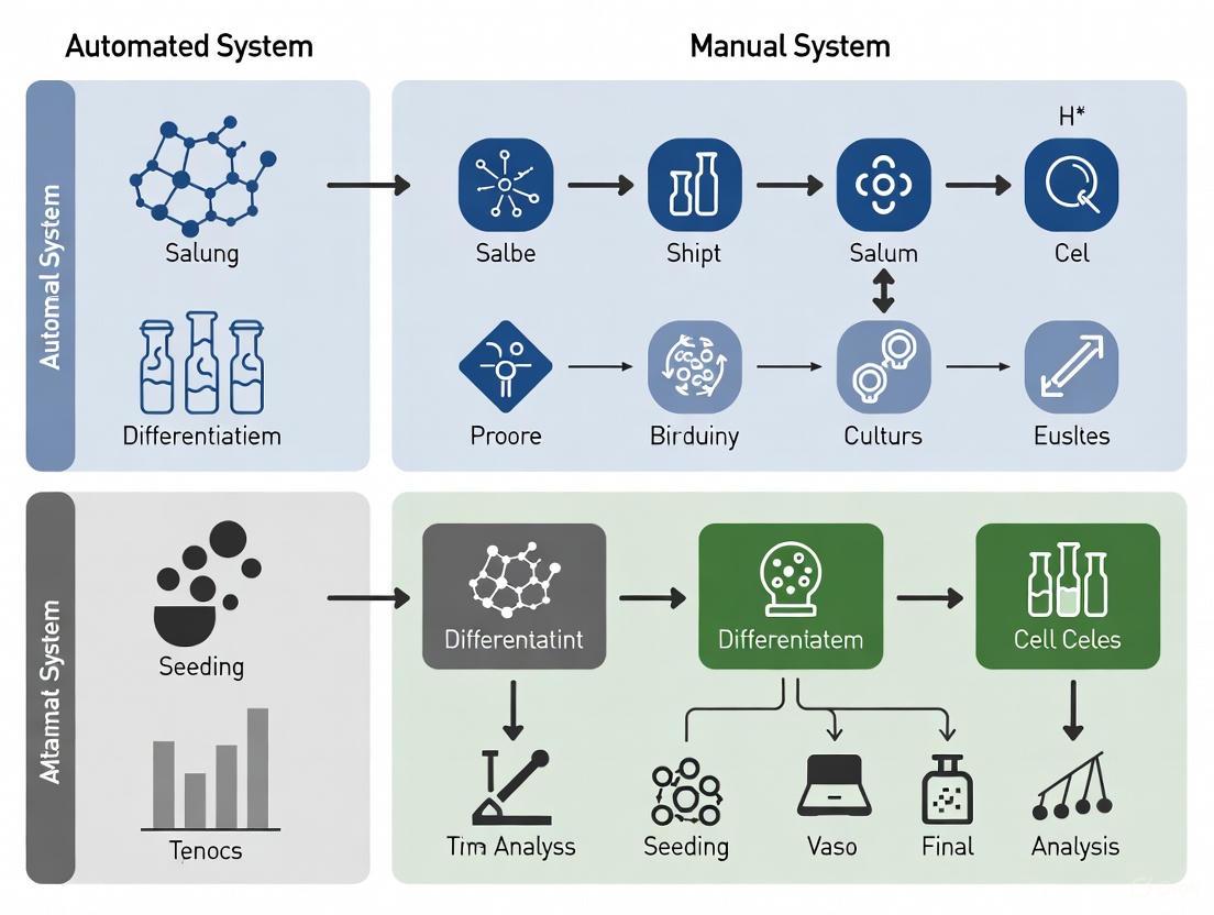
The transition from manual to automated processes is a critical step in developing scalable and robust manufacturing systems for cell-based therapies. For researchers and drug development professionals, selecting the right technology is paramount. This guide objectively compares two pivotal classes of technology—the Sepax automated cell processing system and modern bioreactor platforms—by examining their performance against manual alternatives. The focus is on providing directly comparable experimental data on key performance indicators such as cell yield, viability, and functional characteristics, all within the context of scaling up stem cell culture for Advanced Therapy Medicinal Products (ATMPs) [4] [20] [5].
Sepax System vs. Manual Isolation for MNCs and MSCs
The initial isolation of Mononuclear Cells (MNCs) from source tissues like bone marrow is a fundamental first step in the Mesenchymal Stem Cell (MSC) procurement pipeline. The following section compares the efficacy of automated isolation using the Sepax system against the traditional manual Ficoll method.
Experimental Protocol for Cell Isolation
A 2025 study provided a direct comparison under strict Good Manufacturing Practice (GMP) conditions [4]. The methodology was as follows:
- Source Material: 17 bone marrow aspirates were collected from patients using syringes containing sodium heparin [4].
- Manual Method: 100 mL of undiluted bone marrow was processed over 100 mL of Ficoll-Paque PLUS in 50 mL tubes. Density gradient centrifugation was performed at 300g for 30 minutes at 21°C. The MNC phase was collected, washed, and resuspended in culture medium [4].
- Automated Method: 100 mL of undiluted bone marrow was processed using the Sepax S-100 automated cell processing system and its single-use DGBS/Ficoll CS-900 kit, which is based on density gradient centrifugation. The isolated MNCs were recovered in 50 mL of wash medium [4].
- Downstream MSC Culture: MNCs from both methods were seeded separately at 160,000 cells/cm² in 175 cm² flasks with α-MEM medium supplemented with 20% FBS. Adherent MSCs were detached with trypsin/EDTA after 24 hours and counted [4].
- Analysis: Cell counts were performed using a Sysmex XN-20 hematology analyzer. MSC characterization included colony-forming unit (CFU) assays and differentiation potential into osteocytes, chondrocytes, and adipocytes [4].
Performance Data and Comparison
The table below summarizes the key quantitative findings from the comparative study.
Table 1: Performance Comparison of Manual vs. Sepax Automated MNC Isolation and Subsequent MSC Yield
| Performance Metric | Manual Isolation Method | Sepax Automated System | Notes |
|---|---|---|---|
| MNC Yield | Baseline (Lower) | Slightly Higher [4] | |
| MSC Yield | No significant difference | No significant difference [4] | Measured after 24-hour culture of isolated MNCs. |
| CFU Formation | No significant difference | No significant difference [4] | Indicates equivalent clonogenic potential. |
| Differentiation Potential | Maintained | Maintained [4] | Adipogenic, chondrogenic, and osteogenic potential were all preserved. |
| Process Key Differentiator | Open, multiple handling steps | Functionally closed system, reduced operator-dependent variability [4] | Automated system mitigates contamination risk and improves standardization. |
Workflow Visualization
The following diagram illustrates the comparative pathways for the manual and automated Sepax processes, highlighting the key differences in handling and process closure.
Bioreactor Platforms for Scalable Cell Culture
While the Sepax system automates the initial cell isolation, bioreactors are designed to automate and control the subsequent cell expansion phase. We compare a next-generation automated bioreactor, BECA-Auto, against manual flask culture and examine data on other bioreactor types.
Experimental Protocol for T Cell Culture
A 2025 study demonstrated a seamless transition from manual to automated T cell culture using the Bioreactor with Expandable Culture Area (BECA) platform, a relevant model for automated stem cell processing [5].
- Platform: The BECA platform consists of BECA-S (a single-chamber, open vessel for manual operation) and BECA-Auto (a standalone automated system using a closed version of the BECA-S vessel) [5].
- Culture Vessel: The unique design includes an internal movable wall that expands the culture surface area from 19 cm² to 102.4 cm² [5].
- Automated System (BECA-Auto): This benchtop system contains control units for fluid management (CIFC), automated aseptic sampling (DAAS), actuation of the vessel, and a climate-controlled enclosure that maintains conditions at 37°C, 90% relative humidity, 5% CO₂, and 20% O₂ [5].
- Process: A manual process was developed using BECA-S in a biosafety cabinet. The same process was directly transferred to BECA-Auto without re-optimization, leveraging the identical vessel geometry [5].
Performance Data and Comparison
The table below summarizes the findings from the BECA platform study and includes comparative data on fixed-bed bioreactors used for viral vector production in adherent systems, another critical area in ATMPs.
Table 2: Performance Comparison of Manual vs. Automated Bioreactor-Based Cell Culture
| Culture Platform | Process Description | Key Performance Findings | Critical Differentiators |
|---|---|---|---|
| Manual Flask / BECA-S | Open vessel, handled in BSC [5] | Baseline for cell growth and characteristics [5] | High contamination risk, operator-dependent, low scalability [5] |
| Automated BECA-Auto | Closed, automated system with environmental control [5] | No significant difference in culture outcome vs. manual; enables sterile, automated sampling and fluid handling [5] | Functional closure, process standardization, direct translation from manual protocols [5] |
| iCELLis Nano Bioreactor | Fixed-bed (random PET carriers) for adherent cells [37] | Benchmark for viral vector (lentiviral, adenoviral) production [37] | Controlled, scalable system for adherent culture [37] |
| Scale-X Hydro Bioreactor | Fixed-bed (spiral-wound PET layers) for adherent cells [37] | At least equally efficient or improved vector production vs. iCELLis; more homogeneous cell distribution [37] | Different bed structure potentially enabling better nutrient distribution and cell growth [37] |
Workflow Visualization
The diagram below outlines the logical decision pathway for transitioning from manual culture to an automated bioreactor system, using the BECA platform as a case study.
The Scientist's Toolkit: Essential Research Reagents & Materials
Successful implementation of automated cell processing relies on a foundation of high-quality, standardized reagents. The following table details key materials used in the experiments cited in this guide.
Table 3: Key Reagent Solutions for Automated Cell Processing Workflows
| Reagent / Material | Function in the Protocol | Specific Example (from search results) |
|---|---|---|
| Ficoll-Paque PLUS | Density gradient medium for isolation of Mononuclear Cells (MNCs) from bone marrow or blood [4]. | Cytiva [4] |
| Cell Culture Media | Provides nutrients and environment for cell growth and expansion. Often supplemented with serum and additives. | α-MEM with 20% FBS, Glutamine, Antibiotic-Antimycotic [4] |
| Fetal Bovine Serum (FBS) | Critical supplement providing growth factors, hormones, and attachment factors for cell culture. | Bio-Whittaker [4] |
| Dissociation Agent | Enzyme solution used to detach adherent cells from the culture surface for subculturing or harvest. | Trypsin/EDTA solution [4] |
| Transfection Reagents | For introducing nucleic acids into cells, critical for viral vector or certain therapy production. | PEIpro (Polyplus-transfection) [37] |
| Single-Use Bioreactor Kits | Pre-sterilized, disposable components (vessels, tubing) that eliminate cleaning validation and reduce cross-contamination. | DGBS/Ficoll CS-900 kit for Sepax [4]; BECA-S vessel [5] |
Concluding Analysis for Strategic Decision-Making
The collective data from these studies allows for a clear, evidence-based comparison. The Sepax automated system demonstrates equivalence to manual methods in producing functionally competent MSCs, with the added advantages of slightly higher MNC yield and a closed, standardized process that is vital for GMP manufacturing [4]. Similarly, automated bioreactors like the BECA-Auto and fixed-bed systems such as Scale-X show that automation does not compromise cell growth or product titer and can even enhance performance and homogeneity [37] [5].
The overarching conclusion for researchers and process development scientists is that automated systems are technologically mature to deliver comparable or superior biological outcomes while directly addressing the major pain points of manual processing: contamination risk, process variability, and limited scalability. The choice between systems should be guided by the specific cell type (suspension vs. adherent), the required scale, and the need for a seamless tech transfer from research-scale manual protocols.
Transitioning from 2D Adherent to 3D Suspension Culture Systems
The transition from two-dimensional (2D) adherent to three-dimensional (3D) suspension culture systems represents a paradigm shift in cellular biotechnology. While 2D monolayer cultures on flat surfaces have been the workhorse of laboratories for decades, they involve cells growing on flat, treated plastic surfaces, forming a single layer that alters natural cell morphology and behavior [38] [39]. In contrast, 3D suspension cultures allow cells to grow in all directions, forming tissue-like structures such as spheroids, organoids, or cell aggregates within a supportive matrix or in suspension, which more closely mimics the in vivo environment [40] [41].
This shift is particularly critical in stem cell research and drug development, where physiological relevance is paramount. Cells in 3D cultures exhibit natural cell-cell and cell-extracellular matrix interactions, establish nutrient and oxygen gradients, and maintain phenotypes and gene expression profiles that are often lost in 2D systems [38] [40]. For researchers benchmarking automated versus manual stem cell culture systems, understanding this transition is essential for developing more predictive models and improving the translatability of preclinical data.
Fundamental Comparisons: 2D Adherent vs. 3D Suspension Cultures
Core Structural and Functional Differences
The fundamental differences between these culture systems profoundly impact cellular behavior and experimental outcomes:
Physical Structure and Cell Morphology: In 2D systems, cells are forced into an unnatural flattened state on rigid plastic surfaces, which disrupts their natural architecture and polarity [40] [39]. In 3D suspension systems, cells maintain their natural three-dimensional morphology, with proper polarization and structural organization that resembles native tissue [40] [41].
Cellular Interactions and Microenvironment: 2D cultures offer limited cell-cell interactions primarily in a single plane and essentially no cell-extracellular matrix (ECM) interactions beyond the attachment surface [40]. Conversely, 3D suspension cultures enable multi-directional cell-cell contacts and complex interactions with a 3D ECM scaffold, recreating the tissue-specific niches essential for proper cellular differentiation and function [38] [41].
Gradient Formation and Metabolic Environment: In 2D monolayers, all cells have equal access to oxygen, nutrients, and signaling molecules, creating an artificial homogeneous environment [40]. 3D structures naturally develop physiological gradients of oxygen, nutrients, pH, and metabolic waste products [40] [41]. These gradients, particularly the hypoxic cores in larger spheroids, better mimic conditions found in tissues and tumors in vivo.
Gene Expression and Functional Outcomes: The constrained environment of 2D cultures leads to altered gene expression patterns, mRNA splicing, and cellular biochemistry that can diverge significantly from in vivo conditions [40]. Cells in 3D suspension cultures typically demonstrate more in vivo-like gene expression, protein synthesis, and drug response profiles, making them more predictive for therapeutic screening applications [40] [41].
Quantitative Comparison Table
Table 1: Direct comparison of key parameters between 2D adherent and 3D suspension culture systems.
| Parameter | 2D Adherent Culture | 3D Suspension Culture | References |
|---|---|---|---|
| Culture Formation Time | Minutes to few hours | Several hours to days | [40] |
| Cell Morphology & Polarity | Altered, flattened morphology; loss of native polarity | Preserved 3D morphology; maintained cell polarity | [40] [39] |
| Cell-Cell & Cell-ECM Interactions | Limited to single plane; minimal complex ECM contact | Multi-directional; complex, tissue-like ECM interactions | [38] [40] |
| Nutrient/Oxygen Access | Uniform, unlimited access | Physiochemical gradients (e.g., O₂, nutrients, pH) | [40] [41] |
| Gene Expression Profile | Often altered compared to in vivo | More physiologically relevant, in vivo-like expression | [40] [41] |
| Drug Response | Typically higher sensitivity; may overestimate efficacy | More predictive of in vivo response, including resistance | [38] [41] |
| Scalability | Limited by surface area; requires multiple vessels | Highly scalable in bioreactors; not limited by surface area | [42] [43] |
| Cost & Technical Complexity | Lower cost; simpler, standardized protocols | Higher cost; more complex, variable protocols | [40] [39] |
| Compatibility with High-Throughput Screening | Excellent for most standard assays | Improving with specialized equipment and protocols | [41] [39] |
| Imaging & Analysis | Simple with standard microscopy | Requires advanced (e.g., confocal) microscopy for thick structures | [38] [39] |
Experimental Protocols for 3D Suspension Culture
Protocol for Hematopoietic Progenitor Cell (HPC) Differentiation from hPSCs
Table 2: Step-by-step protocol for generating HPCs from human pluripotent stem cells (hPSCs) using a 3D swirling embryoid body (EB) method, adaptable to automated systems [43].
| Process Parameter | Standard 2D Monolayer Protocol | Modified 3D Suspension Protocol | Critical Notes for Automation |
|---|---|---|---|
| Pre-culture Maintenance | hPSCs maintained in 2D on Matrigel-coated plates in mTeSR Plus. | hPSCs can be pre-adapted to 3D suspension in TeSR-AOF 3D medium. | Automated systems can maintain uniform cell aggregates in controlled bioreactors. |
| Seeding Density | 4-8 x 10³ cells/mL (≈40-80 clumps/mL). | 1.5-2 x 10⁴ cells/mL (≈150-200 clumps/mL). | High-density seeding promotes efficient EB formation. Cell counters can automate density verification. |
| Seeding Method | Clumps (≥50 μm diameter). | Clumps (≥20 μm diameter) in medium supplemented with 10µM Y-27632 (ROCK inhibitor). | ROCK inhibitor reduces apoptosis in early aggregation. Automated dispensers ensure consistent clump size. |
| Culture Vessel & Agitation | Static culture in coated plates. | Orbital shakers (e.g., 70 RPM for 6-well plates) or PBS-MINI bioreactors (e.g., 40 RPM). | Agitation is critical for preventing aggregation and ensuring nutrient/waste exchange. Automated bioreactors control speed and environment. |
| Mesoderm Induction (Day 1-3) | Medium A (Basal Medium + Supplement A). | Same reagents as 2D. 100% medium change on Day 1. | Consistent, timed medium changes are crucial. Automated fluid handling systems improve reproducibility. |
| Hematopoietic Specification (Day 3-10) | Medium B/MK1/E1 (Basal Medium + Supplement B/MK1/E1). | Same reagents as 2D. Feeding strategy may require adjustment based on cell density. | Floating HPCs are released from EBs into suspension from Day 8 onwards. |
| Cell Harvest (Day 10-12) | Enzymatic dissociation of adherent cells required. | Harvest floating HPCs from supernatant using reversible strainers (e.g., 37 μm or 70 μm). | Non-enzymatic harvest is simpler and preserves cell surface markers. Automated filtration systems can be integrated. |
Diagram 1: 3D suspension culture workflow for HPC differentiation.
Protocol for Generating 3D Spheroids via the Hanging Drop Method
The hanging drop method is a scaffold-free technique for generating uniform 3D spheroids, suitable for both manual and automated workflows [39]:
Cell Suspension Preparation: Create a single-cell suspension of the desired cell type at a specific concentration (typically 1x10⁴ to 5x10⁴ cells/mL) in culture medium supplemented with potential ECM components [39].
Drop Formation: Using an automated liquid handler or manual pipette, dispense precise droplets (typically 20-50 µL) of the cell suspension onto the inner surface of a culture dish lid. Surface tension keeps the droplets hanging.
Inverted Incubation: Carefully invert the lid and place it over a tray containing PBS to maintain humidity. Cells settle at the liquid-air interface at the bottom of the droplet and aggregate due to gravity.
Spheroid Formation: Culture the plates for 24-72 hours in a standard CO₂ incubator. During this time, cells self-assemble into a single, compact spheroid within each drop.
Spheroid Harvesting: After spheroid formation, carefully transfer the droplets containing the mature spheroids to a low-attachment plate for long-term culture or direct experimental use. This can be done by pipetting or by flooding the lid with medium.
The Scientist's Toolkit: Essential Reagents and Materials
Successfully establishing a robust 3D suspension culture system requires specific reagents and materials that differ from standard 2D culture.
Table 3: Essential research reagent solutions for establishing 3D suspension cultures, particularly for stem cell applications [43].
| Reagent/Material Category | Specific Examples | Function in 3D Suspension Culture |
|---|---|---|
| Specialized Culture Media | TeSR-AOF 3D; STEMdiff Hematopoietic Kit (Basal Medium & Supplements A, B, MK1, E1) | Provides a defined, animal-component-free environment for maintenance and directed differentiation of hPSCs in 3D. |
| Cell Dissociation Reagents | Gentle Cell Dissociation Reagent; ReLeSR | Enables gentle passaging of hPSCs as clumps, which is critical for maintaining viability and facilitating aggregation in 3D. |
| ROCK Inhibitor | Y-27632 | Significantly improves cell survival after passaging and during the initial aggregation phase by inhibiting apoptosis. |
| Suspension Culture Vessels | 6-Well Flat-Bottom Non-Treated Plates; Nalgene Bottles; PBS-MINI Bioreactor Vessels | Non-treated plastic prevents cell attachment. Bioreactors enable controlled, scalable suspension culture with agitation. |
| Filtration & Harvesting | 37 µm & 70 µm Reversible Strainers | Allows for the gentle separation and collection of floating cells (e.g., HPCs) from larger embryoid bodies or spheroids. |
| Matrix/Scaffold Materials | Matrigel; Hydrogels (e.g., alginate, collagen) | Provides a biomimetic 3D extracellular matrix (ECM) for scaffold-based methods, supporting cell growth and organization. |
| Downstream Differentiation Kits | STEMdiff Microglia, Erythroid, and Megakaryocyte Kits | Used to further differentiate the HPCs produced in 3D suspension into specific, functional hematopoietic cell lineages. |
| Characterization Tools | MethoCult SF (Semi-Solid Medium) | Enables functional assessment of hematopoietic progenitors via colony-forming unit (CFU) assays. |
Comparative Performance Data and Key Challenges
Performance Benchmarking
Transitioning from 2D to 3D systems shows clear biological advantages but also introduces operational complexities. In hematopoietic differentiation, the 3D swirling EB method can generate HPCs that are comparable in quality and differentiation potential to those from 2D systems but with the significant advantage of scalability, as cell production is not limited by the surface area of culture vessels [43]. For drug screening, 3D tumor spheroids consistently demonstrate enhanced predictive power. They model critical tumor characteristics like dormancy, hypoxia, and anti-apoptotic behavior that are absent in 2D monolayers, often leading to more accurate assessments of drug efficacy and penetration [38] [41]. A key technological advancement in automated 3D systems is the ability to monitor and control key process parameters in real-time. Parameters such as glucose, lactate, dissolved oxygen (DO), pH, and viable cell density can be tracked using technologies like Raman spectroscopy and biocapacitance probes, enabling dynamic control of the culture environment for improved consistency and yield [44].
Critical Challenges in Transitioning and Scaling Up
Cell Harvesting Limitations: Retrieving cells from certain 3D systems, particularly fixed-bed bioreactors or dense scaffolds, can be challenging. Enzymatic digestion required for harvest can be harsh, prolong processing time, and risk contaminating the final cell product, which is a critical concern for cell-based therapies [42].
Phenotypic Stability: The 3D structural environment and mechanical forces (e.g., shear stress in stirred-tank bioreactors) can induce unintended cell differentiation or phenotypic drift. For example, stem cells expanded on microcarriers have been shown to lose key pluripotency markers, compromising their therapeutic potential [42].
Process Control and Scalability: While 3D suspension culture in bioreactors is inherently more scalable than 2D monolayers, achieving consistent, large-scale production is non-trivial. Challenges include maintaining uniform aggregate size, ensuring adequate nutrient mixing without damaging cells, and developing standardized, reproducible protocols [42] [45].
Analytical Complexity: Imaging and analyzing 3D structures is significantly more complex than 2D monolayers. It requires advanced techniques like confocal microscopy and specialized software for 3D reconstruction and quantification, posing a barrier to high-throughput analysis [38] [39].
Diagram 2: Key challenges and emerging solutions for 3D suspension culture.
The transition from 2D adherent to 3D suspension culture systems is a critical evolution in cell culture technology, enabling more physiologically relevant models for stem cell research and drug development. While 2D systems remain valuable for high-throughput screening and basic research due to their simplicity and low cost, 3D suspension cultures offer superior predictive power for clinical outcomes by better mimicking the in vivo microenvironment, architecture, and cellular responses [41] [39].
The future of this field lies not in choosing one system over the other, but in developing integrated, hybrid workflows. These workflows leverage the speed and simplicity of 2D for initial screening and the physiological relevance of 3D for validation and deeper mechanistic studies [41]. Furthermore, the integration of advanced process analytical technology (PAT), automated bioreactors, and AI-driven data analytics will be pivotal in overcoming current challenges in scalability, reproducibility, and phenotyping, ultimately solidifying 3D suspension culture as the standard for predictive preclinical research [45] [44].
Organoids are three-dimensional (3D), self-organizing, miniaturized structures derived from stem cells that closely mimic the architecture and functionality of human organs. These advanced in vitro models represent a significant paradigm shift in biomedical research, offering a more physiologically relevant alternative to traditional two-dimensional (2D) cell cultures. By preserving native tissue architecture and critical cellular interactions, organoids provide unprecedented opportunities for studying human development, disease mechanisms, and therapeutic interventions. The emergence of organoid technology is particularly transformative for drug discovery and disease modeling, enabling researchers to bridge the gap between conventional cell culture and in vivo human physiology.
Two primary categories of organoids have emerged as powerful tools: those derived from induced Pluripotent Stem Cells (iPSCs) and Patient-Derived Organoids (PDOs). iPSC-derived organoids are generated from reprogrammed somatic cells that have been returned to a pluripotent state, allowing them to differentiate into virtually any cell type. These models excel at recapitulating developmental processes and genetic disorders. In contrast, PDOs are generated directly from patient tissues and faithfully maintain tissue-specific characteristics and disease phenotypes, making them indispensable for personalized medicine applications. Both model types are revolutionizing preclinical research by providing more human-relevant platforms for evaluating drug efficacy and safety while addressing ethical concerns associated with animal models through the principles of the 3Rs (Replacement, Reduction, and Refinement).
Comparative Analysis of Organoid Types
Types and Characteristics of Organoids
Organoids can be broadly classified based on their cellular origin and methodological approach. Understanding these distinctions is crucial for selecting the appropriate model system for specific research applications in drug discovery and disease modeling.
Table 1: Comparison of Major Organoid Types and Their Characteristics
| Organoid Type | Stem Cell Source | Key Advantages | Primary Limitations | Optimal Applications |
|---|---|---|---|---|
| iPSC-Derived Organoids | Induced Pluripotent Stem Cells | Remarkable plasticity; model wide range of tissues and developmental stages; valuable for genetic disorders and complex diseases [46] | Prolonged differentiation protocols; variability in maturation levels; batch-to-batch variability [46] [47] | Studying early human development, genetic disorders, complex diseases [46] |
| Patient-Derived Organoids (PDOs) | Adult Stem Cells (ASCs) from patient tissues | Faithfully recapitulate tissue-specific characteristics and disease phenotypes; indispensable for personalized medicine [46] | Limited expansion capacity; may not fully capture tissue microenvironment [48] | Personalized drug screening, disease modeling, understanding individualized treatment responses [46] |
| Tumor Organoids | Cancer stem cells from patient tumors or PDX models | Retain original tumor morphology and genetic features; replicate patient response in clinic; high translatability [49] | Typically epithelial origin only; require specific culture conditions [49] | Oncology drug screening, personalized cancer therapy, biomarker discovery |
| Embryonic Stem Cell-Derived Organoids | Human Embryonic Stem Cells (hESCs) | Broast differentiation potential; model early developmental processes | Ethical concerns; limited patient specificity | Human development studies, developmental disorders, tissue morphogenesis |
Organoids vs. Traditional 2D Cultures and 3D Primary Cell Cultures
Organoids represent a significant advancement over traditional 2D cell cultures and other 3D model systems. While 2D cultures grown as flat monolayers on plastic surfaces have been the workhorse of in vitro research for decades, they fail to recapitulate the complex spatial architecture and cell-cell interactions found in native tissues. This limitation reduces their physiological relevance and predictive power for clinical outcomes.
Table 2: Comparison of Organoids with Other Model Systems
| Characteristic | Organoids | Traditional 2D Cultures | 3D Primary Cell Cultures |
|---|---|---|---|
| Architecture | Self-organizing 3D structures resembling mini-organs [46] | Flat monolayers | Simple spheroids via cell-cell adhesion [49] |
| Cellular Complexity | Multiple differentiated cell lineages; stem cell population maintained [49] | Typically homogeneous | Primarily differentiated cells; may contain cancer stem cells in tumorspheres [49] |
| Physiological Relevance | High - preserve native tissue architecture and cellular interactions [46] | Low - lack tissue context and 3D interactions | Moderate - better than 2D but less complex than organoids [49] |
| Long-term Maintenance | Can be maintained long-term with genomic stability [49] | Can be maintained indefinitely but may drift | Limited - cells become senescent or drift over passages [49] |
| Biobanking Potential | Excellent - can be cryopreserved without compromising identity [49] | Good - established cell banks | Poor - difficult to revive successfully [49] |
| Scalability for HTS | High - amenable to scale-up for large-scale screening [49] | High - well established | Limited - more challenging to standardize [49] |
The key distinguishing feature of organoids is their self-organizing capacity, which results in multicellular structures developing from stem or progenitor cells and exhibiting remarkable similarities to in vivo organ architecture [49]. This self-organization occurs through complex developmental processes that mirror organogenesis in vivo, resulting in structures that contain multiple differentiated cell types arranged in a spatially correct manner. In contrast, 3D primary cell cultures typically form through simple cell-cell adhesion when physical or mechanical force is applied, resulting in spheroids that lack the complex architecture and cellular diversity of true organoids [49].
Quantitative Performance Data in Drug Discovery Applications
Efficacy and Toxicity Prediction
Organoid technologies have demonstrated significant advantages in predicting drug efficacy and toxicity, potentially reducing the high attrition rates in drug development. The enhanced physiological relevance of organoids translates to improved predictive power for clinical outcomes.
Table 3: Quantitative Performance Metrics of Organoids in Drug Discovery
| Application Area | Model Type | Key Performance Metrics | Advantages over Traditional Models |
|---|---|---|---|
| Drug Efficacy Screening | Patient-derived tumor organoids (PDTOs) | Retain histological/genomic features of original tumors; predict individual responses to chemotherapy, targeted agents, immunotherapies [47] | Human-specific responses; patient-tailored; more accurate prediction of clinical efficacy [47] |
| Toxicity Testing | hPSC-derived hepatocytes/cardiomyocytes [47] | Better prediction of human toxicity; detect cardiotoxic effects (e.g., doxorubicin) not readily observed in non-human systems [47] | More human-relevant toxicity profiles; reduced reliance on animal models |
| High-Throughput Screening | Various organoid types with automation | 3x increase in screening capacity; 25% reduction in time-to-results reported by biotech firms using robotic platforms [50] | Accelerated pipeline from discovery to clinical trials; cost and time savings |
| Personalized Therapy Selection | Patient-derived organoids (PDOs) | Successful clinical pilots in colorectal, pancreatic, and lung cancers to inform treatment decisions [47] | Enables personalized therapeutic strategies; reduces risk of adverse outcomes |
Economic Impact and Market Adoption
The growing recognition of organoids' value in drug discovery is reflected in market trends and adoption metrics. The global market for organoid culture systems is experiencing robust growth, projected to reach approximately $3.2 billion by 2025, representing a compound annual growth rate of 22.5% from 2020 [48]. The pharmaceutical and biotechnology sectors constitute the largest demand segment, accounting for nearly 45% of the total market share, as these industries increasingly adopt organoid technologies to reduce drug development costs and timelines [48].
Regionally, North America dominates the market with approximately 40% share, followed by Europe (30%) and Asia-Pacific (20%), with the Asia-Pacific region expected to witness the highest growth rate of 25% annually through 2025 [48]. This growth is fueled by increasing research investments in countries like China, Japan, and South Korea, along with the expansion of contract research organizations in these regions.
Experimental Protocols and Methodologies
Organoid Generation and Culture Workflows
The generation of organoids requires specific protocols tailored to the tissue type and stem cell source. Below are detailed methodologies for establishing and maintaining different organoid cultures.
iPSC-Derived Organoid Generation
iPSC-derived organoids are generated through a multi-step differentiation process that mimics embryonic development:
Reprogramming and Pluripotency Maintenance: Human somatic cells (e.g., fibroblasts, blood cells) are reprogrammed into induced pluripotent stem cells using defined transcription factors (OCT4, SOX2, KLF4, c-MYC) [47]. iPSCs are maintained in feeder-free conditions with essential pluripotency factors.
Lineage-Specific Differentiation: iPSCs are directed toward specific germ layers using defined morphogens and small molecules:
- Endodermal lineage: Activin A treatment to induce definitive endoderm
- Mesodermal lineage: BMP4 and FGF2 treatment for mesoderm commitment
- Ectodermal lineage: Dual SMAD inhibition for neural induction
3D Matrix Embedding: Differentiated progenitor cells are embedded in Basement Membrane Extract (e.g., Matrigel) to provide a 3D environment that supports self-organization.
Tissue-Specific Maturation: Organoids are maintained in specialized media containing tissue-specific growth factors and patterning molecules:
Maturation and Analysis: Organoids are cultured for extended periods (weeks to months) to allow functional maturation, with medium changes every 2-4 days.
Patient-Derived Organoid Establishment
PDOs are generated directly from patient tissues, preserving the original tissue architecture and genetic landscape:
Tissue Collection and Processing: Patient tissue samples are obtained via biopsy or surgical resection and immediately placed in cold preservation medium. Tissues are minced into small fragments (0.5-1 mm³) using surgical scalpels.
Enzymatic Digestion: Tissue fragments are digested with collagenase (1-2 mg/mL) or other tissue-specific enzymes at 37°C for 30-60 minutes with gentle agitation.
Stem Cell Enrichment: Digested tissue is filtered through cell strainers (70-100 µm) to remove debris. Epithelial cells or stem cell populations are enriched using differential centrifugation or magnetic-activated cell sorting (MACS).
Matrix Embedding and Culture: Isolated cells are resuspended in Basement Membrane Extract and plated as droplets in pre-warmed culture plates. After matrix polymerization, organoid culture medium is added.
Passaging and Expansion: Organoids are passaged every 1-3 weeks using mechanical disruption or enzymatic digestion (e.g., TrypLE) followed by re-embedding in fresh matrix.
Automated vs. Manual Culture Systems
Automation is increasingly being applied to organoid culture to address challenges of reproducibility and scalability. The following experimental comparison highlights key differences between these approaches.
Diagram 1: Workflow comparison between manual and automated organoid culture systems
A recent study comparing manual and automated methods for isolating mononuclear cells (MNCs) and mesenchymal stem cells (MSCs) using Ficoll density gradient centrifugation provides quantitative insights into the performance differences between these approaches [3] [4]. In this study, seventeen bone marrow samples were processed using both manual methods and the automated Sepax system, with subsequent analysis of MNC and colony-forming unit (CFU) counts, MSC differentiation potential, and phenotypic characterization [4].
Table 4: Experimental Comparison of Manual vs. Automated Cell Culture Methods
| Parameter | Manual Method | Automated Sepax System | Significance |
|---|---|---|---|
| MNC Yield | Baseline | Slightly higher | Automated system demonstrated slightly higher MNC yields [4] |
| CFU Formation | Baseline | No significant difference | No significant differences observed in CFU formation [4] |
| MSC Characteristics | Baseline | No significant difference | No significant differences in differentiation potential or phenotype [4] |
| Reproducibility | Higher variability between operators | Standardized process | Reduced operator-dependent variability in automated system |
| Throughput | Limited by manual labor | Higher processing capacity | Automated systems enable higher throughput |
| Personnel Time | Significant hands-on time | Minimal hands-on time | Automation frees technical staff for other tasks |
While the Sepax system demonstrated slightly higher MNC yields, no significant differences were observed in CFU formation or MSC characteristics compared to manual isolation [4]. This suggests that automated systems can maintain cell quality while improving efficiency and standardization.
Signaling Pathways in Organoid Development and Maintenance
Organoid formation and maintenance rely on precisely regulated signaling pathways that mimic the niche signals present in native tissues. The careful manipulation of these pathways through specific agonists and inhibitors is essential for successful organoid culture.
Diagram 2: Key signaling pathways governing organoid development and maintenance
The intestinal organoid culture system developed by Clevers and colleagues exemplifies the precise manipulation of signaling pathways [51]. This protocol requires the provision of an appropriate niche consisting of Matrigel, epidermal growth factor, WNT agonists (R-spondin-1), BMP inhibitors (Noggin), and other cytokines to support the formation of 3D intestinal organoids from Lgr5+ stem cells [51]. Similar approaches have been adapted for organoids from various tissues, with pathway modulation tailored to specific tissue requirements.
Essential Research Reagents and Materials
Successful organoid culture requires specialized reagents and materials that support stem cell maintenance, differentiation, and 3D structure formation. The following table details key components of organoid culture systems and their functions.
Table 5: Essential Research Reagent Solutions for Organoid Culture
| Reagent Category | Specific Examples | Function | Application Notes |
|---|---|---|---|
| Extracellular Matrices | Matrigel, Cultrex BME, Synthetic hydrogels | Provide 3D scaffolding; present biochemical cues; support polarization | Matrigel is most common but has batch variability; synthetic alternatives in development [48] |
| Basal Media | Advanced DMEM/F12, STEMPRO hESC SFM | Nutrient foundation; osmolarity and pH maintenance | Must be supplemented with specific growth factors and inhibitors |
| Essential Growth Factors | EGF, FGF, Noggin, R-spondin, HGF, Oncostatin M | Activate signaling pathways for proliferation and differentiation | Combinations and concentrations vary by organoid type |
| Pathway Modulators | CHIR99021 (GSK-3 inhibitor), A83-01 (TGF-β inhibitor), Y-27632 (ROCK inhibitor) | Enhance stem cell survival; direct differentiation; prevent anoikis | Small molecule inhibitors often used alongside growth factors |
| Antibiotics/Antimycotics | Penicillin-Streptomycin, Amphotericin B | Prevent microbial contamination | Use may vary based on sterility of initial tissue |
| Dissociation Reagents | Trypsin-EDTA, TrypLE, Accutase, Collagenase | Passage organoids; digest primary tissues | Enzyme selection and incubation time critical for cell viability |
| Serum Alternatives | B27 Supplement, N2 Supplement | Provide defined components for cell growth | Essential for reducing batch variability in culture conditions |
The development of defined, serum-free media formulations has been crucial for advancing organoid technology, reducing variability, and improving reproducibility. These defined systems allow precise control over signaling pathways that govern stem cell maintenance and differentiation. However, challenges remain with batch-to-batch variability of critical components, particularly natural matrices like Matrigel, driving research into synthetic alternatives [48].
Current Challenges and Future Perspectives
Technical Limitations and Ongoing Developments
Despite significant advancements, organoid technology faces several challenges that limit its widespread adoption. Key technical limitations include:
Reproducibility and Standardization: Considerable batch-to-batch variation persists even within the same laboratory settings due to the complex interplay of growth factors, extracellular matrix components, and culture conditions [48]. The field lacks consensus on quality control metrics for defining organoid quality.
Scalability for High-Throughput Applications: Current organoid culture methods are labor-intensive and difficult to scale up while maintaining quality and functionality [48]. This limitation is particularly evident in drug screening applications where high-throughput systems are essential.
Vascularization and Microenvironment Complexity: Most organoids lack proper blood vessel formation, leading to necrotic cores as they grow beyond 300-400 micrometers in diameter [48]. This limits their size, maturation, and long-term viability.
Microenvironment Recapitulation: Many organoid cultures lack components of the native microenvironment, such as immune cells, vasculature, and stromal elements, which can significantly influence therapeutic responses [47].
Promising solutions to these challenges are emerging through interdisciplinary approaches. Automation integration, while still underdeveloped, is progressing with systems emerging for media changes and imaging [48]. Vascularization strategies including co-culture systems and microfluidic devices show promise but require optimization for routine implementation [48]. The integration of organoids with microfluidic "organ-on-chip" platforms enables more accurate modeling of human pharmacokinetics and pharmacodynamics under dynamic flow conditions [47].
Integration with Emerging Technologies
The future of organoid technology lies in its convergence with other cutting-edge technologies:
Artificial Intelligence and Machine Learning: AI applications are being developed for optimizing culture conditions, predicting organoid development trajectories, and analyzing complex datasets from organoid experiments [48] [52]. These approaches promise to enhance standardization and extract more comprehensive information from organoid models.
Advanced Bioengineering: The combination of organoids with high-performance materials, 3D printing technology, and tissue engineering enhances their applications in regenerative medicine [51]. These approaches address limitations in structural complexity and functional integration.
Multi-omics Integration: Combining organoid technology with genomics, transcriptomics, proteomics, and metabolomics provides comprehensive insights into developmental processes, disease mechanisms, and drug responses [47].
Organ-on-Chip Platforms: Integrating organoids with microfluidic systems creates more physiologically relevant models that incorporate fluid flow, mechanical forces, and multi-tissue interactions [47].
As these technologies mature and converge, organoid models are poised to become increasingly sophisticated, bridging the gap between traditional cell culture and in vivo physiology. This progress will enhance their utility in drug discovery, disease modeling, and personalized medicine, ultimately improving the predictive power of preclinical research and accelerating the development of novel therapeutics.
The manufacturing of Advanced Therapy Medicinal Products (ATMPs), including mesenchymal stem/stromal cell (MSC)-based therapies, demands rigorous adherence to Good Manufacturing Practice (GMP) standards to ensure product safety, quality, and efficacy [53]. Traditional manual cell culture methods are increasingly inadequate for meeting clinical-scale production needs due to their labor-intensive nature, pronounced risk of contamination, and inherent process variability [54]. This creates a pressing need for robust scale-up strategies that integrate automated systems, enabling a transition from laboratory-scale processes to commercially viable manufacturing while maintaining full GMP compliance [55]. The evolution from manual to automated processes represents a critical path for the cell therapy industry to achieve the reproducibility, scalability, and cost-effectiveness required for widespread clinical application [56] [53].
This guide objectively compares automated and manual cell culture systems, providing a benchmarking framework grounded in experimental data. It is structured to assist researchers, scientists, and drug development professionals in making informed decisions when designing and scaling GMP-compliant manufacturing processes for cell-based therapies.
Comparative Analysis: Manual vs. Automated Cell Culture Systems
Quantitative Benchmarking of Isolation and Expansion
Direct comparative studies provide the most compelling evidence for evaluating cell culture systems. A seminal study directly compared manual isolation with the automated Sepax S-100 system for processing bone marrow aspirates to isolate mononuclear cells (MNCs), a critical first step in MSC manufacturing [3].
Table 1: Performance Comparison of Manual vs. Automated MNC Isolation and MSC Yield [3]
| Parameter | Manual Method | Automated Sepax S-100 Method |
|---|---|---|
| MNC Yield | Baseline (Reference) | Slightly Higher |
| Colony-Forming Unit (CFU) Formation | No Significant Difference | No Significant Difference |
| MSC Phenotype (CD marker expression) | No Significant Difference | No Significant Difference |
| MSC Differentiation Potential | No Significant Difference | No Significant Difference |
| Process Consistency | Subject to operator variability | High reproducibility, reduced human intervention |
| GMP Compliance Assurance | Relies heavily on rigorous trained operator technique [3] | Enhanced via closed-system processing and automation [3] |
The data demonstrates that automation can achieve equivalent cell quality with potential improvements in initial yield and significant enhancements in process robustness. While the core biological properties of the resulting MSCs were unchanged, the automated method reduces a major source of variability—direct human handling [3].
For the expansion phase, automated bioreactor systems show superior performance in large-scale production. The Quantum Cell Expansion System (Terumo BCT), a hollow fiber bioreactor, provides a scalable, closed-system alternative to manual flask-based culture.
Table 2: Scalability and Efficiency: Flask-Based Culture vs. Automated Bioreactor [53]
| Parameter | Manual Flask-Based Culture | Quantum Cell Expansion System |
|---|---|---|
| Culture Surface Area | ~120 T-175 flasks for equivalent scale | 21,000 cm² (integrated) |
| Manipulation Steps | ~54,400 steps for a typical run | ~133 steps for a typical run |
| Process Type | Primarily open, requiring Grade A/B cleanrooms | Closed system, reducing contamination risk |
| Medium Exchange | Manual, discontinuous | Automated, continuous |
| Yield | Baseline (Reference) | 100–276 × 10⁶ BM-MSCs from a 20 × 10⁶ seed in 7 days [53] |
| Environmental Control | Limited | Direct connection to gases for normoxic/hypoxic control |
Several integrated automated platforms are designed specifically for GMP-compliant cell manufacturing. The table below summarizes key systems and their applications in MSC production.
Table 3: Comparison of Automated Platforms for GMP-Compliant MSC Manufacturing [53]
| Platform (Vendor) | Technology/Process | Key Features and Applications |
|---|---|---|
| Quantum Cell Expansion System (Terumo BCT) | Hollow fiber bioreactor | Closed, automated system; continuous medium exchange; used for BM-MSCs, AT-MSCs, UC-MSCs; supports serum-free culture with hPL [53]. |
| CliniMACS Prodigy (Miltenyi Biotec) | Integrated automated processing | Automates isolation, cultivation, and harvest; uses MSC-Brew GMP medium; processes BM-MSCs, AT-MSCs, UC-MSCs [53]. |
| Xuri Cell Expansion System W25 (Cytiva) | Stirred-tank bioreactor | Scalable wave-mixed bioreactor; suitable for microcarrier-based expansion of adherent cells [53]. |
| Cocoon Platform (Lonza) | End-to-end automated manufacturing | Designed for patient-specific (autologous) cell therapies; integrates all steps from sample to final bag in a single, closed device [53]. |
Experimental Protocols for Benchmarking Studies
Protocol 1: Comparative Isolation of Mononuclear Cells
This protocol is derived from a published methods section that directly compared manual and automated isolation [3].
Objective: To isolate MNCs from bone marrow aspirate using density gradient centrifugation and compare the efficacy and efficiency of manual and automated (Sepax S-100) methods.
Materials and Reagents:
- Source Material: Human bone marrow aspirate.
- Separation Medium: Ficoll-Paque PLUS (Cytiva).
- Wash Medium: α-MEM supplemented with 20% FBS, 2 mM L-glutamine, and 1% antibiotic-antimycotic.
- Equipment: Centrifuge (for manual method); Sepax S-100 automated cell processing system with DGBS/Ficoll CS-900 kit (for automated method).
- Consumables: 50 mL conical tubes (manual); single-use, closed-set tubing (automated).
Experimental Workflow:
Procedure:
- Sample Preparation: Bone marrow aspirates are collected in syringes containing sodium heparin [3].
- Manual Isolation:
- Distribute Ficoll and bone marrow sample across multiple 50 mL tubes.
- Perform density gradient centrifugation at 300g for 30 minutes at 21°C.
- Carefully aspirate the MNC layer post-centrifugation.
- Wash the harvested MNCs by adding wash medium and centrifuging.
- Resuspend the final MNC pellet in a defined volume of medium [3].
- Automated Isolation (Sepax S-100):
- Connect the bone marrow collection bag and solutions to the single-use kit.
- Run the pre-programmed "DGBS/Ficoll" protocol. The system automatically performs density gradient separation, MNC harvesting, and washing.
- Recover the isolated MNCs in a final transfer bag [3].
- Analysis:
- Count MNCs using a hematology analyzer (e.g., Sysmex XN-20).
- Plate MNCs for MSC expansion and compare CFU formation, cell phenotype, and differentiation potential after culture.
Protocol 2: Comparative Expansion in Bioreactor vs. Flasks
Objective: To compare the expansion of MSCs using a traditional flask-based method and an automated bioreactor system (e.g., Quantum System).
Materials and Reagents:
- Cells: Isolated MNCs or early-passage MSCs.
- Culture Medium: Xeno-free, serum-free medium or medium supplemented with Human Platelet Lysate (hPL).
- Substrate: Fibronectin or other recombinant adhesion factors (for coating hollow fiber bioreactor).
- Equipment: T-175 flasks or HYPERflasks; Quantum Cell Expansion System with hollow fiber bioreactor cartridge.
- Analytical Tools: Cell counter, flow cytometer for phenotype, differentiation kits.
Experimental Workflow:
Procedure:
- System Preparation:
- Manual (Flasks): Pre-coat flasks if required.
- Automated (Quantum): Prime and coat the hollow fiber bioreactor with fibronectin per manufacturer's instructions [53].
- Cell Seeding: Seed MSCs at a validated density into both systems.
- Expansion Phase:
- Manual: Culture cells, performing manual medium changes every 2-3 days.
- Automated: The system continuously perfuses fresh medium based on set parameters (e.g., glucose consumption rate).
- Process Monitoring:
- Manual: Monitor via daily microscopy and off-line metabolite analysis (e.g., glucose/lactate).
- Automated: Utilize in-line or at-line sensors for real-time monitoring of metabolites and culture environment [53].
- Cell Harvest:
- Manual: Wash cells, dissociate with trypsin/EDTA, neutralize, and centrifuge.
- Automated: Initiate automated harvest protocol involving enzymatic detachment and collection into a harvest bag [53].
- Product Quality Assessment:
- Determine total cell yield and viability.
- Characterize cells via flow cytometry for standard MSC markers (CD105+, CD73+, CD90+, CD45-, CD34-, etc.).
- Assess functionality through tri-lineage differentiation (adirogenic, osteogenic, chondrogenic) and CFU assays.
The Scientist's Toolkit: Essential Reagents and Materials
Successful and reproducible GMP-compliant manufacturing relies on high-quality, well-defined materials. The following table details key reagents and their critical functions in automated and manual processes.
Table 4: Essential Research Reagent Solutions for MSC Manufacturing [3] [53] [54]
| Reagent/Material | Function | GMP-Compliant Considerations |
|---|---|---|
| Ficoll-Paque PLUS | Density gradient medium for isolation of mononuclear cells from bone marrow or other sources. | Must be GMP-grade. Serves as a critical first step in the manufacturing process [3]. |
| Serum-Free/Xeno-Free Medium | Provides nutrients, growth factors, and hormones for cell growth without animal-derived components. | Essential for avoiding xenoantigens and batch-to-batch variability of FBS. Supports consistent, regulatory-compliant production [53] [54]. |
| Human Platelet Lysate (hPL) | Growth supplement used as a replacement for Fetal Bovine Serum (FBS). | GMP-grade hPL mitigates risks associated with animal sera and can enhance MSC proliferation [53]. |
| Recombinant Trypsin/TrypLE | Enzymatic cell dissociation agent for detaching adherent cells during passaging and harvest. | Preferable to animal-derived trypsin; reduces contamination risk and improves process consistency [54]. |
| Cell Culture Substrates (e.g., Fibronectin) | Coating for surfaces to enhance cell adhesion and growth, critical in bioreactor systems like the Quantum. | Use of recombinant proteins is favored over human-sourced cryoprecipitate to ensure defined composition and reduce adventitious agent risk [53]. |
Strategic Implementation and Compliance Frameworks
Quality by Design and Computer System Validation
Integrating automation into a GMP environment requires more than just purchasing equipment; it demands a strategic approach to process design and compliance.
Quality by Design (QbD): Implementing QbD is crucial for scalable manufacturing. This involves defining a Quality Target Product Profile (QTPP), identifying Critical Quality Attributes (CQAs) of the cell product, and understanding the link between Critical Process Parameters (CPPs) and these CQAs. A well-defined QbD framework ensures that transitioning from a manual to an automated process (or between different automated platforms) does not alter the fundamental quality of the cell product [54].
Computer System Validation (CSV): For any automated system used in GMP production, CSV is mandatory. A scalable CSV strategy should be risk-based and align with facility growth [57]:
- Early Phase: Focus on core systems (e.g., LIMS) using lean but robust validation.
- Mid Phase: Standardize validation templates for new systems like MES and ensure integration between systems is qualified.
- Commercial Phase: Implement a CSV governance framework for ongoing change management and periodic review [57].
Navigating the Regulatory Landscape: GMP vs. cGMP
Understanding regulatory expectations is fundamental. There is a critical distinction between GMP and current GMP (cGMP).
- GMP establishes the baseline requirements for methods, facilities, and controls to ensure product safety and identity.
- cGMP emphasizes that these practices must be current, incorporating the latest technological and scientific advancements, including automation and advanced data analytics [58].
This means that simply having a validated automated system is not enough. Manufacturers must demonstrate continuous improvement and adoption of modern technologies, such as Process Analytical Technology (PAT) for real-time monitoring, to meet cGMP standards [58].
The integration of automated systems into GMP-compliant manufacturing is no longer a luxury but a necessity for the scalable, cost-effective, and reliable production of cell-based therapies. As the data and protocols presented in this guide demonstrate, automation can achieve product quality that is equivalent or superior to manual methods while significantly enhancing process robustness, reproducibility, and scalability. Systems like the Sepax S-100, Quantum, and CliniMACS Prodigy provide viable, closed-system pathways to clinical and commercial manufacturing.
A successful scale-up strategy requires a holistic approach that combines technical selection of appropriate platforms with rigorous experimental benchmarking, a solid QbD foundation, and a proactive cGMP compliance mindset. By adopting these integrated scale-up strategies, researchers and drug development professionals can accelerate the translation of promising cell therapies from the laboratory bench to the patient bedside.
Optimizing Culture Outcomes: AI, Process Control, and Troubleshooting Common Challenges
Leveraging AI and Machine Learning for Predictive Monitoring and Quality Control
The transition from manual to automated systems represents a foundational shift in stem cell biomanufacturing for Advanced Therapy Medicinal Products (ATMPs). This evolution is largely driven by the integration of Artificial Intelligence (AI) and Machine Learning (ML), which are transforming traditional lab practices into data-rich, predictive processes. While manual methods have long been the cornerstone of biological research, they face significant challenges in scalability, reproducibility, and real-time monitoring—critical factors for clinical-grade cell production [25]. Automated systems, enhanced with AI-driven analytics, now offer solutions to these limitations by enabling continuous, non-invasive monitoring and control of Critical Quality Attributes (CQAs) [25]. This guide provides an objective comparison of these approaches, framed within a broader thesis on benchmarking their performance. We present experimental data and detailed methodologies to help researchers, scientists, and drug development professionals make informed decisions in process optimization and technology adoption.
Experimental Protocols: Manual vs. AI-Driven Automated Systems
Protocol for Manual Monitoring and Quality Control
The conventional manual approach relies on periodic sampling and endpoint assays, requiring significant human intervention [25].
- Step 1: Sample Collection. At predefined time points (e.g., every 24 hours), a technician collects small aliquots from the culture vessel under sterile conditions. This process inherently risks contamination and disturbs the culture environment [3] [4].
- Step 2: Cell Viability and Counting. Using trypan blue exclusion or similar methods, cells are counted manually with a hemocytometer or with semi-automated cell counters. This provides a snapshot of viability and concentration but consumes the sample [25].
- Step 3: Flow Cytometry and Immunostaining. Cells are stained for specific surface markers (e.g., CD73, CD90, CD105 for MSCs) and analyzed via flow cytometry to confirm phenotypic identity. This is a destructive, resource-intensive process that offers low temporal resolution [25].
- Step 4: Differentiation Potential Assessment. To monitor differentiation potential, cells are induced toward specific lineages (osteogenic, adipogenic, chondrogenic). The resulting differentiation is typically assessed after 14-21 days using endpoint staining protocols (e.g., Oil Red O for adipocytes, Alizarin Red for osteocytes) [3] [4]. This process is slow and provides only retrospective quality data.
- Step 5: Environmental Monitoring. Parameters like pH and dissolved oxygen are typically monitored using offline samplers or simple in-line probes that lack predictive capability. Adjustments to the incubator or bioreactor are made reactively based on these readings [25].
Protocol for AI-Driven Automated Monitoring and Quality Control
AI-driven systems integrate hardware automation with machine learning algorithms for continuous, predictive quality control [25].
- Step 1: Real-Time Data Acquisition. Integrated sensors continuously collect data on environmental parameters (pH, dissolved O₂, metabolites). Simultaneously, high-resolution time-lapse microscopy captures live-cell images without disturbing the culture [25].
- Step 2: Convolutional Neural Network (CNN) Analysis for Morphology. A CNN model, pre-trained on vast image libraries, analyzes the microscopic images in real-time. It tracks critical morphological features, such as cell confluence, colony formation, and early signs of differentiation or apoptosis, with over 90% accuracy as demonstrated in studies tracking iPSC colonies [25].
- Step 3: Predictive Modeling for Process Control. A predictive algorithm (e.g., a regression model or recurrent neural network) analyzes the stream of sensor data. It forecasts future culture states, such as predicting a critical drop in oxygen saturation hours in advance, allowing for preemptive intervention [25].
- Step 4: Anomaly Detection and Lineage Commitment Tracking. An unsupervised ML model (e.g., an autoencoder) establishes a baseline of "normal" culture behavior. It flags anomalies indicative of contamination or genetic drift. For differentiation cultures, a supervised classifier (e.g., Support Vector Machine) analyzes cellular morphology from brightfield images to identify and quantify lineage-specific commitment stages with high sensitivity [25].
- Step 5: Closed-Loop Feedback Control. Insights from the AI models are fed into a reinforcement learning (RL) controller. This system dynamically adjusts bioreactor setpoints (e.g., gas flow rates, nutrient feed) to maintain CQAs within optimal ranges, creating a fully autonomous control loop [25].
Comparative Performance Analysis
The following tables summarize quantitative and qualitative comparisons between manual and AI-driven automated systems, based on published experimental data.
Table 1: Quantitative Comparison of Key Performance Indicators
| Performance Indicator | Manual Method | AI-Driven Automated System | Experimental Context & Notes |
|---|---|---|---|
| MNC Isolation Yield | Baseline | Slightly Higher [3] [4] | 17 bone marrow samples; automated Sepax system vs. manual Ficoll isolation. |
| MSC Colony Formation (CFU) | No significant difference | No significant difference [3] [4] | Post-isolation culture showed method did not impact final MSC quality. |
| Cell Morphology Analysis Accuracy | ~70-80% (Human expert) | >90% [25] | CNN-based image analysis vs. manual microscopy for iPSC colony formation. |
| Differentiation Stage Classification | N/A (Endpoint only) | >90% Sensitivity [25] | SVM classifier on brightfield images for pancreatic lineage commitment. |
| Process Intervention Lead Time | Reactive (0 hours) | Proactive (Several hours) [25] | Predictive models forecasting O₂ saturation dips. |
| Proliferation Tracking | Destructive, low-resolution | Non-invasive, continuous [25] | AI models infer proliferation from confluency and morphology. |
Table 2: Qualitative Comparison of System Attributes
| Attribute | Manual Method | AI-Driven Automated System |
|---|---|---|
| Scalability | Low; labor-intensive and difficult to scale for large batches [25] [9] | High; designed for scalable, reproducible biomanufacturing [25] [9] |
| Real-Time Capability | Limited to none; relies on destructive endpoint assays [25] | Excellent; enables continuous, non-invasive monitoring of CQAs [25] |
| Reproducibility | Variable; highly dependent on technician skill and consistency [3] [4] | High; reduces human error and standardizes decision-making [25] [9] |
| Contamination Risk | Higher due to frequent manual handling [3] [4] [9] | Lower due to closed-system processing and reduced intervention [9] |
| Depth of Information | Snapshot data; lacks dynamic process understanding [25] | Rich, multi-dimensional data enabling predictive modeling and deep process insight [25] [59] |
| Initial Investment Cost | Lower | Significantly higher (hardware, software, integration) |
| Regulatory Compliance Data | Familiar but voluminous paper trails | Data-rich digital trails, supporting Quality by Design (QbD) [60] |
Workflow Visualization
The fundamental difference between the two approaches is summarized in their operational workflows, as shown in the following diagram.
Diagram Title: Manual vs. Automated Culture Workflows
The Scientist's Toolkit: Essential Research Reagents and Materials
Table 3: Key Reagents and Technologies for Stem Cell Culture and Quality Control
| Item | Function | Application Context |
|---|---|---|
| Ficoll-Paque PLUS | Density gradient medium for isolating mononuclear cells (MNCs) from bone marrow or blood. | Initial cell isolation in both manual and automated (e.g., Sepax system) protocols [3] [4]. |
| α-MEM Supplemented Medium | Basal growth medium for MSC culture, typically supplemented with FBS, glutamine, and antibiotics. | Standard culture medium for expanding MSCs after isolation in both systems [3] [4]. |
| Specific Differentiation Kits (e.g., Adipogenic) | Contains induction and maintenance supplements to direct MSC differentiation into specific lineages. | Functional quality control assay to confirm MSC multipotency post-expansion [3] [4]. |
| Fluorescent-Antibody Panels | Antibodies conjugated to fluorophores for targeting specific cell surface markers (e.g., CD73, CD90, CD105). | Phenotypic characterization via flow cytometry, primarily in manual QC protocols [25]. |
| Live-Cell Imaging Dyes | Non-toxic fluorescent dyes for tracking cell viability, proliferation, or organelle function over time. | Can be used in automated systems for enhanced contrast, though many AI tools use label-free imaging [25]. |
| Convolutional Neural Network (CNN) Model | AI architecture for analyzing high-resolution images to classify morphology and predict cell fate. | Core software component in automated systems for non-invasive, real-time quality monitoring [25]. |
| Predictive Algorithm (e.g., RNN) | Time-series forecasting model for predicting future culture states from sensor data. | Core software component for proactive intervention in bioreactor control [25]. |
| In-line Biosensors (pH, O₂, Metabolites) | Probes integrated into bioreactors for continuous monitoring of the culture microenvironment. | Critical hardware component providing real-time data streams for AI models in automated systems [25]. |
The experimental data and comparative analysis presented in this guide demonstrate that AI-driven automated systems offer a paradigm shift from the static, labor-intensive nature of manual stem cell culture. The primary advantage of automation is not necessarily a higher initial cell yield, as studies show comparable MSC outputs [3] [4], but rather the transformative capability for predictive monitoring, enhanced reproducibility, and scalable production of clinical-grade cells [25] [9]. While the initial investment is substantial, the long-term benefits of increased efficiency, reduced contamination risk, and data-driven compliance with regulatory standards present a compelling case for adoption in translational research and GMP manufacturing [60] [9].
The future of this field lies in the continued refinement of AI models, including the development of "virtual cells" to simulate and optimize processes in silico [61], and the deeper integration of systems biology with AI (SysBioAI) to create a holistic understanding of product and patient performance [59]. As these technologies mature and become more accessible, they will undoubtedly accelerate the clinical translation of safe and effective stem cell therapies, moving the industry closer to the vision of fully autonomous, patient-specific biomanufacturing.
Addressing Batch-to-Batch Variability and Scalability Constraints
The transition from manual to automated cell culture represents a paradigm shift in stem cell research and biomanufacturing. As the field advances toward clinical applications, two critical challenges emerge as significant bottlenecks: batch-to-batch variability and scalability constraints. Traditional manual cultivation methods, while foundational to research, introduce substantial operational inconsistencies that compromise experimental reproducibility and therapeutic quality. Recent comprehensive analyses highlight how automated systems address these limitations through standardized protocols, reduced human intervention, and continuous monitoring capabilities [9] [62]. This comparison guide objectively evaluates the performance of automated versus manual stem cell culture systems by synthesizing experimental data across key parameters including viability, phenotypic stability, production consistency, and operational efficiency, providing researchers with evidence-based criteria for system selection and implementation.
Comparative Performance Analysis of Culture Systems
Quantitative Comparison of Culture Platforms
Table 1: Experimental Performance Metrics Across Culture Systems
| Performance Parameter | Traditional 2D Manual | 3D Hydrogel (Bio-Block) | Automated 3D Suspension |
|---|---|---|---|
| Proliferation Fold-Change | Baseline | ~2-fold higher [26] | 1.4-2.0 daily fold expansion [63] |
| Senescence Reduction | Baseline | 30-37% reduction [26] | Data not available |
| Apoptosis Reduction | Baseline | 2-3-fold decrease [26] | Data not available |
| Secretome Protein Preservation | Declined 35% [26] | Fully preserved [26] | Data not available |
| Extracellular Vesicle Production | Declined 30-70% [26] | Increased ~44% [26] | Data not available |
| Trilineage Differentiation Capacity | Baseline | Significantly higher [26] | Maintained with optimized protocols [63] |
| Batch-to-Batch Consistency | High variability | Improved consistency | Automated systems enhance reproducibility [62] |
| Culture Duration | Limited by passaging | 4 weeks maintained [26] | Continuous long-term culture [63] |
Experimental Data Interpretation
The comparative data reveal distinct advantages of advanced culture systems over traditional 2D manual approaches. The Bio-Block hydrogel platform demonstrates significant improvements in maintaining stem cell functionality, with approximately double the proliferation capacity and substantial reductions in senescence and apoptosis markers after four weeks in culture [26]. Critically, this system preserved secretory function where traditional systems declined, with extracellular vesicle production increasing by 44% while other systems showed declines of 30-70% [26]. For automated 3D suspension cultures, documented performance shows consistent daily expansion rates of 1.4-2.0 fold, enabling scalable production while maintaining pluripotency markers and differentiation potential [63]. Automated systems specifically address batch-to-batch variability through standardized protocols and reduced manual intervention, with integrated monitoring systems tracking key quality attributes including aggregate morphology, viability, and expansion rates at each passage [63] [62].
Detailed Experimental Methodologies
Protocol for 3D Hydrogel Culture Assessment
Table 2: Experimental Protocol for 3D Hydrogel Culture System Evaluation
| Experimental Phase | Protocol Specifications | Quality Assessment Metrics |
|---|---|---|
| Cell Source & Expansion | Human adipose-derived MSCs (ASCs); Initial expansion in 2D with RoosterNourish MSC-XF medium [26] | Phenotype characterization at Passage 1 as baseline [26] |
| 3D System Seeding | Homogeneous cell suspension from single P1 ASC batch; Bio-Block fabrication mimicking adipose tissue mechanical properties [26] | Initial cell number calculated by hemacytometer; dilution to working concentration [26] |
| Culture Conditions | 4-week culture in Bio-Blocks, spheroids, or Matrigel; Serum-free RoosterCollect EV-Pro medium for conditioned media collection [26] | Continuous culture without subculturing; microenvironment control [26] |
| Viability & Function Assessment | Proliferation: Metabolic activity assays; Senescence: β-galactosidase staining; Apoptosis: Caspase activity/flow cytometry [26] | Quantitative comparison to 2D and other 3D systems [26] |
| Secretome Analysis | Conditioned media collection; Protein quantification; EV isolation and characterization [26] | Functional testing on endothelial cells (proliferation, migration, VE-cadherin) [26] |
| Differentiation Potential | Trilineage differentiation induction; Gene expression analysis (LIF, OCT4, IGF1) [26] | Quantitative differentiation assessment; Stem-like marker expression [26] |
Automated 3D Suspension Culture Protocol
The transition to automated 3D suspension culture requires systematic protocol adaptation. The process begins with quality confirmation of hPSCs expanded in defined media such as TeSR-AOF 3D for at least two passages to assess viability, expansion rates, and pluripotency markers [63]. For differentiation protocols, researchers must first validate efficiency in 2D culture using established kits before attempting 3D adaptation, as protocols unsuccessful in 2D are unlikely to succeed in 3D systems [63]. The critical optimization phase occurs at small scale (6-well plates on orbital shakers) before scaling to bioreactor systems, with key parameters including media change strategy, differentiation timing, and seeding density requiring systematic optimization [63]. Successful automation integration incorporates real-time monitoring of pH, oxygen, and metabolite concentrations, with sampling at defined intervals to track differentiation progress and make data-driven adjustments to feeding schedules or aggregate density [63].
Figure 1: Workflow for Transitioning from Manual to Automated 3D Culture Systems
The Researcher's Toolkit: Essential Reagents and Materials
Critical Research Reagent Solutions
Table 3: Essential Materials for Advanced Stem Cell Culture Systems
| Reagent/Material | Specific Function | Application Context |
|---|---|---|
| TeSR-AOF 3D Medium | Animal-origin free formulation for fed-batch 3D suspension culture [63] | Automated 3D hPSC expansion; enhances viral safety and traceability [63] |
| RoosterCollect EV-Pro | Serum-free, low particulate media for conditioned media collection [26] | Secretome and extracellular vesicle production in 3D systems [26] |
| Gentle Cell Dissociation Reagent (GCDR) | Enzyme-free dissociation maintaining cell viability [63] | 3D aggregate passaging; single-cell suspension generation [63] |
| CryoStor CS10 | Optimized cryopreservation medium for cell clumps [63] | Preservation of 3D hPSC aggregates with high post-thaw viability [63] |
| STEMdiff Trilineage Differentiation Kit | Standardized protocol for definitive differentiation [63] | Quality assessment of pluripotency and differentiation fidelity [63] |
| Bio-Block Hydrogel Platform | Tunable biomimetic scaffold with puzzle-piece design [26] | Long-term MSC culture without passaging; enhanced mass transport [26] |
| mTeSR 3D Medium | First media enabling fed-batch workflows for 3D culture [63] | hPSC expansion in suspension with reduced media consumption [63] |
Technological Integration and System Relationships
Figure 2: Interrelationship Between Culture Systems and Performance Outcomes
The comprehensive comparison of stem cell culture systems demonstrates that advanced platforms significantly address the dual challenges of batch-to-batch variability and scalability constraints. Traditional 2D manual culture exhibits fundamental limitations in maintaining consistent quality across batches, with documented declines in secretory function (35% reduction in secretome proteins) and cellular viability during extended culture [26]. Advanced 3D hydrogel systems like the Bio-Block platform demonstrate superior performance in preserving stem cell functionality over four-week cultures, with approximately two-fold higher proliferation rates, 30-37% reduced senescence, and significantly enhanced trilineage differentiation capacity compared to traditional systems [26]. Automated 3D suspension cultures address scalability constraints through standardized protocols and integrated monitoring, enabling consistent daily expansion rates of 1.4-2.0 fold while maintaining pluripotency markers [63]. The integration of AI-driven monitoring systems and quality control protocols within automated platforms further reduces variability through real-time adjustment of culture parameters and objective assessment of critical quality attributes [64] [62]. These technological advancements establish a new benchmark for stem cell culture systems, providing the reproducibility and scale necessary for clinical translation and commercial biomanufacturing applications.
Bayesian Optimization and Active Learning for Culture Media Development
The development of optimized cell culture media is a critical yet resource-intensive challenge in life sciences, biomanufacturing, and regenerative medicine. Traditional methods for media optimization, such as one-factor-at-a-time (OFAT) approaches and statistical Design of Experiments (DoE), struggle with the complexity of biological systems where numerous components interact in nonlinear ways [65]. Within the broader context of benchmarking automated versus manual stem cell culture systems, Bayesian Optimization (BO) and Active Learning have emerged as powerful machine learning frameworks that can dramatically accelerate this process, reduce experimental burden, and improve outcomes [65] [66]. These methodologies represent a paradigm shift towards data-driven, intelligent experimental design, enabling more efficient exploration of vast combinatorial spaces while balancing the trade-offs between exploration of new conditions and exploitation of promising ones.
Core Methodologies: A Comparative Analysis
This section objectively compares the foundational principles, experimental workflows, and performance outcomes of Bayesian Optimization and Active Learning against traditional methods.
Bayesian Optimization in Practice
Bayesian Optimization (BO) is an iterative, model-based approach for optimizing objective functions that are expensive to evaluate. Its core strength lies in using a probabilistic surrogate model, typically a Gaussian Process (GP), to represent the relationship between media components and a target outcome (e.g., cell viability, protein production) [65]. The GP model not only provides predictions but also quantifies uncertainty, allowing the algorithm to balance exploring unknown regions of the design space and exploiting areas known to be high-performing.
Key Implementation Details:
- Surrogate Model: Gaussian Processes are particularly suited for biological applications due to their efficiency with small datasets, ability to incorporate prior beliefs, and inherent handling of process noise [65].
- Acquisition Function: This component guides the selection of the next experiments by using the GP's predictions and uncertainty estimates. Common functions include Expected Improvement (EI), which directs resources towards conditions most likely to outperform the current best.
- Handling Complex Variables: BO frameworks can be designed to accommodate mixed variable types, including continuous (e.g., concentration), discrete, and categorical (e.g., choice of carbon source) factors, which are common in media formulation [65].
A landmark study applying BO to optimize a 14-component media for murine C2C12 cells achieved a media formulation that produced 181% more cells than a common commercial variant. Critically, this was accomplished in 38% fewer experiments than an efficient DoE method [67]. Another study using BO to optimize a blend of commercial media and cytokines for maintaining human peripheral blood mononuclear cells (PBMCs) ex vivo identified superior compositions using 3–30 times fewer experiments than standard DoE, with the reduction factor increasing with the number of design factors [65].
Active Learning for Medium Development
Active Learning is a related machine learning paradigm where the algorithm sequentially selects the most informative data points to be labeled (i.e., experimentally tested) to improve its model most efficiently. In media optimization, this translates to an iterative loop of prediction and experimental validation.
Key Implementation Details:
- Model Choice: While GPs can be used, studies have also successfully employed highly interpretable models like the Gradient-Boosting Decision Tree (GBDT). The "white-box" nature of GBDT allows researchers to glean insights into the contribution of individual medium components [66].
- Workflow: The process begins with an initial dataset. The model then predicts promising medium combinations, which are tested in the lab. The results are added to the training set, and the model is retrained, creating a self-improving cycle [66].
- Fidelity of Data: A "time-saving" mode can be implemented, where a cheaper, faster-to-measure proxy (e.g., cell activity at 96 hours) is used to predict the ultimate outcome (e.g., cell concentration at 168 hours), optimally allocating laboratory resources [66].
Research applying Active Learning to optimize 29 components of a medium for HeLa-S3 cells successfully fine-tuned the formulation to significantly increase the cellular concentration of NAD(P)H, a key indicator of cell vitality. The study demonstrated that the time-saving mode was effective, shortening the optimization process by hundreds of hours without sacrificing final performance [66].
Performance Benchmarking: BO and Active Learning vs. Traditional Methods
The table below summarizes quantitative performance comparisons between these advanced machine learning methods and traditional approaches.
Table 1: Performance Comparison of Media Optimization Methods
| Method | Experimental Efficiency | Key Performance Outcomes | Handling of Complex Design Spaces |
|---|---|---|---|
| Bayesian Optimization | 38% fewer experiments than DoE for a 14-component medium [67]. 3-30x fewer experiments than DoE for PBMC media [65]. | 181% increase in cell yield for C2C12 cells [67]. Identified media for improved PBMC viability and recombinant protein production [65]. | Excellent handling of continuous, discrete, and categorical variables, as well as constrained spaces [65]. |
| Active Learning | Efficient fine-tuning of 29 components within 4 iterative rounds [66]. Time-saving mode reduced total optimization time by hundreds of hours [66]. | Significantly increased cellular NAD(P)H abundance in HeLa-S3 cultures [66]. | High interpretability with models like GBDT, providing insights into component contributions [66]. |
| Design of Experiments (DoE) | Less efficient; requires significant coverage of design space for model robustness [65]. | Often yields suboptimal solutions due to linear/quadratic response surface assumptions [65]. | Limited capability for categorical factors and constrained spaces; scales poorly with many factors [65]. |
| One-Factor-at-a-Time (OFAT) | Highly inefficient and time-consuming for multifactor problems [65] [66]. | Fails to capture component interactions, high risk of missing optimal conditions [66]. | Not designed for complex, multi-factor optimization [65]. |
Detailed Experimental Protocols
To ensure reproducibility and provide a clear technical roadmap, this section outlines the core experimental workflows cited in the research.
Bayesian Optimization Workflow for Media Blending and Cytokine Supplementation
This protocol is adapted from the study that optimized media for PBMC viability and phenotype maintenance [65].
Objective: To find an optimal blend of commercial media and cytokines that maximizes PBMC viability and maintains phenotypic distribution after 72 hours ex vivo.
Materials:
- Cells: Human Peripheral Blood Mononuclear Cells (PBMCs).
- Basal Media: DMEM, AR5, XVIVO, RPMI.
- Supplements: A panel of cytokines and chemokines.
- Key Equipment: Cell culture incubator, cell counter/viability analyzer (e.g., flow cytometer).
Methodology:
- Problem Formulation: Define the design space, including the concentrations of four basal media (with a linear constraint summing to 100%) and the cytokine concentrations. Specify the objective function (e.g., viability at 72h).
- Initial Design: Perform a small initial set of experiments (e.g., 6 conditions) selected via Latin Hypercube Sampling or a space-filling design to build the initial Gaussian Process model.
- Iterative Optimization Loop: a. Model Training: Train the GP surrogate model on all data collected so far. b. Experiment Selection: Use an acquisition function (e.g., Expected Improvement) to select the next batch of experiments (e.g., 6 conditions) that best balance exploration and exploitation. c. Experimental Evaluation: Culture PBMCs in the proposed media conditions for 72 hours and measure cell viability and population distribution via flow cytometry. d. Data Incorporation: Add the new experimental results to the training dataset.
- Termination: Repeat Step 3 for a predefined number of iterations (e.g., 4 rounds) or until model convergence is achieved. The best-performing condition from all experiments is identified as the optimal medium.
Active Learning Protocol for Mammalian Cell Medium Fine-Tuning
This protocol is based on the research that used Active Learning with a GBDT algorithm to optimize a medium for HeLa-S3 cells [66].
Objective: To optimize the concentrations of 29 medium components to maximize the cellular NAD(P)H abundance, measured as absorbance at 450 nm (A450).
Materials:
- Cells: HeLa-S3 cell line.
- Medium Components: 29 components based on Eagle's Minimum Essential Medium (EMEM), excluding phenol red and antibiotics.
- Key Reagent: Cell Counting Kit-8 (CCK-8) to assay NAD(P)H.
- Key Equipment: Plate reader for measuring A450.
Methodology:
- Initial Data Acquisition: Perform cell culture in a large variety (e.g., 232) of medium combinations with component concentrations varied on a logarithmic scale. Measure A450 at the endpoint (e.g., 168h) for the regular mode, or at an earlier timepoint (e.g., 96h) for the time-saving mode.
- Active Learning Loop: a. Model Training: Train a Gradient-Boosting Decision Tree model using the current dataset of medium formulations and their corresponding A450 values. b. Candidate Prediction: Use the trained model to predict a set of new medium combinations (e.g., 18-19) expected to yield the highest A450. c. Experimental Validation: Culture HeLa-S3 cells in the predicted medium combinations and measure the A450. d. Model Update: Add the new experimental results to the training dataset.
- Termination: Repeat the loop for multiple rounds (e.g., 3-4). The process is stopped when the cell culture performance plateaus and model accuracy is high. The best-performing formulation from the final round is the optimized medium.
Workflow Visualization
The following diagram illustrates the core iterative logic shared by both Bayesian Optimization and Active Learning approaches.
The Scientist's Toolkit: Essential Research Reagents and Solutions
The successful implementation of these advanced optimization strategies relies on a foundation of high-quality biological materials and tools. The table below details key reagents and their functions as featured in the cited research.
Table 2: Key Research Reagents for Culture Media Development
| Reagent / Solution | Function in Media Development | Example from Literature |
|---|---|---|
| Basal Media (DMEM, RPMI, etc.) | Provides fundamental nutrients, vitamins, salts, and buffers. Serves as the base for further supplementation and optimization. | Optimized as a blend (DMEM, AR5, XVIVO, RPMI) for PBMC culture [65]. |
| Fetal Bovine Serum (FBS) | Provides a complex, undefined mixture of growth factors, hormones, and proteins to support cell growth. A major cost driver and target for replacement. | Concentration was significantly reduced in Active Learning-optimized media for HeLa-S3 [66]. |
| Cytokines & Growth Factors | Signaling molecules (e.g., interleukins, interferons) that regulate immune cell communication, survival, and differentiation. | Optimized via BO to maintain PBMC phenotypic distribution [65]. |
| Chemically Defined Supplements | Defined components like insulin, transferrin, lipids, and trace elements (e.g., sodium selenite) used to create serum-free, reproducible media. | 14 components, including insulin and transferrin, were optimized using multi-information source BO [67]. |
| Cell Viability/Health Assays | Kits and stains (e.g., CCK-8, AlamarBlue, trypan blue) to measure cell number, viability, and metabolic activity as optimization objectives. | CCK-8 assay measuring NAD(P)H (A450) was used as the objective for Active Learning [66]. Multi-fidelity assays were fused in BO [67]. |
| Recombinant Humanized Microcarriers | Animal-origin-free, biodegradable scaffolds for 3D cell culture, enabling scalable expansion in bioreactors. | Used in an automated 3D bioreactor system for mass-producing high-quality mesenchymal stem cells [8]. |
Bayesian Optimization and Active Learning represent a significant leap forward in the design and optimization of cell culture media. As evidenced by the experimental data, these methods are not merely incremental improvements but are fundamentally more efficient and effective than traditional approaches like OFAT and DoE. They excel in navigating high-dimensional, complex design spaces with both continuous and categorical variables, leading to superior formulations for diverse cell types and objectives—from maintaining primary immune cells to maximizing recombinant protein production in yeast. Within the paradigm of benchmarking automated versus manual systems, these AI-driven methodologies are the intellectual engine that maximizes the return on investment in automation. By coupling intelligent, iterative experimental design with automated, high-throughput robotic systems, researchers can achieve unprecedented levels of precision, scalability, and insight, ultimately accelerating discovery and development in biotechnology and regenerative medicine.
Preventing and Managing Contamination Risks in Automated Workflows
The transition from manual to automated cell culture represents a paradigm shift in stem cell research and biomanufacturing. Within this shift, the control of microbial contamination serves as a critical benchmark for evaluating system performance, directly impacting both scientific reproducibility and clinical safety. Contamination in cell culture remains one of the most persistent challenges, capable of compromising experimental data, ruining valuable cell lines, and leading to catastrophic batch failures in therapeutic development [68]. The nutrient-rich environments ideal for cultivating sensitive stem cells are equally conducive to the rapid proliferation of bacteria, yeast, fungi, and mycoplasma, with contamination sources ranging from human operators and non-sterile reagents to airborne particles and improperly maintained equipment [69] [70].
Automated systems are engineered to mitigate these risks by minimizing human intervention and standardizing aseptic processes. This article provides a comparative analysis of contamination control in automated versus manual stem cell culture workflows. It benchmarks performance through published experimental data, details methodologies for sterility testing, and outlines the essential components of a robust contamination control strategy, providing researchers and drug development professionals with a framework for evaluating and implementing automated solutions.
Benchmarking Contamination: Automated vs. Manual Systems
A direct comparison of contamination rates between manual and automated workflows highlights the tangible benefits of automation for quality assurance. A large-scale, longitudinal study offers compelling quantitative evidence. Over a 25-year period analyzing 11,743 hematopoietic stem cell (HSC) products, a laboratory reorganization that included stricter process controls and trained personnel led to a significant reduction in the contamination rate from 0.5% (Period 1) to 0.2% (Period 2), demonstrating that systematic, well-managed protocols are fundamental to risk reduction [71]. While this study compared different levels of manual process control, it establishes a baseline from which the further advantages of full automation can be inferred.
Automated cell culture systems are designed to address the very sources of contamination identified in manual processes. The following table summarizes the primary contamination risks and how the two approaches differ in their management.
Table 1: Contamination Source Comparison in Manual vs. Automated Workflows
| Contamination Source | Manual Culture Workflow | Automated Culture Workflow |
|---|---|---|
| Human Operator | High risk from improper aseptic technique, shedding of skin particles, and aerosols [69] [68]. | Risk is virtually eliminated by enclosing the process within a HEPA-filtered, sterile environment [72] [73]. |
| Laboratory Environment | Susceptible to airborne contaminants and unclean surfaces; reliant on consistent lab cleanliness [68]. | Controlled via integrated HEPA filtration and, in advanced systems, capability for full system decontamination (e.g., Vaporized Hydrogen Peroxide) [73]. |
| Process Variability | High; dependent on individual technician skill, training, and fatigue, leading to inconsistencies in feeding, passaging, and handling [70]. | Low; standardized, software-defined protocols ensure precise and reproducible liquid handling, media changes, and passaging 24/7 [72] [74]. |
| Cross-Contamination | Risk from shared reagents, pipettes, and equipment in shared cell culture spaces [70] [68]. | Mitigated by using disposable tips, automated tip cleaning, and segregated reagent lines with low dead volumes to prevent carryover [73]. |
The core value proposition of automation lies in its engineering controls. Systems like the CellXpress.ai and Cellmatic are built as enclosed hubs featuring HEPA filtration, positive air pressure, and automated decontamination cycles to maintain a sterile internal environment [72] [73]. They replace the unpredictable element of human technique with robotic precision for tasks like liquid handling and plate lidding, thereby directly targeting the most common vectors of contamination [72] [68]. Furthermore, the integration of real-time, AI-driven image analysis allows for the early detection of phenotypic anomalies that may indicate contamination, enabling proactive intervention before a culture is fully lost [25].
Experimental Protocols for Quantifying Contamination Rates
To objectively benchmark contamination control between manual and automated systems, researchers must employ rigorous, standardized sterility testing protocols. The following methodologies, adapted from clinical and research settings, provide a framework for generating comparable experimental data.
Protocol 1: Sterility Testing of Cell Culture Products
This protocol is based on the methods used in a large clinical study of HSC products and is adaptable for benchmarking cell culture systems [71].
- Objective: To detect microbial contamination (bacteria, fungi) in final cell culture products.
- Materials: Sterile sample collection tubes, BacT/Alert PF pediatric blood culture bottles (or equivalent microbial culture system), biosafety cabinet, incubator.
- Method:
- Sample Collection: At the end of the culture process (e.g., immediately before cryopreservation or at the final harvest), collect a 4-5 mL sample of the cell culture supernatant. If testing an automated system, utilize an integrated sampling port or collect from the output culture vessel under aseptic conditions.
- Inoculation: Aseptically inoculate the sample into a BacT/Alert culture bottle.
- Incubation: Incubate the culture bottle in a dedicated incubator (e.g., BacT/ALERT system) for 5-7 days.
- Data Collection: Monitor the system for automated, continuous reading of microbial growth. Record the time to positivity (TTP) for any contaminated samples and identify the microbial species using standard microbiological techniques (e.g., Gram staining, MALDI-TOF).
- Benchmarking Metric: The primary metric is the contamination rate, calculated as (Number of Culture-Positive Samples / Total Number of Samples Tested) × 100.
Protocol 2: Mycoplasma Detection by PCR
Mycoplasma is a common and insidious contaminant that is not detected by the sterility test above and requires specific screening [69] [68].
- Objective: To detect the presence of Mycoplasma DNA in cultured cells.
- Materials: Cell culture supernatant, mycoplasma PCR kit (e.g., from Thermo Fisher Scientific, Minerva Biolabs), thermal cycler, gel electrophoresis equipment.
- Method:
- Sample Collection: Collect 100-200 µL of cell culture supernatant.
- DNA Extraction: Extract total nucleic acids from the sample according to the PCR kit manufacturer's instructions.
- PCR Amplification: Set up the PCR reaction using primers specific for conserved Mycoplasma genes (e.g., 16S rRNA). Include positive and negative controls.
- Analysis: Run the PCR products on an agarose gel. A positive result is indicated by a band of the expected size.
- Benchmarking Metric: The Mycoplasma positivity rate for each culture method.
AI-Driven Anomaly Detection Protocol
Modern automated systems can leverage integrated AI for non-destructive, real-time monitoring [25].
- Objective: To use AI-based image analysis for the early detection of culture anomalies indicative of contamination or stress.
- Materials: Automated cell culture system with integrated live-cell imaging (e.g., CellXpress.ai), convolutional neural network (CNN) model trained on labeled images of healthy and contaminated cultures.
- Method:
- Image Acquisition: Program the automated system to acquire high-resolution, time-lapse images of cell cultures at regular intervals (e.g., every 4-6 hours).
- AI Analysis: Process the images in real-time using a pre-trained CNN model to classify cell morphology, confluency, and detect abnormal phenotypes (e.g., granularity, cell lysis, unusual structures).
- Alert System: Configure the software to trigger an alert when the analysis predicts a high probability of contamination.
- Benchmarking Metric: Time to Detection - the interval between the first AI-identified anomaly and the point where contamination becomes visually obvious to a human observer.
Table 2: Key Reagent Solutions for Contamination Monitoring and Control
| Research Reagent / Solution | Function in Contamination Control |
|---|---|
| BacT/Alert Culture Bottles | Provides a growth medium and colorimetric sensor for detecting a wide range of aerobic and anaerobic microorganisms from cell culture samples [71]. |
| Mycoplasma PCR Kit | Contains all necessary reagents, including primers and controls, for the specific and sensitive DNA-based detection of Mycoplasma contamination [69] [68]. |
| HEPA Filtration System | Integrated into automated systems to remove airborne particles, including bacteria and fungal spores, from the internal atmosphere, maintaining a Class II or better environment [72] [73]. |
| Vaporized Hydrogen Peroxide (VHP) | A sterilant used for the automated decontamination of the internal chambers of advanced cell culture systems between runs to eliminate any residual microbial life [73]. |
| Fluorescence Stains (e.g., DAPI, Hoechst) | Used in conjunction with microscopy to visually identify microbial DNA or, in specific assays, the characteristic filamentous DNA patterns of Mycoplasma infection on cell surfaces [68]. |
Visualization of Workflow and Risk Mitigation
The logical progression from manual to automated workflows and the corresponding points of risk mitigation can be visualized through the following diagram.
Automated vs Manual Contamination Control
This diagram illustrates the fundamental differences between the two workflows. The automated pathway is characterized by multiple integrated engineering controls that proactively prevent contamination, whereas the manual pathway is more vulnerable to human and environmental factors.
The benchmarking data and protocols presented confirm that automated workflows offer a superior strategy for preventing and managing contamination in stem cell culture. The reduction in human-dependent steps, coupled with a physically enclosed and controlled environment, directly targets the most significant contamination vectors, leading to more reliable and reproducible outcomes. This is critical not only for the integrity of basic research but is a non-negotiable prerequisite for the clinical translation of stem cell therapies, where patient safety is paramount [71] [68].
The future of contamination control in automated systems is increasingly intelligent and proactive. The integration of AI and machine learning for real-time anomaly detection moves the paradigm from simple prevention to predictive monitoring, potentially flagging issues before they become catastrophic [25]. Furthermore, the adoption of single-use technologies within automated platforms and the ongoing development of more robust, closed-system bioreactors will continue to minimize contamination risks during large-scale biomanufacturing [75]. As these technologies mature and become more accessible, they will establish a new, higher benchmark for quality and safety in stem cell research and therapy development.
Real-Time Feedback Systems for Controlling pH, Oxygen, and Metabolites
In the pursuit of manufacturing robust Advanced Therapy Medicinal Products (ATMPs), the transition from manual to automated stem cell culture systems represents a pivotal evolution. Central to this transition is the implementation of real-time feedback systems capable of monitoring and controlling critical process parameters such as pH, oxygen, and metabolite concentrations. Traditional methods relying on intermittent sampling lack the temporal resolution to capture dynamic metabolic changes, potentially allowing detrimental microenvironmental shifts to occur undetected [76]. This comparison guide objectively evaluates the performance of emerging real-time monitoring technologies against conventional methods, providing researchers and drug development professionals with experimental data to inform their bioprocess development strategies.
Performance Benchmarking: Quantitative Comparison of Monitoring Technologies
The following tables summarize experimental data comparing the performance of real-time monitoring systems against traditional methods across key parameters relevant to stem cell culture and metabolic monitoring.
Table 1: Performance Comparison of Real-Time vs. Traditional Monitoring Methods
| Performance Parameter | Traditional Methods | Real-Time Monitoring Systems | Experimental Context |
|---|---|---|---|
| Temporal Resolution | Intermittent (hours/days) | Continuous (seconds/minutes) | Sepsis metabolic monitoring [76] |
| pH Sensitivity | ~59 mV/pH (theoretical Nernstian) [76] | Maintains ~59 mV/pH sensitivity with high stability [76] | Integrated PGFs sensor in subcutaneous tissue [76] |
| Glucose Detection Range | Limited by sampling frequency | 0-25 mmol/L (R² = 0.995) [76] | Animal model implantation [76] |
| Long-Term Stability | Not applicable for single-use | <5% sensitivity decay after 10 days in saline [76] | Physiological saline immersion test [76] |
| In Vivo Sensitivity Retention | 85% after 14 days (non-twisted sensors) [76] | 96% after 14 days (twisted structure) [76] | Subcutaneous mouse implantation [76] |
Table 2: Impact of Monitoring on Bioprocess Outcomes
| Outcome Metric | Manual/Traditional Control | Real-Time Feedback Control | Experimental Context |
|---|---|---|---|
| Xylitol Production | Baseline | 90-fold improvement [77] | E. coli bioconversion with dynamic metabolic control [77] |
| Metabolic Intervention Efficacy | 49.0h survival time [76] | 56.5h survival time (15% improvement) [76] | Septic mouse model with pH/glucose management [76] |
| Cell Isolation Efficiency | Manual MNC isolation [3] [4] | Slightly higher MNC yields with Sepax system [3] [4] | Bone marrow processing (n=17 samples) [3] [4] |
| Alveolar Septal Thickening | Baseline | 30% reduction [76] | Lung tissue in septic animal model [76] |
| Process Consistency | Operator-dependent variability | Automated standardization [9] | Cell therapy manufacturing [9] |
Experimental Protocols for Key Methodologies
Protocol: Fabrication and Implantation of Multifunctional Fiber Sensors (PGFs)
The PGFs sensor represents an integrated approach for simultaneous pH and glucose monitoring, with performance data summarized in Table 1.
- Conductive Substrate Synthesis: Highly conductive carbon nanotube fibers are synthesized via dry spinning technology to serve as a flexible, conductive substrate [76].
- pH Sensor Fabrication: Polyaniline is electrodeposited onto the carbon nanotube fibers to create a highly sensitive pH sensor maintaining theoretical Nernstian response (~59 mV/pH) [76].
- Glucose Sensor Fabrication: Carbon nanotube fibers are coated with a glucose-responsive layer followed by an anti-interference layer, creating a sensor with a wide detection range (0-25 mmol/L) and high selectivity against interfering substances [76].
- Sensor Integration: The pH sensor, glucose sensor, and an Ag/AgCl reference electrode are coaxially twisted into a multifunctional electrode, enabling simultaneous data acquisition from the same site while minimizing spatiotemporal discrepancies [76].
- In Vivo Validation: The integrated PGFs are implanted into subcutaneous tissue of a septic mouse model, with continuous monitoring performed over 14 days to assess stability and correlation with physiological changes [76].
Protocol: Single-Cell Oxygen Consumption Monitoring
This protocol enables real-time measurement of energy metabolism at the single-cell level, addressing cellular heterogeneity.
- Nanosensor Preparation: Prepare core/shell structured luminescence oxygen nanosensors by encapsulating hydrophobic oxygen-sensitive probe Pt(II)meso-tetra(pentafluorophenyl) porphyrin (PtTFPP) within a silica nanoparticle matrix to achieve ultra-high photostability and biocompatibility [78].
- Surface Functionalization: Functionalize the outer silica surface with organelle-targeting groups (e.g., triphenylphosphonium for mitochondria) using well-established silane chemistry to control intracellular distribution [78].
- Cell Loading: Incubate living cells (e.g., PC12 cells) with the functionalized nanosensors to allow cellular uptake and organelle targeting [78].
- Real-Time Monitoring: Measure oxygen consumption rates (OCR) in single cells over extended durations (up to 180 minutes) using time-lapse fluorescence microscopy, utilizing the oxygen-sensitive luminescence properties of the nanosensors [78].
- Metabolic Perturbation: Introduce mitochondrial inhibitors (e.g., rotenone, antimycin A) to assess dynamic changes in OCR and map metabolic pathway activities [78].
Protocol: Automated vs. Manual Mononuclear Cell (MNC) Isolation
This direct comparison protocol evaluates the impact of isolation method on downstream stem cell culture.
- Sample Acquisition: Obtain bone marrow aspirates from human donors (e.g., 17 patients aged 18-65) using syringes containing sodium heparin and antibiotic-antimycotic solution under local anesthesia [3] [4].
- Manual Isolation: Process 100 mL of undiluted bone marrow using density gradient centrifugation with Ficoll-Paque PLUS for 30 minutes at 300g and 21°C. Collect the MNC phase and wash with supplemented minimal essential medium [3] [4].
- Automated Isolation: Process an equivalent 100 mL sample using the Sepax S-100 automated system with the DGBS/Ficoll CS-900 kit, following manufacturer specifications for density gradient separation [3] [4].
- Cell Counting and Analysis: Quantify MNC yields from both methods using a hematology analyzer (e.g., Sysmex XN-20) [3] [4].
- Downstream MSC Culture: Seed MNCs from both isolation methods at 160,000 cells/cm² in culture flasks with supplemented medium. Culture at 37°C with 5% CO₂ for 24 hours, then detach adherent cells (MSCs) for counting and functional assessment [3] [4].
Visualization of Workflows and Metabolic Pathways
Experimental Workflow: Automated vs. Manual Cell Culture Systems
The following diagram illustrates the key differences in workflow and feedback mechanisms between manual and automated approaches to stem cell culture, highlighting where real-time monitoring is integrated.
Metabolic Pathway: pH and Glucose Dysregulation in Sepsis
This diagram visualizes the vicious cycle between pH and glucose dysregulation identified in sepsis research, and how real-time monitoring enables targeted intervention.
The Scientist's Toolkit: Essential Research Reagents and Materials
Table 3: Key Reagents for Real-Time Monitoring and Cell Culture Applications
| Reagent/Material | Function/Application | Example Use Case |
|---|---|---|
| Ficoll-Paque PLUS | Density gradient medium for mononuclear cell isolation | Separation of MNCs from bone marrow aspirates [3] [4] |
| Carbon Nanotube Fibers | Flexible, conductive substrate for sensor fabrication | Core material for multifunctional PGFs sensors [76] |
| Polyaniline | pH-sensitive conducting polymer | Sensing layer in pH sensor fabrication [76] |
| PtTFPP (Pt(II)meso-tetra(pentafluorophenyl) porphyrin) | Oxygen-sensitive luminescent probe | Core sensing element in oxygen nanosensors [78] |
| Tetraethyl Orthosilicate (TEOS) | Precursor for silica nanoparticle synthesis | Formation of protective shell around oxygen nanosensors [78] |
| SspB (Adapter Protein) | Controlled proteolysis in dynamic metabolic control | Targeted degradation of metabolic enzymes in E. coli [77] |
| Durafet III pH Sensor | Solid-state ISFET pH sensor for continuous monitoring | In situ pH measurements in aquatic ecosystems [79] |
The experimental data and performance comparisons presented in this guide demonstrate clear advantages for real-time feedback systems in controlling critical process parameters for stem cell culture and metabolic monitoring. The integrated monitoring of multiple parameters (pH, oxygen, metabolites) enables identification of interconnected dysregulation patterns that would be missed with single-parameter or intermittent monitoring approaches [76]. Automated systems provide not only enhanced process control but also valuable insights into dynamic metabolic pathways, enabling more targeted interventions and potentially improved therapeutic outcomes [76] [77] [9]. For researchers embarking on the transition from manual to automated systems, a phased implementation approach—beginning with pilot studies comparing critical process parameters as outlined in the experimental protocols—can help validate technology performance while building operational expertise in these advanced biomanufacturing platforms.
Direct Comparison and Validation: Assessing Equivalency and Performance Metrics
The transition from manual to automated cell culture systems represents a critical evolution in regenerative medicine and cell therapy manufacturing. This guide provides a objective, data-driven comparison of these methodologies, focusing on core performance metrics essential for research and drug development: mononuclear cell (MNC) yield, cell viability, and colony-forming unit (CFU) capacity [80] [3]. As the industry moves towards greater standardization and scale, understanding the precise performance characteristics of automated systems like the Sepax and Quantum platforms against traditional manual techniques is paramount for developing robust, clinically compliant manufacturing processes [25] [81].
Performance Data Comparison
The following tables consolidate quantitative findings from published studies, offering a direct comparison of key performance indicators between automated and manual methods.
Table 1: Comparison of MNC Isolation and MSC Expansion Outcomes
| Performance Metric | Automated Sepax System | Manual Ficoll Method | Research Context |
|---|---|---|---|
| MNC Yield | Slightly higher [80] [3] | Lower [80] [3] | Bone marrow samples (n=17) [80] [3] |
| CFU Formation | No significant difference [80] [3] | No significant difference [80] [3] | Bone marrow samples (n=17) [80] [3] |
| MSC Characteristics | No significant difference (phenotype, differentiation) [80] [3] | No significant difference (phenotype, differentiation) [80] [3] | Bone marrow samples (n=17) [80] [3] |
| Cell Viability | >96% [81] | >96% [81] | Adipose-derived stromal cells [81] |
| Population Doubling (P1) | ~2.2 [81] | ~1.0 [81] | Adipose-derived stromal cells [81] |
| Process Consistency | High (closed, automated system) [81] [82] | Variable (open, manual steps) [81] | GMP-compliant manufacturing [81] [82] |
Table 2: Impact of Subjective and Procedural Factors on Progenitor Cell Yield
| Factor | Impact on MNC Count | Impact on CFU Formation | Study Details |
|---|---|---|---|
| Increased Patient Age | Statistically significant negative association (r = -0.681, p<0.001) [83] | Statistically significant negative association (r = -0.693, p<0.001) [83] | Retrospective analysis of 58 patients [83] |
| Increased Aspirate Volume | Statistically significant negative association (r = -0.740, p<0.001) [83] | Statistically significant negative association (r = -0.629, p<0.001) [83] | Retrospective analysis of 58 patients [83] |
| Presence of Comorbidities | Significant reduction (p = 0.002) [83] | Significant reduction (p = 0.004) [83] | Retrospective analysis of 58 patients [83] |
| Patient Sex | No significant role (p = 0.092) [83] | No significant role (p = 0.448) [83] | Retrospective analysis of 58 patients [83] |
Detailed Experimental Protocols
To ensure reproducibility and provide context for the comparative data, this section outlines the standard methodologies employed in the cited studies.
Isolation of MNCs via Density Gradient Centrifugation
The density gradient separation of MNCs from bone marrow aspirates using Ficoll-Paque PLUS is a foundational step for both manual and automated processes [80] [3].
- Manual Method: A 100 mL sample of undiluted bone marrow is carefully layered over 100 mL of Ficoll distributed across five 50 mL tubes. The tubes are then centrifuged for 30 minutes at 300g and 21°C. Following centrifugation, the MNC layer at the plasma-Ficoll interface is collected and washed with a supplemented medium (e.g., α-MEM with 20% FBS) via a subsequent centrifugation step (10 min at ~500g) before being resuspended in a final volume of 50 mL [3].
- Automated Sepax Method: A 100 mL bone marrow sample is aseptically connected to the Sepax S-100 system using a single-use kit (e.g., DGBS/Ficoll CS-900). The system automatically performs the density gradient centrifugation, cell separation, and washing steps within a closed pathway. The final MNC product is collected in a 150 mL transfer bag, resuspended in 50 mL of wash medium [3].
Mesenchymal Stem Cell (MSC) Culture and Colony-Forming Unit (CFU) Assay
The functional potential of the isolated MNCs is frequently assessed through their capacity to form MSCs and progenitor colonies.
- MSC Expansion: MNCs obtained from both isolation methods are cultured separately. They are typically seeded at high densities (e.g., 160,000 cells/cm²) in large flasks with culture medium (α-MEM, 20% FBS, glutamine, antibiotics). Adherent cells are maintained at 37°C with 5% CO₂, with medium changes every few days. Upon reaching confluence, MSCs are detached using an enzyme solution like trypsin/EDTA, neutralized, counted, and assessed for yield and viability [3].
- CFU Assay: To quantify progenitor activity, a fixed number of MSCs (e.g., 215 cells/dish) are seeded in low-adherence Petri dishes. After 14 days of culture under standard conditions, the cells are fixed (e.g., with 4% paraformaldehyde) and stained (e.g., with 0.5% cresyl violet). Colonies, defined as clusters of a minimum of 50 cells, are then enumerated manually. Critical considerations for this assay include using low-adherence dishes to prevent inhibition of colony growth by fibroblasts, ensuring proper mixing of cells in the semi-solid medium to achieve even distribution, and maintaining high humidity in the incubator to prevent culture dehydration, which can lead to erroneous counts [3] [84].
Process Workflow and Decision Logic
The following diagrams illustrate the core experimental workflows and the logical relationship between processing choices and cellular outcomes, providing a visual summary of the key concepts in this field.
The Scientist's Toolkit: Essential Research Reagents and Materials
Successful execution of these comparative experiments relies on specific, high-quality reagents and instruments. The following table details key solutions used in the featured protocols.
Table 3: Key Reagents and Equipment for MNC Isolation and CFU Assays
| Item Name | Function / Application | Specific Example / Note |
|---|---|---|
| Ficoll-Paque PLUS | Density gradient medium for isolation of mononuclear cells from bone marrow or blood. [3] | Cytiva; Density of ~1.077 g/mL. [3] |
| α-MEM Medium | Basal medium for the culture and expansion of mesenchymal stem cells. [3] [81] | Often supplemented with 10-20% FBS, L-glutamine, and antibiotics. [3] [81] |
| Fetal Bovine Serum (FBS) | Critical supplement for cell culture media, providing growth factors and adhesion factors. [3] [81] | Batch-to-batch variability is a concern; gamma-irradiated is often used. [81] |
| Trypsin/EDTA or TrypLE | Enzyme solution for detaching adherent cells (e.g., MSCs) from culture surfaces. [3] [81] | TrypLE is a recombinant animal-origin-free alternative. [81] |
| MethoCult Media | Semi-solid methylcellulose-based media for hematopoietic and mesenchymal colony-forming unit (CFU) assays. [84] | Formulations are optimized for specific cell types (human, mouse). [84] |
| Sepax S-100 / Quantum | Automated, closed-system cell processing equipment for MNC isolation and cell expansion. [3] [81] | Sepax for density gradient separation; Quantum for adherent cell expansion in a hollow-fiber bioreactor. [3] [81] |
| Low-Adherence Dishes | Essential for CFU assays to prevent inhibition of colony growth by fibroblast adherence. [84] | Standard tissue culture plastic can inhibit colony growth and visualization. [84] |
This head-to-head analysis demonstrates that while automated systems like Sepax can provide a marginally higher MNC yield and significantly improved process standardization, both manual and automated methods are capable of generating MSCs with equivalent functional potency, as measured by CFU formation, differentiation potential, and surface marker expression [80] [3] [81]. The choice between methodologies must therefore be guided by the specific application requirements, weighing the critical need for process control and reduction of operator-dependent variability against factors like initial capital investment and process flexibility [85] [82]. Furthermore, this comparison highlights that inherent biological variables, such as patient age and aspiration volume, can exert a influence on cellular yield that is equal to or greater than the choice of processing method [83]. Future developments integrating AI-driven real-time monitoring and control are poised to further enhance the consistency and scalability of automated stem cell manufacturing [25].
Comparing Differentiation Potential and Phenotypic Characterization of Output Cells
The translation of stem cell research into reliable clinical and drug discovery applications hinges on the consistent production of high-quality, functionally characterized cells. A central question in this process is whether the method of cell culture and isolation—automated versus manual—impacts the critical biological properties of the resulting cells, particularly their differentiation potential and phenotypic characteristics. This guide objectively compares these systems by synthesizing current experimental data, providing researchers with a evidence-based framework for selecting and benchmarking culture methodologies. The findings are framed within the broader thesis that while automation enhances reproducibility and scale, its ultimate value is determined by its ability to maintain or improve upon the biological fidelity of the cells produced.
Direct Experimental Comparisons: Automated vs. Manual Systems
Bone Marrow Mononuclear Cell (MNC) Isolation and Mesenchymal Stem Cell (MSC) Derivation
A foundational study directly compared the isolation of bone marrow MNCs using a manual Ficoll protocol and the automated Sepax S-100 system. The subsequent derivation and characterization of MSCs from these isolates revealed critical insights [4].
Table 1: Comparison of MSC Output from Manual and Automated Sepax Isolation
| Parameter | Manual Isolation | Automated Sepax Isolation | Significance |
|---|---|---|---|
| MNC Yield | Baseline | Slightly Higher | Not Specified |
| CFU Formation | Present | Present | No Significant Difference |
| MSC Surface Markers | CD73+, CD90+, CD105+ | CD73+, CD90+, CD105+ | No Significant Difference |
| Differentiation Potential | Osteogenic, Chondrogenic, Adipogenic | Osteogenic, Chondrogenic, Adipogenic | No Significant Difference |
Experimental Protocol: For both methods, 100 mL of undiluted bone marrow was processed. The manual method involved layering the sample over Ficoll in 50 mL tubes followed by density gradient centrifugation. The automated method used the Sepax S-100 system with the DGBS/Ficoll CS-900 kit. MSCs were then derived by culturing the isolated MNCs and characterized through colony-forming unit (CFU) assays, flow cytometry for standard MSC surface markers, and in vitro trilineage differentiation assays into osteocytes, chondrocytes, and adipocytes [4].
Conclusion: The isolation method led to no significant differences in the fundamental characteristics of the resulting MSCs, indicating that automation can be adopted for this initial critical step without compromising cell quality [4].
Culture of Human Induced Pluripotent Stem Cells (hiPSCs) and Directed Differentiation
A more comprehensive robotic platform, the CompacT SelecT (CTST), was benchmarked against manual culture for hiPSC maintenance and multi-lineage differentiation [86].
Table 2: Comparison of Robotic and Manual hiPSC Culture and Differentiation
| Parameter | Manual Culture | Robotic CTST Culture | Significance |
|---|---|---|---|
| Culture Variability | Higher (investigator-dependent) | Lower (standardized intervals) | Robotic system superior |
| Metabolite Levels | Higher variability in lactate, glucose | Tighter control of metabolite levels | Robotic system superior |
| Scale-up Capacity | Limited by labor | Billions of cells produced in days | Robotic system superior |
| Neuronal Differentiation | Functional neurons generated | Functional neurons generated | No Functional Difference |
| Cardiomyocyte Function | Electrophysiological activity | Electrophysiological activity | No Functional Difference |
| Hepatocyte Function | Metabolic activity, virus infection | Metabolic activity, virus infection | No Functional Difference |
Experimental Protocol: The study used multiple hiPSC lines cultured in Essential 8 medium on vitronectin. The robotic system automated all steps, including passaging with EDTA and medium changes. The key comparisons included:
- Molecular Phenotyping: Single-cell transcriptomics and mass cytometry were used to analyze population composition and marker expression.
- Functional Differentiation: Both systems were used to differentiate hiPSCs into neurons (characterized by immunostaining and electrophysiology), cardiomyocytes (characterized by beating behavior and transcriptomics), and hepatocytes (characterized by marker expression and metabolic function) [86].
- Process Analysis: Metabolic profiles (Seahorse Analyzer) and spent media analysis (pH, metabolites, ions) were tracked to assess culture condition consistency [86].
Conclusion: The robotic platform provided superior standardization and scalability while generating differentiated cell types that were functionally equivalent to those produced manually [86].
Methodological Deep Dive: Experimental Protocols for Characterization
To ensure the reliability of comparisons, standardized assays for assessing differentiation potential and phenotype are essential. Below are detailed protocols from the cited studies.
Protocol for Trilineage Differentiation of MSCs
This protocol is used to confirm the multipotent differentiation capacity of isolated MSCs, a key quality attribute [4].
- Adipogenic Differentiation: Seed MSCs at a density of 2.1 × 10^4 cells per cm². Culture using a commercial adipogenic differentiation kit (e.g., MSC Differentiation BulletKit from Lonza), which typically involves cycling between adipogenic induction and maintenance media. Differentiated adipocytes can be identified by the accumulation of lipid droplets, visualized by Oil Red O staining.
- Osteogenic Differentiation: Culture MSCs in an osteogenic induction medium supplemented with dexamethasone, ascorbate, and β-glycerophosphate. Differentiated osteocytes will deposit calcium phosphate, which can be stained with Alizarin Red.
- Chondrogenic Differentiation: Pellet MSCs and culture in a chondrogenic induction medium containing TGF-β. The formation of glycosaminoglycans in the resulting chondrogenic pellets can be detected by Alcian Blue staining.
Protocol for Automated Phenotypic Characterization of Cells
Advanced image analysis tools can automate the enumeration and deep phenotypic characterization of output cells, moving beyond simple marker expression [87].
- Sample Preparation and Imaging: Process cells using a platform like the CellSearch system, which immunomagnetically enriches for target cells (e.g., CTCs) and stains them with fluorescent antibodies (e.g., DAPI for nuclei, Cytokeratin for epithelial origin, CD45 for leukocyte exclusion). Automatically capture digital images of all potential cells.
- Automated Image Analysis: Use an open-source tool like ACCEPT (Automated CTC Classification Enumeration and PhenoTyping) to re-analyze all captured images. The software extracts multiple phenotypic parameters for each cell, including nuclear size and shape, cytoplasmic area, and biomarker signal intensity.
- Phenotypic Clustering and Diversity Index Calculation: Apply an algorithm like k-means clustering to group cells based on their extracted phenotypic features. This can reveal distinct subpopulations. To quantify heterogeneity, calculate the Shannon's Diversity Index (SI) for the sample based on the distribution of cells across the different phenotypic clusters. A higher SI indicates greater phenotypic diversity.
The Scientist's Toolkit: Essential Research Reagent Solutions
Table 3: Key Reagents and Tools for Stem Cell Culture and Characterization
| Item | Function | Example Use Case |
|---|---|---|
| Ficoll-Paque PLUS | Density gradient medium for isolating mononuclear cells from bone marrow or blood. | Separation of MNCs from bone marrow aspirates prior to MSC culture [4]. |
| Essential 8 (E8) Medium | Chemically defined, xeno-free medium for the feeder-free culture of pluripotent stem cells. | Maintenance of hiPSCs in an undifferentiated state in both manual and robotic systems [86]. |
| Recombinant Vitronectin (VTN-N) | A defined, human-sourced substrate for coating culture vessels to support pluripotent stem cell attachment and growth. | Used as a standardized coating matrix in automated hiPSC culture systems [86]. |
| CEPT Small Molecule Cocktail | A cytoprotective supplement that enhances cell survival and reduces variability during passaging and freezing. | Improving the robustness and yield of hiPSC expansion in robotic biomanufacturing [86]. |
| ACCENT Image Analysis Tool | Open-source software for automated enumeration and phenotypic characterization of cells from fluorescent microscope images. | Quantifying phenotypic heterogeneity in populations of circulating tumor cells or other rare cells [87]. |
Implementation Guide: Pathways to Adoption
The following diagram illustrates the core workflows and decision-making pathways for implementing manual versus automated systems as revealed by the benchmark studies.
Key Workflow Insights
- Established Processes: For many standard protocols, both manual and automated paths can lead to the generation of viable, functional cells with equivalent core characteristics, as seen in MSC and hiPSC differentiation studies [4] [86].
- Automation Advantage: The primary benefits of automation are realized in process control (reduced variability in metabolite levels and handling timing) and massive scalability (production of billions of cells) [86].
- Next-Level Automation: Advanced systems integrating AI and robotics can transition from simply replicating manual protocols to actively discovering superior conditions. As demonstrated with iPSC-RPE differentiation, a Bayesian optimization search can find culture parameters that yield outputs with enhanced qualities, such as significantly higher pigmentation scores [88].
Emerging Trends: The Role of AI in Quality Control
Beyond physical automation, Artificial Intelligence (AI) is emerging as a transformative tool for the non-invasive, real-time monitoring of critical quality attributes (CQAs) in stem cell cultures [25]. AI-driven strategies include:
- Morphology and Viability Tracking: Using Convolutional Neural Networks (CNNs) to analyze high-resolution images to track cell morphology and predict viability without destructive sampling [25].
- Differentiation Prediction: Employing machine learning models like Support Vector Machines (SVMs) to classify and predict lineage commitment stages from brightfield images or time-lapse data [25].
- Process Control: Leveraging predictive modeling on sensor data (e.g., oxygen, pH) to forecast culture trajectories and dynamically adjust environmental parameters, moving towards fully autonomous biomanufacturing [25].
The collective evidence indicates that well-designed automated and robotic systems can effectively replicate the outputs of manual stem cell culture, producing cells with equivalent differentiation potential and phenotypic profiles. The principal advantages of automation are not necessarily superior biology, but superior process control, reproducibility, and scalability. The future of the field lies in integrating these automated platforms with AI-driven monitoring and optimization, which promises not only to standardize cell production but to unlock new, more efficient protocols and higher-quality cell outputs for research and therapy.
The transition from manual to automated cell culture represents a pivotal shift in regenerative medicine and biopharmaceutical manufacturing. For researchers, scientists, and drug development professionals, selecting the appropriate culture system involves critical trade-offs between economic investment, operational efficiency, and process robustness. Manual cell culture, traditionally conducted in biosafety cabinets using flask-based systems, offers flexibility and lower initial investment but is constrained by scalability limitations and significant labor requirements [89]. In contrast, automated cell culture systems utilize robotic platforms, integrated bioreactors, and environmental controls to standardize processes, reduce human intervention, and enhance reproducibility [7] [9]. This analysis provides a structured comparison of these approaches within a broader benchmarking framework, examining quantitative data on costs, labor efficiency, and throughput to inform strategic decisions in research and therapeutic development.
The drive toward automation is accelerating due to expanding cell and gene therapy pipelines and stringent regulatory requirements for contamination-free manufacturing [7]. With the global cell culture market projected to reach $93.49 billion by 2032, growing at a CAGR of 14.23% [90], and the stem cell culture media segment specifically expected to grow from $2.48 billion in 2025 to $5.28 billion by 2031 [11], understanding these operational parameters becomes essential for maintaining competitive advantage. This guide objectively compares system performance through experimental data, detailed methodologies, and visualization of key relationships, providing a comprehensive toolkit for evaluation.
Comparative Performance Data
Quantitative comparison reveals fundamental differences in the operational and economic profiles of manual versus automated stem cell culture systems. The tables below synthesize key metrics across cost, labor, and throughput dimensions.
Table 1: Economic and Operational Comparison of Culture Systems
| Performance Parameter | Manual Culture Systems | Automated Culture Systems |
|---|---|---|
| Initial Equipment Cost | $25,000 - $100,000 [89] | $2 - 5 million (integrated suites) [7] |
| Recurring Consumable Cost | Media: $30-$100/500mL; FBS: $300-$700/L [89] | Higher per-unit but lower relative to output |
| Labor Requirements | High (hands-on technician time) | 90% reduction in operator touchpoints [7] |
| Throughput Capacity | Limited by technician capacity and space | ~40,000 therapy batches/year (e.g., Cellares Cell Shuttle) [7] |
| Batch Consistency | Higher variability due to human intervention | Improved reproducibility and quality control [9] |
| Contamination Risk | Higher (manual handling) | Reduced through closed systems [7] [9] |
| Scalability | Challenging for large-scale production | Designed for scale-up and scale-out [9] |
| Space Requirements | Moderate (biosafety cabinet, incubators) | Significant footprint for integrated systems |
Table 2: Experimental Outcomes from Comparative Studies
| Experimental Metric | Manual Centrifugation | Automated Non-Centrifugation |
|---|---|---|
| Cell Yield | Standard yield | Greater yield [91] |
| Cell Diameter/Morphology | Comparable to automated [91] | Comparable to manual [91] |
| Pluripotency Marker Expression | Standard expression | Increased expression [91] |
| Differentiation Marker Expression | Higher expression | Decreased expression [91] |
| Process Variability | Higher variability | Decreased variability trend [91] |
| Aggregate Formation Rate | Standard rate | Greater rate [91] |
Table 3: Stem Cell Therapy Manufacturing Costs
| Cost Component | Manual Processing | Automated Processing |
|---|---|---|
| Therapy Cost Range | $5,000 - $50,000 [92] | Higher initial investment but lower COGs at scale |
| Key Cost Drivers | Labor-intensive processes, quality control failures, batch inconsistencies [92] | High capital equipment, specialized personnel [7] |
| Personnel Costs | Significant (technicians, QA/QC) | Reduced labor but higher engineering costs [7] |
| Quality Control Costs | High (batch failure rates) | Lower (integrated process analytical technologies) [9] |
Experimental Protocols for System Comparison
Robust experimental design is essential for objectively comparing manual and automated culture platforms. The following protocols outline standardized methodologies for evaluating system performance.
Protocol 1: Mononuclear Cell (MNC) Isolation Efficiency
Objective: Compare yield, viability, and differentiation potential of MNCs isolated via manual and automated methods [4].
Materials:
- Bone marrow aspirates (100 mL samples)
- Ficoll-Paque PLUS density gradient medium
- Minimal Essential Medium (α-MEM) supplemented with 20% FBS
- Manual: 50 mL centrifuge tubes
- Automated: Sepax S-100 system with DGBS/Ficoll CS-900 kit [4]
Methodology:
- Sample Preparation: Split each bone marrow sample into two equal 100 mL aliquots for parallel processing.
- Manual Isolation:
- Distribute Ficoll evenly across five 50 mL tubes.
- Carefully layer 20 mL bone marrow onto each Ficoll tube.
- Centrifuge at 300g for 30 minutes at 21°C.
- Collect MNC interface layer and wash with supplemented α-MEM.
- Centrifuge at 1,250 rpm for 10 minutes at 21°C.
- Resuspend pellet in 50 mL wash medium [4].
- Automated Isolation:
- Load bone marrow collection bag into Sepax system.
- Connect wash solution bag containing 500 mL supplemented α-MEM.
- Load waste/Ficoll bag with 100 mL Ficoll-Paque PLUS.
- Execute predefined Sepax protocol (CS-900).
- Recover MNCs in 150 mL transfer bag at 50 mL volume [4].
- Analysis:
- Count cells using hematology analyzer (e.g., Sysmex XN-20).
- Assess viability via trypan blue exclusion.
- Culture MSCs at 160,000 cells/cm² for CFU and differentiation assays [4].
Protocol 2: Induced Pluripotent Stem Cell (iPSC) Culture Comparability
Objective: Evaluate morphological, growth, and pluripotency differences in hiPSCs cultured with manual versus automated process steps [91].
Materials:
- Human iPSC line (e.g., VAX001024c07)
- mTeSR or equivalent defined culture medium
- Manual: Centrifuge for passaging
- Automated: CompacT SelecT automated cell culture platform
- Pluripotency markers (e.g., TRA-1-60, OCT4)
- Differentiation markers [91]
Methodology:
- Culture Conditions:
- Split hiPSCs into two parallel culture groups.
- Maintain identical basal media, seeding densities, and environmental conditions (37°C, 5% CO₂).
- Manual Process Arm:
- Passage using standard enzymatic digestion.
- Centrifuge cells for washing and seeding.
- Manual feeding and monitoring daily [91].
- Automated Process Arm:
- Program CompacT SelecT for equivalent passaging protocol.
- Utilize non-centrifugation process steps for cell handling.
- Automated feeding and imaging schedules [91].
- Analysis:
- Document cell morphology and measure cell diameters.
- Quantify cell yields at each passage.
- Analyze pluripotency and differentiation marker expression via flow cytometry.
- Monitor aggregate formation rates.
- Assess population doubling times and culture variability [91].
System Workflow and Decision Pathways
The following diagrams visualize the key processes and decision factors for implementing manual versus automated culture systems.
Stem Cell Culture Process Workflow
Stem Cell Culture Process Workflow
System Selection Decision Pathway
System Selection Decision Pathway
The Scientist's Toolkit: Essential Research Reagents
Successful implementation of either manual or automated culture systems requires specific reagent solutions optimized for stem cell applications. The following table details essential materials and their functions.
Table 4: Essential Research Reagent Solutions for Stem Cell Culture
| Reagent Category | Specific Examples | Function & Application |
|---|---|---|
| Basal Media Formulations | mTeSR Plus [11], DMEM/F12, StemFlex | Provide essential nutrients, vitamins, and salts for stem cell maintenance |
| Specialized Supplements | KnockOut SR, B-27, N-2 | Replace serum and provide defined growth factors for specific lineages |
| Extracellular Matrices | Matrigel, Geltrex, Laminin-521, Vitronectin | Provide substrate for cell attachment and signaling cues for maintenance |
| Dissociation Reagents | Accutase, TrypLE, ReLeSR | Gentle enzymatic or chemical dissociation for passaging while maintaining viability |
| Cryopreservation Media | CryoStor, Bambanker | Protect cells during freeze-thaw cycles with defined cryoprotectants |
| Quality Control Assays | Mycoplasma detection kits, Pluritest, flow cytometry panels | Ensure culture purity, identity, and pluripotency status |
| Cell Culture Media Type | Serum-Free Media [93] [11], Xeno-Free Media [93] [11], Chemically Defined Media [11] | Reduce variability, enhance safety, and ensure reproducibility |
The choice between manual and automated stem cell culture systems represents a strategic decision with significant implications for research and therapeutic development. Manual systems offer lower entry costs and flexibility suitable for exploratory research, small-scale applications, and environments with limited capital resources [89]. Automated platforms provide substantial advantages in throughput consistency, contamination control, and labor efficiency, making them essential for clinical-grade manufacturing and large-scale production [7] [9].
The integration of AI, machine learning, and real-time monitoring technologies is further enhancing automated system capabilities, enabling predictive maintenance and adaptive process control [7] [93]. As the stem cell media market evolves toward serum-free, chemically defined formulations and standardized protocols [93] [11], the operational advantages of automation will likely increase. Researchers and drug development professionals should consider their specific throughput requirements, regulatory constraints, and long-term strategic goals when evaluating these technologies, recognizing that hybrid approaches may offer optimal solutions during transition periods.
Evaluating Reproducibility and Compliance in cGMP Environments
In the field of stem cell research and therapy, achieving robust reproducibility and compliance with current Good Manufacturing Practices (cGMP) is paramount for clinical translation. Traditional manual cell culture methods, while foundational, often introduce significant variability through human error and environmental exposure, presenting challenges for standardized therapeutic product manufacturing [9] [94]. Automated cell culture systems have emerged as a transformative solution, designed to enhance process control, scalability, and documentation—key elements for regulatory compliance and commercial viability [9] [95].
The transition to automation is driven by the pressing need for consistency and traceability in Advanced Therapy Medicinal Products (ATMPs). As cell therapies move from laboratory research to clinical applications, the ability to generate reliable, high-quality cells in sufficient quantities becomes a critical bottleneck. Automated systems address this by reducing manual intervention, thereby minimizing the risk of microbial contamination and operational inconsistencies, which are major concerns in cGMP environments [9]. This guide provides an objective comparison between automated and manual stem cell culture methodologies, evaluating their performance against key benchmarks of reproducibility and compliance essential for drug development professionals and clinical-grade manufacturing.
Performance Comparison: Quantitative Data Analysis
A direct comparison of performance metrics reveals clear operational differences between manual and automated cell culture systems. The following table synthesizes experimental data from controlled studies, highlighting impacts on reproducibility and efficiency.
Table 1: Performance Comparison of Manual vs. Automated Cell Culture Systems
| Performance Metric | Manual Culture Systems | Automated Culture Systems | Data Source/Context |
|---|---|---|---|
| Mononuclear Cell (MNC) Yield | Baseline | Slightly higher (No significant difference in subsequent CFU formation or MSC characteristics) | Bone marrow processing study (n=17 samples) [4] |
| Process Consistency | High variability in colony shape and numbers | More uniform in colony shape and colony numbers | Analysis of human embryonic stem cell expansion [94] |
| Contamination Risk | Higher (Open system, multiple manual interventions) | Reduced (Closed system, minimal intervention) | cGMP manufacturing analysis [9] [95] |
| Hands-on Manufacturing Time | High (Approx. 326.6 hours more for a production batch) | Significantly reduced | Comparative study of MSC manufacturing platforms [95] |
| Cost per Dose | Higher (Approx. $979.41 more per dose) | Lower | Comparative study of MSC manufacturing platforms [95] |
Key Implications of Performance Data
The data demonstrates that automation's primary advantage lies in enhancing process control and economies of scale, rather than drastically altering fundamental biological outcomes. While a study on bone marrow processing found no significant difference in the quality of Mesenchymal Stem Cells (MSCs) obtained from manual or automated methods, the automated Sepax system achieved slightly higher MNC yields [4]. This suggests that the choice of system may be influenced by the specific cell type and the primary goal of the process—whether for research or large-scale cGMP production.
Furthermore, evidence confirms that automated systems significantly improve morphological consistency. A study on human embryonic stem cell (ESC) expansion revealed that automated mechanical passaging produced cells that were more uniform in colony shape and colony numbers compared to manual control [94]. This enhanced reproducibility is critical for manufacturing processes where product consistency is a key component of quality control and regulatory compliance.
Experimental Protocols for Method Comparison
To ensure a fair and accurate comparison, studies must utilize standardized, cGMP-compliant protocols for both manual and automated methods. The following section outlines a representative experimental workflow and a detailed methodology from a published comparison.
Generalized Comparative Workflow
The following diagram illustrates a high-level experimental workflow for a paired comparison study between manual and automated cell culture systems.
Detailed Experimental Protocol: MNC Isolation and MSC Yield
This protocol is adapted from a 2025 study that directly compared manual and automated methods for isolating mononuclear cells (MNCs) from bone marrow, a critical first step in obtaining MSCs, under cGMP conditions [4].
- Sample Collection: Bone marrow aspiration is performed from the iliac crest of patients using syringes containing sodium heparin and an antibiotic-antimycotic solution. All procedures must have appropriate ethical approval and patient informed consent [4].
- Sample Processing (Manual Method):
- A 100 mL sample of undiluted bone marrow is processed.
- The sample is carefully layered over 100 mL of Ficoll-Paque PLUS in five 50 mL tubes.
- Density gradient centrifugation is carried out for 30 minutes at 300g and 21°C.
- The MNC phase is collected and washed with supplemented α-MEM medium, followed by a centrifugation step of 10 minutes at 1,250 rpm [4].
- Sample Processing (Automated Method):
- A 100 mL sample of undiluted bone marrow is processed using the Sepax S-100 automated cell processing system.
- The single-use kit DGBS/Ficoll CS-900 is used, which is based on density gradient centrifugation.
- The collection bag is connected to the kit's input port, along with a wash solution bag and a bag containing lymphocyte separation medium.
- The system runs the pre-programmed isolation protocol, and the isolated MNCs are recovered in a 150 mL transfer bag [4].
- Downstream MSC Culture and Analysis:
- MNCs obtained from both methods are separately cultured in 175 cm² flasks with supplemented α-MEM medium.
- Cells are cultured at 37°C with 5% CO₂.
- After 24 hours, adherent cells (MSCs) are detached using trypsin/EDTA and counted.
- Subsequent analysis includes colony-forming unit (CFU) assays, cell phenotyping, and in vitro differentiation studies (adirogenic, osteogenic, chondrogenic) to compare the quality and functionality of the resulting MSCs [4].
The Scientist's Toolkit: Essential Reagents and Materials
Successful and compliant cell culture, whether manual or automated, relies on a foundation of critical reagents and materials. The following table outlines key solutions used in the featured experimental protocols and their cGMP considerations.
Table 2: Key Research Reagent Solutions for cGMP Cell Culture
| Reagent/Material | Function in Protocol | cGMP & Practical Considerations |
|---|---|---|
| Ficoll-Paque PLUS | Density gradient medium for isolating mononuclear cells (MNCs) from whole tissue like bone marrow. | Must be quality-controlled for consistency. Used in both manual and automated (Sepax) protocols [4]. |
| Cell Culture Media (e.g., α-MEM) | Basal nutrient medium providing essential vitamins, amino acids, and salts for cell survival and growth. | Serum-free, defined formulations are critical for cGMP compliance to avoid unknown variables and pathogen risks [96]. |
| Fetal Bovine Serum (FBS) | Traditional supplement providing growth factors and attachment factors. | High lot-to-lot variability poses a major reproducibility risk. cGMP aims for xeno-free, defined alternatives [96] [97]. |
| Trypsin/EDTA | Enzyme solution used to detach adherent cells from culture surfaces for passaging or harvesting. | Must be sourced as a qualified, high-purity reagent to ensure cell viability and prevent introduction of contaminants. |
| Defined Matrix (e.g., Laminin, Vitronectin) | A substrate that replaces mouse embryonic feeders (MEFs) for pluripotent stem cell culture, providing a surface for attachment and growth. | Essential for a fully defined, xeno-free cGMP process. Redoves variability and safety concerns associated with feeders [96]. |
The objective comparison of manual and automated stem cell culture systems reveals a clear strategic path for organizations operating in or moving toward cGMP environments. While manual methods retain their value for early-stage research and protocol development, the data consistently shows that automation is indispensable for scaling therapeutic production. The principal benefits of automated systems are not necessarily found in altering the fundamental biology of the cells, but in providing the robust process control, reduced contamination risk, and comprehensive traceability required by regulators [9] [4].
The decision to automate should be framed as an investment in product quality and long-term scalability. For drug development professionals, the transition to automated bioprocesses, such as using closed-system 3D bioreactors, mitigates the primary bottlenecks of cost and variability in late-stage clinical trials and commercial manufacturing [95] [97]. Ultimately, integrating automation into cGMP workflows is a critical step in bridging the gap between promising laboratory research and the delivery of safe, effective, and consistent cell therapies to patients.
The Quality by Design (QbD) Principle in Equivalent Manufacturing Processes
The Quality by Design (QbD) principle, as defined by the International Council for Harmonisation (ICH), is "a systematic approach to development that begins with predefined objectives and emphasizes product and process understanding and control based on sound science and quality risk management" [98] [99]. In the context of stem cell manufacturing, QbD provides a robust framework for demonstrating equivalence between different manufacturing processes, such as automated versus manual cell culture systems. The core objective is to ensure that different processes consistently produce cellular products with equivalent Critical Quality Attributes (CQAs), thereby guaranteeing their safety, efficacy, and quality [100].
For researchers and drug development professionals, implementing QbD involves a systematic workflow: defining a Quality Target Product Profile (QTPP), identifying CQAs, conducting risk assessments, establishing a design space through Design of Experiments (DoE), and implementing control strategies [99]. This approach transforms quality assurance from a reactive testing activity to a proactive design process, ensuring that equivalent manufacturing processes deliver biologically equivalent products regardless of the production method [98] [100].
Core QbD Elements for Process Equivalence
Foundational QbD Elements and Their Applications
Table 1: Core QbD Elements and Their Role in Demonstrating Process Equivalence
| QbD Element | Definition | Role in Process Equivalence | Application Example |
|---|---|---|---|
| Quality Target Product Profile (QTPP) | A prospective summary of quality characteristics to ensure safety and efficacy [98]. | Serves as the unified quality target for all compared processes [98] [100]. | Defined hPSC viability >90% and specific pluripotency marker expression for both manual and automated culture [86]. |
| Critical Quality Attributes (CQAs) | Physical, chemical, biological properties within appropriate limits to ensure product quality [98]. | Key metrics for direct comparison between manufacturing processes [98] [99]. | Pluripotency markers (OCT4, NANOG), viability, karyotype, metabolic profile, and differentiation capacity [86] [101]. |
| Critical Process Parameters (CPPs) | Process parameters that significantly impact CQAs and must be controlled [100]. | Identifies which parameters must be matched or controlled to achieve equivalent CQAs [98]. | Medium change intervals, passaging methods, and dissociation reagents [86]. |
| Design Space | Multidimensional combination of input variables proven to assure quality [99]. | Establishes the proven acceptable ranges for operating different processes to achieve equivalent quality [99]. | Defined ranges for cell seeding density, feeding schedules, and passaging timing in both systems [86]. |
| Control Strategy | Planned set of controls from material attributes to process operations [98]. | Ensures both processes remain within their design spaces and consistently produce equivalent products [99]. | Real-time monitoring with PAT and standardized reagents across manual and automated platforms [86] [102]. |
The QbD Workflow for Process Comparison
The following diagram illustrates the systematic QbD approach to establishing equivalence between different manufacturing processes, moving from definition to continuous verification.
Direct Comparison: Automated vs. Manual Stem Cell Culture
Quantitative Performance Benchmarking
Table 2: Experimental Data Comparison of Manual vs. Automated hPSC Culture Systems
| Performance Metric | Manual Culture | Automated Culture (CTST) | Experimental Context & Measurement Method |
|---|---|---|---|
| Production Scale | Limited by labor | 9.07 billion cells in 12 days (from 5.25M initial) [86] | WA09 hPSC line, T175 flasks, passaging at 70-80% confluency [86]. |
| Medium Change Interval | High variability (investigator-dependent) [86] | Tightly controlled and documented [86] | Monitored via live-cell imaging (IncuCyte) for manual; programmed for automated [86]. |
| Metabolite Consistency | Higher deviation in pH, lactate, glucose [86] | Lower deviation from mean values [86] | Analysis of spent media from multiple culture runs [86]. |
| Pluripotency Maintenance | Expression of OCT4, NANOG [86] | Equivalent expression of OCT4, NANOG; normal karyotype [86] | Immunofluorescence, flow cytometry, karyotype analysis [86]. |
| Cell Viability/Health | Variable post-passaging, cellular debris observed [86] | Minimal cell death, robust passaging, reduced debris [86] | Microscopic observation and viability assays, enhanced by CEPT cocktail [86]. |
| Multi-lineage Potential | Capable of differentiation [101] | Successful generation of functional neurons, cardiomyocytes, hepatocytes [86] | Automated multi-lineage differentiation protocols under defined conditions [86]. |
Experimental Protocols for Equivalence Evaluation
Protocol 1: Benchmarking Process Consistency and Product Quality
- Cell Line and Culture: Utilize a standard human pluripotent stem cell (hPSC) line (e.g., WA09) [86]. Culture cells in parallel using both the manual system and the automated platform (e.g., CompacT SelecT) under feeder-free conditions with Essential 8 (E8) medium and recombinant vitronectin [86].
- Process Monitoring: For the manual system, track all interventions (feeding, passaging) using live-cell imaging systems (e.g., IncuCyte) to document inherent variability. For the automated system, use built-in logs to record all actions with timestamps [86].
- Metabolite Analysis: Collect spent media at defined intervals from both systems. Analyze key metabolites and ions (glucose, lactate, oxygen, pH, calcium, sodium, potassium) using a blood gas analyzer or similar instrumentation to quantify process consistency [86].
- Product Quality Assessment:
- Viability and Yield: Perform cell counts and viability assays (e.g., Trypan Blue exclusion) at each passage.
- Phenotype: Assess pluripotency markers (OCT4, NANOG) via flow cytometry and immunofluorescence at passages 3, 6, and 10 [86] [101].
- Functionality: Subject cells from both systems to standardized directed differentiation protocols into the three germ layers (e.g., using specific growth factors and small molecules for endoderm, mesoderm, and ectoderm) [101]. Evaluate the efficiency and functionality of the resulting cells (e.g., electrophysiology for cardiomyocytes, albumin production for hepatocytes) [86].
Protocol 2: Evaluating Impact of Culture Media on Cell Attributes
- Media Comparison: Culture identical donor-derived BM-MSCs in parallel using two different media: Dulbecco’s Modified Eagle Medium (DMEM) and Alpha Minimum Essential Medium (α-MEM), both supplemented with 10% human platelet lysate (hPL) [103].
- Growth Kinetics: Monitor cells until passage 6. Calculate Cell Population Doubling Time (CPDT), time to reach confluency, and expansion ratio at each passage to determine proliferative capacity [103].
- Downstream Product Analysis: Isolate small extracellular vesicles (sEVs) from the conditioned medium of each passage using a standardized method (e.g., Ultracentrifugation - UC, or Tangential Flow Filtration - TFF). Characterize the sEVs for particle yield, size distribution (via Nanoparticle Tracking Analysis - NTA), and standard markers (CD9, CD63, TSG101 via Western Blot) [103].
- Functional Potency: Evaluate the therapeutic potency of the sEVs by applying them to a relevant in vitro disease model, such as hydrogen peroxide-induced damage in ARPE-19 retinal pigment epithelium cells. Measure cell viability and apoptosis (e.g., via flow cytometry) to compare functional efficacy [103].
The Scientist's Toolkit: Essential Reagents and Technologies
Table 3: Key Research Reagent Solutions for QbD-driven Stem Cell Culture
| Reagent / Technology | Function in QbD-based Culture | Relevance to Process Equivalence |
|---|---|---|
| Chemically Defined Media (e.g., Essential 8) | Provides a consistent, xeno-free environment for hPSC growth, minimizing unknown variables [86]. | Essential baseline for comparing manual and automated systems, ensuring media is not a confounding factor [86]. |
| Small Molecule Cocktails (e.g., CEPT) | Enhances cell viability and provides cytoprotection during routine cell passaging, a critical process step [86]. | Reduces a key source of variability (passaging-induced stress), improving robustness and comparability between systems [86]. |
| Recombinant Coating Substrates (e.g., Vitronectin) | Defines the attachment surface for feeder-free culture, ensuring lot-to-lot consistency [86]. | Removes variability introduced by animal-derived substrates like Matrigel, crucial for attributing differences to the process itself [86]. |
| Process Analytical Technology (PAT) | Enables real-time monitoring of critical process parameters and quality attributes [99] [102]. | Allows for dynamic control and provides rich dataset for proving the automated process operates within defined limits [102]. |
| Metabolic Analysis (e.g., Seahorse XF Analyzer) | Measures glycolytic and mitochondrial respiration rates in live cells [86]. | Provides a functional, physiologically relevant CQA to confirm metabolic equivalence between cells from different processes [86]. |
| Tangential Flow Filtration (TFF) | Scalable, consistent method for isolating downstream products like small extracellular vesicles (sEVs) [103]. | When comparing processes, using a superior, consistent isolation method for analytics prevents bias from the analytical method itself [103]. |
The application of the QbD principle provides a powerful, science-based methodology for demonstrating equivalence between disparate manufacturing processes, such as manual and automated stem cell culture systems. The data reveals that while manual processes are susceptible to human-driven variability, automated systems can consistently operate within a predefined design space, delivering equivalent—and in some cases superior—product CQAs at a dramatically increased scale [86]. This equivalence is demonstrable across multiple dimensions, from core pluripotency markers and genetic stability to functional differentiation potential and metabolic health.
For the field of regenerative medicine and drug development, this validates automation as a viable path forward for scaling up production without compromising quality. The integration of QbD does not merely ensure equivalence for a single process but establishes a lifecycle approach to quality. It creates a foundation for continuous improvement, where processes can be systematically refined and optimized within the approved design space, all while maintaining consistent product quality and accelerating the translation of stem cell technologies from the laboratory to the clinic [99] [100].
Conclusion
The benchmarking of automated versus manual stem cell culture systems reveals a critical paradigm shift toward integrated, smart biomanufacturing. While foundational studies demonstrate functional equivalency in critical quality attributes like CFU formation and differentiation potential between methods, automation offers undeniable advantages in scalability, reproducibility, and reduced operational labor for industrial-scale production. The integration of AI-driven quality monitoring, machine learning for media optimization, and real-time sensor data is resolving historical challenges in process control and variability. Future advancements will hinge on the development of fully autonomous biomanufacturing systems, sophisticated digital twins, and robust regulatory frameworks that embrace these technologies. For the field to fully realize the clinical promise of ATMPs, a strategic partnership between quality engineering and automated process engineering is not just beneficial—it is essential.
References
- 1. recent developments and future prospects in stem-cell therapy [https://pmc.ncbi.nlm.nih.gov/articles/PMC11634165/]
- 2. Guidelines — International Society for Stem Cell Research [https://www.isscr.org/guidelines]
- 3. Comparison between manual and automated methods for ... [https://www.frontiersin.org/journals/cell-and-developmental-biology/articles/10.3389/fcell.2025.1556697/full]
- 4. Comparison between manual and automated methods for ... [https://pmc.ncbi.nlm.nih.gov/articles/PMC11969043/]
- 5. Transition from manual to automated processes for ... [https://www.nature.com/articles/s41598-025-00015-4]
- 6. Automated Cell Biology Systems 2025-2033 Analysis [https://www.marketreportanalytics.com/reports/automated-cell-biology-systems-272735]
- 7. Automated Cell Culture Systems Market Size & Share ... [https://www.mordorintelligence.com/industry-reports/automated-cell-culture-systems-market]
- 8. Automated and Enclosed Three-Dimensional ... [https://pmc.ncbi.nlm.nih.gov/articles/PMC12104559/]
- 9. Automated manufacturing of cell therapies [https://www.sciencedirect.com/science/article/pii/S0168365925001701]
- 10. Manual Testing vs Automated Testing: Key Differences [https://www.testrail.com/blog/manual-vs-automated-testing/]
- 11. Stem Cell Culture Media Market Outlook 2025-2032 [https://www.intelmarketresearch.com/global-stem-cell-culture-media-forecast-market-2721]
- 12. Benchmarking Automation vs. Manual Workflows [https://imsnucleii.com/blogs/benchmarking-automation-vs-manual-workflows-this-is-what-you-need-to-know/]
- 13. Induced Pluripotent Stem Cells Production Market 2025 [https://www.custommarketinsights.com/report/induced-pluripotent-stem-cells-production-market/]
- 14. Automated and Closed Cell Therapy Processing Systems ... [https://www.biospace.com/press-releases/automated-and-closed-cell-therapy-processing-systems-market-size-to-hit-usd-11-36-billion-by-203]
- 15. Bioprocess Automation Market Uplifts 12.04% CAGR by 2034 [https://www.towardshealthcare.com/insights/bioprocess-automation-market-sizing]
- 16. BioPharma By the Numbers: Rise of Bioprocessing ... [https://www.biopharminternational.com/view/biopharma-by-the-numbers-rise-of-bioprocessing-automation]
- 17. Bioprocess Automation and Control Software Market [https://www.einpresswire.com/article/866593066/bioprocess-automation-and-control-software-market-to-reach-us-13-59-billion-by-2032-datam-intelligence]
- 18. Global Bioprocess Automation Market Size & Industry Report [https://www.rootsanalysis.com/reports/bioprocess-controllers-and-automation-systems-market.html]
- 19. 3 dimensional cell cultures: a comparison between ... [https://pmc.ncbi.nlm.nih.gov/articles/PMC4960154/]
- 20. Comparability of scalable, automated hMSC culture using ... [https://www.sciencedirect.com/science/article/pii/S1369703X15300097]
- 21. Intelligent automation: transforming bioprocessing through ... [https://www.bioindustry.org/resource/intelligent-automation-transforming-bioprocessing-through-data-driven-control.html]
- 22. Understanding Critical Quality Attributes (CQAs) in T-Cell ... [https://www.atlantisbioscience.com/blog/understanding-critical-quality-attributes-cqas-in-t-cell-and-msc-therapy/?srsltid=AfmBOopQdBYZZ9IcNw9MRt64Y7v4NUo3VO1Cty6MSmA5mxMDePGLS9Zl]
- 23. Critical Quality Attributes (CQAs): Know Their Importance & ... [https://www.roosterbio.com/blog/critical-quality-attributes-cqas-know-their-importance-limitations-in-product-process-development/]
- 24. A Novel Approach for Determining the Critical Quality ... [https://pmc.ncbi.nlm.nih.gov/articles/PMC10021496/]
- 25. AI‐Driven Quality Monitoring and Control in Stem Cell Cultures [https://pmc.ncbi.nlm.nih.gov/articles/PMC12336434/]
- 26. Comparison of 3D culture systems on mesenchymal stem cell ... [https://pmc.ncbi.nlm.nih.gov/articles/PMC12562725/]
- 27. Navigating stem cell culture: insights, techniques, challenges ... [https://pmc.ncbi.nlm.nih.gov/articles/PMC11586186/]
- 28. Quantitative analysis of operators' flow line in the cell ... [https://www.sciencedirect.com/science/article/pii/S2352320418300695]
- 29. Manufacture of a human mesenchymal stem cell ... [https://pmc.ncbi.nlm.nih.gov/articles/PMC2289788/]
- 30. Why the scale-up of cell culture is essential for ... [https://greenelephantbiotech.com/blog/why-the-scale-up-of-cell-culture-is-essential-for-manufacturing-cell-and-gene-therapies/]
- 31. Scaling up stem cell production: harnessing the potential of ... [https://www.sciencedirect.com/science/article/abs/pii/S0734975023001787]
- 32. Validated methods for isolation and qualification of ... [https://translational-medicine.biomedcentral.com/articles/10.1186/s12967-025-06972-8]
- 33. Protocol for Mesenchymal Stem Cell Isolation [https://fujifilmbiosciences.fujifilm.com/protocol-for-mesenchymal-stem-cell-isolation]
- 34. Isolation and Expansion of Mesenchymal Stem/Stromal Cells ... [https://pmc.ncbi.nlm.nih.gov/articles/PMC4927767/]
- 35. Procedure for the Expansion of Human Mesenchymal Stem ... [https://www.rndsystems.com/resources/protocols/procedure-expansion-human-mesenchymal-stem-cells]
- 36. Mesenchymal stem cells: opening a new chapter in the ... [https://pmc.ncbi.nlm.nih.gov/articles/PMC12465317/]
- 37. Benchmarking of Scale-X Bioreactor in Lentiviral and... System [https://pmc.ncbi.nlm.nih.gov/articles/PMC7087403/]
- 38. and 2 Cell D 3 D Culture Comparison [https://www.news-medical.net/life-sciences/2D-and-3D-Cell-Culture-Comparison.aspx]
- 39. 2D vs 3D Cell Culture: Advantages, Limitations, and Lab ... [https://www.astorscientific.us/blogs/news/2d-vs-3d-cell-culture?srsltid=AfmBOoqz4TylXLL7LSY1Oa3EcjEXCFQHOOtC_ojf4ubYCYn6-VjqL5hN]
- 40. and 2 cell D – a 3 of different types of cancer... D cultures comparison [https://pmc.ncbi.nlm.nih.gov/articles/PMC6040128/]
- 41. 3D vs 2D Cell Culture: Which Model Suits Your Lab in 2025? [https://www.pharmanow.live/knowledge-hub/research/3d-vs-2d-cell-culture-comparison]
- 42. 2D vs 3D cell culture: What's best for scalable ... [https://greenelephantbiotech.com/blog/2d-vs-3d-cell-culture-what-s-best-for-scalable-biopharmaceutical-production/]
- 43. Generation of Human Blood Cells and Microglia from hPSCs [https://www.stemcell.com/generation-of-human-blood-cells-and-microglia-from-hpscs.html]
- 44. Cell Culture Trends To Watch In 2025 [https://www.bioprocessonline.com/doc/cell-culture-trends-to-watch-in-0001]
- 45. 3D Cell Culture Market Size, Growth & Share Analysis by ... [https://www.metastatinsight.com/report/3d-cell-culture-market]
- 46. iPSC-derived and Patient-Derived Organoids: Applications ... [https://www.sciencedirect.com/science/article/pii/S2666027X24000501]
- 47. Stem Cells and Organoids: A Paradigm Shift in Preclinical ... [https://pmc.ncbi.nlm.nih.gov/articles/PMC12299584/]
- 48. Examining the Market for Organoid Culture Systems in 2025 [https://eureka.patsnap.com/report-examining-the-market-for-organoid-culture-systems-in-2025]
- 49. How Are Organoids Different from 3D Primary Cell Cultures? [https://blog.crownbio.com/how-are-organoids-different-from-3d-primary-cell-cultures]
- 50. in the Real World: 5 Uses You'll Actually See... Automated Cell Culture [https://www.linkedin.com/pulse/automated-cell-culture-real-world-5-tr0tf]
- 51. Organoids: development and applications in disease models ... [https://pmc.ncbi.nlm.nih.gov/articles/PMC11416091/]
- 52. Organoid Culture Market : Trends And Outlook 5031 [https://www.linkedin.com/pulse/organoid-culture-market-trends-outlook-5031-cjdue]
- 53. Automated Manufacturing Processes and Platforms for ... [https://pmc.ncbi.nlm.nih.gov/articles/PMC11872983/]
- 54. Advanced cell-based products generated via automated ... [https://pmc.ncbi.nlm.nih.gov/articles/PMC10205494/]
- 55. Strategies for Scalable and GMP-Compliant Drug ... [https://www.bioanalysis-zone.com/from-lab-to-launch-strategies-for-scalable-and-compliant-drug-development-with-lenora-dieyi/]
- 56. What Are the Best GMP-Compliant Cell Culture Systems for ... [https://synapse.patsnap.com/article/what-are-the-best-gmp-compliant-cell-culture-systems-for-cell-therapy]
- 57. CSV Strategies for a Growing Biotech Facility [https://assureallc.com/scaling-up-gmp-manufacturing-csv-strategies-for-a-growing-biotech-facility/]
- 58. GMP vs cGMP: A Comparison of Good Manufacturing ... [https://www.qualityze.com/blogs/gmp-vs-cgmp]
- 59. Artificial intelligence and systems biology analysis in stem cell ... [https://pmc.ncbi.nlm.nih.gov/articles/PMC12476622/]
- 60. Artificial Intelligence for Drug Development [https://www.fda.gov/about-fda/center-drug-evaluation-and-research-cder/artificial-intelligence-drug-development]
- 61. Arc Institute launches its inaugural “virtual cell” competition ... [https://arcinstitute.org/news/virtual-cell-challenge-2025]
- 62. Automated Cell Culture [https://www.moleculardevices.com/applications/automated-cell-culture]
- 63. Scaling Up and Differentiating hPSCs in 3D Suspension ... [https://www.isctglobal.org/telegrafthub/blogs/ken-ip1/2025/03/12/scaling-up-and-differentiating-hpscs-in-3d-suspens]
- 64. AI-Driven Quality Monitoring and Control in Stem Cell ... [https://pubmed.ncbi.nlm.nih.gov/40785233/]
- 65. Accelerating cell culture media development using ... [https://www.nature.com/articles/s41467-025-61113-5]
- 66. Employing active learning in the optimization of culture ... [https://www.nature.com/articles/s41540-023-00284-7]
- 67. Multi‐information source Bayesian optimization of culture ... [https://pmc.ncbi.nlm.nih.gov/articles/PMC9541924/]
- 68. Cell culture challenges: Contamination & prevention [https://greenelephantbiotech.com/blog/cell-culture-challenges-contamination-prevention/]
- 69. Microbial and viral contamination of animal and stem ... cell cultures [https://medcraveonline.com/JSRT/microbial-and-viral-contamination-of-animal-and-stem-cell-cultures-common-contaminants-detection-and-elimination.html]
- 70. A Beginner's Guide to Cell Culture: Practical Advice for ... [https://pmc.ncbi.nlm.nih.gov/articles/PMC10000895/]
- 71. Reducing Microbial Contamination in Hematopoietic Stem ... Cell [https://pmc.ncbi.nlm.nih.gov/articles/PMC10151283/]
- 72. CellXpress.ai Automated Cell Culture System [https://www.moleculardevices.com/products/3d-biology/cellxpress-ai-automated-cell-culture-system]
- 73. Cellmatic - Automated Cell Culture [https://formulatrix.com/automated-cell-culture/]
- 74. Automated Cell Culture Systems and Their Applications to ... [https://www.sciencedirect.com/science/article/pii/S247263032201247X]
- 75. Cell Culture Automation System Future-Proof Strategies [https://www.marketreportanalytics.com/reports/cell-culture-automation-system-26916]
- 76. A Twisted-Integrated Multifunctional Fiber Sensor for Real ... [https://pmc.ncbi.nlm.nih.gov/articles/PMC12635969/]
- 77. Dynamic control over feedback regulatory mechanisms ... [https://pmc.ncbi.nlm.nih.gov/articles/PMC12521971/]
- 78. Real-time measurement of a single living cell energy ... [https://www.sciencedirect.com/science/article/abs/pii/S0925400525001959]
- 79. Insights from high-resolution time series of pH measured in ... [https://www.sciencedirect.com/science/article/abs/pii/S0043135423008631]
- 80. between Comparison and manual methods for the... automated [https://pubmed.ncbi.nlm.nih.gov/40191228/]
- 81. expansion of adipose derived stromal Culture . cells [https://translational-medicine.biomedcentral.com/articles/10.1186/s12967-016-1080-9]
- 82. Human Mesenchymal Stem Processing for Clinical Applications... Cell [https://www.jove.com/t/64707/human-mesenchymal-stem-cell-processing-for-clinical-applications]
- 83. Factors Influencing the Yield of Progenitor Cells in Bone ... [https://www.mdpi.com/2227-9059/11/3/738]
- 84. Frequently Asked Questions on Performing... | STEMCELL Technologies [https://www.stemcell.com/frequently-asked-questions-cfu-assay.html]
- 85. Automated Cell Culture Market Size, Share & Growth ... [https://www.researchnester.com/reports/automated-cell-culture-market/4105]
- 86. Robotic high-throughput biomanufacturing and functional ... [https://pmc.ncbi.nlm.nih.gov/articles/PMC8693769/]
- 87. Automated enumeration and phenotypic characterization of ... [https://www.nature.com/articles/s41391-020-00304-1]
- 88. Robotic search for optimal cell culture in regenerative ... [https://elifesciences.org/articles/77007]
- 89. How Much Does Cell Culture Equipment Cost? [https://www.excedr.com/blog/how-much-does-cell-culture-equipment-cost]
- 90. Cell Culture Market is expected to generate a revenue of [https://www.globenewswire.com/news-release/2025/10/06/3161885/0/en/Cell-Culture-Market-is-expected-to-generate-a-revenue-of-USD-93-49-Billion-by-2032-Globally-at-14-23-CAGR-Verified-Market-Research.html]
- 91. Comparability of automated human induced pluripotent ... [https://www.stemcell.com/comparability-of-automated-human-induced-pluripotent-stem-cell-culture-a-pilot-study.html]
- 92. Stem Cell Therapy Cost (2025 Update) [https://www.dvcstem.com/post/stem-cell-therapy-cost-2020]
- 93. Stem Cell Media Market Insights & Growth Outlook 2025– ... [https://www.congruencemarketinsights.com/report/stem-cell-media-market]
- 94. Robotics in stem cells research: a high-return investment [https://www.tecan.com/blog/robotics-in-stem-cells-research-a-high-return-investment]
- 95. Generation of Current Good Manufacturing Practices ... [https://pmc.ncbi.nlm.nih.gov/articles/PMC11940689/]
- 96. cGMP-Manufactured Human Induced Pluripotent Stem ... [https://pmc.ncbi.nlm.nih.gov/articles/PMC4624993/]
- 97. Stem cell bioprocess engineering towards cGMP ... [https://pmc.ncbi.nlm.nih.gov/articles/PMC4158018/]
- 98. Understanding Pharmaceutical Quality by Design - PMC [https://pmc.ncbi.nlm.nih.gov/articles/PMC4070262/]
- 99. Aspects and Implementation of Pharmaceutical Quality by ... [https://pmc.ncbi.nlm.nih.gov/articles/PMC12114689/]
- 100. Rethinking Pharmaceutical Industry with Quality by Design [https://www.pharmaexcipients.com/news/rethinking-pharmaceutical-industry-qbd/]
- 101. A Comparative Study of Cell Culture Conditions during ... [https://pmc.ncbi.nlm.nih.gov/articles/PMC9219795/]
- 102. Product Attribute Control and Its' role in Quality by Design [https://www.lonza.com/knowledge-center/Blogs/product-attribute-control]
- 103. Comparison of production methods for mesenchymal stem ... [https://www.nature.com/articles/s41598-025-21218-9]