Clinical-Grade Cryopreservation of Allogeneic Mesenchymal Stromal Cells: Protocols, Challenges, and Functional Validation
The transition of allogeneic Mesenchymal Stromal Cell (MSC) therapies from research to clinical practice hinges on robust cryopreservation protocols that ensure cell viability, functionality, and therapeutic potency upon thawing.
Clinical-Grade Cryopreservation of Allogeneic Mesenchymal Stromal Cells: Protocols, Challenges, and Functional Validation
Abstract
The transition of allogeneic Mesenchymal Stromal Cell (MSC) therapies from research to clinical practice hinges on robust cryopreservation protocols that ensure cell viability, functionality, and therapeutic potency upon thawing. This article provides a comprehensive resource for researchers and drug development professionals, covering foundational principles, advanced methodological workflows, and troubleshooting strategies for clinical-grade cryopreservation. It further examines the critical impact of cryopreservation on MSC immunophenotype, paracrine function, and in vivo efficacy, synthesizing current evidence to guide the development of reliable 'off-the-shelf' MSC products for regenerative medicine and immunomodulation.
The Science of MSC Cryopreservation: From Basic Principles to Clinical Rationale
The development of allogeneic mesenchymal stromal cell (MSC) therapies requires rigorous standardization to ensure product safety, efficacy, and quality. Clinical-grade MSCs must adhere to well-defined characterization criteria established by the International Society for Cell & Gene Therapy (ISCT), particularly when intended for cryopreserved allogeneic products that enable immediate "off-the-shelf" access to treatments [1]. The field has recently achieved significant milestones, including the first United States Food and Drug Administration (FDA) approval of an allogeneic bone marrow-derived MSC product (Ryoncil/remestemcel-L) for pediatric steroid-refractory acute graft-versus-host disease in December 2024 [2] [3]. This approval underscores the importance of standardized characterization and manufacturing processes. However, a recent scoping review revealed that only 18% of published MSC studies explicitly referenced the ISCT minimal criteria, highlighting significant reporting inconsistencies that hinder reproducibility and clinical translation [4]. This Application Note details the definitive standards for characterizing clinical-grade MSCs according to ISCT guidelines, documents optimal tissue sources for allogeneic therapies, and provides standardized protocols for cryopreservation to support rigorous research and therapeutic development.
Defining MSCs: ISCT Minimal Criteria
The ISCT established minimal criteria for defining human MSCs in 2006, providing a foundational framework that remains essential for clinical-grade characterization [5] [6]. These criteria encompass plastic adherence, specific surface marker expression, and multipotent differentiation capacity, forming the cornerstone of MSC identity and quality control.
Table 1: ISCT Minimal Criteria for Defining Human MSCs
| Criterion | Requirement | Technical Specifications | Clinical Significance |
|---|---|---|---|
| Plastic Adherence | Ability to adhere to plastic culture surfaces under standard culture conditions | • Maintain adherent growth pattern through serial subculturing• Document morphology (typically spindle-shaped, fibroblast-like) | Confirms fundamental MSC growth behavior and excludes non-adherent cell types |
| Surface Marker Expression | ≥95% positive for CD105, CD73, CD90≤2% positive for hematopoietic markers (CD45, CD34, CD14/CD11b, CD79α/CD19) and HLA-DR | • Flow cytometry analysis with validated antibodies• Appropriate isotype controls• Documentation of percentage positive populations | Ensures immunophenotypic purity and absence of hematopoietic cell contamination |
| Multipotent Differentiation | In vitro differentiation into osteoblasts, adipocytes, and chondroblasts under standard inducing conditions | • Osteogenic: Mineralization confirmed by Alizarin Red staining• Adipogenic: Lipid vacuoles confirmed by Oil Red O staining• Chondrogenic: Proteoglycans confirmed by Alcian Blue or Safranin O staining | Verifies functional stem cell potency and differentiation capacity |
The immunophenotypic criteria specifically require expression of CD105 (endoglin), CD73 (ecto-5'-nucleotidase), and CD90 (Thy-1) while lacking expression of hematopoietic markers CD45 (pan-leukocyte marker), CD34 (hematopoietic stem/progenitor cells), CD14/CD11b (monocytes/macrophages), CD79α/CD19 (B cells), and HLA-DR (antigen-presenting cells) [6]. This specific marker profile distinguishes MSCs from hematopoietic and other immune cells, confirming their mesenchymal origin and stromal characteristics.
MSCs can be isolated from multiple tissue sources, each offering distinct advantages for allogeneic therapeutic development. The selection of tissue source significantly impacts cell yield, proliferation capacity, differentiation potential, and immunomodulatory properties, all critical considerations for clinical application.
Table 2: Comparison of MSC Sources for Allogeneic Therapeutic Development
| Tissue Source | Isolation Method | Advantages | Limitations | Clinical Status |
|---|---|---|---|---|
| Bone Marrow (BM-MSCs) | Bone marrow aspiration from iliac crest | • Most extensively characterized• Proven clinical efficacy (e.g., SR-aGVHD)• Established international standards (ISO/TS 24651:2022) | • Invasive harvesting procedure• Declining cell quality with donor age• Limited cell numbers requiring extensive expansion | First FDA-approved allogeneic MSC product (Ryoncil) [2] [6] |
| Adipose Tissue (AD-MSCs) | Liposuction procedure | • Abundant tissue availability• High cell yield (up to 1 billion cells from 300g tissue)• Less invasive harvesting• Superior bone regeneration and skin healing capacity | • Donor age and metabolic health may influence quality• Requires plastic surgery procedure for collection | Multiple clinical trials; approved products in specific markets [6] [7] |
| Umbilical Cord (UC-MSCs) | Isolation from Wharton's jelly | • High proliferation and migratory capacity• Non-invasive collection from medical waste• Immunologically naive with low immunogenicity• Standardized isolation (ISO/TS 22859-1:2022) | • Limited donor screening opportunities• Perinatal factors may influence quality | Extensive clinical investigation; approved products internationally [6] |
| Placenta (PMSCs) | Isolation from amnion, chorionic frondosum, or basal decidua | • Superior proliferative capacity• Potent immunomodulatory effects on dendritic and T cells• Non-invasive collection from medical waste | • Complex tissue composition challenges purification• Potential ethical considerations in some regions | Preclinical and clinical investigation stage [6] |
| Umbilical Cord Blood (UCB-MSCs) | Isolation from cord blood units | • Delayed cellular senescence• Enhanced anti-inflammatory function• Non-invasive collection• Biological advantages over adult sources | • Low frequency and variable success in isolation• Limited cell numbers without expansion | Limited but growing clinical application [6] |
The emergence of induced pluripotent stem cell-derived MSCs (iMSCs) represents a promising advancement for overcoming limitations associated with primary tissue sources. iMSCs offer enhanced consistency, scalability, and reduced batch-to-batch variability [3]. Currently, several companies are developing iMSC therapeutics, with clinical trials underway for conditions including high-risk acute graft-versus-host disease (NCT05643638) [3].
Cryopreservation Methods and Impact on MSC Functionality
Cryopreservation is essential for clinical-grade allogeneic MSC products, enabling off-the-shelf availability, quality testing before release, and logistical flexibility [8] [1]. However, the cryopreservation process can significantly impact MSC viability, recovery, and functionality, necessitating optimized protocols.
Cryopreservation Techniques
Table 3: Comparison of MSC Cryopreservation Methods
| Parameter | Slow Freezing | Vitrification |
|---|---|---|
| Mechanism | Controlled-rate freezing induces gradual cellular dehydration, minimizing intracellular ice crystal formation [5] | Ultra-rapid cooling solidifies cells and extracellular environment into glassy state without ice crystal formation [5] |
| Cooling Rate | -1°C/min to -3°C/min, typically using controlled-rate freezers [5] | Extremely high cooling rates (>20,000°C/min) achieved by direct liquid nitrogen exposure [5] |
| CPA Concentration | Low to moderate (typically 5-10% DMSO) [5] | High (typically 30-40% total CPA concentration) [5] |
| Technical Requirements | Programmable freezing equipment or passive freezing devices | Minimal equipment; direct liquid nitrogen contact |
| Cell Survival | 70-80% post-thaw viability [5] | Potentially higher but technique-dependent |
| Clinical Adoption | Widely adopted for clinical MSC products [5] [1] | Limited clinical application for MSC products |
| Advantages | Standardized, scalable, suitable for large volumes | Rapid processing, avoids ice crystal damage |
| Limitations | Requires specialized equipment, potential for ice crystal damage if suboptimal | CPA toxicity concerns, challenging CPA removal, limited sample volume |
Cryopreservation Workflow for Clinical-Grade MSCs
Functional Impacts of Cryopreservation
Research demonstrates that cryopreservation significantly influences MSC functionality. While basic MSC characteristics typically remain intact, specific functional attributes may be altered:
Viability and Recovery: Optimized freezing and thawing protocols can yield superior viability (>80%) and cell recovery [1]. The thawing method is critical, with rapid warming in a 37°C water bath until ice crystals dissolve recommended (at rates >100°C/min) [5].
Immunophenotype: MSC surface marker expression (CD105, CD73, CD90) is generally preserved after cryopreservation, maintaining adherence to ISCT criteria [1].
Differentiation Potential: Multipotent differentiation capacity into osteogenic, adipogenic, and chondrogenic lineages typically remains unaltered after thawing [1].
Immunomodulatory Function: Cryopreserved and thawed MSCs may exhibit reduced performance in in vitro immunosuppression assays. One study reported approximately 50% reduction in suppression of T-cell proliferation, particularly affecting the indoleamine 2,3-dioxygenase (IDO) pathway [1]. This impairment may be temporary, with function recovering after post-thaw culture.
Freezing-Thawing Cycles: While 1-2 freezing steps in early passages are generally feasible, exhaustive freezing cycles (≥4) may induce premature senescence and alter functional properties [1].
Experimental Protocols
Protocol 1: Cryopreservation of Clinical-Grade MSCs Using Slow Freezing Method
Principle: Controlled-rate freezing facilitates gradual cellular dehydration, minimizing lethal intracellular ice crystal formation through the use of cryoprotective agents (CPAs) [5].
Materials:
- Biological Material: Expanded MSCs (P2-P4) at 70-90% confluence
- Cryoprotectant Solution: Clinical-grade DMSO (5-10%) in suitable cryomedium (e.g., containing human serum albumin or platelet lysate)
- Equipment: Controlled-rate freezer or isopropanol freezing container, -80°C mechanical freezer, liquid nitrogen storage tank, 37°C water bath
Procedure:
- Cell Preparation: Harvest MSCs using standard detachment procedure (e.g., trypsin/EDTA or enzyme-free alternative). Determine cell count and viability, ensuring >90% viability pre-cryopreservation.
- CPA Addition: Resuspend cells at 1-5 × 10^6 cells/mL in pre-chilled (2-8°C) cryoprotectant solution. Mix gently but thoroughly.
- Aliquoting: Dispense cell suspension into cryogenic vials (1.0-2.0 mL per vial). Label vials with complete identification information.
- Freezing Phase:
- Option A (Controlled-Rate Freezer): Place vials in controlled-rate freezer programmed to cool at -1°C/min to -40°C, then -5°C/min to -100°C, followed by transfer to liquid nitrogen vapor phase (-135°C to -150°C) or liquid phase (-196°C).
- Option B (Passive Freezing): Place vials in isopropanol freezing container at -80°C for 24 hours, then transfer directly to long-term storage in liquid nitrogen.
- Storage: Maintain vials in liquid nitrogen storage system for long-term preservation.
- Thawing: Remove vial from storage and immediately place in 37°C water bath with gentle agitation until small ice crystal remains.
- CPA Removal: Transfer cell suspension to pre-warmed culture medium (10x volume) and centrifuge at 300-400 × g for 5-7 minutes. Discard supernatant and resuspend in appropriate medium for subsequent applications.
- Post-Thaw Assessment: Determine cell count, viability, and functionality according to quality control specifications.
Quality Control:
- Post-thaw viability should exceed 70% (typically 80-90% with optimized protocols) [5] [1]
- Maintain adherence to ISCT criteria after thawing
- Confirm absence of microbial contamination
Protocol 2: Assessment of MSC Immunomodulatory Function Post-Thaw
Principle: Evaluate the immunosuppressive capacity of cryopreserved MSCs through co-culture with activated peripheral blood mononuclear cells (PBMCs), measuring T-cell proliferation suppression [1].
Materials:
- Test Cells: Cryopreserved MSCs (thawed using Protocol 1) and fresh MSCs from same donor/passage as control
- Responder Cells: PBMCs from healthy donor
- Activation Agent: Mitogen (e.g., phytohemagglutinin) or CD3/CD28 antibodies
- Detection Reagents: CFSE cell proliferation dye, flow cytometry antibodies for T-cell subsets
Procedure:
- MSC Preparation: Thaw cryopreserved MSCs according to Protocol 1. Allow cells to recover for 4-24 hours in culture medium under standard conditions.
- PBMC Isolation: Isolate PBMCs from healthy donor blood using density gradient centrifugation.
- PBMC Labeling: Label PBMCs with CFSE dye according to manufacturer's instructions.
- Co-culture Establishment: Plate MSCs in 96-well plates at varying ratios (typically 1:10 to 1:100 MSC:PBMC). Add CFSE-labeled PBMCs with or without activation agent.
- Incubation: Culture cells for 3-5 days under standard conditions.
- Analysis: Harvest cells and analyze CFSE dilution by flow cytometry to determine T-cell proliferation rates.
- Calculation: Determine percentage suppression of T-cell proliferation compared to PBMC-only controls.
Interpretation:
- Cryopreserved MSCs typically show 30-50% reduced immunosuppressive capacity immediately post-thaw compared to fresh counterparts [1]
- Function may recover after 24-48 hours of post-thaw culture
- Consider including MSC functional potency assays in quality control testing
The Scientist's Toolkit: Essential Reagents and Materials
Table 4: Essential Research Reagents for Clinical-Grade MSC Characterization
| Reagent Category | Specific Examples | Application | Clinical-Grade Considerations |
|---|---|---|---|
| Cell Culture Media | α-MEM, DMEM-low glucose | MSC expansion and maintenance | Xeno-free formulations (e.g., with human platelet lysate instead of FBS) [1] |
| Cryoprotective Agents | DMSO, glycerol, sucrose, trehalose | Cryopreservation of MSC products | Clinical-grade, GMP-compliant sources; DMSO concentration optimization (typically 5-10%) [8] [5] |
| Characterization Antibodies | CD105, CD73, CD90, CD45, CD34, CD14, CD19, HLA-DR | Immunophenotyping by flow cytometry | Validated antibody clones; GMP-grade when available [4] [6] |
| Differentiation Kits | Osteogenic, adipogenic, chondrogenic induction media | Multipotency assessment | Defined, serum-free formulations for standardized differentiation |
| Cell Viability Assays | Trypan blue exclusion, flow cytometry with viability dyes, automated cell counters | Quality control pre- and post-cryopreservation | Validated methods; correlation with functional potency |
| Cell Dissociation Reagents | Trypsin/EDTA, trypsin alternatives, enzyme-free cell dissociation buffers | Cell harvesting | GMP-grade, animal-origin-free formulations preferred |
The standardized characterization of clinical-grade MSCs according to ISCT criteria is fundamental to advancing allogeneic MSC therapies. The recent first FDA approval of an allogeneic MSC product marks a significant milestone for the field, validating years of research and development efforts [2]. Successful implementation requires meticulous attention to tissue source selection, optimized cryopreservation protocols that maintain functional properties, and comprehensive characterization throughout product development. While cryopreservation enables practical allogeneic therapies, researchers must acknowledge and address its impact on MSC functionality, particularly immunomodulatory potency. The continued development of standardized protocols, improved cryopreservation methodologies with reduced CPA toxicity, and implementation of predictive potency assays will further enhance clinical translation. As the field progresses toward anticipated global regulatory approvals—with projections of 50 approved MSC-based products by 2040—adherence to these fundamental principles of characterization and preservation will ensure the development of safe, efficacious, and consistent MSC therapies [7].
The development of 'off-the-shelf' allogeneic cell products represents a paradigm shift in regenerative medicine and immunotherapy, moving away from patient-specific (autologous) treatments toward standardized, readily available therapeutics. Cryopreservation serves as the critical enabling technology for this transition, allowing for the long-term storage of cell banks that can treat multiple patients from a single manufacturing batch [9]. For allogeneic Mesenchymal Stem Cells (MSCs), effective cryopreservation is particularly vital as it facilitates the creation of master and working cell banks that ensure consistent quality, reduce manufacturing variability, and maintain product availability for clinical use [10] [5]. Without robust cryopreservation protocols, the scalable, cost-effective, and standardized production necessary for widespread clinical application of allogeneic MSCs would not be feasible.
This application note details the essential methodologies, quality control measures, and practical considerations for implementing clinical-grade cryopreservation of allogeneic MSCs, providing researchers and drug development professionals with the technical foundation for developing standardized 'off-the-shelf' cellular therapeutics.
Quantitative Data on Cryopreservation Efficacy
Post-Thaw Viability and Functional Retention of MSCs
Table 1: Viability and functional recovery of MSCs post-cryopreservation
| Cell Type / Product | Cryopreservation Method | Storage Duration | Post-Thaw Viability | Key Functional Retention | Citation |
|---|---|---|---|---|---|
| Bone Marrow MSCs (MSCTRAIL) | 5% DMSO in HSA | >1 week (LN₂) | 85.7 ± 0.4% | - TRAIL expression- Tumor cell killing- Migration capacity | [11] |
| Bone Marrow Aspirate Concentrate (BMAC) | 10% DMSO in autologous plasma, -80°C | 4 weeks | Not significantly different from fresh | - Proliferation- Chondrogenic differentiation- Cartilage repair in vivo | [12] |
| Human Bone Tissue-derived MSCs | CELLBANKER, -80°C | 20 years | Decreased with storage time | - Osteogenic & adipogenic differentiation- No accelerated senescence | [13] |
| Amniotic Fluid MSCs (AF-MSCs) | Three-tier banking system | Long-term (LN₂) | Maintained | - Genomic stability- Differentiation capacity- Morphology | [10] |
Temperature Ranges for Storage of Biological Materials
Table 2: Temperature specifications for storing cell therapy products
| Storage Temperature | Typical Range | Suitability for Cell Therapies | Key Considerations |
|---|---|---|---|
| Cryogenic | -150°C to -196°C | Long-term storage of MSCs and CAR-T cells; halts all metabolic activity [14] [15] | Liquid nitrogen (vapor or liquid phase); gold standard for long-term preservation. |
| Ultra-Low | -70°C to -80°C | Short to mid-term storage; some mRNA/AAV vectors [14] | Ultra-low freezers; suitable for products stable at slightly higher temperatures. |
| Refrigerated | 2°C to 8°C | Short-term holding of reagents or products pre-use [15] | Standard medical refrigerators; not for long-term cell storage. |
| Controlled Room Temp | 15°C to 25°C | Products stable at room temperature; short-term handling [14] | Temperature-controlled rooms/cabinets. |
Experimental Protocols for Clinical-Grade Cryopreservation
Protocol 1: Slow-Freezing Cryopreservation of Allogeneic MSCs
This protocol, adapted from established methodologies, is suitable for creating a Master Cell Bank of allogeneic MSCs [10] [11] [5].
3.1.1 Materials and Reagents
- Cells: Mesenchymal Stem Cells (e.g., AF-MSCs, BM-MSCs) at 70-80% confluence, passage 3-5.
- Basal Medium: α-MEM or DMEM.
- Cryoprotectant Solution: 5-10% (v/v) Dimethyl Sulfoxide (DMSO) in 90-95% (v/v) Human Serum Albumin (HSA, 4.5-5%) or autologous plasma [11] [12]. Note: FBS is not recommended for clinical-grade applications.
- Equipment: Controlled-rate freezer, cryogenic vials, isopropanol freezing container (e.g., "Mr. Frosty"), -80°C freezer, liquid nitrogen tank.
3.1.2 Procedure
- Cell Harvesting: Wash the cell monolayer with PBS and dissociate using a clinical-grade enzyme (e.g., TrypLE). Neutralize the enzyme with serum-containing medium.
- Cell Counting and Preparation: Centrifuge the cell suspension (300-400 x g for 5 min). Resuspend the cell pellet in a cold basal medium to achieve a concentration of 5-10 x 10^6 cells/mL.
- Mixing with Cryoprotectant: Slowly and dropwise, add an equal volume of cold 2X cryoprotectant solution (e.g., 10% DMSO in HSA) to the cell suspension, gently mixing to achieve a final concentration of 1-10 x 10^6 cells/mL in 5% DMSO and 95% HSA/plasma. Keep the mixture on ice.
- Aliquoting: Dispense 1.0-1.5 mL of the cell-cryoprotectant mixture into labeled cryogenic vials.
- Controlled-Rate Freezing:
- Option A (Preferred): Place vials in a controlled-rate freezer and cool at a rate of -1°C/min from +4°C to -80°C [14] [5].
- Option B (Passive Cooling): Place vials in an isopropanol freezing container and transfer directly to a -80°C freezer for 18-24 hours. This apparatus approximates a cooling rate of -1°C/min.
- Long-Term Storage: After 24 hours, promptly transfer the cryovials to the vapor phase of a liquid nitrogen tank (-135°C to -196°C) for long-term storage [14].
3.1.3 Thawing and Post-Thaw Processing
- Rapid Thawing: Retrieve a vial from liquid nitrogen and immediately place it in a 37°C water bath with gentle agitation until only a small ice crystal remains (approximately 2-3 minutes) [11] [5].
- Dilution and Centrifugation: Decontaminate the vial exterior. Gently transfer the thawed cell suspension to a tube containing 9-10 mL of pre-warmed complete culture medium. This step dilutes the DMSO to a less toxic concentration.
- CPA Removal: Centrifuge the cell suspension at 300-400 x g for 5 minutes. Carefully aspirate the supernatant containing the DMSO.
- Resuspension and Culture: Resuspend the cell pellet in fresh, pre-warmed complete culture medium and seed into culture vessels at a density of 5,000-10,000 cells/cm². A medium change after 24 hours can remove non-adherent, non-viable cells.
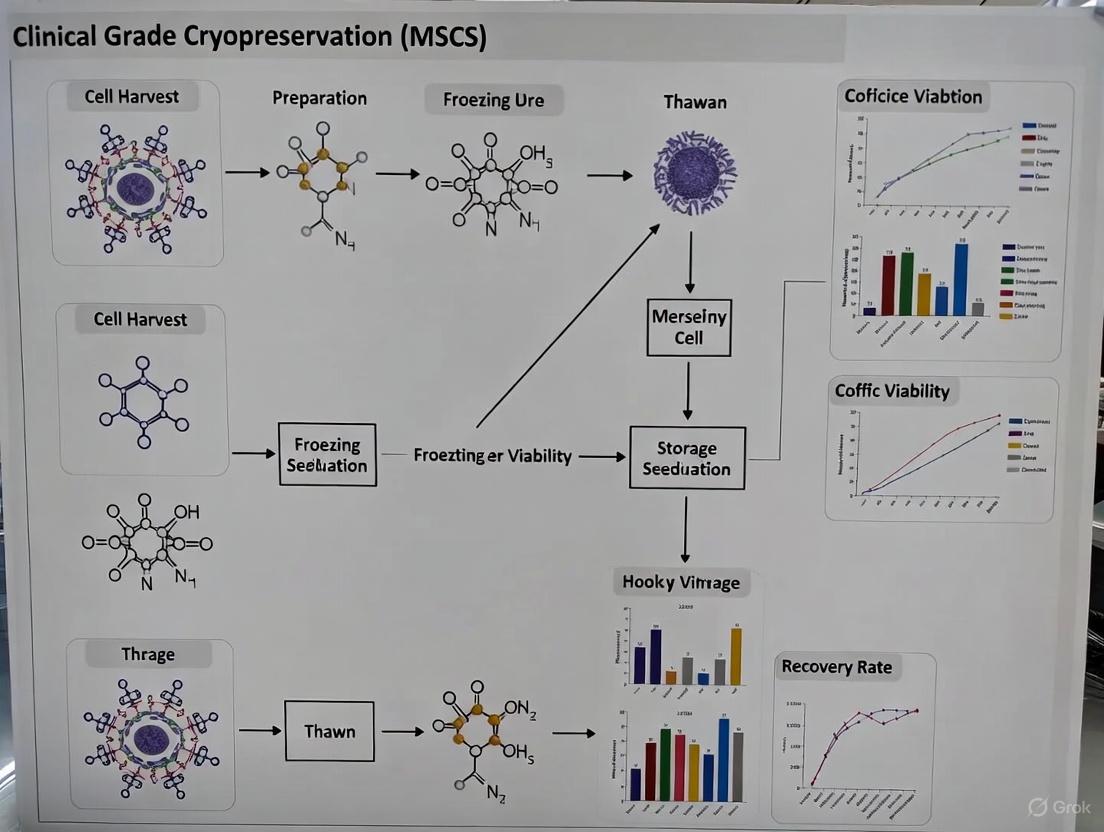
Diagram 1: Slow-freezing and thawing workflow for MSCs.
Protocol 2: Establishment of a Three-Tiered Cell Bank
A tiered banking system is a regulatory requirement for clinical-grade allogeneic cell products, ensuring traceability and consistent quality [10].
3.2.1 Cell Bank Tiers
- Initial Clone Bank (ICB): Generated from a single, fully characterized and qualified clonal cell population. This is the foundational bank from which all subsequent banks are derived.
- Master Cell Bank (MCB): Produced by expanding cells from one or more vials of the ICB at a predefined passage (e.g., passage 4). The MCB is thoroughly tested for identity, purity, potency, and safety. It serves as the source for all Working Cell Banks.
- Working Cell Bank (WCB): Generated by expanding cells from one vial of the MCB to a later passage (e.g., passage 9). The WCB is the direct source of cells for clinical production lots.
3.2.2 Banking Procedure
- Donor Screening: Adhere to national and international regulatory standards (e.g., GTP, FDA/EMA guidelines). This includes comprehensive donor medical history, behavioral assessment, and testing for relevant infectious agents (HBV, HCV, HIV, HTLV, syphilis, etc.) [10].
- Cell Line Establishment: Isolate and culture MSCs from the donor tissue (e.g., amniotic fluid, bone marrow). For AF-MSCs, high clonogenicity allows for the establishment of homogeneous cell lines from a single cell [10].
- Bank Creation: Cryopreserve a sufficient number of vials for each bank tier (ICB, MCB, WCB) using the slow-freezing protocol described in Section 3.1. All vials must be meticulously labeled with a unique identifier, passage number, date, and cell count.
- Quality Control: Perform rigorous testing on the MCB and WCB, including:
- Viability and Cell Count: Post-thaw viability should typically exceed 80% [11].
- Identity: Flow cytometry for positive (CD73, CD90, CD105) and negative (CD34, CD45, HLA-DR) MSC markers [10] [5].
- Potency: In vitro trilineage differentiation (osteogenic, adipogenic, chondrogenic) and/or other relevant functional assays (e.g., immunomodulation) [10] [13] [12].
- Safety: Sterility, mycoplasma, and endotoxin testing. Karyotype analysis to confirm genetic stability [10].
The Scientist's Toolkit: Essential Research Reagent Solutions
Table 3: Key reagents and materials for MSC cryopreservation
| Reagent / Material | Function / Purpose | Clinical-Grade Considerations |
|---|---|---|
| Dimethyl Sulfoxide (DMSO) | Penetrating cryoprotectant; reduces ice crystal formation and cellular dehydration [11] [5]. | Use high-purity, compendial (e.g., Ph. Eur.) grade. Final concentration typically 5-10%. Associated with patient adverse events, so post-thaw removal is critical [11]. |
| Human Serum Albumin (HSA) | Non-penetrating cryoprotectant; provides extracellular matrix and mitigates osmotic shock [11]. | Preferred over Fetal Bovine Serum (FBS) to avoid xenogeneic components and regulatory complications. Used at 4.5-5% concentration. |
| Autologous Plasma | Alternative to HSA; serves as a protein base for the cryoprotectant solution [12]. | Sourced from the same donor (if autologous) or a qualified allogeneic donor. Must be tested for pathogens. |
| Controlled-Rate Freezer | Equipment that ensures a consistent, optimal cooling rate (e.g., -1°C/min) [14]. | Critical for process standardization and reproducibility. Passive cooling containers are a lower-cost alternative but offer less control. |
| Liquid Nitrogen Storage | Provides long-term cryogenic storage (-150°C to -196°C) to suspend all biological activity [14] [15]. | Use vapor phase to minimize risk of cross-contamination. Requires continuous monitoring and validated backup systems. |
| CELLBANKER / Proprietary Media | Commercial, ready-to-use cryopreservation solutions [13]. | Often serum-free and DMSO-containing. Must be thoroughly validated for the specific cell type and clinical application. |
Critical Considerations for Protocol Implementation
Navigating Immunogenicity and Allorejection
A primary challenge for 'off-the-shelf' allogeneic cell products is host immune rejection. While MSCs are considered immunoprivileged due to low MHC class II expression, they do express MHC class I, which can lead to recognition and elimination by the recipient's immune system [10] [16]. Strategies to mitigate this include:
- Genetic Engineering: Using tools like CRISPR/Cas9 to knock out key molecules in the Major Histocompatibility Complex (MHC), such as Beta-2-microglobulin (B2M), to create universal donor cells [17] [16].
- Selection of Low-Immunogenicity Sources: Amniotic fluid-derived MSCs (AF-MSCs) naturally express HLA-G, an immunosuppressive molecule, which may enhance their persistence post-transplantation [10].
Cold Chain and Storage Logistics
Maintaining the integrity of cryopreserved products from the manufacturing facility to the clinic is paramount. Key elements include:
- Robust Packaging: Use of validated cryogenic shippers that can maintain temperatures below -150°C for up to 14 days during transit [14].
- Real-Time Monitoring: Implementing 21 CFR Part 11 compliant monitoring systems to track temperature, location, and shock during shipment, providing a continuous chain of custody and identity [14].
- Contingency Planning: Developing protocols for managing temperature excursions and other logistical disruptions, such as weather emergencies or geopolitical issues [14].
Diagram 2: Clinical-grade allogeneic MSC production and supply chain.
Cryopreservation is not merely a storage step but a fundamental pillar supporting the entire ecosystem of 'off-the-shelf' allogeneic cell therapies. The methodologies outlined herein—from controlled-rate freezing and tiered banking to rigorous quality control and cold chain management—provide a framework for developing robust, clinically applicable MSC products. As the field advances, ongoing research into DMSO-free cryoprotectants, optimized thawing protocols, and strategies to combat allorejection will further enhance the safety, efficacy, and accessibility of these transformative therapeutics, ultimately fulfilling their promise in regenerative medicine.
For researchers developing allogeneic mesenchymal stromal cell (MSC) therapies, cryopreservation is not merely a storage technique but a critical determinant of therapeutic efficacy and regulatory compliance. The transition from research to clinical application hinges on the ability to reliably preserve "off-the-shelf" cell products without compromising their viability, functionality, or safety profile. Within this framework, two principal cryopreservation methodologies—slow freezing and vitrification—embody distinct biophysical approaches to stabilizing living cells at cryogenic temperatures. This application note examines the core principles governing these techniques, providing detailed protocols and analytical frameworks to inform method selection for clinical-grade MSC manufacturing.
The imperative for robust cryopreservation strategies in allogeneic MSC therapy stems from both practical and biological considerations. Logistically, cryopreservation enables the creation of cell banks that facilitate thorough quality control testing and ensure immediate product availability for acute conditions [18]. Biologically, it circumvents the detrimental effects of continuous cell passaging, including epigenetic alterations, telomere shortening, and random genomic losses [5]. Understanding the fundamental mechanisms underlying slow freezing and vitrification is therefore essential for optimizing cryopreservation outcomes in clinical settings.
Fundamental Mechanisms of Cryopreservation
The Challenge of Cryoinjury
Cells encounter three primary forms of damage during cryopreservation: osmotic damage, mechanical damage, and oxidative damage [19]. During freezing, extracellular ice formation increases solute concentration in the unfrozen fraction, creating osmotic gradients that drive water efflux from cells. If unchecked, this dehydration leads to lethal hypertonicity. Mechanical damage occurs when intracellular ice crystals form, physically disrupting membranes and organelles. Simultaneously, the cryopreservation process generates reactive oxygen species (ROS) that oxidize lipids, proteins, and nucleic acids [19]. Both slow freezing and vitrification address these challenges through distinct physical approaches.
Table 1: Primary Types of Cryodamage and Their Mechanisms
| Cryodamage Type | Primary Cause | Cellular Consequences |
|---|---|---|
| Osmotic Damage | Extracellular ice formation increases solute concentration, causing osmotic dehydration [19]. | Cell shrinkage; membrane damage; hypertonic stress [19]. |
| Mechanical Damage | Intracellular ice crystal formation during cooling or thawing [19]. | Physical disruption of membranes and organelle structures [19]. |
| Oxidative Damage | Generation of Reactive Oxygen Species (ROS) during freezing/thawing [19]. | Oxidation of lipids, proteins, and nucleic acids [19]. |
Core Principles of Slow Freezing
Slow freezing operates on the principle of controlled dehydration. By gradually reducing temperature at precisely controlled rates (typically -1°C/min to -3°C/min), the technique allows sufficient time for water to exit cells before intracellular freezing occurs [5]. This gradual cooling minimizes intracellular ice formation by promoting ice crystallization in the extracellular space, thereby progressively concentrating solutes outside the cell and osmotically drawing water out through the membrane. The process typically involves cooling cells in stages: first to 4°C, then to -80°C, and finally transferring to liquid nitrogen at -196°C for long-term storage [5]. Cryoprotective agents (CPAs) like dimethyl sulfoxide (DMSO) play a crucial role in this process by penetrating cells, reducing the freezing point of water, and improving membrane permeability to water [5].
Core Principles of Vitrification
Vitrification takes a fundamentally different approach by achieving a glass-like solidification without ice crystal formation. This technique uses high concentrations of CPAs combined with ultra-rapid cooling rates to dramatically increase solution viscosity until molecular motion effectively ceases, forming an amorphous glass [5]. Two methodological variants exist: equilibrium vitrification, where cells reach osmotic equilibrium with specific CPA formulations before freezing, and non-equilibrium vitrification, which prioritizes extreme cooling rates and high CPA concentrations to achieve the glassy state almost instantaneously [5]. Both approaches fundamentally aim to avoid the phase transitions that cause mechanical damage in conventional freezing.
The following diagram illustrates the fundamental mechanistic differences between slow freezing and vitrification:
Comparative Technical Analysis: Slow Freezing vs. Vitrification
Method Workflows and Procedural Requirements
The practical implementation of slow freezing and vitrification differs significantly in equipment needs, procedural complexity, and handling requirements. Slow freezing employs programmable controlled-rate freezers that execute precise cooling profiles, typically progressing from room temperature to -80°C before final transfer to liquid nitrogen [5]. This method utilizes relatively low CPA concentrations (commonly 5-10% DMSO), which reduces direct chemical toxicity but necessitates careful controlled-rate equipment [5] [19]. The thawing process involves rapid warming in a 37°C water bath followed by stepwise removal of CPAs to minimize osmotic shock during rehydration [5].
Vitrification requires specialized carriers (e.g., Cryotop, CryoLoop) that facilitate ultra-rapid cooling by minimizing sample volume and maximizing surface area-to-volume ratios [20]. These systems enable cooling rates exceeding -100°C/min when plunged directly into liquid nitrogen. The technique demands high CPA concentrations (often 6-8 M combined permeating and non-permeating agents) to achieve the glassy state, creating greater toxicity concerns that must be managed through precise exposure timing [5]. Warming is similarly rapid, typically accomplished by directly immersing the sample into pre-warmed media, with subsequent CPA removal steps.
Quantitative Performance Comparison
Direct comparative studies reveal significant differences in post-preservation outcomes between the two methods. In embryonic cell research, vitrification has demonstrated superior survival rates (96.9% vs. 82.8%) and better preservation of excellent morphology (91.8% vs. 56.2%) compared to slow freezing [20]. These cellular-level advantages translated to improved clinical outcomes, with higher clinical pregnancy rates (40.5% vs. 21.4%) and implantation rates (16.6% vs. 6.8%) [20]. For MSCs specifically, slow freezing typically achieves 70-80% cell survival when optimized [5], while vitrification protocols continue to be refined for these adherent cell systems.
Table 2: Technical and Outcome Comparison Between Slow Freezing and Vitrification
| Parameter | Slow Freezing | Vitrification |
|---|---|---|
| Cooling Rate | Slow (-1°C/min to -3°C/min) [5] | Ultra-rapid (>100°C/min) [5] |
| CPA Concentration | Low (e.g., 5-10% DMSO) [5] [19] | High (e.g., 6-8 M total CPAs) [5] |
| Ice Formation | Extracellular ice crystals form [5] | No ice crystal formation [5] |
| Primary Equipment | Programmable freezer [5] | Specialized carriers (e.g., Cryotop) [20] |
| Typical Survival (MSCs) | 70-80% [5] | Protocol-dependent; can exceed slow freezing [20] |
| Key Advantages | Standardized, suitable for large volumes [5] | Superior survival in validated systems [20] |
| Key Limitations | Time-consuming; intracellular ice with improper cooling [5] | CPA toxicity; sample volume restrictions [5] |
The following workflow diagram compares the key procedural stages for both methods:
Protocols for Clinical-Grade MSC Cryopreservation
Detailed Protocol: Slow Freezing of Allogeneic MSCs
Principle: Preserve cell viability through controlled dehydration and minimal intracellular ice formation [5].
Materials:
- Clinical-grade MSCs (passage 3-5, >80% viability)
- Cryopreservation medium: Clinical-grade DMSO (5-10%) in human platelet lysate (hPL)-based media [18]
- Controlled-rate freezer
- Cryogenic vials
- Liquid nitrogen storage system
- Water bath (37°C)
- Washing medium: hPL-based expansion media
Procedure:
- Harvesting: Detach MSC monolayers using clinical-grade enzymes. Quench enzyme activity with hPL-based media.
- Formulation: Centrifuge cell suspension (300 × g, 5 min). Resuspend cell pellet in precooled (4°C) cryopreservation medium at 1-5 × 10^6 cells/mL.
- Packaging: Aseptically aliquot 1-2 mL cell suspension into cryogenic vials.
- Freezing: Place vials in controlled-rate freezer and execute program:
- Start at 4°C
- Cool at -1°C/min to -20°C
- Cool at -3°C/min to -80°C
- Hold at -80°C for 2 hours
- Transfer to liquid nitrogen vapor phase (-135°C to -150°C) for long-term storage [5]
- Thawing: Retrieve vial from storage. Immediately transfer to 37°C water bath with gentle agitation until last ice crystal disappears (approximately 1-2 minutes).
- CPA Removal: Transfer cell suspension to 15 mL centrifuge tube. Slowly add 10 mL pre-warmed washing medium dropwise over 5 minutes. Centrifuge (300 × g, 5 min). Discard supernatant and resuspend in fresh culture medium.
- Assessment: Determine viability via trypan blue exclusion and flow cytometry. Verify identity (CD73+, CD90+, CD105+, CD45-) and differentiation potential post-thaw [21].
Detailed Protocol: Vitrification of Allogeneic MSCs
Principle: Achieve glass-like solidification using high CPA concentrations and ultra-rapid cooling [5].
Materials:
- Clinical-grade MSCs
- Equilibrium solution: 1.5-2.0 M DMSO + 0.5 M sucrose in base medium
- Vitrification solution: 6-8 M total CPAs (e.g., DMSO + ethylene glycol + sucrose) in base medium
- Vitrification devices (e.g., Cryotop)
- Liquid nitrogen
- Warming solution: 1.0 M sucrose in base medium
- Dilution solution: 0.5 M sucrose in base medium
- Washing solution: Base medium
Procedure:
- Harvesting: Detach MSCs to create single-cell suspension. Concentrate to high density (1-5 × 10^7 cells/mL).
- Equilibration: Suspend cells in equilibrium solution for 3-5 minutes at room temperature.
- Vitrification: Transfer cells to vitrification solution for <1 minute at room temperature.
- Loading: Immediately place small volume (≤1 µL) containing 5-20 × 10^3 cells onto vitrification device.
- Cooling: Immediately plunge device directly into liquid nitrogen within 60 seconds of vitrification solution exposure.
- Storage: Transfer to sealed container for long-term storage in liquid nitrogen.
- Warming: Retrieve device from storage. Quickly immerse in pre-warmed (37°C) warming solution for 1 minute.
- CPA Removal: Transfer cells through dilution series: dilution solution for 3 minutes, then washing solution for 5 minutes.
- Assessment: Determine survival, identity, and functionality as for slow freezing protocol.
Critical Considerations: Strict timing is essential during CPA exposure steps. Sample volume must be minimized to achieve required cooling rates. Consider implementing aseptic closed-system devices for clinical applications.
The Scientist's Toolkit: Essential Reagents and Materials
Successful implementation of clinical-grade cryopreservation requires carefully selected reagents and materials that comply with regulatory standards.
Table 3: Essential Research Reagents for Clinical-Grade MSC Cryopreservation
| Reagent/Material | Function/Purpose | Clinical-Grade Considerations |
|---|---|---|
| Dimethyl Sulfoxide (DMSO) | Permeating CPA; reduces ice crystal formation [5] [19]. | Pharmaceutical grade; minimize residual concentration (<5%) due to patient adverse effects [19] [18]. |
| Sucrose/Trehalose | Non-permeating CPA; osmotic buffer during freezing/thawing [5] [19]. | Reduces required DMSO concentration; human-approved sources [19]. |
| Human Platelet Lysate (hPL) | Serum alternative in freeze media; supports MSC viability [18]. | Xenogeneic-free; standardized composition; pathogen testing required [18]. |
| Programmable Freezer | Controlled cooling for slow freezing [5]. | Calibrated, validated cooling rates; GMP-compliant documentation. |
| Vitrification Carriers | Enable ultra-rapid cooling [20]. | Sterile, closed systems preferred for clinical use. |
| Hydrogel Microcapsules | 3D scaffold providing cryoprotection [22]. | Enables DMSO reduction to 2.5% while maintaining >70% viability [22]. |
Clinical Translation Considerations for Allogeneic MSCs
Regulatory and Safety Framework
The transition to clinical application necessitates strict adherence to current Good Manufacturing Practices (cGMP) throughout the cryopreservation process [18]. Key considerations include comprehensive donor screening, validated manufacturing protocols, and rigorous quality control testing. For allogeneic products, regulators emphasize demonstrating consistent cell viability, identity, potency, and purity post-thaw [21]. Safety concerns specific to cryopreserved products include DMSO toxicity in patients, with reported adverse effects including nausea, vomiting, arrhythmias, and neurotoxicity [19] [22]. These risks drive efforts to reduce or eliminate DMSO from final formulations through technologies like hydrogel microencapsulation, which enables DMSO reduction to 2.5% while maintaining viability above the 70% clinical threshold [22].
Functional Assessment Post-Cryopreservation
Beyond simple viability metrics, clinical-grade MSCs must retain critical biological functions after thawing. Essential quality control measures include:
- Immunophenotype: Confirmation of CD73, CD90, and CD105 expression (>95%) with absence of hematopoietic markers (CD45, CD34, CD14) [5] [21]
- Differentiation Potential: Demonstrated trilineage differentiation into osteocytes, adipocytes, and chondrocytes [21] [23]
- Immunomodulatory Capacity: Validated suppression of lymphocyte proliferation [24]
- Karyotypic Stability: Absence of chromosomal abnormalities after freeze-thaw cycling [18]
For allogeneic applications, particular attention should be paid to how cryopreservation affects immunogenicity, as some evidence suggests that frozen-thawed allogeneic MSCs may elicit immune responses upon repeated administration [24].
The selection between slow freezing and vitrification for clinical-grade allogeneic MSCs involves balancing multiple technical and regulatory considerations. Slow freezing offers operational simplicity, scalability, and well-established regulatory pathways, making it the current mainstream approach for large-volume clinical applications. Vitrification demonstrates theoretical advantages for preserving membrane integrity and cellular function through complete avoidance of ice crystallization, though technical challenges regarding standardization and scalability remain. Emerging technologies like hydrogel-based 3D cryopreservation present promising avenues for reducing CPA toxicity while maintaining high viability. Ultimately, method selection should be guided by target product profile requirements, with rigorous pre-clinical validation ensuring that cryopreserved allogeneic MSCs maintain their critical quality attributes throughout their shelf life and upon administration.
The successful cryopreservation of allogeneic mesenchymal stromal cells (MSCs) is a critical determinant for their clinical application in regenerative medicine and cell-based therapies. Cryoprotectant Agents (CPAs) are essential components that mitigate freezing-induced damage, ensuring post-thaw viability, potency, and functionality of these therapeutic products [25]. For decades, dimethyl sulfoxide (DMSO) has been the cornerstone CPA in biobanking and cell therapy manufacturing. However, growing concerns regarding its toxicity profile for both cells and patients have accelerated the development of DMSO-free alternatives [26]. This application note delineates the mechanisms of action and toxicity profiles of DMSO versus emerging DMSO-free CPAs, providing structured data, validated protocols, and practical tools to guide researchers in the field of clinical-grade allogeneic MSC cryopreservation.
Mechanisms of Action: A Comparative Analysis
Cryoprotectants function through distinct yet complementary mechanisms to protect cells during the freeze-thaw cycle. The table below categorizes and compares the primary mechanisms of traditional and novel CPAs.
Table 1: Mechanisms of Action of Various Cryoprotectant Agents
| Cryoprotectant Category | Specific Examples | Primary Mechanism of Action | Cellular Interaction |
|---|---|---|---|
| Penetrating CPAs | DMSO, Glycerol, Ethylene Glycol [25] | Depresses freezing point colligatively; penetrates cell membrane to prevent intracellular ice formation (IIF) and excessive dehydration [25]. | Intracellular |
| Non-Penetrating CPAs | Sucrose, Trehalose, Hydroxyethyl Starch (HES) [25] [27] | Induces osmotic dehydration prior to freezing; promotes vitrification (glassy state) via high glass transition temperature; stabilizes membranes via water substitution hypothesis [25]. | Extracellular |
| Bio-Inspired CPAs | Antifreeze Proteins (AFPs) [28] | Binds to specific ice crystal planes, inhibiting ice recrystallization (IRI) and exhibiting thermal hysteresis (TH) activity [28]. | Extracellular/Ice-Binding |
| Deep Eutectic Solvents (DES) | Choline Chloride-Glycerol, Proline-Glycerol [29] [28] | Forms extensive hydrogen-bonding networks with water, depressing freezing point and stabilizing membranes/proteins via viscous, glass-forming behavior [29]. | Primarily Extracellular |
The following diagram illustrates the synergistic workflow of how these diverse mechanisms protect a cell during cryopreservation.
Toxicity and Performance Profiles
The drive toward DMSO-free formulations is primarily fueled by toxicity concerns. DMSO's cytotoxicity is well-documented, affecting cell differentiation, epigenetic profiles, and causing pharmacological side effects in patients, such as nausea, vomiting, and cardiovascular events [25] [26]. While a dose of 1 g DMSO/kg body weight is generally accepted in hematopoietic stem cell transplantation, the administered dose with MSC products is typically 2.5–30 times lower, mitigating but not eliminating risks [26]. Post-thaw washing to remove DMSO adds complexity, risks cell loss, and can compromise the therapeutic product [25] [26].
Recent multicenter studies demonstrate that DMSO-free alternatives can achieve comparable, and in some aspects superior, post-thaw outcomes for MSCs. The data below summarizes key performance metrics from a recent international collaborative study.
Table 2: Post-Thaw MSC Performance: DMSO vs. DMSO-Free (SGI) Solution Data synthesized from an international multicenter study [27]
| Performance Metric | Fresh MSCs (Pre-Freeze) | DMSO-Based Solution | DMSO-Free Solution (SGI) |
|---|---|---|---|
| Average Viability | 94.3% | 89.8% (Δ -4.5%) | 82.9% (Δ -11.4%) |
| Viable Cell Recovery | 100% (Baseline) | ~94.4% (Δ -5.6%) | 92.9% (Δ -7.1%) |
| Immunophenotype (CD73, CD90, CD105) | Compliant with ISCT criteria | Maintained, no significant difference from fresh | Maintained, no significant difference from DMSO |
| Global Gene Expression | Baseline Profile | Comparable to fresh | Comparable to DMSO-preserved cells |
Beyond the SGI solution, other innovative formulations show promise. For instance, cryopreservation of platelets using a choline chloride-glycerol Deep Eutectic Solvent (DES) in a controlled-rate freezer demonstrated post-thaw recovery >85% and maintained functional integrity, highlighting the potential of designer solvents [29].
Detailed Protocol: DMSO-Free Cryopreservation of Allogeneic MSCs
The following protocol is adapted from a recent international multicenter study that validated a DMSO-free solution for MSCs [27].
Materials and Reagents
Table 3: The Scientist's Toolkit: Essential Reagents for DMSO-Free Cryopreservation
| Item | Function / Description | Example / Composition |
|---|---|---|
| DMSO-Free Cryoprotectant (SGI Solution) | A non-penetrating CPA cocktail that protects cells via osmotic dehydration and vitrification. | Sucrose, Glycerol, and Isoleucine in Plasmalyte A base [27]. |
| Controlled-Rate Freezer (CRF) | Equipment that precisely controls cooling rate, critical for reproducible ice formation and cell recovery. | Standard laboratory CRF. |
| Liquid Nitrogen Storage System | For long-term storage of cryopreserved cell products at or below -135°C. | Liquid nitrogen vapor phase freezer. |
| Plasmalyte A | An isotonic, balanced salt solution serving as the base for the SGI cryoprotectant solution. | Commercially available infusion solution. |
| Cell Viability Assay | To quantify post-thaw cell viability and recovery. | Trypan Blue exclusion, Flow cytometry with 7-AAD/Annexin V. |
Step-by-Step Procedure
- Preparation of DMSO-Free SGI Solution: Prepare the cryoprotectant solution containing sucrose, glycerol, and isoleucine (SGI) in a base of Plasmalyte A. Filter-sterilize the solution (0.22 µm) prior to use.
- MSC Harvest and Suspension: Harvest allogeneic MSCs (from bone marrow or adipose tissue) at the desired passage using standard methods (e.g., trypsinization). Perform a final cell count and viability assessment on the harvested cell suspension. Centrifuge the cell suspension and resuspend the cell pellet in the pre-chilled SGI solution to a clinically relevant final concentration (e.g., 5-20 x 10^6 cells/mL).
- Aliquoting and Packaging: Aseptically aliquot the cell suspension into cryogenic vials or bags. Ensure proper labeling in accordance with Good Manufacturing Practice (GMP).
- Controlled-Rate Freezing: Transfer the aliquoted samples to a controlled-rate freezer. Initiate the following freezing program:
- Start temperature: 4°C.
- Cooling rate 1: -1°C per minute to a seeding temperature (e.g., -5°C).
- Induce ice nucleation (seeding): Hold for 5-10 minutes at the seeding temperature to initiate controlled extracellular ice formation.
- Cooling rate 2: -1°C per minute to -40°C.
- Cooling rate 3: -5°C to -10°C per minute to -80°C to -100°C.
- Transfer to Long-Term Storage: Immediately after the freezing program completes, transfer the vials/bags to a liquid nitrogen storage tank for long-term preservation in the vapor phase (-135°C to -196°C).
- Post-Thaw Assessment: To assess the product, rapidly thaw a representative vial in a 37°C water bath with gentle agitation. Dilute the thawed cell product in a pre-warmed culture medium or plasma to reduce osmotic stress. Centrifuge and resuspend the cells in an appropriate buffer for subsequent analyses:
- Viability & Recovery: Determine using an automated cell counter or flow cytometry.
- Immunophenotype: Confirm expression of CD73, CD90, CD105 and lack of CD45 via flow cytometry.
- Potency Assays: Perform functional assays relevant to the clinical application (e.g., immunomodulation, trilineage differentiation).
The landscape of CPA development is dynamically evolving toward safer and more effective DMSO-free solutions. Robust, multicenter data now confirms that novel formulations, such as the SGI solution, can preserve the viability, recovery, and critical identity markers of allogeneic MSCs at a clinically acceptable level [27]. The emergence of bio-inspired agents like Antifreeze Proteins (AFPs) and Natural Deep Eutectic Solvents (NADES) further expands the chemical toolbox, offering highly specific mechanisms of action and enhanced biocompatibility [28]. For clinical-grade allogeneic MSC research, adopting these DMSO-free protocols mitigates patient safety risks associated with DMSO and streamlines the therapeutic workflow by potentially obviating post-thaw washing steps. As the field advances, the integration of these next-generation CPAs will be instrumental in standardizing manufacturing, ensuring product consistency, and ultimately fulfilling the transformative promise of MSC-based therapies.
GMP Workflows: From Cell Expansion to Fill-and-Finish Cryopreservation
The production of clinical-grade allogeneic mesenchymal stromal cells (MSCs) requires scalable, automated, and well-controlled expansion processes to generate therapeutically relevant cell numbers—often ranging from 10^6 to 10^9 cells per dose—while maintaining consistent quality and functionality [30] [31]. Automated bioreactor systems have emerged as superior alternatives to traditional planar culture, offering enhanced control, reduced contamination risk, and significantly improved scalability [30] [32]. Among these, hollow-fiber and packed-bed bioreactors represent advanced platforms for the intensive expansion of adherent cells like MSCs. This application note details protocols and performance data for these systems within the context of clinical-grade allogeneic MSC manufacturing, providing a framework for their implementation in a Good Manufacturing Practice (GMP) environment.
Key Bioreactor Platforms for MSC Expansion
Hollow-fiber bioreactors (HFBs) consist of a cartridge containing thousands of semi-permeable capillary membranes, creating a large surface area for cell growth and two separate flow compartments: the intracapillary (IC) space (where cells reside) and the extracapillary (EC) space [32]. This configuration allows for continuous media exchange, efficient nutrient delivery, and waste removal while retaining cells and secreted proteins [32] [33]. The system provides a protective, low-shear environment conducive to high cell density cultures [33].
Packed-bed bioreactors (PBRs) are fixed-bed systems where cells adhere to solid carriers or scaffolds packed within the bioreactor vessel [34] [35]. Culture medium is perfused through the bed to nourish the immobilized cells. This design also offers low shear forces and high cell density capacity, though cell retrieval can be more challenging compared to other systems [34] [35].
Table 1: Comparative Analysis of Hollow-Fiber and Packed-Bed Bioreactor Systems
| Feature | Hollow-Fiber Bioreactor (e.g., Quantum) | Packed-Bed Bioreactor |
|---|---|---|
| Principle | Cells grow on semi-permeable hollow fibers; continuous medium perfusion between IC and EC spaces [32]. | Cells adhere to stationary solid carriers or scaffolds; medium perfused through the packed bed [34] [35]. |
| Scalability | Modular design; scalable by adding larger cartridges [33]. | Scalable by increasing bed volume, but fluid distribution challenges may arise [34]. |
| Shear Stress | Very low, protects sensitive cells [33]. | Low, due to minimal mechanical agitation [34]. |
| Cell Density | Very high (e.g., peak densities of 4 × 10^7 cells/mL reported for suspension cells) [32]. | High, supported by large surface area of packing material [34]. |
| Cell Harvesting | Enzymatic detachment and flushing from fibers; requires optimization [30]. | Can be challenging; may require enzymatic treatment and mechanical flushing to dislodge cells from carriers [34] [35]. |
| Process Monitoring | Limited direct sampling; relies on in-line sensors and metabolite analysis of circulating media [35]. | Parameters like DO and pH can be monitored in-line; sampling of cells may require disrupting the bed [36]. |
| Typical Applications | Expansion of adherent MSCs [30] and high-density suspension cells [32]. | Expansion of adherent cells, including MSCs and stem cells, often used with microcarriers [35]. |
Quantitative Performance Data
Independent studies have demonstrated the robust performance of these automated systems for expanding various cell types, consistently outperforming traditional flask-based cultures in terms of final cell yield and efficiency.
Table 2: Experimental Cell Yields from Automated Bioreactor Systems
| Cell Type | Bioreactor System | Scale/Model | Expansion Time | Seeding Density | Final Yield / Density | Reference |
|---|---|---|---|---|---|---|
| Bone Marrow MSCs (BM-MSCs) | Quantum HFB | 21,000 cm² surface area | 7 days | 20 × 10^6 cells | 100–276 × 10^6 cells | [30] |
| Adipose-derived Stem Cells (ASCs) | Quantum HFB | Not specified | Multi-harvest protocol | Not specified | Phenotype and function maintained post-cryo [37] | |
| Mouse Erythroleukemia (MEL) Cells | Quantum HFB | Not specified | 29 days | 5 × 10^7 cells | 2.5 × 10^10 total cells; peak density 4 × 10^7 cells/mL | [32] |
| Human Liver Stem Cells (HLSCs) | Xpansion Multi-Plate PBR | XPN10 | Not specified | 4,000 cells cm⁻² | 94 ± 8 × 10^3 cells cm⁻² | [36] |
| Human MSCs (hMSCs) | Stirred-Tank w/ Microcarriers & ATF Perfusion | 1.8 L working volume | 5-7 days | Not specified | ≈2.9 × 10^6 cells mL⁻¹; expansion factor of 41–57 | [35] |
Diagram 1: Automated MSC Expansion and Cryopreservation Workflow. This chart outlines the key stages from bioreactor preparation to final cryopreservation of the cell product.
Detailed Experimental Protocols
Protocol for MSC Expansion in a Hollow-Fiber Bioreactor (Quantum System)
Objective: To achieve large-scale, automated expansion of allogeneic MSCs in a closed and controlled system, generating cells that meet release criteria for clinical cryopreservation.
Materials and Reagents:
- Bioreactor System: Quantum Cell Expansion System (Terumo BCT) with appropriate Cell Expansion Set [30] [32].
- Cells: MSCs (e.g., Bone Marrow-derived) at Passage 2-4.
- Culture Medium: Alpha-MEM or DMEM, supplemented with 5-10% (v/v) human Platelet Lysate (hPL) or other GMP-compliant growth supplement [30] [35].
- Coating Solution: Recombinant human fibronectin (e.g., 5 µg/cm²) or other GMP-compliant adhesion substrate [30].
- Harvesting Solution: GMP-grade Trypsin/EDTA or TrypLE Select [35].
Method:
- System Setup and Coating:
- Install the pre-sterilized hollow-fiber bioreactor cartridge and tubing set according to the manufacturer's instructions.
- Circulate the coating solution through the intracapillary (IC) space and incubate as required (e.g., 2 hours at 37°C or overnight at 4°C). Remove the coating solution and rinse the circuit with PBS before cell loading [30].
Cell Seeding:
Automated Expansion:
- Initiate continuous perfusion of fresh medium through the extracapillary (EC) space. The initial IC inlet flow rate can be set to 0.2 mL/min and the EC inlet to 0.0 mL/min [32].
- Set the bioreactor to periodically recirculate cells within the IC loop (e.g., 4 minutes every 24 hours) to ensure homogeneous distribution [32].
- Monitor key metabolites (glucose and lactate) daily. Gradually increase the IC and EC perfusion rates (e.g., to 0.4 mL/min and 0.3 mL/min, respectively) as cell density and metabolic consumption increase [30] [32].
- Maintain culture parameters at 37°C, pH 7.2-7.4, and dissolved oxygen (DO) at 20-50% air saturation.
Cell Harvest:
- Once the target cell yield is reached (typically after 7-14 days), stop medium perfusion.
- Drain the culture medium from the circuit and introduce the pre-warmed harvesting solution into the IC space. Incubate with periodic circulation to facilitate detachment.
- Flush the detached cells from the IC space using a buffer like PBS supplemented with serum or albumin. The harvested cell suspension is collected into a sterile bag [30] [35].
Protocol for MSC Expansion in a Packed-Bed Bioreactor (e.g., Fixed-Bed System)
Objective: To expand MSCs on microcarriers within a packed-bed configuration, leveraging the high surface area and low-shear environment for intensive cultivation.
Materials and Reagents:
- Bioreactor System: Packed-bed or fixed-bed bioreactor (e.g., systems utilizing non-instrumented stirred tanks with packed beds or specialized fixed-bed reactors) [34] [31].
- Microcarriers: GMP-compliant macroporous microcarriers (e.g., Cytodex 1, CultiSpher-S).
- Cells and Culture Medium: As described in Section 3.1.
Method:
- Bioreactor and Microcarrier Preparation:
- Hydrate and sterilize the microcarriers according to the manufacturer's instructions.
- Load the prepared microcarriers into the bioreactor's packed bed to create the fixed bed.
Cell Seeding:
- Prepare a suspension of MSCs and circulate it through the packed bed containing the microcarriers. Use an intermittent flow strategy (e.g., 3 minutes of circulation followed by 2.5 hours without flow) to facilitate cell attachment [36].
- Repeat the circulation cycles to maximize seeding efficiency.
Expansion with Perfusion:
- Once cells are attached, initiate continuous perfusion of fresh medium through the packed bed.
- Monitor and control process parameters (DO, pH, temperature) online. Monitor glucose and lactate levels off-line to guide perfusion rate adjustments [36] [35].
- The perfusion rate should be optimized to maintain glucose concentration above a critical level (e.g., 2 g/L) and prevent the accumulation of inhibitory metabolites like lactate and ammonia [36].
Cell Harvest:
- This is a critical step for packed-bed systems. Stop the perfusion and drain the culture medium.
- Introduce a harvesting solution (e.g., TrypLE Select) and circulate it through the bed with possible pauses for incubation. The external cell retention device (e.g., an Alternating Tangential Flow (ATF) system) used during perfusion can be repurposed to separate detached cells from microcarriers and concentrate the harvest [35].
- The resulting cell suspension is collected for further downstream processing.
The Scientist's Toolkit: Essential Research Reagents and Materials
The following table lists key materials required for establishing an automated MSC expansion process in hollow-fiber or packed-bed bioreactors.
Table 3: Essential Research Reagents and Materials for Automated MSC Expansion
| Item | Function / Purpose | Example Products / Notes |
|---|---|---|
| GMP-Grade Culture Medium | Base nutrient source for cell growth. | Alpha-MEM, DMEM; must be xeno-free for clinical work [36] [35]. |
| Human Platelet Lysate (hPL) | Growth supplement; serum-free alternative to FBS. | Must be clinically qualified; enhances MSC proliferation in bioreactors [30]. |
| GMP-Grade Recombinant Trypsin | Enzymatic cell detachment from microcarriers or fibers during harvest. | TrypLE Select is a gentler, animal-origin-free alternative [35]. |
| Microcarriers | Provide surface for cell adhesion in packed-bed and stirred-tank bioreactors. | Cytodex, CultiSpher; choose based on size, material, and surface properties [35] [31]. |
| Coating Substrate | Enhances initial cell attachment to hollow fibers. | Recombinant human Fibronectin; a GMP-compliant coating is critical [30]. |
| Cell Detachment Solution | Enzymatic cell detachment from microcarriers or fibers during harvest. | TrypLE Select is a gentler, animal-origin-free alternative [35]. |
| Activated Charcoal Cartridge | Removal of residual detergents (e.g., SDS) in downstream purification; used in decellularization protocols. | Adsorba cartridge; for ensuring purity and removing cytotoxic agents [38]. |
Hollow-fiber and packed-bed bioreactor systems are robust, automated platforms that effectively address the critical need for scalable upstream manufacturing of clinical-grade allogeneic MSCs. The detailed protocols and performance data provided in this application note demonstrate their capacity to produce therapeutically relevant cell numbers while maintaining quality attributes. Successful implementation of these systems, coupled with rigorous quality control and an optimized cryopreservation strategy, is foundational for advancing reliable and efficacious allogeneic MSC therapies from research into clinical practice.
The transition of Mesenchymal Stromal Cell (MSC)-based therapies from research to clinical application hinges on effective cryopreservation. For allogeneic MSC therapies, cryopreservation enables the creation of "off-the-shelf" products, allowing for complete quality control testing before release and immediate availability for patient treatment [39]. The formulation of cryopreservation media is a critical determinant of post-thaw cell viability, functionality, and ultimately, clinical efficacy. This application note provides a detailed overview of the composition, serum alternatives, and cryoprotectant agent (CPA) selection for formulating clinical-grade cryopreservation media for allogeneic MSCs, consolidating current research and standardized protocols.
Cryopreservation Media Composition and CPA Selection
Cryopreservation media are designed to protect cells from the physical and chemical stresses of freezing and thawing. The core components can be categorized as penetrating CPAs, non-penetrating CPAs, and a base carrier solution.
Table 1: Common Components of Cryopreservation Media for MSCs
| Component Type | Example Agents | Common Concentrations | Function | Clinical Considerations |
|---|---|---|---|---|
| Penetrating CPA | Dimethyl Sulfoxide (DMSO) [5] [39] | 5-10% (v/v) [39] [27] | Lowers freezing point, reduces intracellular ice crystal formation [5] | Potential patient toxicity (allergic reactions); intrinsic cell toxicity [5] [40] |
| Propylene Glycol (PG), Ethylene Glycol (EG) [5] [41] | 7.5-10% (v/v) [41] | Alternative penetrating agents; cell toxicity lower than DMSO [5] | Glycerol resulted in poor cryopreservation effect in one study [5] | |
| Non-Penetrating CPA | Sucrose [23] [42] [27] | 0.1 M - 0.2 M [23] [42] | Induces osmotic dehydration, stabilizes cell membranes [5] | Reduces required concentration of penetrating CPAs [23] |
| Trehalose, Hydroxyethyl Starch [5] | Varies | Functions as a saccharide-based stabilizer [5] | Often used in combination with other CPAs | |
| Base Solution | Saline, Plasmalyte A [41] [27] | N/A | Isotonic foundation for the cryomedium | Provides a defined, serum-free environment |
| Protein Stabilizer | Human Serum Albumin (HSA) [39] [42] [41] | 2-5% (v/v) or specific concentrations like 4 mg/mL [42] [41] | Mitigates osmotic shock, replaces serum [39] | Clinical-grade, xeno-free alternative to serum |
DMSO-Free Formulations
Due to the toxicity concerns associated with DMSO, significant effort has been dedicated to developing DMSO-free cryopreservation solutions. These formulations often rely on combinations of non-penetrating CPAs and alternative penetrating agents.
- Sucrose-Glycerol-Isoleucine (SGI): An international multicenter study demonstrated that a DMSO-free solution containing sucrose, glycerol, and isoleucine (SGI) in a Plasmalyte A base was comparable to DMSO-containing solutions. MSCs cryopreserved in SGI showed slightly lower post-thaw viability (a decrease of 11.4% vs. 4.5% for DMSO) but had better recovery of viable cells (92.9% vs. lower by 5.6% for DMSO) and comparable immunophenotype and gene expression profiles [27].
- Compatible Solutes: Research has identified ectoin as a promising non-toxic alternative. In serum-free conditions, cryopreservation with ectoin achieved post-thaw cell survival of up to 72%, outperforming glycerol and proline, which resulted in complete cell death or poor survival (22%), respectively [40].
- Commercial Solutions: Novel commercial DMSO-free solutions like XT-Thrive have shown superior performance compared to 10% DMSO (CryoStor10). Studies report higher pre-freeze viability after 24-hour incubation (~93% vs. ~61%) and higher post-thaw viability (~87% vs. ~63%). Cells preserved in XT-Thrive also exhibited improved expansion capability, particularly in serum-free microcarrier cultures [43].
Serum Alternatives in Cryopreservation Media
The use of fetal bovine serum (FBS) in clinical-grade MSC manufacturing is undesirable due to risks of xenogenic immunoreactions and pathogen transmission. Human-derived components are the standard for clinical formulations.
- Human Serum Albumin (HSA): HSA is the most common serum replacement in clinical-grade cryopreservation protocols. It is used as a defined, clinical-grade protein stabilizer in concentrations such as 2% in saline or 4 mg/mL in specific media [39] [42] [41].
- Platelet Lysate and Human Serum: For MSC expansion, human platelet lysate is a widely adopted serum-free alternative [39]. In cryopreservation media, human serum itself can be used as a component, providing a natural combination of proteins and nutrients [23].
- Chemically Defined Formulations: The trend is moving toward fully defined, xeno-free, and protein-free formulations. Solutions like Plasmalyte A-based SGI medium represent this approach, eliminating both serum and DMSO to enhance product safety and regulatory compliance [27].
Detailed Experimental Protocols
Standard Slow Freezing Protocol for MSCs in Suspension
This protocol is widely used for cryopreserving clinical-grade MSCs and is considered the recommended technique due to its ease of operation and low contamination risk [5] [39].
Key Steps:
- Cell Preparation: Detach MSCs using a clinical-grade enzyme (e.g., TrypLE Select), wash with DPBS, and perform a final cell count and viability assessment [39] [41].
- CPA Addition: Resuspend the cell pellet in ice-cold cryopreservation medium. The cell density is typically adjusted to 1-5 x 10^6 cells/mL [39] [41]. For the SGI solution, the final formulation is Sucrose, Glycerol, and Isoleucine in Plasmalyte A [27].
- Freezing: Use a controlled-rate freezer, cooling at approximately 1°C/min. Alternatively, place vials in an insulated container at -80°C for 24 hours to achieve a similar cooling rate before transfer to long-term storage [5] [27].
- Storage: Transfer cryovials to the vapor or liquid phase of nitrogen for long-term storage [5].
Thawing and CPA Removal Protocol
Rapid thawing and careful CPA removal are critical to minimize osmotic shock and DMSO toxicity.
Key Steps:
- Thawing: Thaw cryovials quickly in a 37°C water bath until no ice is visible [5] [41]. To enhance sterility, consider using dry heating equipment instead of a water bath [5].
- Dilution and Washing: Immediately after thawing, dilute the cell suspension dropwise or by transferring it into a large volume (e.g., 10 volumes) of pre-warmed washing/thawing solution. A typical solution is saline containing 2.5% HSA and 5% anticoagulant citrate-dextrose solution (ACD-A) [41]. This step gradually reduces the external CPA concentration, preventing excessive cell swelling and lysis [5].
- Centrifugation: Centrifuge the diluted suspension (e.g., at 400g for 5 minutes) to pellet the cells and remove the CPA-containing supernatant [41].
- Resuspension and Assessment: Resuspend the final cell pellet in the desired medium or infusion solution. Perform a cell count and viability check (e.g., using trypan blue exclusion or a NucleoCounter) [39] [41].
Advanced Protocol: Cryopreservation of MSCs in a 3D Bioscaffold
Cryopreserving MSCs within 3D structures like the PRP-Synovial Fluid (PRP-SF) bioscaffold requires optimized protocols to ensure CPA penetration.
- Scaffold Formation: Embed MSCs within the PRP-SF biomimetic bioscaffold [23].
- CPA Incubation: Immerse the cell-loaded bioscaffold in a cryoprotectant solution. The optimal solutions identified are 10% DMSO or a combination of 10% DMSO and 0.2 M sucrose in an appropriate base [23].
- Freezing: Use a slow freezing protocol, similar to the one for cells in suspension, to cool the constructs to -80°C before transfer to liquid nitrogen [23].
- Thawing and Washing: Rapidly thaw the scaffolds at 37°C and wash thoroughly with culture medium to remove CPAs before in vitro or in vivo application [23].
The Scientist's Toolkit: Essential Research Reagents
Table 2: Key Reagents for Clinical-Grade MSC Cryopreservation
| Reagent / Material | Function / Application | Example Usage |
|---|---|---|
| DMSO (CryoSure) | Penetrating cryoprotectant [5] [39] | Used at 5-10% in freezing medium for slow freezing [39] [41] |
| Human Serum Albumin (HSA) | Protein stabilizer; serum replacement [39] [41] | Used at 2-5% in cryomedium to reduce osmotic shock [39] [41] |
| Sucrose | Non-penetrating cryoprotectant [23] [42] | Combined with DMSO (0.1-0.2M) or glycerol for synergistic effect [23] [27] |
| Platelet Lysate | Serum-free supplement for MSC expansion [39] | Used in culture medium prior to cryopreservation; provides growth factors |
| Controlled-Rate Freezer | Equipment for standardized freezing [42] [27] | Essential for implementing optimized slow-freezing curves (e.g., 1°C/min) [5] |
| XT-Thrive | Commercial DMSO-free cryopreservation solution [43] | Ready-to-use solution for freezing MSCs, aiming to reduce toxicity [43] |
| CryoStor CS10 | Commercial DMSO-containing cryopreservation solution [43] [41] | A common, well-characterized control solution in comparative studies [43] [41] |
| TrypLE Select | Animal-origin-free enzyme for cell detachment [39] | Used to harvest adherent MSCs before cryopreservation [39] |
Formulating effective cryopreservation media for clinical-grade allogeneic MSCs requires a careful balance between cell protection and clinical safety. While slow freezing with DMSO-based media remains a widely used and effective standard, the field is actively moving toward safer, defined alternatives. The emergence of DMSO-free formulations, such as those based on Sucrose-Glycerol-Isoleucine (SGI) or ectoin, shows great promise, demonstrating comparable post-thaw recovery and function with reduced toxicity risks. The successful cryopreservation of MSCs, whether in suspension or within advanced 3D bioscaffolds, relies not only on the medium composition but also on rigorously optimized and standardized protocols for freezing, thawing, and CPA removal. Adopting these advanced formulations and protocols is crucial for ensuring the consistent quality, safety, and efficacy of off-the-shelf allogeneic MSC therapies.
This application note provides a detailed protocol for the clinical-grade cryopreservation of allogeneic Mesenchymal Stromal Cells (MSCs), a critical process in ensuring the stability, viability, and functionality of these Advanced Therapy Medicinal Products (ATMPs) [44]. The transition of MSC therapies from research to clinical practice demands robust, reproducible, and well-documented cryopreservation processes that comply with Good Manufacturing Practice (GMP) standards. Effective cryopreservation enables rigorous quality control testing, long-term storage, and off-the-shelf availability, thereby extending the geographic and temporal reach of viable cell therapies [45]. This protocol emphasizes the use of controlled-rate freezing and automated systems to standardize the process, minimize operator-dependent variability, and enhance the safety profile of the final product by addressing concerns associated with cryoprotectant agents like Dimethyl Sulfoxide (DMSO) [27] [45].
Materials and Reagents
Research Reagent Solutions
The following table lists essential materials and reagents required for the cryopreservation process, along with their specific functions.
Table 1: Essential Reagents and Materials for Clinical-Grade MSC Cryopreservation
| Item | Function/Application | Clinical-Grade Considerations |
|---|---|---|
| Cryostor CS-10 [46] | A defined, GMP-compliant cryopreservation solution containing 10% DMSO. Protects cells from cryo-injury. | Pre-formulated, serum-free solution reduces batch variability and improves regulatory compliance. |
| DMSO-Free Cryoprotectant [27] | Alternative solution containing Sucrose, Glycerol, and Isoleucine (SGI) in Plasmalyte A base. | Mitigates potential DMSO-related toxicity in patients; viability remains clinically acceptable (>80%). |
| Human Platelet Lysate (hPL) [44] [46] | Growth supplement used in culture media and as a buffer component (e.g., 2% in dilution buffer). | Humanized alternative to Fetal Bovine Serum (FBS), reducing xenogenic risks and aligning with GMP standards. |
| FINIA Tubing Set [46] | Single-use, closed-system disposable set for automated processing. Includes mixing bag, QC bag, and storage bags. | Ensures a sterile, closed processing pathway, critical for maintaining product safety and integrity. |
| Controlled-Rate Freezer [46] | Programmable equipment to standardize and record the freezing procedure. | Provides process control and documentation, which are essential for reproducibility and cGMP compliance. |
Equipment
- Automated Fill-Finish System (e.g., Finia Fill and Finish System) [46]
- Controlled-Rate Freezer (e.g., models from Thermo Fisher Scientific) [46]
- Liquid Nitrogen storage tank (vapor phase)
- Laminar flow hood (if using open-step manipulations)
- Cell counting and viability analyzer
Methodologies
Pre-freeze Cell Processing and Formulation
- Cell Harvest and Washing: Harvest MSCs (e.g., from umbilical cord, bone marrow, or adipose tissue) at the target passage number using a GMP-compliant dissociation reagent like TrypLE Express. Terminate the reaction with a culture medium supplemented with human platelet lysate (hPL). Centrifuge the cell suspension and wash the pellet with a suitable buffer, such as PBS containing 2% hPL [46].
- Cell Counting and Viability Assessment: Perform a cell count and viability check using an automated cell counter or trypan blue exclusion. Ensure cell viability exceeds a predetermined threshold (typically >90%) before proceeding to formulation [46].
- Formulation with Cryoprotectant: The final formulation and aliquoting of the cell product can be performed manually or, preferably, using an automated system like the Finia Fill and Finish System.
- Automated Method: Program the Finia system to cool the cell suspension and the cryopreservation solution (e.g., Cryostor CS-10 or a DMSO-free alternative) to a specified temperature (e.g., 4°C). The system then mixes the cells and cryoprotectant in a predefined ratio and aliquots the final formulation into product bags. This method offers high accuracy in volume aliquoting and maintains temperature control throughout, supporting post-thaw viabilities of >90% [46].
- Manual Method: Resuspend the cell pellet stepwise in the chilled cryopreservation solution to the target cell concentration (e.g., 1–10 × 10^6 cells/mL). Gently mix to ensure homogeneity. Manually aliquot the cell suspension into cryobags or cryovials.
Controlled-Rate Freezing and Seeding
The freezing process is critical to prevent the formation of intracellular ice crystals, which are lethal to cells. A controlled-rate freezer ensures a reproducible and optimized cooling profile.
- Loading: Place the aliquoted product bags or cryovials into the controlled-rate freezer chamber, ensuring proper spacing for uniform heat transfer.
- Program Execution: Initiate the following standardized freezing program. The "seeding" step is crucial for inducing ice formation in the extracellular solution in a controlled manner, preventing supercooling.
Table 2: Standardized Controlled-Rate Freezing Program
| Step | Description | Rate | Target Temperature | Hold Time |
|---|---|---|---|---|
| 1 | Initial Cooling | -1°C to -3°C per minute | +4°C to -5°C | None |
| 2 | Seeding | N/A | -5°C to -7°C | 5-10 minutes |
| 3 | Further Cooling | -1°C per minute | -40°C to -50°C | None |
| 4 | Rapid Cooling | -5°C to -10°C per minute | ≤ -90°C | None |
The following diagram illustrates the workflow and the critical freezing profile.
Transfer to Long-Term Storage
- Immediately upon completion of the freezing program, transfer the product bags from the controlled-rate freezer to a pre-chilled storage rack.
- Quickly move the bags into the vapor phase of a liquid nitrogen storage system for long-term preservation. The vapor phase (typically -135°C to -150°C) is preferred over the liquid phase to minimize the risk of microbial contamination and bag seal compromise.
- Record the storage location and all relevant batch information in a controlled log or database.
Technical Specifications and Data
The selection of cryoprotectant and processing method directly impacts critical quality attributes of the cryopreserved MSCs. The data below compares two main approaches.
Table 3: Post-Thaw MSC Quality Attributes: DMSO vs. DMSO-Free Cryoprotectants
| Parameter | 5-10% DMSO (In-House Solution) [27] | Novel DMSO-Free (SGI) Solution [27] | Hydrogel Microencapsulation with 2.5% DMSO [47] |
|---|---|---|---|
| Average Post-Thaw Viability | ~89.8% | ~82.9% | >70% |
| Average Viable Cell Recovery | ~87.3% | ~92.9% | Not Specified |
| Immunophenotype | Normal expression of CD73, CD90, CD105; lack of CD45 | Comparable to DMSO controls | Unaltered |
| Differentiation Potential | Preserved (Data inferred from functional testing) [12] | Preserved | Retained |
| Key Advantage | Established, high viability | Avoids DMSO patient toxicity; superior recovery | Drastically reduces DMSO requirement |
Discussion
Process Workflow and Decision Logic
The successful cryopreservation of clinical-grade MSCs relies on a series of critical decisions, each of which impacts the quality, safety, and efficacy of the final product. The following diagram outlines the key decision points and the logical flow for selecting the optimal strategy based on specific requirements.
Critical Considerations for Clinical Translation
- Cryoprotectant Selection: While DMSO is the current standard, evidence supports the clinical acceptability of DMSO-free SGI solution, which shows slightly lower viability but better cell recovery and comparable phenotype and gene expression profiles [27]. For products where DMSO must be used, strategies like post-thaw washing or novel technologies like hydrogel microencapsulation can reduce the final DMSO concentration administered to patients [45] [47].
- Automation vs. Manual Processing: Automated systems like the Finia Fill and Finish System and controlled-rate freezers are highly recommended for clinical manufacturing. They provide a closed system, reduce operator error and contamination risk, ensure high accuracy in volume aliquoting, and generate electronic records for process traceability, which is a cornerstone of cGMP [46].
- Quality Control: Rigorous pre- and post-thaw quality control is mandatory. This includes assessment of cell viability, recovery, immunophenotype (confirming expression of CD73, CD90, CD105 and lack of hematopoietic markers), and potency (e.g., differentiation capacity or immunomodulatory function) [44] [48]. Post-thaw viability should meet a pre-defined release specification, often above 70-80% for clinical use [27] [47].
This protocol provides a robust, step-by-step framework for the controlled-rate freezing, seeding, and liquid nitrogen storage of allogeneic MSCs, designed to meet the stringent requirements of clinical-grade manufacturing. By integrating advanced cryoprotectant solutions, automated processing platforms, and a standardized freezing profile, researchers and manufacturers can enhance the consistency, safety, and efficacy of MSC-based therapies, thereby accelerating their translation into routine clinical practice.
The transition of mesenchymal stromal cells (MSCs) from research tools to clinical-grade advanced therapy medicinal products (ATMPs) necessitates robust, reproducible cryopreservation and post-thaw protocols [49]. While cryopreservation enables off-the-shelf availability for allogeneic therapies, the thawing and immediate post-thaw phases represent critical windows where cell viability, recovery, and therapeutic potency can be significantly compromised [50] [5]. Variations in reconstitution solutions, handling practices, and DMSO removal methods introduce substantial heterogeneity in final cell products, directly impacting clinical trial outcomes and therapeutic efficacy. This Application Note synthesizes recent, evidence-based findings to standardize thawing and post-thaw processing of allogeneic MSCs, providing detailed protocols designed to maximize cell recovery and ensure compliance with Good Manufacturing Practice (GMP) standards for clinical applications.
Critical Parameters in Post-Thaw Cell Recovery
Successful post-thaw recovery is contingent upon managing several critical parameters that directly impact cell viability and function. The quantitative impact of these factors is summarized in Table 1.
Table 1: Quantitative Impact of Post-Thaw Processing Parameters on MSC Recovery
| Parameter | Suboptimal Condition | Optimal Condition | Impact on Viability/Recovery | Reference |
|---|---|---|---|---|
| Reconstitution Solution | Protein-free PBS or saline | Isotonic saline with 2% HSA | >40% cell loss vs. >90% viability | [50] |
| Post-Thaw Cell Concentration | <1 x 10^5 cells/mL | ≥5 x 10^6 cells/mL | Instant >40% cell loss prevented | [50] |
| Post-Thaw Storage Duration | >1 hour in suboptimal solution | ≤4 hours in optimal solution | <80% viability vs. >90% viability maintained | [50] |
| DMSO Concentration | 10% (standard) | 2.5% (with hydrogel) | Viability maintained >70% (clinical threshold) | [22] |
| Post-Thaw Washing | Rapid centrifugation | Osmotically-balanced dilution | Reduced osmotic stress and mechanical damage | [5] [51] |
The Necessity of Protein in Reconstitution Solutions
The composition of the solution used to reconstitute and dilute cells after thawing is a primary determinant of cell survival. Studies demonstrate that using protein-free vehicles, such as plain phosphate-buffered saline (PBS) or saline, induces significant and immediate cell loss, exceeding 40% [50]. This damage occurs because the absence of proteins like human serum albumin (HSA) fails to protect cells from osmotic stress and mechanical shear during the critical transition from the cryopreserved state.
The addition of 2% Human Serum Albumin (HSA) to isotonic saline or other balanced solutions has been proven essential. HSA acts as a protective colloid, stabilizing the cell membrane and preventing instant cell loss. Notably, simple isotonic saline supplemented with 2% HSA demonstrates excellent performance, ensuring >90% viability with no significant cell loss for at least 4 hours at room temperature, providing a clinically compatible and simple formulation [50].
Cell Concentration and DMSO Toxicity
Reconstituting MSCs to excessively low concentrations (e.g., below 1 x 10^5 cells/mL) in protein-free vehicles results in instant cell loss exceeding 40% and reduced viability below 80% [50]. Maintaining a higher cell concentration, such as 5 x 10^6 cells/mL, is recommended to enhance cell stability post-thaw.
Simultaneously, mitigating the cytotoxicity of dimethyl sulfoxide (DMSO), the most common cryoprotectant, is crucial. While clinical doses in MSC products are typically lower than the accepted thresholds for hematopoietic stem cell transplants, DMSO can still cause adverse effects [45]. Strategies to address this include:
- Post-thaw washing to remove DMSO before administration.
- Reducing DMSO concentration in the cryopreservation medium. Emerging technologies, such as hydrogel microencapsulation, enable a drastic reduction of DMSO concentration to 2.5% while maintaining viability above the 70% clinical threshold and preserving MSC phenotype and differentiation potential [22].
Recommended Workflow and Protocols
The following section outlines a standardized, end-to-end workflow for the thawing and post-thaw handling of clinical-grade MSCs, integrating the critical parameters discussed above.
Detailed Thawing and Reconstitution Protocol
This protocol is optimized for MSCs cryopreserved in a DMSO-based solution.
Research Reagent Solutions & Essential Materials
| Item | Function/Explanation | Clinical-Grade Consideration |
|---|---|---|
| Water Bath | Provides rapid and uniform warming to 37°C. | Use a validated, cleanable bath. Prefer a dry-heating device to avoid contamination risk from water [5]. |
| Thawing/Reconstitution Solution | Isotonic solution (e.g., saline) with 2% Human Serum Albumin (HSA). Protects cells from osmotic shock and mechanical damage during/after thawing [50]. | Must be GMP-grade. HSA is typically sourced as a clinical-grade formulation (e.g., Albutein) [52]. |
| Centrifuge | Gently pellets cells for DMSO removal. | Use a validated, calibrated instrument. |
| Final Formulation Vehicle | The solution for final resuspension before administration (e.g., Saline + 2% HSA). Ensures stability during storage and infusion [50]. | Must be compatible with intravenous infusion and GMP-compliant. |
Step-by-Step Procedure:
- Preparation: Pre-warm the thawing/reconstitution solution (e.g., saline with 2% HSA) to room temperature (15-25°C). Prepare all necessary sterile tubes and equipment under a laminar flow hood.
- Thawing: Remove the cryovial from liquid nitrogen storage and immediately place it in a 37°C water bath. Gently agitate the vial until only a small ice crystal remains (typically 1-2 minutes). Critical: Avoid submerging the vial cap to maintain sterility [5].
- Initial Dilution: Transfer the vial to the biosafety cabinet. Wipe the exterior with sterile 70% ethanol. Aseptically transfer the thawed cell suspension into a tube containing a pre-measured volume of the pre-warmed thawing/reconstitution solution. To minimize osmotic shock, add the solution drop-wise or slowly (e.g., over 1-2 minutes) while gently agitating the tube. A 1:10 dilution ratio is a common starting point (e.g., 1 mL of cell suspension into 9 mL of solution) [51].
- DMSO Removal: Centrifuge the cell suspension at 300-400 x g for 5 minutes at room temperature. Gently aspirate and discard the supernatant, which contains the majority of the DMSO.
- Final Formulation: Gently resuspend the cell pellet in the chosen final formulation vehicle (e.g., saline with 2% HSA). Critical: Adjust the final cell concentration to at least 1 x 10^6 cells/mL, preferably around 5 x 10^6 cells/mL, to prevent dilution-induced cell death [50].
- Post-Thaw Storage: If not used immediately, keep the final cell product on wet ice (2-8°C). The product should be administered within 4 hours of thawing when formulated in an optimized solution like saline with HSA [50].
Protocol for Functional Validation: Immunosuppressive Assay
To ensure that the thawing process preserves not only viability but also MSC functionality, an in vitro immunosuppressive assay is recommended. This protocol assesses the capacity of post-thaw MSCs to suppress T-cell proliferation [52].
Step-by-Step Procedure:
- T-Cell Activation: Isolate peripheral blood mononuclear cells (PBMCs) from a healthy donor. Label the PBMCs with a cell proliferation dye (e.g., CFSE). Activate the T-cells within the PBMC population using a mitogen like phytohemagglutinin (PHA) or anti-CD3/CD28 antibodies.
- Co-Culture Setup: Plate the activated PBMCs in a culture plate. Add the post-thaw, washed MSCs to the wells at varying MSC:PBMC ratios (e.g., 1:5, 1:10, 1:20). Include control wells with activated PBMCs alone (positive control for proliferation) and non-activated PBMCs (negative control).
- Incubation and Analysis: Culture the cells for 3-5 days. Harvest the cells and analyze T-cell proliferation by flow cytometry, measuring the dilution of the CFSE dye. The immunosuppressive capacity of the MSCs is calculated as the percentage reduction in T-cell proliferation compared to the positive control.
Concluding Remarks
The path to successful clinical-grade allogeneic MSC therapies is paved with standardized, robust manufacturing and handling protocols. The thawing and post-thaw stages are not merely technical steps but are critical processes that define the quality of the final cellular product. By adhering to evidence-based practices—specifically, the use of protein-containing reconstitution solutions, maintaining adequate cell concentrations, and employing gentle processing techniques—researchers and clinicians can significantly enhance MSC viability, recovery, and functional integrity. Implementing the detailed protocols and validation assays provided herein will contribute to greater reproducibility, improved therapeutic outcomes, and accelerated advancement in the field of MSC-based regenerative medicine.
Overcoming Critical Challenges: Viability, Potency, and Process Optimization
The clinical application of allogeneic Mesenchymal Stem Cells (MSCs) necessitates effective cryopreservation strategies to ensure cell viability, functional potency, and therapeutic efficacy post-thaw. Cryopreservation, while enabling long-term storage and "off-the-shelf" availability of clinical-grade MSC products, exposes cells to significant stresses that can induce cryo-injury [53] [54]. These injuries manifest at molecular, structural, and functional levels, with particular impact on cell membrane integrity, cellular senescence, and the induction of programmed cell death pathways such as apoptosis [55] [54]. For clinical-grade allogeneic MSCs, where reproducible product characteristics and predictable in-vivo function are paramount, understanding and mitigating these injuries is essential. This document details the primary mechanisms of cryo-injury and provides standardized protocols for its assessment and mitigation within a Good Manufacturing Practice (GMP) framework, supporting the broader objective of developing robust and reliable cellular therapeutics.
Key Mechanisms of Cryo-Injury
Cryo-injury during low-temperature preservation is a multi-factorial process. The dominant mechanisms can be broadly categorized into physical damage from ice crystals and osmotic stress, and biochemical activation of specific cell death and stress pathways.
Physical and Osmotic Stress
The formation of ice crystals, both extracellular and intracellular, is a primary cause of physical damage. During slow freezing, extracellular ice formation increases solute concentration in the unfrozen fraction, creating a hypertonic environment that drives water out of the cell, leading to cell dehydration and shrinkage [54]. If the cooling rate is too rapid, intracellular water does not have time to efflux, resulting in lethal intracellular ice formation (IIF) that physically disrupts membranes and organelles [54]. Conversely, during thawing, cells are subjected to osmotic swelling as the extracellular environment becomes hypotonic. These volumetric fluctuations can exceed the elastic limits of the cell membrane, causing lysis. The "two-factor hypothesis" of cryo-injury elegantly describes the interplay between cooling rate and the resultant damage from IIF and solute effects (SEs) [54].
Molecular Pathways of Apoptosis and Necroptosis
Sublethal cryo-injury can trigger programmed cell death. Apoptosis, a controlled and energy-dependent process, is a significant contributor to post-thaw cell death [54]. It is characterized by caspase activation, phosphatidylserine externalization, and DNA fragmentation. Necroptosis, a form of regulated necrosis with necrotic morphology, can also be initiated by cryopreservation stresses [54]. The triggering factors include mitochondrial membrane perturbation, oxidative stress from reactive oxygen species (ROS), and activation of death receptors. Studies on sea urchin embryonic cells confirmed that freezing-thawing increases the number of apoptotic cells with activated caspases, though physical cell disruption may be the predominant cause of death in some systems [55].
Senescence and Functional Attenuation
Beyond acute cell death, cryopreservation can induce cellular senescence, a state of irreversible growth arrest. While senescent cells remain metabolically active, they adopt a distinctive Senescence-Associated Secretory Phenotype (SASP), secreting pro-inflammatory factors that can impair the regenerative and immunomodulatory functions of the surrounding MSC population [53]. This functional attenuation is a critical concern for clinical efficacy, as the therapeutic action of MSCs relies heavily on their paracrine signaling and immunomodulatory capacity. Evidence suggests that cryopreserved MSCs may exhibit a diminished response to pro-inflammatory cytokines and altered secretion profiles compared to their freshly cultured counterparts, potentially impacting their in-vivo performance [56].
Quantitative Data on Cryo-Injury
The following tables summarize key quantitative findings from cryopreservation studies, highlighting the impact on viability, apoptosis, and cellular functions.
Table 1: Impact of Cryopreservation on Cell Viability and Recovery
| Cell Type | Cryopreservation Method | Cryoprotectant | Post-Thaw Viability | Cell Recovery | Reference |
|---|---|---|---|---|---|
| Sea Urchin Embryonic Cells | Slow freezing | Various CPAs | ~75-78% (vs. unfrozen control) | N/R | [55] |
| Bone Marrow MSCs (GMP) | Slow freezing | 10% DMSO | No significant impact | No significant impact | [57] |
| Adipose MSCs | Slow freezing | 3% Trehalose + 5% Dextran 40 + 4% PEG | ~95% | ~95% | [45] |
| Umbilical Cord MSCs | Vitrification | 0.5M Trehalose + 2.0M 1,2-Propanediol + 2.0M EG | ~72% | N/R | [45] |
| MSCs (Various) | Fresh (Plasma storage) | Human Plasma (4 days, 5°C) | >80% | >80% confluency in culture | [56] |
Table 2: Apoptosis and Functional Markers Post-Cryopreservation
| Assessment Parameter | Finding | Cell Type | Significance | Reference |
|---|---|---|---|---|
| Caspase Activity | Increased after freezing-thawing | Sea Urchin Cells | Induces apoptosis | [55] |
| Annexin V Staining | Unsuitable (High background in controls) | Sea Urchin Cells | Method-dependent reliability | [55] |
| Immunomodulatory Capacity | No alteration post-cryopreservation | GMP-MSCs | Maintained immunosuppressive function | [57] |
| IDO Expression | Dramatically higher in fresh MSCs vs. freeze-thawed (24h post-stimulation) | MSCs (Clinical dose) | Reduced initial immunomodulatory potential in thawed cells | [56] |
| Complement-mediated Lysis | Freeze-thawed MSCs more susceptible | MSCs (Clinical dose) | Impacts immediate post-transfusion survival | [56] |
| Osteogenic/Adipogenic Potential | Preserved after 3-day storage in plasma | MSCs (Adipose, BM, WJ) | Stemness characteristics maintained | [56] |
| Chromosomal Aberrations | Not consistently observed | GMP-MSCs | No cryopreservation-induced malignant transformation | [57] |
Experimental Protocols for Assessing Cryo-Injury
Protocol 1: Assessment of Apoptosis via Caspase Activity and Flow Cytometry
This protocol provides a robust method to quantify apoptosis in thawed MSC populations, using a combination of caspase activity measurement and flow cytometric analysis.
- Principle: Activated caspases are key executioners of apoptosis. This assay detects their activity spectrophotometrically and couples it with flow cytometry to identify the proportion of apoptotic cells in a heterogeneous population.
- Materials:
- MSC single-cell suspension post-thaw
- Caspase assay kit (e.g., Caspase-3/7)
- Flow cytometry buffer (PBS + 1% BSA)
- Annexin V-FITC / Propidium Iodide (PI) apoptosis detection kit
- Spectrophotometer or microplate reader
- Flow cytometer
- Method:
- Cell Preparation: Thaw MSCs using a standard 37°C water bath protocol. Wash twice with culture medium to remove residual CPAs. Count and adjust cell concentration.
- Caspase Activity Assay:
- Lyse a defined number of cells (e.g., 1x10^6) from both thawed and fresh control samples.
- Incubate the lysate with caspase-specific substrates as per the manufacturer's instructions.
- Measure the cleavage product spectrophotometrically at the recommended wavelength (e.g., 405 nm for p-nitroaniline).
- Express caspase activity as a fold-change relative to the fresh control.
- Annexin V/PI Staining for Flow Cytometry:
- Resuspend 1x10^5 cells in Annexin V binding buffer.
- Add Annexin V-FITC and PI to the cell suspension and incubate for 15 minutes in the dark at room temperature.
- Add additional binding buffer and analyze by flow cytometry within 1 hour.
- Use the following gating: Viable (Annexin V-/PI-), Early Apoptotic (Annexin V+/PI-), Late Apoptotic (Annexin V+/PI+), Necrotic (Annexin V-/PI+).
- Data Interpretation: A high percentage of Annexin V+ cells, coupled with elevated caspase activity, confirms the induction of apoptosis following cryopreservation. This method overcomes the potential non-specificity of Annexin V alone, as seen in some cell types like sea urchin cells [55].
Protocol 2: Evaluation of Post-Thaw MSC Immunomodulatory Potency
This protocol tests a critical quality attribute of clinical-grade MSCs—their ability to suppress immune cell proliferation—after cryopreservation.
- Principle: Cryopreserved MSCs are co-cultured with mitogen-stimulated peripheral blood mononuclear cells (PBMCs). The suppression of PBMC proliferation is a direct measure of retained immunomodulatory potency.
- Materials:
- Cryopreserved and fresh control MSCs
- Allogeneic PBMCs from healthy donor
- Mitogen (e.g., Phytohemagglutinin (PHA))
- Co-culture medium (e.g., RPMI-1640 + 10% FBS)
- Cell proliferation dye (e.g., CFSE) or ^3H-thymidine
- Flow cytometer or scintillation counter
- Method:
- Cell Preparation: Thaw and rest MSCs for 24 hours. Isolate PBMCs from donor blood using Ficoll density gradient centrifugation.
- Co-culture Setup:
- Seed MSCs in a 96-well plate and allow to adhere overnight.
- Label PBMCs with CFSE according to standard protocols.
- Add CFSE-labeled PBMCs to the MSC monolayer at a defined ratio (e.g., 10:1 PBMCs:MSCs).
- Stimulate the co-culture with PHA.
- Include controls: PBMCs alone (unstimulated and stimulated), MSCs alone.
- Proliferation Analysis:
- After 3-5 days of co-culture, harvest the non-adherent PBMCs.
- Analyze CFSE dilution by flow cytometry. Alternatively, pulse cultures with ^3H-thymidine for the last 18 hours and measure incorporated radioactivity.
- Calculate the percentage suppression of PBMC proliferation compared to the stimulated PBMC-only control.
- Data Interpretation: Effective cryopreservation should result in no significant difference in the immunosuppressive capacity of thawed MSCs compared to fresh controls. A study on GMP-MSCs confirmed that cryopreservation did not alter their ability to suppress mitogen-stimulated PBMC proliferation [57].
Signaling Pathways in Cryo-Injury
The following diagrams illustrate the key molecular pathways of apoptosis and necroptosis implicated in cryo-injury.
Apoptosis Pathway in Cryo-Injury
Cryo-Induced Apoptosis Pathway. Diagram illustrating the intrinsic apoptosis pathway triggered by cryopreservation stresses, leading to mitochondrial dysfunction and caspase activation.
Necroptosis Pathway in Cryo-Injury
Cryo-Induced Necroptosis Pathway. Diagram showing the regulated necroptosis pathway activated by cryo-stress, involving RIPK1, RIPK3, and MLKL, resulting in inflammatory cell death.
The Scientist's Toolkit: Essential Reagents for Cryo-Injury Research
Table 3: Key Research Reagent Solutions for Cryo-Injury Studies
| Reagent/Material | Function & Application | Example Use in Protocol |
|---|---|---|
| Dimethyl Sulfoxide (DMSO) | Penetrating cryoprotectant; reduces ice crystal formation. | Standard CPA in slow-freezing protocols for MSCs at 5-10% concentration [53] [45]. |
| Trehalose | Non-penetrating cryoprotectant; stabilizes membranes and proteins. | Component of DMSO-free freeze media; used in vitrification solutions [45]. |
| Annexin V-FITC / PI Kit | Fluorescent probes to distinguish apoptotic and necrotic cells. | Flow cytometry-based apoptosis detection post-thaw (Protocol 1) [55]. |
| Caspase Activity Assay Kit | Spectrophotometric or fluorometric measurement of caspase activation. | Quantifying apoptosis induction in thawed cell populations (Protocol 1) [55]. |
| Human Platelet Lysate (hPL) | Xenogen-free supplement for MSC culture and freeze medium. | Supports post-thaw recovery and proliferation in GMP-compliant manufacturing [57]. |
| Peripheral Blood Mononuclear Cells (PBMCs) | Allogeneic immune cells for co-culture potency assays. | Assessing immunomodulatory function of MSCs post-thaw (Protocol 2) [57]. |
| Cell Proliferation Dye (e.g., CFSE) | Fluorescent dye to track and quantify cell division. | Measuring suppression of PBMC proliferation in co-culture with MSCs [57]. |
| Trypan Blue | Dye exclusion test for basic cell viability assessment. | Initial, rapid evaluation of post-thaw cell membrane integrity and viability [55]. |
The therapeutic efficacy of allogeneic mesenchymal stromal cells (MSCs) in clinical applications hinges upon their immunomodulatory capacity and secretory functions, which are mediated through paracrine signaling and direct cell-to-cell interactions [48] [58]. These critical attributes are profoundly vulnerable to cryopreservation-induced stress, which can compromise MSC functionality and ultimately diminish therapeutic outcomes [52] [5]. The growing demand for off-the-shelf MSC products in regenerative medicine and immunomodulatory therapies necessitates robust cryopreservation protocols that maintain not only cell viability but also functional potency [59] [58]. This application note provides a comprehensive framework of evidence-based strategies and standardized protocols to preserve the immunomodulatory and secretory capacity of clinical-grade allogeneic MSCs throughout the cryopreservation workflow, from pre-freeze processing to post-thaw recovery and assessment.
Impact of Cryopreservation on MSC Functionality
Functional Compromises Post-Cryopreservation
Conventional cryopreservation methods can significantly impair MSC functionality through multiple mechanisms. The freeze-thaw process induces cellular stress that dampens anti-inflammatory and immunomodulatory activity, even after a reactivation period [52] [41]. Research demonstrates that cryopreservation alters the secretory profile of MSCs, potentially reducing the production of critical therapeutic factors such as indoleamine-2,3-dioxygenase (IDO), transforming growth factor-β1 (TGF-β1), prostaglandin E2 (PGE2), and interleukin-6 (IL-6) [60] [5]. Furthermore, cryopreservation can disrupt homing molecule expression and receptor functionality, particularly affecting surface markers essential for MSC migration to inflammation sites [52] [61].
Clinical Evidence of Functional Impact
Registry-based clinical studies provide compelling evidence of the functional consequences of cryopreservation. An Italian registry study (GITMO) on allogeneic stem cell transplantation revealed that cryopreserved grafts were associated with delayed hematopoietic recovery, evidenced by lower day 30 incidence of neutrophil engraftment (adjusted sHR = 0.8, p = 0.031) and platelet engraftment (adjusted sHR = 0.7, p < 0.001) compared to fresh grafts [62]. Although primary graft failure rates were similar at day +30 (4% vs. 5%, p = 0.337), the cryopreserved group demonstrated a shorter overall survival (adjusted HR = 1.2, p = 0.038), underscoring the potential clinical implications of impaired cellular function [62].
Table 1: Clinical Outcomes of Cryopreserved vs. Fresh Allogeneic Cell Products
| Parameter | Cryopreserved Grafts | Fresh Grafts | Statistical Significance |
|---|---|---|---|
| Neutrophil Engraftment (Day 30) | Lower incidence | Higher incidence | adjusted sHR = 0.8, p = 0.031 |
| Platelet Engraftment (Day 30) | Lower incidence | Higher incidence | adjusted sHR = 0.7, p < 0.001 |
| Primary Graft Failure (Day 30) | 4% | 5% | p = 0.337 |
| Overall Survival | Shorter | Longer | adjusted HR = 1.2, p = 0.038 |
| Grade II-IV Acute GVHD (Day 100) | Comparable | Comparable | adjusted sHR = 1.2, p = 0.194 |
Strategic Approaches for Functional Preservation
Pre-Cryopreservation Conditioning
Cytokine Priming Protocol
Cytokine priming represents a powerful strategy to enhance MSC functionality and reduce donor-dependent heterogeneity prior to cryopreservation [60]. This approach involves preconditioning MSCs with proinflammatory cytokines to activate their immunomodulatory pathways and mimic the "licensing" that occurs in inflammatory environments.
Detailed Experimental Protocol:
- Cell Preparation: Culture MSCs (bone marrow or adipose tissue-derived) to 70-80% confluence in complete growth medium [60].
- Priming Cocktail Preparation: Prepare priming medium consisting of:
- IFN-γ: 20 ng/mL
- TNF-α: 10 ng/mL
- IL-1β: 20 ng/mL
- Diluted in complete culture medium [60].
- Stimulation: Harvest MSCs using standard dissociation reagents and seed at a density of 5×10^5 cells per culture vessel. After 24 hours of attachment, replace medium with the priming cocktail [60].
- Incubation: Maintain cells in priming medium for 24 hours at 37°C in a humidified 5% CO₂ atmosphere [60].
- Harvesting for Cryopreservation: After priming, wash cells twice with DPBS and proceed to cryopreservation using optimized freezing solutions.
This priming approach has demonstrated significant reduction in inter-donor variability and enhanced immunomodulatory capacity against NK cells and dendritic cells, while maintaining T cell immunomodulatory functions [60]. The effects of priming persist through cryopreservation and remain stable even after a secondary inflammatory challenge, making this strategy particularly valuable for clinical applications [60].
mRNA Engineering for Functional Enhancement
mRNA engineering offers a novel approach to equip MSCs with enhanced homing and immunomodulatory capabilities before cryopreservation [61]. This technique enables transient genetic modification without genomic integration, making it suitable for clinical applications.
Detailed Experimental Protocol:
- mRNA Construct Design: Design mRNA constructs encoding:
- CXCR4: To enhance homing to inflammatory sites
- Specific T cell engagers: e.g., targeting B cell maturation antigen (BCMA) for specific applications
- Immunomodulatory cytokines: e.g., interleukin-12 (IL-12) to potentiate pro-immune responses [61]
- mRNA Transfection: Utilize commercially available mRNA transfection systems optimized for MSCs.
- Validation: Confirm protein expression using flow cytometry (for surface markers like CXCR4) and ELISA (for secreted factors like IL-12) before cryopreservation [61].
- Functional Assessment: Perform in vitro migration assays to validate enhanced homing capacity prior to processing for cryopreservation.
This engineering approach creates MSCs with tunable expression of therapeutic genes, supporting a predictable pharmacokinetic profile post-thaw [61]. The engineered MSCs (DC-25) have demonstrated potent efficacy in preclinical models, surpassing protein infusion therapies [61].
Optimized Cryopreservation Formulations
The composition of cryopreservation solutions significantly impacts post-thaw MSC functionality. Research has systematically evaluated multiple freezing solutions to identify formulations that preserve immunomodulatory capacity [52] [41].
Table 2: Evaluation of Cryopreservation Solutions for Functional MSC Preservation
| Freezing Solution Composition | Post-Thaw Viability | Immunomodulatory Capacity | Key Advantages |
|---|---|---|---|
| Saline + 10% DMSO + 2% HSA | >90% | Moderate (Baseline) | Standard approach, widely available |
| Saline + 5% DMSO + 5% PEG + 2% HSA | >90% | Enhanced | Reduced DMSO toxicity, improved function |
| Saline + 7.5% PG + 2.5% PEG + 2% HSA | >85% | Good | Further reduced chemical toxicity |
| NutriFreez D10 | >90% | Enhanced | Serum-free, proprietary formulation |
| CryoStore CS10 | >95% | Best | Animal component-free, optimized for function |
Optimized Cryopreservation Protocol:
- Post-Priming Processing: After functional enhancement (priming or engineering), harvest cells using gentle dissociation reagents [52].
- Cryopreservation Solution Preparation: Prepare selected freezing solution (recommended: CryoStore CS10 or formulation with 5% DMSO + 5% PEG + 2% HSA) and maintain on ice [52] [41].
- Cell Resuspension: Resuspend cell pellet in ice-cold freezing solution at optimal density:
- Standard density: 2×10^6 cells/mL
- High density: 5×10^6 cells/mL (for resource conservation) [52]
- Controlled-Rate Freezing: Transfer cryovials to a controlled-rate freezing device maintaining -1°C/min cooling rate, or use a -80°C freezing container overnight [41] [5].
- Long-Term Storage: After 24 hours, transfer vials to liquid nitrogen vapor phase (-196°C) for long-term storage [5].
Post-Thaw Recovery and Reactivation
The post-thaw recovery phase is critical for functional restoration. Implement these steps to maximize recovery of immunomodulatory and secretory functions:
Detailed Recovery Protocol:
- Rapid Thawing: Thaw cryovials quickly in a 37°C water bath until only a small ice crystal remains (approximately 2-3 minutes) [5].
- Controlled Dilution: Immediately transfer cell suspension to 10 volumes of pre-warmed (37°C) thawing solution composed of:
- Saline solution
- 2.5% HSA
- 5% anticoagulant citrate-dextrose solution (ACD-A) [41]
- Gentle Centrifugation: Centrifuge at 400g for 5 minutes to remove cryoprotectants [41].
- Viability Assessment: Determine post-thaw viability using trypan blue exclusion (>80% acceptable, >90% target) [52].
- Functional Reactivation: Culture recovered MSCs in complete medium for 6-24 hours before therapeutic application to allow functional recovery and membrane repair [52] [5].
Quality Assessment and Functional Validation
Potency Assays for Immunomodulatory Capacity
Rigorous potency assessment is essential for validating post-thaw MSC functionality. Implement the following standardized assays:
T Lymphocyte Suppression Assay:
- Co-culture Setup: Seed post-thaw MSCs at varying ratios (1:1 to 1:10) with peripheral blood mononuclear cells (PBMCs) activated with anti-CD3/CD28 beads or phytohemagglutinin [60].
- Proliferation Measurement: After 72-96 hours, measure T cell proliferation using:
- ³H-thymidine incorporation
- CFSE dilution by flow cytometry
- ATP quantification assays [60]
- Acceptance Criteria: >40% suppression of T cell proliferation at 1:5 (MSC:PBMC) ratio compared to PBMC-only controls.
IDO Activity Assessment:
- Stimulation: Culture post-thaw MSCs with IFN-γ (100 U/mL) for 24 hours [60].
- Metabolite Measurement: Quantify kynurenine production in supernatants using spectrophotometric methods (absorbance at 360nm) or HPLC.
- Acceptance Criteria: Significant kynurenine production (>5μM) following IFN-γ stimulation.
Cytokine Secretion Profile:
- Multiplex Analysis: Use Luminex or ELISA-based multiplex assays to quantify secretion of:
- Immunosuppressive factors: PGE2, TGF-β1, HLA-G, IL-10
- Pro-inflammatory mediators: IL-6, IL-8 (context-dependent) [60]
- Functional Validation: Correlate secretory profiles with functional outcomes in suppression assays.
Secretory Function Assessment
Paracrine Factor Quantification:
- Conditioned Media Collection: Culture post-thaw MSCs for 24 hours in serum-free medium, then collect conditioned media [48] [58].
- Growth Factor Analysis: Quantify key regenerative factors:
- VEGF (angiogenesis)
- HGF (tissue repair)
- FGF-2 (proliferation and repair)
- SDF-1 (homing and recruitment) [58]
- Extracellular Vesicle Characterization: Isolate and characterize MSC-derived EVs using:
- Nanoparticle tracking analysis (size and concentration)
- Western blot for markers (CD63, CD81, CD9)
- microRNA profiling for therapeutic miRNAs [58]
The Scientist's Toolkit: Essential Research Reagents
Table 3: Key Research Reagents for Functional MSC Cryopreservation Studies
| Reagent Category | Specific Products | Function in Protocol | Experimental Notes |
|---|---|---|---|
| Cryoprotectants | DMSO (CryoSure), PEG, Propylene Glycol | Prevent ice crystal formation, maintain membrane integrity | DMSO concentration critical (5-10%); consider toxicity-function balance |
| Priming Cytokines | Recombinant human IFN-γ, TNF-α, IL-1β | Enhance immunomodulatory capacity pre-freeze | Optimal concentrations: IFN-γ (20ng/mL), TNF-α (10ng/mL), IL-1β (20ng/mL) |
| Cryopreservation Media | CryoStore CS10, NutriFreez D10 | Optimized formulations for functional preservation | Commercial media show superior results in functional assays |
| mRNA Engineering Kits | mRNA transcription kits, transfection reagents | Introduce homing receptors or therapeutic proteins | Transient expression ideal for clinical applications |
| Viability Assays | Trypan blue, flow cytometry with Annexin V/PI | Assess post-thaw recovery and apoptosis | Combine with functional assays for comprehensive assessment |
| Functional Assay Kits | IDO activity assays, T cell suppression kits, multiplex cytokine panels | Quantify immunomodulatory capacity | Essential for potency assessment and batch consistency |
Workflow Visualization
Diagram 1: Comprehensive workflow for functional MSC cryopreservation (55 characters)
Diagram 2: Cytokine priming mechanism and outcomes (48 characters)
The successful clinical translation of allogeneic MSC therapies necessitates a paradigm shift from merely preserving cell viability to maintaining critical immunomodulatory and secretory functions throughout the cryopreservation workflow. The integrated strategies presented—including pre-cryopreservation cytokine priming, optimized cryoprotectant formulations, and comprehensive post-thaw functional validation—provide a robust framework for ensuring therapeutic potency. Implementation of these evidence-based protocols will enhance batch-to-batch consistency, reduce donor-dependent variability, and ultimately improve clinical outcomes for MSC-based therapies. As the field advances, continued refinement of these approaches, coupled with standardized potency assays, will be essential for realizing the full therapeutic potential of cryopreserved allogeneic MSCs in regenerative medicine and immunomodulation.
The advancement of allogeneic mesenchymal stromal cell (MSC) therapies represents a frontier in regenerative medicine and immunomodulatory treatment. Within this landscape, engineered fucosylated MSCs (FucMSCs) have emerged as a particularly promising therapeutic candidate due to their enhanced homing capabilities to inflammation sites via enforced E-selectin ligand (HCELL) expression [63] [41] [52]. The transition from research to clinical application necessitates robust cryopreservation protocols that maintain not only cell viability but, crucially, the functional potency of these living biodrugs. Cryopreservation enables critical stability in storage and transport from Good Manufacturing Practice (GMP) facilities to point-of-care administration [63] [52]. However, conventional freezing and thawing processes can significantly dampen the immunomodulatory and anti-inflammatory activity of MSCs, even after standard reconditioning steps [63] [64]. This application note synthesizes recent research insights to provide detailed protocols for optimizing the cryopreservation of fucosylated human MSCs, ensuring their therapeutic efficacy is preserved for clinical use.
The Impact of Cryopreservation on MSC Biology and Function
Understanding the biological consequences of cryopreservation is foundational to developing optimized protocols. The freeze-thaw process imposes multiple stresses on cells, including osmotic shock, ice crystal formation, and oxidative stress, leading to a phenomenon known as cryopreservation-induced delayed-onset cell death (CIDOCD) [65]. For MSCs specifically, immediate post-thaw analysis reveals significant functional deficits:
- Diminished Surface Marker Expression: Freshly thawed (FT) MSCs show decreased expression of key surface markers like CD44 and CD105, which are partially restored after a 24-hour acclimation period [64].
- Reduced Proliferative and Clonogenic Capacity: FT MSCs exhibit significantly lower cell proliferation and colony-forming unit capacity [64].
- Increased Apoptosis: Apoptosis is significantly elevated immediately post-thaw, with a concomitant decrease in metabolic activity [64].
- Altered Gene Expression: Key regenerative, angiogenic, and anti-inflammatory genes are downregulated in FT MSCs [64].
Importantly, while immunomodulatory function is partially maintained immediately post-thaw, a 24-hour acclimation period allows MSCs to "reactivate" and recover their full functional potency, including significantly enhanced ability to arrest T-cell proliferation [64]. These findings underscore that viability alone is an insufficient metric for judging cryopreservation success; functional potency must be explicitly evaluated and preserved.
Optimized Cryopreservation Workflow for Fucosylated MSCs
The following section outlines a comprehensive, optimized workflow for the cryopreservation of fucosylated MSCs, from initial cell preparation through post-thaw assessment. This workflow integrates the most effective methods identified through systematic comparison of freezing solutions, cell densities, and thawing conditions [41] [52].
Experimental Workflow Diagram
The diagram below illustrates the complete experimental workflow for the isolation, engineering, and cryopreservation of MSCs for allogeneic clinical use.
Detailed Methodological Protocols
MSC Isolation, Expansion, and Exofucosylation
Protocol 1: Isolation of Human MSCs from Bone Marrow and Adipose Tissue
- Tissue Source: Bone marrow obtained by iliac crest aspiration; adipose tissue from lipoaspirates [41] [52].
- Bone Marrow Processing: Density gradient centrifugation over Ficoll-Paque at 540g for 20 minutes at room temperature [41] [52].
- Adipose Tissue Processing: Mechanical disaggregation followed by enzymatic digestion in DMEM containing 2 mg/mL collagenase type I for 45 minutes at 37°C with constant agitation [41] [52].
- Primary Culture: Plate mononuclear cells at 1.6×10⁵ cells/cm² in α-MEM supplemented with 10% fetal bovine serum (FBS), 1% GlutaMAX, and 1% penicillin/streptomycin. Incubate at 37°C with 5% CO₂ [41] [52].
- Expansion: Culture cells to 70-80% confluence, passage using TrypLE Express, and expand to passages 3-4 for experimentation [41] [52].
Protocol 2: Exofucosylation to Generate FucMSCs
- Cell Preparation: Resuspend MSCs at 2×10⁷ cells/mL in exofucosylation reaction buffer [Hanks' Balanced Salt Solution (HBSS)] containing [41] [52]:
- 40 μg/mL fucosyltransferase VII (FTVII)
- 10 mmol/L HEPES
- 0.1% human serum albumin (HSA)
- 1 mmol/L guanosine 5'-diphosphate (GDP)-fucose
- Reaction Conditions: Treat cell suspension at 37°C for 1 hour with gentle shaking [41] [52].
- Post-reaction Processing: Collect cells by centrifugation, wash twice with DPBS [41] [52].
- Quality Control: Assess viability via trypan blue exclusion (typically >90%) and exofucosylation efficacy using phycoerythrin-labeled anti-sLeX (HECA-452) antibody by flow cytometry [41] [52].
Systematic Cryopreservation Optimization
Protocol 3: Comparative Cryopreservation in Multiple Freezing Solutions
- Cell Preparation: After exofucosylation, resuspend FucBMMSCs and FucAdMSCs in ice-cold freezing solutions at two densities: 2×10⁶ cells/mL and 5×10⁶ cells/mL [41] [52].
- Freezing Solutions Evaluated [41] [52]:
- Saline solution + 10% DMSO + 2% HSA
- Saline solution + 5% DMSO + 5% PEG + 2% HSA
- Saline solution + 7.5% propylene glycol + 2.5% PEG + 2% HSA
- NutriFreez D10 (commercial serum-free medium)
- CryoStore CS10 (animal component-free medium)
- Freezing Protocol: Transfer cell suspensions to cryovials, store at -80°C overnight in a controlled-rate freezing container (e.g., Corning CoolCell) achieving approximately 1°C/min cooling rate, then transfer to liquid nitrogen for long-term storage (≥1 month) [41] [52].
Protocol 4: Thawing and Post-Thaw Processing
- Thawing: Rapidly warm cryovials in a 37°C water bath until just thawed [41] [5].
- CPA Removal: Immediately dilute cell suspension with 10 volumes of pre-warmed thawing solution [saline solution containing 2.5% HSA and 5% anticoagulant citrate-dextrose solution (ACD-A)] [41].
- Centrifugation: Centrifuge at 400g for 5 minutes, gently resuspend in growth culture medium [41].
- Post-Thaw Acclimation: For functional recovery, plate thawed cells at appropriate density and allow 24-hour acclimation in standard culture conditions before functional assessment or administration [64].
Quantitative Comparison of Cryopreservation Outcomes
Solution Efficacy and Functional Recovery Data
Table 1: Comparison of Cryopreservation Solutions and Their Impact on MSC Properties
| Cryopreservation Solution | Post-Thaw Viability (%) | Immunosuppressive Properties | Phenotype Maintenance | Clinical Compatibility |
|---|---|---|---|---|
| Saline + 10% DMSO + 2% HSA | High (>90% with proper protocols) [41] [66] | Maintained, especially after 24h acclimation [41] [64] | Good CD44, CD105 expression after recovery [64] | DMSO concerns require post-thaw washing [65] |
| Saline + 5% DMSO + 5% PEG + 2% HSA | High, potentially reduced DMSO toxicity [41] | Maintained [41] | Good | Reduced DMSO exposure [41] |
| NutriFreez D10 / CryoStore CS10 | High with commercial optimized formulations [41] | Maintained [41] | Good | GMP-grade, serum-free advantages [41] |
| Sucrose (0.1M) + DMSO (10%) | Very high (87 ± 5%) with slow freezing [66] | Not explicitly tested in source | Excellent marker retention [66] | Reduced DMSO concentration possible |
Table 2: Impact of Post-Thaw Processing on Functional Recovery of MSCs
| Post-Thaw Protocol | Viability / Apoptosis | Immunomodulatory Function | Gene Expression Profile | Clinical Recommendation |
|---|---|---|---|---|
| Immediate Use (Freshly Thawed) | ↑ Apoptosis, ↓ Metabolic activity [64] | Maintained but suboptimal T-cell inhibition [64] | ↓ Angiogenic/anti-inflammatory genes [64] | Not recommended for critical applications |
| 24-Hour Acclimation | ↓ Apoptosis, normalized metabolism [64] | Significantly enhanced potency [64] | ↑ Key regenerative/immunomodulatory genes [64] | Recommended for maximal therapeutic efficacy |
| Post-thaw viability >80% | N/A | 3.44% improvement in LVEF in cardiac patients [67] | N/A | Critical quality release criterion [67] |
The Scientist's Toolkit: Essential Reagents and Materials
Table 3: Key Research Reagent Solutions for Clinical-Grade MSC Cryopreservation
| Reagent / Material | Function / Application | Specific Examples & Considerations |
|---|---|---|
| Cryoprotectant Agents (CPAs) | Prevent ice crystal formation, reduce osmotic stress [65] [5] | DMSO (gold standard, but cytotoxic [65]); Sucrose/Trehalose (non-permeating CPAs [66]); PEG (alternative permeating CPA [41]) |
| Freezing Media Base | Provide osmotic stability, protein carrier | Saline solution with HSA [41]; Commercial GMP-grade media (NutriFreez D10, CryoStore CS10 [41]) |
| Enzymatic Engineering Kit | Generate FucMSCs with enhanced homing | Fucosyltransferase VII (FTVII) + GDP-fucose in HBSS/HEPES/HSA buffer [41] [52] |
| Controlled-Rate Freezer | Ensure reproducible cooling rate (~1°C/min) | Programmable freezer or passive devices (e.g., Corning CoolCell [41]) |
| Cryogenic Storage | Long-term preservation at <-130°C | Liquid nitrogen (vapor phase, -156°C) prevents recrystallization [65] |
| Post-Thaw Assessment Tools | Validate viability, phenotype, and function | Flow cytometry (CD44, CD105, CD73, CD90 [64]); Functional assays (T-cell inhibition [64]); Metabolic assays (Resazurin reduction [64]) |
Post-Thaw Recovery and Functional Validation
The Critical Acclimation Period
The requirement for a 24-hour post-thaw acclimation period represents a paradigm shift in the clinical application of cryopreserved MSCs. The recovery process following this period includes:
- Membrane Repair and Receptor Re-expression: Restoration of surface markers like CD44 and CD105 to pre-freeze levels [64].
- Metabolic Reactivation: Normalization of metabolic activity and reduction in apoptosis markers [64].
- Gene Expression Rebound: Upregulation of key angiogenic (VEGF, HGF) and anti-inflammatory (TSG-6, PGE2) genes [64].
- Functional Potency Recovery: Enhanced immunomodulatory capacity, with significantly improved T-cell proliferation inhibition compared to freshly thawed cells [64].
This recovery period is essential for ensuring that administered MSCs exhibit their full therapeutic potential upon delivery to patients.
Post-Thaw Recovery Pathway
The diagram below illustrates the molecular and cellular recovery processes during the critical 24-hour post-thaw acclimation period.
The optimization of cryopreservation protocols for fucosylated MSCs is not merely a technical consideration but a fundamental determinant of therapeutic efficacy in clinical settings. The integration of enzymatic fucosylation with optimized cryopreservation methodologies creates a powerful synergy that maintains the enhanced homing capability of these engineered cells while ensuring their functional potency upon administration. Key takeaways for clinical translation include:
- Freezing Solution Selection: Commercial GMP-grade, serum-free cryopreservation media or carefully formulated solutions containing reduced DMSO concentrations with supplementary cryoprotectants like PEG provide optimal balance between viability maintenance and minimization of toxic effects [41].
- Post-Thaw Viability Threshold: Maintaining post-thaw viability >80% is clinically significant, correlating with improved functional outcomes, as evidenced by a 3.44% improvement in left ventricular ejection fraction in cardiac patients [67].
- Mandatory Acclimation Period: A 24-hour post-thaw acclimation period is essential for recovery of full immunomodulatory potency and should be incorporated into clinical protocols where logistically feasible [64].
- Comprehensive Quality Assessment: Release criteria should extend beyond simple viability measures to include phenotypic marker analysis and, when possible, functional potency assays [64].
The protocols and data presented herein provide a roadmap for implementing robust, clinically-relevant cryopreservation strategies for fucosylated MSCs and other engineered cell therapeutics. By addressing both the cellular stress of cryopreservation and the specific functional requirements of immunomodulatory applications, these methods enable the reliable translation of advanced MSC-based therapies from manufacturing facilities to patient bedside.
For clinical-grade cryopreservation of allogeneic mesenchymal stromal cells (MSCs), establishing robust quality control (QC) points is paramount to ensuring product consistency, safety, and efficacy. Cryopreservation, while enabling "off-the-shelf availability", introduces variabilities that can significantly impact critical quality attributes (CQAs) if not properly monitored and controlled [68] [69]. A comprehensive QC strategy must therefore be implemented to quantitatively assess the impact of the freeze-thaw process on cell quality. This application note delineates the essential assays for evaluating three fundamental CQAs of thawed MSCs: viability, phenotype, and differentiation potential, providing standardized protocols tailored for researchers and drug development professionals in the Advanced Therapy Medicinal Product (ATMP) sector.
Key Post-Thaw Quality Attributes and Assessment Timelines
The process of cryopreservation and thawing imposes significant stress on cells, leading to a range of injuries including membrane damage, osmotic shock, and induction of apoptosis. A 24-hour post-thaw assessment period is critical, as cellular recovery is dynamic and immediate measurements can be misleading [68] [70]. The table below outlines the core quality attributes to be measured and their recommended assessment timelines.
Table 1: Key Post-Thaw Quality Attributes and Assessment Schedule
| Quality Attribute | Key Parameters | Recommended Post-Thaw Assessment Time Points | Rationale |
|---|---|---|---|
| Viability & Recovery | Membrane integrity, early/late apoptosis, total live cell recovery | 0 h, 2 h, 4 h, 24 h [68] [69] | Captures delayed-onset apoptosis and initial recovery phase. 0h data alone can yield false positives [70]. |
| Cell Phenotype | Expression of CD73, CD90, CD105; lack of CD45, CD34, CD14, CD19, HLA-DR [5] | 24 h [68] [1] | Allows cells to recover from thawing stress and re-express surface markers. |
| Differentiation Potential | Adipogenic, osteogenic, and chondrogenic lineage potential [71] | >24 h (after recovery culture) | Assesses long-term functional potency, requiring cells to be fully recovered and proliferative. |
| Functional Potency | Immunosuppressive capacity (e.g., T-cell proliferation inhibition) [1] | 24 h [69] [1] | Evaluates a key mechanistic functionality, which may be impaired post-thaw. |
The following workflow diagram illustrates the logical sequence and timing for the comprehensive assessment of these attributes.
Detailed Experimental Protocols for Key Assays
Protocol 1: Post-Thaw Viability and Recovery Assessment
Principle: This protocol uses a combination of dye exclusion and flow cytometry to distinguish between live, apoptotic, and dead cells, providing a quantitative measure of survival and recovery after thawing [68] [72].
Materials:
- Cells: Cryopreserved MSC vial (e.g., 1x10^6 cells/mL in 10% DMSO)
- Reagents: Trypan Blue (0.4%), Annexin V Binding Buffer, FITC-conjugated Annexin V, Propidium Iodide (PI) or 7-AAD, Complete Culture Medium (e.g., DMEM with 10% FBS/platelet lysate)
- Equipment: Hemocytometer or automated cell counter (e.g., Vi-CELL BLU), Flow cytometer, 37°C water bath, Centrifuge, CO₂ incubator
Procedure:
- Thawing: Rapidly thaw the MSC vial in a 37°C water bath for 1-2 minutes until only a small ice crystal remains [68] [69].
- Dilution & Washing: Aseptically transfer the cell suspension to a tube containing 9 mL of pre-warmed complete medium. This 1:10 dilution reduces CPA concentration. Centrifuge at 200-400 x g for 5 minutes. Discard the supernatant [68].
- Resuspension & Plating: Resuspend the cell pellet in 10 mL of complete medium. Perform a cell count (Step 4). Seed an appropriate number of cells into a culture flask and place in a 37°C, 5% CO₂ incubator for recovery.
- Time-Point Sampling & Staining:
- Immediate (0 h): Immediately after resuspension, take an aliquot of cells.
- Trypan Blue (TB) Assay: Mix 10 µL of cells with 10 µL of 0.4% Trypan Blue. Incubate for 1-2 minutes and load onto a hemocytometer. Count live (unstained) and dead (blue) cells. Calculate viability:
% Viability = (Live Cells / Total Cells) x 100[69] [72]. - Annexin V/PI Staining: Wash 1x10^5 cells in Annexin V Binding Buffer. Resuspend in 100 µL Binding Buffer containing FITC-Annexin V (per manufacturer's instructions) and PI (or 7-AAD). Incubate for 15 minutes in the dark at room temperature. Add 400 µL of Binding Buffer and analyze by flow cytometry within 1 hour [69].
- Trypan Blue (TB) Assay: Mix 10 µL of cells with 10 µL of 0.4% Trypan Blue. Incubate for 1-2 minutes and load onto a hemocytometer. Count live (unstained) and dead (blue) cells. Calculate viability:
- Post-Incubation (2 h, 4 h, 24 h): At each time point, harvest the recovering cells from the culture flask using a gentle detachment agent like TrypLE Select. Repeat the TB and Annexin V/PI staining as described for the 0 h time point [68].
- Immediate (0 h): Immediately after resuspension, take an aliquot of cells.
- Calculations:
- Viable Cell Recovery:
(Total Live Cells at Time Point / Total Cells Frozen) x 100[69]. - Apoptosis/Necrosis: Analyze flow cytometry data to determine the percentage of cells in each quadrant: Annexin V-/PI- (viable), Annexin V+/PI- (early apoptotic), Annexin V+/PI+ (late apoptotic), Annexin V-/PI+ (necrotic).
- Viable Cell Recovery:
Protocol 2: Immunophenotypic Characterization by Flow Cytometry
Principle: Confirms MSC identity according to International Society for Cell & Gene Therapy (ISCT) standards by detecting the presence of positive and absence of negative surface markers after post-thaw recovery [68] [5].
Materials:
- Cells: MSCs recovered for 24 hours post-thaw.
- Antibodies: Fluorochrome-conjugated monoclonal antibodies against CD73, CD90, CD105, CD45, CD34, CD14, CD19, HLA-DR. Include appropriate isotype controls.
- Reagents: Flow Cytometry Staining Buffer (PBS with 1-2% FBS), Fixation Buffer (optional, 1-4% PFA).
- Equipment: Flow cytometer, Refrigerated centrifuge, 5 mL Polystyrene round-bottom tubes.
Procedure:
- Cell Harvest: Harvest the 24-hour post-thaw MSCs using a gentle cell dissociation reagent. Wash and resuspend in staining buffer at a concentration of 1x10^7 cells/mL.
- Staining: Aliquot 100 µL of cell suspension (1x10^6 cells) into separate flow cytometry tubes. Add the recommended volume of each antibody or isotype control to the respective tubes. Vortex gently.
- Incubation: Incubate the tubes for 30 minutes in the dark at 4°C.
- Washing: Add 2 mL of staining buffer to each tube and centrifuge at 400 x g for 5 minutes. Carefully decant the supernatant.
- Fixation (Optional): For delayed analysis, resuspend cells in 200-500 µL of fixation buffer (e.g., 1% PFA).
- Acquisition & Analysis: Resuspend cells in 300-500 µL of staining buffer. Acquire data on a flow cytometer, collecting a minimum of 10,000 events per sample. Analyze using flow cytometry software. The population should be ≥95% positive for CD73, CD90, and CD105, and ≤2% positive for hematopoietic markers [5].
Protocol 3: Trilineage Differentiation Potential Assay
Principle: Verifies the functional potency of post-thaw MSCs by demonstrating their capacity to differentiate into adipocytes, osteocytes, and chondrocytes in vitro, a defining characteristic of MSCs [68] [71].
Materials:
- Cells: MSCs that have undergone at least one passage after thawing to ensure full recovery.
- Media: Commercial or in-house prepared adipogenic, osteogenic, and chondrogenic induction media, and corresponding control maintenance media.
- Stains: Oil Red O (for lipids), Alizarin Red S (for calcium), Alcian Blue or Safranin O (for proteoglycans).
- Equipment: 6-well or 12-well tissue culture plates, Chondrogenic differentiation requires polypropylene tubes for pellet culture, Humidified CO₂ incubator.
Procedure:
- Cell Seeding:
- Adipogenic & Osteogenic Differentiation: Seed MSCs at a density of 2x10^4 cells/cm² in well plates and culture until 100% confluent. Replace medium with induction media for the respective lineages. Change the media every 3-4 days for 21 days [71].
- Chondrogenic Differentiation: Pellet 2.5x10^5 MSCs in a 15 mL polypropylene tube by centrifugation at 500 x g for 5 minutes. Culture the pellet in chondrogenic induction media without disturbing. Change media every 3-4 days for 21-28 days.
- Fixation and Staining:
- Adipogenesis: After 21 days, wash cells with PBS, fix with 4% PFA for 10-20 minutes, and stain with Oil Red O working solution for 30-60 minutes to visualize lipid droplets.
- Osteogenesis: After 21 days, wash, fix, and stain with 2% Alizarin Red S (pH 4.1-4.3) for 20-45 minutes to detect calcium deposits.
- Chondrogenesis: After 21-28 days, carefully harvest pellets, fix, dehydrate, paraffin-embed, and section. Stain sections with Alcian Blue or Safranin O to visualize sulfated proteoglycan matrix.
- Analysis: Qualitatively assess differentiation via microscopy. For quantitative analysis, extracted dyes can be measured spectrophotometrically (e.g., Oil Red O in isopropanol at 520 nm, Alizarin Red S with cetylpyridinium chloride at 562 nm) [71].
Quantitative Data and Benchmarking
Data from controlled studies provide essential benchmarks for evaluating the success of a cryopreservation process. The following tables summarize key quantitative findings from recent research.
Table 2: Impact of Cryopreservation on MSC Attributes Over Time (Based on [68])
| Cell Attribute | 0-4 Hours Post-Thaw | 24 Hours Post-Thaw | Beyond 24 Hours (Long-Term) |
|---|---|---|---|
| Viability | Significantly reduced | Recovers to near pre-freeze levels | Variable by cell line |
| Apoptosis Level | Significantly increased | Drops considerably | Stabilizes |
| Metabolic Activity | Impaired | Remains lower than fresh cells | Recovers with culture |
| Adhesion Potential | Impaired | Remains lower than fresh cells | Recovers with culture |
| Proliferation Rate | Not assessed at early time points | Not assessed | Generally comparable to fresh |
| CFU-F Ability | Not assessed | Not assessed | Reduced in some cell lines |
| Differentiation Potential | Not assessed | Not assessed | Variably affected (adipogenic, osteogenic) |
Table 3: Comparison of Cryopreservation Solutions (Based on [69] [27])
| Cryopreservation Solution | Approx. Viability (Post-Thaw) | Approx. Viable Cell Recovery | Key Findings / Notes |
|---|---|---|---|
| 10% DMSO (Standard Control) | ~85-90% | Varies | Common baseline; associated with cytotoxicity [69]. |
| 5% DMSO | ~80% (decreasing trend over 6h) | Lower than 10% DMSO | Shows a decreasing trend in viability and recovery [69]. |
| PHD10 (Plasmalyte/HA/10% DMSO) | Comparable to 10% DMSO standard | Comparable to 10% DMSO standard | Clinical-ready formulation [69]. |
| Novel DMSO-Free (SGI) | >80% (slightly lower than DMSO) | ~93% (better than DMSO controls) | Sucrose, Glycerol, Isoleucine; comparable phenotype and gene expression [27]. |
The Scientist's Toolkit: Essential Research Reagents and Materials
Table 4: Key Reagents and Materials for Post-Thaw QC Assays
| Category / Item | Specific Examples | Function / Application |
|---|---|---|
| Cryopreservation Solutions | 5-10% DMSO in PlasmaLyte A + Human Albumin (PHD10) [69]; CryoStor CS10/CS5 [69]; Novel DMSO-free SGI solution [27] | Protects cells from cryo-injury during freezing and thawing. |
| Viability Assay Reagents | Trypan Blue [69] [72]; Annexin V / Propidium Iodide (PI) / 7-AAD [69] [72] | Distinguishes live, apoptotic, and necrotic cell populations. |
| Phenotyping Antibodies | Anti-human CD73, CD90, CD105 (Positive); CD45, CD34, CD14, CD19, HLA-DR (Negative) [68] [5] | Confirms MSC identity per ISCT criteria via flow cytometry. |
| Differentiation Media | Adipogenic, Osteogenic, Chondrogenic Induction Media (Commercial or formulated) [68] [71] | Induces trilineage differentiation to confirm functional potency. |
| Differentiation Stains | Oil Red O (Lipids); Alizarin Red S (Calcium); Alcian Blue / Safranin O (Proteoglycans) [71] | Histochemical staining to visualize successful differentiation. |
A rigorous, multi-parametric QC strategy is non-negotiable for the clinical-grade development of allogeneic MSC therapies. The assays detailed herein—spanning viability, phenotype, and differentiation—provide a critical framework for evaluating product quality post-thaw. Adhering to standardized protocols and using quantitative benchmarks allows researchers to accurately discern the impact of cryopreservation, ensuring that only MSCs with proven attributes move forward in the therapeutic pipeline. This approach is fundamental to overcoming the bottlenecks in regenerative medicine and delivering safe, effective, and consistent "off-the-shelf" cellular products to patients.
Functional and Clinical Validation: Assessing the Post-Thaw MSC Product
The transition from freshly cultured to cryopreserved mesenchymal stem cells (MSCs) is a critical step in developing practical, "off-the-shelf" cellular therapies for clinical applications. This analysis synthesizes current evidence comparing the functional outcomes of cryopreserved versus fresh MSCs, providing evidence-based protocols and standardized methodologies to ensure consistent cell quality and therapeutic performance. For researchers and drug development professionals, this document offers a framework for implementing cryopreservation strategies that maintain MSC potency, viability, and functionality across the cellular therapy supply chain.
Quantitative Outcomes Comparison
In Vivo Functional Efficacy
Analysis of 257 in vivo preclinical efficacy experiments across inflammatory disease models revealed minimal significant differences between cryopreserved and freshly cultured MSCs.
Table 1: Summary of In Vivo Preclinical Efficacy Outcomes [73]
| Outcome Category | Total Experiments | Significant Differences (p<0.05) | Favored Fresh MSCs | Favored Cryopreserved MSCs |
|---|---|---|---|---|
| All In Vivo Measures | 257 | 6 (2.3%) | 2 | 4 |
| Function & Tissue Composition | 101 distinct measures | Not separately quantified | Not separately quantified | Not separately quantified |
| Protein Expression & Secretion | 101 distinct measures | Not separately quantified | Not separately quantified | Not separately quantified |
In Vitro Potency and Cartilage Repair
Recent investigations into specific therapeutic applications further support the functional equivalence of cryopreserved MSCs.
Table 2: In Vitro and Cartilage Repair Outcomes [12]
| Assessment Type | Specific Outcome | Fresh MSCs | Cryopreserved MSCs | Statistical Significance |
|---|---|---|---|---|
| In Vitro Potency | Total experiments | 68 | 68 | N/A |
| Significant differences | 7 favored fresh | 2 favored cryopreserved | 13% (9/68) | |
| Cartilage Repair | ICRS Histology Score | Significantly improved vs. control | Significantly improved vs. control | No significant difference between groups |
| Proliferation Capacity | Colony Forming Units | Baseline | Preserved after 4 weeks at -80°C | No significant difference |
| Multilineage Differentiation | Chondrogenic, adipogenic, osteogenic | Baseline | Preserved after 4 weeks at -80°C | No significant difference |
Experimental Protocols
Standardized Cryopreservation Methodology
Protocol 1: Slow Freezing Method for Clinical-Grade MSCs [5]
- Objective: To preserve MSC viability and functionality using a controlled-rate freezing protocol suitable for clinical applications.
- Materials:
- MSC suspension (passage 3-6, 80-90% confluency)
- Cryoprotective Agent (CPA): Clinical-grade DMSO (5-10%) or DMSO-free alternatives (e.g., SGI solution)
- Base medium: Plasmalyte A or clinical-grade saline
- Programmable controlled-rate freezer
- Cryogenic vials or bags
- Liquid nitrogen storage system
- Procedure:
- Harvesting: Detach MSCs using validated methods (e.g., trypsin-EDTA) and quantify viable cell concentration and viability via trypan blue exclusion.
- CPA Preparation: Prepare cryopreservation solution containing 5-10% DMSO in base medium or equivalent concentration of alternative CPAs. Solution must be sterile-filtered (0.22 μm) and kept chilled (2-8°C).
- Cell Suspension: Centrifuge harvested MSCs (300-400 × g for 5-10 minutes), discard supernatant, and resuspend cell pellet in pre-chilled CPA solution to achieve final concentration of 1-10 × 10^6 cells/mL.
- Aliquoting: Aseptically dispense cell suspension into cryogenic vials or bags (typically 1-2 mL/vial).
- Controlled-Rate Freezing:
- Place vials/bags in controlled-rate freezer.
- Initiate program: -1°C/min to -4°C → -25°C/min to -40°C → -10°C/min to -90°C.
- Alternatively, use a passive freezing container placed at -80°C for 24 hours (less optimal but acceptable).
- Long-Term Storage: Immediately transfer frozen samples to vapor phase liquid nitrogen (-135°C to -196°C) for long-term storage.
- Quality Control: Post-thaw viability should exceed 70-80%, with recovery of ≥90% of viable cells. Immunophenotype (CD73+, CD90+, CD105+, CD45-) and differentiation capacity must be maintained [5] [27].
Thawing and Post-Thaw Processing
Protocol 2: Rapid Thaw Method for Cryopreserved MSCs [5]
- Objective: To recover cryopreserved MSCs while minimizing osmotic stress and cytotoxic effects of CPAs.
- Materials:
- Pre-warmed complete culture medium (37°C)
- Water bath or dry bath (37°C)
- Centrifuge
- DMSO removal solution (e.g., medium containing 5-10% serum)
- Procedure:
- Rapid Thawing: Retrieve vial from liquid nitrogen and immediately place in 37°C water bath with gentle agitation until only a small ice crystal remains (approximately 2-3 minutes).
- CPA Dilution: Wipe vial with 70% ethanol, transfer cell suspension to 15 mL centrifuge tube, and slowly add 10 mL pre-warmed dropwise (1-2 mL initially, then remaining volume over 5-10 minutes).
- Centrifugation: Centrifuge cell suspension at 300-400 × g for 5-10 minutes to pellet cells and remove CPA-containing supernatant.
- Resuspension: Gently resuspend cell pellet in fresh complete culture medium.
- Assessment: Determine post-thaw viability and cell count using trypan blue exclusion or automated cell counters.
- Culture: Plate cells at desired density for immediate experimentation or allow 24-hour recovery period before functional assays.
- Critical Steps: Rapid thawing minimizes recrystallization damage. Slow dilution of CPA prevents osmotic shock. For clinical applications, consider using automated, closed-system thawing devices to enhance reproducibility and safety [5].
Functional Potency Assays
Protocol 3: Assessment of Immunomodulatory Capacity [73]
- Objective: To evaluate the retention of immunomodulatory function in cryopreserved MSCs compared to fresh controls.
- Materials:
- Fresh and cryopreserved MSCs from same donor/passage
- Peripheral blood mononuclear cells (PBMCs) from healthy donors
- Mitogens (e.g., PHA, ConA)
- Cytokine analysis kits (ELISA or multiplex)
- Procedure:
- Co-culture Setup: Plate MSCs (fresh or cryopreserved) in 96-well plates and allow to adhere overnight.
- PBMC Activation: Add PBMCs to MSC cultures at various ratios (e.g., 10:1 PBMC:MSC) along with T-cell mitogen.
- Incubation: Co-culture for 3-5 days under standard conditions.
- Functional Readouts:
- T-cell Proliferation: Measure via 3H-thymidine incorporation or CFSE dilution.
- Cytokine Secretion: Quantify IFN-γ, TNF-α, IL-10, and TGF-β in supernatant.
- Immune Cell Phenotyping: Analyze T-regulatory cell (CD4+CD25+FoxP3+) expansion via flow cytometry.
- Interpretation: Cryopreserved MSCs should demonstrate equivalent suppression of T-cell proliferation and similar cytokine secretion profiles compared to fresh controls [73].
Workflow and Decision Framework
Experimental and Clinical Translation Workflow
Cryoprotectant Selection Decision Pathway
The Scientist's Toolkit: Essential Research Reagents
Table 3: Critical Reagents for Cryopreservation Studies [5] [27]
| Reagent Category | Specific Examples | Function & Application Notes |
|---|---|---|
| Cryoprotective Agents | DMSO (5-10%), Sucrose-Glycerol-Isoleucine (SGI), Trehalose | Protect cells from ice crystal formation; DMSO is standard but SGI offers DMSO-free alternative with comparable phenotype and function |
| Base Media | Plasmalyte A, Saline, Culture Medium (α-MEM, DMEM) | Provide ionic and osmotic stability during freezing process; Plasmalyte A is preferred for clinical applications |
| Cell Separation | Ficoll Gradient, MSC Separation Filter Devices | Isolate mononuclear cells; filter devices provide 2.5x higher yield in closed system vs. conventional density centrifugation [74] |
| Quality Assessment | Flow Cytometry Panels (CD73, CD90, CD105, CD45, CD34), Differentiation Kits (osteogenic, adipogenic, chondrogenic) | Verify MSC identity and functionality pre-/post-cryopreservation; must meet ISCT criteria |
| Viability Assays | Trypan Blue Exclusion, Automated Cell Counters, Annexin V/Propidium Iodide | Quantify cell survival and recovery; target >80% viability post-thaw for clinical applications |
| Functional Assays | T-cell Suppression Kits, Cytokine Multiplex Panels, Colony Forming Unit-Fibroblast (CFU-f) Assays | Evaluate immunomodulatory capacity and clonogenic potential; critical for potency assessment |
The comprehensive analysis of current evidence demonstrates that cryopreserved MSCs largely maintain the functional characteristics of their freshly cultured counterparts, with most studies showing no significant differences in in vivo efficacy. The minimal differences observed in a small percentage of in vitro potency measures (13%) do not appear to translate to meaningful clinical outcomes in preclinical models. For clinical-grade allogeneic MSC therapies, optimized cryopreservation protocols utilizing controlled-rate freezing and appropriate cryoprotectants—whether traditional DMSO-containing solutions or emerging DMSO-free alternatives—enable the development of practical "off-the-shelf" cellular products. Standardized post-thaw assessment of both viability and functional potency remains essential for ensuring batch-to-batch consistency and therapeutic reliability in clinical applications.
The manufacturing of allogeneic mesenchymal stromal cells (MSCs) for clinical applications presents a critical challenge in regenerative medicine. As these therapies progress toward commercial reality, the transition from traditional planar culture flasks to scalable bioreactor-based expansion systems becomes essential to meet clinical-scale cell quantities. However, this shift introduces potential variations in post-thaw cell characteristics - a crucial consideration for "off-the-shelf" therapies where cryopreservation is integral to product distribution and administration.
This Application Note examines the impact of two expansion systems - tissue culture polystyrene (TCP) flasks and various bioreactor platforms - on critical quality attributes of MSCs following cryopreservation and thawing. We provide comparative data and detailed protocols to support researchers in designing manufacturing processes that maintain cell potency, phenotype, and functionality after freeze-thaw cycles, ensuring compliance with regulatory standards for advanced therapy medicinal products (ATMPs).
Comparative Analysis of Expansion Platforms
Traditional TCP Flasks represent the historical standard for MSC expansion, offering simplicity and low entry costs but limited scalability due to surface area constraints and intensive manual handling [75]. In contrast, Bioreactor Systems provide automated, closed environments capable of volumetric expansion with continuous monitoring and control of critical parameters [30].
Multiple bioreactor configurations have been developed for MSC manufacturing:
- Hollow Fiber Bioreactors (HFB): Utilize semi-permeable membranes to create high-surface-area environments for cell attachment with continuous medium perfusion [75] [76]
- Stirred-Tank Bioreactors (STR): Employ microcarriers as attachment surfaces suspended in culture medium via impeller agitation [77] [78]
- Vertical-Wheel Bioreactors (VWBR): Provide gentle, homogeneous mixing with reduced shear stress through a vertically rotating wheel design [79]
- Fixed-Bed Bioreactors: Support high-density cell cultures with uniform nutrient perfusion and minimal shear stress [80]
Table 1: Scalability Comparison of Expansion Systems
| Expansion System | Max Culture Surface | Typical Cell Yield | Manual Handling Requirements | Process Control Capabilities |
|---|---|---|---|---|
| T-Flasks (Multilayer) | ~0.175 m² per T175 | ~1-2×10⁶ cells per flask | High (open operations) | Limited (static environment) |
| Hollow Fiber Bioreactor | ~21,000 cm² (Quantum) | 100-276×10⁶ cells [30] | Low (closed system) | Medium (perfusion control) |
| Stirred-Tank Bioreactor | Scalable with microcarriers | ~9.5×10⁸ cells in 3L STR [78] | Low (closed system) | High (DO, pH, temperature) |
| Vertical-Wheel Bioreactor | 100 mL to 500 L scale | ~4.1×10¹¹ EV particles [79] | Low (closed system) | High (homogeneous mixing) |
Impact on Post-Thaw Cell Characteristics
The expansion system significantly influences critical quality attributes (CQAs) of MSCs following cryopreservation. Recent comparative studies demonstrate system-specific effects on phenotype, functionality, and subpopulation distributions post-thaw.
Immunophenotype Stability: While most MSC markers remain stable across systems post-thaw, CD105 expression shows system-dependent variation. TCP-expanded cells exhibit a significant decrease in CD105 expression after freeze-thaw (from >95% to ~75% positive cells), whereas HFB-expanded cells maintain stable CD105 expression [75]. CD274 (PD-L1) demonstrates differential expression patterns, with significantly lower pre-freeze expression in HFB systems that normalizes post-thaw to match TCP levels [75].
Functional Potency: Despite phenotypic variations, functional characteristics appear comparable between systems post-thaw. Both TCP and HFB-expanded MSCs maintain trilineage differentiation potential (adipogenic, osteogenic, chondrogenic), colony-forming unit (CFU) capacity, and paracrine effects on fibroblast migration (wound healing assays) with no statistically significant differences [75]. Similarly, MSCs expanded in stirred-tank bioreactors on microcarriers demonstrate equivalent immunomodulatory potential and response to IFNγ stimulation compared to flask-expanded counterparts [81].
Subpopulation Heterogeneity: Expansion systems support distinct immunophenotypic subpopulations that respond differently to cryopreservation. TCP cultures favor CD73+CD90+CD105+ subpopulations, while HFB systems maintain different heterogeneity patterns. Post-thaw, TCP-expanded cells become less variable while HFB-expanded cells show increased heterogeneity [75].
Table 2: Post-Thaw Characteristics of MSCs from Different Expansion Systems
| Critical Quality Attribute | TCP Flask Expansion | Bioreactor Expansion | Significance |
|---|---|---|---|
| Post-Thaw Viability | >90% [75] | >90% [75] [82] | Comparable between systems |
| CD105 Expression Post-Thaw | Significant decrease (to ~75%) [75] | Maintained stable [75] [78] | System-dependent variation |
| CD73/CD90 Expression | Maintained >95% [75] | Maintained >95% [75] | Consistent across systems |
| Trilineage Differentiation | Maintained [75] | Maintained [75] [78] | Functionally comparable |
| CFU Capacity | Preserved [75] | Potentially higher (NS) [75] | Comparable with trend |
| Immunomodulatory Function | Preserved [81] | Preserved [81] | Therapeutically equivalent |
| Growth Kinetics Post-Thaw | No significant difference [75] | No significant difference [75] | Comparable recovery |
Experimental Protocols
Parallel Expansion in Flask vs. Bioreactor Systems
Objective: To compare post-thaw characteristics of MSCs expanded in TCP flasks versus bioreactor systems while controlling for population doublings.
Materials:
- Cryopreserved MSC seed stock (P2)
- Tissue culture flasks (T175)
- Hollow Fiber Bioreactor (Quantum) or Stirred-Tank Bioreactor
- MSC culture medium (alpha-MEM + 15% FBS or serum-free alternatives)
- Microcarriers (for STR systems)
- Dissociation reagent (TrypLE or Trypsin/EDTA)
Protocol:
- Thaw and Pre-culture: Thaw cryopreserved MSCs and culture in T-flasks for one passage to ensure recovery and adherence [81]
- System Inoculation:
- Expansion Conditions:
- Harvest and Cryopreservation:
- Harvest TCP cells at P4 (achieving equivalent population doublings to HFB P1) [75]
- Harvest bioreactor cells at confluence (typically 5-7 days)
- Use dissociation enzymes appropriate for system (TrypLE for microcarriers)
- Resuspend in cryopreservation medium (90% FBS + 10% DMSO or serum-free alternatives)
- Freeze using controlled-rate freezer at -1°C/min to -80°C followed by liquid nitrogen storage
- Post-Thaw Analysis: Thaw cells after minimum 1-week storage and assess viability, phenotype, and functionality
Assessment of Post-Thaw Characteristics
Immunophenotyping by Flow Cytometry:
- Staining Protocol:
- Aliquot 1×10⁵ post-thaw cells per staining condition
- Incubate with antibodies against CD73, CD90, CD105, CD34, CD45, HLA-DR
- Include viability dye (DAPI or propidium iodide) to exclude dead cells
- Analyze using flow cytometer with appropriate compensation controls [75]
- Subpopulation Analysis:
- Use sequential gating to identify co-expression patterns
- Focus on triple-positive population (CD73+CD90+CD105+) as primary MSC phenotype
- Document shifts in subpopulation distributions pre- vs. post-cryopreservation [75]
Functional Potency Assays:
- Trilineage Differentiation:
- Colony-Forming Unit (CFU) Assay:
- Seed post-thaw cells at low density (100-500 cells per 10cm dish)
- Culture for 10-14 days without disturbance
- Fix and stain with crystal violet, count colonies >50 cells [75]
- Immunomodulatory Potency:
- Stimulate post-thaw MSCs with IFNγ (50ng/mL, 24h)
- Measure IDO activity or PD-L1 expression
- Perform T-cell suppression assays using CFSE-labeled PBMCs [81]
Workflow and Signaling Pathways
Experimental Workflow for Comparative Analysis
Signaling Pathways in MSC Potency Regulation
Research Reagent Solutions
Table 3: Essential Materials for Comparative Expansion Studies
| Reagent/Consumable | Function | Example Products | Considerations for Clinical Translation |
|---|---|---|---|
| Serum-Free Medium | Cell nutrition without animal components | PRIME-XV SFM, MSC-Brew GMP | Reduced variability, regulatory compliance [82] [30] |
| Human Platelet Lysate | Xeno-free culture supplement | UltraGRO-PURE, hPL commercially available | Avoids FBS concerns, enhances proliferation [79] [30] |
| Microcarriers | 3D substrate for bioreactor culture | Cytodex, Plastic P-102L, dissolvable MCs | Surface chemistry affects cell attachment [82] [81] |
| Dissociation Reagents | Cell harvesting | TrypLE, Trypsin/EDTA | Enzyme selection impacts surface marker integrity [82] [78] |
| Cryopreservation Medium | Cell freezing and storage | CS10, Serum-free cryomedium | DMSO concentration affects post-thaw recovery [82] |
| Phenotypic Antibodies | Quality control staining | CD73, CD90, CD105, CD34, CD45, HLA-DR | ISCT compliance for MSC identification [75] [30] |
The expansion system selection significantly impacts post-thaw MSC characteristics, with bioreactor platforms demonstrating advantages in scalability and process control while maintaining critical quality attributes. Although subtle differences in immunophenotype and subpopulation distributions exist between TCP and bioreactor-expanded cells, functional potency remains comparable across systems post-thaw. Successful implementation of bioreactor-based manufacturing requires careful attention to system-specific processing parameters and harvesting techniques to ensure consistent product quality. The provided protocols and analytical frameworks support the development of robust, scalable manufacturing processes for clinical-grade allogeneic MSC therapies.
Mesenchymal stem cells (MSCs) have emerged as a highly promising strategy in regenerative medicine due to their self-renewal, pluripotency, and immunomodulatory properties [48]. These nonhematopoietic, multipotent stem cells can differentiate into various mesodermal lineages and potently modulate the immune system [48]. The therapeutic potential of MSCs from different tissues has been widely explored in preclinical models and clinical trials for human diseases, ranging from autoimmune diseases and inflammatory disorders to neurodegenerative diseases and orthopedic injuries [48]. The therapeutic effects of MSCs can be mediated through the release of bioactive molecules, including growth factors, cytokines, and extracellular vesicles, which play crucial roles in modulating the local cellular environment, promoting tissue repair, angiogenesis, and cell survival, and exerting anti-inflammatory effects [48]. This review delves into the molecular mechanisms, signaling pathways, and clinical applications of MSC-based therapies across three key therapeutic areas: graft-versus-host disease (GVHD), cardiovascular diseases, and orthopedic disorders, with particular emphasis on the impact of cryopreservation within clinical-grade manufacturing protocols.
Mechanisms of Action
Paracrine Signaling and Immunomodulation
The therapeutic potential of MSCs is not primarily restricted to mediating differentiation through the release of paracrine factors; these paracrine factors also participate in the immune modulation of niche microenvironments [58]. MSC-derived soluble factors suppress activation and maturation of innate immune cells and skew early innate reactions toward an anti-inflammatory phenotype [58]. Unlike traditional cell therapies that rely on engraftment, MSCs primarily function through paracrine signaling—secreting bioactive molecules like vascular endothelial growth factor (VEGF), transforming growth factor-beta (TGF-β), and exosomes [83]. These factors contribute to tissue repair, promote angiogenesis, and modulate immune responses in damaged or inflamed tissues [83].
In terms of immunomodulation, MSCs interact with both innate and adaptive immune systems to help restore immune balance. They inhibit T-cell proliferation through the secretion of immunosuppressive agents such as prostaglandin E2 (PGE2), indoleamine 2,3-dioxygenase (IDO), and programmed death-ligand 1 (PD-L1), thereby tempering overactive immune responses [83]. Moreover, MSCs guide macrophage polarization by converting pro-inflammatory M1 macrophages into anti-inflammatory M2 phenotypes through signaling molecules like interleukin-10 (IL-10) and transforming growth factor-beta (TGF-β) [83]. This shift plays a critical role in autoimmune conditions, where MSCs also promote the expansion of regulatory T cells (Tregs) to enhance immune tolerance [83].
Mitochondrial Transfer: A Novel Mechanism
Recent research has uncovered an innovative mechanism by which mesenchymal stem cells (MSCs) facilitate tissue repair: the transfer of mitochondria [83]. Through the development of tunneling nanotubes—slender, dynamic membrane structures—MSCs can deliver healthy mitochondria directly to damaged cells, thereby restoring cellular energy production in compromised tissues [83]. This mechanism has shown significant potential in conditions characterized by mitochondrial dysfunction, such as acute respiratory distress syndrome (ARDS) and myocardial ischemia [83]. In ARDS, MSCs have been observed to transfer mitochondria to alveolar epithelial cells, resulting in increased ATP generation, decreased oxidative stress, and improved survival outcomes in preclinical models [83]. Similarly, in the context of myocardial ischemia, mitochondrial transfer to cardiomyocytes helps counteract ischemia-reperfusion injury by stabilizing mitochondrial membrane potential and reducing cell death [83].
MSC Homing and Engineering Solutions
Following intravenous administration, MSC migration to inflamed/damaged tissue(s) is very limited, and only a small proportion of systemically administered MSCs reach their intended target(s) due to mechanical entrapment in small diameter pulmonary and hepatic blood vessels [52]. This shortfall has been overcome by cell surface enzymatic fucosylation to engender expression of the potent E-selectin ligand HCELL [52]. Additionally, mRNA engineering is being used to create advanced off-the-shelf immunotherapies. One such approach, termed DC-25, consists of a mesenchymal stem cell armed with three designed mRNA constructs encoding CXCR4 to direct migration, a T cell engager specific for B cell maturation antigen to target plasma cells involved in cancer and autoimmunity, and interleukin-12 to potentiate pro-immune responses [61].
Figure 1: MSC Therapeutic Mechanisms. This diagram illustrates the three primary mechanisms by which MSCs exert their therapeutic effects: paracrine signaling, mitochondrial transfer, and immunomodulation, ultimately leading to tissue repair, angiogenesis, and anti-inflammatory outcomes.
Therapeutic Applications and Evidence
Graft-Versus-Host Disease (GVHD)
Clinical Evidence: Mesenchymal stem cells have demonstrated significant clinical benefit in autoimmune and inflammatory diseases, particularly in graft-versus-host disease [83]. In a phase III trial of Remestemcel-L, an MSC product derived from bone marrow, infusions markedly alleviated symptoms in pediatric patients with steroid-refractory acute GVHD, with an overall response rate of 70.4% at day 28 and durable benefit [83]. This has led to the recent approval of an MSC therapy for pediatric graft-versus-host disease in the United States, marking the first MSC therapy approved by the U.S. Food and Drug Administration [58]. Notably, there has been a recent approval of an MSC therapy for pediatric graft-vs.-host disease in the United States, marking the first MSC therapy approved by the U.S. Food and Drug Administration [58].
Experimental Protocol for GVHD Treatment:
- Cell Source: Bone marrow-derived MSCs (BM-MSCs) manufactured under GMP conditions [83] [58].
- Dosage and Administration: 2 × 10^6 cells per kilogram of body weight, administered intravenously in a series of infusions [83].
- Treatment Schedule: Twice weekly for 4 consecutive weeks [83].
- Endpoint Assessment: Response evaluation at day 28 following initial infusion, with assessment of overall response rate and durable benefit [83].
- Concomitant Medication: Patients typically have steroid-refractory acute GVHD and may continue baseline immunosuppressive therapy [83].
Table 1: Clinical Evidence for MSC Therapy in GVHD
| Trial/Study | Phase | Patient Population | Cell Source | Key Efficacy Outcomes | Reference |
|---|---|---|---|---|---|
| Remestemcel-L | Phase III | Pediatric steroid-refractory aGVHD | Bone Marrow | 70.4% overall response rate at day 28; durable benefit | [83] |
| MSC for GVHD | N/A | Pediatric aGVHD | Bone Marrow | First FDA-approved MSC therapy in US | [58] |
Cardiovascular Applications
Clinical Evidence: In the realm of cardiovascular medicine, MSCs play a pivotal role [83]. Studies like the PARACCT trial report that allogeneic MSCs help reduce scar formation and enhance ejection fraction in patients recovering from myocardial infarction (MI) [83]. Furthermore, MSC-secreted factors contribute to the attenuation of adverse ventricular remodeling in heart failure, helping to maintain cardiac function [83]. The therapeutic benefits in cardiovascular conditions are largely attributed to paracrine effects and the recently discovered mechanism of mitochondrial transfer [83].
Experimental Protocol for Myocardial Infarction:
- Cell Source: Allogeneic bone marrow or umbilical cord-derived MSCs [83].
- Cell Preparation: Cells harvested at 70-80% confluence, washed, and resuspended in saline solution with human serum albumin [83].
- Delivery Method: Intracoronary infusion or transendocardial injection guided by electromechanical mapping [83].
- Dosage: Typically 20-100 million cells per patient, depending on the infarct size and trial protocol [83].
- Endpoint Assessment: Cardiac MRI at baseline and follow-up (typically 6-12 months) to assess scar mass reduction, left ventricular ejection fraction (LVEF) improvement, and left ventricular end-systolic volume reduction [83].
Table 2: Clinical Evidence for MSC Therapy in Cardiovascular Diseases
| Trial/Study | Phase | Patient Population | Cell Source | Key Efficacy Outcomes | Reference |
|---|---|---|---|---|---|
| PARACCT Trial | Clinical Trial | Myocardial Infarction | Allogeneic MSCs | Reduced scar formation, enhanced ejection fraction | [83] |
| MSC for Heart Failure | Clinical Trial | Heart Failure | Allogeneic MSCs | Attenuation of adverse ventricular remodeling | [83] |
Orthopedic Applications
Clinical Evidence: The orthopedic application segment accounted for the largest revenue share in 2024 due to high demand for non-invasive cartilage and tissue repair treatments [84]. The growth of regenerative orthopedics, particularly age-related cartilage, tendon, and ligament repair, leverages the aging global population's need for non-invasive care options [84]. MSCs have been shown to have a role in repairing damaged tissues and organs in animal models and human clinical assays, with early studies indicating that MSCs can recognize sites of injury and aid in functional repair [58].
Experimental Protocol for Cartilage Repair:
- Cell Source: Autologous or allogeneic MSCs from bone marrow or adipose tissue [84] [58].
- Cell Expansion: Culture expansion to achieve sufficient cell numbers (typically 10-50 million cells) [58].
- Delivery Scaffolds: Often combined with biomaterials such as hydrogels, collagen scaffolds, or hyaluronic acid matrices to enhance retention and integration [84].
- Implantation Method: Direct injection into joint space or implantation with scaffold during surgical procedure [84] [58].
- Post-treatment Assessment: Evaluation through MRI for cartilage volume, histological analysis when possible, and clinical scoring systems (e.g., WOMAC, IKDC) for pain and function improvement [58].
Impact of Cryopreservation on MSC Structure and Function
Cryopreservation Effects on MSC Properties
The cryopreservation process significantly impacts MSC structure and function, which has important implications for clinical efficacy. Quantitative studies show that cryopreservation reduces cell viability, increases apoptosis level and impairs hBM-MSC metabolic activity and adhesion potential in the first 4 h after thawing [85]. At 24 h post-thaw, cell viability recovered, and apoptosis level dropped but metabolic activity and adhesion potential remained lower than fresh cells, suggesting that a 24-h period is not enough for a full recovery [85]. Beyond 24 h post-thaw, the observed effects are variable for different cell lines [85]. While no difference is observed in the pre- and post-cryopreservation proliferation rate, cryopreservation reduced the colony-forming unit ability and variably affected the adipogenic and osteogenic differentiation potentials [85].
Experimental Protocol for Assessing Cryopreservation Impact:
- Cell Lines: Human bone marrow-derived MSCs (hBM-MSCs) from multiple donors [85].
- Cryopreservation Method: Slow freezing at -1°C/min in fetal bovine serum (FBS) supplemented with 10% (v/v) dimethylsulfoxide (DMSO) using a Mr. Frosty freezing container, then transfer to liquid nitrogen after 24 hours [85].
- Thawing Protocol: Vials removed from LN2 and immediately placed in a water bath at 40°C for exactly 1 minute, then diluted in warm complete medium and centrifuged to remove DMSO [85].
- Assessment Time Points: Immediately (0 h), 2 h, 4 h, and 24 h post-thaw [85].
- Parameters Measured: Cell viability (trypan blue exclusion), apoptosis level (Annexin V assay), metabolic activity (MTS assay), adhesion potential (adhesion assay), phenotypic marker expression (flow cytometry for CD73, CD90, CD105), proliferation rate, colony-forming unit ability, and differentiation potentials [85].
Table 3: Quantitative Impact of Cryopreservation on MSC Properties
| Cell Attribute | Immediate Post-Thaw (0-4 h) | 24 h Post-Thaw | Long-Term Impact (>24 h) | Reference |
|---|---|---|---|---|
| Viability | Reduced | Recovered | Variable recovery | [85] |
| Apoptosis | Increased | Decreased but above fresh levels | Returns to baseline | [85] |
| Metabolic Activity | Impaired | Remains lower than fresh | Variable impact | [85] |
| Adhesion Potential | Impaired | Remains lower than fresh | Not fully characterized | [85] |
| Proliferation Rate | Not assessed | Not assessed | No significant difference | [85] |
| CFU-F Ability | Not assessed | Not assessed | Reduced in some cell lines | [85] |
| Differentiation Potential | Not assessed | Not assessed | Variably affected | [85] |
Optimizing Cryopreservation Protocols
Optimizing cryopreservation conditions is essential for maintaining MSC functionality. Studies have employed a variety of methods to cryopreserve MSCs, evaluating their immunosuppressive properties, cell viability, morphology, proliferation kinetics, immunophenotype, senescence, and differentiation potential [52]. Different freezing solutions were evaluated for optimizing cryopreservation conditions, including saline solution containing 10% dimethyl sulfoxide (DMSO) and 2% human serum albumin (HSA) [52]. The standard slow freezing method involves mixing MSCs with cryoprotective agents (CPAs), cooling at -1°C/min to -80°C, then transferring to liquid nitrogen at -196°C [5]. Approximately 70–80% of cells survive when employing this gradual freezing procedure [5].
Figure 2: Clinical-Grade MSC Cryopreservation Workflow. This diagram outlines the standardized protocol for cryopreserving and thawing MSCs for clinical applications, highlighting key steps in the process from initial cell harvest through final clinical application.
The Scientist's Toolkit: Research Reagent Solutions
Table 4: Essential Research Reagents for MSC Cryopreservation and Characterization
| Reagent/Material | Function/Application | Example Specifications | Reference |
|---|---|---|---|
| DMSO (Dimethyl Sulfoxide) | Cryoprotective agent | 10% (v/v) in FBS or saline with HSA | [85] [5] |
| Human Serum Albumin (HSA) | Protein stabilizer in freezing media | 2% in saline with 10% DMSO | [52] |
| FTVII (Fucosyltransferase VI) | Enzymatic fucosylation to enhance homing | 40 μg/mL in exofucosylation reaction buffer | [52] |
| GDP-fucose | Donor substrate for fucosylation | 1 mmol/L in exofucosylation reaction | [52] |
| CD73, CD90, CD105 Antibodies | MSC positive marker characterization | Flow cytometry confirmation ≥95% expression | [48] [83] |
| CD34, CD45, CD14, HLA-DR Antibodies | MSC negative marker characterization | Flow cytometry confirmation ≤2% expression | [48] [83] |
| TrypLE Express | Cell detachment reagent | 5 min incubation at 37°C | [52] |
| α-MEM Medium | MSC expansion medium | Supplemented with 10% FBS, 1% GlutaMAX, 1% P/S | [52] |
| Ficoll-Paque | Density gradient separation | Bone marrow mononuclear cell isolation | [52] |
| Collagenase Type I | Tissue digestion | 2 mg/mL for adipose tissue processing | [52] |
The preclinical and clinical evidence supporting MSC therapy in GVHD, cardiovascular, and orthopedic applications continues to grow, with an increasing number of advanced clinical trials demonstrating promising results. The recent FDA approval of an MSC product for pediatric GVHD marks a significant milestone in the field and paves the way for further regulatory approvals. However, the efficacy of MSC therapies remains variable across clinical trials, highlighting the need for standardized protocols and better understanding of critical quality attributes. The impact of cryopreservation on MSC structure and function represents a crucial consideration in clinical-grade manufacturing, as freezing and thawing processes can significantly alter cell viability, metabolic activity, adhesion potential, and differentiation capacity. Optimized cryopreservation protocols that maintain MSC potency and functionality are essential for the successful clinical translation of these promising cellular therapies. Future research should focus on developing improved cryopreservation methods, standardized potency assays, and personalized approaches to enhance the therapeutic efficacy of MSC-based treatments across diverse clinical applications.
The COVID-19 pandemic created unprecedented challenges for allogeneic hematopoietic stem cell transplantation (allo-HSCT), disrupting the carefully coordinated logistics of infusing fresh donor cells into recipients on scheduled transplantation days [86]. In response, scientific societies, donor registries, and regulatory authorities worldwide recommended cryopreserving grafts before initiating conditioning regimens, despite limited data on the clinical impact of this practice [86]. This case study examines findings from a comprehensive registry-based analysis conducted by the Italian Group for Bone Marrow Transplantation (GITMO), which evaluated the safety and efficacy of cryopreserved allografts in 3,492 patients [86]. The insights gained from this large-scale investigation provide crucial guidance for clinical practice and inform the broader field of clinical-grade cryopreservation of allogeneic mesenchymal stem cells (MSCs).
Key Findings from the GITMO Registry Analysis
Study Design and Patient Population
The GITMO conducted a registry-based study with participation from 44 adult and pediatric transplant centers [86]. The investigation included 3,492 patients who underwent allo-HSCT between March 2018 and September 2021 [86]. The cryopreserved cohort (n = 976) consisted of patients who received cryopreserved grafts during the pandemic period and was compared to a historical cohort (n = 2,516) who received fresh grafts before March 2020 [86]. The study employed multivariable analysis adjusted for factors including age, HLA match, stem cell source, conditioning regimen, disease status, T-cell depletion, Karnofsky score, and comorbidities [86].
Table 1: Primary Outcomes - Engraftment and Graft Failure
| Outcome Measure | Cryopreserved Cohort | Historical Cohort | Adjusted Analysis | P-value |
|---|---|---|---|---|
| Neutrophil Engraftment (Day 30) | Lower incidence | Higher incidence | sHR = 0.8 | 0.031 |
| Platelet Engraftment (Day 30) | Lower incidence | Higher incidence | sHR = 0.7 | <0.001 |
| Primary Graft Failure (Day +30) | 4% | 5% | RR = 1.19 | 0.337 |
Secondary Clinical Outcomes
The investigation extended to critical secondary endpoints including graft-versus-host disease (GVHD), relapse, survival, and non-relapse mortality [86]. The impact of cryopreservation on chronic GVHD demonstrated age-dependent effects, with higher incidence in patients aged <18 years but lower incidence in those aged 18-55 years [86]. Notably, the analysis revealed a statistically significant shorter overall survival in the cryopreserved group, even while other endpoints like relapse-free survival showed no significant difference [86].
Table 2: Secondary Outcomes - GVHD, Survival, and Complications
| Outcome Measure | Cryopreserved Cohort | Historical Cohort | Adjusted Analysis | P-value |
|---|---|---|---|---|
| Acute GVHD (Grade II-IV, Day 100) | Comparable | Comparable | sHR = 1.2 | 0.194 |
| Chronic GVHD (Patients <18 years) | Higher incidence | Lower incidence | sHR = 3.9 | 0.002 |
| Chronic GVHD (Patients 18-55 years) | Lower incidence | Higher incidence | sHR = 0.7 | 0.008 |
| Relapse Incidence | Comparable | Comparable | sHR = 1.0 | 0.943 |
| Non-Relapse Mortality | Comparable | Comparable | sHR = 1.1 | 0.196 |
| Relapse-Free Survival | Comparable | Comparable | sHR = 1.1 | 0.197 |
| Overall Survival | Inferior | Better | HR = 1.2 | 0.038 |
Experimental Protocols and Methodologies
GITMO Registry Study Design
The registry-based study established rigorous methodologies for data collection and analysis [86]. All clinical data were extracted from the GITMO registry, with patients providing formal consent for data collection [86]. The statistical approach employed cumulative incidence estimation using the Kalbfleisch and Prentice method for engraftment, GVHD, relapse, and non-relapse mortality, while Kaplan-Meier methods assessed overall and relapse-free survival [86]. Multivariable analyses utilized Fine & Gray regression models for competing risks, with results reported as sub-hazard ratios (sHR) and 95% confidence intervals [86].
Cryopreservation Techniques for Stem Cells
The cryopreservation methodologies relevant to stem cell products encompass two primary techniques: slow freezing and vitrification [5].
Slow Freezing Protocol: This method involves cooling cells at a controlled rate of approximately -1°C to -3°C per minute [5]. Cells are mixed with cryoprotective agents (CPAs), placed in cryopreservation tubes, and initially cooled to -20°C [5]. The temperature is gradually reduced to -80°C before long-term storage in liquid nitrogen at -196°C [5]. This approach achieves approximately 70-80% cell survival and remains the preferred method for clinical MSC cryopreservation due to operational simplicity and low contamination risk [5].
Vitrification Protocol: This technique utilizes high concentrations of CPAs and rapid cooling rates to transform the cellular environment directly into a glassy state without ice crystal formation [5]. Two approaches exist: equilibrium vitrification (balancing cells with specific CPA formulations before freezing) and non-equilibrium vitrification (using high CPA concentrations with immediate liquid nitrogen immersion) [5].
Thawing Process: Cryopreserved cells are typically thawed by rapid warming in a 37°C water bath until ice crystals dissolve [5]. Centrifugation follows to remove CPAs, particularly toxic agents like DMSO [5]. To enhance safety, drying heating equipment may be preferable to water baths due to potential microbial contamination [5].
Cryopreservation Workflow: This diagram illustrates the two primary pathways for stem cell cryopreservation and subsequent thawing processes.
Quality Assessment and Characterization
For MSC products specifically, quality control assessments follow International Society for Cell and Gene Therapy (ISCT) standards, including evaluation of cell surface markers (CD105, CD73, CD90 positive; CD45, CD34, CD14, CD11b, CD79a, CD19, HLA-DR negative) and differentiation potential into osteoblasts, adipocytes, and chondroblasts [10] [5]. Additional safety testing includes mycoplasma detection, endotoxin testing, and microbiological growth assessment [87].
The Scientist's Toolkit: Essential Research Reagents
Table 3: Essential Reagents for Clinical-Grade Stem Cell Cryopreservation
| Reagent/Category | Function/Purpose | Examples/Specifics |
|---|---|---|
| Cryoprotective Agents (CPAs) | Protect cells from ice crystal damage during freezing/thawing | Dimethyl sulfoxide (DMSO), glycerol, ethylene glycol, propylene glycol, sucrose, trehalose [5] |
| Culture Media | Support cell growth and maintenance prior to cryopreservation | Minimum Essential Medium α (α-MEM), Dulbecco's Modified Eagle Medium (DMEM) [10] |
| Serum Supplements | Provide essential growth factors and adhesion molecules | Fetal bovine serum (FBS), embryonic stem cell-qualified FBS [10] [87] |
| Characterization Reagents | Verify MSC identity and quality per ISCT standards | Antibodies for CD105, CD73, CD90 (positive markers) and CD45, CD34, CD14, CD11b, CD79a, CD19, HLA-DR (negative markers) [10] [5] |
| Differentiation Kits | Assess multilineage differentiation potential | Osteogenic, adipogenic, and chondrogenic induction media [10] [5] |
| Safety Testing Reagents | Ensure product sterility and safety | Mycoplasma detection kits, endotoxin testing systems, microbiological culture media [87] |
Implications for Allogeneic MSC Cryopreservation
The findings from the GITMO registry analysis provide valuable insights for the broader field of clinical-grade allogeneic MSC cryopreservation, despite focusing on hematopoietic stem cells. The observed delayed engraftment with cryopreserved products highlights the importance of optimizing cryopreservation protocols to maintain cell viability and function [86]. The age-dependent effects on chronic GVHD suggest that patient-specific factors may influence responses to cryopreserved cellular products, warranting consideration in MSC therapy applications [86].
The statistically significant shorter overall survival in the cryopreserved group, despite similar relapse rates and non-relapse mortality, indicates that cryopreservation may impact subtle aspects of cellular function that influence long-term patient outcomes without affecting initial engraftment or disease control [86]. This finding underscores the necessity for comprehensive functional assessment of cryopreserved MSC products beyond simple viability measures.
Recent research on cryopreserved menstrual blood-derived MSCs (MenSCs) demonstrates that properly optimized cryopreservation protocols can maintain therapeutic efficacy, with cryopreserved MenSCs retaining their biological properties and exerting therapeutic effects in experimental acute respiratory distress syndrome [87]. Similarly, studies establishing allogeneic amniotic fluid MSC banks show that carefully controlled cryopreservation systems can produce high-quality, homogeneous MSC populations suitable for clinical applications [10].
The GITMO registry analysis provides crucial evidence that cryopreservation significantly impacts clinical outcomes in allo-HSCT, with effects on engraftment dynamics, GVHD manifestations, and overall survival [86]. These findings highlight the necessity for transplant centers to carefully balance the logistical benefits of cryopreservation against potential clinical drawbacks when making procedural decisions [86]. For the broader field of allogeneic MSC therapeutics, this large-scale analysis underscores the importance of developing optimized, validated cryopreservation protocols that maintain consistent product potency and efficacy. Future work should focus on refining cryopreservation methodologies, identifying critical quality attributes for cryopreserved cellular products, and establishing standardized reporting criteria for clinical trials utilizing cryopreserved MSC products [88].
Conclusion
Clinical-grade cryopreservation is a cornerstone for the viable commercialization of allogeneic MSC therapies, but it is not a simple freezing process. The evidence synthesized indicates that while cryopreservation can delay engraftment and impact certain functions, optimized protocols can yield cells that retain critical immunomodulatory and therapeutic properties. Successful translation requires an integrated approach, combining advanced automated manufacturing, optimized freeze-thaw cycles with reduced DMSO toxicity, and rigorous functional validation post-thaw. Future efforts must focus on standardizing potency assays, developing predictive biomarkers for post-thaw performance, and conducting large-scale clinical studies to definitively correlate specific cryopreservation parameters with long-term therapeutic outcomes in patients.
References
- 1. The Utilization of Freezing Steps in Mesenchymal Stromal ... [https://www.frontiersin.org/journals/immunology/articles/10.3389/fimmu.2019.01627/full]
- 2. committee statement on the US FDA approval of allogenic... ISCT MSC [https://pubmed.ncbi.nlm.nih.gov/39864015/]
- 3. Current Landscape of FDA Stem Cell Approvals and Trials ... [https://www.reprocell.com/blog/current-landscape-of-fda-stem-cell-approvals-and-trials-2023-2025]
- 4. Definition and Characteristics of Mesenchymal Stromal Cells ... [https://pmc.ncbi.nlm.nih.gov/articles/PMC8895491/]
- 5. Effects, methods and limits of the cryopreservation on ... [https://stemcellres.biomedcentral.com/articles/10.1186/s13287-024-03954-3]
- 6. Mesenchymal stem cells: opening a new chapter in the ... [https://pmc.ncbi.nlm.nih.gov/articles/PMC12465317/]
- 7. Mesenchymal Stem Cells/Medicinal Signaling ... [https://www.businesswire.com/news/home/20251009529610/en/Mesenchymal-Stem-CellsMedicinal-Signaling-Cells-Advances-Applications-Report-2025-1670-Trials-12-Approvals-Worldwide-and-Path-to-50-Approvals-by-2040---ResearchAndMarkets.com]
- 8. Cryostorage of Mesenchymal Stem Cells and Biomedical Cell ... [https://pmc.ncbi.nlm.nih.gov/articles/PMC9454587/]
- 9. Scaling Allogeneic Cell Therapy Manufacturing [https://www.patheon.com/us/en/insights-resources/blog/scaling-allogeneic-cell-therapy-manufacturing.html]
- 10. Clinical-grade allogeneic amniotic fluid stem cell banking [https://pmc.ncbi.nlm.nih.gov/articles/PMC12465728/]
- 11. Cryopreservation of human mesenchymal stromal cells ... [https://pmc.ncbi.nlm.nih.gov/articles/PMC4906234/]
- 12. Short-Term Cryopreservation Preserved the Function of ... [https://pmc.ncbi.nlm.nih.gov/articles/PMC12524150/]
- 13. Differentiation and proliferation potencies of human bone ... [https://pmc.ncbi.nlm.nih.gov/articles/PMC8473671/]
- 14. Cell & Gene Therapy Cold Chain: Ensuring Product Integrity [https://www.cellandgene.com/topic/cell-gene-therapy-cold-chain]
- 15. Safe Storage Temperatures for Biological Materials [https://www.azenta.com/learning-center/blog/safe-storage-temperatures-biological-materials]
- 16. Managing allorejection in off-the-shelf CAR-engineered ... [https://www.sciencedirect.com/science/article/pii/S1525001624007627]
- 17. Allogeneic CART progress: platforms, current ... [https://pmc.ncbi.nlm.nih.gov/articles/PMC12198129/]
- 18. Current good manufacturing practice considerations for ... [https://pmc.ncbi.nlm.nih.gov/articles/PMC9934414/]
- 19. Principles and Protocols For Post-Cryopreservation Quality ... [https://pmc.ncbi.nlm.nih.gov/articles/PMC9113563/]
- 20. Vitrification versus slow freezing gives excellent survival ... [https://pmc.ncbi.nlm.nih.gov/articles/PMC2729856/]
- 21. Standards of clinical-grade mesenchymal stromal cell ... [https://www.sciencedirect.com/science/article/pii/S2324242622000596]
- 22. Hydrogel microcapsulation technology reduces the DMSO ... [https://pmc.ncbi.nlm.nih.gov/articles/PMC12639263/]
- 23. Cryopreservation of Human Mesenchymal Stem Cells in an ... [https://www.nature.com/articles/s41598-017-16134-6]
- 24. vs. autologous mesenchymal stem/stromal cells in their... Allogeneic [https://pmc.ncbi.nlm.nih.gov/articles/PMC8561357/]
- 25. Dimethyl sulfoxide-free cryopreservation for cell therapy [https://www.sciencedirect.com/science/article/abs/pii/S0011224020300936]
- 26. Dimethyl sulfoxide in cryopreserved mesenchymal stromal cell ... [https://translational-medicine.biomedcentral.com/articles/10.1186/s12967-025-06807-6]
- 27. Cryopreservation of mesenchymal stem/stromal cells using ... [https://www.sciencedirect.com/science/article/abs/pii/S1465324924007734]
- 28. Bio-based cryoprotectants, mechanisms, and advanced ... [https://www.sciencedirect.com/science/article/pii/S0924224425003000]
- 29. Innovations in Platelet Cryopreservation: Evaluation of DMSO ... [https://pmc.ncbi.nlm.nih.gov/articles/PMC12563202/]
- 30. Automated Manufacturing Processes and Platforms for ... [https://pmc.ncbi.nlm.nih.gov/articles/PMC11872983/]
- 31. Manufacturing human mesenchymal stem cells at clinical ... [https://link.springer.com/article/10.1007/s00253-018-8912-x]
- 32. Large-Scale Expansion of Suspension Cells in an ... [https://www.mdpi.com/2306-5354/12/6/644]
- 33. Comparative Analysis of Hollow-Fiber and Traditional ... [https://hollow-fiber.com/blog/comparative-analysis-of-hollow-fiber-and-traditional-bioreactors-in-cell-culture/]
- 34. Bioreactor - an overview [https://www.sciencedirect.com/topics/agricultural-and-biological-sciences/bioreactor]
- 35. Cell retention in scalable, perfusion-based mesenchymal ... [https://www.frontiersin.org/journals/bioengineering-and-biotechnology/articles/10.3389/fbioe.2025.1611703/full]
- 36. Large-Scale Expansion of Human Liver Stem Cells Using ... [https://www.mdpi.com/2306-5354/11/7/692]
- 37. Comparative analysis of cryopreserved adipose stem cells ... [https://pmc.ncbi.nlm.nih.gov/articles/PMC11686135/]
- 38. Development and qualification of clinical grade ... [https://www.nature.com/articles/s41598-023-45610-5]
- 39. The Utilization of Freezing Steps in Mesenchymal Stromal ... [https://pmc.ncbi.nlm.nih.gov/articles/PMC6646664/]
- 40. Alternatives to dimethylsulfoxide for serum-free ... [https://pubmed.ncbi.nlm.nih.gov/20669142/]
- 41. Optimizing cryopreservation conditions for use ... [https://pmc.ncbi.nlm.nih.gov/articles/PMC11007032/]
- 42. Optimization of freezing and thawing protocols for human ... [https://www.sciencedirect.com/science/article/pii/S0011224025000513]
- 43. 138 Mesenchymal Stem/Stromal Cells XT-THRIVE, A NON- ... [https://www.sciencedirect.com/science/article/pii/S1465324925002026]
- 44. Automated Manufacturing Processes and Platforms for ... [https://link.springer.com/article/10.1007/s12015-024-10812-5]
- 45. Dimethyl sulfoxide in cryopreserved mesenchymal stromal cell ... [https://pmc.ncbi.nlm.nih.gov/articles/PMC12362920/]
- 46. Streamlined Methods for Processing and Cryopreservation ... [https://en.bio-protocol.org/en/bpdetail?id=4900&type=0]
- 47. Hydrogel microcapsulation technology reduces the DMSO ... [https://www.sciencedirect.com/science/article/pii/S2352320425002123]
- 48. Mesenchymal stem cells in treating human diseases [https://www.nature.com/articles/s41392-025-02313-9]
- 49. Optimization of Mesenchymal Stromal Cell (MSC ... [https://www.frontiersin.org/journals/immunology/articles/10.3389/fimmu.2022.918565/full]
- 50. Reconstitution and post-thaw storage of cryopreserved ... [https://www.sciencedirect.com/science/article/pii/S2352320423000275]
- 51. Optimizing PBMC Viability and Recovery: Updated Best ... [https://cgt.global/pbmc-viability-and-recovery/]
- 52. Optimizing cryopreservation conditions for use of ... [https://www.frontiersin.org/journals/immunology/articles/10.3389/fimmu.2024.1385691/full]
- 53. Effects, methods and limits of the cryopreservation on ... [https://pmc.ncbi.nlm.nih.gov/articles/PMC11441116/]
- 54. Biomolecular Pathways of Cryoinjuries in Low-Temperature ... [https://pmc.ncbi.nlm.nih.gov/articles/PMC9598205/]
- 55. The contribution of apoptosis and necrosis in freezing ... [https://www.sciencedirect.com/science/article/abs/pii/S0011224016300803]
- 56. A novel method for maintaining the stability of freshly cultured ... [https://transmedcomms.biomedcentral.com/articles/10.1186/s41231-021-00103-3]
- 57. Cryopreservation does not alter main characteristics of ... [https://www.sciencedirect.com/science/article/abs/pii/S1465324914008433]
- 58. Mesenchymal stromal cell therapy: Progress to date and ... [https://www.sciencedirect.com/science/article/pii/S1525001625000930]
- 59. Clinical-grade allogeneic amniotic fluid stem cell banking [https://stemcellres.biomedcentral.com/articles/10.1186/s13287-025-04654-2]
- 60. Improving the therapeutic profile of MSCs: Cytokine ... [https://www.frontiersin.org/journals/immunology/articles/10.3389/fimmu.2025.1473788/full]
- 61. mRNA engineering of allogeneic mesenchymal stem cells ... [https://www.nature.com/articles/s41551-025-01552-z]
- 62. Clinical Impact of Graft Cryopreservation on Allogeneic ... [https://pubmed.ncbi.nlm.nih.gov/40456127/]
- 63. Optimizing cryopreservation conditions for use of fucosylated human mesenchymal stromal cells in anti-inflammatory/immunomodulatory therapeutics - PubMed [https://pubmed.ncbi.nlm.nih.gov/38605955/]
- 64. Cryopreserved mesenchymal stem cells regain functional ... [https://translational-medicine.biomedcentral.com/articles/10.1186/s12967-019-2038-5]
- 65. Pros and Cons of Cryopreserving Allogeneic Stem Cell ... [https://www.mdpi.com/2073-4409/13/6/552]
- 66. The assessment of cryopreservation conditions for human umbilical cord stroma-derived mesenchymal stem cells towards a potential use for stem cell banking - PubMed [https://pubmed.ncbi.nlm.nih.gov/23270628/]
- 67. Clinical experience with cryopreserved mesenchymal stem ... [https://pmc.ncbi.nlm.nih.gov/articles/PMC11947892/]
- 68. Quantitative assessment of the impact of cryopreservation ... [https://pmc.ncbi.nlm.nih.gov/articles/PMC7734731/]
- 69. Key quality parameter comparison of mesenchymal stem ... [https://pmc.ncbi.nlm.nih.gov/articles/PMC11324487/]
- 70. Post-Thaw Culture and Measurement of Total Cell ... [https://pmc.ncbi.nlm.nih.gov/articles/PMC7362331/]
- 71. Phenotypic and Functional Characterization of Long-Term ... [https://www.nature.com/articles/srep09596]
- 72. Assessment and comparison of viability assays for cellular ... [https://www.sciencedirect.com/science/article/pii/S1465324923011003]
- 73. Comparison of freshly cultured versus cryopreserved ... [https://pmc.ncbi.nlm.nih.gov/articles/PMC9286731/]
- 74. Original paper Mesenchymal stromal cells Genomic and ... [https://www.sciencedirect.com/science/article/abs/pii/S146532491400838X]
- 75. Comparative analysis of cryopreserved adipose stem cells ... [https://www.nature.com/articles/s41598-024-83255-0]
- 76. A comprehensive review of quantum bioreactor cell ... [https://www.sciencedirect.com/science/article/pii/S146532492300097X]
- 77. Scalable, High‐Density Expansion of Human Mesenchymal ... [https://pmc.ncbi.nlm.nih.gov/articles/PMC12417783/]
- 78. Comparability exercise of critical quality attributes ... [https://www.sciencedirect.com/science/article/pii/S1465324923010393]
- 79. Scalable Production of Human Mesenchymal Stromal Cell ... [https://www.frontiersin.org/journals/cell-and-developmental-biology/articles/10.3389/fcell.2020.553444/full]
- 80. A scalable platform for EPSC-Induced MSC extracellular ... [https://stemcellres.biomedcentral.com/articles/10.1186/s13287-025-04507-y]
- 81. Preparing for cell culture scale-out: establishing parity of ... [https://translational-medicine.biomedcentral.com/articles/10.1186/s12967-019-1989-x]
- 82. Expansion, harvest and cryopreservation of human ... [https://pmc.ncbi.nlm.nih.gov/articles/PMC5029583/]
- 83. From bench to bedside: translating mesenchymal stem cell ... [https://pmc.ncbi.nlm.nih.gov/articles/PMC12344367/]
- 84. Mesenchymal Stem Cells Market Report 2025–2035 [https://www.marketbusinessinsights.com/mesenchymal-stem-cells-market]
- 85. Quantitative assessment of the impact of cryopreservation on ... [https://stemcellres.biomedcentral.com/articles/10.1186/s13287-020-02054-2]
- 86. Clinical Impact of Graft Cryopreservation on Allogeneic Stem ... [https://pmc.ncbi.nlm.nih.gov/articles/PMC12232522/]
- 87. Safety and efficacy of clinical-grade, cryopreserved ... [https://www.frontiersin.org/journals/cell-and-developmental-biology/articles/10.3389/fcell.2023.1031331/full]
- 88. Defining minimal criteria for peer-reviewed reporting of ... [https://www.sciencedirect.com/science/article/pii/S1465324925007443]