DMSO vs. DMSO-Free Cryoprotectants for MSCs: A Comprehensive Analysis for Clinical Translation
This article provides a critical comparison of DMSO-containing and DMSO-free cryoprotectants for Mesenchymal Stromal Cell (MSC) therapy, tailored for researchers and drug development professionals.
DMSO vs. DMSO-Free Cryoprotectants for MSCs: A Comprehensive Analysis for Clinical Translation
Abstract
This article provides a critical comparison of DMSO-containing and DMSO-free cryoprotectants for Mesenchymal Stromal Cell (MSC) therapy, tailored for researchers and drug development professionals. We explore the foundational rationale for seeking DMSO alternatives, including its documented cytotoxicity and patient side effects. The review details current methodological approaches, from novel cryoprotectant formulations like SGI (sucrose, glycerol, isoleucine) to advanced intracellular delivery techniques for trehalose. We address key troubleshooting and optimization strategies for post-thaw viability and function. Finally, we present a rigorous validation and comparative analysis of post-thaw cell characteristics, drawing on recent multicenter clinical studies and meta-analyses to guide the selection of cryopreservation protocols for clinical-grade MSC manufacturing.
The Critical Need for DMSO Alternatives in MSC Therapeutics
The Established Role and Mechanisms of DMSO in Cell Cryopreservation
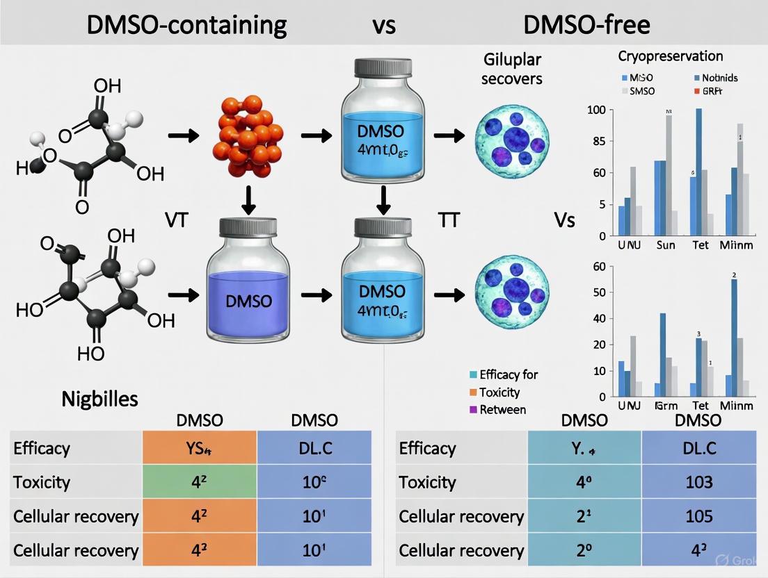
Cryopreservation is a cornerstone technology for enabling the widespread use of mesenchymal stem/stromal cells (MSCs) in research and clinical therapy. This process allows for long-term storage, rigorous quality control, and the creation of "off-the-shelf" cellular products that are readily available for patient treatment [1] [2]. The fundamental challenge of cryopreservation lies in preventing the lethal intracellular ice crystal formation and osmotic stress that occur during freezing and thawing [3] [4]. For decades, dimethyl sulfoxide (DMSO) has been the predominant cryoprotective agent (CPA) used to mitigate these damages. However, growing concerns over its potential toxicity have spurred the development of DMSO-free alternatives [5] [6]. This guide provides an objective, data-driven comparison of DMSO-containing and DMSO-free cryoprotectants, offering researchers a clear framework for selecting and optimizing cryopreservation protocols for MSCs.
The Established Role and Mechanism of DMSO
DMSO functions as a permeating cryoprotectant due to its small molecular size and amphiphilic nature, allowing it to easily cross cell membranes [3]. Its mechanism is twofold. Primarily, DMSO depresses the freezing point of water and strongly hydrogen bonds with water molecules, thereby disrupting the formation of ice crystals. This promotes an amorphous, glassy state (vitrification) rather than a crystalline solid, preventing mechanical damage to cellular structures [3] [4]. Secondly, by increasing the intracellular solute concentration, DMSO reduces the osmotic gradient across the cell membrane during freezing. This minimizes the efflux of water and the subsequent cell shrinkage and damage that would otherwise occur [3]. The efficacy of DMSO is concentration-dependent. While low concentrations (e.g., 5-10%) are protective, concentrations as high as 40% can cause lipid bilayer disintegration, and even at standard concentrations, DMSO can induce pore formation in biological membranes [3].
Table 1: Common Permeating Cryoprotectants and Their Properties
| Cryoprotectant | Molecular Weight (Da) | Common Concentration | Key Characteristics |
|---|---|---|---|
| Dimethyl Sulfoxide (DMSO) | 78.1 | 5-10% | Rapid membrane penetration; effective ice crystal inhibition [3]. |
| Glycerol (GLY) | 92.1 | ~10% | Slower membrane penetration; lower cellular toxicity [3] [2]. |
| Ethylene Glycol (EG) | 62.1 | Varies | Rapid membrane penetration; often used in vitrification mixtures [3]. |
| Propylene Glycol (PG) | 76.1 | Varies | Similar structure to EG; demonstrated cell toxicity [2]. |
Performance Comparison: DMSO vs. DMSO-Free Solutions
Recent multicenter studies have provided robust, quantitative data comparing traditional DMSO-based cryopreservation with emerging DMSO-free solutions. The performance of these solutions is typically evaluated based on post-thaw cell viability and recovery of viable cells.
A significant international, multicenter study compared a novel DMSO-free solution (SGI: containing Sucrose, Glycerol, and Isoleucine in Plasmalyte A) with in-house DMSO-containing solutions (5-10% DMSO) from seven different centers [5] [7]. The results, summarized in Table 2, indicate a trade-off: MSCs cryopreserved in the SGI solution showed a slightly larger decrease in viability post-thaw but demonstrated better recovery of viable cells compared to those frozen in DMSO-based solutions [5]. Critically, the immunophenotype (expression of CD73, CD90, CD105) and global gene expression profiles of the MSCs were comparable between the two groups, suggesting core cellular identity is maintained with the DMSO-free protocol [5].
Table 2: Multicenter Comparison of DMSO vs. DMSO-Free (SGI) Cryopreservation for MSCs
| Parameter | Fresh MSCs (Pre-freeze) | In-House DMSO Solution | SGI DMSO-Free Solution |
|---|---|---|---|
| Average Viability | 94.3% [5] | Decrease of 4.5% from fresh [5] | Decrease of 11.4% from fresh [5] |
| Viable Cell Recovery | Not Applicable | 87.3% (92.9% - 5.6%) [5] | 92.9% [5] |
| Immunophenotype | Not Applicable | Comparable to fresh cells [5] | Comparable to fresh cells, no significant difference vs. DMSO [5] |
| Global Gene Expression | Not Applicable | Baseline | No significant difference vs. DMSO [5] |
Another independent study compared a wider array of clinical-grade cryopreservation solutions, including NutriFreez (10% DMSO), an in-house PHD10 solution (Plasmalyte A/5% Human Albumin/10% DMSO), and CryoStor solutions (CS5 and CS10 with 5% and 10% DMSO, respectively) [4]. This research highlighted that the choice of solution and cell concentration can significantly impact post-thaw outcomes. Solutions with 10% DMSO generally showed comparable and stable viabilities up to 6 hours post-thaw, whereas a decreasing trend in viability and recovery was noted with 5% DMSO (CS5) [4]. Furthermore, MSCs cryopreserved at high concentrations (e.g., 9 million cells/mL) and then diluted after thawing showed improved viability over time, though this could come at the cost of reduced initial cell recovery [4]. Proliferative capacity after a 6-day culture was also affected, with MSCs in NutriFreez and PHD10 performing similarly, while those in CryoStor solutions showed a marked reduction [4].
Experimental Protocols for Performance Comparison
To ensure the reproducibility of cryopreservation studies, detailed methodologies are essential. Below is a synthesis of the key protocols from the cited literature.
- Cell Source and Culture: MSCs were isolated from bone marrow or adipose tissue and cultured ex vivo according to local protocols at each of the seven participating international centers.
- Cryopreservation Solutions:
- Test Solution: DMSO-free SGI solution (Sucrose, Glycerol, Isoleucine in Plasmalyte A) prepared at the University of Minnesota.
- Control Solutions: In-house DMSO-containing solutions (5-10% DMSO) prepared at each center.
- Freezing Process: The cell suspension was aliquoted into vials or bags. For six of the seven centers, vials/bags were placed in a controlled-rate freezer before transfer to liquid nitrogen for at least one week.
- Post-Thaw Assessment:
- Viability & Recovery: Measured using flow cytometry or dye exclusion methods.
- Immunophenotype: Analyzed by flow cytometry for surface markers (CD45, CD73, CD90, CD105).
- Gene Expression: Global transcriptional profiles were assessed.
- Cell Source: Human bone marrow-derived MSCs from commercial suppliers and local donors with ethical approval.
- Cryopreservation Solutions:
- NutriFreez: Commercial solution with 10% DMSO.
- PHD10: In-house formulation (Plasmalyte A, 5% Human Albumin, 10% DMSO).
- CryoStor CS5 & CS10: Commercial solutions with 5% and 10% DMSO, respectively.
- Freezing Process: MSCs were cryopreserved at three different concentrations (3, 6, and 9 million cells/mL) using a standard slow-freezing protocol, ultimately stored in liquid nitrogen.
- Thawing and Dilution: Cells were thawed in a 37°C water bath. To mitigate DMSO toxicity, cells frozen at high concentrations were diluted post-thaw (1:1 or 1:2) with Plasmalyte A/5% HA to a uniform concentration.
- Post-Thaw Assessment:
- Viability: Measured at 0, 2, 4, and 6 hours post-thaw using Trypan blue exclusion and Annexin V/PI staining.
- Recovery: Calculated as (post-thaw cell count / pre-freeze cell count) × 100.
- Proliferation: Assessed by measuring cell growth after a 6-day recovery culture.
- Potency: Evaluated via T cell inhibition assays and monocytic phagocytosis tests.
Diagram 1: Generic Experimental Workflow for MSC Cryopreservation Studies
The Scientist's Toolkit: Key Reagents and Materials
Successful cryopreservation relies on a suite of specialized reagents and equipment. The table below details essential items used in the featured experiments.
Table 3: Essential Research Reagents and Solutions for MSC Cryopreservation
| Reagent/Solution | Function / Key Feature | Example Use in Research |
|---|---|---|
| DMSO (Dimethyl Sulfoxide) | Permeating CPA; prevents intracellular ice crystallization [3]. | Standard CPA at 5-10% concentration in slow-freezing protocols [4]. |
| SGI Solution | DMSO-free CPA; contains Sucrose, Glycerol, Isoleucine [5]. | Tested as a clinically safer alternative to DMSO for MSC cryopreservation [5]. |
| CryoStor CS5/CS10 | Commercial, cGMP-manufactured freezing media with defined DMSO levels [4]. | Used as a standardized, regulatory-compliant control in comparative studies [4]. |
| Plasmalyte A | Isotonic, balanced salt solution; serves as a base solution for in-house CPA formulations [5] [4]. | Base for PHD10 and SGI cryopreservation solutions [5] [4]. |
| Human Serum Albumin (HA) | Protein stabilizer; provides oncotic pressure and can mitigate cell agglomeration [4]. | Component of in-house PHD10 freezing medium at 5% concentration [4]. |
| Controlled-Rate Freezer | Equipment that ensures a consistent, optimal cooling rate (e.g., -1°C/min) [8]. | Used in the majority of centers in the multicenter study for standardized freezing [5]. |
The established role of DMSO as a highly effective cryoprotectant for MSCs is undeniable, supported by decades of successful use. Its primary mechanism, involving membrane permeation and ice crystal inhibition, provides robust protection against cryo-injury. However, quantitative data from recent, well-designed studies demonstrate that DMSO-free solutions, such as the SGI formulation, can achieve post-thaw outcomes that are comparable and in some aspects (like viable cell recovery) potentially superior [5]. The choice between DMSO-containing and DMSO-free solutions is not absolute but should be guided by the specific requirements of the research or therapy. Factors such as the acceptable threshold of DMSO-related toxicity, desired post-thaw viability and recovery, necessary cell functionality, and regulatory considerations must all be weighed. As the field advances, the development and validation of DMSO-free protocols will be crucial for enhancing the safety and efficacy of MSC-based therapies.
Dimethyl sulfoxide (DMSO) is one of the most widely used cryoprotective agents (CPAs) in the cryopreservation of mesenchymal stem cells (MSCs) for cell-based therapies. While effective at preventing ice crystal formation during freezing, its application in clinical settings is accompanied by documented concerns regarding cellular toxicity and patient adverse effects. This comparison guide objectively examines the risks associated with DMSO-containing cryoprotectants versus emerging DMSO-free alternatives, providing researchers and drug development professionals with experimental data to inform cryopreservation protocol decisions. The ongoing development of cellular therapies necessitates a critical evaluation of all components, with cryoprotectant selection representing a crucial factor in balancing cell viability, functionality, and patient safety.
DMSO Cytotoxicity: Mechanisms and Cellular-Level Evidence
Documented Cellular Damage and Functional Impairment
DMSO mitigates cryo-injury by disrupting ice crystallization and preventing dangerous intra- and extracellular solute concentration increases during freezing [9]. However, this protective function comes with significant cellular-level trade-offs:
- Membrane and Cytoskeleton Instability: At high concentrations (e.g., 40%), DMSO can interrupt cell membrane stability [9]. The process of freezing and thawing itself, even with DMSO present, subjects the cell membrane-cytoskeleton complex to significant stress [5].
- Altered Cellular Function: Studies report that MSCs cryopreserved in certain DMSO-containing solutions like CryoStor CS5 and CS10 showed a 10-fold reduction in proliferative capacity after a 6-day recovery culture compared to those preserved in other formulations [9].
- Induction of Apoptosis: In vitro studies on cancer cell lines suggest that DMSO can induce apoptosis by interacting with apoptotic proteins and potentially elevating reactive oxygen species (ROS) production, affecting mitochondrial function [10].
- Concentration-Dependent Effects: Research indicates that DMSO's cytotoxic effects are variable depending on cell type and exposure duration. While concentrations around 0.3% showed minimal cytotoxicity across most cell lines tested, higher concentrations demonstrated significant toxic effects [10].
Table 1: Comparative Cytotoxicity of DMSO in Various Experimental Models
| Cell Type/Model | DMSO Concentration | Exposure Duration | Observed Effects | Citation |
|---|---|---|---|---|
| Various Cancer Cell Lines | 0.3125% | 24-72 hours | Minimal cytotoxicity in most lines | [10] |
| Various Cancer Cell Lines | >0.3125% | 24-72 hours | Concentration-dependent cytotoxicity | [10] |
| Bone Marrow MSCs | 5-10% (cryopreservation) | Post-thaw | 10-fold reduced proliferation in some formulations | [9] |
| MSCs (in silico analysis) | N/A | N/A | Binds to apoptotic and membrane proteins | [10] |
Molecular Mechanisms of DMSO Toxicity
Experimental and in silico studies have provided insights into the molecular mechanisms underlying DMSO cytotoxicity:
Diagram 1: Molecular pathways of DMSO-induced cytotoxicity. DMSO exposure triggers multiple mechanisms that can compromise cellular function and viability, particularly at higher concentrations.
Clinical Adverse Effects of DMSO in Patients
Documented Patient Reactions to DMSO-Containing Products
The administration of DMSO-cryopreserved cellular products has been associated with various adverse reactions in patients, as documented in clinical settings and systematic reviews:
- Gastrointestinal Reactions: A systematic review of 109 studies found gastrointestinal reactions to be the most commonly reported adverse effects, with nausea occurring in approximately 12% of patients and vomiting in 7% of patients receiving DMSO. These effects were more frequent with intravenous administration (17% and 11% for nausea and vomiting, respectively) compared to transdermal application [11].
- Systemic Reactions: DMSO may induce histamine release, which can lead to reactions such as flushing, dyspnea, abdominal cramps, and cardiovascular effects [11]. Characteristic garlic- or oyster-like breath odor frequently occurs due to pulmonary excretion of the DMSO metabolite dimethyl sulfide [11] [1].
- Serious Adverse Events: Although rare, serious adverse events have been reported, including encephalopathy, cardiac arrest, respiratory depression, cerebral infarction, and severe neurotoxicity [12]. The vast majority of cardiac side effects, however, are self-limiting and not usually associated with serious morbidity and mortality [12].
Table 2: Documented Adverse Reactions to DMSO in Clinical Applications
| Reaction Category | Specific Adverse Effects | Reported Incidence | Severity |
|---|---|---|---|
| Gastrointestinal | Nausea, vomiting, abdominal cramps | Up to 35-50% of patients [12]; Nausea: 12% overall, 17% IV [11] | Mostly mild to moderate |
| Dermatological | Skin rash, flushing, itching | Not quantified overall | Mild to moderate |
| Neurological | Headache, dizziness, seizures, encephalopathy | Rare for serious events | Mild to severe |
| Cardiovascular | Bradycardia, tachycardia, hypertension, hypotension | Common but usually self-limiting [12] | Mild to moderate |
| Respiratory | Dyspnea, cough, hypoxia | Not quantified overall | Moderate to severe |
| General | Unpleasant taste, garlic-like breath odor | Very common [12] | Mild but bothersome |
Dose-Dependent Toxicity and Risk Management
A clear relationship exists between DMSO dose and the occurrence of adverse reactions [11]. In hematopoietic stem cell transplantation, a maximum dose of 1 g DMSO/kg body weight per infusion is generally considered acceptable [1] [12]. For MSC therapies, the doses of DMSO delivered via intravenous administration are typically 2.5-30 times lower than this accepted threshold [1]. Risk management strategies include:
- Premedication: Administration of antihistamines and corticosteroids before infusion can help prevent or reduce the severity of reactions, though this does not completely prevent anaphylaxis [12].
- Dilution and Washing: Removing DMSO before infusion through single or multiple washes can reduce side effects but may cause cell damage and loss [1] [12].
- Infusion Rate Control: Starting infusions slowly and increasing as tolerated helps identify susceptible patients and manage reactions [1].
DMSO-Free Alternatives: Experimental Evidence and Performance
Emerging DMSO-Free Cryoprotectant Formulations
Research into DMSO-free cryopreservation strategies has accelerated, with several promising alternatives demonstrating efficacy in preserving MSC viability and function:
- Sucrose-Glycerol-Isoleucine (SGI) Formulation: A novel DMSO-free solution containing sucrose, glycerol, and isoleucine in a Plasmalyte A base was tested in an international multicenter study. MSCs cryopreserved in SGI showed slightly lower cell viability (decrease of 11.4% from pre-freeze viability) compared to those in DMSO-containing solutions (decrease of 4.5%), but better recovery (92.9% for SGI vs. lower by 5.6% for in-house DMSO solutions) [7] [5].
- Urea-Glucose Combinations: Research has identified synergistic cryoprotective activity of urea and glucose at equimolar concentrations in human MSCs. The addition of sugars like mannitol and sucrose to the formulation, along with pre-incubation with trehalose, further enhanced cell viability after freeze-thaw stress [13].
- Sugar-Based Formulations: Various sugars and sugar alcohols (e.g., sucrose, trehalose, glucose, mannitol) have been explored as cryoprotectants, though their protective effects alone have been limited and not entirely satisfactory without combination strategies [13].
Comparative Analysis: DMSO vs. DMSO-Free Formulations
Table 3: Performance Comparison of DMSO vs. DMSO-Free Cryoprotectants for MSCs
| Parameter | DMSO-Based Formulations | DMSO-Free Alternatives | Experimental Evidence |
|---|---|---|---|
| Post-Thaw Viability | Decrease of 4.5% from pre-freeze viability [7] | Decrease of 11.4% from pre-freeze viability (SGI solution) [7] | Multicenter study [7] [5] |
| Cell Recovery | Lower by 5.6% compared to SGI [7] | 92.9% viable cell recovery (SGI solution) [7] | Multicenter study [7] [5] |
| Immunophenotype | Maintained surface marker expression [9] | Comparable expression of CD73, CD90, CD105 [7] [5] | Flow cytometry analysis [9] [7] |
| Proliferation Capacity | Variable (10-fold reduction in some formulations) [9] | Not fully reported for all formulations | In vitro culture [9] |
| Immunomodulatory Function | Preserved in some formulations [9] | Comparable in tested formulations [9] | T-cell inhibition assays [9] |
| Gene Expression | Potential alterations [9] [13] | Comparable global gene expression profiles [7] [5] | Microarray/transcriptional analysis [7] |
Experimental Protocols for Cryoprotectant Comparison
Standardized Methodology for Cryoprotectant Evaluation
To ensure comparable results across studies, researchers should adhere to standardized experimental protocols when evaluating cryoprotectant formulations:
Sample Preparation and Cryopreservation
- Isolate and culture MSCs according to established protocols [9] [7]
- Cryopreserve cells at concentrations typically ranging from 3-9 million cells/mL [9]
- Utilize controlled-rate freezing protocols before transfer to liquid nitrogen [7]
- Store frozen cells for at least one week before thawing and testing [7]
Thawing and Post-Thaw Assessment
- Rapidly thaw cells in a 37°C water bath for approximately 2 minutes [9]
- Consider dilution strategies (no dilution, 1:1, or 1:2 dilution) with appropriate solutions such as Plasmalyte A with human albumin [9]
- Assess cell viability at multiple time points (0, 2, 4, and 6 hours post-thaw) using Trypan blue exclusion and Annexin V/PI staining [9]
- Calculate viable cell recovery by dividing the total number of live cells counted by the number of cells originally cryopreserved [9]
Functional Assays
- Evaluate immunophenotype through flow cytometry analysis of MSC surface markers (CD73, CD90, CD105, CD14, CD19, CD34, CD45) [9] [7]
- Assess proliferative capacity through 6-day recovery cultures [9]
- Test immunomodulatory function via T-cell proliferation inhibition assays and monocytic phagocytosis assays [9]
- Analyze global gene expression profiles to identify cryoprotectant-induced alterations [7] [5]
Diagram 2: Experimental workflow for comparing cryoprotectant formulations. The standardized methodology enables objective assessment of both DMSO-containing and DMSO-free solutions across multiple parameters.
The Scientist's Toolkit: Essential Research Reagents
Table 4: Key Reagents for Cryoprotectant Research
| Reagent/Category | Specific Examples | Function/Application | Experimental Notes |
|---|---|---|---|
| Penetrating CPAs | DMSO (5-10%) | Prevents intracellular ice formation; reduces osmotic stress | Clinical standard but with toxicity concerns [9] [1] |
| Non-Penetrating CPAs | Sucrose, trehalose, glucose | Provides extracellular protection; stabilizes cell membranes | Often used in combination [7] [13] |
| Novel CPA Formulations | SGI solution (sucrose-glycerol-isoleucine) | DMSO-free alternative with comparable efficacy | Slightly lower viability but better recovery [7] [5] |
| Synergistic Combinations | Urea-glucose mixtures | Mimics natural cryoprotection in hibernating organisms | Shows promise for specific cell types [13] |
| Base Solutions | Plasmalyte A | Isotonic solution for cryoprotectant formulation | Provides physiological ion balance [9] [7] |
| Viability Assessment | Trypan blue, Annexin V/PI staining | Differentiates live, apoptotic, and necrotic cells | Essential for post-thaw quality assessment [9] |
| Phenotypic Validation | CD73, CD90, CD105 antibodies | Confirms MSC identity post-preservation | Critical for functional characterization [9] [7] |
The choice between DMSO-containing and DMSO-free cryoprotectants for MSC preservation involves careful consideration of competing priorities. While DMSO remains the clinical standard with proven efficacy in maintaining cell viability during cryopreservation, its documented cytotoxicity at both cellular and patient levels necessitates continued research into safer alternatives. Current evidence suggests that emerging DMSO-free formulations, particularly those combining multiple cryoprotective mechanisms, show promising results with post-thaw viability rates above clinically acceptable thresholds (typically >80%) and comparable immunophenotype and gene expression profiles. For researchers and therapy developers, the selection of cryoprotectants should be guided by comprehensive assessment of not only immediate post-thaw viability but also long-term functionality, potency, and ultimately, patient safety profile. As the field advances, standardized testing protocols and multicenter validation studies will be crucial for establishing the next generation of clinically viable, DMSO-free cryopreservation solutions for cell-based therapies.
Regulatory and Manufacturing Drivers for Chemically-Defined, DMSO-Free Media
The adoption of chemically-defined, DMSO-free cryopreservation media is accelerating, driven by patient safety concerns, stringent regulatory guidelines, and the need for scalable, reproducible manufacturing in cell and gene therapy. For Mesenchymal Stromal Cells (MSCs), recent international multicenter studies demonstrate that DMSO-free formulations achieve comparable cell recovery, viability, and functional phenotypes to traditional DMSO-containing media, with the significant advantage of eliminating cytotoxic risks and simplifying production workflows. This guide provides an objective, data-driven comparison to inform research and development strategies.
Market and Regulatory Landscape
The shift toward DMSO-free solutions is a definitive trend within the biopreservation market, influenced by both clinical and commercial factors.
- Market Growth: The global DMSO-free freezing culture media market, valued at approximately USD 950 million in 2025, is projected to grow at a CAGR of 7.5%, reaching nearly USD 1.7 billion by 2033 [14]. This robust growth is fueled by the expanding cell therapy pipeline and increased R&D expenditure.
- Regulatory Drivers: Regulatory bodies like the FDA and EMA are increasingly advocating for serum-free and chemically-defined formulations in clinical applications [15]. This push aims to reduce batch-to-batch variability, eliminate animal-derived components, and minimize the risk of adventitious agents, thereby ensuring greater product consistency and safety [14] [16].
- Manufacturing Efficiency: DMSO-free media streamline the manufacturing process by eliminating the need for post-thaw wash steps. This reduces cell loss, damage, and operational complexity, while also cutting down on equipment, labor, and time requirements [17]. This simplification is critical for scaling up production to meet clinical demand.
Performance Data: DMSO-Free vs. DMSO-Containing Media
Objective evaluation of experimental data is crucial for selecting a cryopreservation protocol. The following tables summarize key quantitative findings from a recent international multicenter study and other performance analyses.
Table 1: Post-Thaw Viability and Recovery of MSCs in a Multicenter Study [7] [5]
| Cryopreservation Solution | Average Post-Thaw Viability | Average Recovery of Viable MSCs | Key Components |
|---|---|---|---|
| DMSO-Free (SGI Solution) | 82.9% | 92.9% | Sucrose, Glycerol, Isoleucine in Plasmalyte A |
| In-House DMSO Solution | 89.8% | 87.3% | 5-10% DMSO |
Table 2: Comparative Analysis of Commercial DMSO-Free Media Performance
| Performance Metric | DMSO-Free Media (e.g., NB-KUL DF) | Traditional DMSO-Based Media |
|---|---|---|
| Cell Viability | Equivalent or slightly lower for some cell types, but consistently >80% (clinically acceptable) [7] [18] | High, but with risk of cytotoxicity during freeze/thaw [17] |
| Cell Recovery | Better recovery of viable cells demonstrated in controlled studies [7] | Standard, but can be impacted by DMSO toxicity |
| Immunophenotype | Comparable, no significant differences in MSC surface markers (CD73, CD90, CD105) [7] | Comparable, maintains standard immunophenotype |
| Global Gene Expression | No significant differences observed [7] [5] | No significant differences observed [7] [5] |
| Patient Safety | High; no risk of DMSO-induced adverse reactions (nausea, hypotension, arrhythmias) [17] | Requires risk mitigation; associated with adverse reactions [1] |
Key Experimental Findings
- Multicenter Study Conclusions: MSCs cryopreserved in the novel DMSO-free (SGI) solution showed slightly lower cell viability (a decrease of 11.4% from fresh vs. 4.5% for DMSO), but better recovery of viable cells (92.9% vs. 87.3%) and fully comparable immunophenotype and global gene expression profiles post-thaw compared to DMSO-cryopreserved cells [7] [5]. The authors concluded that the viability in the DMSO-free solution was "likely clinically acceptable" [7].
- Commercial Formulation Performance: Independent testing of commercial DMSO-free media, such as NB-KUL DF, shows it can provide equivalent performance to DMSO-based media like CryoStor CS5 for MSCs, peripheral blood mononuclear cells (PBMCs), and T cells [18]. This demonstrates the viability of DMSO-free alternatives across multiple human cell types relevant to therapy.
Detailed Experimental Protocol
To ensure reproducibility, the following details the core methodology from the international multicenter study that generated the comparative data in Table 1 [7] [5].
Key Reagents and Materials
Table 3: Research Reagent Solutions for MSC Cryopreservation
| Reagent/Material | Function in the Protocol | Example/Note |
|---|---|---|
| DMSO-Free Cryoprotectant | Prevents ice crystal formation using non-penetrating agents. | SGI Solution: Sucrose, Glycerol, Isoleucine in Plasmalyte A [7]. |
| DMSO-Based Cryoprotectant | Penetrating cryoprotectant; standard control. | 5-10% DMSO in culture medium [7] [5]. |
| Plasmalyte A | Base solution for the SGI formulation; provides physiological pH and electrolytes. | Used as the solvent for the SGI components [7]. |
| Controlled-Rate Freezer | Ensures consistent, optimized cooling rate to minimize cryoinjury. | Used by 6 out of 7 centers in the multicenter study [7]. |
| Liquid Nitrogen | Long-term storage at ultra-low temperatures (-196°C). | Ensures long-term viability of cryopreserved cells [7] [19]. |
The Scientist's Toolkit: Essential Research Reagents
Beyond the specific reagents used in the cited study, the following table outlines key solutions and materials essential for MSC cryopreservation research.
Table 4: Essential Reagents for MSC Cryopreservation Research
| Reagent Solution | Function & Importance | Commercial Examples |
|---|---|---|
| Chemically-Defined, DMSO-Free Media | Provides a non-toxic, serum-free, and consistent formulation for clinical-grade cryopreservation. | Gibco Synth-a-Freeze [15], NB-KUL DF [17], CryoStor platform [15] |
| Serum-Free Culture Media | Supports the expansion of MSCs without animal serum, reducing variability and contamination risk. | Various specialized media from STEMCELL Technologies, Bio-Techne, etc. [16] |
| Cell Dissociation Agents | Used to detach adherent MSCs from culture flasks into a single-cell suspension for freezing. | Trypsin-EDTA, TrypLE, enzyme-free solutions [5] |
| Flow Cytometry Antibody Panels | Validates MSC immunophenotype pre- and post-thaw (positive for CD73, CD90, CD105; negative for CD45). | Antibodies from BD Biosciences, BioLegend, etc. [7] [5] |
| Viability Assay Kits | Quantifies cell viability and recovery post-thaw (e.g., via dye exclusion). | Trypan Blue, 7-AAD, Annexin V/PI apoptosis kits [7] |
Strategic Implementation Guide
Choosing a cryopreservation strategy requires a balanced consideration of scientific and regulatory needs. The following diagram outlines a decision-making workflow.
Guidance for Selection
- For Clinical Therapy Development: The strong regulatory and safety drivers make chemically-defined, DMSO-free media the preferred choice. Their use simplifies the regulatory filing process, enhances patient safety, and supports scalable GMP manufacturing [17] [15].
- For Foundational Research: While DMSO-based media remain a cost-effective and familiar option, transitioning to DMSO-free formulations future-proofs research programs and ensures better alignment with the direction of clinical translation [14].
- Validation is Critical: Regardless of the chosen path, thorough in-house validation is essential. Data from multicenter studies provide strong evidence, but confirming performance with specific MSC sources (bone marrow, adipose) and laboratory protocols is necessary for success [7] [5].
Innovative Formulations and Protocols for DMSO-Free Cryopreservation
The cryopreservation of mesenchymal stem/stromal cells (MSCs) is a critical step in ensuring the widespread availability and stability of these cells for clinical applications in regenerative medicine and cellular therapy [7] [5]. Currently, the standard of practice involves cryopreservation using solutions containing dimethyl sulfoxide (DMSO), which presents significant challenges for clinical translation. While effective for cell preservation, DMSO is associated with dose-dependent toxicity in patients, potentially causing adverse effects ranging from mild nausea and vomiting to severe cardiovascular, respiratory, and neurological reactions upon infusion [20] [21]. Furthermore, DMSO exposure affects cellular processes in vitro, including disruptions in DNA methylation mechanisms, dysregulation of gene expression, and potential induction of unwanted differentiation [21]. These concerns have driven the search for effective DMSO-free alternatives that maintain cell viability, recovery, and function while eliminating DMSO-related toxicity.
Multi-component cryoprotectant solutions represent an innovative approach inspired by natural systems. Various organisms survive freezing temperatures through the use of combinations of protective osmolytes [22] [21]. The solution containing sucrose, glycerol, and isoleucine (SGI) in a base of Plasmalyte A has emerged as a promising candidate, demonstrating comparable performance to DMSO-containing cryoprotectants in recent international multicenter studies [7] [5] [23]. This review provides a comprehensive comparison of this novel SGI formulation against traditional DMSO-containing cryoprotectants, presenting experimental data and methodologies to inform researchers, scientists, and drug development professionals in the field of MSC research.
Comparative Performance Data: SGI Versus DMSO-Containing Cryoprotectants
Quantitative Comparison of Post-Thaw Outcomes
An international multicenter study conducted by the Production Assistance for Cellular Therapies (PACT) and Biomedical Excellence for Safer Transfusion (BEST) Collaborative directly compared a novel DMSO-free SGI solution with traditional DMSO-containing cryoprotectants (5-10% DMSO) across seven participating centers in the United States, Australia, and Germany [7] [5] [23]. MSCs were isolated from bone marrow or adipose tissue and cultured following local protocols at each center, providing robust data across different laboratory conditions. The table below summarizes the key post-thaw outcomes from this comprehensive study:
| Performance Metric | Fresh MSCs (Pre-Freeze) | SGI Solution (DMSO-Free) | DMSO-Containing Solutions (5-10%) | Statistical Significance |
|---|---|---|---|---|
| Average Viability | 94.3% (95% CI: 87.2-100%) | 82.9% (decrease of 11.4%) | 89.8% (decrease of 4.5%) | P < 0.001 (SGI) vs P: 0.049 (DMSO) |
| Viable Cell Recovery | Baseline | 92.9% (95% CI: 85.7-100.0%) | 87.3% (5.6% lower than SGI) | P < 0.013 |
| Immunophenotype | Expected CD45-/CD73+/CD90+/CD105+ | Maintained expected expression | Maintained expected expression | No significant difference |
| Global Gene Expression | Baseline profile | Comparable to pre-freeze profile | Comparable to pre-freeze profile | No significant difference |
The data demonstrates that while MSCs cryopreserved in the SGI solution showed a statistically significant greater decrease in viability compared to those frozen in DMSO, the average post-thaw viability remained above the 80% threshold generally considered clinically acceptable [7] [5]. Importantly, the SGI solution demonstrated significantly better recovery of viable cells, indicating that a greater proportion of cells survive the freeze-thaw process with this DMSO-free formulation [23]. Both cryoprotectant methods successfully maintained critical quality attributes, including characteristic immunophenotype (CD45, CD73, CD90, and CD105 expression) and global gene expression profiles, suggesting comparable biological fidelity after thawing [7].
Mechanism of Action and Comparative Advantages
The SGI solution employs a multi-component approach where each constituent provides distinct protective functions that collectively enhance cell survival during cryopreservation:
- Sucrose: As a non-penetrating disaccharide, sucrose functions as an extracellular cryoprotectant that stabilizes the cell membrane and moderates osmotic stress during freezing and thawing [22] [21]. It interacts with water molecules via hydrogen bonding, altering solidification patterns and reducing ice crystal formation [22].
- Glycerol: This penetrating sugar alcohol serves as an intracellular cryoprotectant that readily crosses cell membranes. Glycerol interacts strongly with water through hydrogen bonding, lowers the freezing point of intracellular water, and helps stabilize proteins, thereby preventing denaturation during freezing [22] [21].
- Isoleucine: This amino acid plays a crucial role in stabilizing the cryoprotectant mixture during freezing, preventing precipitation of sugar components, and contributing to the overall cryoprotective effect through synergistic interactions with sucrose and glycerol [22].
- Plasmalyte A Base: This balanced electrolyte solution serves as the carrier, providing a physiologically compatible environment that maintains optimal osmotic conditions and pH stability throughout the cryopreservation process [7] [5].
The complementary actions of these components create a cryoprotective system that addresses both intracellular and extracellular damage mechanisms, effectively replacing the dual protective functions of DMSO without its associated toxicity concerns.
Experimental Protocols and Methodologies
International Multicenter Study Design
The pivotal study comparing SGI and DMSO-containing cryoprotectants employed a rigorous, standardized methodology across multiple international sites to ensure robust and generalizable results [7] [5] [23]. The experimental workflow encompassed cell preparation, cryopreservation, storage, thawing, and comprehensive post-thaw assessment, as illustrated in the following diagram:
Detailed Methodological Parameters
Cell Preparation and Cryopreservation:
- MSCs were isolated from bone marrow or adipose tissue and cultured ex vivo according to standardized local protocols at each participating center [7] [5].
- The DMSO-free cryoprotectant solution containing sucrose, glycerol, and isoleucine (SGI) in Plasmalyte A was prepared at the University of Minnesota, while cryoprotectant solutions containing 5-10% DMSO (in-house formulations) were prepared at each of the seven participating centers [23].
- Cells in suspension were aliquoted into cryovials or cryobags, with six of the seven centers using controlled-rate freezing and one center employing a -80°C freezer overnight before transfer to liquid nitrogen for storage [7] [5].
Freezing Protocol:
- The standard controlled-rate freezing protocol included: (1) start at 20°C, (2) cool at -10°C/min to 0°C, (3) hold at 0°C for 15 minutes, (4) cool at -1°C/min to -8°C, (5) rapid cooling at -50°C/min to -45°C, (6) rewarming at +15°C/min to -12°C, (7) cool at -1°C/min to -60°C, and (8) final cooling at -10°C/min to -100°C [7] [5] [22].
- This sophisticated protocol incorporates deliberate nucleation steps to initiate controlled ice formation in the extracellular solution, minimizing supercooling effects and enhancing reproducibility across samples.
Post-Thaw Assessment Methods:
- Cell Viability: Measured using fluorescent staining with calcein-AM and propidium iodide (PI) to distinguish live from dead cells, with quantification via fluorescence microscopy or flow cytometry [7] [22].
- Cell Recovery: Calculated as the ratio of the number of live cells post-thaw to the number of seeded live cells pre-freeze, providing a measure of the total viable cell yield [7] [5].
- Immunophenotype: Characterized by flow cytometric analysis of characteristic MSC surface markers (CD45, CD73, CD90, and CD105) to confirm maintenance of phenotypic identity after cryopreservation [7] [23].
- Gene Expression Profiling: Assessed using transcriptional profiling and microarray analysis to evaluate potential changes in global gene expression patterns resulting from cryopreservation method [7] [5].
The Scientist's Toolkit: Essential Research Reagents
Successful implementation of DMSO-free cryopreservation protocols requires specific reagents and materials. The following table details key research reagent solutions and their functions based on the methodologies employed in the cited studies:
| Reagent/Material | Function and Purpose | Application Notes |
|---|---|---|
| Sucrose | Non-penetrating cryoprotectant that stabilizes cell membranes and reduces extracellular ice formation | Pharmaceutical grade; typically used at concentrations of 30-150 mM in combination with other osmolytes [22] |
| Glycerol | Penetrating cryoprotectant that reduces intracellular ice formation and stabilizes proteins | Humco or pharmaceutical grade; typically used at 5% concentration in multi-component formulations [7] [24] |
| Isoleucine | Amino acid that stabilizes cryoprotectant mixture and enhances post-thaw recovery through synergistic effects | Sigma-Aldrich or equivalent; typically used at 7.5 mM concentration [7] [22] |
| Plasmalyte A | Balanced electrolyte solution used as base for cryoprotectant formulation | Provides physiological ions and pH stability; superior to simple saline solutions [7] [5] |
| Calcein-AM/PI | Fluorescent viability stain (calcein-AM for live cells, PI for dead cells) | Standardized quantification of post-thaw viability; preferred over trypan blue for accuracy [7] [22] |
| Controlled-Rate Freezer | Programmable freezer that ensures reproducible cooling rates | Planer Kryo 10 or equivalent; essential for standardized freezing protocols across laboratories [7] [5] |
| Antibody Panels | Flow cytometry antibodies for CD45, CD73, CD90, CD105 | Quality control assessment of MSC phenotype maintenance post-thaw [7] [5] |
The comprehensive multicenter evaluation of the SGI cryoprotectant solution demonstrates that DMSO-free cryopreservation of MSCs is achievable with performance characteristics comparable to traditional DMSO-containing methods. While the SGI formulation resulted in slightly lower post-thaw viability compared to DMSO controls (82.9% vs 89.8%), it exhibited superior recovery of viable cells (92.9% vs 87.3%) and comparable maintenance of immunophenotype and global gene expression profiles [7] [5] [23].
These findings have significant implications for both research and clinical applications of MSCs. The elimination of DMSO addresses important safety concerns associated with patient administration of cryopreserved cell products, including potential adverse reactions and effects on cellular epigenetics [20] [21]. For the research community, the SGI formulation provides a valuable tool for MSC preservation without the confounding effects of DMSO on cellular processes, differentiation potential, and gene expression patterns [21].
Future research directions should focus on optimizing component ratios for specific MSC sources (bone marrow, adipose tissue, umbilical cord), evaluating long-term functionality post-thaw, and validating the formulation across diverse clinical-grade manufacturing environments. The development of effective DMSO-free cryoprotectants like the SGI solution represents a significant advancement in cellular therapy, potentially enabling safer and more effective MSC-based treatments for conditions such as graft-versus-host disease, cardiovascular disease, stroke, and acute respiratory distress syndrome [7] [5].
The cryopreservation of mesenchymal stromal cells (MSCs) is a critical prerequisite for their widespread application in regenerative medicine and cellular therapies. For decades, dimethyl sulfoxide (DMSO) has served as the predominant cryoprotectant in both research and clinical settings. However, growing evidence of its cytotoxicity and potential patient side effects has stimulated the urgent development of DMSO-free preservation strategies [1]. Among promising alternatives, trehalose—a natural, nontoxic disaccharide—has emerged as a leading candidate due to its exceptional biostabilizing properties.
A significant challenge in utilizing trehalose for mammalian cell cryopreservation is its inherent impermeability to cell membranes, necessitating innovative delivery strategies to achieve protective intracellular concentrations [25]. This article provides a comprehensive comparison of intracellular trehalose delivery via ultrasound and microbubbles against established cryopreservation methods, presenting quantitative data and experimental protocols to inform researchers and drug development professionals in their pursuit of safer, more effective cell preservation platforms.
Cryoprotectant Performance Comparison
Quantitative Analysis of Cryoprotectant Efficacy
Table 1: Comparative performance of cryoprotectants in MSC preservation
| Cryoprotectant & Method | Post-Thaw Viability | Viable Cell Recovery | Intracellular [Trehalose] | Key Functional Outcomes |
|---|---|---|---|---|
| 10% DMSO (Standard) | ~90% [26] | Not Reported | Not Applicable | Maintains immunomodulatory gene expression (IDO1, TSG6) [26] |
| Trehalose + Electroporation | Comparable to DMSO [26] | Not Reported | 50-90 mM [26] | Maintains osteogenic and adipogenic differentiation potential [26] |
| Trehalose + Ultrasound/Microbubbles | Maintains membrane integrity and viability [25] | Not Reported | Confirmed via rhodamine-labeled trehalose [25] | Preserves multipotency; successful lyophilization [25] |
| Sucrose/Glycerol/Isoleucine (SGI) | ~82.9% (decrease from fresh) [7] | ~92.9% [7] | Not Applicable | Comparable immunophenotype (CD73, CD90, CD105) and global gene expression profiles [7] |
Safety Profile Assessment
Table 2: Safety and clinical applicability comparison
| Parameter | DMSO-Based Cryopreservation | Trehalose-Based Cryopreservation |
|---|---|---|
| Cytotoxicity | Cytotoxic, especially during long-term exposure; associated with epigenetic changes, cell dysfunction [25] | Nontoxic, biocompatible; FDA-approved for food, vaccines [25] |
| Patient Side Effects | Nausea, vomiting, diarrhea, hemolysis, renal failure, hypertension, pulmonary edema [26] | No significant toxicity concerns reported |
| Clinical Administration Concerns | Infusion-related reactions; requires premedication and potential washing steps [1] | Reduced safety concerns; no complex removal procedures needed |
| Serum Requirement | Often requires FBS or human blood derivatives [26] | Compatible with xeno-free formulations |
Ultrasound-Mediated Trehalose Delivery: Mechanism and Workflow
Fundamental Principles and Mechanisms
Ultrasound-mediated trehalose delivery leverages acoustic cavitation to transiently permeabilize cell membranes. When ultrasound is applied in the presence of microbubbles, the oscillating bubbles generate shear stresses and microstreaming forces that temporarily disrupt lipid bilayers, creating nanopores that facilitate trehalose entry into the cytoplasm [25] [27]. This process, known as sonoporation, enables intracellular delivery of this membrane-impermeant disaccharide without significant cytotoxicity.
The protective mechanism of trehalose operates through multiple pathways. During freezing and dehydration, trehalose stabilizes membranes and proteins by replacing water molecules through hydrogen bonding with phospholipid head groups and biomolecular surfaces [25]. This water substitution effect prevents denaturation and maintains structural integrity. Additionally, trehalose forms a vitrified glassy state at low hydration levels, immobilizing cellular components and dramatically reducing destructive biochemical reactions [27].
Experimental Workflow for Ultrasound-Mediated Trehalose Delivery
The standardized protocol for ultrasound-mediated trehalose delivery in MSCs involves a sequence of carefully optimized steps to maximize loading efficiency while maintaining cell viability. The process begins with cell preparation and culminates in post-treatment assessment of cryoprotective efficacy.
Detailed Experimental Protocols
Ultrasound-Mediated Trehalose Loading for MSCs
Cell Preparation: Culture MSCs in standard DMEM media supplemented with 10% FBS. At 80-90% confluency, harvest cells using trypsin-EDTA and resuspend in trehalose solution (50-1000 mM in DMEM without phenol red) at a density of 1×10⁶ cells/mL [25].
Ultrasound Parameters: Transfer cell suspension to a 2.0 mL Eppendorf tube. Add 1% (v/v) SonoVue microbubbles. Expose samples to ultrasound using a 500 kHz source at 0.25 MPa peak negative pressure, with 100 ms pulse length and 2 s pulse repetition period for 5 minutes total exposure time. Maintain temperature at 35±1°C [25].
Post-Treatment Processing: Following sonication, incubate cells for 30-60 minutes at 37°C to facilitate pore resealing and intracellular trehalose distribution. Cryopreserve using controlled-rate freezing and store in liquid nitrogen [25].
Electroporation-Based Trehalose Loading Protocol
Cell Preparation: Harvest MSCs and resuspend in electroporation buffer containing 250 mM trehalose [26].
Electroporation Parameters: Apply reversible electroporation pulses using conditions optimized for MSC types (typically multiple square-wave pulses of 1-1.5 kV/cm for 1-5 ms duration) [26].
Post-Electroporation Processing: Immediately transfer cells to culture media and incubate at 37°C for membrane recovery. Determine intracellular trehalose concentration via HPLC or enzymatic assays, typically achieving 20-90 mM depending on parameters [26].
Functional Assessment of Cryopreserved MSCs
Viability and Recovery Analysis: Assess post-thaw viability using trypan blue exclusion or flow cytometry with Annexin V/PI staining. Calculate viable cell recovery by comparing pre-freeze and post-thaw counts of viable cells [7].
Phenotypic Characterization: Evaluate MSC surface markers (CD73, CD90, CD105) via flow cytometry to confirm maintained immunophenotype after cryopreservation [7].
Multipotency Assessment: Differentiate thawed MSCs into adipogenic, osteogenic, and chondrogenic lineages using standard induction media. Confirm successful differentiation via Oil Red O (adipocytes), Alizarin Red S (osteocytes), and Alcian Blue (chondrocytes) staining [26] [25].
Immunomodulatory Function: Analyze expression of immunomodulatory genes (IDO1, TSG6) in thawed MSCs following IFN-γ stimulation using RT-qPCR to confirm preserved immunomodulatory capacity [26].
The Scientist's Toolkit: Essential Research Reagents
Table 3: Key reagents and materials for ultrasound-mediated trehalose delivery
| Reagent/Material | Function/Purpose | Example Specifications |
|---|---|---|
| D-(+)-Trehalose Dihydrate | Cryoprotective disaccharide | High purity, low endotoxin [25] |
| SonoVue Microbubbles | Ultrasound contrast agent for cavitation nucleation | Sulfur hexafluoride microbubbles, 1% (v/v) [25] |
| Ultrasound System | Controlled membrane permeabilization | 0.5 MHz frequency, 0.25 MPa pressure [25] |
| DMEM without Phenol Red | Cell suspension medium during sonication | Eliminates interference with viability assays [25] |
| Microfluidic Device (Optional) | High-throughput processing | Spiral channel design for continuous flow [28] |
Ultrasound and microbubble-mediated trehalose delivery represents a promising DMSO-free alternative for MSC cryopreservation, demonstrating comparable efficacy to conventional DMSO-based methods while eliminating associated toxicity concerns. The technique enables efficient intracellular delivery of trehalose through transient membrane permeabilization, achieving cytoprotective concentrations that maintain cell viability, recovery, and critical biological functions post-preservation.
When selecting cryopreservation strategies, researchers must balance efficiency, practicality, and clinical translatability. Ultrasound-mediated trehalose loading offers significant advantages for applications prioritizing patient safety and long-term storage stability, particularly in clinical settings where DMSO toxicity presents unacceptable risks. As protocol standardization improves and scalability challenges are addressed, this technology holds strong potential to become a cornerstone of next-generation biopreservation platforms for cellular therapeutics and regenerative medicine applications.
The cryopreservation of mesenchymal stromal cells (MSCs) is a critical step in ensuring the availability and stability of cellular products for clinical applications, including regenerative medicine and treatment of graft-versus-host disease [5]. The conventional cryopreservation method relies on dimethyl sulfoxide (DMSO) as a penetrating cryoprotectant, but concerns about its potential toxicity to both cells and patients have driven research into alternative strategies [1]. This has led to increased interest in combining permeating cryoprotectants like glycols with non-penetrating agents such as polymers and amino acids to create effective DMSO-free solutions. These combinations aim to provide both intracellular and extracellular protection during the freeze-thaw cycle, mitigating ice crystal formation and osmotic stress while maintaining cell viability, recovery, and functionality [7] [5]. The development of such cryoprotectant formulations represents an active area of research in cellular therapy, balancing the need for effective cryopreservation with enhanced safety profiles.
Comparative Performance of Cryoprotectant Formulations
Quantitative Analysis of DMSO vs. DMSO-Free Solutions
Recent multicenter studies have directly compared the performance of traditional DMSO-containing cryoprotectants with novel DMSO-free solutions. The international PACT/BEST collaborative study evaluated a DMSO-free solution containing sucrose, glycerol, and isoleucine (SGI) in Plasmalyte A against various in-house DMSO solutions (5-10% DMSO) across seven research centers [7] [5]. The study employed standardized methodologies where MSCs from bone marrow or adipose tissue were cryopreserved in either the SGI or DMSO solutions, frozen using controlled-rate freezers (with one center using a -80°C freezer), stored in liquid nitrogen for at least one week, and subsequently assessed for viability, recovery, immunophenotype, and gene expression profiles [5].
Table 1: Comparative Performance of DMSO vs. DMSO-Free Cryoprotectants for MSCs
| Performance Metric | DMSO-Containing Solutions | DMSO-Free SGI Solution | Statistical Significance |
|---|---|---|---|
| Pre-cryo viability | 94.3% (95% CI: 87.2-100%) | 94.3% (95% CI: 87.2-100%) | Baseline measurement |
| Post-thaw viability decrease | 4.5% (95% CI: 0.03-9.0%) | 11.4% (95% CI: 6.9-15.8%) | P: 0.049 (DMSO) vs. P<0.001 (SGI) |
| Viable cell recovery | Lower by 5.6% (95% CI: 1.3-9.8%) | 92.9% (95% CI: 85.7-100.0%) | P<0.013 |
| Immunophenotype | Expected expression of CD45, CD73, CD90, CD105 | Comparable expression to DMSO | No significant difference |
| Global gene expression | Reference profile | Comparable to reference | No significant difference |
| Clinical acceptability | Established standard | >80% viability, likely acceptable | Meets clinical thresholds |
The data reveal a trade-off between viability maintenance and cell recovery. While DMSO solutions better maintained cell viability post-thaw, the SGI solution demonstrated significantly better recovery of viable cells [7]. Both cryoprotectant formulations preserved critical MSC immunophenotype markers (CD45, CD73, CD90, CD105) and global gene expression profiles, suggesting that cellular identity and function remain intact with either approach [5].
Toxicity and Safety Considerations
The safety profile of cryoprotectants encompasses both cellular effects and potential patient risks. DMSO has been associated with various adverse effects when administered to patients, including infusion-related reactions, gastrointestinal symptoms, and cardiovascular effects, which are often attributed to DMSO-induced histamine release [1]. In hematopoietic stem cell transplantation, a maximum dose of 1 g DMSO/kg body weight is generally considered acceptable, with MSC therapies typically delivering DMSO doses 2.5-30 times lower than this threshold [1].
Polyethylene glycols (PEGs), commonly used as non-penetrating cryoprotectants, demonstrate molecular-weight-dependent toxicity profiles. A comprehensive comparative study investigating cellular effects of PEGs with molecular weights ranging from 200 to 20,000 found that osmolality and cytotoxicity showed significant correlation with PEG structure, while autophagosome formation and early apoptotic cell proportions showed no statistical correlation [29]. This highlights the importance of testing PEGs individually for biological effects rather than relying solely on molecular weight estimations.
Table 2: Toxicity Profiles of Common Cryoprotectant Components
| Component | Molecular Weight Range | Reported Toxicities | Safety Considerations |
|---|---|---|---|
| DMSO | 78.13 g/mol | Infusion reactions, gastrointestinal symptoms, cardiovascular effects, characteristic breath odor | Dose-dependent toxicity; max 1 g/kg for HSC transplantation |
| PEGs | 200-20,000 g/mol | Osmotic stress, cytotoxicity (MW-dependent), renal effects at high doses | Lower MW PEGs generally more cytotoxic; tissue accumulation concerns |
| Amino Acids | Varies by type | Minimal reported toxicity at cryoprotective concentrations | Biocompatible and metabolizable |
| Sucrose | 342.3 g/mol | Minimal cytotoxicity, primarily osmotic effects | Established safety profile in biologics |
| Glycerol | 92.09 g/mol | Minimal reported toxicity at standard concentrations | Well-characterized safety profile |
For DMSO-containing MSC products administered intravenously, adequate premedication has been shown to minimize infusion-related reactions, with only isolated cases reported in the literature [1]. When considering topical applications of DMSO-cryopreserved MSC products, available evidence suggests that DMSO concentrations are unlikely to cause significant local adverse effects, and even under worst-case scenarios assuming complete systemic absorption, exposure levels would be approximately 55 times lower than the accepted intravenous dose of 1 g/kg [1].
Experimental Protocols for Cryoprotectant Evaluation
Standardized Multicenter Testing Protocol
The international multicenter study that compared DMSO-containing and DMSO-free cryoprotectants employed a rigorous methodology to ensure reproducible results across different research centers [5]. The experimental workflow can be summarized as follows:
Cell Culture and Cryopreservation: MSCs were isolated from bone marrow or adipose tissue and cultured ex vivo according to local protocols at each participating center. The cells in suspension were frozen by aliquoting into vials or bags, with six of the seven centers using a controlled-rate freezer and one center placing them at -80°C overnight before transfer to liquid nitrogen [5].
Test Solutions: The DMSO-free cryoprotectant solution contained sucrose, glycerol, and isoleucine (SGI) in a base of Plasmalyte A, prepared at the University of Minnesota. Cryoprotectant solutions containing 5-10% DMSO (in-house formulations) were prepared at each of the seven participating centers [7].
Assessment Parameters: Pre- and post-thaw assessments included cell viability and recovery, immunophenotype (CD45, CD73, CD90, CD105), and transcriptional and gene expression profiles. Linear regression, mixed effects models, and two-sided t-tests were applied for statistical analysis [5].
Cytotoxicity Screening Protocol
The comprehensive assessment of PEG cytotoxicity provides a methodology for evaluating polymer-based cryoprotectants [29]. This protocol enables systematic screening of multiple compounds with varying molecular weights:
Materials Preparation: Eleven PEGs with molecular weights ranging from 200 to 20,000 were dissolved in PBS at 30 w/v% concentration. All test solutions were freshly prepared immediately before each experiment. Sorbitol solutions dissolved in PBS served as osmotic controls [29].
Cell Culture Maintenance: Caco-2 cell lines were cultured in DMEM supplemented with 10% FBS, non-essential amino acids, and penicillin-streptomycin at 37°C in 5% CO2 atmosphere. Experiments were conducted on cultures between passage numbers 25-40 [29].
Assessment Methods:
- Cytotoxicity was evaluated using MTT and Neutral Red (NR) assays
- Cell death mechanisms were analyzed via flow cytometry with propidium iodide and annexin V staining
- Autophagosome formation was detected using CYTO-ID Autophagy Detection Kit
- Osmolality measurements were performed with a vapor pressure osmometer
- In vivo toxicity was assessed using G. mellonella larvae injection models [29]
Statistical Correlation: Researchers calculated statistical correlations to describe molecular weight dependence of different measured effects, including osmolality, cytotoxicity, apoptosis, and in vivo toxicity [29].
The Scientist's Toolkit: Essential Research Reagents
Table 3: Key Reagent Solutions for Cryoprotectant Research
| Reagent Category | Specific Examples | Research Function | Experimental Notes |
|---|---|---|---|
| Penetrating CPAs | DMSO (5-10%), Glycerol, PEG 200-400 | Intracellular cryoprotection, prevent ice crystal formation | DMSO concentration typically 5-10% in clinical formulations; dose-dependent toxicity |
| Non-Penetrating CPAs | Sucrose, PEG 1000-20,000, Amino acids (Isoleucine) | Extracellular protection, osmotic stabilization | Sucrose provides osmotic buffer; amino acids offer biocompatible alternative |
| Basal Solutions | Plasmalyte A, PBS, DMEM | Cryoprotectant vehicle, maintain physiological conditions | Provide ionic balance and pH stability during freeze-thaw process |
| Assessment Tools | MTT assay, Neutral Red assay, Flow cytometry with propidium iodide/annexin V | Viability, cytotoxicity, and apoptosis assessment | Multiparametric approach recommended for comprehensive safety profile |
| In Vivo Models | G. mellonella larvae, Caco-2 cell lines | Preliminary toxicity screening | G. mellonella offers intermediate between in vitro and mammalian models |
Molecular Mechanisms and Formulation Strategies
Cryoprotectant Composition and Function
The composition of combined cryoprotectant formulations targets multiple protective mechanisms during the freeze-thaw process. Permeating cryoprotectants like DMSO and glycerol function by entering cells and disrupting water molecule organization, thereby reducing intracellular ice crystal formation and stabilizing cellular membranes and proteins [1]. Non-penetrating agents such as high molecular weight PEGs, sucrose, and amino acids remain extracellular, creating an osmotic gradient that promotes gentle cellular dehydration before freezing, minimizing mechanical damage from intracellular ice formation [29].
The functional relationships between cryoprotectant components and their cellular effects can be visualized as follows:
Amino acids in cryoprotectant formulations serve multiple functions beyond osmotic regulation. Their amphoteric properties help buffer pH changes during temperature shifts, while specific amino acids like proline demonstrate membrane-stabilizing effects through interactions with phospholipid head groups [30]. The combination of permeating and non-penetrating agents creates a synergistic protective environment that addresses both intracellular and extracellular challenges during cryopreservation.
Emerging Alternatives and Innovation Directions
Research into novel cryoprotectant strategies continues to advance, with several promising alternatives emerging. PAS (proline/alanine/serine) biopolymers represent a biologically produced alternative to PEG, offering similar hydrophilicity and hydrodynamic volume while being enzymatically biodegradable and monodisperse [30]. These recombinant polypeptides can be secreted in high yields (≥4 g/L) using Corynebacterium glutamicum expression systems, providing a sustainable production platform for clinical-grade material [30].
Other innovative approaches include the development of ice-binding proteins that inhibit ice recrystallization, bioinspired cryoprotectants mimicking natural antifreeze compounds, and advanced delivery systems that temporally control cryoprotectant exposure to minimize toxicity while maximizing efficacy. The ongoing refinement of DMSO-free formulations like the SGI solution demonstrates the potential for combining established cryoprotectant agents in novel configurations that maintain efficacy while reducing potential risks [7] [5].
The comparative analysis of cryoprotectant formulations reveals a dynamic landscape in MSC research, with combined approaches using permeating and non-penetrating agents offering viable alternatives to traditional DMSO-based cryopreservation. While DMSO-containing solutions currently demonstrate slightly better post-thaw viability maintenance, advanced DMSO-free formulations like the SGI solution show comparable cell recovery, immunophenotype preservation, and gene expression profiles, with the significant advantage of eliminating DMSO-associated toxicity concerns [7] [5]. The optimal cryoprotectant strategy depends on specific application requirements, balancing efficacy, safety, regulatory considerations, and practical implementation constraints. As research continues, the development of increasingly sophisticated cryoprotectant combinations promises to enhance both the safety and efficacy of MSC-based therapies, supporting their expanded clinical application in regenerative medicine and cellular therapy.
Overcoming Practical Challenges in DMSO-Free MSC Preservation
The cryopreservation of mesenchymal stromal cells (MSCs) is a critical process in regenerative medicine, enabling the development of off-the-shelf cellular therapies for a wide range of diseases. The balance between effective cryoprotection and minimizing toxic effects represents a fundamental challenge in the field. Dimethyl sulfoxide (DMSO) has served as the cornerstone cryoprotective agent (CPA) for decades, leveraging its ability to penetrate cells and prevent ice crystal formation through strong hydrogen bonding with water molecules. However, growing recognition of DMSO-associated cytotoxicity and patient side effects has accelerated the development of DMSO-free alternatives. This comparison guide objectively evaluates the performance of DMSO-containing versus DMSO-free cryoprotectants for MSC research, providing experimental data and methodologies to inform researcher selection criteria. As the field advances toward clinical applications, understanding the nuanced tradeoffs between cryoprotective efficacy and cellular safety becomes paramount for both basic research and therapeutic development.
Performance Comparison of Cryoprotectants
Quantitative Analysis of Post-Thaw Outcomes
Table 1: Comparative Performance of DMSO vs. DMSO-Free Cryoprotectants for MSCs
| Cryoprotectant Solution | Composition | Post-Thaw Viability (%) | Viable Cell Recovery (%) | Phenotype Maintenance | Key Findings |
|---|---|---|---|---|---|
| 5-10% DMSO (In-house) | 5-10% DMSO in various base solutions | 89.8 (95% CI: 87.2-100%) [7] | 87.3 (95% CI: 85.7-100%) [7] | Comparable immunophenotype [7] | Considered current clinical standard [20] |
| SGI Solution | Sucrose, glycerol, isoleucine in Plasmalyte A | 82.9 (decrease of 11.4% from pre-freeze) [7] | 92.9 (95% CI: 85.7-100%) [7] | Comparable immunophenotype and global gene expression [7] | Better recovery than in-house DMSO solutions (P<0.013) [7] |
| NutriFreez D10 | 10% DMSO proprietary formulation | >90% at 0h post-thaw [4] | High with minimal decline over 6h [4] | Maintained surface markers and immunomodulatory function [4] | Similar performance to PHD10 [4] |
| PHD10 | Plasmalyte A + 5% HA + 10% DMSO | >90% at 0h post-thaw [4] | High with minimal decline over 6h [4] | Maintained surface markers and immunomodulatory function [4] | Similar performance to NutriFreez [4] |
| CryoStor CS5 | 5% DMSO proprietary formulation | Decreasing trend over 6h post-thaw [4] | Decreasing trend over 6h post-thaw [4] | Maintained surface markers [4] | 10-fold less proliferative capacity at 3-6 M/mL [4] |
| 10% Glycerol (in MEM) | 10% glycerol in Minimum Essential Medium | ~70% [31] | Similar adhesion to DMSO-preserved cells [31] | Favorable marker expression profile [31] | Higher proliferation rate compared to DMSO [31] |
Table 2: Safety and Toxicity Profile Comparison
| Parameter | DMSO-Based Cryoprotectants | DMSO-Free Alternatives |
|---|---|---|
| In vivo toxicity concerns | Associated with toxicity; debated for MSC therapies [20] | Designed to eliminate DMSO-related toxicity [6] |
| Clinical dose guidance | Maximum 1 g/kg in HSC transplantation [20] | No DMSO-related limitations [18] |
| Infusion-related reactions | Isolated reactions with adequate premedication [20] | Potentially reduced risk profile [18] |
| Systemic exposure risk | 2.5-30x lower than HSC limit in MSC products [20] | No DMSO systemic exposure [6] |
| Cell functionality impact | Potential epigenetic changes, differentiation effects [6] | Preserved differentiation capacity [7] [31] |
| Post-thaw processing | Often requires washing steps to remove DMSO [20] | Can eliminate washing steps for some formulations [31] |
Experimental Protocols and Assessment Methodologies
Multicenter SGI Formulation Protocol
The Production Assistance for Cellular Therapies (PACT) and Biomedical Excellence for Safer Transfusion (BEST) collaborative study conducted a comprehensive comparison of a novel DMSO-free solution against DMSO-containing cryoprotectants across seven international centers [7]. The experimental methodology followed standardized procedures:
Cell Preparation: MSCs were isolated from bone marrow or adipose tissue and cultured ex vivo according to local protocols at each participating center [7].
Cryopreservation Solutions: The DMSO-free cryoprotectant contained sucrose, glycerol, and isoleucine (SGI) in a base of Plasmalyte A, while control solutions contained 5-10% DMSO prepared in-house at each center [7].
Freezing Protocol: Cell suspensions were aliquoted into vials/bags, placed in a controlled rate freezer (with one center using a -80°C freezer overnight), then transferred to liquid nitrogen for at least one week before thawing and testing [7].
Assessment Parameters: Pre- and post-thaw evaluations included cell viability and recovery, immunophenotype (CD45, CD73, CD90, CD105), and transcriptional/gene expression profiles [7].
Statistical Analysis: Researchers applied linear regression, mixed effects models, and two-sided t-tests for statistical analysis of results [7].
Cryopreservation Formulation Comparison Protocol
Tan et al. (2024) conducted a systematic evaluation of four different cryopreservation regimens using rigorous experimental conditions [4]:
Cell Culture: Bone marrow-derived MSCs were cultured in Nutristem XF complete media and cryopreserved at passage 4 [4].
Tested Solutions: Evaluation included (1) NutriFreez (10% DMSO), (2) PHD10 (plasmalyte-A + 5% human albumin + 10% DMSO), (3) CryoStor CS5 (5% DMSO), and (4) CryoStor CS10 (10% DMSO) [4].
Freezing Conditions: Cells were frozen at concentrations of 3, 6, and 9 million cells/mL in each solution [4].
Thawing and Dilution: Post-thaw, cells cryopreserved at 6 M/mL underwent 1:1 dilution with PLA/5%HA, while cells at 9 M/mL were diluted 1:2 to achieve a uniform final concentration of 3 M/mL [4].
Viability Assessment: Cell viability was measured at 0-, 2-, 4-, and 6-h post-thaw using Trypan blue exclusion and Annexin V/PI staining with flow cytometry analysis [4].
Functional Assays: Recovery calculations, proliferation capacity after 6-day culture, immunomodulatory function (T cell inhibition, monocytic phagocytosis), and phenotype characterization were performed [4].
Decision Framework for Cryoprotectant Selection
Cryoprotectant Selection Decision Pathway
The decision framework above illustrates the strategic selection process between DMSO-containing and DMSO-free cryoprotectants based on research objectives and application requirements. For clinical applications where regulatory acceptance and established protocols are paramount, DMSO-containing cryoprotectants remain the standard choice, particularly with adequate premedication to mitigate infusion-related reactions [20]. When moving to research applications, the priority shifts to specific experimental needs—maximum viability favors established DMSO formulations, while superior recovery rates may direct selection toward newer DMSO-free alternatives like the SGI formulation [7].
For studies requiring preserved cellular function without DMSO-associated interference, emerging technologies such as ultrasound-mediated trehalose delivery offer promising alternatives [25]. The framework emphasizes that cryoprotectant selection is context-dependent, requiring researchers to weight viability, recovery, functional preservation, and toxicity concerns according to their specific experimental or clinical objectives.
The Scientist's Toolkit: Essential Research Reagents
Table 3: Key Reagents for Cryopreservation Research
| Reagent / Solution | Composition | Primary Function | Application Notes |
|---|---|---|---|
| SGI Solution | Sucrose, glycerol, isoleucine in Plasmalyte A | DMSO-free cryoprotection | Multicenter validated; better recovery than DMSO [7] |
| NutriFreez D10 | 10% DMSO proprietary formulation | Standardized DMSO cryopreservation | Maintains immunomodulatory function post-thaw [4] |
| PHD10 | Plasmalyte A + 5% human albumin + 10% DMSO | In-house clinical formulation | Comparable to NutriFreez in viability and function [4] |
| CryoStor CS5/CS10 | 5% or 10% DMSO proprietary formulations | Commercial DMSO solutions | CS5 showed decreasing viability trend over 6h [4] |
| Glycerol MEM Formulation | 10% glycerol in Minimum Essential Medium | DMSO alternative for specific MSCs | Higher proliferation rate post-thaw [31] |
| Trehalose with UMT | 50-1000mM trehalose with ultrasound | Non-toxic cryoprotection | Requires ultrasound + microbubbles for delivery [25] |
| SonoVue Microbubbles | Sulfur hexafluoride microbubbles | Ultrasound-mediated delivery | Enables trehalose intracellular delivery [25] |
The evolving landscape of MSC cryopreservation reflects a strategic balance between the well-established efficacy of DMSO-containing cryoprotectants and the emerging safety profile of DMSO-free alternatives. Current evidence indicates that DMSO remains a viable option, particularly in clinical settings where administered doses are significantly below safety thresholds and its cryoprotective efficacy is well-documented. However, the promising performance of DMSO-free formulations—demonstrating comparable phenotype maintenance, superior recovery rates in some cases, and eliminated risk of DMSO-related toxicity—positions them as legitimate contenders for both research and clinical applications. The optimal selection depends critically on specific research requirements, cell type characteristics, and application context, with the decision framework provided serving as a guide for evidence-based protocol development. As innovation continues in cryoprotectant formulations and delivery technologies, particularly with advanced methods like ultrasound-mediated trehalose transport, the field moves progressively toward optimized preservation strategies that maximize both cell viability and patient safety.
In the field of mesenchymal stem cell (MSC) research, cryopreservation is a critical process that enables the long-term storage and off-the-shelf availability of these therapeutic cells [9]. The choice between controlled-rate freezing and passive cooling methods represents a significant technical decision that directly impacts cell viability, recovery, and functionality post-thaw. This comparison is particularly relevant within the broader context of evaluating DMSO-containing versus DMSO-free cryoprotectants, as the cooling method can influence cryoprotectant performance and toxicity. The optimization of cooling rates is not merely a technical consideration but a fundamental aspect of ensuring consistent, high-quality MSC products for research and clinical applications [32]. This guide objectively examines the performance characteristics of these two freezing methodologies, supported by experimental data and practical implementation considerations.
Fundamental Principles of Cell Freezing
Cryopreservation-induced damage can occur through multiple mechanisms, including the formation of intracellular ice crystals that mechanically damage cell membranes, and osmotic imbalances caused by increasing solute concentrations in unfrozen parts of the solution as water forms crystalline structures [9]. During freezing, cells experience substantial stress that can compromise their structural integrity and biological function. Cryoprotective agents (CPAs), such as dimethyl sulfoxide (DMSO) and various DMSO-free alternatives, mitigate these effects through several mechanisms. Permeating CPAs like DMSO function by forming strong hydrogen bonds with water molecules, thereby disrupting ice crystallization and preventing dangerous increases in solute concentration [9]. Non-permeating CPAs, including sugars like trehalose and sucrose, typically function by stabilizing cell membranes through interactions with phospholipid head groups and providing extracellular protection [25].
The cooling rate profoundly influences the balance between these damaging mechanisms. If cells are cooled too rapidly, intracellular ice formation occurs, causing mechanical damage to cellular structures. If cooled too slowly, cells experience prolonged exposure to hypertonic conditions, leading to excessive dehydration and solution-effects damage [32]. Thus, identifying the optimal cooling rate for specific cell types is essential for maximizing post-thaw recovery.
Table: Mechanisms of Cryoinjury and Cryoprotectant Counterrmeasures
| Cryoinjury Mechanism | Resultant Cell Damage | Cryoprotectant Countermeasure |
|---|---|---|
| Intracellular Ice Formation | Mechanical damage to membrane and organelles | Permeating CPAs (DMSO, glycerol) depress freezing point and limit ice formation [9] |
| Solution Effects | Osmotic imbalance and dehydration | Non-permeating CPAs (trehalose, sucrose) stabilize membranes and modulate water behavior [9] [25] |
| Eutectic Formation | Damage from salt crystallization | CPAs limit freeze-concentration of solutes [33] |
Controlled-Rate Freezing vs. Passive Cooling: A Technical Comparison
The core distinction between controlled-rate freezing and passive cooling lies in the precision and reproducibility of the thermal profile during the critical phase change. Controlled-rate freezing utilizes specialized equipment to precisely lower the sample temperature at a predetermined, consistent rate (typically quoted in °C per minute). This method programs specific cooling profiles tailored to the thermodynamic requirements of specific cell types [34]. In contrast, passive cooling methods, such as the use of isopropanol (IPA) freezing containers, rely on placing samples in a mechanically refrigerated -80°C freezer. The cooling rate is not actively controlled but is determined by the insulating properties of the container and the thermal mass of the sample, resulting in a less reproducible, non-linear cooling curve [34].
Table: Performance Comparison of Controlled-Rate Freezing vs. Passive Cooling
| Parameter | Controlled-Rate Freezer | Passive Cooling (IPA Container) |
|---|---|---|
| Control over Freezing Process | Precise, programmable control over cooling rate [34] | Minimal control; rate determined by container and freezer [34] |
| Reproducibility | High; ensures uniform freezing across samples and batches [34] | Low to moderate; susceptible to variability [34] |
| Sample Throughput | Suitable for large batches with consistent results [34] | Best for smaller volumes; larger quantities become cumbersome [34] |
| Initial Cost | High initial investment [34] | Low cost and readily accessible [34] |
| Data Logging | Built-in data logging for traceability and compliance [34] | Not available |
| Optimal Use Case | Sensitive cells, high-value samples, regulated environments (GMP) [34] | Research settings with robust protocols, less sensitive cell types [7] |
Impact on MSC Cryopreservation with DMSO and DMSO-Free Cryoprotectants
The interaction between cooling rate and cryoprotectant formulation is complex. For MSCs cryopreserved in traditional DMSO-containing solutions, controlled cooling is often employed to mitigate the known biochemical toxicity of DMSO by ensuring a predictable osmotic response. Recent multicenter studies investigating DMSO-free alternatives highlight the continued importance of optimized cooling. One international study found that MSCs cryopreserved in a novel DMSO-free solution (containing sucrose, glycerol, and isoleucine in Plasmalyte A) using controlled-rate freezing achieved post-thaw viabilities above 80%, which was deemed clinically acceptable despite being slightly lower than those achieved with DMSO controls [7].
Another study comparing different cryopreservation solutions, including NutriFreez (10% DMSO) and CryoStor CS10 (10% DMSO), utilized controlled freezing protocols to ensure consistent assessment of the formulations themselves. The results demonstrated that MSCs cryopreserved in solutions with 10% DMSO displayed comparable viabilities and recoveries up to 6 hours after thawing [9]. This consistency is critical for evaluating the true performance of cryoprotectants, as variable cooling rates can introduce significant confounding factors.
(Figure 1: Decision workflow illustrating how the choice of freezing method impacts the quality and consistency of the final thawed MSC product.)
Experimental Data and Comparative Analysis
Recent studies provide quantitative data on post-thaw outcomes for MSCs under different preservation regimens. A 2024 multicenter collaborative study published in Cytotherapy offers a direct comparison relevant to both cryoprotectant and cooling method discussions. In this study, MSCs from bone marrow or adipose tissue were cryopreserved at multiple international centers. The protocol involved aliquoting cell suspensions into vials/bags, which were then frozen. Six out of seven centers placed the vials/bags in a controlled rate freezer before transfer to liquid nitrogen, while one center used a -80°C freezer overnight (a form of passive cooling) [7]. This methodology allows for observation of outcomes across different cooling approaches.
The results demonstrated that pre-freeze MSC viability averaged 94.3%. After thawing, viability decreased by 11.4% for cells in the DMSO-free SGI solution and 4.5% for those in standard in-house DMSO solutions. Notably, the average recovery of viable MSCs cryopreserved in the SGI solution was 92.9%, and it was lower by 5.6% for the in-house DMSO solution [7]. This highlights that viability and recovery are distinct metrics, both critical for assessing overall success.
Another 2024 study in Frontiers in Bioengineering and Biotechnology further elucidates the interaction of solution and method. Researchers cryopreserved MSCs in various solutions (NutriFreez, PHD10, CryoStor CS5, CS10) at concentrations of 3, 6, and 9 million cells/mL. While the specific cooling device is not stated, the experimental protocol for thawing and dilution is highly detailed: after storage in liquid nitrogen, vials were thawed in a 37°C water bath for 2 minutes. Cells cryopreserved at high concentrations (6 and 9 M/mL) were then diluted (1:1 or 1:2) with Plasmalyte A/5% Human Albumin to achieve a uniform testing concentration of 3 M/mL [9]. This standardized post-thaw handling is essential for obtaining comparable data.
Table: Key Parameter Comparison from MSC Cryopreservation Studies
| Study & Cryoprotectant | Cooling Method | Post-Thaw Viability | Viable Cell Recovery | Phenotype/Potency |
|---|---|---|---|---|
| Mamo et al. (2024) [7] | ||||
| › DMSO-free (SGI) | Controlled-Rate (6/7 sites) | ~82.9% (reduced 11.4%) | 92.9% | Comparable immunophenotype and global gene expression [7] |
| › DMSO-containing (In-house) | Controlled-Rate (6/7 sites) | ~89.8% (reduced 4.5%) | ~87.3% (reduced 5.6%) | Comparable immunophenotype and global gene expression [7] |
| PMC (2024) [9] | ||||
| › 10% DMSO (NutriFreez, PHD10) | Not Specified | Comparable up to 6h post-thaw | Comparable up to 6h post-thaw | Similar immunomodulatory potency (T-cell inhibition) [9] |
| › 5% DMSO (CryoStor CS5) | Not Specified | Decreasing trend over 6h | Decreasing trend over 6h | 10-fold less proliferative capacity in some scenarios [9] |
Practical Implementation and Protocols
Standardized Protocol for Controlled-Rate Freezing of MSCs
A standardized protocol derived from recent literature ensures consistency in cryopreservation outcomes [7] [9]:
- Pre-freeze Processing: Harvest and process MSCs according to established local protocols. Annotate collection parameters meticulously (e.g., anticoagulant, volume, processing time, centrifugation force) to identify sources of variability [32]. Confirm cell viability exceeds a minimum threshold (e.g., >90%) before cryopreservation.
- Cryoprotectant Formulation: Suspend MSCs in the chosen cryoprotectant solution. For in-house formulations like PHD10, use Plasmalyte A as a base, supplemented with 5% Human Albumin and 10% DMSO [9]. For DMSO-free options, solutions may contain specified concentrations of sucrose, glycerol, and isoleucine in Plasmalyte A [7]. To mitigate osmotic stress and biochemical toxicity, consider stepwise addition of the cryoprotectant solution or using a syringe pump for slow addition, limiting the total incubation time before freezing to less than 30 minutes [32].
- Freezing Execution: Aliquot the cell suspension into cryovials or bags. Program the controlled-rate freezer with an appropriate cooling profile. A common profile involves initiating the freeze at a specific temperature, then cooling at a controlled rate (e.g., -1°C/min) through the critical phase change temperature, followed by a faster cooling rate to the final temperature (e.g., -40°C to -100°C) before transfer to long-term storage in liquid nitrogen [7] [34].
- Thawing and Dilution: Rapidly thaw cells by placing vials in a 37°C water bath with gentle agitation for approximately 2 minutes [9]. For products cryopreserved at high cell concentrations or in high DMSO concentrations, immediate dilution is necessary. Use a solution like Plasmalyte A/5% Human Albumin [9]. To reduce osmotic shock during DMSO removal, consider using a washing solution with slightly higher osmolarity for an initial, slow dilution step [32].
The Scientist's Toolkit: Essential Reagents and Materials
Table: Key Research Reagent Solutions for MSC Cryopreservation
| Reagent / Material | Function / Description | Example Use Case |
|---|---|---|
| Plasmalyte A | A balanced, physiological base solution for preparing in-house cryomedium [7] [9]. | Serves as the base for PHD10 and the SGI DMSO-free formulation [7] [9]. |
| Human Serum Albumin (HSA) | Provides colloidal osmotic pressure, stabilizes cell membranes, and can mitigate some stresses of freezing [9]. | Used at 5% in PHD10 formulation [9]. |
| DMSO (Cell Culture Grade) | Permeating cryoprotectant; depresses freezing point and reduces intracellular ice formation [9]. | Used at 5-10% in standard cryomedia (e.g., PHD10, NutriFreez) [7] [9]. |
| Sucrose | Non-permeating cryoprotectant; provides extracellular stabilization and modulates osmotic pressure [7]. | A component of the DMSO-free SGI solution [7]. |
| Trehalose | Non-permeating, biocompatible disaccharide; stabilizes membranes by replacing water molecules [25]. | Requires specialized delivery (e.g., ultrasound) for intracellular effect in mammalian cells [25]. |
| Controlled-Rate Freezer | Equipment that ensures a precise, reproducible, and programmable cooling profile [34]. | Essential for minimizing variability in research and for compliance in clinical-grade production [34]. |
| Isopropanol (IPA) Container | A passive cooling device providing an approximate cooling rate when placed in a -80°C freezer [34]. | A low-cost alternative for research settings where sample variability is more acceptable [7] [34]. |
(Figure 2: Mechanisms of action for key cryoprotectant components, highlighting how permeating and non-permeating agents work synergistically to protect cells.)
The choice between controlled-rate freezing and passive cooling is fundamentally a trade-off between precision and practicality. Controlled-rate freezing delivers superior consistency, reproducibility, and control, making it the unequivocal method for clinical-grade manufacturing, sensitive cell types, and research requiring minimal inter-batch variability [34]. Passive cooling with devices like IPA containers offers a cost-effective and accessible alternative for basic research where absolute consistency may be less critical [7] [34].
This decision cannot be divorced from the choice of cryoprotectant. The move toward DMSO-free formulations, driven by toxicity concerns [1], introduces new complexities. While DMSO-free solutions like SGI show promising results with good recovery and maintained phenotype [7], their performance, and that of all cryoprotectants, is inextricably linked to the freezing protocol used. Therefore, optimizing cooling rates is not a standalone process but a critical component of a holistic cryopreservation strategy that includes cryoprotectant selection, pre-freeze processing, and post-thaw handling. For researchers, the optimal path involves aligning the freezing method with the specific requirements of their MSCs, the stringency of their application, and the formulation of their chosen cryoprotectant to ensure the delivery of viable, functional, and consistent cell products.
Comparative Performance of DMSO-Free vs. DMSO-Containing Cryoprotectants
| Performance Metric | DMSO-Free Solution (SGI) | Traditional DMSO (5-10%) | Experimental Details |
|---|---|---|---|
| Average Post-Thaw Viability | 82.9% (95% CI: 76.8–89.0%) [5] | 89.8% (95% CI: 87.2–92.4%) [5] | International multicenter study; viability measured post-thaw [5]. |
| Viable Cell Recovery | 92.9% (95% CI: 85.7–100.0%) [5] | Lower by 5.6% (P < 0.013) than SGI [5] | Recovery of viable cells after thawing [5]. |
| Immunophenotype | Comparable expression of CD73, CD90, CD105; no significant difference [5] | Comparable expression of standard MSC markers; no significant difference [5] | Flow cytometry analysis of surface markers post-thaw [5]. |
| Global Gene Expression | No significant difference in profiles [5] | No significant difference in profiles [5] | Transcriptional analysis of cryopreserved MSCs [5]. |
| Post-Thaw Immunomodulatory Potency | Comparable to fresh cells in suppressing T-cell proliferation and enhancing monocyte phagocytosis in vitro and in vivo [35] | Comparable to fresh cells; no significant difference from DMSO-free in direct comparison [35] | In vitro potency assays and in vivo model of polymicrobial sepsis [35]. |
Detailed Experimental Methodologies for Key Comparisons
International Multicenter Study on DMSO-Free SGI Solution
- Cell Culture: MSCs were isolated from bone marrow or adipose tissue and cultured ex vivo according to local protocols at seven participating international centers [5].
- Cryopreservation: The novel DMSO-free solution contained sucrose, glycerol, and isoleucine (SGI) in a base of Plasmalyte A. The control was each center's in-house cryoprotectant containing 5-10% DMSO [5].
- Freezing Protocol: Cell suspensions were aliquoted into vials/bags. For six out of seven centers, these were placed in a controlled-rate freezer before transfer to liquid nitrogen. One center used a -80°C freezer overnight [5].
- Post-Thaw Assessment: After a minimum of one week in storage, cells were thawed. Assessments included cell viability and recovery, immunophenotype, and transcriptional/gene expression profiles [5].
Protocol for Thawing and Reconstitution without Washing
- Critical Step - Thawing Solution: Cryopreserved MSCs must be thawed directly into a protein-containing solution. Using protein-free vehicles can result in a loss of up to 50% of cells [36].
- Reconstitution Solution: For post-thaw storage and administration, reconstituting MSCs in simple isotonic saline (e.g., 0.9% Sodium Chloride) has been shown to ensure >90% viability with no significant cell loss for at least 4 hours at room temperature [36].
- Cell Concentration: Diluting MSCs to concentrations that are too low (e.g., < 10^5 cells/mL) in protein-free solutions causes instant cell loss (>40%) and reduced viability (<80%). Addition of clinical-grade Human Serum Albumin (HSA) can prevent this thawing- and dilution-induced cell loss [36].
Workflow Visualization: Traditional vs. Simplified Processing
The diagrams below contrast the complex, multi-step traditional protocol with the modern, streamlined workflow enabled by DMSO-free cryoprotectants.
Functional Recovery Post-Thaw
It is important to note that while some cellular functions may be temporarily impaired immediately after thawing, a recovery period can restore full potency. One study demonstrated that cryopreserved MSCs which showed reduced clonogenic capacity and gene expression immediately after thawing (FT) regained their functional potency, including enhanced immunomodulatory effects, following a 24-hour acclimation period (TT) in culture [37].
The Scientist's Toolkit: Essential Reagents for Simplified Workflows
| Reagent / Solution | Function & Importance | Application Note |
|---|---|---|
| DMSO-Free Cryomedium (e.g., SGI) | Chemically defined cryoprotectant; eliminates DMSO toxicity and the need for post-thaw washing [5] [17]. | Enables direct thaw-and-administer protocols. The SGI solution contains sucrose, glycerol, and isoleucine in Plasmalyte A [5]. |
| Human Serum Albumin (HSA) | Protein source; prevents cell loss during thawing and dilution by mitigating osmotic stress and providing membrane stabilization [36] [21]. | Critical additive to thawing and reconstitution solutions when using low cell concentrations or protein-free vehicles [36]. |
| Isotonic Saline (0.9% NaCl) | Clinically compatible suspension vehicle; provides optimal post-thaw stability for MSCs for several hours [36]. | Superior to PBS or culture medium for short-term post-thaw storage before administration [36]. |
| Controlled-Rate Freezer | Equipment that provides a consistent, optimal cooling rate (typically -1°C/min); maximizes cell viability and recovery reproducibility [5]. | Standard for clinical-grade manufacturing. Some protocols validate passive freezing devices at -80°C [5]. |
Head-to-Head Comparison: Viability, Recovery, and Functional Potency
Cryopreservation is a critical step in ensuring the off-the-shelf availability of mesenchymal stromal cells (MSCs) for clinical and research applications. For decades, dimethyl sulfoxide (DMSO) has been the predominant cryoprotectant agent (CPA) used in freezing cellular therapies. However, concerns over its potential toxicity for both patients and the MSC product have driven the search for DMSO-free alternatives [7] [5] [1]. Framed within the broader thesis of comparing DMSO-containing versus DMSO-free cryoprotectants for MSCs, this guide objectively presents the results of an international, multicenter study that evaluated a novel DMSO-free solution against traditional DMSO-containing formulas.
Experimental Design and Methodology
Study Design and Participant Centers
This investigation was conducted as a collaborative effort by the Production Assistance for Cellular Therapies (PACT) and the Biomedical Excellence for Safer Transfusion (BEST) group. The study was designed to mirror real-world variability in MSC manufacturing, involving seven independent centers across the United States, Australia, and Germany [7] [5]. Each center followed its local protocols for MSC isolation and expansion, using cells derived from either bone marrow or adipose tissue. This design ensures that the findings are robust and applicable across different laboratory settings.
Preparation of Cryoprotectant Solutions
The cryoprotectant solutions compared in this study were:
- Novel DMSO-free Solution (SGI): Developed at the University of Minnesota, this solution contains Sucrose, Glycerol, and Isoleucine in a base of Plasmalyte A [7] [5].
- Control DMSO-containing Solutions (in-house): Prepared individually at each of the seven participating centers, these solutions contained DMSO at concentrations ranging from 5% to 10%, reflecting standard practices in many labs [7].
Cryopreservation and Thawing Protocol
A standardized cryopreservation workflow was followed across centers:
- Preparation: MSCs in suspension were aliquoted into cryovials or bags.
- Freezing: For six of the seven centers, the aliquots were frozen using a controlled-rate freezer. One center used a passive method, placing vials at -80°C overnight [7] [5].
- Storage: All samples were transferred to liquid nitrogen for storage for a minimum of one week.
- Thawing and Analysis: Post-thaw, cells were assessed for key quality metrics.
Post-Thaw Assessment Metrics
A comprehensive panel of tests was performed on the pre-fresh and post-thaw MSCs to evaluate the performance of the two cryoprotectant types:
- Cell Viability: Measured using standard assays.
- Recovery of Viable Cells: A critical metric for estimating the final usable cell dose.
- Immunophenotype: Confirmation of standard MSC surface markers (CD45, CD73, CD90, CD105).
- Transcriptional and Gene Expression Profiles: Global gene expression analysis to detect any cryoprotectant-induced functional changes [7] [5].
Table 1: Key Reagents and Materials in the Featured Study
| Reagent/Material | Function in the Experiment |
|---|---|
| Mesenchymal Stem/Stromal Cells (MSCs) | The primary cell type being cryopreserved, sourced from bone marrow or adipose tissue. |
| SGI Cryoprotectant | The novel DMSO-free solution containing Sucrose, Glycerol, and Isoleucine in Plasmalyte A. |
| DMSO (5-10%) | The standard penetrating cryoprotectant used in the control in-house solutions. |
| Controlled-Rate Freezer | Equipment used to ensure a consistent and optimal cooling rate during the freezing process. |
| Liquid Nitrogen | Medium for long-term storage of cryopreserved cell samples at ultra-low temperatures. |
| Flow Cytometer | Instrument used for immunophenotyping analysis of MSC surface markers. |
Diagram 1: Experimental workflow for the multicenter cryoprotectant comparison study.
Comparative Performance Data
The following tables summarize the key quantitative outcomes from the international multicenter study, providing a direct, data-driven comparison between the SGI and DMSO-based cryoprotectants.
Table 2: Comparison of Post-Thaw Cell Viability and Recovery
| Performance Metric | SGI (DMSO-Free) Solution | DMSO-Containing (In-House) Solutions | Statistical Significance |
|---|---|---|---|
| Average Pre-Freeze Viability | 94.3% (95% CI: 87.2-100%) [7] | 94.3% (95% CI: 87.2-100%) [7] | Not Applicable (Same baseline) |
| Viability Decrease Post-Thaw | 11.4% (95% CI: 6.9-15.8%) [7] | 4.5% (95% CI: 0.03-9.0%) [7] | P < 0.001 for SGI decrease [7] |
| Average Post-Thaw Viability | ~82.9% (Calculated) | ~89.8% (Calculated) | SGI decrease was greater [7] |
| Recovery of Viable MSCs | 92.9% (95% CI: 85.7-100.0%) [7] | 87.3% (Calculated from data) [7] | P < 0.013 (SGI recovery was higher) [7] |
Table 3: Comparison of Phenotypic and Functional Characteristics
| Characteristic | SGI (DMSO-Free) Solution | DMSO-Containing (In-House) Solutions | Interpretation |
|---|---|---|---|
| Immunophenotype (CD73, CD90, CD105) | Expected level of expression [7] | Expected level of expression [7] | Comparable; both solutions maintained standard MSC surface markers. |
| Immunophenotype (CD45) | Expected level of expression (low/negative) [7] | Expected level of expression (low/negative) [7] | Comparable; both solutions maintained lack of hematopoietic marker. |
| Global Gene Expression Profile | No significant difference [7] [5] | No significant difference [7] [5] | Comparable; cryopreservation with SGI did not alter transcriptional profiles. |
Analysis of Results and Safety Profile
Interpreting the Key Findings
The data reveals a nuanced performance profile. The SGI solution demonstrated a statistically significant higher recovery of viable cells (92.9% vs. ~87.3%) compared to the in-house DMSO solutions. This is a crucial metric in clinical and biobanking settings, as it directly impacts the final yield of functional cells available for therapy or experimentation [7] [5].
While the SGI solution resulted in a greater absolute decrease in viability post-thaw, the resulting average viability of over 80% is generally considered clinically acceptable [7]. Most importantly, the two methods yielded cells with comparable immunophenotype and global gene expression, indicating that the DMSO-free solution does not negatively impact the critical identity and functional potential of the MSCs [7] [5].
The DMSO Safety Context
The drive to develop DMSO-free alternatives is largely motivated by safety concerns. DMSO is associated with patient side effects, ranging from mild symptoms like nausea and a characteristic garlic-like odor to more serious adverse reactions such as respiratory depression and neurotoxicity [1]. Furthermore, DMSO can exert toxic effects on the cell product itself, potentially compromising its function [7] [1]. A 2025 review concluded that while DMSO in MSC products may not pose significant safety risks when used according to standard protocols, the continued exploration of alternatives is warranted to eliminate this variable entirely from the safety profile of cell therapies [1].
Diagram 2: Strategies to address DMSO-related toxicity in cryopreserved cell therapies.
The international multicenter study provides robust evidence that the DMSO-free SGI cryoprotectant is a viable alternative to traditional DMSO-containing solutions for freezing MSCs. While it may result in a slightly lower post-thaw viability, its superior cell recovery and preservation of key phenotypic and genetic characteristics make it a compelling option.
Future work in this field, as suggested by the study authors, should focus on evaluating the post-thaw functional capacities of SGI-cryopreserved MSCs, such as their immunosuppressive potency, differentiation potential, and efficacy in in vivo models [7]. Other parallel strategies, like using hydrogel microencapsulation to enable cryopreservation with very low concentrations of DMSO (as low as 2.5%), also show significant promise [38]. Collectively, these advancements are paving the way for safer, more standardized, and effective cellular therapies.
The cryopreservation of mesenchymal stem/stromal cells (MSCs) is a critical process for enabling their "off-the-shelf" availability in clinical applications, including regenerative medicine and cellular therapy [5] [4]. Dimethyl sulfoxide (DMSO) has been the conventional cryoprotectant of choice for decades, functioning as a penetrating agent that prevents intracellular ice crystal formation and mitigates osmotic imbalances during freeze-thaw cycles [4]. However, concerns regarding DMSO-related toxicity for both the cellular product and the patient have driven extensive research into optimizing its concentration and developing DMSO-free alternatives [5] [20] [1].
This guide objectively compares the performance of lower DMSO concentrations against standard formulations and novel DMSO-free solutions, synthesizing data from recent multicenter studies, meta-analyses, and primary research. The objective is to provide researchers and drug development professionals with a clear, evidence-based overview of how these different cryopreservation strategies impact key quality parameters of MSCs, such as viability, recovery, phenotype, and functionality, to inform protocol development and regulatory discussions.
Quantitative Data Comparison
The following tables consolidate key quantitative findings from recent studies, enabling a direct comparison of how different DMSO concentrations and DMSO-free formulations affect post-thaw MSC quality.
Table 1: Impact of Cryoprotectant Formulation on MSC Viability and Recovery
| Cryoprotectant Formulation | Post-Thaw Viability (%) | Viable Cell Recovery (%) | Key Study Findings |
|---|---|---|---|
| 10% DMSO (Standard Control) | 81.2 ± 0.001 [39] | Not Reported | Population Doubling Time (PDT): 38.2 ± 11.9 h [39] |
| 7.5% DMSO | 78.9 ± 0.001 [39] | Not Reported | PDT: 43.1 ± 14.8 h [39] |
| 7.5% DMSO + 0.015M Sucrose | 86.1 ± 0.001 [39] | Not Reported | PDT similar to non-cryopreserved MSCs (33.2 ± 10.5 h) [39] |
| 5% DMSO (CryoStor CS5) | Decreased trend over 6h [4] | Decreased trend [4] | 10-fold less proliferative capacity after 6-day culture [4] |
| DMSO-Free (SGI Solution) | ~82.9% (approx. 11.4% decrease from fresh) [5] [7] | 92.9% [5] [7] | Comparable immunophenotype and global gene expression to DMSO controls [5] |
| 2.5% DMSO with Microencapsulation | >70% (meets clinical threshold) [38] | Not Reported | Retained multidifferentiation potential and enhanced stemness genes [38] |
Table 2: Clinical Safety and Functional Outcomes of DMSO in Cell Therapies
| Parameter | Standard DMSO (10%) | Reduced DMSO (5%) | Evidence and Context |
|---|---|---|---|
| Reported Adverse Effects | Nausea, vomiting, arrhythmias, neurotoxicity [38] | Lower rates of adverse side effects [40] | In autologous hematopoietic stem cell transplantation (AHCT) [40] |
| Systemic Exposure from MSC Products | Doses 2.5–30 times lower than the 1 g/kg accepted for HSC transplantation [20] [1] | N/A | With adequate premedication, only isolated infusion-related reactions were reported [20] [1] |
| Post-Thaw Immunomodulatory Function | Comparable to fresh MSCs in some studies [4] | Similar inhibition of T-cell proliferation and monocytic phagocytosis as 10% DMSO formulations [4] | Varies with solution composition; e.g., CryoStor CS5 showed reduced proliferation [4] |
Detailed Experimental Protocols
To ensure the reproducibility of key findings, this section outlines the methodologies employed in the cited studies.
International Multicenter Study on DMSO-Free SGI Solution
- Cell Sources and Culture: MSCs were isolated from human bone marrow or adipose tissue and cultured ex vivo according to local protocols at seven participating centers across the USA, Australia, and Germany [5] [7].
- Cryopreservation Solutions Tested:
- Experimental: A novel DMSO-free solution (SGI) containing Sucrose, Glycerol, and Isoleucine in a base of Plasmalyte A.
- Control: In-house cryoprotectant solutions containing 5–10% DMSO.
- Freezing Protocol: The cell suspension was aliquoted into vials/bags. For six of the seven centers, these were frozen using a controlled-rate freezer before transfer to liquid nitrogen; one center used a -80°C freezer overnight [5].
- Post-Thaw Assessment: After a minimum of one week in storage, cells were thawed and assessed for:
Evaluation of Clinical-Ready Cryopreservation Formulations
- Cell Source: Human bone marrow-derived MSCs from commercial suppliers and consented donors [4].
- Cryopreservation Solutions Tested:
- NutriFreez (10% DMSO)
- PHD10 (Plasmalyte-A/5% Human Albumin/10% DMSO)
- CryoStor CS5 (5% DMSO)
- CryoStor CS10 (10% DMSO)
- Freezing and Thawing Protocol: Cells were cryopreserved at three concentrations (3, 6, and 9 million cells/mL). Post-thaw, cells from higher concentrations were diluted (1:1 or 1:2) with Plasmalyte-A/5% HA to a uniform testing concentration [4].
- Post-Thaw Assessment:
- Viability: Measured at 0-, 2-, 4-, and 6-hours post-thaw using Trypan blue exclusion and Annexin V/PI staining by flow cytometry.
- Recovery: Calculated as the number of live cells counted divided by the number originally cryopreserved.
- Phenotype and Potency: Surface marker expression, proliferation capacity after 6 days in culture, and immunomodulatory function (inhibition of T-cell proliferation and enhancement of monocytic phagocytosis) [4].
Hydrogel Microencapsulation with Low-Concentration DMSO
- Cell Source: Human umbilical cord-derived MSCs (hUC-MSCs) [38].
- Encapsulation Technique: hUC-MSCs were encapsulated in alginate hydrogel microspheres using a high-voltage electrostatic coaxial spraying device [38].
- Cryopreservation Protocol: The fabricated MSCs-laden microcapsules were cryopreserved using five different concentrations of DMSO (0%, 1.0%, 2.5%, 5.0%, 10.0% (v/v)) to identify the minimum effective concentration [38].
- Post-Thaw Assessment:
- Viability
- Morphology and Phenotype
- Expression of stemness-related genes
- Multidirectional differentiation potential (osteogenic, adipogenic, chondrogenic) [38].
Technical and Technological Innovations
Beyond simple concentration reduction, novel technologies and formulations are being developed to overcome the limitations of DMSO.
DMSO-Free Formulations
The international multicenter study validated a DMSO-free solution (SGI) as a clinically acceptable alternative. While it resulted in a slightly greater decrease in viability from fresh cells compared to DMSO controls (11.4% vs. 4.5%), it demonstrated superior recovery of viable cells (92.9%) and, crucially, comparable immunophenotype and global gene expression profiles [5] [7]. This suggests that the SGI solution effectively preserves MSC identity and potential functionality post-thaw.
Hydrogel Microencapsulation
This technology offers a physical strategy to protect cells from cryo-injury. By encapsulating MSCs in alginate hydrogel microcapsules, researchers created a protective 3D environment that enabled successful cryopreservation with DMSO concentrations as low as 2.5% while maintaining viability above the 70% clinical threshold [38]. Furthermore, this method did not alter the MSC phenotype or differentiation potential and even enhanced the expression of stemness-related genes, likely due to the beneficial 3D culture environment [38].
Diagram: Strategies to Reduce DMSO Toxicity and Their Impact on MSC Quality. Reducing DMSO concentration or using DMSO-free SGI solution can improve patient safety and maintain cell recovery, phenotype, and function. Hydrogel microencapsulation is a technological innovation that enables the use of very low DMSO concentrations while maintaining and even enhancing key cell qualities.
The Scientist's Toolkit: Essential Research Reagents
Table 3: Key Reagents and Materials for MSC Cryopreservation Research
| Reagent / Material | Function in Cryopreservation | Example Use Case |
|---|---|---|
| DMSO (Dimethyl Sulfoxide) | Penetrating cryoprotectant; reduces ice crystal formation and osmotic stress. | Standard control (5-10%) in most studies [5] [4] [41]. |
| Sucrose | Non-penetrating cryoprotectant; provides extracellular cryoprotection and stabilizes cell membranes. | Component of DMSO-free SGI solution [5]; used as an additive with 7.5% DMSO [39]. |
| Glycerol | Penetrating cryoprotectant; functions similarly to DMSO but with slower membrane permeability. | Component of DMSO-free SGI solution [5] [7]. |
| Isoleucine | Amino acid; may act as a small-molecule additive to enhance cryoprotection, though its precise mechanism is under investigation. | Component of DMSO-free SGI solution [5] [7]. |
| Plasmalyte A | Isotonic, buffered base solution; serves as a physiologically compatible vehicle for cryoprotectants. | Base for in-house PHD10 formulation and the SGI solution [5] [4]. |
| Human Serum Albumin (HA) | Protein additive; provides oncotic pressure, stabilizes cell membranes, and reduces mechanical stress during freezing. | Component of the PHD10 (5% HA) formulation and used in post-thaw dilution [4]. |
| Sodium Alginate | Natural polymer; forms hydrogel microcapsules for 3D cell encapsulation, providing a physical barrier against cryo-injury. | Used in microencapsulation technology to enable low-DMSO cryopreservation [38]. |
| Controlled-Rate Freezer | Equipment; ensures a consistent, optimized cooling rate during the freezing process, which is critical for cell survival. | Used by six out of seven centers in the multicenter study [5]. |
The collective evidence indicates that reducing DMSO concentration from 10% to 5% in MSC cryopreservation can mitigate patient safety risks without substantially compromising engraftment in the context of hematopoietic stem cell transplantation [40]. For the MSC product itself, outcomes are more variable and highly dependent on the specific formulation, with some 5% DMSO solutions showing reduced post-thaw performance and proliferative capacity [4]. The emergence of a DMSO-free SGI solution demonstrates viability above the 80% clinical benchmark with superior cell recovery and maintained phenotypic and genomic profiles [5] [7]. Furthermore, technological advancements like hydrogel microencapsulation present a promising path to drastically reduce DMSO use to as low as 2.5% while meeting clinical viability standards and preserving cellular function [38].
For researchers and clinicians, the choice of cryopreservation strategy should be guided by a balanced consideration of target cell quality parameters, intended clinical application, and the associated logistical and regulatory pathways. The data supports a continued shift away from high-concentration DMSO towards optimized, safer, and equally effective alternatives.
Cryopreservation stands as a pivotal technology in the development and commercialization of cellular therapeutic products, enabling long-term storage and ensuring availability of mesenchymal stromal cells (MSCs) for clinical applications [6]. As MSCs continue to demonstrate promise in treating diverse conditions from graft-versus-host disease to Alzheimer's disease and respiratory disorders, the methods employed to preserve these cells directly impact their critical quality attributes (CQAs) and ultimately their therapeutic efficacy [42] [43]. Traditional cryopreservation protocols have relied heavily on dimethyl sulfoxide (DMSO) as a penetrating cryoprotectant due to its ability to restrict ice nucleation and promote post-thaw viability [6]. However, concerns over DMSO's potential toxicity for both patients and the MSC product itself have prompted rigorous investigation into DMSO-free alternatives [42] [7] [6].
This comparison guide objectively assesses the performance of DMSO-containing versus DMSO-free cryoprotectants by examining their impacts on three fundamental CQAs: immunophenotype, differentiation potential, and gene expression profiles of MSCs. The evaluation is framed within the context of advancing MSC therapies from research to clinical applications, where consistent product quality and safety are paramount. Through systematic analysis of experimental data and methodologies, this guide provides researchers and drug development professionals with evidence-based insights for selecting cryopreservation strategies that maintain MSC therapeutic potency while minimizing potential risks associated with traditional cryoprotectants.
Experimental Design & Methodologies for CQA Assessment
Cryoprotectant Formulations and Cell Processing
The foundational methodology for comparing cryoprotectant efficacy employs standardized preparation of MSC suspensions across multiple research centers. In a significant international multicenter study, a novel DMSO-free cryoprotectant solution containing sucrose, glycerol, and isoleucine (SGI) in a base of Plasmalyte A was compared against traditional DMSO-containing solutions (ranging from 5-10% DMSO) [7] [5]. MSCs were isolated from either bone marrow or adipose tissue and cultured ex vivo according to local protocols at each participating center [5]. The cells in suspension were aliquoted into vials or bags, frozen using controlled rate freezers in six of seven centers (with one center using a -80°C freezer overnight), then transferred to liquid nitrogen for storage for at least one week before thawing and analysis [5].
Pre- and post-thaw assessments consistently measured three primary CQAs: (1) cell viability and recovery using flow cytometry with Annexin V/propidium iodide staining; (2) immunophenotype analysis through surface marker expression (CD45, CD73, CD90, CD105); and (3) transcriptional and gene expression profiles via microarray or RNA sequencing techniques [7] [5] [37]. This standardized approach across multiple independent laboratories strengthens the validity of comparative findings and reduces center-specific bias in evaluating cryoprotectant performance.
Assessment Workflows and Analytical Techniques
The experimental workflow for evaluating cryoprotectant impacts on MSC CQAs follows a systematic process from cell preparation through post-thaw analysis, as visualized below:
Viability and recovery assessments employ multiple complementary methods. Flow cytometry with Annexin V/propidium iodide staining distinguishes live, early apoptotic, and necrotic cell populations [37]. Metabolic activity is measured using resazurin reduction assays (Vybrant assays), where viable cells convert non-fluorescent resazurin to red-fluorescent resorufin, quantified at wavelengths of 563/587 nm [37]. DNA concentration measurements using Quant-iT PicoGreen assay provide additional proliferation data [37].
Immunophenotype analysis utilizes flow cytometry with antibody cocktails against standard MSC-positive markers (CD90-FITC, CD105-PerCP-Cy5.5, CD73-APC) and negative markers (CD45-PE, CD34-PE, CD11b-PE, CD19-PE, HLA-DR-PE) per International Society for Cell & Gene Therapy guidelines [5] [37]. Cells are incubated with staining buffer containing bovine serum albumin and Fc blocker to reduce non-specific binding before antibody application [37].
Multipotent differentiation capacity is evaluated using commercially available differentiation kits (StemPro Differentiation Kits, Thermo Fisher Scientific) [37]. For osteogenic differentiation, cells are cultured in osteogenic media for 21 days with calcium deposits quantified by alizarin red staining. For chondrogenic differentiation, cell micromasses are induced using chondrogenic differentiation media for 14 days with sulfated proteoglycans detected by Alcian Blue staining [37].
Gene expression profiles are assessed through global transcriptional analysis using microarrays or RNA sequencing, with particular attention to genes associated with angiogenesis, immunomodulation, and stemness [7] [5] [37]. Functional potency is further evaluated through T-cell proliferation suppression assays and cytokine secretion profiles to confirm immunomodulatory capabilities [37].
Comparative Performance Analysis of Cryoprotectants
Impact on Cell Viability, Recovery, and Immunophenotype
The comparative analysis of DMSO-containing versus DMSO-free cryoprotectants reveals distinct patterns in their effects on fundamental MSC characteristics. The international multicenter study demonstrated that while DMSO-free solutions (specifically the SGI formulation) resulted in slightly lower post-thaw viability compared to DMSO-containing solutions, they exhibited superior recovery of viable cells [7] [5]. Fresh MSCs showed an average viability of 94.3% before cryopreservation, which decreased by 4.5% for MSCs cryopreserved in DMSO-containing solutions compared to an 11.4% decrease for those in the SGI solution [7]. However, the average recovery of viable MSCs cryopreserved in the SGI formulation was 92.9% - significantly higher than the recovery rate for DMSO-preserved cells, which was lower by 5.6% [7].
Table 1: Viability and Recovery Metrics of Cryopreserved MSCs
| Parameter | Fresh MSCs (Pre-cryopreservation) | DMSO-Containing Cryoprotectants | DMSO-Free Cryoprotectants (SGI) |
|---|---|---|---|
| Average Viability | 94.3% (95% CI: 87.2-100%) | Decreased by 4.5% (95% CI: 0.03-9.0%; P: 0.049) | Decreased by 11.4% (95% CI: 6.9-15.8%; P<0.001) |
| Viable Cell Recovery | - | Lower by 5.6% (95% CI: 1.3-9.8%, P<0.013) compared to SGI | 92.9% (95% CI: 85.7-100.0%) |
| Clinical Acceptability | - | Viability generally maintained above 85% | Average viability >80%, considered clinically acceptable |
Regarding immunophenotype, both cryoprotectant types effectively maintained standard MSC surface marker expression profiles. MSCs cryopreserved in either DMSO-containing or SGI solutions showed expected level of expressions for CD45, CD73, CD90, and CD105, with no significant differences observed between groups [7] [5]. This preservation of characteristic immunophenotype indicates that both cryoprotectant strategies adequately maintain the fundamental cellular identity of MSCs post-thaw, a critical requirement for regulatory approval and therapeutic consistency.
Differentiation Potential and Gene Expression Profiles
The differentiation capacity and genomic stability of MSCs following cryopreservation represent crucial quality attributes for their therapeutic application. Studies evaluating multipotent differentiation capacity demonstrate that MSCs cryopreserved in both DMSO-containing and DMSO-free solutions retain their ability to differentiate along osteogenic and chondrogenic lineages [7] [37]. No substantial differences in differentiation potential were observed between the two cryopreservation approaches, as assessed by alizarin red staining of calcium deposits (osteogenesis) and Alcian Blue staining of sulfated proteoglycans (chondrogenesis) [37].
Table 2: Functional Potency Assessment of Cryopreserved MSCs
| Quality Attribute | DMSO-Containing Cryoprotectants | DMSO-Free Cryoprotectants | Assessment Method |
|---|---|---|---|
| Osteogenic Differentiation | Preserved capacity | Preserved capacity | Alizarin Red staining after 21-day induction |
| Chondrogenic Differentiation | Preserved capacity | Preserved capacity | Alcian Blue staining after 14-day induction |
| Immunomodulatory Function | T-cell proliferation arrest maintained | Comparable T-cell proliferation arrest | T-cell suppression assays |
| Global Gene Expression | Baseline expression patterns | No significant difference from DMSO-preserved cells | Microarray/RNA sequencing analysis |
| Anti-inflammatory Gene Expression | Variable patterns post-thaw | Upregulation observed after 24h acclimation | PCR analysis of angiogenic and anti-inflammatory genes |
Global gene expression profiling reveals no significant differences between MSCs cryopreserved in DMSO-containing versus DMSO-free solutions [7] [5]. Comprehensive transcriptional analysis demonstrates that the SGI formulation maintains genomic stability and does not induce aberrant expression patterns compared to traditional DMSO-based cryopreservation. However, research indicates that a 24-hour acclimation period post-thaw enables MSCs to recover diminished function and upregulate key regenerative genes, particularly in angiogenic and anti-inflammatory pathways [37]. This recovery period appears beneficial regardless of cryoprotectant type, though the specific gene expression patterns may vary.
Practical Implementation in MSC Research & Manufacturing
Research Reagent Solutions for Cryopreservation Studies
The transition toward DMSO-free cryopreservation requires familiarity with emerging reagent systems and their applications. The table below summarizes key solutions documented in recent research:
Table 3: Research Reagent Solutions for DMSO-Free Cryopreservation
| Product/Formulation | Composition | Primary Application | Documented Outcomes |
|---|---|---|---|
| SGI Solution | Sucrose, Glycerol, Isoleucine in Plasmalyte A | MSC Cryopreservation | Comparable immunophenotype and gene expression to DMSO; slightly lower viability but better recovery [7] [5] |
| Pentahibe Base | Carbohydrate-based (pentaisomaltose) | T cells, MSCs, CAR-T workflows | Enables reduction to 2% DMSO while maintaining function; animal-origin-free [44] |
| NB-KUL DF | Chemically defined, proprietary | MSCs, PBMCs, T cells | Performance comparable to CryoStor CS5 for MSCs [18] |
| StemCell Keep | Polyampholyte-based | hiPSCs, hESCs, MSCs | Adsorbs to cell membrane providing surface protection [6] |
| Osmolyte-Based Solutions | Sucrose, creatine, isoleucine, mannitol blends | Mesenchymal stromal cells | Retains differentiation capacity and modulates cytosine-phosphate-guanine epigenome [6] |
Strategic Considerations for Protocol Implementation
Implementing DMSO-free cryopreservation protocols requires careful consideration of several factors beyond formulation composition. First, the freezing methodology significantly impacts outcomes. While controlled-rate freezing remains standard, emerging techniques like the "Cells Alive System" using magnetic field vibration to prevent water clustering show promise for DMSO-free preservation [6]. Similarly, slow vitrification methods at specific cooling rates (4.9-10.8°C/min) for MSC monolayers using polyampholyte-based solutions have demonstrated significantly improved viability [6].
Second, post-thaw handling procedures require optimization. Research indicates that a 24-hour acclimation period post-thaw enables MSCs to recover functional potency, with significant improvements in metabolic activity, reduced apoptosis, and upregulation of key regenerative genes [37]. This acclimation period reverses cryopreservation-induced alterations in immunomodulatory function and enhances T-cell suppression capability [37].
Third, regulatory compliance must guide formulation selection. The movement toward animal-origin-free (AOF) components represents a critical consideration for clinical applications. Combinations such as Pentahibe Base with Optibumin 25 (recombinant human serum albumin) provide fully AOF solutions that align with global regulatory requirements while enabling low-DMSO cryopreservation [44].
The following diagram illustrates the decision pathway for implementing DMSO-free cryopresentation in MSC research and manufacturing:
The comprehensive assessment of critical quality attributes reveals that DMSO-free cryoprotectants, particularly advanced formulations like the SGI solution, represent a viable alternative to traditional DMSO-based cryopreservation for MSC therapeutics. While minor differences exist in specific parameters—with DMSO-free solutions showing slightly reduced viability but superior cell recovery—the fundamental characteristics of immunophenotype, differentiation potential, and gene expression profiles remain comparable between the two approaches [7] [5]. The documented safety concerns associated with DMSO, including potential patient toxicity and effects on cellular function, provide compelling rationale for the transition to DMSO-free strategies [42] [6].
For researchers and drug development professionals implementing DMSO-free cryopreservation, success depends on strategic formulation selection matched to specific application requirements, careful attention to freezing and thawing protocols, and incorporation of adequate post-thaw acclimation periods to ensure functional recovery [37]. The growing availability of commercial DMSO-free solutions and custom formulation options provides multiple pathways for adoption across both research and clinical settings [44] [18] [6]. As MSC therapies continue to advance through clinical trials for conditions including Alzheimer's disease, graft-versus-host disease, and respiratory disorders, the implementation of optimized, safe cryopreservation methods will play an increasingly critical role in ensuring consistent product quality and therapeutic efficacy [42] [43].
Conclusion
The transition to DMSO-free cryopreservation for MSCs is both feasible and increasingly necessary for advancing cell therapy. Recent evidence confirms that novel solutions, such as SGI, can provide clinically acceptable post-thaw viability (>80%), excellent cell recovery, and preserved immunophenotype, positioning them as viable replacements for many applications. While a slight viability trade-off may exist compared to some DMSO protocols, this is counterbalanced by significant gains in patient safety, streamlined manufacturing, and reduced side effects. Future work must focus on standardizing DMSO-free protocols across cell sources and donors, and critically, on validating the long-term functional potency—including immunomodulatory capacity and in vivo efficacy—of MSCs preserved with these next-generation solutions to fully unlock their clinical potential.
References
- 1. Dimethyl sulfoxide in cryopreserved mesenchymal stromal cell ... [https://translational-medicine.biomedcentral.com/articles/10.1186/s12967-025-06807-6]
- 2. Effects, methods and limits of the cryopreservation on ... [https://stemcellres.biomedcentral.com/articles/10.1186/s13287-024-03954-3]
- 3. Cryopreservation: An Overview of Principles and Cell- ... [https://pmc.ncbi.nlm.nih.gov/articles/PMC7995302/]
- 4. Key quality parameter comparison of mesenchymal stem ... [https://www.frontiersin.org/journals/bioengineering-and-biotechnology/articles/10.3389/fbioe.2024.1412811/full]
- 5. Cryopreservation of mesenchymal stem/stromal cells using ... [https://www.sciencedirect.com/science/article/abs/pii/S1465324924007734]
- 6. Strategies in developing dimethyl sulfoxide (DMSO)-free ... [https://www.frontiersin.org/journals/immunology/articles/10.3389/fimmu.2022.1030965/full]
- 7. Cryopreservation of mesenchymal stem/stromal cells using a ... [https://pubmed.ncbi.nlm.nih.gov/39066775/]
- 8. Freezing Cells in DMSO: Benefits & Process Explained [https://strexcell.com/blog/freezing-cells-in-dmso-the-benefits-and-process-explained/]
- 9. Key quality parameter comparison of mesenchymal stem ... [https://pmc.ncbi.nlm.nih.gov/articles/PMC11324487/]
- 10. Optimizing Cell Density and Unveiling Cytotoxic Profiles of ... [https://pmc.ncbi.nlm.nih.gov/articles/PMC12388702/]
- 11. Adverse reactions of dimethyl sulfoxide in humans [https://pmc.ncbi.nlm.nih.gov/articles/PMC6707402/]
- 12. 1832-Hypersensitivity reaction to dimethyl sulfoxide (DMSO) [https://www.eviq.org.au/clinical-resources/side-effect-and-toxicity-management/immunological/1832-hypersensitivity-reaction-to-dimethyl-sulfoxi]
- 13. Cryoprotection in Human Mesenchymal Stromal/Stem Cells [https://www.sciencedirect.com/science/article/abs/pii/S0022354923000564]
- 14. DMSO-free Freezing Culture Media Trends and Forecast ... [https://www.datainsightsmarket.com/reports/dmso-free-freezing-culture-media-1170387]
- 15. Top 10 Companies in the DMSO-free Freezing Culture Media ... [https://liveindustryinsights.com/2025/08/top-10-companies-in-the-dmso-free-freezing-culture-media-industry-2025-pioneers-in-cell-preservation-technology/]
- 16. Serum Free Stem Cell Medium Charting Growth Trajectories [https://www.archivemarketresearch.com/reports/serum-free-stem-cell-medium-284177]
- 17. Importance Of Chemically-Defined, DMSO-Free ... [https://nucleusbiologics.com/blog/nb-kul-df-importance-of-chemically-defined-dmso-free-cryopreservation-media/]
- 18. Performance Of A Customizable DMSO-Free ... [https://www.cellandgene.com/doc/performance-of-a-customizable-dmso-free-cryopreservation-media-formulation-0001]
- 19. Cell Freezing Media Market to Grow 8.6% CAGR till 2035 [https://www.towardshealthcare.com/insights/cell-freezing-media-market-sizing]
- 20. Dimethyl sulfoxide in cryopreserved mesenchymal stromal cell ... [https://pmc.ncbi.nlm.nih.gov/articles/PMC12362920/]
- 21. Effects of storage media, supplements and ... [https://pmc.ncbi.nlm.nih.gov/articles/PMC8474714/]
- 22. Characterizing the “sweet spot” for the preservation of a T- ... [https://www.nature.com/articles/s41598-018-34638-7]
- 23. Cryopreservation of mesenchymal stem/stromal cells using ... [https://experts.umn.edu/en/publications/cryopreservation-of-mesenchymal-stemstromal-cells-using-a-dmso-fr]
- 24. DMSO-free cryopreservation of hiPSC-derived cardiomyocytes [https://stemcellres.biomedcentral.com/articles/10.1186/s13287-025-04384-5]
- 25. Mesenchymal stem cell cryopreservation with cavitation- ... [https://www.nature.com/articles/s44172-024-00265-6]
- 26. Intracellular delivery of trehalose renders mesenchymal ... [https://pmc.ncbi.nlm.nih.gov/articles/PMC8047578/]
- 27. Sonoporation enables high-throughput loading of ... [https://www.sciencedirect.com/science/article/abs/pii/S0011224020306829]
- 28. Ultrasound-Assisted Molecule Delivery Looks to Preserve ... [https://publishing.aip.org/publications/latest-content/ultrasound-assisted-molecule-delivery-looks-to-preserve-blood-for-years/]
- 29. Comparative Investigation of Cellular Effects ... [https://pmc.ncbi.nlm.nih.gov/articles/PMC8779311/]
- 30. Efficient secretory production of proline/alanine/serine (PAS ... [https://microbialcellfactories.biomedcentral.com/articles/10.1186/s12934-022-01948-5]
- 31. Alternative cryoprotective agent for corneal stroma-derived ... [https://www.nature.com/articles/s41598-024-65469-4]
- 32. The role of preservation in the variability of regenerative ... [https://pmc.ncbi.nlm.nih.gov/articles/PMC7677879/]
- 33. Reassessing DMSO–lipid interactions: Improved AMBER ... [https://www.sciencedirect.com/science/article/abs/pii/S0005273625000756]
- 34. Freeze Down Methods featuring Controlled Rate Freezers [https://strexcell.com/blog/freeze-down-methods-for-cryopreservation/]
- 35. Thawed Mesenchymal Stem Cell Product Shows ... [https://www.nature.com/articles/s41598-019-54462-x]
- 36. Reconstitution and post-thaw storage of cryopreserved human ... [https://pmc.ncbi.nlm.nih.gov/articles/PMC10154666/]
- 37. Cryopreserved mesenchymal stem cells regain functional ... [https://translational-medicine.biomedcentral.com/articles/10.1186/s12967-019-2038-5]
- 38. Hydrogel microcapsulation technology reduces the DMSO ... [https://pmc.ncbi.nlm.nih.gov/articles/PMC12639263/]
- 39. Cryopreservation of mesenchymal stem cells (MSCs) ... [https://www.sciencedirect.com/science/article/abs/pii/B9780323912518000131]
- 40. a systematic review and meta-analysis of controlled clinical ... [https://pubmed.ncbi.nlm.nih.gov/38416086/]
- 41. Cryopreservation of equine mesenchymal stem cells in 95 ... [https://stemcellres.biomedcentral.com/articles/10.1186/s13287-015-0230-y]
- 42. Dimethyl sulfoxide in cryopreserved mesenchymal stromal ... [https://pubmed.ncbi.nlm.nih.gov/40826088/]
- 43. Allogeneic mesenchymal stem cell therapy with ... [https://www.nature.com/articles/s41591-025-03559-0]
- 44. Collaboration with InVitria Delivers Strong T Cell Outcomes ... [https://pentahibe.com/news/collaboration-with-invitria-delivers-strong-t-cell-outcomes-in-animal-origin-free/]