Feeder-Free Human ESC Culture: Protocols, Matrices & Media for Defined Conditions in 2024
This comprehensive guide explores feeder-free culture systems for human embryonic stem cells (hESCs), addressing key needs of researchers and drug developers.
Feeder-Free Human ESC Culture: Protocols, Matrices & Media for Defined Conditions in 2024
Abstract
This comprehensive guide explores feeder-free culture systems for human embryonic stem cells (hESCs), addressing key needs of researchers and drug developers. We cover the foundational rationale for moving away from mouse feeder layers, detail current methodological protocols using defined matrices and media, provide troubleshooting for common challenges like spontaneous differentiation and low viability, and validate these systems through direct comparison with traditional methods. The article synthesizes the latest advancements to support scalable, reproducible, and clinically-compliant hESC maintenance.
Why Go Feeder-Free? Defining the Rationale and Core Components for hESC Culture
1. Application Notes
The transition from feeder-dependent to feeder-free culture systems represents a critical advancement in human embryonic stem cell (hESC) research. Feeder-dependent systems, while historically foundational, introduce significant constraints that impede standardized, clinically relevant research and scalable biomanufacturing. These limitations are primarily categorized into three areas: variability, xenogenicity, and scalability.
- Variability: Feeder cells (e.g., mouse embryonic fibroblasts, MEFs) are primary cell lines subject to batch-to-batch variability in isolation, expansion, and mitotic inactivation (via irradiation or mitomycin-C treatment). This inconsistency directly impacts hESC morphology, pluripotency marker expression, and differentiation potential, leading to irreproducible experimental outcomes.
- Xenogenicity: The use of non-human feeder cells and associated animal-derived components (e.g., bovine serum) risks the transfer of immunogenic non-human sialic acids (e.g., Neu5Gc) and adventitious agents (viruses, prions) to hESCs. This renders the resulting cells unsuitable for clinical applications due to safety concerns and immune rejection risks.
- Scalability: The feeder layer system is labor-intensive, requiring periodic preparation of feeder cells. It is not amenable to large-scale, high-density culture in bioreactors or automated platforms, limiting its utility for drug screening or cellular therapy production where vast, uniform cell numbers are required.
Recent data (2023-2024) underscore these points. A comparative analysis of hESC lines cultured under feeder-dependent and defined feeder-free conditions reveals stark differences in key performance indicators.
Table 1: Comparative Analysis of hESC Culture Systems
| Parameter | Feeder-Dependent System | Defined Feeder-Free System | Implication |
|---|---|---|---|
| Pluripotency Marker Expression (OCT4+) | 85% ± 12% (n=15 batches) | 98% ± 3% (n=15 batches) | High variability vs. consistent potency. |
| Karyotype Stability (Passages 20-30) | 60% stable (40% aberrant) | 95% stable (5% aberrant) | Feeders may select for genetically variant cells. |
| Population Doubling Time (hours) | 36 ± 8 | 24 ± 2 | Unpredictable growth hinders planning. |
| Cost per 10⁶ cells (USD, recurrent) | ~$120 | ~$45 | Feeders incur high material/labor costs. |
| Suitability for Automated Passaging | Low | High | Critical for scale-up. |
2. Experimental Protocols
Protocol A: Quantifying Batch Variability in Feeder Cell Performance Objective: To assess the batch-to-batch variability of MEFs in supporting hESC pluripotency. Materials: See "Research Reagent Solutions" below. Procedure:
- MEF Preparation: Isolate MEFs from three separate timed-pregnant mice (batches A, B, C). Expand to P3, irradiate (35 Gy), and plate at a standardized density (5.5x10⁴ cells/cm²) on gelatin-coated 6-well plates.
- hESC Co-culture: Seed a single, early-passage hESC line (e.g., H9) at equal density onto each MEF batch in triplicate. Use standard MEF-conditioned media supplemented with bFGF.
- Assessment at P1: After 5 days, harvest cells from one well per batch for analysis.
- Flow Cytometry: Dissociate to single cells, fix, permeabilize, and stain for intracellular pluripotency markers (OCT4, NANOG). Analyze using a flow cytometer. Record the percentage of double-positive cells.
- qPCR: Extract RNA, synthesize cDNA, and perform qPCR for POUSF1 (OCT4) and NANOG. Normalize to GAPDH and express relative to a reference sample.
- Data Analysis: Calculate the mean and standard deviation of pluripotency metrics across the three MEF batches. Statistical significance can be tested using one-way ANOVA.
Protocol B: Detecting Xenogenic Contamination in hESCs Objective: To screen for the presence of non-human sialic acid Neu5Gc in hESCs cultured on MEFs. Materials: See "Research Reagent Solutions" below. Procedure:
- Cell Culture: Maintain hESCs on MEFs (test) and on a defined, xenofree matrix (control) for 5 passages.
- Cell Lysis: Harvest and lyse cells in RIPA buffer containing protease inhibitors.
- Immunoblotting: Resolve 20 µg of total protein per sample by SDS-PAGE. Transfer to a PVDF membrane.
- Detection: Block membrane with 5% BSA (ensure it is Neu5Gc-free). Probe with a primary chicken anti-Neu5Gc antibody (1:1000) overnight at 4°C. Incubate with HRP-conjugated anti-chicken secondary antibody (1:5000) for 1 hour. Develop using chemiluminescent substrate.
- Analysis: A positive signal in the test sample indicates incorporation of xenogenic material from the feeder system.
3. Signaling Pathways & Workflows
Title: Feeder System Limitations and the Feeder-Free Solution
Title: Signaling in Feeder vs. Defined Culture
4. The Scientist's Toolkit: Research Reagent Solutions
| Reagent/Material | Function & Rationale |
|---|---|
| Vitronectin (VTN-N) | Defined, recombinant human protein used to coat cultureware. Provides a consistent, xeno-free substrate for hESC adhesion via integrin binding, replacing variable feeder-derived extracellular matrix. |
| E8 Medium | A defined, minimal essential medium formulation containing only 8 components (incl. bFGF and TGFβ1). Supports robust hESC self-renewal in feeder-free conditions, eliminating the need for conditioned media. |
| Rho-associated kinase (ROCK) inhibitor (Y-27632) | A small molecule added during passaging. Inhibits apoptosis in single dissociated hESCs, enabling high-efficiency cloning and survival in feeder-free systems where cell-cell contacts are minimal. |
| Anti-Neu5Gc Antibody | Specific antibody used in ELISA or immunoblotting to detect the incorporation of non-human sialic acid, a key marker of xenogenic contamination from feeders/serum. |
| Truncated Recombinant Human Laminin-521 (LN-521) | A defined, human-derived basement membrane protein. Binds strongly to integrins α6β1, promoting optimal hESC adhesion, survival, and pluripotency in feeder-free systems. |
| Geltrex/Matrigel (Comparative Control) | A complex, tumor-derived basement membrane extract. Often used in early feeder-free protocols but is ill-defined, variable, and contains animal proteins, serving as a benchmark for improvement. |
Feeder-free culture systems for human pluripotent stem cells (hPSCs), including embryonic stem cells (ESCs), represent a critical advancement for rigorous research and therapeutic applications. By eliminating murine or human feeder cells, these systems minimize xenogeneic contamination, increase experimental reproducibility, and enable precise dissection of the factors governing self-renewal. This application note details the core principles and protocols for maintaining hPSCs in an undifferentiated, self-renewing state without supportive feeder cells, framed within a thesis on standardized culture conditions.
Core Principles: Signaling Pathways for Self-Renewal
Feeder-free maintenance of hPSCs requires exogenous activation of key signaling pathways that are otherwise provided by feeder cell secretions. The two primary pathways are the FGF/MEK/ERK pathway and the TGF-β/Activin/Nodal pathway. Inhibition of differentiation-inducing pathways, particularly GSK3β, is also employed.
Table 1: Key Signaling Pathways in Feeder-Free hPSC Culture
| Pathway | Primary Exogenous Activator/Inhibitor | Molecular Target | Primary Effect on hPSCs |
|---|---|---|---|
| FGF/MEK/ERK | Recombinant human bFGF (FGF2) | FGFR → MEK1/2 → ERK1/2 | Promotes proliferation, suppresses spontaneous differentiation. |
| TGF-β/Activin/Nodal | TGF-β1 or Activin A | Type I/II Receptors → SMAD2/3 | Sustains expression of core pluripotency transcription factors (OCT4, NANOG). |
| WNT/β-Catenin | GSK3β inhibitor (e.g., CHIR99021) | Inhibition of GSK3β → β-catenin stabilization | Supports self-renewal in concert with other pathways; concentration-dependent. |
The Scientist's Toolkit: Essential Reagents for Feeder-Free Culture
Table 2: Research Reagent Solutions for Feeder-Free hPSC Culture
| Reagent Category | Example Product(s) | Critical Function |
|---|---|---|
| Basal Medium | mTeSR Plus, StemFlex, E8 medium | Chemically defined, xeno-free liquid base containing salts, vitamins, and essential nutrients. |
| Extracellular Matrix (ECM) | Matrigel (GFR), Vitronectin (recombinant), Laminin-521 | Provides a surrogate adhesion substrate for cell attachment, spreading, and survival, signaling through integrins. |
| Key Growth Factors | Recombinant human bFGF (FGF2), Recombinant human TGF-β1/Activin A | Activates core self-renewal signaling pathways (see pathways above). |
| Small Molecule Inhibitors | CHIR99021 (GSK3βi), Y-27632 (ROCKi) | CHIR: Enhances self-renewal via WNT pathway. Y-27632: Improves single-cell survival after passaging (anti-apoptosis). |
| Passaging Enzymes/Dissociation Agents | ReLeSR, Accutase, Gentle Cell Dissociation Reagent | Enzymatically or chemically dissociates colonies into small clumps or single cells while preserving viability. |
| Cell Culture Supplements | Albumin (human recombinant), Insulin, Ascorbic Acid | Provides carrier function, metabolic support, and antioxidant activity in defined formulations. |
Detailed Protocols
Protocol 4.1: Coating Culture Vessels with Recombinant Vitronectin
Objective: To prepare a consistent, defined substrate for hPSC attachment in feeder-free systems.
- Thaw a vial of recombinant human vitronectin (VTN-N) stock solution (0.5 mg/mL) on ice.
- Dilute VTN-N in cold DMEM/F-12 or sterile PBS to a working concentration of 5 µg/mL.
- Add enough diluted solution to cover the culture surface (e.g., 0.5 mL for a well of a 6-well plate).
- Incubate plates at room temperature for 1 hour or at 4°C overnight (seal plate to prevent evaporation).
- Immediately before plating cells, aspirate the coating solution. Do not allow the surface to dry. Plates can be used directly or stored sealed at 4°C for up to one week.
Protocol 4.2: Routine Passaging of hPSCs Using EDTA-Based Solution
Objective: To subculture hPSCs as small clumps while maintaining high viability and undifferentiated state.
- Prepare: Warm EDTA solution (0.5 mM EDTA in PBS) and complete feeder-free medium (e.g., mTeSR Plus).
- Aspirate & Rinse: Aspirate spent medium from hPSC cultures. Gently rinse cells with DMEM/F-12 or PBS.
- Dissociate: Add enough EDTA solution to cover the surface (e.g., 0.5 mL/well of 6-well plate). Incubate at 37°C for 5-7 minutes.
- Monitor: Observe edges of colonies under a microscope. Cells should slightly retract and colonies show "haloing."
- Quench & Dislodge: Aspirate EDTA. Gently add 2 mL of fresh, warm medium per well. Using a serological pipette, carefully stream medium over the surface to dislodge colonies into small clumps (target size: 50-100 cells).
- Seed: Transfer cell clump suspension to a VTN-N coated well containing fresh, pre-warmed medium. Distribute evenly. A typical split ratio is between 1:6 and 1:12.
- Incubate: Place plate in a 37°C, 5% CO₂ incubator. Change medium daily.
Protocol 4.3: Quantitative Assessment of Pluripotency Marker Expression via Flow Cytometry
Objective: To quantify the percentage of cells expressing core pluripotency transcription factors.
- Harvest Cells: Dissociate a well of hPSCs to a single-cell suspension using Accutase (5-10 min, 37°C). Neutralize with complete medium.
- Fix & Permeabilize: Pellet 1x10⁶ cells (300g, 5 min). Resuspend in 100 µL of fixation buffer (e.g., 4% PFA, 15 min, RT). Wash, then permeabilize with 100 µL ice-cold 90% methanol for 30 min on ice.
- Stain: Pellet cells, block with 3% BSA in PBS for 15 min. Centrifuge and resuspend in 100 µL of primary antibody solution (e.g., anti-OCT4, anti-NANOG diluted in blocking buffer) for 1 hour at RT.
- Wash & Secondary Stain: Wash twice with PBS + 1% BSA. Incubate with appropriate fluorophore-conjugated secondary antibody (30 min, RT, protected from light).
- Acquire Data: Wash twice, resuspend in flow cytometry buffer. Analyze on a flow cytometer using appropriate isotype controls to set gates. Calculate the percentage of positive cells.
Table 3: Quantitative Benchmarks for Healthy Feeder-Free hPSC Cultures
| Parameter | Target Benchmark | Measurement Method | Notes |
|---|---|---|---|
| Doubling Time | 18 - 24 hours | Cell counting over 3-5 days | Significantly longer or shorter times may indicate stress or adaptation. |
| Pluripotency Marker Expression (e.g., OCT4) | >90% positive cells | Flow cytometry (intracellular stain) | Should be assessed at least every 5 passages. |
| Karyotypic Normality | 100% normal (46, XY or XX) | G-band karyotyping (every 10-15 passages) | Essential for long-term culture and downstream applications. |
| Colony Morphology | Tight, flat colonies with prominent nuclei | Phase-contrast microscopy daily | Differentiated cells appear as dense, 3D areas or flattened, spread-out cells. |
| Spontaneous Differentiation | <10% of colony area | Microscopy assessment or SSEA-1 flow cytometry | Varies by cell line and culture density. |
Application Notes
Within feeder-free culture systems for human pluripotent stem cells (hPSCs), the extracellular matrix (ECM) is a critical determinant of cell survival, proliferation, and pluripotency. This note compares three core matrices: natural proteins (Laminin-521, Vitronectin) and a synthetic peptide (Synthemax).
Laminin-521 (LN-521), a major component of the natural stem cell niche, engages integrins α6β1 and αVβ5, activating focal adhesion kinase (FAK) and downstream PI3K/Akt signaling to promote survival and self-renewal. Vitronectin (VTN), a serum protein, primarily binds integrin αVβ5, supporting attachment but may require additional ligands for optimal signaling. Synthemax, a synthetic acrylate copolymer coated with a specific peptide ligand, is designed to mimic the integrin-binding site of LN-521, offering a defined, xeno-free alternative.
Key metrics from recent studies (2023-2024) are summarized below:
Table 1: Comparative Matrix Performance in Feeder-Free hESC Culture
| Component | Type | Key Ligand(s) | Typical Coating Conc. | Attachment Efficiency (%)* | Pluripotency Marker (OCT4+) Maintenance* | Cost per cm² (Relative) |
|---|---|---|---|---|---|---|
| Laminin-521 | Natural Protein | Integrins α6β1, αVβ5 | 0.5 - 1 µg/cm² | >90% (24h) | >95% (Passage 10) | High (5) |
| Vitronectin | Natural Protein | Integrin αVβ5 | 0.25 - 0.5 µg/cm² | 85-90% (24h) | >90% (Passage 10) | Medium (3) |
| Synthemax | Synthetic Peptide | Peptide-Integrin αVβ5/α5β1 | Ready-to-use surface | 80-85% (24h) | >85% (Passage 10) | Low (1) |
*Data represent aggregated averages from recent publications; actual performance varies by cell line and media formulation.
Table 2: Functional Characteristics & Practical Considerations
| Component | Lot-to-Lot Variability | Risk of Xenogenic Contaminants | Defined Composition | Scalability for Bioprocessing | Ease of Use |
|---|---|---|---|---|---|
| Laminin-521 | High | Possible (source-dependent) | No | Moderate | Requires coating |
| Vitronectin | Moderate | Possible (source-dependent) | No | High | Requires coating |
| Synthemax | Very Low | No | Yes | Very High | Pre-coated vessels only |
Experimental Protocols
Protocol 1: Standard Coating & Plating for LN-521 and VTN
- Materials: Recombinant Human LN-521 or VTN, PBS without Ca²⁺/Mg²⁺, Pluripotent Stem Cell (PSC)-Qualified Basement Membrane Matrix Diluent (e.g., 1% BSA in PBS), hPSCs, EDTA or enzyme-free dissociation reagent, complete feeder-free culture medium.
- Procedure:
- Coating: Dilute LN-521 to 0.5-1 µg/mL or VTN to 0.25-0.5 µg/mL in cold PBS. Add sufficient volume to cover culture surface (e.g., 0.1 mL/cm²). Incubate at room temperature for 1 hour or at 4°C overnight.
- Preparation: Aspirate coating solution. Rinse once with PBS. Do not let surface dry. Use immediately.
- Cell Plating: Dissociate hPSCs to single cells using a gentle dissociation reagent. Neutralize with complete medium. Count cells.
- Centrifuge cell suspension at 300 x g for 5 minutes. Resuspend in fresh, pre-warmed complete medium supplemented with a ROCK inhibitor (Y-27632, 10 µM).
- Plate cells at recommended density (e.g., 10,000-30,000 cells/cm²) onto the coated surface.
- Incubate at 37°C, 5% CO₂. Change medium daily, without ROCK inhibitor, after 24 hours.
Protocol 2: Passaging hPSCs on Synthemax-Coated Vessels
- Materials: Pre-coated Synthemax cultureware, hPSCs, EDTA or enzyme-free dissociation reagent, complete feeder-free culture medium.
- Procedure:
- Vessel Preparation: Aseptically open pre-coated Synthemax plate. No pre-rinsing required.
- Cell Dissociation: Follow Step 3 from Protocol 1.
- Centrifuge and resuspend cells in complete medium without ROCK inhibitor (as per some manufacturer guidelines for Synthemax).
- Plate cells directly onto the Synthemax surface at recommended density.
- Incubate at 37°C, 5% CO₂. Change medium daily starting 24 hours post-plating.
The Scientist's Toolkit: Essential Reagent Solutions
| Item | Function in Feeder-Free Culture |
|---|---|
| Recombinant Laminin-521 | Gold-standard natural matrix providing full-length, bioactive signaling for robust attachment and pluripotency. |
| Truncated Vitronectin | Recombinant, animal-free fragment supporting high-efficiency cell attachment via αVβ5 integrin. |
| Synthemax Surface | Defined, synthetic, xeno-free surface for scalable and consistent cell culture manufacturing. |
| ROCK Inhibitor (Y-27632) | Critical for enhancing single-cell survival during subculture, reducing anoikis. |
| Defined, Xeno-Free Culture Medium | Chemically formulated medium (e.g., E8, mTeSR) providing precise growth factors and nutrients. |
| EDTA or Enzyme-Free Dissociation Reagent | Gentle method for generating single-cell suspensions while minimizing surface protein damage. |
Pathway and Workflow Diagrams
Matrix Signaling Pathways Comparison
Culture Workflow: Natural vs. Synthetic Matrices
Application Notes
Defining the Culture System
Feeder-free, chemically defined culture systems are essential for the robust and reproducible expansion of human Embryonic Stem Cells (hESCs). These systems eliminate variability introduced by feeder cells or undefined components like serum, enhancing experimental consistency and enabling molecular-scale analysis of cell behavior. The core of such systems is a basal medium (e.g., DMEM/F12 or commercial equivalents like mTeSR or E8) supplemented with precisely defined key growth factors and small molecules that maintain pluripotency and inhibit differentiation. This approach is critical for downstream applications such as disease modeling, drug screening, and regenerative medicine.
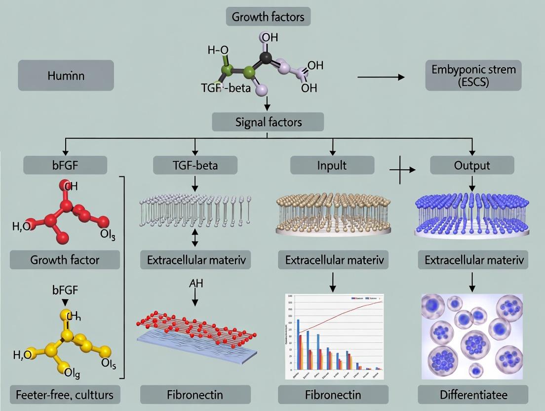
The Central Role of bFGF and TGF-β/Activin/Nodal Signaling
Two primary signaling pathways, governed by specific growth factors, are indispensable for sustaining hESC self-renewal in feeder-free conditions.
- Basic Fibroblast Growth Factor (bFGF/FGF-2): bFGF is a critical mitogen and survival factor. It activates the MAPK/ERK and PI3K/Akt pathways, promoting proliferation and preventing apoptosis. In feeder-free systems, significantly higher concentrations (e.g., 100 ng/mL) are required compared to feeder-supported cultures, as feeders themselves produce bFGF. It works synergistically with TGF-β signaling to stabilize the pluripotent state.
- Transforming Growth Factor-Beta (TGF-β) Superfamily (TGF-β1/Activin/Nodal): This pathway, primarily through SMAD2/3 phosphorylation, directly upregulates the expression of core pluripotency transcription factors like NANOG and OCT4. In defined media, recombinant TGF-β1 or the related ligand Activin A is used to activate this pathway, which suppresses differentiation cues and maintains the undifferentiated phenotype.
Strategic Use of Small Molecules
Small molecules provide enhanced control, stability, and cost-effectiveness over recombinant proteins. They are used to either inhibit differentiation-inducing pathways or to activate/genergate key pluripotency pathways.
- Rho-Associated Kinase (ROCK) Inhibitor (Y-27632): Routinely added during cell passaging (single-cell dissociation) to dramatically improve cell survival and cloning efficiency by inhibiting apoptosis.
- GSK-3β Inhibitors (e.g., CHIR99021): These molecules activate Wnt/β-catenin signaling, which in conjunction with TGF-β signaling, reinforces pluripotency and can enhance cell proliferation.
- MEK/ERK Pathway Inhibitors (e.g., PD0325901): Used in "2i" or "3i" culture regimens, these inhibitors reduce spontaneous differentiation by modulating MAPK signaling, often combined with other inhibitors for ground-state pluripotency.
Quantitative Analysis of Media Components
The following table summarizes the typical concentration ranges and functions of key components in defined feeder-free media formulations.
Table 1: Key Components in Defined Feeder-Free hESC Media
| Component | Type | Typical Concentration | Primary Function in hESC Culture |
|---|---|---|---|
| bFGF (FGF-2) | Recombinant Protein | 80 – 120 ng/mL | Activates MAPK/PI3K pathways; promotes proliferation & survival. |
| TGF-β1 / Activin A | Recombinant Protein | 1 – 2 ng/mL / 10 – 20 ng/mL | Activates SMAD2/3; upregulates NANOG/OCT4; maintains pluripotency. |
| Insulin | Recombinant Protein | ~20 µg/mL | Activates PI3K/Akt pathway; promotes metabolic activity and growth. |
| Y-27632 (ROCKi) | Small Molecule | 5 – 10 µM | Inhibits ROCK; reduces dissociation-induced apoptosis (used during passaging). |
| CHIR99021 | Small Molecule | 3 – 6 µM | Inhibits GSK-3β; activates Wnt signaling; supports pluripotency. |
| PD0325901 | Small Molecule | 0.5 – 1 µM | Inhibits MEK; suppresses differentiation; part of "2i" protocols. |
| Ascorbic Acid | Small Molecule | 50 – 100 µg/mL | Antioxidant; improves cell viability and collagen synthesis. |
| Lithium Chloride | Small Molecule | 0.5 – 1 mM | GSK-3β inhibitor; synergizes with CHIR99021; promotes survival. |
Experimental Protocols
Protocol 1: Routine Maintenance of hESCs in Defined Medium
Objective: To passage and maintain pluripotent hESCs in a feeder-free, defined culture system. Materials:
- hPSCs (e.g., H1, H9 lines)
- Defined culture medium (e.g., mTeSR Plus, StemFlex, or E8 medium)
- Vitronectin-coated 6-well plates
- DPBS, without Ca2+/Mg2+
- Gentle Cell Dissociation Reagent (GCDR) or EDTA (0.5 mM)
- ROCK inhibitor (Y-27632)
- Centrifuge
- Incubator at 37°C, 5% CO2
Procedure:
- Pre-coating: Thaw vitronectin solution and coat plates per manufacturer's instructions.
- Feeding: Prior to passaging, replace spent medium with fresh, pre-warmed defined medium daily.
- Passaging (at ~80% confluency): a. Aspirate medium and wash cells once with DPBS. b. Add 1 mL of GCDR or 0.5 mM EDTA per well of a 6-well plate. Incubate at 37°C for 5-8 minutes. c. Monitor under microscope until cell borders brighten and cells begin to detach. d. Aspirate dissociation reagent. Gently add 2 mL of defined medium supplemented with 10 µM Y-27632. e. Pipette gently to create a single-cell suspension or small clusters. f. Transfer cell suspension to a conical tube, centrifuge at 300 x g for 5 minutes. g. Aspirate supernatant and resuspend pellet in fresh defined medium with Y-27632. h. Plate cells onto the pre-coated plate at the desired split ratio (typically 1:10 to 1:20).
- Post-passage: 24 hours after plating, replace medium with fresh defined medium without Y-27632. Continue daily feeding.
Protocol 2: Evaluating Pluripotency via Immunocytochemistry
Objective: To confirm the maintenance of pluripotency in hESCs cultured under defined conditions. Materials:
- hESC cultures on coated plates
- 4% Paraformaldehyde (PFA)
- Permeabilization buffer (0.1% Triton X-100 in PBS)
- Blocking buffer (3% BSA in PBS)
- Primary antibodies: OCT4, NANOG, SOX2
- Fluorescent-conjugated secondary antibodies
- DAPI nuclear stain
- Fluorescence microscope
Procedure:
- Fixation: Aspirate medium, wash with PBS, and fix cells with 4% PFA for 15 minutes at room temperature.
- Permeabilization & Blocking: Wash with PBS. Permeabilize cells for 10 minutes. Wash, then add blocking buffer for 30-60 minutes.
- Primary Antibody Incubation: Dilute primary antibodies in blocking buffer. Incubate cells overnight at 4°C.
- Secondary Antibody Incubation: Wash 3x with PBS. Add fluorophore-conjugated secondary antibodies (diluted in blocking buffer). Incubate for 1 hour at room temperature in the dark.
- Nuclear Stain & Imaging: Wash 3x with PBS. Add DAPI solution for 5 minutes. Wash and image using a fluorescence microscope. Expected Outcome: >95% of cells should show strong nuclear expression of OCT4, NANOG, and SOX2.
Protocol 3: Testing the Effect of bFGF/TGF-β Concentration on Proliferation
Objective: To quantitatively assess hESC proliferation in response to varying concentrations of key growth factors. Materials:
- Defined basal medium (without bFGF/TGF-β)
- Recombinant human bFGF stock
- Recombinant human TGF-β1 stock
- 96-well plates, vitronectin-coated
- Cell Counting Kit-8 (CCK-8)
Procedure:
- Prepare Media Conditions: Prepare 5 different media formulations:
- Condition A: Basal only (negative control).
- Condition B: Basal + 100 ng/mL bFGF + 2 ng/mL TGF-β1 (positive control).
- Condition C: Basal + 50 ng/mL bFGF + 2 ng/mL TGF-β1.
- Condition D: Basal + 100 ng/mL bFGF + 0.5 ng/mL TGF-β1.
- Condition E: Basal + 50 ng/mL bFGF + 0.5 ng/mL TGF-β1.
- Cell Seeding: Seed a single-cell suspension of hESCs at 5,000 cells/well in 100 µL of each test medium containing Y-27632.
- Culture: After 24h, replace medium with the same test medium but without Y-27632. Culture for 72 hours with daily medium changes.
- Proliferation Assay: Add 10 µL of CCK-8 reagent to each well. Incubate for 2-4 hours at 37°C. Measure absorbance at 450 nm using a plate reader.
- Analysis: Plot absorbance (proxy for cell number) against media condition. Compare proliferation rates.
The Scientist's Toolkit
Table 2: Essential Research Reagents for Defined Feeder-Free hESC Culture
| Item | Function & Rationale |
|---|---|
| Vitronectin (VTN-N) or Recombinant Laminin-521 | Defined, xeno-free extracellular matrix (ECM) that replaces Matrigel. Provides essential adhesion signals via integrins (αVβ5, α6β1). |
| Chemically Defined Basal Medium (e.g., DMEM/F12) | Nutrient foundation. Must be free of serum or undefined components to ensure reproducibility. |
| Albumin, Human Recombinant | Carrier protein that stabilizes growth factors, buffers media, and provides essential lipids and trace elements. |
| Recombinant Human bFGF (FGF-2) | The primary mitogen and survival factor. High purity and activity are critical for consistent results. |
| Recombinant Human TGF-β1 or Activin A | Activates SMAD2/3 pathway to sustain core pluripotency transcription factor network. |
| ROCK Inhibitor (Y-27632 dihydrochloride) | Crucial for clonal survival after enzymatic dissociation. Reduces anoikis, enabling efficient single-cell passaging. |
| GSK-3β Inhibitor (CHIR99021) | Small molecule used to activate canonical Wnt signaling, supporting self-renewal and proliferation in specific protocols. |
| Gentle Cell Dissociation Reagent (GCDR) | Enzyme-free, EDTA-based chelating agent for gentle detachment, preserving surface proteins and cell viability better than trypsin. |
Visualizations
Title: Signaling Pathways Maintaining hESC Pluripotency
Title: Routine hESC Maintenance Protocol Flowchart
The establishment of human embryonic stem cell (hESC) culture has been a journey defined by the quest for defined, reproducible, and xeno-free conditions. The initial reliance on mouse embryonic fibroblast (mEF) feeders provided a stable, albeit complex and ill-defined, microenvironment for hESC self-renewal. This progression to first-generation feeder-free formulations marked a pivotal shift, enabling higher experimental consistency and paving the way for translational applications in drug development and regenerative medicine.
Comparative Analysis of Culture Systems
Table 1: Key Characteristics of mEF vs. First-Generation Feeder-Free Systems
| Feature | mEF Feeder-Based Culture (c. 1998-2001) | First-Generation Feeder-Free Formulations (c. 2001-2006) |
|---|---|---|
| Substrate | Gelatin-coated plates with live, mitotically inactivated mEFs. | Defined extracellular matrix: Matrigel or laminin-511. |
| Medium Formulation | Serum-containing or Serum Replacement (SR) supplemented with basic FGF (bFGF). | Defined media: e.g., mTeSR1, StemPro, X-VIVO 10. bFGF concentration increased (40-100 ng/mL vs. 4-8 ng/mL on feeders). |
| Key Signaling Pathways | TGF-β/Activin/Nodal (via mEF-secreted factors) and bFGF. | Exogenous TGF-β/Activin/Nodal supplementation (in media) and high bFGF. |
| Typical Doubling Time | ~36-48 hours | ~30-40 hours |
| Advantages | Supported initial derivations; robust maintenance of pluripotency. | Defined, scalable, easier for downstream assays; reduced pathogen risk. |
| Disadvantages | Xenogenic contaminants, variable mEF batches, labor-intensive, obscures secreted factor analysis. | High cost; matrix variability (Matrigel); required adaptation of cell lines; residual animal components. |
Table 2: Quantitative Comparison of Common First-Generation Media Formulations
| Media (Commercial) | Key Defined Components (Beyond Base) | Typical bFGF (ng/mL) | Pluripotency Marker Expression (Typical % Oct4+) | Recommended Matrix |
|---|---|---|---|---|
| mTeSR1 | TGFβ1, LiCl, GABA, Pipecolic Acid | 100 | >95% | Matrigel, Laminin |
| StemPro hESC SFM | FGF2, TGFβ1, NEAA, β-mercaptoethanol | 40 | >90% | Matrigel, Vitronectin |
| X-VIVO 10 | FGF2, TGFβ1 (in early protocols) | 80 | >85% | Matrigel |
Detailed Experimental Protocols
Protocol 1: Transitioning hESCs from mEF Feeders to Feeder-Free Matrigel
Objective: Adapt and maintain hESC lines on a feeder-free Matrigel substrate using a defined medium.
Materials (Research Reagent Solutions):
- hESCs maintained on mEF feeders.
- mTeSR1 Medium (StemCell Technologies, #85850): Defined, feeder-free culture medium.
- Growth Factor Reduced Matrigel (Corning, #354230): Basement membrane matrix providing adhesion and signaling cues.
- DMEM/F-12 (Gibco, #11330): Medium for diluting Matrigel.
- Y-27632 ROCK inhibitor (Tocris, #1254): Enhances single-cell survival.
- Dispase (StemCell Technologies, #07913) or Gentle Cell Dissociation Reagent.
- hESC-Qualified PBS (without Ca2+/Mg2+).
Method:
- Matrigel Coating:
- Thaw Matrigel on ice overnight at 4°C. Pre-chill pipettes and tubes.
- Dilute Matrigel 1:100 in cold DMEM/F-12. Aliquot and store at -20°C.
- Coat culture plates with diluted Matrigel (e.g., 0.5 mL/well of 6-well plate). Incubate at 37°C for at least 1 hour.
- Passaging hESCs from mEFs:
- Aspirate medium from mEF culture. Wash with PBS.
- Add Dispase (1 mg/mL in DMEM/F-12) or Gentle Cell Dissociation Reagent. Incubate until colony edges begin to detach (5-10 min).
- Aspirate enzyme. Gently wash with PBS.
- Add fresh mTeSR1 medium. Gently scrape and collect cell clusters.
- Transfer clusters to a conical tube and allow to settle by gravity (5-10 min). Aspirate supernatant.
- Seeding on Matrigel:
- Resuspend cell clusters in mTeSR1 supplemented with 10 µM Y-27632.
- Aspirate Matrigel coating solution from plate. Do not let wells dry.
- Seed cell clusters onto the Matrigel-coated plate. Distribute evenly.
- Place plate in a 37°C, 5% CO2 incubator. Change medium daily with fresh mTeSR1 (without Y-27632) after 24 hours.
- Maintenance:
- Passage cells every 5-7 days using Gentle Cell Dissociation Reagent or ReLeSR for clump passaging when colonies are large and centers become dense.
Protocol 2: Characterizing Pluripotency in Feeder-Free Cultures
Objective: Assess the undifferentiated state of hESCs maintained under feeder-free conditions via immunocytochemistry.
Materials:
- Feeder-free hESC cultures on Matrigel-coated coverslips or plate.
- 4% Paraformaldehyde (PFA): Fixative.
- Permeabilization Buffer (0.5% Triton X-100 in PBS).
- Blocking Buffer (5% normal goat serum/1% BSA in PBS).
- Primary Antibodies: Anti-OCT4 (Abcam, #ab19857), Anti-SOX2 (R&D Systems, #MAB2018), Anti-NANOG (ReproCELL, #RCAB002P).
- Fluorescently-labeled Secondary Antibodies.
- DAPI nuclear stain.
- Mounting Medium.
Method:
- Fixation: Aspirate medium. Wash cells with PBS. Fix with 4% PFA for 15 min at RT. Wash 3x with PBS.
- Permeabilization & Blocking: Permeabilize with 0.5% Triton X-100 for 15 min. Wash. Incubate with Blocking Buffer for 1 hour.
- Primary Antibody Incubation: Incubate with primary antibodies (diluted in Blocking Buffer) overnight at 4°C.
- Secondary Antibody Incubation: Wash 3x with PBS. Incubate with appropriate fluorescent secondary antibodies (protected from light) for 1 hour at RT.
- Nuclear Stain & Mounting: Wash 3x. Incubate with DAPI (1 µg/mL) for 5 min. Wash. Mount coverslip onto slide.
- Analysis: Image using a fluorescence microscope. >85% nuclear co-localization of OCT4, SOX2, and NANOG indicates robust pluripotency.
Signaling Pathways and Workflow Visualizations
The Scientist's Toolkit: Key Research Reagent Solutions
| Reagent / Material | Function / Role in Feeder-Free Culture | Example Product/Catalog # |
|---|---|---|
| Defined Culture Medium | Provides essential nutrients, salts, and specific growth factors (TGFβ, bFGF) to maintain pluripotency in absence of feeders. | mTeSR1 (StemCell Tech, #85850), StemPro hESC SFM (Gibco, #A1000701) |
| Synthetic ROCK Inhibitor | Selectively inhibits Rho-associated kinase (ROCK), dramatically improving survival of dissociated hESCs during passaging. | Y-27632 (Tocris, #1254) |
| Recombinant Laminin-521 | Defined, xeno-free human recombinant substrate promoting integrin-mediated adhesion and signaling for hESCs. | Laminin-521 (BioLamina, #LN521) |
| Gentle Cell Dissociation Reagent | Enzyme-free, gentle buffer for passaging hESCs as small clumps, minimizing differentiation and maintaining cell health. | Gentle Cell Dissociation Reagent (StemCell Tech, #07174) |
| Growth Factor-Reduced Matrigel | Complex basement membrane extract from Engelbreth-Holm-Swarm tumor; provides adhesion proteins (laminin, collagen) but is ill-defined. | Corning Matrigel GFR (Corning, #354230) |
| Basic Fibroblast Growth Factor (bFGF) | Critical mitogen and signaling molecule; concentration is significantly elevated in feeder-free systems to sustain self-renewal pathways. | Recombinant Human FGF-basic (PeproTech, #100-18B) |
| Pluripotency Marker Antibodies | Essential tools for validating the undifferentiated state of hESCs via immunostaining or flow cytometry. | Anti-OCT4 (Abcam, #ab19857), Anti-TRA-1-60 (StemCell Tech, #60064) |
Step-by-Step Protocols: Establishing and Maintaining hESCs in Feeder-Free Conditions
Within the context of developing robust, feeder-free culture conditions for human embryonic stem cells (hESCs), the use of defined extracellular matrix (ECM) coatings is a critical foundational step. This protocol details the preparation of culture surfaces coated with defined ECM proteins, such as recombinant laminin isoforms, vitronectin, and defined synthetic polymers, which replace undefined substrates like mouse embryonic fibroblasts (MEFs) or Matrigel. This standardization is essential for reproducibility, xeno-free conditions, and precise analysis of signaling pathways governing pluripotency and differentiation in downstream thesis experiments.
Research Reagent Solutions
| Reagent/Material | Function in Coating Protocol |
|---|---|
| Recombinant Human Laminin-521 (LN-521) | A defined, xeno-free isoform that interacts with α6β1 integrins on hESCs, promoting robust adhesion and pluripotency via integrin-FAK signaling. |
| Recombinant Human Vitronectin (VTN-N) | A cost-effective, defined alternative that supports hESC attachment and growth through αVβ5 integrin binding. |
| Synthetic Polymer (e.g., poly-[acrylamide-co-propargyl acrylamide]) | A fully defined, synthetic substrate offering tunable mechanical and chemical properties for studying mechanotransduction. |
| DPBS, Ca²⁺/Mg²⁺-free | Used for diluting and handling ECM proteins without causing premature polymerization or degradation. |
| Tissue Culture-Grade Plate | Typically 6-well, 12-well, or 24-well plates, treated for optimal cell adhesion. |
| Albumin, Human Recombinant | Used as a blocking agent to passivate any uncoated plastic surfaces after ECM coating. |
Current research in feeder-free hESC culture identifies optimal coating concentrations and resulting cell behavior metrics. The following table summarizes key data for common defined matrices.
Table 1: Comparative Performance of Defined ECM Coatings for Feeder-Free hESC Culture
| ECM Coating | Recommended Coating Concentration (µg/cm²) | Working Diluent | Incubation Time/Temp | Key Supported hESC Features (Pluripotency Marker % >95%) | Primary Cell-Binding Integrin |
|---|---|---|---|---|---|
| Laminin-521 | 0.5 - 1.0 | DPBS (-/-), 4°C O/N | 2h, 37°C or O/N, 4°C | Colony growth, Genomic stability, Clonal survival | α6β1, αVβ5 |
| Vitronectin (Truncated) | 0.25 - 0.5 | DPBS (-/-) | 1h, RT | Single-cell passaging efficacy, Cost-effective scale-up | αVβ5 |
| Laminin-511 E8 Fragment | 0.25 - 0.5 | DPBS (-/-) | 2h, 37°C | High cloning efficiency, Defined conditions | α6β1 |
| Synthetic Peptide Polymer | As per mfr. (e.g., 2-4% w/v) | Water or buffer | 1h, RT, then UV crosslink | Mechanobiology studies, Fully defined chemistry | Varies by ligand |
Detailed Experimental Protocols
Protocol 1: Coating with Recombinant Human Laminin-521 (LN-521)
Objective: To create a defined, xeno-free substrate for long-term maintenance of undifferentiated hESCs. Materials:
- Recombinant Human Laminin-521 (e.g., Biolamina, #LN521)
- DPBS, without calcium and magnesium
- Tissue culture plates (e.g., 6-well)
- Sterile pipettes and tips
- Refrigerator (4°C) or incubator (37°C)
Procedure:
- Dilution: Thaw LN-521 stock solution (typically 1 mg/mL) slowly on ice. Dilute to a final working concentration of 0.5 µg/cm² in cold DPBS (-/-). For a standard 6-well plate (well growth area ~9.5 cm²), use 1 mL of a 4.75 µg/mL LN-521 solution per well.
- Coating: Immediately add the calculated volume of diluted LN-521 solution to each well of the culture plate. Ensure the liquid covers the entire growth surface by gentle rocking.
- Incubation: Seal the plate with parafilm to prevent evaporation. Incubate for a minimum of 2 hours at 37°C or, preferably, overnight at 4°C for more even coating.
- Preparation for Use: Just before plating cells, aspirate the LN-521 solution. Do not let the coated surface dry out. The coated wells can be used immediately or stored at 4°C for up to 1 week sealed with parafilm.
- Optional Blocking: Rinse the coated well once with DPBS (-/-). For some applications, a 30-minute incubation with a 0.1-1% solution of recombinant human albumin at room temperature can be used to block non-specific binding sites. Aspirate and rinse before use.
Protocol 2: Coating with Recombinant Human Vitronectin (VTN-N)
Objective: To provide a cost-effective, defined substrate suitable for single-cell passaging of hESCs. Materials:
- Recombinant Human Vitronectin (Truncated) (e.g., Thermo Fisher, #A14700)
- DPBS, without calcium and magnesium
- Tissue culture plates
Procedure:
- Dilution: Prepare a vitronectin working solution at 0.5 µg/mL in DPBS (-/-). For a 6-well plate, add 1 mL per well.
- Coating: Add the solution to each well, ensuring complete coverage.
- Incubation: Incubate the plate for 1 hour at room temperature (20-25°C) in a sterile environment.
- Preparation for Use: Aspirate the coating solution. Do not rinse the wells. Use the coated plates immediately. Do not allow to dry.
Signaling Pathways in hESC-ECM Interaction
The adhesion of hESCs to defined ECM coatings initiates critical intracellular signaling cascades that sustain self-renewal.
Diagram 1: Key survival and pluripotency signals from ECM-integrin binding.
Experimental Workflow for Coating Validation
A logical workflow for preparing and validating coated plates within a thesis project.
Diagram 2: Workflow for defined ECM plate coating and validation.
Within feeder-free culture systems for human embryonic stem cells (hESCs), the choice of dissociation method is critical for maintaining pluripotency, genomic stability, and high cell viability. Standard passaging techniques primarily involve enzymatic dissociation (using proteases like Accutase or Trypsin) or non-enzymatic, EDTA-based dissociation. This application note, framed within a thesis on optimizing feeder-free conditions, provides detailed protocols and a comparative analysis to guide researchers and drug development professionals in selecting the appropriate method for their experimental objectives.
Key Research Reagent Solutions
| Reagent/Material | Function in Feeder-Free hESC Culture |
|---|---|
| mTeSR Plus or Essential 8 Medium | Defined, feeder-free culture medium providing essential nutrients and growth factors to maintain pluripotency. |
| Recombinant Human Laminin-521 | Recombinant basement membrane matrix that replaces feeder cells, providing crucial adhesion and signaling for hESC attachment and survival. |
| Accutase | Mild enzymatic cell dissociation solution containing proteolytic and collagenolytic enzymes. Ideal for generating single-cell suspensions for consistent seeding and cryopreservation. |
| Trypsin-EDTA (0.05%) | Proteolytic enzyme for rapid single-cell dissociation. Requires precise timing and neutralization with serum or inhibitors to prevent over-digestion. |
| EDTA (0.5 mM) | Calcium and magnesium chelator. Weakens cell-cell and cell-matrix adhesions, facilitating passaging as small clumps with minimal perturbation to surface receptors. |
| Y-27632 (ROCK inhibitor) | Small molecule inhibitor of Rho-associated kinase. Dramatically improves survival of single hESCs post-dissociation by inhibiting apoptosis. |
| DMEM/F-12 | Basal medium used for washing cells and diluting dissociation reagents. |
Comparative Data: Enzymatic vs. EDTA-Based Dissociation
Table 1: Quantitative Comparison of Key Passaging Outcomes
| Parameter | Enzymatic Dissociation (Accutase) | EDTA-Based Dissociation |
|---|---|---|
| Dissociation Outcome | Single-cell suspension | Small clumps (3-20 cells) |
| Typical Seeding Density | 10,000 - 50,000 cells/cm² | 5,000 - 15,000 cell clumps/cm² |
| Post-Passage Viability | 85-95% (with ROCKi) | 90-98% |
| Attachment Time | 6-12 hours | 2-6 hours |
| Population Doubling Time | ~24 hours | ~28-36 hours |
| Spontaneous Differentiation Rate | Low-Moderate (requires careful control) | Very Low (clump method preserves niche) |
| Genomic Instability Risk | Slightly elevated with prolonged use | Minimal |
| Ideal Application | Scalable expansion, clonal selection, CRISPR editing | Routine maintenance, banking, differentiation initiation |
Detailed Experimental Protocols
Protocol 1: Enzymatic Passaging with Accutase for Single-Cell Seeding
Objective: To generate a single-cell suspension for uniform seeding and quantitative expansion in feeder-free conditions.
Materials:
- hESCs cultured on Laminin-521 in mTeSR Plus.
- DMEM/F-12, Accutase, mTeSR Plus supplemented with 10µM Y-27632 (ROCKi).
- Centrifuge, 37°C incubator.
Method:
- Pre-treatment: Add 10µM Y-27632 to culture medium 1 hour prior to passaging.
- Wash: Aspirate medium and wash cells once with 2 mL DMEM/F-12 per well of a 6-well plate.
- Dissociation: Add 1 mL of pre-warmed Accutase. Incubate at 37°C for 5-7 minutes.
- Neutralization: Gently pipette the Accutase solution over the cell layer until a single-cell suspension is achieved. Transfer to a conical tube containing 2 mL of DMEM/F-12.
- Centrifugation: Spin at 300 x g for 5 minutes. Aspirate supernatant.
- Resuspension & Counting: Resuspend pellet in mTeSR Plus + Y-27632. Count using an automated cell counter or hemocytometer.
- Seeding: Seed cells at desired density (e.g., 20,000 viable cells/cm²) onto fresh Laminin-521 coated plates. Ensure even distribution.
- Medium Change: After 24 hours, replace medium with fresh mTeSR Plus without Y-27632. Change medium daily thereafter.
Protocol 2: EDTA-Based Passaging for Clump Dissociation
Objective: To passage hESCs as small, uniform clumps to minimize dissociation-induced stress and preserve pluripotency.
Materials:
- hESCs cultured on Laminin-521 in mTeSR Plus.
- DPBS (without Ca²⁺/Mg²⁺), 0.5 mM EDTA solution, mTeSR Plus.
Method:
- Wash: Aspirate culture medium and wash cells gently with 2 mL DPBS.
- EDTA Incubation: Add 1 mL of pre-warmed 0.5 mM EDTA solution. Incubate at 37°C for 5-8 minutes. Observe edges of colonies under a microscope; cells should begin to detach as sheets or curl at the borders.
- Clump Generation: Carefully aspirate EDTA. Gently add 2 mL of mTeSR Plus to the well. Using a sterile pipette tip or cell scraper, dislodge colonies by directing medium stream. Triturate 3-5 times with a 1 mL pipette to break into small clumps.
- Seeding: Transfer the cell suspension (clumps in medium) directly to a new Laminin-521 coated plate. No centrifugation is required. Distribute clumps evenly.
- Settling: Allow the plate to sit undisturbed in the incubator for 20-30 minutes to facilitate clump attachment.
- Medium Top-Up: Gently add an additional 1-2 mL of fresh mTeSR Plus to the well. Change medium daily.
Signaling Pathways and Experimental Workflow
Title: hESC Passaging Decision Workflow for Feeder-Free Culture
Title: Dissociation Method Impact on hESC Survival Signaling
Application Notes
Within feeder-free culture systems for human embryonic stem cells (hESCs), daily maintenance is critical for maintaining pluripotency, genomic stability, and experimental reproducibility. The absence of feeder cells places the entire burden of support on the defined extracellular matrix and the precisely formulated medium, making consistent daily protocols non-negotiable. Key objectives include maintaining optimal nutrient and growth factor concentrations, preventing spontaneous differentiation triggered by over-confluence or metabolic stress, and early detection of culture anomalies. Successful daily management directly impacts downstream applications in disease modeling, drug screening, and developmental biology.
Table 1: Key Parameters for Daily hESC Maintenance in Feeder-Free Culture
| Parameter | Optimal Range | Monitoring Frequency | Consequence of Deviation |
|---|---|---|---|
| Cell Density | 50-80% confluence | Daily (pre-media change) | <50%: Reduced paracrine signaling; >80%: Increased differentiation risk, nutrient depletion. |
| Media Change Interval | Every 24 hours | Fixed daily schedule | Extended intervals: Nutrient depletion (e.g., Glucose <17.5 mM), acidification (pH <7.2), growth factor degradation. |
| Colony Morphology | Compact, well-defined borders, high nucleus-to-cytoplasm ratio | Daily microscopic inspection | Irregular borders, flattened cells: Onset of differentiation. |
| Media Color (phenol red) | Peach/Orange (pH ~7.4) | Visual check at change | Yellow (acidic): Over-confluence or contamination. Purple (basic): Rare, CO2 imbalance. |
| Doubling Time | ~20-24 hours | Assess every 2-3 passages | Prolongation: Suboptimal conditions or senescence. |
Table 2: Common Differentiation Markers and Associated Morphological Cues
| Morphological Cue | Potential Lineage Bias | Key Marker to Assess |
|---|---|---|
| Flattened, spread-out colony edge | Primitive Endoderm | GATA6, SOX17 |
| Elongated, spindle-shaped cells | Mesoderm | BRA (T), HAND1 |
| Dark, clustered, multi-layered nodules | Ectoderm (Neural) | PAX6, SOX1 |
| Loosened, non-adherent cells in center | Trophoblast | CDX2, hCG |
Detailed Experimental Protocols
Protocol 1: Daily Media Change and Morphological Assessment
Objective: To replenish nutrients and signaling factors while systematically assessing colony health and density.
Materials:
- Pre-warmed, complete feeder-free hESC medium (e.g., mTeSR Plus, E8).
- Sterile DPBS (-/- Ca2+/Mg2+).
- 37°C, 5% CO2 incubator.
- Inverted phase-contrast microscope.
Procedure:
- Pre-Change Inspection: Using the microscope, observe cultures at 4x and 10x magnification. Systematically scan the entire vessel.
- Document colony density, estimating overall confluence.
- Assess colony morphology: note sharpness of borders, cell packing, and any regions with atypical, differentiated morphology.
- Look for spontaneous differentiation as per Table 2.
- Check for any signs of contamination (cloudy media, unexplained particles).
- Media Aspiration: Carefully aspirate the spent medium from the side of the well without touching the coated surface.
- Rinse (Optional): For cultures approaching high density or showing slight acidity, gently add 1 mL of pre-warmed DPBS per well of a 6-well plate to remove metabolic waste. Aspirate immediately.
- Media Addition: Gently add the required volume of pre-warmed complete medium down the side of the well (e.g., 2 mL/well for a 6-well plate). Avoid disturbing colonies.
- Post-Change Documentation: Briefly note observations (confluence estimate, morphology score, any concerns) in the laboratory logbook.
- Return to Incubator: Place the culture vessel gently back into the 37°C, 5% CO2 incubator.
Protocol 2: Management Based on Density Assessment
Objective: To decide and execute the appropriate downstream process (passage, continue culture, or quality control) based on daily confluence evaluation.
Procedure:
- Following the morphological assessment in Protocol 1, determine the approximate confluence.
- Decision Tree:
- If confluence is 50-80%: Return culture to incubator. Continue daily media changes.
- If confluence is >85%: Proceed to passage within 24 hours. Schedule passaging for the same or next day. For critical experiments, consider a emergency same-day passage to prevent differentiation.
- If confluence is <40% for proliferating lines: Ensure medium quality and coating efficacy. Consider increasing the passaging ratio to achieve higher post-passage density for better survival.
- Action for Differentiated Regions:
- If differentiation is localized and minimal (<5% of culture area), marks can be made on the underside of the plate, and the differentiated regions can be manually removed via aspiration with a pipette tip or scraping under a microscope before passaging.
- If differentiation is widespread (>10-15%), the culture should be terminated or subjected to fluorescence-activated cell sorting (FACS) for pluripotency marker-positive cells to reclaim the line.
Protocol 3: Routine Quality Control Monitoring
Objective: To periodically verify pluripotency and genomic integrity as part of maintenance records.
Schedule:
- Pluripotency Marker Check (Immunofluorescence or Flow Cytetry): Every 5-10 passages. Assess OCT4, NANOG, SSEA-4, TRA-1-60 expression. Expect >95% positivity in a healthy culture.
- Karyotype Analysis (G-banding or SNP array): Every 15-20 passages or before commencing a major new project.
Diagrams
Title: Daily hESC Maintenance Decision Workflow
Title: Core Signaling in Feeder-Free hESC Culture
The Scientist's Toolkit: Research Reagent Solutions
Table 3: Essential Materials for Feeder-Free hESC Daily Maintenance
| Item | Function & Rationale |
|---|---|
| Defined, Xeno-Free Culture Medium (e.g., mTeSR Plus, Essential 8) | Provides a precisely formulated, consistent blend of basal nutrients, vitamins, and recombinant human proteins (bFGF, TGF-β1) essential for pluripotency without animal-derived components. |
| Synthetic Extracellular Matrix (e.g., Geltrex, Vitronectin XF, Synthemax) | Defined, recombinant coating substrate that replaces mouse embryonic fibroblasts (MEFs). Provides adhesion ligands (e.g., laminin, vitronectin) for integrin-mediated cell attachment and survival. |
| ROCK Inhibitor (Y-27632) | Small molecule added briefly after passaging. Inhibits Rho-associated kinase, dramatically reducing anoikis (cell death due to detachment) and improving single-cell survival. |
| Gentle Cell Dissociation Reagent (e.g., ReLeSR, Accutase) | Enzyme-free or mild protease solutions for passaging. Allow clump-wise or single-cell detachment while preserving surface receptors and viability better than traditional trypsin. |
| Pluripotency Marker Antibody Panel | Includes antibodies against intracellular (OCT4, NANOG) and surface (SSEA-4, TRA-1-60) markers for routine quality control via immunofluorescence or flow cytometry. |
| Metabolite-Glo or Similar Assay | Luciferase-based kit to quantitatively measure glucose, lactate, and glutamine levels in spent media, providing a direct readout of metabolic activity and media exhaustion. |
Protocol for Cryopreservation and Recovery in Feeder-Free Systems
Within the broader thesis on optimizing feeder-free culture for human embryonic stem cells (hESCs), the development of robust cryopreservation and recovery protocols is critical. This ensures genetic stability, phenotypic fidelity, and experimental reproducibility. This application note details standardized protocols for the cryopreservation and revival of hESCs maintained under defined, feeder-free conditions.
Successful feeder-free cryopreservation hinges on controlled-rate freezing and the use of specialized recovery media to minimize spontaneous differentiation and maximize cell survival. Key quantitative benchmarks from recent literature are summarized below.
Table 1: Comparative Performance of Cryopreservation Solutions in Feeder-Free hESC Culture
| Cryopreservation Solution | Post-Thaw Viability (%) (Mean ± SD) | Recovery Time to 80% Confluence (Days) | Spontaneous Differentiation Rate (%) | Key Reference |
|---|---|---|---|---|
| Commercial Serum-Free Freeze Medium (e.g., mFreSR) | 85 ± 5 | 4-5 | <10% | Wakeman et al., 2023 |
| 10% DMSO in Defined Culture Medium | 70 ± 8 | 6-7 | 15-25% | Chen & Li, 2022 |
| 5% DMSO + 5% Ethylene Glycol | 78 ± 6 | 5-6 | 10-15% | Gupta et al., 2023 |
| ROCK Inhibitor (Y-27632) Supplemented Medium | 92 ± 4* | 3-4 | <5% | Park et al., 2024 |
*Viability measured 24 hours post-plating with ROCK inhibitor in recovery medium.
Detailed Experimental Protocols
Protocol: Cryopreservation of Feeder-Free hESCs
Objective: To preserve hESC colonies in a viable state with high recovery potential and maintained pluripotency.
Materials: See "The Scientist's Toolkit" section.
Method:
- Pre-Freeze Preparation: Ensure hESCs are in log-phase growth, with colonies displaying typical compact morphology and minimal differentiation. Pre-warm necessary reagents.
- Harvesting Cells: Aspirate culture medium and gently wash with 2 mL of DPBS without Ca2+/Mg2+.
- Cell Dissociation: For clump freezing, add 1 mL of Gentle Cell Dissociation Reagent (or 0.5 mM EDTA in DPBS). Incubate at 37°C for 5-7 minutes until colony edges begin to detect. Gently rinse cells off using 2 mL of defined culture medium (e.g., mTeSR Plus). Triturate gently 2-3 times with a P1000 pipette to create small clumps (50-100 cells).
- Preparation for Freezing: Centrifuge the cell suspension at 200 x g for 4 minutes. Aspirate supernatant completely.
- Resuspension in Cryomedium: Resuspend the cell pellet in ice-cold, serum-free cryopreservation medium at a density of 1-2 x 10^6 cells/mL or 200-500 clumps/mL. Gently mix.
- Aliquoting: Quickly aliquot 1 mL of cell suspension per cryovial. Place vials on ice.
- Controlled-Rate Freezing: Transfer vials to a pre-cooled (4°C) isopropanol freezing container or a controlled-rate freezer. Place at -80°C for 24 hours.
- Freezing Program (if using controlled-rate freezer): Cool at -1°C/min to -50°C, then rapid cool to -150°C.
- Long-Term Storage: After 24 hours at -80°C, promptly transfer vials to liquid nitrogen vapor phase for long-term storage.
Protocol: Thawing and Recovery of Feeder-Free hESCs
Objective: To efficiently recover viable, pluripotent hESCs from cryopreservation with minimal differentiation.
Method:
- Preparation: Pre-coat culture vessel with appropriate substrate (e.g., Geltrex, Matrigel) for at least 1 hour at 37°C. Thaw defined culture medium supplemented with 10 µM ROCK inhibitor (Y-27632) and warm to 37°C.
- Rapid Thaw: Retrieve cryovial from liquid nitrogen. Thaw quickly in a 37°C water bath with gentle agitation until only a small ice crystal remains (~1.5 minutes).
- Dilution: Immediately transfer the thawed cell suspension to a 15 mL conical tube containing 9 mL of pre-warmed, ROCK inhibitor-supplemented defined medium. Gently mix by pipetting.
- Centrifugation: Centrifuge at 200 x g for 4 minutes to pellet cells/clumps. Aspirate supernatant carefully to remove residual DMSO.
- Resuspension & Seeding: Resuspend the pellet in 2 mL of fresh, ROCK inhibitor-supplemented defined medium. Plate the entire suspension onto the pre-coated culture vessel.
- Initial Recovery (Day 1-2): Place vessel in a 37°C, 5% CO2 incubator. Do not disturb for the first 24 hours. Change to fresh defined medium without ROCK inhibitor after 24 hours.
- Monitoring & Passaging: Monitor daily. Colonies should be visible within 2-3 days. Passage cells when they reach 70-80% confluence, typically 4-6 days post-thaw, using standard feeder-free passaging techniques.
Signaling Pathways & Workflow Visualizations
Diagram 1: Cryopreservation Workflow
Diagram 2: Thaw & Recovery with ROCK Inhibitor
The Scientist's Toolkit
Table 2: Essential Research Reagent Solutions for Feeder-Free hESC Cryopreservation
| Item | Function & Rationale |
|---|---|
| Defined, Serum-Free Culture Medium (e.g., mTeSR Plus, E8) | Provides consistent, xeno-free conditions for maintenance and as a base for cryopreservation solutions. Essential for preserving defined state. |
| Rho-associated Kinase (ROCK) Inhibitor (Y-27632) | Critical additive to recovery medium. Inhibits apoptosis (anoikis) induced by single-cell/clump dissociation and freeze-thaw stress, dramatically improving attachment and survival. |
| Serum-Free Cryopreservation Medium | A defined, protein-rich solution containing 10% DMSO as a cryoprotectant. Minimizes ice crystal formation and osmotic shock. Prevents differentiation associated with serum. |
| Synthetic Substrate (e.g., Geltrex, Vitronectin, Laminin-521) | Provides a defined, reproducible extracellular matrix for cell attachment and survival post-thaw, replacing mouse embryonic feeders. |
| Gentle Cell Dissociation Reagent (e.g., EDTA, enzyme-free solutions) | Allows harvesting of hESCs as small clumps, which improves survival post-thaw compared to single cells, while maintaining colony integrity. |
| Controlled-Rate Freezing Container | Ensures an optimal, reproducible cooling rate of approximately -1°C/minute, which is crucial for cell viability during the freezing process. |
Within the framework of feeder-free culture conditions for human embryonic stem cells (hESCs), the transition from multi-well plates to larger culture vessels is a critical step for generating sufficient cell numbers for downstream applications in drug screening, disease modeling, and potential therapeutic development. This protocol outlines scalable, feeder-free methodologies that maintain pluripotency and genomic stability.
Application Notes: Key Considerations for Scale-Up
Successful scaling hinges on replicating the microenvironment of small-scale culture. Key parameters shift from merely increasing surface area to maintaining critical signaling, metabolite, and gas exchange dynamics.
Quantitative Scaling Parameters
Table 1: Comparative Parameters Across Culture Vessels
| Vessel Type | Typical Surface Area (cm²) | Recommended Seeding Density (cells/cm²) | Working Media Volume (mL) | Key Scaling Challenge |
|---|---|---|---|---|
| 6-well Plate | 9.5 | 15,000 - 20,000 | 2.0 | Baseline control |
| 10 cm Dish | 58 | 15,000 - 20,000 | 10.0 | Edge effect mitigation |
| T-75 Flask | 75 | 15,000 - 20,000 | 15.0 | Gas exchange gradient |
| Cell Factory (1-layer) | 600 | 12,000 - 18,000 | 200.0 | Nutrient/waste distribution |
| Roller Bottle (850 cm²) | 850 | 10,000 - 15,000 | 100-150 | Uniform cell attachment |
Table 2: Media Component Adjustments for Scale-Up
| Component | 6-Well Concentration | Large-Scale (T-75+) Adjustment | Rationale |
|---|---|---|---|
| bFGF | 100 ng/mL | Increase by 10-20% or add twice daily | Mitigates growth factor instability |
| TGF-β/Activin A | As per commercial media | Monitor; may require slight increase | Maintains SMAD signaling |
| ROCKi (Y-27632) | 10 µM (passaging only) | Standard protocol applies | Consistent apoptosis inhibition |
| Glucose | Standard (e.g., ~17.5 mM in DMEM/F12) | Monitor depletion; may require supplementation | Higher metabolic demand |
Detailed Experimental Protocols
Protocol 1: Direct Scale-Up from 6-Well to 10 cm Dish Format
Objective: Expand hESCs while maintaining >85% expression of OCT4 and NANOG. Materials:
- Feeder-free, defined culture medium (e.g., mTeSR Plus, StemFlex).
- Recombinant human bFGF.
- ROCK inhibitor Y-27632.
- EDTA or gentle cell dissociation reagent.
- Matrigel or Recombinant Laminin-521 coated 10 cm dishes.
Methodology:
- Pre-coating: Coat 10 cm dishes with Matrigel (1:100 dilution in DMEM/F12) for 1 hour at room temperature or Laminin-521 (0.5 µg/cm²) for 2 hours at 37°C.
- Cell Dissociation (Donor Plate): Aspirate medium from a confluent 6-well (~9.5 cm²). Wash with 1 mL DPBS. Add 0.5 mL of 0.5 mM EDTA (or 1 mL gentle dissociation reagent) and incubate at 37°C for 5-7 minutes. Monitor until colonies detach at edges.
- Cell Collection: Gently aspirate dissociation reagent. Add 2 mL of fresh, pre-warmed medium containing 10 µM ROCKi. Gently pipette to create a single-cell suspension or small clusters. Transfer to a 15 mL conical tube.
- Seeding Calculation & Execution: Perform a cell count. The target seeding density is 15,000-20,000 viable cells/cm². For a 10 cm dish (~58 cm²), this equals ~0.87 - 1.16 million cells/dish.
- Example: If your cell count yields 3.5 million cells in 2 mL, you can seed three 10 cm dishes with ~1.0 million cells each in a final volume of 10 mL medium + ROCKi.
- Seeding: Add the calculated cell suspension to the pre-coated, pre-equilibrated 10 cm dish. Rock gently to distribute evenly. Place in a 37°C, 5% CO₂ incubator.
- Media Change: After 24 hours, aspirate medium containing ROCKi and replace with 10 mL of fresh, pre-warmed standard medium. Change medium daily thereafter.
- Monitoring: Monitor daily for colony morphology, confluency, and differentiation. Passage at ~80% confluency, typically every 4-5 days.
Protocol 2: Adaptation to Multi-Layer Vessels (Cell Factory)
Objective: Achieve large-scale expansion with consistent cell quality. Key Adaptation: Ensure uniform cell distribution and media exchange.
- Coating: Introduce coating solution (e.g., Laminin-521) into the vessel, lay it flat, and rock systematically to ensure all surfaces are covered. Incubate.
- Seeding: Use a peristaltic pump to transfer a well-mixed, high-density cell suspension (targeting 12,000-18,000 cells/cm²) into the vessel. Gently rock and tilt the vessel repeatedly for 15 minutes post-seeding to ensure even distribution before static incubation.
- Feeding: For feeding, use a closed sterile tubing system connected to a media reservoir and waste bag, or perform careful manual exchange on a perfectly level surface. Agitation (e.g., on a rocking platform) between feeds can improve metabolite homogeneity.
Signaling Pathways in Feeder-Free hESC Culture
The maintenance of pluripotency during scale-up depends on tightly regulated signaling pathways.
Diagram Title: Signaling in Feeder-Free hESC Scale-Up
Workflow for Scaling Culture Vessels
A systematic workflow is essential for successful transition.
Diagram Title: Systematic hESC Scale-Up Workflow
The Scientist's Toolkit: Research Reagent Solutions
Table 3: Essential Materials for Feeder-Free hESC Scale-Up
| Reagent/Material | Function in Scale-Up | Key Consideration for Large Vessels |
|---|---|---|
| Recombinant Laminin-521 (or -511) | Defined extracellular matrix for adhesion. Promotes integrin signaling. | Cost-effective at scale. Requires optimization for uniform coating of complex surfaces. |
| Chemically Defined Media (mTeSR Plus, StemFlex) | Provides consistent, xeno-free nutrients and growth factors. | Pre-formulated stability eases use. Monitor glucose/lactate in high-density cultures. May require custom supplementation. |
| Recombinant Human bFGF | Primary mitogen supporting self-renewal via MAPK pathway. | Highly labile. Requires increased concentration, more frequent feeding, or stabilized analogs in large, static vessels. |
| ROCK Inhibitor (Y-27632) | Inhibits Rho-associated kinase. Reduces apoptosis post-dissociation. | Critical for single-cell seeding efficiency. Use standardized at 10 µM for 24h post-seeding only. |
| Gentle Cell Dissociation Reagent | Enzyme-free, EDTA-based solution. Detaches cells as small clusters. | Minimizes shear stress on large cell populations. Yields more uniform seeding than trypsin. |
| Closed-System Sterile Connectors & Tubing | Enables aseptic media exchange and cell harvesting in multi-layer stacks/roller bottles. | Reduces contamination risk during manual handling of large volumes. |
| Programmable Rocking Platform | Provides gentle, consistent agitation for roller bottles or stacked vessels. | Improves nutrient/waste distribution and gas exchange, mimicking small-scale homogeneity. |
| Portable Metabolite Analyzer (e.g., Nova Bioprofile) | Monitors key media components (glucose, lactate, pH) in real-time. | Essential data for optimizing feed schedules and predicting harvest times in large volumes. |
Solving Common Challenges: A Troubleshooting Guide for Feeder-Free hESC Culture
Within feeder-free culture systems for human embryonic stem cells (hESCs), spontaneous differentiation represents a persistent challenge to maintaining pluripotency. This application note details the primary causes and evidence-based corrective actions to preserve undifferentiated cultures.
Primary Causes of Spontaneous Differentiation
Recent research (2023-2024) identifies key mechanistic drivers under feeder-free conditions.
Table 1: Quantitative Drivers of Spontaneous Differentiation
| Cause Category | Specific Factor | Measured Impact (Typical Range) | Key Reference/Model |
|---|---|---|---|
| Growth Factor Signaling | Reduced bFGF (FGF2) concentration | < 20 ng/mL leads to >40% differentiation in 5 days | Chen et al., 2023; WA09 hESCs |
| Cell Density | Seeding density below critical threshold | < 15,000 cells/cm² increases diff. markers 3-5 fold | Singh et al., Stem Cell Rep., 2024 |
| Matrix Composition | Suboptimal vitronectin/integrin engagement | Laminin-511 > Vitronectin > Matrigel for clonality | Ludwig et al., 2023 Commercial E8 |
| Metabolic Stress | Glucose depletion from medium | [Glucose] < 15 mM triggers Sox2 downregulation | Data from BioProfile analyzer studies |
| Cell-Cell Contact | Disruption of E-cadherin mediated adhesion | Inhibition increases OCT4− cells by 60% in 72h | Mellough et al., Sci. Adv., 2023 |
Corrective Action Protocols
Protocol 3.1: Daily Morphology Assessment & Pre-emptive Correction
Purpose: To identify early signs of differentiation and intervene before colony compromise. Materials: Phase-contrast microscope, pre-warmed complete feeder-free medium (e.g., mTeSR Plus, E8), recombinant FGF2 (155 aa), Y-27632 (ROCKi). Workflow:
- Daily Imaging: Capture 4 representative 10x images per well of a 6-well plate.
- Morphology Scoring: Assess for loss of tight colony borders, increased cytoplasmic/nuclear ratio, and appearance of heterogeneous cell morphology.
- Threshold Action: If >5% of colony area shows altered morphology: a. Immediate Medium Change: Aspirate old medium and replace with fresh, pre-warmed complete medium supplemented with an additional 20 ng/mL FGF2. b. Optional ROCKi Addition: If differentiation appears stress-related (e.g., from low density), add 10 µM Y-27632 for 24 hours. c. Re-assess after 24 hours.
- Targeted Passaging: If specific colonies are differentiated but overall culture is >90% undifferentiated, manually scrape away differentiated regions using a pulled glass pipette or microtool before passaging.
Protocol 3.2: High-Density Re-seeding for Colony Reformation
Purpose: To rescue cultures experiencing widespread, diffuse differentiation by re-establishing optimal cell-cell contact. Method:
- Harvest Cells: Use gentle cell dissociation reagent (e.g., ReLeSR, Gentle Cell Dissociation Reagent) to create a single-cell suspension. Avoid accutase for this rescue, as it increases stress.
- Count and Concentrate: Centrifuge at 200g for 5 minutes. Resuspend in fresh medium supplemented with 10 µM Y-27632.
- High-Density Plating: Seed cells at 50,000 – 70,000 cells/cm² onto fresh, matrix-coated plates (recommend Geltrex or Laminin-521).
- Enhanced Medium: Supplement standard feeder-free medium with 100 ng/mL FGF2 and 10 ng/mL Activin A for the first 48 hours.
- Medium Schedule: Perform 100% medium change daily. On day 3, transition back to standard growth factor concentrations.
- Assessment: On day 5, assess for the reformation of compact, OCT4-positive colonies. Passage normally once colonies reach optimal size.
Protocol 3.3: Fluorescence-Activated Cell Sorting (FACS) for Pluripotency Marker Positive Selection
Purpose: To physically isolate and recover the undifferentiated cell population from a partially differentiated culture. Staining Protocol:
- Prepare a single-cell suspension using EDTA (0.5 mM) or enzyme-free dissociation buffer to preserve surface epitopes.
- Wash cells twice in PBS containing 0.5% BSA (FACS buffer).
- Resuspend ~1x10⁶ cells in 100 µL FACS buffer with a directly conjugated antibody (e.g., anti-SSEA-4-PE, anti-TRA-1-60-FITC) at manufacturer's recommended dilution. Use an IgG isotype control.
- Incubate for 30 minutes at 4°C in the dark.
- Wash twice with 2 mL FACS buffer and resuspend in 500 µL buffer containing 1 µM Y-27632.
- Sorting: Use a 100 µm nozzle, low pressure (20-25 psi). Gate on live, single cells, then select the top 10-20% brightest marker-positive population.
- Collection: Collect sorted cells into medium with 20% KSR and 10 µM Y-27632.
- Post-Sort Culture: Plate sorted cells at high density (as in Protocol 3.2) onto fresh matrix. Resume standard culture after first passage.
Signaling Pathways in Maintenance vs. Differentiation
Experimental Workflow for Identifying Differentiation Cause
The Scientist's Toolkit: Essential Reagents for Feeder-Free Culture Maintenance
Table 2: Key Research Reagent Solutions
| Item | Example Product/Catalog # | Function & Rationale |
|---|---|---|
| Defined Culture Medium | mTeSR Plus, StemFlex, Essential 8 | Serum-free, xeno-free formulations with optimized [FGF2] and [TGFβ] to maintain pluripotency. |
| Recombinant Human FGF2 (155 aa) | PeproTech 100-18B, R&D Systems 233-FB | The primary pluripotency-sustaining factor in feeder-free systems. Use at 50-100 ng/mL for maintenance. |
| Synthetic Matrix | Vitronectin (VTN-N), Recombinant Laminin-521 (LN521), Synthemax | Defined, xeno-free substrates for integrin-mediated adhesion, replacing mouse embryonic fibroblasts (MEFs). |
| ROCK Inhibitor | Y-27632 (Tocris 1254), Thiazovivin | Enhances single-cell survival post-passage, critical for maintaining high density in feeder-free systems. |
| Gentle Dissociation Reagent | ReLeSR, Gentle Cell Dissociation Reagent (GCDR) | Enzyme-free or mild enzyme blends for passaging small clumps, preserving E-cadherin and viability. |
| Pluripotency Marker Antibodies | Anti-OCT4 (AF1759), Anti-SSEA-4 (MC-813-70), Anti-TRA-1-60 (MAB4360) | For immunocytochemistry or FACS to quantitatively assess undifferentiated state. |
| Small Molecule Inhibitors (for studies) | SB431542 (TGFβi), LDN-193189 (BMPi), PD0325901 (MEKi) | Tool compounds to dissect signaling contributions to differentiation in controlled experiments. |
| Cell Count/Viability Kit | NucleoCounter NC-250, Trypan Blue | Essential for precise seeding at optimal densities to prevent density-driven differentiation. |
Improving Cell Attachment and Survival Post-Passage
Application Notes
Within the broader thesis investigating optimized feeder-free culture conditions for human embryonic stem cells (hESCs), robust post-passage recovery is a critical bottleneck. Successful dissociation into single cells or small clumps induces significant mechanical and metabolic stress, leading to anoikis and reduced pluripotency. This document outlines the key challenges and evidence-based solutions to enhance cell attachment and survival after passaging in feeder-free systems.
The primary challenge is the disruption of cell-matrix and cell-cell adhesions. In feeder-free cultures, this reliance on a defined matrix is absolute. Recent research underscores the role of ROCK inhibition in suppressing actomyosin hyperactivation, thereby preventing apoptosis. Furthermore, the precise composition of the matrix and the supplementation of media with pro-survival factors immediately post-passage are determinative for colony formation and maintenance of an undifferentiated state.
Quantitative data from recent studies highlight the efficacy of various interventions:
Table 1: Efficacy of Post-Passage Survival Interventions in Feeder-Free hESC Culture
| Intervention | Concentration / Type | Survival Rate Increase (vs. Control) | Key Outcome Measurement | Reference Year |
|---|---|---|---|---|
| ROCK inhibitor (Y-27632) | 10 µM | ~30-50% | Apoptosis reduction at 24h post-passage | 2023 |
| Laminin-521 Matrix | 0.5 µg/cm² | ~40% | Attachment efficiency at 6h | 2024 |
| Synthemax II-S | 1:100 dilution | ~35% | Colony formation efficiency at day 5 | 2023 |
| EDTA-based Passaging | 0.5 mM | ~25%* | Viability post-dissociation (vs. enzymatic) | 2024 |
| Lipid Supplement (AlbuMAX) | 1% | ~15-20% | Clonal growth from single cells | 2023 |
| *Comparison to standard TrypLE treatment. EDTA preserves more cell-surface proteins. |
Table 2: Media Supplementation Strategy Post-Passage (First 48 Hours)
| Supplement | Function | Recommended Duration |
|---|---|---|
| ROCK inhibitor (Y-27632) | Inhibits apoptosis, promotes adhesion | 24-48 hours |
| RevitaCell Supplement | Anti-oxidant, ROCK inhibitor, apoptosis suppressor | 24 hours |
| bFGF (FGF-2) | Maintains pluripotency signaling | Continuous |
| TGF-β1/Activin A | Supports self-renewal via SMAD2/3 | Continuous |
| Low Serum (e.g., 2% KnockOut SR) | Provides undefined adhesion factors | 24 hours, then remove |
Experimental Protocols
Protocol 1: Optimized Passaging and Plating for Maximal Attachment
This protocol is designed for hESCs maintained on a defined, feeder-free substrate like Laminin-521.
Materials:
- hESCs in log-phase growth
- Dulbecco’s Phosphate-Buffered Saline (DPBS), without Ca2+/Mg2+
- Gentle cell dissociation reagent (e.g., 0.5 mM EDTA in DPBS or gentle protease)
- Pre-warmed complete hESC medium (e.g., mTeSR Plus or E8)
- Supplemented medium: Complete medium + 10 µM Y-27632 (ROCKi)
- Defined matrix (e.g., Laminin-521, Vitronectin, Synthemax), coated plate
- Centrifuge
Method:
- Pre-coat Culture Vessel: Coat the new culture vessel with the chosen matrix per manufacturer's instructions (e.g., Laminin-521 at 0.5 µg/cm² for 1 hour at 37°C or 2 hours at RT). Aspirate coating solution just before use.
- Cell Dissociation:
- Aspirate medium from culture and wash once with DPBS.
- Add enough gentle dissociation reagent (e.g., 0.5 mM EDTA) to cover the surface.
- Incubate at 37°C for 3-7 minutes. Monitor until colonies begin to round up and edges detach.
- Aspirate the dissociation reagent carefully.
- Gently wash cells off the surface using pre-warmed supplemented medium (with ROCKi). Use a pipette to generate a suspension of small clumps (≈50-100 cells). Avoid creating a single-cell suspension unless required.
- Cell Collection & Seeding:
- Transfer the cell suspension to a conical tube. If necessary, a brief, low-speed centrifugation (200 x g, 3 min) can be performed to pellet cells. Resuspend the pellet thoroughly in a known volume of supplemented medium.
- Perform a cell count using trypan blue to assess viability.
- Seed cells onto the pre-coated vessel at a density 1.3-1.5x higher than the final desired density (to account for attachment loss). A typical range is 1-2 x 10^4 cells/cm².
- Distribute cells evenly and place the vessel in a 37°C, 5% CO2 incubator.
- Post-Seeding Medium Change:
- After 24 hours, carefully aspirate the medium containing ROCKi and dead floating cells.
- Replace with fresh, complete medium without ROCKi. If viability was particularly low, a second 24-hour period with ROCKi may be beneficial.
- Proceed with standard feeding regimen.
Protocol 2: Quantitative Assessment of Attachment Efficiency
This protocol allows for the precise measurement of post-passage attachment success.
Materials:
- Cell suspension post-dissociation (from Protocol 1, step 3)
- Pre-coated 24-well plate
- Hemocytometer or automated cell counter
- Fluorescent live/dead stain (e.g., Calcein AM / Ethidium homodimer-1)
- Plate reader or fluorescence microscope
Method:
- Baseline Count: After preparing the cell suspension in supplemented medium (Protocol 1, step 3), take an aliquot for a precise count of total and viable cells (C_initial).
- Seed for Assay: Seed cells into multiple pre-coated wells of a 24-well plate at a standardized density.
- Incubate: Place plate in the incubator for a defined attachment period (e.g., 4h, 6h).
- Wash & Count Attached Cells: After the incubation, gently aspirate the medium. Wash each well once with DPBS to remove non-adherent cells. Add trypsin/EDTA to detach the adherent cells from the well. Neutralize, collect, and count the cells (C_attached).
- Calculate: Attachment Efficiency (%) = (Cattached / Cinitial) x 100.
- Viability Staining (Optional): At the attachment time point, add fluorescent live/dead stain directly to the well (without washing). Incubate and image with a fluorescence microscope. Calcein-positive (green) cells are live/attached; EthD-1-positive (red) cells are dead, often rounded and poorly attached.
The Scientist's Toolkit: Research Reagent Solutions
| Item | Function in Post-Passage Context |
|---|---|
| ROCK Inhibitor (Y-27632) | A small molecule that inhibits Rho-associated kinase (ROCK), dramatically reducing apoptosis (anoikis) triggered by cell detachment. Essential for single-cell or clump passaging. |
| Laminin-521 (LN-521) | A recombinant human laminin isoform that binds strongly to hESC integrins α6β1 and αVβ5, providing an optimal adhesion signal for attachment, spreading, and survival. |
| Vitronectin (VTN-N) | A truncated recombinant human vitronectin peptide that supports hESC attachment via integrins αVβ5 and αVβ3. A cost-effective, defined alternative to Matrigel. |
| mTeSR Plus / E8 Medium | Chemically defined, feeder-free media formulations optimized for hESC/iPSC culture. Contain essential lipids, TGF-β, and FGF to support survival and pluripotency post-passage. |
| RevitaCell Supplement | A cocktail containing a ROCK inhibitor, anti-oxidants, and other molecules designed to improve cell recovery after passaging or thawing. |
| Gentle Cell Dissociation Reagent (GCDR) | Enzyme-free, EDTA-based solutions that cleave calcium-dependent cell-cell adhesions, preserving surface proteins and resulting in healthier clumps for replating. |
| Synthemax II-S | A synthetic, acrylate-based copolymer substrate that presents defined peptide motifs for integrin binding, enabling consistent, xeno-free adhesion. |
| CloneR Supplement | A supplement containing lipids, anti-oxidants, and other components specifically formulated to enhance single-cell cloning efficiency. |
Visualizations
Title: Signaling Pathways in Post-Passage Survival and Intervention
Title: Optimized Workflow for Post-Passage Recovery
Managing Culture Heterogeneity and Atypical Colony Formation
Application Notes
Within the paradigm of feeder-free culture systems for human pluripotent stem cells (hPSCs), managing spontaneous differentiation and morphological heterogeneity remains a critical challenge for reproducible research and downstream applications. This protocol outlines a systematic approach for identifying, quantifying, and correcting atypical colony formation in feeder-free cultures, a core component of ensuring genomic and phenotypic stability in hESC research.
Quantitative Assessment of Colony Heterogeneity
Systematic daily observation and periodic quantitative analysis are required. Key metrics are summarized below.
Table 1: Quantitative Metrics for Assessing Colony Heterogeneity
| Metric | Target Phenotype (Undifferentiated) | Atypical/ Differentiated Indicators | Measurement Method |
|---|---|---|---|
| Colony Morphology | Compact, multicellular, smooth, Refractile borders | Flat, spread, fibroblastic, irregular borders, necrotic centers | Brightfield microscopy; scoring >50 colonies per well. |
| Nuclear-to-Cytoplasmic Ratio | High (~0.9) | Decreased | Fluorescent imaging (DAPI/Phalloidin) & image analysis. |
| Pluripotency Marker Expression | >95% OCT4+/NANOG+ nuclei | Focal loss or diffuse, weak staining | Immunocytochemistry (ICC) quantification. |
| Spontaneous Differentiation | <10% colony area showing lineage markers (e.g., SOX17, TBXT) | Patches >10% colony area positive for early lineage markers | ICC for ectoderm (PAX6), mesoderm (TBXT), endoderm (SOX17). |
Table 2: Common Atypical Colony Types and Corrective Actions
| Atypical Type | Probable Cause | Immediate Action | Long-term Correction |
|---|---|---|---|
| Differentiated Center | Overcrowding, depleted media factors | Mechanically remove affected colonies; mark for monitoring. | Optimize passaging density; ensure daily medium change. |
| Flat & Spread | Overdigestion during passaging, suboptimal matrix | Do not passage; monitor adjacent colonies. | Shorten enzymatic digestion time; validate matrix coating. |
| Spontaneous Cysts | Primed towards extra-embryonic differentiation | Excise and remove. | Screen for karyotype abnormalities; use ROCK inhibitor post-passage. |
Protocol 1: Daily Morphological Screening and Targeted Removal
Objective: To identify and manually remove atypical colonies to prevent overgrowth of differentiated cells.
Materials:
- Feeder-free hESC cultures on recommended matrix (e.g., Geltrex, Vitronectin).
- Pre-warmed culture medium with appropriate small molecule inhibitors (e.g., ROCK inhibitor Y-27632 for passaging only).
- Stereomicroscope in a sterile laminar flow hood.
- Low-attachment 200 µL pipette tips or a 25G needle attached to a syringe.
Procedure:
- Under a stereomicroscope in the flow hood, systematically scan each well of the culture plate.
- Identify colonies with clear atypical morphology (see Table 1).
- For localized differentiation: Use a pipette tip or needle to gently scrape and aspirate the atypical portion of the colony. Avoid disturbing the intact, undifferentiated edges.
- For wholly atypical colonies: Aspirate the entire colony.
- Replace the culture medium with fresh, pre-warmed complete medium.
- Document the location and type of atypical colonies for trend analysis.
Protocol 2: Quantitative Immunocytochemical Analysis of Heterogeneity
Objective: To quantify the percentage of cells expressing pluripotency and early differentiation markers.
Materials:
- Cultured hESCs in a multi-well imaging plate.
- 4% Paraformaldehyde (PFA) in PBS.
- Permeabilization/Wash Buffer (0.1% Triton X-100 in PBS).
- Blocking Buffer (3% BSA in PBS).
- Primary Antibodies: Anti-OCT4 (Pluripotency), Anti-SOX17 (Endoderm), Anti-TBXT/Brachyury (Mesoderm).
- Fluorescently-labeled secondary antibodies.
- Hoechst 33342 nuclear stain.
- Fluorescence microscope or high-content imaging system.
Procedure:
- Fixation: Aspirate medium and fix cells with 4% PFA for 15 min at RT. Wash 3x with PBS.
- Permeabilization & Blocking: Permeabilize with 0.1% Triton X-100 for 10 min. Wash. Incubate with Blocking Buffer for 1 hour.
- Primary Antibody Incubation: Incubate with primary antibodies diluted in Blocking Buffer overnight at 4°C.
- Secondary Antibody Incubation: Wash 3x. Incubate with fluorophore-conjugated secondary antibodies and Hoechst (1:5000) for 1 hour at RT in the dark. Wash 3x.
- Imaging & Analysis: Image entire wells or random fields (≥20). Use image analysis software to calculate:
- Percentage of OCT4+ nuclei.
- Colony area positive for differentiation markers.
- Correlation between loss of OCT4 and gain of lineage markers.
The Scientist's Toolkit: Essential Reagents for Managing Heterogeneity
Table 3: Key Research Reagent Solutions
| Reagent/Material | Function in Managing Heterogeneity | Example/Notes |
|---|---|---|
| ROCK Inhibitor (Y-27632) | Improves single-cell survival post-passage, reducing stress-induced differentiation. | Use at 10 µM for 24h after passaging only. |
| Clonal Density Matrix | Supports growth from single cells, enabling clonal selection of optimal colonies. | Recombinant Laminin-521. |
| Small Molecule Inhibitors | Channels cells toward ground-state pluripotency, reducing lineage priming. | CHIR99021 (GSK3βi), PD0325901 (MEKi). |
| High-Quality Growth Factors | Maintain consistent activation of pluripotency pathways (e.g., FGF2/TGFβ1). | Use recombinant human FGF2 (bFGF) at validated concentrations (e.g., 100 ng/mL). |
| Defined Culture Medium | Eliminates batch variability; allows precise modulation of signaling pathways. | mTeSR Plus, E8 medium. |
| Live-Cell Stain | Enables real-time identification of differentiated cells for sorting/removal. | Cell Surface Markers (e.g., SSEA-5 for live sorting of pluripotent cells). |
Visualizations
Title: Workflow for Managing Colony Heterogeneity
Title: Signaling in Self-Renewal vs. Atypical Formation
Optimizing Cell Seeding Density for Specific Matrices and Media
Within the broader thesis on establishing robust, feeder-free culture conditions for human Embryonic Stem Cells (hESCs), the optimization of initial cell seeding density emerges as a critical, yet often empirically determined, variable. The shift from feeder-dependent to defined, feeder-free systems (e.g., using vitronectin, laminin-521, or synthetic polymers) necessitates precise calibration of cell-matrix and cell-cell interactions from the outset. Seeding density directly influences key parameters central to the thesis: pluripotency maintenance, homogeneous expansion, differentiation efficiency, and experimental reproducibility. This application note provides detailed protocols and data for determining the optimal seeding density for hESCs on common matrices in defined media, a foundational step for downstream applications in disease modeling and drug development.
Research Reagent Solutions Toolkit
| Item | Function & Rationale |
|---|---|
| Defined hESC Culture Medium (e.g., mTeSR Plus, StemFlex, E8) | Chemically defined, xeno-free media supporting feeder-free growth. Eliminates batch variability and unknown factors. |
| Recombinant Human Vitronectin (VTN-N) | A defined, cost-effective matrix supporting integrin-mediated adhesion and pluripotency in feeder-free culture. |
| Recombinant Human Laminin-521 (LN-521) | A physiological basement membrane component promoting high clonal survival and adhesion via α6β1 integrin. |
| Synthetic Peptide Matrix (e.g., Synthemax II, CELLstart) | Defined, animal-free substrate designed to mimic cell adhesion motifs, offering consistency and scalability. |
| Rho-associated Kinase (ROCK) Inhibitor (Y-27632) | Enhances single-cell survival post-dissociation, crucial for accurate seeding density experiments. |
| Accutase or Recombinant Trypsin | Gentle, defined enzymes for generating single-cell suspensions for precise counting and seeding. |
| Automated Cell Counter (or Hemocytometer) | Essential for obtaining accurate live cell counts prior to seeding. |
| Pluripotency Marker Antibodies (OCT4, SOX2, NANOG) | For immunocytochemistry (ICC) or flow cytometry to assess pluripotency status post-expansion. |
Table 1: Comparative Outcomes of hESC Seeding Densities on Different Matrices in Defined Media (E8/mTeSR Plus)
| Matrix | Seeding Density (cells/cm²) | Day 3 Colony Morphology | Confluency Day 5 | Pluripotency Marker Expression (Day 5) | Notes / Optimal Use Case |
|---|---|---|---|---|---|
| Vitronectin | 15,000 | Small, dispersed colonies | ~60% | >95% (OCT4+) | Ideal for routine, cost-effective maintenance. |
| Vitronectin | 30,000 (Recommended) | Medium, well-defined colonies | ~80% | >98% (OCT4+) | Optimal balance for expansion and homogeneity. |
| Vitronectin | 60,000 | Large, crowded, multilayered | 100% (premature) | ~90% (OCT4+) | Risk of spontaneous differentiation at edges. |
| Laminin-521 | 10,000 | Large, expansive colonies | ~70% | >99% (NANOG+) | Excellent for clonal expansion and single-cell cloning. |
| Laminin-521 | 20,000 (Recommended) | Uniform, monolayer-like growth | ~85% | >99% (NANOG+) | Optimal for highly uniform, high-quality cultures. |
| Synthetic Peptide | 20,000 | Small to medium colonies | ~65% | >95% (SOX2+) | Good for scalable, animal-free processes. |
| Synthetic Peptide | 40,000 (Recommended) | Dense, uniform coverage | ~90% | >97% (SOX2+) | Required for robust growth on less adhesive surfaces. |
Detailed Experimental Protocols
Protocol 4.1: Systematic Optimization of Seeding Density
Objective: To determine the optimal seeding density for a specific hESC line on a chosen matrix/media combination.
Materials:
- hPSCs (e.g., H9 or iPSC line)
- Defined medium (e.g., mTeSR Plus)
- Recombinant Vitronectin (5 µg/cm²)
- ROCK inhibitor (Y-27632, 10 µM)
- Accutase
- DMEM/F-12 + 0.5% BSA (quenching medium)
- 6-well or 24-well culture plate
- Automated cell counter
Method:
- Matrix Coating: Coat plate wells with vitronectin (diluted in PBS without Ca²⁺/Mg²⁺) for 1 hour at room temperature. Aspirate immediately before use.
- Cell Preparation: a. Aspirate medium from a confluent well of hESCs (80-90% confluent). b. Wash once with PBS. c. Add Accutase (1 mL/well of 6-well) and incubate at 37°C for 5-7 min until single cells. d. Quench with 2 volumes of DMEM/F-12 + 0.5% BSA. e. Centrifuge at 300 x g for 5 min. Aspirate supernatant. f. Resuspend pellet in 1 mL of defined medium + 10 µM Y-27632. g. Perform a live cell count using trypan blue exclusion.
- Seed Density Gradient: a. Prepare a cell suspension in medium + ROCK inhibitor at a master concentration. b. Seed cells in the coated plate at a gradient of densities (e.g., 10k, 20k, 40k, 60k cells/cm²). For a 6-well plate (9.6 cm²/well), this equals 96k, 192k, 384k, 576k cells/well in 2 mL medium. c. Gently rock plate to distribute evenly.
- Culture & Analysis: a. Place in 37°C, 5% CO₂ incubator. b. Day 1: Replace medium with fresh defined medium without ROCK inhibitor. c. Day 3 & 5: Capture brightfield images of 3-5 random fields per well. Monitor colony size, morphology, and confluence. d. Day 5: Harvest cells for flow cytometry analysis of OCT4 (or NANOG) expression. Alternatively, fix for immunocytochemistry.
- Assessment: The optimal density yields ~80% confluency by day 5-6, with uniform colonies showing high nuclear pluripotency marker expression and minimal spontaneous differentiation.
Protocol 4.2: Assessment of Pluripotency via Flow Cytometry
Objective: Quantitatively assess the percentage of pluripotent cells after expansion at different seeding densities.
Materials:
- Cells from Protocol 4.1, Day 5
- Dissociation reagent (e.g., Accutase)
- Flow cytometry buffer (PBS + 2% FBS or BSA)
- Fixation/Permeabilization kit (e.g., Foxp3/Transcription Factor Staining Buffer Set)
- Primary antibody: Anti-OCT4 conjugated to AF488 (or isotype control)
- 5 mL round-bottom FACS tubes
- Flow cytometer
Method:
- Cell Harvest: Dissociate cells to single-cell suspension using Accutase. Neutralize, centrifuge, and resuspend in flow buffer. Count.
- Fixation/Permeabilization: Aliquot 0.5-1 x 10⁶ cells per tube. Centrifuge. Resuspend in 1 mL of fix/permeabilization working solution. Incubate 30-60 min at 4°C in dark.
- Staining: Wash twice with 1x permeabilization buffer. Centrifuge at 600 x g for 5 min. Resuspend cell pellet in 100 µL permeabilization buffer containing pre-titrated OCT4-AF488 antibody. Incubate 30 min at 4°C in dark.
- Analysis: Wash twice with permeabilization buffer, then once with flow buffer. Resuspend in 300 µL flow buffer. Analyze on flow cytometer within 24 hours. Gate on single, live cells and compare fluorescence to isotype control.
Visualizations
Diagram 1 Title: Workflow for hESC Seeding Density Optimization
Diagram 2 Title: Signaling and Fate Outcomes by Seeding Density
Preventing Contamination and Maintaining Medium Stability
Within the broader thesis on establishing robust, feeder-free culture conditions for human embryonic stem cells (hESCs), preventing contamination and maintaining medium stability are not merely supportive practices but foundational pillars. The absence of feeder layers eliminates a potential biological buffer against contaminants and metabolic shifts, placing direct emphasis on aseptic technique and precise medium formulation. This application note provides detailed protocols and data to ensure the integrity of hESC cultures in feeder-free systems, which is critical for reproducible self-renewal, directed differentiation, and reliable downstream applications in drug development and disease modeling.
Key Contaminants and Their Impact on hESC Culture
Table 1: Primary Contaminants in Feeder-Free hESC Culture
| Contaminant Type | Common Sources | Impact on hESCs | Detection Method |
|---|---|---|---|
| Microbial (Bacterial) | Improper aseptic technique, contaminated reagents. | Rapid acidification of medium, cell death, release of endotoxins. | Visual turbidity, pH shift, microbiological culture. |
| Mycoplasma | Fetal bovine serum (FBS), cell stocks, lab personnel. | Alters metabolism, gene expression, and growth; often covert. | PCR, fluorescence staining (Hoechst), ELISA. |
| Chemical/Endotoxin | Water, serum replacements, plasticware, labware detergents. | Induces differentiation, triggers inflammatory responses, reduces clonogenicity. | Limulus Amebocyte Lysate (LAL) assay. |
| Cross-Cell | Aerosols or droplets from other cell lines in the lab. | Leads to misidentified cultures, unreliable genetic data. | STR profiling, species-specific PCR. |
Application Notes & Protocols
Protocol: Routine Mycoplasma Testing via PCR
Objective: To routinely screen hESC cultures and culture reagents for mycoplasma contamination. Materials:
- Test samples (conditioned medium or cell pellet supernatant).
- Mycoplasma PCR detection kit (e.g., Takara Bio, Thermo Fisher).
- Positive and negative control templates.
- Thermal cycler.
- Gel electrophoresis equipment.
Methodology:
- Sample Collection: Collect 500 µL of conditioned medium from a 48-hour hESC culture. Centrifuge at 12,000g for 5 min to pellet any cells/debris.
- DNA Extraction: Use the kit's lysis buffer to extract DNA from 200 µL of supernatant. Incubate at 95°C for 10 min, then centrifuge.
- PCR Setup: Prepare master mix per kit instructions. Load samples: Test sample, kit positive control, nuclease-free water (negative control).
- Amplification: Run PCR with recommended cycling conditions (typically 30-35 cycles).
- Analysis: Run products on a 1.5% agarose gel. A band at the expected size (e.g., ~500 bp) in the test sample indicates contamination.
Protocol: Assessing Medium Stability and Performance
Objective: To quantitatively determine the functional shelf-life of prepared hESC medium under different storage conditions. Experimental Design:
- Group A: Freshly prepared medium.
- Group B: Medium stored at 4°C for 2 weeks.
- Group C: Medium aliquoted and stored at -20°C for 1 month.
- Group D: Medium stored at 4°C for 2 weeks with light exposure.
Table 2: Medium Stability Assessment Parameters
| Assessment Parameter | Method | Acceptance Criterion for Use |
|---|---|---|
| pH | pH meter or indicator phenol red (target: 7.2-7.4). | Deviation ≤ ±0.2 |
| Osmolality | Freezing-point depression osmometer (target: ~340 mOsm/kg). | Deviation ≤ ±5% |
| Growth Factor Stability | ELISA for bFGF (included in medium). | Concentration ≥ 90% of fresh prep |
| Functional Performance | Seeding clonally single hESCs (500 cells/cm²). Measure colony formation efficiency (CFE) after 7 days. | CFE ≥ 70% of control (Fresh Medium) |
Methodology for Colony Formation Efficiency (CFE):
- Cell Preparation: Accutase-dissociated hESCs are passed through a 40 µm strainer to ensure a single-cell suspension. Viable cells are counted.
- Plating: Plate 500 cells/cm² in mTeSR1 or equivalent onto Matrigel-coated 6-well plates in triplicate for each medium group.
- Culture: Feed daily with respective medium groups. Do not disturb for first 48 hours.
- Fix & Stain: On day 7, rinse with PBS, fix with 4% PFA, and stain with 0.1% Crystal Violet for 30 min.
- Analysis: Wash, air dry, and manually count colonies (>50 cells). CFE = (No. of colonies / No. of cells seeded) * 100%.
Protocol: Aseptic Technique for Feeder-Free Medium Exchange
Objective: To minimize contamination risk during routine feeding. Critical Steps:
- Perform all work in a certified Class II biosafety cabinet, sterilized with 70% ethanol and UV for 20 min prior to use.
- Warm only the volume of medium required for the day's work (e.g., in a 37°C bead bath, NOT a water bath).
- Never pour from stock bottles. Use sterile serological pipettes to withdraw medium. Never let the pipette touch the neck or cap of any bottle.
- Work quickly but deliberately. Avoid passing hands or objects over open vessels.
- Cap or close all vessels immediately after use. Do not leave plates open for extended periods during microscopic inspection inside the cabinet.
Visualization of Concepts and Workflows
Title: Vulnerabilities in Feeder-Free hESC Culture
Title: Medium Preparation and Storage Workflow
The Scientist's Toolkit: Essential Research Reagent Solutions
Table 3: Key Reagents for Contamination Prevention and Medium Stability
| Reagent/Material | Primary Function in Feeder-Free hESC Culture | Critical Note |
|---|---|---|
| Defined, Xeno-Free Culture Medium (e.g., mTeSR Plus, E8) | Provides optimized, stable baseline nutrients and growth factors. Eliminates animal-sourced components that introduce variability and contamination risk. | "Plus" formulations often contain high-density lipoproteins for increased stability. |
| Recombinant Human bFGF (FGF-2) | Key growth factor sustaining pluripotency signaling via MAPK/ERK and PI3K pathways. | Most labile component. Aliquot in carrier protein (e.g., BSA), store at -80°C. Avoid repeated freeze-thaw. |
| ROCK Inhibitor (Y-27632) | Improves single-cell survival after passaging, reducing selective pressure and culture recovery time. | Use only during passaging (24-48 hrs). Not a substitute for good technique. |
| Penicillin-Streptomycin (Pen-Strep) | Broad-spectrum antibiotic to prevent bacterial growth. | Use at 1x concentration. Note: Masks low-level contamination; periodic antibiotic-free culture is recommended. |
| Normocin or Plasmocin | Antimycoplasmal agents for prophylaxis or treatment of contaminated stocks. | For treatment, not routine use. Treated lines should be quarantined and re-tested. |
| 0.22 μm PES Syringe Filters | Sterile filtration of prepared medium or supplements. | Low protein binding (PES) is crucial to avoid growth factor loss. |
| Endotoxin-Free Water (≤0.001 EU/mL) | Solvent for all medium and supplement reconstitution. | Endotoxins are potent inducers of hESC differentiation. |
| Single-Use, Sterile Serological Pipettes | For all medium handling. Eliminates risk of cross-contamination from glass pipette washers. | Essential for maintaining sterility of stock bottles. |
| Matrigel or Recombinant Laminin-521 | Defined extracellular matrix for feeder-free cell attachment. | Thaw on ice, aliquot, store at -80°C. Avoid polymerization during handling. |
Feeder-Free vs. Feeder-Dependent: A Comparative Analysis of hESC Quality and Utility
I. Introduction & Application Notes
Within the broader thesis on optimizing feeder-free culture conditions for human embryonic stem cells (hESCs), quantitative assessment of core pluripotency markers is non-negotiable. OCT4 (POUSF1), NANOG, and SSEA-4 represent a triad of critical indicators: OCT4 and NANOG are core transcription factor networks maintaining the pluripotent state, while SSEA-4 is a cell surface glycolipid antigen readily accessible for live-cell analysis. In feeder-free systems, where extrinsic signals are precisely defined, monitoring these markers provides a sensitive readout of culture health, pluripotency stability, and potential drift. This document provides application notes and standardized protocols for their comparative analysis.
II. Quantitative Data Summary: Expression Under Feeder-Free Conditions
Table 1: Typical Expression Metrics for Pluripotency Markers in Undifferentiated hESCs
| Marker | Type | Detection Method | Expected Expression Level (Undifferentiated) | Notes on Variability |
|---|---|---|---|---|
| OCT4 | Nuclear Transcription Factor | Immunocytochemistry (ICC), Flow Cytometry, qRT-PCR | >95% positive nuclei (ICC/Flow). High mRNA expression. | Expression levels are tightly regulated; small decreases can signal early differentiation. |
| NANOG | Nuclear Transcription Factor | ICC, Flow Cytometry, qRT-PCR | >90% positive nuclei (ICC/Flow). High mRNA expression. | More variable than OCT4; sensitive to culture conditions. A key indicator of naive-like state. |
| SSEA-4 | Cell Surface Glycolipid | Live-Cell ICC, Flow Cytometry | >85% positive cells (Flow Cytometry). | High, uniform surface expression is characteristic. Rapid downregulation upon differentiation. |
Table 2: Comparative Advantages of Detection Methods
| Method | Throughput | Quantitative Output | Live Cell? | Primary Use |
|---|---|---|---|---|
| qRT-PCR | Medium-High | Precise mRNA levels | No | Bulk population analysis, sensitive detection of changes. |
| Flow Cytometry | High | Percentage positive & fluorescence intensity | Yes (for surface, fixed for intracellular) | Quantitative population analysis, sorting. |
| Immunocytochemistry | Low | Qualitative/ Semi-quantitative (Image Analysis) | No (fixed) | Morphological context, co-localization studies. |
III. Detailed Experimental Protocols
Protocol 1: Quantitative Flow Cytometry for OCT4, NANOG, and SSEA-4
Objective: To simultaneously quantify the percentage of cells expressing pluripotency markers within a feeder-free hESC culture.
- Cell Harvest: Dissociate feeder-free hESC colonies using gentle cell dissociation reagent. Quench with culture medium. Centrifuge (300 x g, 5 min).
- Fixation & Permeabilization: Resuspend cell pellet in 4% paraformaldehyde (PFA). Fix for 15 min at RT. Centrifuge, wash with PBS. For SSEA-4 (surface): Proceed to step 3. For OCT4/NANOG (intracellular): Permeabilize cells in 90% ice-cold methanol for 30 min on ice. Wash twice with Flow Staining Buffer (FSB: PBS + 2% FBS).
- Antibody Staining: Aliquot cells (1x10^6 per test). Resuspend in FSB with primary antibodies (see Toolkit). Use isotype controls. Incubate 45-60 min, RT, protected from light. For intracellular stains, include 0.1% saponin in FSB.
- Secondary Staining (if needed): Wash cells 2x with FSB. Resuspend in appropriate fluorophore-conjugated secondary antibody (diluted in FSB) for 30 min, RT, protected from light. Wash 2x.
- Analysis: Resuspend in FSB with DAPI (1 µg/mL) for viability gating. Analyze on a flow cytometer. Gate on single, live cells. Collect >10,000 events.
Protocol 2: Immunocytochemistry (ICC) for Co-localization Analysis
Objective: To visualize the spatial expression and nuclear co-localization of OCT4 and NANOG.
- Cell Seeding: Culture feeder-free hESCs on Geltrex-coated glass-bottom culture dishes.
- Fixation: At ~70% confluency, aspirate medium. Wash with PBS. Fix with 4% PFA for 15 min at RT.
- Permeabilization & Blocking: Wash with PBS. Permeabilize/block with a solution of PBS containing 5% normal serum, 0.3% Triton X-100 for 60 min.
- Primary Antibody Incubation: Prepare primary antibody cocktail (mouse anti-OCT4, rabbit anti-NANOG) in PBS with 1% BSA and 0.1% Triton X-100. Apply to cells. Incubate overnight at 4°C in a humidified chamber.
- Secondary Antibody & Counterstain: Wash 3x with PBS. Apply fluorophore-conjugated secondary antibodies (e.g., anti-mouse 488, anti-rabbit 594) and DAPI (300 nM) in PBS with 1% BSA for 60 min at RT, protected from light.
- Imaging: Wash 3x with PBS. Image using a confocal or epifluorescence microscope with appropriate filter sets.
IV. Signaling Pathways and Workflow Visualizations
Title: Core Pluripotency Network in Feeder-Free Culture
Title: Tri-Method Assessment Workflow for Pluripotency
V. The Scientist's Toolkit: Essential Research Reagents
Table 3: Key Reagents for Pluripotency Marker Analysis
| Item | Function in Experiment | Example/Note |
|---|---|---|
| Feeder-Free Culture Medium | Basal defined medium (e.g., mTeSR Plus, E8) providing essential growth factors (FGF2, TGF-β) to maintain pluripotency. | Eliminates variability from feeders. |
| Synthetic Extracellular Matrix | Coating substrate (e.g., Geltrex, Vitronectin) for cell adhesion in feeder-free systems. | Provides essential adhesion signals. |
| Gentle Dissociation Reagent | Enzyme-free solution (e.g., EDTA-based, ReLeSR) to dissociate hESC colonies into single cells or clumps. | Preserves surface antigens like SSEA-4. |
| Flow Cytometry Antibody: SSEA-4 | Directly conjugated primary antibody (e.g., APC anti-SSEA-4) for live-cell surface staining. | Clone: MC-813-70. No permeabilization needed. |
| Flow Cytometry Antibody: OCT4 | Conjugated antibody for intracellular staining (e.g., Alexa Fluor 488 anti-OCT4). | Clone: 9E3 or 40/Oct-3. Requires permeabilization. |
| Immunocytochemistry Antibodies | Validated antibody pairs for co-staining (e.g., mouse anti-OCT4, rabbit anti-NANOG). | Critical for assessing nuclear co-localization. |
| qPCR Assays | Validated primer-probe sets or SYBR Green assays for POUSF1 (OCT4), NANOG, and housekeeping genes (e.g., GAPDH, HPRT1). | Use assays spanning exon-exon junctions. |
Karyotypic Stability and Genetic Integrity Assessment Over Long-Term Culture
Application Notes
Maintaining karyotypic stability and genetic integrity in human embryonic stem cells (hESCs) during long-term feeder-free culture is a critical prerequisite for their use in research, regenerative medicine, and drug development. Recent studies indicate that while feeder-free systems offer defined conditions, they can impose selective pressures leading to genomic aberrations, commonly including gains on chromosomes 1, 12, 17, and 20. Regular monitoring is essential.
Key Quantitative Data Summary
Table 1: Common Karyotypic Aberrations in hESCs Under Long-Term Feeder-Free Culture
| Chromosomal Abnormality | Frequency in Affected Lines (%) | Associated Culture Factor | Potential Functional Impact |
|---|---|---|---|
| Trisomy 12 | 30-40% | Adaptation to single-cell passaging, enzymatic dissociation | Increased proliferation, survival advantage |
| Trisomy 17 | 15-20% | Unknown; possibly related to pluripotency network | Unknown |
| Trisomy 20 | 10-15% | Adaptation to culture conditions | Unknown |
| Amplification 1q | 20-25% | Extended culture (>P50) | Altered differentiation capacity |
| Method of Detection | Sensitivity | Turnaround Time | Primary Use Case |
| Karyotyping (G-banding) | >5-10 Mb | 7-10 days | Routine screening, identifies balanced/unbalanced changes |
| SNP Microarray | 50 kb - 5 Mb | 3-5 days | High-resolution detection of CNVs, LOH |
| qPCR for Common Aberrations | Single copy | 1 day | Rapid, targeted screening of known hotspots |
Table 2: Comparison of Genetic Integrity Assessment Methods
| Assay | Resolution | Genome-Wide? | Detects Point Mutations? | Cost |
|---|---|---|---|---|
| Karyotyping (G-banding) | ~5-10 Mb | Yes | No | $$ |
| SNP/Karyotype Microarray | 50 kb - 1 Mb | Yes | No (except LOH) | $$$ |
| Whole Genome Sequencing (WGS) | Single base | Yes | Yes | $$$$ |
| Targeted NGS Panel | Single base | No (targeted) | Yes | $$$ |
| qPCR for Hotspots | Single copy | No | No | $ |
Detailed Experimental Protocols
Protocol 1: Routine Metaphase Spread Preparation and G-Banding for hESCs Objective: To generate metaphase chromosomes for karyotypic analysis.
- Culture & Colecemid Treatment: Grow hESCs to ~70% confluency in feeder-free conditions. Add colcemid (final concentration 0.1 µg/mL) directly to the culture medium. Incubate for 45-60 minutes at 37°C to arrest cells in metaphase.
- Cell Harvest: Dissociate cells using routine enzymatic method (e.g., Accutase). Transfer cell suspension to a conical tube and centrifuge at 200 x g for 5 minutes. Aspirate supernatant.
- Hypotonic Treatment: Resuspend cell pellet gently in 5-10 mL of pre-warmed 0.075 M KCl. Incubate in a 37°C water bath for 12-15 minutes. This swells the cells.
- Fixation: Add 1 mL of fresh, ice-cold fixative (3:1 methanol:glacial acetic acid) dropwise while gently vortexing. Centrifuge at 200 x g for 5 min. Aspirate supernatant. Resuspend pellet in 5 mL ice-cold fixative, incubate on ice for 20 min. Repeat centrifugation and fixation step twice more.
- Slide Preparation: Drop fixed cell suspension from a height of 20-30 cm onto clean, wet microscope slides. Air dry overnight.
- G-Trypsin Banding: Age slides at 60°C for 1 hour. Treat with 0.025% Trypsin-EDTA solution for 30-60 seconds. Rinse in PBS, then stain in 5% Giemsa solution for 7-10 minutes. Rinse with distilled water and air dry.
- Analysis: Visualize under oil immersion. Analyze 20 metaphase spreads per sample for chromosomal count and structure.
Protocol 2: DNA Extraction for SNP Microarray Analysis Objective: To obtain high-quality, high-molecular-weight genomic DNA.
- Cell Lysis: Harvest ~1-5 x 10^6 hESCs. Wash pellet with PBS. Resuspend pellet in 200 µL PBS. Add 20 µL Proteinase K (20 mg/mL) and 200 µL Buffer AL (from DNeasy Blood & Tissue Kit, Qiagen). Mix thoroughly by vortexing.
- Incubation: Incubate at 56°C for 10 minutes. Briefly centrifuge to remove droplets from lid.
- Ethanol Precipitation: Add 200 µL of 100% ethanol. Mix by vortexing. Briefly centrifuge.
- Column Binding: Transfer mixture to a DNeasy Mini spin column. Centrifuge at ≥6000 x g for 1 min. Discard flow-through.
- Washes: Add 500 µL Buffer AW1. Centrifuge for 1 min. Discard flow-through. Add 500 µL Buffer AW2. Centrifuge for 3 min. Discard flow-through and collection tube.
- Elution: Place column in a clean 1.5 mL microcentrifuge tube. Add 100-200 µL Buffer AE directly onto the membrane. Incubate at room temp for 5 min. Centrifuge at 6000 x g for 1 min to elute DNA.
- QC: Measure DNA concentration and purity (A260/A280 ratio ~1.8) via spectrophotometry.
Protocol 3: Periodic Monitoring via Targeted qPCR for Common Aneuploidies Objective: Rapid, low-cost screening for trisomies 12, 17, and 20.
- Primer Design: Design TaqMan assays for target genes on chromosomes 12 (e.g., NANOG), 17 (e.g., TP53), and 20 (e.g., BMP2), plus a reference gene on a stable chromosome (e.g., chromosome 5).
- qPCR Reaction: Prepare reaction mix per manufacturer's protocol (e.g., TaqMan Copy Number Assay). Use 20 ng of gDNA per reaction. Run in triplicate on a real-time PCR system.
- Data Analysis: Use the ∆∆Cq method. Calculate copy number relative to the diploid reference gene. A ∆Cq (Target - Reference) ~0 indicates two copies; ∆Cq ~ -1 indicates three copies (trisomy).
Mandatory Visualizations
Title: hESC Long-Term Culture Genetic Monitoring Workflow
Title: Pathways to Genomic Instability in Cultured hESCs
The Scientist's Toolkit: Essential Research Reagents & Materials
Table 3: Key Reagents for Karyotypic and Genetic Integrity Assessment
| Reagent/Material | Function/Application | Key Consideration |
|---|---|---|
| Colcemid (KaryoMAX) | Microtubule inhibitor; arrests cells in metaphase for chromosome spreading. | Optimize concentration and time to avoid over-condensation. |
| Giemsa Stain | Chromatin dye for G-banding; creates characteristic light/dark band patterns on chromosomes. | Requires precise timing with trypsin pretreatment for banding quality. |
| DNeasy Blood & Tissue Kit (Qiagen) | Silica-membrane based spin-column system for high-quality gDNA extraction. | Essential for microarray and NGS applications. Eliminates RNA/protein contaminants. |
| TaqMan Copy Number Assays | Fluorogenic probe-based qPCR assays for targeted, quantitative detection of specific chromosomal regions. | Ideal for rapid, routine screening of known aberration hotspots. |
| CytoScan HD or Infinium SNP Array | High-density oligonucleotide microarray for genome-wide detection of CNVs and LOH. | Provides high-resolution, standardized data suitable for regulatory documentation. |
| Gelatine-Coated or Laminin-521 Plates | Defined, feeder-free substrates for hESC culture prior to analysis. | Maintains pluripotency while eliminating murine feeder cell contamination for genetic assays. |
| Karyotyping Software (e.g., Ikaros, MetaSystems) | Automated image capture and analysis of metaphase spreads. | Increases throughput and objectivity of karyotype analysis. |
Application Notes: Feeder-Free Culture as a Foundation for Differentiation
Within the broader thesis on feeder-free culture conditions for human embryonic stem cells (hESCs), this protocol focuses on the critical next step: evaluating the efficiency of directed differentiation into definitive lineages. Feeder-free systems, using defined matrices and media, remove confounding variables from feeder cells, enabling precise comparisons of differentiation protocols. Consistent, high-yield differentiation is essential for disease modeling, drug screening, and regenerative medicine applications.
Experimental Protocols for Efficiency Comparison
Protocol 1: Quantitative Assessment of Mesoderm Differentiation (Cardiomyocyte Lineage)
Aim: To compare the efficiency of two leading mesoderm induction protocols (BMP4/Wnt activation vs. Small Molecule-based) in feeder-free hESCs.
Materials:
- Cell Source: hESCs maintained in feeder-free conditions (e.g., on Matrigel in mTeSR Plus).
- Protocol A (Growth Factor): RPMI/B27 medium, Recombinant Human BMP4, CHIR99021 (Wnt activator).
- Protocol B (Small Molecule): RPMI/B27 medium, CHIR99021, IWP2 (Wnt inhibitor).
Method:
- Culture hESCs to 90% confluence in a 12-well plate.
- Day 0: Switch to RPMI/B27 minus insulin.
- Protocol A: Add 10 ng/mL BMP4 and 6 µM CHIR99021.
- Protocol B: Add 12 µM CHIR99021 only.
- Day 3: For both protocols, replace medium with RPMI/B27 (with insulin).
- Protocol A: Continue without additional factors.
- Protocol B: Add 5 µM IWP2.
- Day 5: Switch to RPMI/B27 with insulin. Refresh medium every 3 days.
- Day 12-15: Analyze.
Quantification: Dissociate cells and stain for TNNT2 (cardiac troponin T). Analyze via flow cytometry. Calculate efficiency as: (TNNT2+ cells / total live cells) * 100%.
Protocol 2: Quantitative Assessment of Ectoderm Differentiation (Neuronal Progenitor Lineage)
Aim: To compare dual-SMAD inhibition efficiency using single versus combination small molecule approaches.
Materials:
- Cell Source: Feeder-free hESCs.
- Basal Medium: DMEM/F12 with N2 supplement.
- Inhibitors: SB431542 (TGF-β inhibitor), LDN-193189 (BMP inhibitor), Dorsomorphin (BMP inhibitor).
Method:
- Culture hESCs to near confluence.
- Day 0: Switch to neuronal induction medium (DMEM/F12 + N2).
- Protocol C (Dual-SMAD): Add 10 µM SB431542 and 100 nM LDN-193189.
- Protocol D (Alternative): Add 10 µM SB431542 and 2 µM Dorsomorphin.
- Culture for 10 days, changing medium daily.
- Day 10: Analyze.
Quantification: Fix cells and immunostain for PAX6 (neuroectoderm marker). Acquire 5 random 20x fields per well. Calculate efficiency as: (PAX6+ nuclei / total DAPI+ nuclei) * 100%.
Data Presentation
Table 1: Quantitative Comparison of Directed Differentiation Efficiency
| Target Lineage | Protocol Name | Key Inducing Factors | Reported Efficiency (Mean ± SD) | Time to Phenotype (Days) | Key Quality Marker Assessed |
|---|---|---|---|---|---|
| Cardiomyocytes (Mesoderm) | Growth Factor (BMP4/CHIR) | BMP4, CHIR99021, IWP2 (sequential) | 85% ± 5% TNNT2+ | 12-15 | TNNT2, cTNT, beating areas |
| Small Molecule (CHIR/IWP) | CHIR99021, IWP2 (sequential) | 90% ± 4% TNNT2+ | 10-12 | TNNT2, sarcomeric structure | |
| Neuronal Progenitors (Ectoderm) | Dual-SMAD Inhibition | SB431542, LDN-193189 | 92% ± 3% PAX6+ | 7-10 | PAX6, SOX1, Nestin |
| Alternative Inhibition | SB431542, Dorsomorphin | 88% ± 6% PAX6+ | 10-12 | PAX6, FOXG1 | |
| Definitive Endoderm | Activin A High Dose | Activin A, CHIR99021, PI3K inhibitor | 95% ± 2% SOX17+ | 5 | SOX17, FOXA2, CXCR4 |
| Wnt3a & Activin | Wnt3a, Activin A | 88% ± 5% SOX17+ | 5-6 | SOX17, FOXA2 |
Visualization: Signaling Pathways and Workflows
Diagram 1: Cardiomyocyte Differentiation Signaling Pathway (76 chars)
Diagram 2: Differentiation Protocol Comparison Workflow (58 chars)
Diagram 3: Neural Differentiation via Dual-SMAD Inhibition (75 chars)
The Scientist's Toolkit: Research Reagent Solutions
Table 2: Essential Materials for Feeder-Free Differentiation Studies
| Item | Function & Relevance to Differentiation |
|---|---|
| Defined Culture Matrix (e.g., Geltrex, Vitronectin) | Provides a consistent, xeno-free substrate for hESC attachment and growth, removing variability from feeder cells. Essential for baseline standardization. |
| Feeder-Free hESC Maintenance Medium (e.g., mTeSR Plus, E8) | Chemically defined medium for the consistent, undifferentiated expansion of hESCs. Provides a uniform starting population. |
| Lineage-Specific Differentiation Kits (e.g., Cardiomyocyte, Neural) | Pre-optimized, factor-rich media and protocols that reduce optimization time and improve reproducibility between labs. |
| Recombinant Growth Factors (BMP4, Activin A, Wnt3a) | High-purity proteins for precise activation of key developmental signaling pathways (BMP, Nodal/Activin, Wnt). |
| Small Molecule Pathway Modulators (CHIR99021, SB431542, LDN-193189) | Chemically defined, stable alternatives to recombinant proteins. Allow precise temporal control of signaling pathways (Wnt, TGF-β, BMP). |
| Flow Cytometry Antibodies (e.g., anti-TNNT2, anti-SOX17, anti-PAX6) | Conjugated antibodies for the quantitative, single-cell analysis of differentiation efficiency via intracellular staining. |
| Live Cell Imaging System | Enables longitudinal, non-destructive monitoring of morphological changes (e.g., beating cardiomyocytes, neurite outgrowth). |
| qPCR Assays for Pluripotency & Lineage Markers (OCT4, NANOG, MESP1, SOX1) | Molecular validation of differentiation progression and the loss of pluripotency. |
Application Notes: Economic and Operational Considerations for Feeder-Free hESC Culture
The shift from feeder-dependent to feeder-free culture systems for human Embryonic Stem Cells (hESCs) represents a critical advancement for scalable and standardized research and drug development. This analysis quantifies the key cost, labor, and scalability factors, providing a framework for laboratory decision-making.
Quantitative Cost-Benefit Analysis
Table 1: Annualized Direct Reagent Cost Comparison (Per Cell Line)
| Cost Component | Feeder-Dependent System (MEFs) | Feeder-Free System (Commercial Matrix) | Feeder-Free System (Synthetic Matrix) |
|---|---|---|---|
| Basal Medium | $1,200 | $1,200 | $1,200 |
| Growth Supplement | $2,800 (KSR) | $3,600 (Defined Supplement) | $3,600 (Defined Supplement) |
| Extracellular Matrix | $450 (Gelatin) | $4,800 (Laminin-521/Matrigel) | $1,500 (Synthetic Peptide) |
| Feeder Cells | $2,500 | $0 | $0 |
| Total Annual Reagent Cost | $6,950 | $9,600 | $6,300 |
Table 2: Labor Time Analysis (Hours Per Week)
| Activity | Feeder-Dependent System | Feeder-Free System | Time Saved |
|---|---|---|---|
| Feeder Cell Thawing/Plating | 2.5 | 0.0 | 2.5 |
| hESC Passaging (Manual) | 3.0 | 2.0 | 1.0 |
| Medium Preparation | 2.0 | 2.0 | 0.0 |
| Quality Control (Microscopy) | 1.5 | 1.5 | 0.0 |
| Total Weekly Labor | 9.0 | 5.5 | 3.5 |
Table 3: Scalability and Consistency Metrics
| Parameter | Feeder-Dependent System | Feeder-Free System |
|---|---|---|
| Standardization Potential | Low (MEF batch variability) | High (Defined components) |
| Ease of Scale-Up | Difficult (2D surface limitation) | High (Adaptable to 3D bioreactors) |
| Typical Passage Ratio | 1:3 to 1:6 | 1:10 to 1:20 |
| Automation Compatibility | Low | High |
| Xeno-Free/GMP Potential | No | Yes (with selected components) |
Experimental Protocols
Protocol 1: Cost-Per-Passage Calculation for Feeder-Free hESC Culture
Objective: To determine the direct reagent cost for a single passage of hESCs in a 6-well plate format. Materials: See "The Scientist's Toolkit" below. Method:
- Calculate Coating Cost: Divide the total cost of the matrix vial by the number of wells it coats. Example: $500 vial / 50 wells = $10/well.
- Calculate Medium Cost: Determine cost per mL of complete medium (Basal + Supplement). Multiply by volume per well per feeding cycle, then by the number of days between passages. Example: $0.5/mL * 2 mL/well * 2 feedings/day * 5 days = $10/well.
- Calculate Dissociation Reagent Cost: Cost per use divided by number of wells processed.
- Sum: Add costs from steps 1-3 for total cost per well per passage. Multiply by number of wells to get total passage cost.
Protocol 2: Labor Time-Motion Study for Routine Passaging
Objective: Quantify hands-on labor time for feeder-free vs. feeder-dependent passaging. Method:
- Define Tasks: Break down passaging into discrete steps: Pre-work preparation, cell washing, enzyme application, quenching, centrifugation, trituration, counting, seeding.
- Time Measurement: Using a calibrated stopwatch, measure the hands-on time (operator actively handling materials) for each step across 10 consecutive passages.
- Exclude Incubation Times: Only measure active labor.
- Data Analysis: Calculate mean time per step and total time per passage for each system. Perform a two-tailed t-test to determine significance (p<0.05).
Protocol 3: Scalability Assessment via Adaptation to 3D Microcarrier Culture
Objective: Evaluate the feasibility and cost of scaling feeder-free hESC culture. Method:
- Microcarrier Preparation: Hydrate and coat 1g of synthethic microcarriers with 10 µg/mL laminin-521 in PBS overnight at 4°C.
- Cell Seeding: Seed 2x10^6 hESCs per 100 mg coated microcarriers in a 125 mL spinner flask with 50 mL of defined, feeder-free medium.
- Culture Conditions: Maintain at 37°C, 5% CO2 with intermittent stirring (60 rpm for 5 min, off for 55 min). Perform 50% medium exchange daily.
- Monitoring: Sample daily for cell counting (via nuclei count after crystal violet staining) and viability assessment.
- Cost Projection: Scale up reagent volumes linearly from 2D plate costs to calculate cost per million cells in 3D culture.
Diagrams
The Scientist's Toolkit: Key Research Reagent Solutions
Table 4: Essential Materials for Defined, Feeder-Free hESC Culture
| Item | Example Product(s) | Function in Feeder-Free Culture |
|---|---|---|
| Defined Basal Medium | mTeSR Plus, StemFlex, E8 Basal Medium | Provides essential nutrients, vitamins, and salts in a consistent, xeno-free formulation. |
| Defined Growth Supplement | mTeSR Plus Supplement, StemFlex Supplement, E8 Supplement | Contains precise concentrations of key recombinant proteins (FGF2, TGF-β1/Activin A) to maintain pluripotency. |
| Recombinant Matrix | Laminin-521 (LN-521), Vitronectin (VTN-N), Recombinant Laminin-511 | Defined substratum replacing MEFs; engages specific integrins to promote adhesion, survival, and self-renewal signaling. |
| Gentle Cell Dissociation Reagent | ReLeSR, Gentle Cell Dissociation Reagent, Accutase | Enzyme-free or mild protease solutions for efficient single-cell or clump passaging with high viability. |
| ROCK Inhibitor | Y-27632 (Ri) | Small molecule added transiently after passaging to inhibit apoptosis (anoikis) in single hESCs, improving seeding survival. |
| Pluripotency Marker Antibodies | Anti-OCT4, Anti-SOX2, Anti-NANOG, Anti-SSEA-4 | For immunocytochemistry or flow cytometry to validate pluripotent state in the absence of feeder cells. |
Feeder-free culture of human embryonic stem cells (hESCs) has become the cornerstone for robust, standardized, and translationally relevant research. By eliminating murine feeder layers and undefined components, these systems provide a clean, xeno-reduced environment that enhances experimental reproducibility. This purity is critical for downstream applications where genetic background, signaling pathway fidelity, and consistent differentiation potential are paramount. This article details application notes and protocols for leveraging feeder-free hESC cultures in CRISPR editing, disease modeling, and high-throughput drug screening.
Application Note 1: CRISPR-Cas9 Genome Editing in Feeder-Free hESCs
Feeder-free conditions, utilizing defined matrices like Geltrex or Vitronectin, are ideal for CRISPR-Cas9 editing due to reduced risk of microbial contamination and the absence of confounding animal-derived nucleic acids. Single-cell passaging with ROCK inhibitors ensures high viability of transfected or electroporated clones.
Key Quantitative Data: Editing Efficiency in Feeder-Free vs. Feeder-Dependent Systems
Table 1: Comparative CRISPR-Cas9 Editing Metrics in hESCs under Different Culture Conditions
| Culture Condition | Average Transfection Efficiency (%) | Single-Cell Cloning Survival (%) | HDR-Mediated Knock-in Efficiency (%) | Karyotypically Normal Edited Clones (%) |
|---|---|---|---|---|
| Feeder-Free (Defined Matrix) | 75-90 | 20-35 | 10-25 | >85 |
| Feeder-Dependent (MEFs) | 50-70 | 5-15 | 5-15 | ~70 |
Protocol: Ribonucleoprotein (RNP) Electroporation in Feeder-Free hESCs
Materials: Feeder-free maintained hESC line (e.g., WA09/H9), defined matrix (e.g., Vitronectin), defined culture medium (e.g., mTeSR Plus), Neon Transfection System or similar, Alt-R CRISPR-Cas9 RNP complex, synthetic single-stranded DNA donor (ssODN) if applicable.
Procedure:
- Culture Preparation: Maintain hESCs in feeder-free conditions, passaging as single cells using Accutase and 10µM Y-27632 ROCK inhibitor.
- RNP Complex Formation: For a 10µL reaction, combine 3µL Alt-R S.p. Cas9 Nuclease V3 (62µM) with 3µL Alt-R CRISPR-CrRNA (100µM) and 3µL Alt-R CRISPR-tracrRNA (100µM). Incubate at room temperature for 10-20 minutes.
- Cell Harvesting: Harvest ~1x10^6 log-phase cells with Accutase. Wash once in PBS and resuspend in "R" Buffer (Neon System) to a density of 1x10^7 cells/mL.
- Electroporation: Mix 10µL cell suspension (1x10^5 cells) with 1µL RNP complex (and 1-2µL of 100µM ssODN for HDR). Electroporate using Neon System (Pulse: 1200V, Width: 20ms, Number: 2).
- Recovery & Cloning: Immediately transfer cells to a pre-warmed well containing mTeSR Plus with 10µM Y-27632. After 72 hours, re-plate as single cells at clonal density (500-1000 cells/10cm dish) in CloneR supplement-enriched medium.
- Screening: Allow colonies to form for 10-14 days. Manually pick >50 clones for genomic DNA extraction and analysis via PCR and Sanger sequencing. Confirm karyotype stability.
Application Note 2: Disease Modeling via Directed Differentiation
Feeder-free hESCs provide a uniform starting population for differentiating into disease-relevant cell types (e.g., cardiomyocytes, neurons). Isogenic CRISPR-corrected/corrected lines derived under feeder-free conditions serve as perfect controls.
Key Quantitative Data: Differentiation Efficiency
Table 2: Differentiation Outcomes from Feeder-Free hESCs
| Differentiation Target | Protocol Duration (Days) | Marker Expression (%) | Functional Assay Readiness |
|---|---|---|---|
| Cardiomyocytes (Metabolic Selection) | 12-15 | cTnT+ (>85%) | Calcium imaging, MEA |
| Cortical Neurons (Dual-SMAD Inhibition) | 35-50 | PAX6+/SOX1+ (~70%) | Patch clamp, Multi-electrode array |
| Hepatocyte-like Cells | 20-25 | AFP+/ALB+ (60-80%) | CYP450 activity, Albumin secretion |
Protocol: Rapid Monolayer Cardiomyocyte Differentiation
Materials: Feeder-free hESCs, RPMI 1640 medium, B-27 supplements (minus and plus insulin), CHIR99021 (GSK3 inhibitor), IWP-2 (Wnt inhibitor).
Procedure:
- Seed hESCs: Seed single-cell hESCs on a defined matrix-coated plate at a high density (1.5-2x10^5 cells/cm²) in mTeSR Plus with Y-27632. Culture until 95% confluent.
- Initiate Differentiation (Day 0): Replace medium with RPMI/B-27 minus insulin containing 6-8µM CHIR99021. Incubate for 24 hours.
- Wnt Inhibition (Day 3): Replace medium with RPMI/B-27 minus insulin. On Day 5, add medium containing 5µM IWP-2 for 48 hours.
- Metabolic Selection (Day 7 onward): From Day 7, maintain cells in RPMI/B-27 plus insulin, changing media every 3 days. Between Days 10-15, switch to glucose-depleted RPMI (with lactate) for 3-5 days to metabolically select for contracting cardiomyocytes.
- Analysis: Assess beating areas by Day 12-15. Analyze by flow cytometry for cardiac Troponin T (cTnT) or NKX2.5.
Application Note 3: High-Throughput Drug Screening
The scalability and consistency of feeder-free hESC-derived cells enable their use in high-content screening (HCS) and high-throughput screening (HTS) platforms.
Key Quantitative Data: Screening Assay Parameters
Table 3: Typical Screening Assay Metrics Using Feeder-Free hESC-Derived Cardiomyocytes
| Parameter | Value | Note |
|---|---|---|
| Assay Format | 384-well plate | 96-well for high-content imaging |
| Cell Density per Well | 5,000-10,000 cells | For monolayer assays |
| Z'-Factor | 0.5 - 0.8 | Indicator of assay robustness |
| DMSO Tolerance | Up to 0.3% | Critical for compound libraries |
| Throughput | 10,000 - 100,000 compounds/week | Varies by automation level |
Protocol: High-Content Imaging Assay for Cardiomyocyte Hypertrophy
Materials: Feeder-free hESC-derived cardiomyocytes, 384-well imaging plates, automated liquid handler, high-content imager, phenylephrine (positive control), test compounds, fixation/permeabilization buffers, anti-actinin antibody, nuclear stain.
Procedure:
- Cell Plating: Using an automated dispenser, plate cardiomyocytes into matrix-coated 384-well plates at 8,000 cells/well in RPMI/B-27 plus insulin. Culture for 48 hours.
- Compound Treatment: Using a pin tool or acoustic dispenser, transfer test compounds from library plates to assay plates. Include positive (10µM phenylephrine) and negative (0.1% DMSO) controls. Incubate for 48-72 hours.
- Fixation and Staining: Automatically fix cells with 4% PFA for 15 min, permeabilize with 0.1% Triton X-100, and block. Stain with anti-α-actinin antibody (1:500) and Hoechst 33342 (1:5000) for 1 hour.
- Imaging and Analysis: Image each well using a 20x objective (≥4 fields/well). Use analysis software to quantify parameters: cell size (α-actinin area), nuclear count, and sarcomere organization (texture analysis).
- Hit Identification: Normalize data to controls. A compound causing a >20% increase in cell area with a p-value <0.01 versus DMSO control is considered a preliminary hit.
The Scientist's Toolkit: Research Reagent Solutions
Table 4: Essential Materials for Feeder-Free hESC Downstream Applications
| Reagent/Kit | Supplier Examples | Primary Function in Downstream Applications |
|---|---|---|
| Vitronectin (VTN-N) | Thermo Fisher Scientific | Defined, xeno-free substrate for feeder-free hESC adhesion and maintenance. |
| mTeSR Plus / E8 Medium | STEMCELL Technologies | Chemically defined, complete medium for consistent feeder-free hESC culture. |
| CloneR Supplement | STEMCELL Technologies | Enhances survival of single hESCs during cloning after genome editing. |
| Alt-R CRISPR-Cas9 RNP | Integrated DNA Technologies (IDT) | High-efficiency, ready-to-use complex for precise genome editing with minimal off-target effects. |
| Neon Transfection System | Thermo Fisher Scientific | Electroporation device optimized for high efficiency in hard-to-transfect cells like hESCs. |
| PSC-Derived Cardiomyocyte Kit | FUJIFILM Cellular Dynamics | Ready-to-use, consistent cardiomyocytes derived from feeder-free iPSCs for screening. |
| CellEvent Caspase-3/7 Detection Reagent | Thermo Fisher Scientific | Fluorescent probe for live-cell imaging of apoptosis in toxicity screening. |
| ImageXpress Micro Confocal | Molecular Devices | High-content imaging system for automated analysis of cell morphology and signaling. |
Visualizations
Title: Feeder-Free hESC Downstream Application Workflow
Title: Key Signaling Pathways in hESC Fate Control
Conclusion
Feeder-free culture systems for hESCs represent a critical advancement towards standardized, scalable, and clinically relevant stem cell research. By providing a defined environment, they reduce variability and xenogenic risks inherent in feeder-dependent methods. Successful implementation hinges on selecting the appropriate matrix-media combination, meticulous protocol adherence, and proactive troubleshooting. While challenges like differentiation sensitivity persist, optimized modern systems robustly maintain pluripotency and genetic stability. The future lies in further refining these defined conditions, potentially incorporating novel synthetic substrates and small molecules, to fully realize the promise of hESCs in regenerative medicine, high-throughput toxicology, and personalized cell therapies.