GMP Standards for Clinical-Grade MSC-EV Production: A Comprehensive Guide for Researchers and Biotech Professionals
This article provides a comprehensive, up-to-date guide to Good Manufacturing Practice (GMP) standards for producing Mesenchymal Stromal Cell-derived Extracellular Vesicles (MSC-EVs) for clinical applications.
GMP Standards for Clinical-Grade MSC-EV Production: A Comprehensive Guide for Researchers and Biotech Professionals
Abstract
This article provides a comprehensive, up-to-date guide to Good Manufacturing Practice (GMP) standards for producing Mesenchymal Stromal Cell-derived Extracellular Vesicles (MSC-EVs) for clinical applications. Targeting researchers, scientists, and drug development professionals, it systematically covers the fundamental rationale for GMP compliance, detailed methodologies for scalable and reproducible manufacturing, critical troubleshooting strategies for common production challenges, and rigorous approaches for product validation and comparability assessment. By integrating the latest regulatory perspectives with practical implementation strategies, this guide aims to bridge the gap between bench-scale MSC-EV research and the development of standardized, clinically viable therapeutic products.
Why GMP is Non-Negotiable: The Foundational Principles of Clinical-Grade MSC-EV Manufacturing
The transition from Research-Grade to Clinical-Grade Mesenchymal Stromal Cell-Extracellular Vesicles (MSC-EVs) is pivotal for therapeutic applications. The core distinction lies in the implementation of Good Manufacturing Practice (GMP) standards, which ensure safety, purity, potency, and identity of the final product. Research-grade EVs facilitate biological discovery, while clinical-grade EVs are manufactured as active pharmaceutical ingredients (APIs) for human administration.
Comparative Analysis: Research vs. Clinical-Grade Production
The table below summarizes the critical differences across key production parameters.
Table 1: Key Distinctions Between Research and Clinical-Grade MSC-EV Production
| Parameter | Research-Grade MSC-EVs | Clinical-Grade MSC-EVs |
|---|---|---|
| Primary Objective | Mechanistic understanding, proof-of-concept | Safety and efficacy in humans (Therapeutic product) |
| Cell Source | Research cell lines, often poorly characterized | Fully traceable, qualified Master Cell Bank (MCB) under GMP |
| Culture System | Fetal Bovine Serum (FBS), 2D flasks | Xeno-free, serum-free media; often 3D bioreactors |
| Process Control | Variable, lot-to-lot inconsistency | Fully defined, validated, and consistent SOPs |
| Purification | Research kits (e.g., precipitation), ultracentrifugation | Scalable, closed-system methods (e.g., TFF, SEC, AEX) |
| Characterization | Minimal (protein, particle count), research-focused | Extensive and mandated: Identity (CD63/81/CD9, neg for GM130), Potency, Purity (host cell protein/DNA), Safety (sterility, endotoxin) |
| Quality System | Laboratory notebook | Pharmaceutical Quality System (PQS) with full traceability (batch records) |
| Documentation | Experimental protocols | Chemistry, Manufacturing, and Controls (CMC) dossier for regulatory submission |
| Storage & Stability | -80°C, short-term stability studies | Defined formulation, validated long-term stability under GMP conditions |
Essential Protocols for Clinical-Grade MSC-EV Development
Protocol 3.1: Establishment of a GMP-Compliant MSC Master Cell Bank
Objective: To create a characterized, cryopreserved MCB from a qualified donor tissue source for all future EV production.
- Source Material: Obtain human tissue (e.g., bone marrow, umbilical cord) with full donor consent, screening, and traceability.
- Isolation & Expansion: Isolate MSCs using a xeno-free, enzymatic digestion or explant method. Perform initial expansion in GMP-grade, serum-free, chemically defined medium.
- Banking: At population doubling level (PDL) 3-4, harvest cells, formulate in a GMP-grade cryoprotectant (e.g., Human Serum Albumin + DMSO), and fill into validated cryovials. Create 50-100 vials as the MCB. Cryopreserve in a validated rate-controlled freezer, then transfer to liquid nitrogen vapor phase.
- Characterization: Test MCB vials for:
- Identity: ≥95% positive for CD73, CD90, CD105; ≤5% positive for CD34, CD45, CD11b, CD19, HLA-DR (Flow Cytometry, ISCT criteria).
- Viability: ≥90% post-thaw viability.
- Sterility: Negative for bacterial/fungal growth (BacT/ALERT).
- Mycoplasma: Negative (e.g., PCR-based assay).
- Adventitious Viruses: Test per ICH Q5A(R1) guidelines.
- Karyotype: Normal diploid karyotype.
Protocol 3.2: Scalable EV Production in a Bioreactor System
Objective: To produce large quantities of MSC-EVs under controlled, monitored, and reproducible conditions.
- Thaw and Expansion: Thaw one MCB vial and expand cells in stacked culture vessels or a seed train to generate sufficient biomass.
- Bioreactor Inoculation and Culture: Inoculate cells into a controlled bioreactor (e.g., hollow-fiber or stirred-tank) with GMP-grade, serum-free, EV-depleted medium. Set parameters: pH 7.2-7.4, DO 30-60%, temperature 37°C. Monitor glucose/lactate.
- Conditioned Media Harvest: Initiate a perfusion or fed-batch process. Once cells reach >80% confluency, replace medium with a production medium (may be reduced volume/serum-free). Harvest conditioned media (CM) continuously or at set intervals (e.g., every 48-72h) while maintaining cell viability >85%.
- Clarification: Pool harvested CM and perform immediate low-speed centrifugation (e.g., 2000 x g, 30 min, 4°C) followed by 0.22 µm filtration to remove cells and large debris.
Protocol 3.3: Tangential Flow Filtration (TFF) for EV Purification
Objective: To concentrate and purify EVs from clarified CM using a scalable, closed-system method.
- System Setup: Assemble a sterile, single-use TFF cassette (e.g., 300-500 kDa MWCO) and tubing set. Flush system with GMP-grade Water for Injection (WFI), followed by equilibration buffer (e.g., sterile PBS).
- Concentration & Diafiltration: Load clarified CM into the feed reservoir. Concentrate the retentate to ~1/50th of the starting volume. Initiate diafiltration with 5-10 volumes of PBS or the final formulation buffer to exchange the medium and remove soluble contaminants.
- Final Recovery: Recover the concentrated retentate containing EVs. Flush the system with formulation buffer to maximize yield.
- Sterile Filtration: Pass the final retentate through a 0.22 µm PES syringe filter into a sterile product container.
Protocol 3.4: Potency Assay: EV-Mediated Macrophage Polarization
Objective: To quantify the biological activity (potency) of MSC-EVs by their capacity to induce anti-inflammatory macrophages.
- THP-1 Cell Differentiation: Culture THP-1 monocytes with 100 ng/mL PMA for 48h to differentiate into M0 macrophages. Wash and rest for 24h in RPMI with 10% exosome-depleted FBS.
- EV Treatment & Polarization: Seed M0 macrophages. Treat cells with a standardized dose of MSC-EVs (e.g., 1e9 particles/mL) or reference standard. Co-stimulate with 20 ng/mL IFN-γ and 100 ng/mL LPS to induce pro-inflammatory (M1) polarization. Incubate for 48h.
- Flow Cytometry Analysis: Harvest cells. Stain for surface markers: CD206 (M2 marker) and CD86 (M1 marker). Include isotype controls.
- Data Interpretation: Calculate the ratio of %CD206+ to %CD86+ cells. A higher ratio indicates greater anti-inflammatory potency. The assay must meet pre-defined acceptance criteria for a batch to be released (e.g., ratio > 2.0 relative to a negative control).
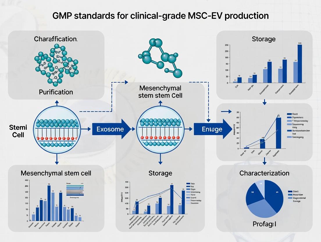
Visualizing Key Concepts and Workflows
Title: Roadmap from Research to Clinical MSC-EV Product
Title: Mechanism of MSC-EV Potency Assay
The Scientist's Toolkit: Key Reagents & Materials for Clinical-Grade EV Work
Table 2: Essential Toolkit for Clinical-Grade MSC-EV Development
| Category | Item | Function & Rationale |
|---|---|---|
| Cell Source | GMP-Grade Human Tissue (e.g., Bone Marrow) | Starting material with full traceability and donor screening. |
| Cell Culture | Xeno-Free, Serum-Free MSC Medium (e.g., STEMxyme MSC, PRIME-XV) | Eliminates animal-derived components, ensures consistency, and prevents bovine EV contamination. |
| Banking | GMP-Grade DMSO & Human Serum Albumin | Cryoprotectant components for creating Master Cell Banks under GMP. |
| Production | Single-Use Bioreactor (e.g., hollow-fiber, stirred tank) | Enables scalable, controlled, and closed-system expansion of MSCs for EV production. |
| Harvest | 0.22 µm PES Sterile Filters | Clarification of conditioned media to remove cells and large debris. |
| Purification | Tangential Flow Filtration (TFF) Cassette (300-500 kDa) | Scalable concentration and buffer exchange; key GMP-compatible purification step. |
| Characterization | Tunable Resistive Pulse Sensing (TRPS) System (e.g., qNano) | Measures particle concentration and size distribution with high resolution. |
| Characterization | CD63/CD81/CD9 ELISA Kit (GMP-compliant) | Quantifies EV-associated tetraspanins for identity testing. |
| Characterization | Recombinant Protein Standards (e.g., for Flow Cytometry) | Critical for quantitative assay calibration and validation. |
| Safety Testing | Limulus Amebocyte Lysate (LAL) Assay Kit | Quantifies endotoxin levels to ensure product safety (<0.5 EU/mL for injectables). |
The regulatory landscape for clinical-grade Mesenchymal Stromal Cell-derived Extracellular Vesicle (MSC-EV) production is defined by foundational guidelines from the U.S. Food and Drug Administration (FDA), the European Medicines Agency (EMA), and the International Council for Harmonisation (ICH). These frameworks, applied within a GMP (Good Manufacturing Practice) context, ensure product quality, safety, and efficacy from donor to final product.
Table 1: Key Regulatory Guideline Comparison for MSC-EV Production
| Aspect | FDA (CBER) | EMA (ATMP Regulation) | ICH Quality Guidelines |
|---|---|---|---|
| Legal Basis | PHS Act 351; 21 CFR Parts 210, 211, 1271 | Regulation (EC) 1394/2007; Directive 2001/83/EC | Harmonised technical requirements adopted by regions. |
| Classification | Somatic Cell Therapy Product / Biologic (BLA pathway) | Advanced Therapy Medicinal Product (ATMP) | Not a legal authority; provides harmonised standards. |
| GMP Foundation | 21 CFR 210 & 211; Guidance for Human Cells, Tissues, and Cellular and Tissue-Based Products (HCT/Ps) | EudraLex Volume 4, Part IV (ATMP GMP) | ICH Q7: GMP for Active Pharmaceutical Ingredients. |
| Quality System | Quality by Design (QbD) principles encouraged. | Comprehensive Quality System mandated. | ICH Q9: Quality Risk Management. ICH Q10: Pharmaceutical Quality System. |
| Critical Focus for MSC-EVs | Identity, purity, potency, safety (adventitious agents), manufacturing consistency. | Risk-based approach, characterization, stability, traceability. | Application of Q9 risk assessment and Q10 management review across product lifecycle. |
| Stability Requirement | Real-time data for shelf-life determination. | Real-time stability studies under recommended storage conditions. | Covered under ICH Q1 and Q5C, referenced by Q10. |
| Key Recent Guidance | CMC Information for Human Gene Therapy INDs (2020); Potency Assurance (2021). | Guideline on quality, non-clinical & clinical aspects of ATMPs (2023). | Ongoing revisions; Q9(R1) revised in 2023 emphasizing formality in risk management. |
Table 2: Quantitative Quality Attribute Targets for MSC-EVs (Illustrative)
| Quality Attribute | Analytical Method | Proposed Target Specification | Associated ICH/FDA/EMA Concept |
|---|---|---|---|
| Particle Concentration | Nanoparticle Tracking Analysis (NTA) | ≥ 1.0 x 10^10 particles/mL; CV < 20% batch-to-batch | ICH Q6B: Test Procedures and Acceptance Criteria |
| Particle Size (Mode) | NTA / Tunable Resistive Pulse Sensing | 80 - 200 nm | ICH Q6B: Characterization |
| EV-Specific Markers | Flow Cytometry (CD9, CD63, CD81) | Positive for ≥ 2 transmembrane markers | FDA Potency: Link to mechanism |
| MSC-Specific Markers | Flow Cytometry (CD73, CD90, CD105) | Positive (>90% population) | Identity (ICH Q6B) |
| Negative Markers | Flow Cytometry (CD14, CD45, HLA-DR) | Negative (<5% population) | Purity (ICH Q6B) |
| Protein Contaminant | BCA / ELISA (e.g., Apolipoprotein B) | ≤ 5% of total protein | Purity; Risk of co-isolation (ICH Q9) |
| Bioburden | Sterility test (EP 2.6.1 / USP <71>) | Sterile | EMA GMP Annex 1; ICH Q6A |
| Endotoxin | LAL test | < 0.5 EU/mL | FDA Pyrogen Test; EP 2.6.14 |
Experimental Protocols
Protocol 1: Risk Assessment for MSC-EV Manufacturing Process (Based on ICH Q9(R1))
Objective: To systematically identify, analyze, and evaluate critical risks in the MSC-EV manufacturing workflow from cell banking to final fill.
Materials:
- Risk Assessment Team (Multi-disciplinary: Process Development, QC, QA, Regulatory).
- Process Flow Diagram (See Diagram 1).
- Risk Assessment Tool (e.g., Failure Mode and Effects Analysis - FMEA template).
- ICH Q9(R1) Guideline.
Procedure:
- Define Risk Question: "What are the risks to the Critical Quality Attributes (CQAs: identity, purity, potency, safety) of the final MSC-EV product?"
- Assemble Team & Define Scope: Include members with expertise in MSC biology, EV isolation, analytics, and GMP. Scope: All steps from Thawing of MSC Working Cell Bank to Final Filled EV Product.
- Hazard Identification: Using the process map, brainstorm potential failure modes for each unit operation (e.g., "During cell expansion: low cell viability").
- Risk Analysis (Severity x Occurrence):
- Severity (S): Score 1 (negligible) to 5 (catastrophic) impact on CQAs/patient safety.
- Occurrence (O): Score 1 (rare) to 5 (very likely) based on historical or development data.
- Calculate Preliminary Risk Priority Number (RPN) = S x O.
- Risk Evaluation: Prioritize risks. Example: "Introduction of mycoplasma during cell culture" (S=5, O=2, RPN=10) is high severity and requires control.
- Risk Control: For unacceptable risks, define control measures. Example: Mitigation for mycoplasma: use of End-of-Production Cell banking with full compendial testing (ICH Q5A, EP 2.6.7).
- Risk Review: Document assessment. Re-evaluate risks post-mitigation and at defined milestones (per ICH Q10, Management Review).
Protocol 2: Critical Quality Attribute (CQA) Assessment for Potency (Linking FDA/EMA/ICH)
Objective: To establish a quantitative, mechanism-based potency assay for MSC-EVs intended for an immunomodulatory indication.
Materials:
- Test Article: Purified MSC-EV batch.
- Cells: Human peripheral blood mononuclear cells (PBMCs), activated with e.g., anti-CD3/CD28 beads.
- Assay Reagents: IL-2, IFN-γ ELISA kits; cell culture media.
- Equipment: CO2 incubator, ELISA plate reader.
Procedure:
- Principle: MSC-EVs should suppress T-cell activation/proliferation, a key mechanism of action (MOA). Assay measures inhibition of IFN-γ release.
- PBMC Activation:
- Isolate PBMCs from healthy donor leukopak.
- Seed 2 x 10^5 cells/well in a 96-well plate.
- Add T-cell activation beads at a pre-optimized cell:bead ratio.
- EV Treatment:
- Prepare a dilution series of MSC-EVs (e.g., 1e7 to 1e9 particles/well) in culture medium.
- Add EV dilutions to activated PBMCs. Include controls: activated PBMCs only (max IFN-γ), non-activated PBMCs (background), and a reference MSC-EV standard if available.
- Incubate for 72 hours at 37°C, 5% CO2.
- Analysis:
- Centrifuge plate, collect supernatant.
- Perform IFN-γ ELISA per manufacturer's protocol.
- Calculate % inhibition relative to activated control for each EV concentration.
- Data Interpretation & Specification:
- Plot dose-response curve. Determine IC50 (concentration causing 50% inhibition).
- Establish Provisional Potency Specification: e.g., "The test article shall inhibit IFN-γ production by ≥ 40% at a concentration of 1e8 particles/well relative to the activated control." This links the product attribute (EV concentration) directly to a biological function, addressing FDA/EMA potency expectations and ICH Q6B.
Visualizations
Diagram 1 Title: MSC-EV GMP Production and Control Workflow
Diagram 2 Title: ICH Q10 Pharmaceutical Quality System for MSC-EVs
The Scientist's Toolkit: Essential Research Reagent Solutions
Table 3: Key Reagents for MSC-EV Process Development & QC
| Reagent / Material | Function in MSC-EV Workflow | Key Quality / Regulatory Consideration |
|---|---|---|
| GMP-grade Human MSC Cell Bank | Starting material for production. Provides genetic stability and defined identity. | Must be qualified per ICH Q5D (Derivation, History, Testing). Traceable, non-xenogeneic origin preferred. |
| Xeno-free, Chemically-defined Cell Culture Medium | Supports expansion of MSCs without introducing animal-derived contaminants. | Eliminates risk from FBS-derived EVs and adventitious agents. Essential for clinical-grade production (EMA CAT guideline). |
| Serum Albumin (Human), GMP-grade | May be used as a stabilizer or medium component. | Sourced from approved vendors, with full traceability and viral safety data (ICH Q5A). |
| DNase I, RNase-free, GMP-grade | Treatment of conditioned media to reduce viscosity from cell debris DNA. | Must be well-characterized, low endotoxin. Validated for lack of adverse impact on EV yield/function. |
| Protease Inhibitor Cocktail | Added during conditioned media harvest to prevent EV protein degradation. | Composition should be defined. Avoids inhibitors that interfere with subsequent functional assays. |
| Size-Exclusion Chromatography (SEC) Columns | Critical for EV purification from soluble protein contaminants. | Column must be validated for EV separation efficiency, non-binding properties, and cleaned/sanitized per bioburden control (GMP). |
| Particle Size & Count Standard (e.g., Silica Beads) | Calibration of NTA or Flow Cytometry instruments for EV quantification. | Traceable to international standards (e.g., NIST). Required for assay qualification (ICH Q2). |
| EV Characterization Antibody Panel (CD9, CD63, CD81, etc.) | Confirmation of EV identity via flow cytometry (MISEV guidelines). | Critical reagents: require clonality, specificity testing, and lot-to-lot consistency. |
| LAL Endotoxin Assay Kit | Detection of Gram-negative bacterial endotoxins in final product. | Must be validated for use with the specific EV product (inhibition/enhancement testing per USP <85>, EP 2.6.14). |
| Reference Standard EV Material | Well-characterized EV batch used for assay system suitability and trending. | Ideally an in-house primary standard from a GMP-like process. Characterized for key attributes (Identity, Potency, Purity). |
Application Note: Integrating QbD into MSC-EV Process Development
Objective: To establish a systematic Quality by Design (QbD) framework for the development of a scalable, clinical-grade MSC-EV production process, ensuring critical quality attributes (CQAs) are met.
Key Concepts & Data:
- Quality Target Product Profile (QTPP): A predefined summary of the quality characteristics for MSC-EVs intended for clinical use.
- Critical Quality Attributes (CQAs): Physical, biochemical, or functional properties that must be within appropriate limits to ensure product safety and efficacy.
- Critical Process Parameters (CPPs): Process variables that directly impact CQAs and must be monitored and controlled.
Table 1: Exemplary QbD Elements for Clinical-Grade MSC-EV Production
| QbD Element | Definition & Example for MSC-EVs | Typical Target/Control Range |
|---|---|---|
| QTPP | Intended Use: Immunomodulation for Acute Respiratory Distress Syndrome (ARDS). | N/A |
| CQAs | Particle Concentration: EVs/µL. | > 5e8 EVs/µL (final product) |
| Size Distribution (Mean): nm by NTA. | 80 - 200 nm | |
| Purity (Protein/particle ratio): µg protein / 1e9 particles. | < 150 µg protein / 1e9 particles | |
| Potency Marker: IDO activity (nmol/hr/mg protein) or tetraspanin positivity (%) | > 80% CD63/CD81 positive | |
| Sterility: Bacterial/fungal growth. | No growth (compendial test) | |
| Critical Material Attributes (CMAs) | MSC Donor Age/Population Doublings: Influences EV yield and potency. | PDL < 15 |
| Serum-Free Media Lot: Impacts cell growth and EV secretion. | Qualified vendor, performance-tested lot | |
| CPPs | Cell Seeding Density: Cells/cm² at initiation of EV production. | 70-90% confluency at harvest |
| Hypoxia Conditioning: % O₂ during EV production phase. | 1-5% O₂ for 24-48h | |
| Harvest Time (Conditioned Media): Hours post-media change. | 48 ± 2 hours | |
| Tangential Flow Filtration (TFF) Shear Rate: s⁻¹. | Optimized to minimize aggregate formation |
Protocol 1.1: Establishing a Design Space for EV Harvesting
- Objective: Determine the optimal interaction between cell confluence at harvest and hypoxic conditioning time to maximize EV yield (CQA: particle concentration) while maintaining potency (CQA: IDO activity).
- Materials: Passage 5 human bone marrow-derived MSCs, qualified serum-free media, hypoxia chamber (1% O₂), normoxia incubator (21% O₂), nanoparticle tracking analyzer (NTA), HPLC for IDO (kynurenine) assay.
- Method:
- Seed MSCs in T-175 flasks at densities to achieve 60%, 80%, and 100% confluence at the start of harvest.
- Replace media with production media. For each density, expose flasks to either normoxia (21% O₂) or hypoxia (1% O₂).
- Collect conditioned media at 24h, 48h, and 72h time points for each confluence/O₂ combination (n=3 flasks per condition).
- Process conditioned media through sequential centrifugation (300 × g, 2000 × g) and 0.22 µm filtration.
- Concentrate and buffer exchange using a 100 kDa TFF system.
- Analyze concentrate for: a) Particle concentration and size (NTA), b) Total protein (BCA), c) IDO activity.
- Data Analysis: Use multivariate analysis (e.g., Response Surface Methodology) to model the relationship between CPPs (confluence, O₂ %, time) and CQAs. The operable region where all CQAs meet specifications defines the design space.
Diagram 1: QbD Framework for MSC-EV Process Development
Application Note: Implementing Risk Management (ICH Q9) in MSC-EV Workflows
Objective: To apply a formal risk management process to identify, analyze, evaluate, and control potential sources of variability and failure in MSC-EV manufacturing and testing.
Protocol 2.1: Failure Mode and Effects Analysis (FMEA) for EV Isolation
- Objective: Prioritize risks associated with the Tangential Flow Filtration (TFF) and Size Exclusion Chromatography (SEC) isolation steps.
- Method:
- Assemble Team: Process development scientist, analytical scientist, quality assurance.
- Define Process Steps: List each sub-step (e.g., TFF system setup, concentration, diafiltration, SEC column equilibration, fraction collection).
- Identify Failure Modes: For each step, brainstorm what could go wrong (e.g., TFF membrane fouling, incorrect buffer pH, SEC fraction mis-collection).
- Score Severity (S), Occurrence (O), Detection (D): Use a 1-10 scale. S: Impact on CQAs. O: Likelihood. D: Chance of detecting before release.
- Calculate RPN: Risk Priority Number = S × O × D.
- Plan Mitigations: For high RPN items, define corrective actions (e.g., implement in-line pH monitoring, validate cleaning-in-place protocol).
Table 2: FMEA Excerpt for MSC-EV Isolation Process
| Process Step | Potential Failure Mode | Potential Effect on CQAs | S | O | D | RPN | Mitigation Action |
|---|---|---|---|---|---|---|---|
| TFF Concentration | High shear rate | EV aggregation, loss of potency, increased size | 8 | 3 | 2 | 48 | Validate and fix optimal shear rate (CPP); implement sensor control. |
| Buffer Exchange | Incorrect diafiltration buffer pH | EV stability compromised, protein degradation | 9 | 2 | 6 | 108 | Use pre-qualified buffer bags; perform in-process pH check. |
| SEC Fraction Collection | Incorrect fraction window (too broad) | Low purity (soluble protein contamination) | 7 | 4 | 3 | 84 | Validate fraction collection based on UV/RI trace; automate collection. |
Diagram 2: ICH Q9 Risk Management Process Flow
Application Note: Ensuring Complete Traceability in MSC-EV Research
Objective: To establish a chain of identity and chain of custody from the originating mesenchymal stromal cell (MSC) donor through to the final MSC-EV batch, including all materials, processes, and data.
Protocol 3.1: Implementing a Unique Identifier (UID) System for EV Batches
- Objective: Generate a UID that links an EV batch to all its antecedents.
- Method:
- Donor/Source Cell ID: Assign a UID to the primary MSC donor/tissue (e.g.,
DON-BM-001). - Cell Bank ID: Assign a UID to each master/working cell bank derived (e.g.,
DON-BM-001-MCB-P5). - Production Run ID: Assign a UID to each EV production run, incorporating the cell bank ID and run number (e.g.,
EV-RUN-DON-BM-001-MCB-P5-024). - Material Linkage: Log all critical reagent lot numbers (media, supplements, columns) against the Production Run ID.
- Data Linkage: All analytical files (NTA, protein, potency) must be saved with the Production Run ID in the filename and metadata.
- Storage Linkage: Final product vials and retain samples must be labeled with the UID and stored in a location mapped in an inventory system.
- Donor/Source Cell ID: Assign a UID to the primary MSC donor/tissue (e.g.,
Table 3: Traceability Matrix for a Single MSC-EV Batch
| UID | Entity | Key Linked Attributes | Storage Location |
|---|---|---|---|
| DON-BM-001 | Bone Marrow Donor | Donor consent, age, screening tests | Donor file, secure server |
| DON-BM-001-MCB-P5 | Master Cell Bank (Passage 5) | Vial numbers, viability, sterility, identity (flow cytometry) | Liquid N2 Tank A, Rack 3 |
| MED-SFM-XX-123 | Serum-Free Media | Vendor, lot number, CoA, endotoxin test | -20°C Freezer F12 |
| EV-RUN-DON-BM-001-MCB-P5-024 | EV Production Batch | Harvest date, CPP logs (confluence, O2%), operator | 4°C Cold Room, Shelf B2 |
| DATA-EV-RUN-024-NTA | Analytical Result | NTA report file, instrument ID, analyst | LIMS Project "EV-ARDS" |
Diagram 3: Chain of Identity and Custody for MSC-EVs
The Scientist's Toolkit: Essential Reagents & Materials for GMP-Compliant MSC-EV Research
Table 4: Key Research Reagent Solutions for Clinical-Grade MSC-EV Workflows
| Item | Function & GMP Relevance | Example (for informational purposes) |
|---|---|---|
| Xeno-Free, Serum-Free MSC Media | Supports MSC expansion and EV production without animal-derived components, reducing pathogen risk and lot variability. Essential for clinical compliance. | StemMACS MSC EV-XF, TheraPEAK MSCGM-CD. |
| Defined Attachment Substrate | Provides a consistent, recombinant surface for MSC adhesion, replacing animal-sourced coatings like gelatin. Enhances process control. | CELLstart, Recombinant Human Vitronectin. |
| Biocompatible TFF Membranes | For gentle concentration and buffer exchange of conditioned media. Cassettes with appropriate molecular weight cut-offs (e.g., 100-300 kDa) are critical for EV yield and purity. | Pellicon Cassettes (100 kDa), mPES hollow fiber filters. |
| GMP-Grade Size Exclusion Columns | For high-resolution purification of EVs from soluble proteins. Pre-packed, sanitizable columns ensure reproducibility and reduce endotoxin/particle contamination. | qEVoriginal columns (Izon), HiPrep Sephacryl S-500 HR. |
| Particle & Protein Standards | Essential for calibrating analytical instruments (NTA, SEC, protein assays) to ensure accurate quantification of CQAs. | Silica microspheres (NTA), Protein Standard (BCA). |
| Mycoplasma Detection Kit | A validated, highly sensitive assay for detecting mycoplasma contamination in cell cultures and EV products. A mandatory release test. | MycoAlert PLUS (Lonza). |
| Endotoxin Detection Assay | A chromogenic LAL assay to quantify bacterial endotoxins in final EV preparations, a critical safety specification. | Endosafe Neogen (Charles River). |
| Tetraspanin Detection Antibodies | Fluorescently labelled, validated antibodies for characterizing EV surface markers (e.g., CD63, CD81, CD9) via flow cytometry (MACSQuant) or ELISA. | Anti-human CD63-APC, CD81-FITC. |
Within a Good Manufacturing Practice (GMP) framework for clinical-grade Mesenchymal Stromal Cell-derived Extracellular Vesicle (MSC-EV) production, the foundation lies in the rigorous characterization and standardization of the starting cellular material. Variability in MSC source directly impacts EV yield, cargo, and therapeutic potency. This document details critical application notes and protocols for donor screening, tissue origin selection, and Master Cell Bank (MCB) establishment to ensure a reproducible, safe, and efficacious MSC starting population.
Application Note: Donor Screening Criteria
Donor selection is the first critical control point. Comprehensive screening mitigates risks of adventitious agent transmission and ensures donor health parameters align with intended therapeutic use.
Table 1: Mandatory Donor Screening Assays
| Screening Category | Specific Test/Parameter | Acceptance Criteria | Rationale |
|---|---|---|---|
| Infectious Disease | HIV-1 & HIV-2 (RNA/PCR) | Non-reactive | Prevents viral contamination of cell bank. |
| Hepatitis B (HBsAg, Anti-HBc, HBV DNA) | Non-reactive | Prevents viral contamination. | |
| Hepatitis C (Anti-HCV, HCV RNA) | Non-reactive | Prevents viral contamination. | |
| Treponema pallidum (Syphilis) | Non-reactive | Ensures donor health status. | |
| Health & Genetics | Karyotype (G-banding) | Normal (46, XX or XY) | Ensures genomic stability. |
| Short Tandem Repeat (STR) Profiling | Completed for identity testing | Provides unique donor/cell line fingerprint. | |
| Mycoplasma (PCR/Culture) | Negative | Prevents microbial contamination. | |
| MSC Potency | In vitro trilineage differentiation (Adipogenic, Osteogenic, Chondrogenic) | Positive by staining & qPCR | Confirms MSC multipotency. |
| Immunosuppression Assay (e.g., T-cell proliferation) | >30% inhibition of proliferation | Confirms a key functional potency. | |
| Surface Marker Profile (Flow Cytometry) | ≥95% positive for CD73, CD90, CD105; ≤2% positive for CD34, CD45, HLA-DR | Confirms immunophenotype per ISCT criteria. |
Experimental Protocol: MSC Isolation & Initial Characterization from Tissue
Protocol 2.1: Explant Isolation and Culture of Bone Marrow-derived MSCs (BM-MSCs)
- Objective: To isolate and establish primary MSC cultures from bone marrow aspirate using the explant method.
- Materials:
- GMP-grade α-MEM medium, supplemented with 5% human platelet lysate (hPL), 1% L-glutamine, 1% penicillin/streptomycin.
- Phosphate-Buffered Saline (PBS), without Ca²⁺/Mg²⁺.
- GMP-grade trypsin/EDTA or recombinant trypsin.
- T-75 culture flasks.
- Sterile 100 µm cell strainers.
- Bone marrow aspirate (5-10 mL) from screened donor.
- Procedure:
- Dilute the bone marrow aspirate 1:2 with PBS.
- Slowly layer the diluted aspirate over an equal volume of Ficoll-Paque Premium in a 50 mL conical tube.
- Centrifuge at 400 x g for 30 minutes at room temperature with the brake OFF.
- Aspirate the mononuclear cell (MNC) layer at the interface and transfer to a new tube.
- Wash MNCs with 3x volume of PBS. Centrifuge at 300 x g for 10 minutes. Repeat wash.
- Resuspend cell pellet in complete medium. Pass through a 100 µm cell strainer.
- Seed cells at a density of 5 x 10⁴ cells/cm² in a T-75 flask.
- Incubate at 37°C, 5% CO₂. Perform first medium change at 72 hours to remove non-adherent cells, then change medium twice weekly.
- Passage cells at ~80% confluence using trypsin. This is designated as Passage 0 (P0).
Protocol 2.2: Immunophenotypic Characterization by Flow Cytometry
- Objective: To confirm MSC surface marker expression per ISCT guidelines.
- Materials:
- Flow cytometry buffer (PBS + 2% FBS).
- Antibody panel: CD73-APC, CD90-FITC, CD105-PE, CD45-PerCP, CD34-PE-Cy7, HLA-DR-BV421.
- Appropriate isotype controls.
- Flow cytometer with 488 nm and 633 nm lasers.
- Procedure:
- Harvest MSCs at P2-P3 using trypsin. Wash 2x with PBS. Count cells.
- Aliquot 1 x 10⁵ cells per tube (test and isotype controls).
- Resuspend cells in 100 µL flow buffer. Add recommended volume of antibody. Vortex gently.
- Incubate for 30 minutes at 4°C in the dark.
- Add 2 mL flow buffer, centrifuge at 300 x g for 5 minutes. Aspirate supernatant.
- Resuspend in 300 µL flow buffer. Analyze immediately on flow cytometer.
- Analysis: Gate on viable, single cells. Compare fluorescence to isotype controls. ≥95% positivity for CD73, CD90, CD105 and ≤2% positivity for CD34, CD45, HLA-DR is required.
Application Note: Tissue Origin Comparative Analysis
The tissue source influences MSC expansion potential, secretome, and immunomodulatory profile.
Table 2: Comparative Analysis of Common MSC Tissue Sources
| Parameter | Bone Marrow (BM) | Adipose Tissue (AT) | Umbilical Cord (UC) | Dental Pulp (DP) |
|---|---|---|---|---|
| Typical Yield (Cells per gram) | 5 x 10³ - 6 x 10³ | 5 x 10⁵ - 2 x 10⁶ | 1 x 10⁴ - 5 x 10⁴ | 1 x 10³ - 3 x 10³ |
| Population Doubling Time (Early passages) | ~40 hours | ~30 hours | ~25 hours | ~35 hours |
| Senescence Limit (Population doublings) | ~30-40 | ~40-50 | ~50-60 | ~30-40 |
| Key Secretome/EV Advantages | High angiogenic potential; robust immunomodulation. | High VEGF & HGF secretion; abundant source. | High proliferation; strong anti-inflammatory profile (TSG-6). | High neuroregenerative potential. |
| Key Challenges for GMP | Invasive harvest; donor age effects on potency. | Liposurgery variability; need for collagenase digestion. | Fetal tissue ethics/logistics; perinatal screening. | Limited starting material; heterogeneity. |
Protocol: Master Cell Bank (MCB) Generation & Qualification
Protocol 4.1: MCB Generation and Cryopreservation
- Objective: To create a characterized, homogeneous MCB from a validated MSC working cell bank (WCB) at low passage.
- Materials:
- MSCs at P3-P4 (from WCB).
- GMP-grade cryopreservation medium (e.g., 5% DMSO in hPL).
- Controlled-rate freezer.
- Cryogenic vials.
- Liquid nitrogen storage system.
- Procedure:
- Culture MSCs to 80% confluence. Harvest using trypsin and perform a viable cell count.
- Centrifuge at 300 x g for 5 minutes. Aspirate supernatant completely.
- Resuspend cell pellet in cold cryopreservation medium to a final concentration of 1-2 x 10⁶ cells/mL.
- Aliquot 1.0 mL per labeled cryovial.
- Place vials in a controlled-rate freezer: Cool at -1°C/min to -40°C, then at -10°C/min to -90°C.
- Transfer vials immediately to the vapor phase of liquid nitrogen for long-term storage.
- The MCB size should be sufficient for the entire product development lifecycle (e.g., 100-200 vials).
Protocol 4.2: MCB Release Testing
- Objective: To perform quality control tests on a representative vial from the MCB.
- Tests: Post-thaw viability (≥80%), sterility (bacteria/fungi, 14-day culture), mycoplasma (PCR), endotoxin (LAL, <0.5 EU/mL), identity (STR match to donor), purity (flow cytometry, per Table 1), and potency (e.g., trilineage differentiation or immunosuppression assay).
The Scientist's Toolkit: Essential Research Reagents & Materials
Table 3: Key Reagent Solutions for GMP-Compliant MSC Source Work
| Item | Function/Application | Key Consideration for GMP |
|---|---|---|
| Human Platelet Lysate (hPL) | Serum replacement for xeno-free MSC expansion. | Must be gamma-irradiated, pathogen-tested, and from accredited donors. Quality impacts expansion and EV profile. |
| GMP-Grade Trypsin | Enzymatic detachment of adherent MSCs. | Recombinant, animal-origin-free versions reduce contamination risks and lot variability. |
| Defined Cryopreservation Medium | Long-term storage of MCB. | Chemically defined, DMSO-containing, with optimized carrier protein. Ensures post-thaw viability and function. |
| Flow Cytometry Antibody Panel | Immunophenotypic characterization. | Clone specificity and validation for human MSCs is critical. Use GMP-grade or RUO antibodies with strict validation protocols. |
| Trilineage Differentiation Kits | Assessment of multipotency. | Use GMP-grade or well-validated, defined media components. Standardize quantification (e.g., qPCR for lineage genes). |
| PCR Mycoplasma Detection Kit | Screening for mycoplasma contamination. | Must have high sensitivity (<10 CFU/mL). Used for in-process and MCB release testing. |
Visualizations: Workflow and Strategy
Within a thesis on GMP standards for clinical-grade Mesenchymal Stromal Cell-Extracellular Vesicle (MSC-EV) production, defining Critical Quality Attributes (CQAs) is paramount. CQAs are measurable properties that directly link to product safety, efficacy, and quality. For MSC-EVs as investigational therapeutic agents, four core CQA categories emerge: Identity, Purity, Potency, and Safety. This document provides detailed application notes and protocols for their assessment, forming the analytical bedrock for GMP-compliant research and development.
CQA 1: Identity
Identity confirms the product is derived from MSCs and is indeed an EV preparation. It involves source cell characterization and vesicle-specific profiling.
Application Notes: Identity assays verify the presence of expected markers and the physical attributes of EVs. A multi-parametric approach is required, as no single marker is definitive.
Protocol 1.1: Nanoparticle Tracking Analysis (NTA) for Size and Concentration
- Objective: Determine particle size distribution and concentration in particles/mL.
- Materials: Purified EV sample in PBS, syringe, 0.1 µm filtered PBS, NTA instrument (e.g., Malvern NanoSight NS300).
- Procedure:
- Dilute EV sample in 0.1 µm filtered PBS to achieve 20-100 particles per frame.
- Load sample into instrument syringe.
- Capture five 60-second videos at camera level 13-16 and detection threshold 5.
- Process videos with instrument software to calculate mode and mean diameter (D50, D90) and particle concentration.
- Data Interpretation: Expect a mode size between 70-200 nm. A primary peak outside this range may indicate impurity or aggregation.
Protocol 1.2: Transmission Electron Microscopy (TEM) for Morphology
- Objective: Visualize cup-shaped vesicle morphology.
- Materials: Formvar/carbon-coated grids, purified EV sample, 2% uranyl acetate, TEM.
- Procedure:
- Load 5-10 µL of EVs onto grid for 20 min.
- Wash with distilled water, negative stain with 2% uranyl acetate for 1 min.
- Image using TEM at 80-100 kV.
Protocol 1.3: Surface Marker Profiling via Flow Cytometry (Capture Bead Assay)
- Objective: Semi-quantitatively detect presence of EV tetraspanins (CD9, CD63, CD81) and MSC markers (CD29, CD44, CD73, CD90, CD105).
- Materials: Aldehyde/sulfate latex beads (4% w/v, 4 µm), purified EVs, PBS-BSA (0.1%), antibodies against target antigens, flow cytometer.
- Procedure:
- Dilute beads 1:1000 in PBS, mix with equal volume of EV sample, incubate 15 min.
- Add PBS-BSA to 2 mL final, incubate 90 min at RT with rotation.
- Block with glycine (100 mM) for 30 min.
- Wash beads, incubate with fluorescent-conjugated primary antibodies for 40 min.
- Analyze on flow cytometer. Use isotype and bead-only controls.
Table 1: Identity CQA Specifications
| Attribute | Recommended Method | Target Specification | Justification |
|---|---|---|---|
| Size Distribution | Nanoparticle Tracking Analysis (NTA) | Mode: 70-200 nm | Characteristic of small EVs (exosomes). |
| Morphology | Transmission Electron Microscopy (TEM) | Cup-shaped vesicles observed | Confirms EV structure. |
| Concentration | NTA or Tunable Resistive Pulse Sensing (TRPS) | Report particles/mL & µg protein | Batch consistency metric. |
| EV Surface Markers | Flow Cytometry (Bead-based) | Positive for ≥2 of: CD9, CD63, CD81 | MISEV2018 guidelines. |
| Parent MSC Markers | Flow Cytometry (Bead-based) | Positive for ≥3 of: CD73, CD90, CD105, CD44 | Confirms MSC origin. |
Diagram Title: Analytical Workflow for Identity CQAs
CQA 2: Purity
Purity assesses the degree of contamination from non-EV components, primarily residual proteins from culture medium or cell debris.
Application Notes: Purity is a ratio, not an absolute measure. Key metrics include particle-to-protein ratio and assessment of specific contaminants like lipoproteins or apoptotic bodies.
Protocol 2.1: Particle-to-Protein Ratio
- Objective: Calculate a high-fidelity purity index (particles/µg protein).
- Materials: EV sample, BCA or Micro BCA Protein Assay Kit, NTA instrument.
- Procedure:
- Determine particle concentration (particles/mL) via NTA (Protocol 1.1).
- Determine total protein concentration (µg/mL) via BCA assay, using BSA standard curve.
- Calculate ratio: (Particles/mL) / (Protein µg/mL).
- Data Interpretation: Higher ratios (>1e10 particles/µg) suggest purer EV preparations with less co-isolated soluble protein.
Protocol 2.2: Western Blot for Negative Markers
- Objective: Detect absence of common contaminant proteins.
- Materials: EV lysate, SDS-PAGE gel, antibodies against Apolipoprotein A1/ B (lipoproteins), Calnexin/GM130 (endoplasmic reticulum/Golgi).
- Procedure:
- Load 10-20 µg EV protein and parent MSC whole cell lysate (positive control) per lane.
- Perform SDS-PAGE and transfer to PVDF membrane.
- Block, incubate with primary antibody (e.g., anti-ApoA1, 1:1000) overnight.
- Incubate with HRP-conjugated secondary, develop.
- Data Interpretation: Contaminant markers should be absent or significantly reduced (>90%) in EV lysate compared to cell lysate.
Table 2: Purity CQA Specifications
| Attribute | Recommended Method | Target Specification | Justification |
|---|---|---|---|
| Particle-to-Protein Ratio | NTA + BCA Assay | > 3.0 x 10¹⁰ particles/µg | Indicator of low soluble protein contamination. |
| Negative Markers | Western Blot | Absent or minimal: Apolipoprotein A1/B, Calnexin | Absence of lipoprotein & intracellular organelle contamination. |
| Lipoprotein Contamination | SEC-UV/Vis (A280/A260) | A280/A260 ratio > 0.6 | Lower ratio suggests nucleic acid or lipoprotein carryover. |
CQA 3: Potency
Potency is the quantitative measure of biological activity linked to the relevant therapeutic mechanism of action (MoA).
Application Notes: Potency assays must be mechanism-based, quantitative, and validated. Common MoAs for MSC-EVs include immunomodulation, angiogenesis, and tissue repair.
Protocol 3.1: T-Cell Proliferation Suppression Assay (Immunomodulation)
- Objective: Quantify EV ability to suppress activated immune cell proliferation.
- Materials: Human PBMCs, CFSE dye, anti-CD3/CD28 activation beads, purified MSC-EVs, flow cytometer.
- Procedure:
- Isolate CD3+ T-cells from PBMCs, label with CFSE (5 µM).
- Activate cells with anti-CD3/CD28 beads.
- Co-culture with MSC-EVs (e.g., 1e9 particles/well) for 72-96 hrs.
- Analyze CFSE dilution via flow cytometry. Calculate % suppression vs. activated control without EVs.
- Data Interpretation: A dose-dependent suppression of T-cell proliferation indicates immunomodulatory potency.
Protocol 3.2: Endothelial Tube Formation Assay (Angiogenesis)
- Objective: Measure pro-angiogenic activity.
- Materials: HUVECs, Matrigel, purified MSC-EVs, tubule imaging system.
- Procedure:
- Seed HUVECs on Matrigel-coated plates.
- Treat with MSC-EVs (e.g., 1e10 particles/mL) in endothelial basal medium.
- Incubate 4-8 hrs, fix, stain with calcein-AM.
- Image and quantify total tubule length, number of nodes.
- Data Interpretation: Increased tubule formation vs. vehicle control indicates angiogenic potency.
Table 3: Example Potency CQA Specifications
| Therapeutic Indication | Potency Assay | Quantitative Readout | Target Specification (Example) |
|---|---|---|---|
| GvHD / Inflammation | T-Cell Proliferation Assay | % Suppression of proliferation | IC₅₀ ≤ 1 x 10⁹ particles/mL |
| Myocardial Infarction | Endothelial Tube Formation | % Increase in total tubule length | ED₅₀ ≤ 5 x 10⁹ particles/mL |
| Wound Healing | Fibroblast Migration (Scratch) | % Wound closure at 24h | ≥40% enhancement vs. control |
Diagram Title: Example EV Potency Pathway in Tissue Repair
CQA 4: Safety
Safety CQAs ensure the product is free from harmful contaminants that pose risk to patients, including endotoxins, mycoplasma, and replicating viruses.
Protocol 4.1: Endotoxin Testing (LAL Assay)
- Objective: Detect and quantify bacterial endotoxins.
- Materials: EV sample, Kinetic Chromogenic LAL Assay kit, pyrogen-free tubes, microplate reader.
- Procedure:
- Dilute EV sample in endotoxin-free water.
- Follow kit protocol: mix sample with LAL reagent, incubate at 37°C.
- Add chromogenic substrate, measure absorbance at 405 nm over time.
- Calculate EU/mL from standard curve.
- Data Interpretation: Must meet limit of <5 EU/kg/hr for parenteral administration (FDA guideline).
Protocol 4.2: Sterility Testing (Bacteriostasis/Fungistasis)
- Objective: Demonstrate absence of viable bacteria and fungi.
- Materials: EV sample, Fluid Thioglycollate Medium (FTM), Soybean-Casein Digest Medium (SCDM).
- Procedure:
- Inoculate 10 mL FTM (for aerobes/anaerobes) and SCDM (for fungi) with 1 mL of EV product.
- Incubate FTM at 30-35°C and SCDM at 20-25°C for 14 days.
- Observe daily for turbidity, indicating microbial growth.
- Data Interpretation: No growth in media after 14 days indicates sterility.
Table 4: Safety CQA Specifications
| Attribute | Recommended Method | Target Specification | Justification |
|---|---|---|---|
| Endotoxin | Limulus Amebocyte Lysate (LAL) Assay | < 2.0 EU/mL (or per dose) | Compliance with parenteral drug standards. |
| Sterility | Direct Inoculation / Membrane Filtration | No growth in 14 days | USP <71>; ensures absence of microbiological contamination. |
| Mycoplasma | PCR-based or Culture-based Assay | Negative | Essential for products derived from cell culture. |
| Residual Host Cell DNA | qPCR (e.g., Alu-repeat) | < 10 ng/dose (WHO recommendation) | Minimizes oncogenic/immunogenic risk. |
The Scientist's Toolkit: Key Research Reagent Solutions
| Item | Function & Application | Example Vendor/Product |
|---|---|---|
| qEV size-exclusion columns | Isolate EVs with high purity and recovery from conditioned medium. | Izon Science, qEVoriginal 35nm |
| Total Exosome Isolation Reagent | Precipitation-based EV enrichment from serum-free or serum-containing media. | Thermo Fisher Scientific, 4478359 |
| CD9/CD63/CD81 Antibody Cocktail | Detection of EV tetraspanins for characterization via flow cytometry or WB. | System Biosciences, EXOAB-KIT-1 |
| Recombinant Human TGF-β1 | Positive control for potency assays involving SMAD signaling pathways. | PeproTech, 100-21 |
| Lonza Kinetic-QCL LAL Kit | Sensitive, quantitative detection of endotoxin for safety testing. | Lonza, 50-650U |
| MycoAlert Detection Kit | Rapid, bioluminescent detection of mycoplasma contamination. | Lonza, LT07-318 |
| HUVECs & EGM-2 BulletKit | Primary cells and optimized media for angiogenesis potency assays. | Lonza, CC-2517 & CC-3162 |
| CellTrace CFSE Cell Proliferation Kit | Fluorescent dye for tracking and quantifying lymphocyte proliferation. | Thermo Fisher Scientific, C34554 |
From Flask to Vial: Step-by-Step GMP Methods for Scalable MSC-EV Production
The transition from research to clinical-grade mesenchymal stromal cell (MSC) production for extracellular vesicle (EV) harvest requires strict adherence to Good Manufacturing Practice (GMP). This mandates a shift from open, xeno-containing systems to closed, automated, and chemically defined platforms. The core triad for GMP-compliant expansion comprises: (1) Closed Processing Systems to minimize contamination, (2) Xeno-Free/Serum-Free Media to eliminate undefined components and batch variability, and (3) Bioreactor Technologies for scalable, monitored, and reproducible culture. Implementing this triad is critical for generating MSC-EVs with consistent quality, potency, and safety profiles for therapeutic applications.
Key Protocols and Methodologies
Protocol 2.1: Seeding and Expansion in a Closed System Stirred-Tank Bioreactor
- Objective: To expand MSCs from passage 2 (P2) master cell bank vials to harvest-ready quantities in a GMP-compliant, controlled bioreactor.
- Materials: See Scientist's Toolkit (Table 1).
- Pre-Culture: Thaw a P2 MSC vial in a closed thawing device. Transfer cells to a pre-coated T-flask using a sterile tubing welder/connector. Expand in xeno-free medium in a closed, multi-layer flask stack system until sufficient inoculum is achieved (e.g., 1–2 x 10^8 cells).
- Bioreactor Setup & Seeding:
- Sterilize the bioreactor vessel (e.g., 2L working volume) in situ via autoclave or SIP (Steam-In-Place).
- Connect all media, harvest, and sampling lines via sterile connectors within a closed pathway.
- Prime the system with pre-warmed, equilibrated xeno-free medium.
- Detach cells from flask stacks using a closed-system detachment device and transfer cell suspension to the bioreactor inoculum bag via peristaltic pump.
- Seed cells into the bioreactor at a density of 1–2 x 10^4 cells/cm² (microcarrier surface area) or 2–4 x 10^5 cells/mL (aggregate culture).
- Process Parameters:
- Temperature: 37°C ± 0.2°C
- pH: 7.2–7.4 (controlled via CO₂ and base addition)
- Dissolved Oxygen (DO): 30–50% air saturation (cascaded control with O₂, N₂, and air)
- Agitation: 60–100 rpm (intermittent or continuous, depending on microcarrier type) to prevent aggregation and ensure nutrient homogeneity.
- Feeding & Harvest: Perform medium exchange (50–80%) every 48-72 hours via a closed drain-and-fill protocol. Monitor glucose/lactate to guide feeding strategy. At harvest (typically day 7-10), stop agitation, allow microcarriers/cell aggregates to settle, and drain spent medium. Wash with PBS, then add dissociation reagent. Transfer cell slurry to a closed harvest bag via in-line filtration to remove microcarriers.
Protocol 2.2: In-Process Quality Control (QC) Testing
- Objective: To monitor critical quality attributes (CQAs) during expansion.
- Sampling: Use aseptic, closed sampling systems integrated into the bioreactor line.
- Daily Tests:
- Cell Count & Viability: Use an automated cell counter with trypan blue exclusion. Withdraw a 1 mL sample, lyse microcarriers if necessary, and analyze.
- Metabolite Analysis: Use a blood gas/biochemistry analyzer for daily glucose, lactate, and glutamine measurement from 0.5 mL supernatant.
- Microbiology: Inoculate 10 mL samples into BacT/ALERT culture bottles for rapid sterility testing.
- Endpoint Tests (Pre-Harvest):
- Identity: Analyze surface markers (CD73+, CD90+, CD105+, CD45-, CD34-, HLA-DR-) via flow cytometry using a closed-tube staining system.
- Potency: Measure immunosuppressive capacity via a closed-system co-culture assay (e.g., inhibition of PHA-stimulated PBMC proliferation) or cytokine secretion profile (IDO, PGE2).
- Karyotyping: Send a cell sample for G-band karyotype analysis to confirm genetic stability.
Table 1: Comparison of Expansion Platforms for MSCs
| Platform | Max Scale (Cells) | Volumetric Productivity (Cells/mL/day) | Key Process Control Parameters | Suitability for GMP EV Production |
|---|---|---|---|---|
| Multi-layer Flasks | 1–2 x 10^9 | 1–3 x 10^4 | Temperature, CO₂ | Low-scale seed train; not final expansion |
| Fixed-bed Bioreactor | 5 x 10^9 | 2–5 x 10^4 | pH, DO, Flow Rate | High; integrated harvesting, good for adherent cells |
| Stirred-Tank w/ Microcarriers | 1 x 10^11 | 2–6 x 10^4 | pH, DO, Agitation, Temperature | Very High; highly scalable, proven in industry |
| Vertical-Wheel Bioreactor | 5 x 10^10 | 3–8 x 10^4 | pH, DO, Shear Stress | High; low-shear, suitable for aggregates & microcarriers |
Table 2: Key Performance Indicators in Xeno-Free Media vs. FBS-Containing Media
| Parameter | FBS-Based Media (Typical Range) | Xeno-Free/Serum-Free Media (Typical Range) | Impact on MSC-EV Production |
|---|---|---|---|
| Population Doubling Time (hrs) | 30–48 | 24–40 | Faster expansion may alter EV miRNA cargo. |
| Max Cumulative Population Doublings | 25–35 | 20–30 | Genetic stability threshold must be defined for EV consistency. |
| CD Marker Expression (% positive) | >95% (Tri-positive) | >95% (Tri-positive) | Confirms identity is maintained. |
| Immunosuppressive Activity (e.g., % T-cell inhibition) | 60–80% | 65–85% | Potency must be maintained or enhanced. |
| EV Yield (Particles/Cell) | 1,000–5,000 | 2,000–8,000 | Xeno-free media can enhance specific productivity. |
Diagrams
Title: GMP MSC Expansion Workflow for EV Production
Title: Bioreactor Process Control Loop
The Scientist's Toolkit: Research Reagent Solutions
Table 1: Essential Materials for GMP-Compliant MSC Expansion
| Item | Function in Protocol | Example Product/Category | Critical GMP Attribute |
|---|---|---|---|
| Xeno-Free MSC Medium | Provides defined nutrients & growth factors for proliferation. | StemMACS MSC XF, PPRF-msc6, TheraPEAK MSCGM-CD | Chemically defined, animal component-free, TSE/BSE-free, GMP-manufactured. |
| Human-Derived Attachment Factor | Coats surfaces for cell adhesion in place of FBS. | Recombinant Human Vitronectin, Laminin-521, Collagen I (Human) | Defined, pathogen-tested, xeno-free origin. |
| Microcarriers | Provides high surface area for adherent growth in stirred-tank bioreactors. | Solohill PrimeSurface, Cytodex 3, Plastic Porous Microcarriers. | Sterile, non-animal derived, validated for compatibility with xeno-free media. |
| Closed System Connectors | Enables aseptic connections between bags, bioreactors, and tubing. | Colder AseptiQuik, CPC Quick Disconnect, Sterile Welding. | Sterility assurance, steam-sterilizable, validated for zero bioburden ingress. |
| Single-Use Bioreactor | Pre-assembled, sterile culture vessel for scalable expansion. | Mobius (Merck), Xcellerex (Cytiva), BIOSTAT STR (Sartorius). | Eliminates cleaning validation, integrated sensors, scalable from 1L to 2000L. |
| In-Process Monitoring System | Measures critical parameters (pH, DO, Glucose) in real-time. | BioProfile FLEX2, SGA (DASGIP), Raman Spectroscopic Probes. | PAT (Process Analytical Technology) enabler, supports quality by design (QbD). |
| Closed Harvest System | Filters cells from microcarriers and concentrates harvest. | kSep or Centritech closed centrifugation, in-line depth filtration. | Maintains closed processing, reduces shear stress on cells for optimal EV release. |
The translation of Mesenchymal Stromal Cell-derived Extracellular Vesicles (MSC-EVs) from research tools to clinical-grade therapeutics requires adherence to Good Manufacturing Practice (GMP) principles. Within this broader thesis framework, this document provides detailed Application Notes and Protocols focusing on the upstream optimization of EV biogenesis. Critical input parameters—cell conditioning strategies and culture physicochemical environment—directly influence the yield, purity, and functional potency of the EV product, impacting downstream processing and final compliance with GMP standards for identity, safety, and efficacy.
Application Notes: Key Conditioning Strategies & Parameters
Optimizing EV biogenesis involves manipulating the cellular microenvironment to modulate endosomal sorting complexes required for transport (ESCRT)-dependent and -independent pathways, ceramide-mediated budding, and cellular stress responses. The following parameters are critical.
Table 1: Conditioning Strategies for Enhancing MSC-EV Biogenesis & Potency
| Strategy Category | Specific Condition | Typical Parameters / Reagents | Proposed Effect on EV Biogenesis & Cargo | Key Considerations for GMP Translation |
|---|---|---|---|---|
| Hypoxia | Reduced O2 tension | 1-5% O2 for 24-72h | ↑ EV yield via HIF-1α activation; ↑ angiogenic & immunomodulatory miRNAs. | Requires validated, calibrated incubators; process consistency is critical. |
| Serum Deprivation | Nutrient Stress | Serum-free medium for 24-48h | ↑ EV release as stress response; alters lipid and protein cargo. | Risk of increased apoptosis; must define optimal window to balance yield and cell health. |
| Small Molecule Induction | ESCRT/Pathway Modulators | GW4869 (nSMase2 inhibitor) - Note: used for inhibition studies. Trehalose, Dimethyloxalylglycine (DMOG). | Inhibition: Used as a negative control. Induction: Trehalose induces autophagy-EV crosstalk; DMOG stabilizes HIF-1α. | Reagent purity (e.g., DMSO quality); need for removal during downstream processing. |
| 3D Culture & Scaffolds | Spheroids/Bioreactors | Low-attachment plates, microcarriers, hollow-fiber bioreactors. | Mimics niche, ↑ cell-cell contact; ↑ EV yield and stemness-related cargo vs. 2D monolayers. | Scalability; extraction complexity; potential for contaminating materials (e.g., microcarrier debris). |
| Cytokine/Priming | Inflammatory Priming | IFN-γ, TNF-α, LPS (low dose) for 6-24h. | ↑ EV immunomodulatory cargo (e.g., PD-L1, IDO); shifts EV function towards anti-inflammatory. | Defining "Clinical-Grade" cytokine sources; exact priming conditions must be standardized. |
Table 2: Critical Physicochemical Culture Parameters
| Parameter | Optimal / Tested Range | Impact on EV Biogenesis | Monitoring & Control for GMP |
|---|---|---|---|
| pH | 7.2 - 7.6 | Deviations alter membrane fluidity & ESCRT function; can induce stress response. | In-line sensors in bioreactors; buffering capacity of medium must be defined. |
| Temperature | 37°C ± 0.5°C | Crucial for membrane lipid ordering and enzyme kinetics; lower temps may inhibit budding. | Continuous monitoring with alarms; uniformity mapping of incubators/chambers. |
| Dissolved O2 (DO) | Varies by strategy (e.g., 5% for hypoxia). | Directly regulates HIF signaling pathways; affects mitochondrial function and redox cargo. | Calibrated DO probes; control via gas mixing in bioreactors. |
| Glucose/Lactate | Glucose: Maintain >1 g/L; Lactate: Monitor accumulation. | Nutrient exhaustion stresses cells; high lactate can inhibit proliferation and alter EV release. | Off-line analyzers or in-line biosensors; fed-batch strategies to maintain levels. |
| OsmoMolarity | 280 - 320 mOsm/kg | Hyperosmotic stress can enhance EV release via ESCRT-III activation. | Must be tightly controlled; affected by media supplements and metabolite shifts. |
Detailed Experimental Protocols
Protocol 1: Optimization of Hypoxic Conditioning for MSC-EVs Objective: To produce a high yield of EVs with enhanced angiogenic cargo from human bone marrow-derived MSCs under hypoxic conditions. Materials: GMP-grade MSCs (P3-P5), validated serum-free MSC medium, triple-gas incubator (O2, CO2, N2 control), PBS, 0.22 µm PES filters, evLY-64 EV labeling kit, nanoparticle tracking analysis (NTA) instrument, BCA protein assay kit. Procedure:
- Cell Preparation: Seed MSCs at 5,000 cells/cm² in T-225 flasks. Expand to 70-80% confluence in normoxia (37°C, 21% O2, 5% CO2).
- Conditioning: Replace medium with fresh, pre-warmed, serum-free medium.
- Experimental Group: Transfer flasks to hypoxic incubator set to 1% O2, 5% CO2, balance N2.
- Control Group: Maintain in normoxic incubator (21% O2, 5% CO2).
- Conditioned Media (CM) Harvest: After 48 hours, collect CM from both groups.
- CM Processing: Centrifuge CM at 2,000 x g for 30 min at 4°C to remove cells. Transfer supernatant and centrifuge at 10,000 x g for 45 min at 4°C to remove debris. Filter supernatant through a 0.22 µm PES filter.
- EV Isolation: Concentrate EV-containing filtrate using tangential flow filtration (TFF) with a 100 kDa cutoff membrane, followed by size-exclusion chromatography (SEC) using qEVoriginal columns.
- Analysis:
- Yield: Use NTA to determine particle concentration (particles/mL) and mode size.
- Protein: Perform BCA assay on SEC fractions.
- Potency Marker: Analyze EV samples via western blot for HIF-1α-induced miRNAs (e.g., miR-210) by RT-qPCR.
Protocol 2: Monitoring and Controlling Physicochemical Parameters in a Bioreactor Objective: To maintain culture parameters within defined limits during EV production in a stirred-tank bioreactor. Materials: Benchtop bioreactor system (with pH, DO, temperature probes), MSC microcarriers, perfusion or fed-batch medium set, base (e.g., NaHCO3) and acid (e.g., CO2) for pH control, N2/O2/CO2 gas tanks, offline blood gas/glucose analyzer. Procedure:
- Bioreactor Setup & Calibration: Calibrate pH and DO probes according to manufacturer instructions. Set initial parameters: pH=7.4 (deadband ±0.1), DO=50% air saturation (or target for hypoxia), Temperature=37.0°C.
- Inoculation: Seed MSCs onto microcarriers and transfer to bioreactor vessel.
- Process Control:
- pH: Controlled via automatic addition of CO2 (to lower pH) or base (to raise pH).
- DO: Maintained by cascading control of stirrer speed (primary) and gas blending (secondary, e.g., N2/O2 mix).
- Temperature: Controlled by heated jacket.
- Sampling & Feeding: Take 5 mL samples every 12 hours for offline analysis of glucose, lactate, and osmolality. Implement a perfusion or bolus feed strategy based on glucose consumption rate to maintain glucose >1 g/L.
- Harvest Initiation: Begin continuous harvest of conditioned medium via an overflow filter when cell viability remains >90% and glucose consumption stabilizes. Process harvested medium immediately through downstream clarification (as in Protocol 1, Step 4).
Visualizations (Generated via Graphviz DOT Language)
Diagram 1: Key Signaling Pathways in EV Biogenesis Modulation
Title: Signaling Pathways in EV Biogenesis Modulation
Diagram 2: GMP Workflow for Conditioning & EV Production
Title: GMP Workflow for Conditioned EV Production
The Scientist's Toolkit: Research Reagent Solutions
Table 3: Essential Materials for EV Biogenesis Optimization Research
| Reagent / Material | Supplier Examples | Function in EV Research | GMP-Translation Consideration |
|---|---|---|---|
| GMP-grade MSC Medium (Xeno-free) | Thermo Fisher, Lonza, Merck | Provides defined, consistent, animal-origin-free expansion of clinical-grade MSCs. | Essential; formulation must be fully disclosed and compliant. |
| Triple-Gas Incubator | Baker, Thermo Fisher | Precise control of O2, CO2, and N2 for reproducible hypoxic conditioning studies. | Requires IQ/OQ/PQ validation; continuous data logging. |
| Serum Replacement / EV-Depleted FBS | Thermo Fisher, System Biosciences | Provides growth factors while minimizing contaminating bovine EV background in conditioned media. | Must be thoroughly characterized; lot-to-lot consistency is critical. |
| GW4869 | Cayman Chemical, Sigma | Inhibits neutral sphingomyelinase 2 (nSMase2), used as a critical negative control to demonstrate ceramide-dependent EV biogenesis. | Research tool only; not for use in clinical production. Purity for reliable results. |
| Nanoparticle Tracking Analyzer (NTA) | Malvern Panalytical, Particle Metrix | Gold-standard for determining EV particle concentration and size distribution in suspension. | Key QC instrument; requires strict SOPs and reference standards for calibration. |
| Size-Exclusion Chromatography (SEC) Columns (qEV) | Izon Science | Gentle, size-based separation of EVs from soluble proteins and aggregates for high-purity isolates. | Columns are for single-use; scalable GMP-compatible alternatives (e.g., hollow fiber + FPLC) needed. |
| CD63/CD81/CD9 ELISA/ MACSPlex Exosome Kit | System Biosciences, Miltenyi Biotec | Multiplexed capture and analysis of EV surface epitopes for phenotypic characterization. | Assay suitability must be confirmed for MSC-EVs; used for identity testing. |
| Tangential Flow Filtration (TFF) System | Repligen, Spectrum Labs | Scalable concentration and buffer exchange of large volumes of conditioned media. | Core GMP technology; material compatibility (silicon-free) and sanitization validation required. |
Within the development of clinical-grade Mesenchymal Stromal Cell-derived Extracellular Vesicles (MSC-EVs), the initial harvesting and clarification steps are critical determinants of yield, purity, and compliance with Good Manufacturing Practice (GMP). Conditioned media (CM) is the starting material containing the EV product of interest, alongside cellular debris, apoptotic bodies, and soluble proteins. Scalable, closed-system methods for CM collection and primary clarification are essential to ensure process consistency, minimize batch-to-batch variability, and reduce the risk of contamination—core tenets of GMP manufacturing for therapeutic applications.
Application Notes: Scalable Collection Strategies
Harvest Parameters & Conditioning
Optimal CM collection balances MSC health, EV yield, and the minimization of impurities. Key parameters are summarized in Table 1.
Table 1: Optimization Parameters for Conditioned Media Harvesting
| Parameter | Typical Range (for MSC-EVs) | Rationale & GMP Consideration |
|---|---|---|
| Cell Confluence at Harvest | 70-90% | Prevents over-confluence-induced stress/apoptosis, reducing contaminating debris. Must be standardized. |
| Serum Deprivation Period | 24-48 hours | Uses EV-depleted serum or serum-free media to reduce bovine EV contaminants. Requires pre-qualified media lots. |
| Conditioning Time | 24-72 hours | Longer times increase yield but risk nutrient depletion and cell death. Must be validated for each cell line. |
| Collection Temperature | 2-8°C | Slows metabolic activity and protease function, preserving EV integrity post-secretion. |
| Bioreactor vs. Flask | Microcarriers/Suspension vs. 2D Layers | Scalable bioreactors (e.g., fixed-bed, hollow fiber) enable continuous harvest, aligning with large-scale GMP needs. |
Primary Clarification Techniques
Initial clarification removes cells and large debris. Scalability and closed-processing are paramount.
Table 2: Comparison of Primary Clarification Methods
| Method | Throughput | EV Recovery Estimate* | Suitability for GMP Scale-Up | Key Limitation |
|---|---|---|---|---|
| Low-Speed Centrifugation | Medium | ~95-100% | Low; open handling risks. Simple but not easily closed for large volumes. | Poor removal of small debris/apoptotic bodies. |
| Depth Filtration | High | ~85-95% | High; integrates into closed single-use systems. | Filter adsorption can cause EV loss; requires validation. |
| Tangential Flow Filtration (TFF) | High | ~90-98% | Very High; ideal for continuous processing & diafiltration. | Initial set-up complexity; potential shear stress. |
| Sequential Filtration (e.g., 5µm → 0.8µm) | Medium-High | ~80-90% | Medium; uses disposable filters in series. | Multiple steps increase adsorption loss points. |
*Recovery estimates are system-dependent and must be empirically determined.
Detailed Protocols
Protocol 3.1: Scalable Harvest from Multilayer Flasks with Depth Filtration
Objective: To collect and primarily clarify CM from MSCs grown in Cell Factory systems.
Materials (Research Reagent Solutions):
- Multilayer Cell Factories (e.g., 10-layer): For scalable 2D MSC expansion.
- GMP-grade, EV-depleted/XF Serum-Free Media: Pre-qualified lot to reduce contaminant load.
- Peristaltic Pump & Single-Use Tubing Set: Enables closed fluid transfer.
- Single-Use Depth Filter Capsule (e.g., 5-10 µm pore size): For inline primary clarification.
- Temperature-Controlled Collection Vessel: Maintains 2-8°C.
Methodology:
- Media Exchange & Conditioning: Aspirate growth media from confluent (80%) MSCs. Wash twice with DPBS. Add pre-warmed, EV-depleted conditioning media.
- Incubate: Condition cells for 48 hours at 37°C, 5% CO₂.
- Harvest (Closed System): a. Connect the Cell Factory outlet to the peristaltic pump tubing. b. Connect the pump outlet to the inlet of the pre-primed depth filter capsule. c. Connect the filter outlet to a sterile, chilled (4°C) collection bag. d. Pump CM from the Cell Factory, through the depth filter, into the collection bag. Maintain a controlled, low shear flow rate (e.g., 100 mL/min).
- Immediate Processing: Process clarified CM for secondary concentration/purification (e.g., TFF, UC) within 24 hours, storing at 4°C if necessary.
Protocol 3.2: Continuous Harvest and Clarification Using Tangential Flow Filtration (TFF)
Objective: To enable continuous CM harvest and clarification from a bioreactor system.
Materials (Research Reagent Solutions):
- Hollow Fiber or Fixed-Bed Bioreactor: For adherent MSC culture at scale.
- TFF System with 500 kDa - 0.8 µm MWCO/Pore Cassette: For simultaneous clarification and concentration.
- pH & Metabolite Sensors: For real-time monitoring of conditioning media.
- Feed and Permeate Collection Bags: Single-use, sterile.
Methodology:
- System Set-Up: Configure the bioreactor in a feed-and-bleed mode. Connect the harvest line to the TFF system's feed port. The retentate line returns to the bioreactor or a holding bag; the permeate line is the clarified CM product.
- Continuous Operation: a. Continuously pump fresh conditioning media into the bioreactor at a defined rate (e.g., 1 reactor volume per day). b. Simultaneously, harvest an equivalent volume of spent CM from the bioreactor, passing it through the TFF module. c. The TFF cassette retains cells and large debris, returning them to the bioreactor (retentate), while the clarified CM (permeate) is collected cold. d. Maintain system parameters (transmembrane pressure, shear rate) within validated ranges to preserve EV integrity.
- Collection: Pool the clarified permeate from the collection bag at defined intervals for downstream processing.
The Scientist's Toolkit: Essential Research Reagent Solutions
| Item | Function in CM Harvest/Clarification |
|---|---|
| EV-Depleted Fetal Bovine Serum (FBS) | Provides growth factors during expansion while minimizing contaminating bovine EVs. Must be ultracentrifuged or commercially sourced. |
| Defined, Serum-Free, Xeno-Free Media | Eliminates animal components entirely for GMP-compliant production, reducing immunogenicity risks. |
| Single-Use, Closed Fluid Transfer Systems | Maintains sterility, reduces adventitious agent risk, and is essential for current GMP. |
| Pre-filtration Membranes (5 µm, 0.8 µm) | Used in series for gentle removal of larger particulates prior to ultrafiltration. |
| Benchtop & Floor Model Centrifuges with CHC | For low-speed clarification; refrigerated chambers with contained heating centrifuges (CHC) protect the operator. |
| Protease & Phosphatase Inhibitor Cocktails | Added to chilled CM immediately post-harvest to preserve EV cargo integrity. |
| Bioreactor with Perfusion Capability | Allows for continuous media exchange and harvest, maximizing cell health and volumetric EV yield. |
| Online pH/DO/Glucose/Lactate Sensors | Monitor conditioning media health to determine optimal harvest windows and ensure process consistency. |
Visualizations
Workflow for Scalable MSC-EV Conditioned Media Collection
Clarification Methods and Target Contaminant Removal
Application Notes: Purification Strategies for GMP-Compliant MSC-EV Production
The transition from research-scale to clinical-grade mesenchymal stromal cell-derived extracellular vesicle (MSC-EV) production necessitates stringent purification protocols compliant with Good Manufacturing Practice (GMP) standards. The primary goal is to isolate EVs with high yield, purity, and potency while removing contaminants like soluble proteins, lipoproteins, and cell debris. This note evaluates three core techniques within a GMP framework.
Tangential Flow Filtration (TFF) is favored for its scalability and closed-system potential, crucial for GMP. It efficiently concentrates and diafiltrates large-volume conditioned media, enabling buffer exchange into formulation buffers. Size-Exclusion Chromatography (SEC) is the gold standard for high-purity EV isolation post-concentration. It effectively separates EVs from co-isolated soluble proteins based on hydrodynamic radius, preserving vesicle integrity and biological activity. Ultracentrifugation (UC) Alternatives address UC's limitations: low scalability, potential vesicle damage, and co-precipitation of contaminants. Alternatives like TFF and SEC are more suitable for reproducible, large-scale GMP processes.
Comparative Performance Data: The following table summarizes key quantitative metrics for each method, derived from recent comparative studies in MSC-EV purification.
Table 1: Comparative Analysis of EV Purification Methods for MSC-EVs
| Parameter | Ultracentrifugation (UC) | Tangential Flow Filtration (TFF) | Size-Exclusion Chromatography (SEC) |
|---|---|---|---|
| EV Yield | Moderate to Low (10-25%) | High (>80%) | Moderate (50-70%) |
| Protein Contamination | High (Protein:EV ratio >100) | Moderate (Protein:EV ratio ~50) | Low (Protein:EV ratio ~20) |
| Lipoprotein Removal | Poor | Moderate | Good (with optimized columns) |
| Process Time | Long (6-24 hours) | Medium (2-5 hours) | Fast (1-2 hours) |
| Scalability | Poor | Excellent | Good (for pilot scale) |
| GMP Compliance Potential | Low (open system, hard to validate) | High (closed system, scalable) | High (reproducible, validated columns) |
| EV Integrity/Function | Often compromised | Well-preserved | Best preserved |
Detailed Experimental Protocols
Protocol 1: Two-Step TFF-SEC for GMP-Grade MSC-EV Purification
This protocol describes a scalable, closed-system workflow for purifying MSC-EVs from conditioned media.
Research Reagent Solutions & Essential Materials: Table 2: Key Reagent Solutions for TFF-SEC Protocol
| Item | Function |
|---|---|
| Serum-free, chemically-defined MSC media | Cell culture medium to produce EV-containing conditioned media without serum-derived contaminants. |
| 0.1 μm PES TFF Cassette | For initial clarification and removal of large debris and apoptotic bodies. |
| 500 kDa MWCO PES TFF Cassette | For concentration and diafiltration of clarified conditioned media to retain EVs. |
| Diafiltration Buffer (e.g., PBS, 0.22 μm filtered) | For buffer exchange into a physiologically compatible solution. |
| qEVoriginal / 70 nm SEC Columns (Izon Science) | For high-resolution separation of EVs from soluble proteins. |
| EV Storage Buffer (e.g., PBS with 1% HSA) | For stabilizing purified EVs post-isolation. |
| 0.22 μm PES Sterile Filters | For terminal sterilization of buffers and final EV product (if applicable). |
Methodology:
- Conditioned Media Harvest: Culture MSCs to 80% confluence in T-flasks or bioreactors. Replace with serum-free media. Collect conditioned media after 48 hours. Centrifuge at 2,000 x g for 30 min at 4°C to remove cells.
- Initial Clarification (TFF): Assemble a 0.1 μm pore size TFF system per manufacturer instructions. Pump the 2,000 x g supernatant through the system in concentration mode until volume is reduced by 50%. Perform diafiltration with 5 volumes of cold PBS.
- EV Concentration & Buffer Exchange (TFF): Switch the retentate to a 500 kDa Molecular Weight Cut-Off (MWCO) TFF cassette. Concentrate the sample to ~1-2 mL. Perform diafiltration with 10 volumes of cold, sterile PBS.
- High-Resolution Purification (SEC): Equilibrate a qEVoriginal column with 20 mL of filtered PBS. Load the concentrated TFF retentate (≤ 1% of column volume). Collect 0.5 mL fractions. EVs typically elute in fractions 7-9 (determined empirically via nanoparticle tracking analysis).
- Concentration & Sterilization: Pool EV-rich SEC fractions. Concentrate using a 100 kDa MWCO centrifugal concentrator if needed. Pass through a 0.22 μm sterile filter for sterilization (validating filter does not retain EVs). Aliquot and store at -80°C.
Protocol 2: Size-Exclusion Chromatography (SEC) Optimization for MSC-EVs
Methodology:
- Column Preparation: Pack a glass column (e.g., 10 x 300 mm) with Sepharose CL-2B or Sephacryl S-400 resin. Equilibrate with 50 mL of 0.22 μm-filtered PBS or 0.9% NaCl.
- Sample Preparation: Pre-concentrate conditioned media (from Protocol 1, Step 1) using a 100 kDa MWCO centrifugal device to a volume of ≤ 500 μL.
- Fractionation: Carefully load the sample onto the column. Elute with PBS at a flow rate of 0.5 mL/min. Collect 0.5 mL fractions automatically.
- Analysis: Measure the absorbance at 280 nm (protein) and 260 nm (nucleic acid) for each fraction. Analyze fractions 4-12 for EV presence via NTA (particle concentration), BCA (protein), and western blot for EV markers (CD63, CD81, TSG101).
Visualizations
Title: GMP MSC-EV Purification Workflow
Title: Method Performance Comparison Graph
Within the framework of Good Manufacturing Practice (GMP) standards for clinical-grade mesenchymal stromal cell-derived extracellular vesicle (MSC-EV) production, the downstream processes of concentration, formulation, and final fill are critical determinants of product quality. These unit operations must preserve EV integrity, biological activity, and sterility while ensuring formulation stability for clinical storage and administration. This application note details protocols and considerations for these pivotal steps, integrating current regulatory and scientific best practices.
Concentration and Diafiltration: Achieving Target Potency and Buffer Exchange
Concentration reduces processing volumes to a manageable scale for formulation, while diafiltration exchanges the EV suspension into the final formulation buffer. Tangential Flow Filtration (TFF) is the industry-preferred scalable method.
Protocol 2.1: TFF for EV Concentration and Buffer Exchange
- Objective: Concentrate clarified MSC-EV conditioned medium and perform buffer exchange into the final formulation buffer.
- Materials: TFF system (e.g., KrosFlo or Pellicon), hollow fiber or cassette (typically 500-750 kDa molecular weight cutoff), peristaltic pump, pressure gauges, final formulation buffer (e.g., PBS with cryoprotectant), conductivity meter.
- Procedure:
- System Preparation: Aseptically install and flush the TFF membrane with formulation buffer according to manufacturer instructions. Ensure integrity testing is performed.
- Loading: Aseptically transfer the clarified, sterile-filtered (0.22 µm) EV harvest into the feed reservoir.
- Concentration: Initiate recirculation. Maintain constant transmembrane pressure (TMP) by adjusting retentate and permeate valves. Concentrate to the target volume (typically 10-50x concentration).
- Diafiltration: Initiate continuous diafiltration. Add formulation buffer to the feed reservoir at the same rate as permeate generation. Perform 5-10 volume exchanges, monitoring conductivity until it matches that of the formulation buffer.
- Final Concentration & Recovery: Concentrate to the final target volume. Recover the retentate. Flush the system with formulation buffer to maximize product recovery ("flush recovery").
- Cleaning: Immediately clean the TFF system with appropriate cleaning agents (e.g., NaOH) to prevent fouling.
Table 1: Key TFF Operational Parameters and Target Values
| Parameter | Typical Target Range | Justification |
|---|---|---|
| Molecular Weight Cutoff (MWCO) | 500 - 750 kDa | Retains EVs (>100 nm), passes contaminants (proteins, media components). |
| Transmembrane Pressure (TMP) | 1 - 5 psi | Minimizes shear stress and membrane fouling while maintaining flux. |
| Cross-flow Rate | Maintains wall shear rate of ~3000-5000 s⁻¹ | Ensures sufficient scouring of membrane surface to reduce fouling. |
| Concentration Factor | 10x - 50x | Balances process time with achieving potent, manageable volume. |
| Diafiltration Volume | 5 - 10x sample volume | Ensures >99% exchange of original buffer components. |
Formulation Development for Stability
Formulation is essential for maintaining EV physical stability, preventing aggregation, and preserving biological function during storage.
Protocol 3.1: Formulation Screening for Storage Stability
- Objective: Screen candidate formulation buffers for their ability to maintain EV particle concentration, size distribution, and protein activity during storage.
- Materials: Concentrated EVs in diafiltration buffer, candidate formulation buffers (see Table 2), sterile vials, -80°C freezer, 4°C refrigerator, nanoparticle tracking analysis (NTA) system, protein assay kit.
- Procedure:
- Aliquot: Subdivide the concentrated EV pool into equal volumes.
- Formulation: Dilute each aliquot 1:1 into a 2x concentrated candidate formulation buffer (final concentration: 1x). Mix gently.
- Storage: Fill 0.5 mL of each formulated EV sample into 2 mL cryovials. Store samples at -80°C, -20°C, and 4°C. Include a "Time 0" analysis point.
- Stability Testing: At predetermined intervals (e.g., 1 week, 1 month, 3 months, 6 months), thaw/retrieve samples (n=3 per condition).
- Analysis: Analyze each sample for: a) Particle concentration and mode size via NTA, b) Particle size distribution via Dynamic Light Scattering (DLS), c) Total protein or specific marker content (e.g., CD81, Alix) via ELISA.
- Criteria: The optimal formulation minimizes particle loss, size distribution change, and protein degradation across the intended storage temperature and duration.
Table 2: Common Formulation Buffer Components for MSC-EVs
| Component | Example & Typical Concentration | Function & Rationale |
|---|---|---|
| Bulking Agent | Trehalose (5-10% w/v), Sucrose (5-10% w/v) | Cryoprotectant; stabilizes membrane and proteins by water replacement and vitrification. |
| Buffer Salt | Phosphate-Buffered Saline (PBS), HEPES (10-25 mM) | Maintains physiological pH and osmolarity to prevent aggregation and lysis. |
| Surfactant | Polysorbate-20/80 (0.001-0.01% w/v) | Minimates surface adsorption and aggregation at interfaces. |
| Antioxidant | L-Methionine (0.1-0.5% w/v) | Prevents oxidation of lipids and proteins in the EV membrane. |
| Carrier Protein | Human Serum Albumin (HSA, 0.1-1%) | Can stabilize against shear and surface adsorption; use GMP-grade, human-derived. |
Final Fill: Aseptic Processing and Container Closure
The final fill step must be performed as an aseptic process to maintain sterility, with strict control over fill volume, container closure, and labeling.
Protocol 4.1: Aseptic Final Fill into Clinical Vials
- Objective: To aseptically dispense the formulated, filtered EV drug substance into final primary containers (vials) within a controlled GMP environment.
- Materials: Formulated EV bulk, sterile 0.22 µm PVDF syringe filter, peristaltic or piston pump fill system, sterilized vials (e.g., 2R or 5R), sterilized stoppers, aluminum crimp seals, laminar airflow hood or isolator, balance.
- Procedure:
- Line Setup & Pre-Filtration: Aseptically connect the bulk container to the fill system via a sterile, integrity-tested 0.22 µm filter. Prime the filling line with product to waste.
- Weight-Based Filling: Place empty, sterile vial on a calibrated balance. Tare balance. Initiate fill. Stop fill when target fill weight (accounting for overage) is achieved. Verify fill accuracy (typically ±1%).
- Stoppering: Immediately place sterile stopper into the vial opening, partially seated.
- Capping: Transfer filled vials to a capping station. Apply and crimp sterile aluminum seals to ensure container closure integrity.
- Labeling & Storage: Apply pre-printed labels with product name, batch number, fill volume, storage conditions, and expiry date. Transfer vials to final storage conditions (e.g., -80°C±10°C) in a monitored freezer.
- In-Process Controls: Document fill weight checks, environmental monitoring data (viable and non-viable particles), and filter integrity test results (post-fill).
The Scientist's Toolkit: Key Research Reagent Solutions
| Item | Function in Concentration/Formulation/Fill |
|---|---|
| Tangential Flow Filtration (TFF) System | Scalable, closed-system platform for gentle concentration and buffer exchange of EV suspensions with minimal shear stress and loss. |
| 500 kDa MWCO Hollow Fiber | TFF membrane designed to retain particles >~50 nm (EVs) while allowing smaller proteins and media components to pass through. |
| GMP-grade Trehalose | Non-reducing disaccharide used as a cryo- and lyo-protectant in formulation to stabilize EV integrity during freezing and storage. |
| Polysorbate 80 (GMP-grade) | Non-ionic surfactant used in formulation (at low concentrations) to prevent EV aggregation and adsorption to container surfaces. |
| Sterile, Low-Protein-Bind 0.22 µm Filter | Used for final sterilization of formulated bulk prior to fill; material (e.g., PES, PVDF) minimizes adsorptive loss of EVs. |
| 2R Sterile Cryovials | Primary container for final product; suitable for -80°C or liquid nitrogen storage; compatible with automated filling systems. |
| Vial Crimper | Tool to apply aluminum seals, ensuring container closure integrity (CCI) and preventing contamination during storage and transport. |
Visualizations
Diagram 1: Downstream Workflow for MSC-EVs
Diagram 2: EV Stability in Formulation Screening
Process Analytical Technologies (PAT) for In-Line Monitoring and Control
Within the framework of Good Manufacturing Practice (GMP) standards for clinical-grade Mesenchymal Stromal Cell-derived Extracellular Vesicle (MSC-EV) production, Process Analytical Technology (PAT) is a critical system for ensuring quality, consistency, and regulatory compliance. PAT enables real-time in-line monitoring and control of Critical Process Parameters (CPPs) to maintain Critical Quality Attributes (CQAs) within predefined limits. This approach aligns with the FDA’s and EMA’s guidance on quality by design (QbD), moving from traditional end-product testing to continuous quality assurance.
Key CQAs and Corresponding PAT Tools for MSC-EV Production
The implementation of PAT requires mapping analytical technologies to specific CQAs of MSC-EVs.
Table 1: MSC-EV CQAs and Associated PAT Tools
| Critical Quality Attribute (CQA) | PAT Technology | Measurement Principle | Typical In-Line/On-Line Application |
|---|---|---|---|
| Particle Concentration & Size Distribution | Dynamic Light Scattering (DLS) / Multi-Angle Light Scattering (MALS) | Scattering intensity fluctuations / Angular dependence of scattered light | On-line sampling from bioreactor for vesicle count and mean diameter (e.g., 70-200 nm). |
| Vesicle Surface Marker Profile | Flow Cytometry (PAT-FC) | Light scattering & fluorescence from antibody-conjugated probes | At-line, rapid analysis of CD63, CD81, CD9, and MSC markers (e.g., CD90, CD105). |
| Biochemical Composition (Total Protein, RNA) | UV-Vis Spectroscopy | Absorbance at specific wavelengths (e.g., 280 nm, 260 nm) | In-line flow cell for gross contamination check or harvest trigger. |
| Process Contaminants (Cell Debris, Apoptotic Bodies) | Microscopy with Image Analysis (e.g., HSAM) | Automated digital image processing | At-line, for culture health and harvest timing. |
| Metabolic Environment (Glucose, Lactate, pH, DO) | Bioanalyzer (e.g., Blood Gas Analyzer) / Electrochemical Sensors | Enzymatic/electrochemical detection | In-line sensors in bioreactor for cell culture phase monitoring. |
Detailed Experimental Protocols
Protocol 3.1: In-Line Monitoring of Particle Size and Concentration Using Flow-Through DLS/MALS
Objective: To continuously monitor the size distribution and concentration of EVs during the tangential flow filtration (TFF) concentration step.
Materials:
- PAT-enabled TFF system with integrated flow cell.
- DLS/MALS probe (e.g., VASCO Flex, Wyatt Technologies).
- Process buffer (PBS, pH 7.4).
- Data acquisition and analysis software.
Procedure:
- System Setup: Sterilize the flow cell and integrate it into the TFF retentate loop. Calibrate the DLS/MALS system using a 100 nm polystyrene size standard.
- Baseline Acquisition: Initiate TFF with process buffer only. Record the baseline scattering signal for 10 minutes to establish background.
- Process Monitoring: Introduce the MSC-EV harvest into the TFF system. Start continuous measurement.
- Data Acquisition: Set the software to record hydrodynamic radius (Rh) distribution and derived particle concentration every 30 seconds.
- Control Logic: Program a feedback control to adjust TFF transmembrane pressure (TMP) or diafiltration rate if the polydispersity index (PdI) exceeds a set threshold (e.g., >0.25), indicating potential aggregation.
- Termination: Stop monitoring upon completion of the concentration/diafiltration process. Clean the flow cell with 0.5M NaOH.
Protocol 3.2: At-Line PAT-Flow Cytometry for Surface Marker Analysis
Objective: To rapidly assess the immunophenotype of produced EVs during the purification process.
Materials:
- Microfluidic or nanoscale flow cytometer (e.g., NanoFCM, Apogee A60-Micro).
- Antibody kits: FITC-anti-CD63, PE-anti-CD81, APC-anti-CD9, and appropriate isotype controls.
- Stain buffer (PBS with 1% BSA).
- 0.22 µm filtered sheath fluid.
Procedure:
- Sample Point: Install an at-line sampling port post-chromatography column.
- Staining: Automatically mix 10 µL of sample with 5 µL of each antibody cocktail. Incubate at 4°C for 15 min in a dark, automated sampler.
- Dilution: Automatically dilute the stained sample 1:100 in stain buffer.
- Analysis: Inject sample into the flow cytometer. Acquire a minimum of 10,000 vesicle-gated events.
- Data Processing: Software automatically calculates the percentage of particles positive for each marker and the relative fluorescence intensity.
- Process Decision: If the triple-positive (CD63+/CD81+/CD9+) population falls below 70%, the system flags the batch for review or triggers recycling through the purification step.
Visualizations
Diagram 1: PAT Control Loop for GMP MSC-EV Production (97 chars)
Diagram 2: PAT QbD Feedback Principle (44 chars)
The Scientist's Toolkit: Research Reagent & Essential Materials
Table 2: Key Reagents and Materials for PAT Implementation in MSC-EV Research
| Item | Function in PAT Context | Example/Note |
|---|---|---|
| Size Standard Nanoparticles | Calibration of DLS/MALS/NTA instruments for accurate size and concentration measurements. | Polystyrene beads (e.g., 100 nm, 200 nm). Must be NIST-traceable. |
| Fluorescent Antibody Panels | Labeling EVs for surface marker analysis via PAT-flow cytometry. | Anti-tetraspanin clones (CD63, CD81, CD9) conjugated to distinct fluorophores. |
| Sensor-ready Bioreactor Probes | In-line monitoring of culture CPPs. | Sterilizable pH, dissolved oxygen (DO), and glucose biosensor probes. |
| Sterile, Single-Use Flow Cells | Enable aseptic integration of optical probes into process streams. | Customizable flow cells for UV-Vis, fluorescence, or light scattering. |
| PAT Data Acquisition Software | Unifies data from disparate sensors, enables real-time analysis and control algorithms. | Platforms like UNICORN, DeltaV, or custom LabVIEW applications. |
| Standard Reference EV Material | System suitability testing for the entire analytical workflow. | Well-characterized MSC-EV preparation from a reference cell line. |
Solving Production Hurdles: Troubleshooting and Optimizing Your MSC-EV GMP Process
Within the framework of producing clinical-grade Mesenchymal Stromal Cell-derived Extracellular Vesicles (MSC-EVs) under Good Manufacturing Practice (GMP) standards, the expansion phase of MSCs is a critical determinant of product quality, potency, and safety. Three interconnected pitfalls—phenotypic drift, replicative senescence, and microbial contamination—can compromise the entire pipeline. This document provides application notes and detailed protocols to address these challenges, ensuring a robust starting cell population for EV production.
Maintaining Phenotypic Identity During Expansion
Application Notes
Prolonged in vitro culture leads to selective pressure, often resulting in the loss of standard MSC surface markers (e.g., CD73, CD90, CD105) and gain of undesirable markers. This drift directly impacts the biological function and consistency of derived EVs.
Table 1: Quantitative Impact of Passage Number on MSC Phenotype (Representative Data)
| Passage Number | % CD73+/CD90+/CD105+ (Avg ± SD) | % Positive for Non-MSC Marker (e.g., CD45) | Doubling Time (Hours) |
|---|---|---|---|
| P3 | 98.5 ± 1.2 | 0.5 ± 0.2 | 36 ± 4 |
| P5 | 95.1 ± 2.8 | 1.2 ± 0.5 | 42 ± 5 |
| P8 | 82.4 ± 5.6 | 8.7 ± 2.1 | 58 ± 7 |
| P10 | 65.3 ± 8.9 | 15.3 ± 4.5 | 85 ± 12 |
Protocol: Flow Cytometric Phenotype Monitoring at Critical Passages
Objective: To quantitatively assess MSC surface marker expression at each expansion milestone. Materials: See "Scientist's Toolkit" below. Procedure:
- Harvest MSC culture at ~80% confluence using TrypLE Select.
- Wash cells twice with PBS + 0.5% BSA (FACS Buffer).
- Aliquot 1x10^5 cells per tube for staining.
- Add fluorochrome-conjugated antibodies against human CD73, CD90, CD105, and lineage-negative cocktail (CD34, CD45, CD11b, CD19, HLA-DR). Include matched isotype controls.
- Incubate for 30 minutes at 4°C in the dark.
- Wash twice with FACS Buffer, resuspend in PBS.
- Analyze on a flow cytometer. A compliant MSC population for GMP-expansion must be >90% positive for CD73, CD90, CD105 and <5% positive for lineage markers.
Diagram Title: MSC Phenotypic Drift Monitoring Workflow
Preventing Replicative Senescence
Application Notes
Cellular senescence leads to altered EV cargo (pro-senescent, pro-inflammatory), reducing therapeutic efficacy. Key indicators include increased SA-β-Gal activity, morphological flattening, and elevated p21/p53 expression.
Table 2: Senescence Markers Across Passages
| Senescence Assay | P3 (Baseline) | P6 (Warning) | P8 (Critical) |
|---|---|---|---|
| SA-β-Gal+ Cells (%) | <5% | 15-25% | >40% |
| Population Doublings (Cumulative) | ~10 | ~25 | ~35 |
| p21 mRNA (Fold Change) | 1.0 | 3.5 ± 0.8 | 8.2 ± 1.5 |
| Secreted IL-6 (pg/mL/24h) | 50 ± 15 | 220 ± 45 | 650 ± 120 |
Protocol: SA-β-Galactosidase Staining Assay
Objective: To detect senescence-associated β-galactosidase activity. Procedure:
- Culture MSCs in a 6-well plate to ~70% confluence.
- Wash cells 2x with PBS.
- Fix with 2% formaldehyde/0.2% glutaraldehyde in PBS for 5 minutes at room temperature.
- Wash 3x with PBS.
- Incubate with fresh SA-β-Gal staining solution (1 mg/mL X-Gal, 40 mM citric acid/Na phosphate buffer pH 6.0, 5 mM potassium ferrocyanide, 5 mM potassium ferricyanide, 150 mM NaCl, 2 mM MgCl2) at 37°C (no CO2) for 12-16 hours.
- Observe under a brightfield microscope. Senescent cells stain blue.
- Count positive cells in at least three random fields. A threshold of >20% positive cells suggests the culture should be retired from GMP EV production.
Diagram Title: Senescence Signaling in MSCs
Avoiding Microbial Contamination
Application Notes
Contamination (bacterial, fungal, mycoplasma) is a critical failure point, necessitating stringent aseptic technique and regular testing. Mycoplasma is a predominant concern due to its small size and lack of visual cytopathic effect.
Table 3: Contamination Testing Schedule & Methods
| Test | Frequency | Method | Acceptance Criteria |
|---|---|---|---|
| Sterility (Bacteria/Fungi) | Each production lot (Final product & in-process) | Automated culture (e.g., BacT/ALERT) or direct inoculation per USP <71> | No growth after 14 days |
| Mycoplasma | Each Master/Working Cell Bank & each production lot | PCR-based assay AND indicator cell culture (e.g., Vero cells with DAPI stain) per EP 2.6.7 | Negative by both methods |
| Endotoxin | Final EV product | LAL assay (kinetic chromogenic) | <0.5 EU/mL |
Protocol: Mycoplasma Detection by PCR
Objective: Rapid, sensitive detection of mycoplasma DNA in culture supernatants. Procedure:
- Collect 500 µL of supernatant from a 48-72 hour MSC culture.
- Extract DNA using a commercial silica-membrane-based kit.
- Perform PCR using universal mycoplasma primers (e.g., targeting 16S rRNA gene: Forward 5'-GGGAGCAAACAGGATTAGATACCCT-3', Reverse 5'-TGCACCATCTGTCACTCTGTTAACCTC-3').
- Include controls: positive control (mycoplasma DNA), negative control (water), and internal control for extraction/PCR inhibition.
- Run products on a 1.5% agarose gel. A band at ~500 bp indicates contamination. The culture must be discarded immediately, and the incubator decontaminated.
Diagram Title: GMP Contamination Control Workflow
The Scientist's Toolkit: Key Reagent Solutions
| Reagent/Material | Function & Rationale for GMP Compliance |
|---|---|
| Xeno-Free, Chemically Defined Media (e.g., TheraPEAK MSCGM-CD, StemPro MSC SFM) | Eliminates batch variability and immunogenic risks associated with FBS; supports consistent expansion while maintaining phenotype. |
| Recombinant Human FGF-2 (bFGF) | Critical growth supplement to extend MSC lifespan, delay senescence, and maintain differentiation potential during expansion. |
| TrypLE Select or Recombinant Trypsin | Animal-origin-free enzymes for cell passaging, reducing risk of adventitious agent transmission compared to porcine trypsin. |
| Flow Cytometry Antibody Panel (CD73, CD90, CD105, CD34, CD45, HLA-DR) | Validated, directly conjugated antibodies for identity and purity testing as per ISCT criteria. GMP-grade reagents are preferred. |
| Mycoplasma Detection Kit (PCR-based, e.g., MycoAlert PLUS) | Validated, high-sensitivity assay for routine screening. Must be supplemented with culture-based method for GMP lot release. |
| SA-β-Gal Staining Kit | Standardized, ready-to-use reagents for reliable detection of senescent cells, ensuring culture quality control. |
| Sterile, Single-Use Bioreactors (e.g., hollow-fiber or microcarrier-based systems) | Scalable, closed-system expansion platforms that minimize contamination risk and improve process consistency for clinical-grade production. |
Within a GMP framework for clinical-grade MSC-EV production, achieving high yield while maintaining stringent quality attributes is paramount. Traditional large-scale culture often leads to nutrient depletion and metabolic waste accumulation, which can induce cellular stress, alter EV cargo, and impact therapeutic potency. This application note details strategies to modulate feeding regimens and metabolic stressors to enhance EV productivity without compromising critical quality criteria, aligning with the broader thesis on establishing robust, standardized GMP processes.
Table 1: Impact of Feeding Strategies on MSC-EV Yield and Quality
| Strategy | Glucose Level (mM) | Feeding Interval | Cumulative EV Yield (particles/cell) | Key Quality Marker (CD81/CD63 ratio) | Reference (Example) |
|---|---|---|---|---|---|
| Batch (Standard) | 17.5 (Initial) | None | 1,200 ± 150 | 0.95 ± 0.08 | Haraszti et al., 2018 |
| Fed-Batch (Boosted) | Maintained > 5 | 48h Supplement | 3,500 ± 400 | 1.02 ± 0.05 | Patel et al., 2021 |
| Perfusion (Continuous) | Maintained 8-10 | Continuous | 5,800 ± 600 | 1.10 ± 0.03 | de Almeida et al., 2023 |
| Nutrient Restriction | < 2.5 (Low) | None | 2,100 ± 300 | 0.85 ± 0.10 | Chen et al., 2020 |
Table 2: Effects of Specific Metabolic Stressors on EV Characteristics
| Stressor Type | Intensity/Duration | EV Yield Change | EV miRNA Cargo Shift (Example) | Functional Assay Outcome (Angiogenesis) | GMP Compatibility Note |
|---|---|---|---|---|---|
| Hypoxia | 1% O₂, 48h | +180% ↑ | miR-210 ↑ | Enhanced tube formation | Requires validated O₂ control |
| Serum Starvation | 0% FBS, 24h | +150% ↑ | let-7 family ↑ | Variable; requires QC | Xeno-free media essential |
| Low pH (Acidosis) | pH 6.8, 24h | +120% ↑ | Inflammatory miR-155 ↑ | May induce pro-inflammatory effects | Bioreactor pH control needed |
| Mitochondrial Inhibition (Rot/AA) | 1µM, 24h | +220% ↑ | Metabolic miR-378 ↑ | Potentiated metabolic rescue | Must be fully removed post-treatment |
Detailed Experimental Protocols
Protocol 1: Optimized Fed-Batch for High-Yield EV Production
Objective: To maintain MSC metabolic health and boost EV production via nutrient supplementation. Materials: Human bone marrow-derived MSCs (P4-P6), Xeno-free MSC expansion media, Glucose assay kit, Bioreactor or multilayer flasks. Procedure:
- Seed MSCs at 5,000 cells/cm² in a controlled bioreactor system.
- Monitor glucose and lactate levels daily using assay kits or biosensors.
- When glucose concentration drops to 5 mM, initiate fed-batch supplement. The supplement is 10x concentrated glucose and amino acids in base media (no serum).
- Add supplement volume to restore glucose to 10 mM. Calculate using: Volume_supplement = (V_culture * (10 - [Glucose]_current)) / [Glucose]_supplement.
- Continue culture for 72-96 hours post-confluence to maximize EV secretion.
- Collect conditioned media every 48h for EV isolation.
Protocol 2: Induction of Transient Hypoxic Stress for EV Potentiation
Objective: To enhance EV yield and pro-angiogenic cargo via controlled hypoxia. Materials: Hypoxia chamber or tri-gas incubator, Pre-conditioned normoxic MSCs, Blood gas analyzer. Procedure:
- Culture MSCs to 80-90% confluence under standard conditions (21% O₂, 5% CO₂).
- Replace media with fresh, serum-free, EV-production defined medium.
- Transfer cells to a hypoxia chamber pre-equilibrated to 1% O₂, 5% CO₂, balance N₂. Verify O₂ level with a sensor.
- Incubate for 48 hours.
- Collect conditioned media under hypoxia (use ports if using a chamber) to avoid re-oxygenation effects during harvest.
- Return a control plate to normoxia for parallel analysis. Isolate EVs immediately.
Visualizations
Feeding and Stress Impact on EV Output
EV Production Workflow: Two Strategies
The Scientist's Toolkit: Research Reagent Solutions
| Item / Reagent | Function in EV Yield/Quality Research | GMP-Grade Consideration |
|---|---|---|
| Xeno-Free, Chemically Defined MSC Medium | Provides consistent basal nutrition without animal-derived variables, essential for standardized stress induction. | Mandatory for clinical-grade production. |
| Glucose & Lactate Assay Kits (Biochemical or Biosensor) | Enables real-time monitoring of metabolic flux, critical for triggering fed-batch supplements. | Assays must be validated for process analytics (PAT). |
| Tri-Gas Incubator / Hypoxia Chamber | Provides precise, controllable low-oxygen environment for hypoxia-based stress protocols. | Requires calibration and mapping per GMP guidelines. |
| Tangential Flow Filtration (TFF) System | For gentle concentration of large-volume conditioned media prior to EV purification, maximizing recovery. | Systems must be compatible with single-use, closed-system processing. |
| Size Exclusion Chromatography (SEC) Columns | High-resolution purification of EVs from soluble proteins and aggregates, ensuring product quality. | Use of GMP-compliant, medical-grade columns (e.g., Izon qEV) is recommended. |
| NTA / TRPS Instrument (e.g., Nanosight, qNano) | Provides quantitative particle concentration and size distribution for yield and QC metrics. | Instrument qualification and SOPs are required for GMP. |
| CD81/CD63/CD9 ELISA or Multiplex Bead Assay | Quantifies specific EV surface tetraspanins for identity and quality assessment. | Assay must be validated, reference standards needed. |
Within the framework of current Good Manufacturing Practice (GMP) standards for clinical-grade Mesenchymal Stromal Cell-derived Extracellular Vesicle (MSC-EV) production, the selection of separation technologies is paramount. The core challenge lies in balancing the imperative for high-purity EVs (devoid of protein aggregates, lipoproteins, and other contaminants) with the need for sufficient yield to meet therapeutic dosing requirements. This application note provides a structured approach to selecting and validating these critical technologies, ensuring product consistency, safety, and potency for clinical research and development.
Comparative Analysis of Key EV Separation Technologies
The following table summarizes quantitative performance metrics for prevalent technologies, based on recent benchmarking studies. Data is synthesized to reflect typical outcomes when processing conditioned medium from GMP-compliant human MSC cultures.
Table 1: Quantitative Comparison of MSC-EV Separation Technologies
| Technology | Typical Purity (Particle/Protein Ratio) | Yield (Recovery of Initial Particles) | Processing Time (Hours) | Scalability (Relative) | Key Contaminants |
|---|---|---|---|---|---|
| Ultracentrifugation (UC) | 2.0e9 ± 5.0e8 particles/µg protein | 5-25% | 5-8 | Low-Medium | Protein aggregates, Apolipoproteins |
| Size-Exclusion Chromatography (SEC) | 3.5e9 ± 1.0e9 particles/µg protein | 40-70% | 1-2 | Medium | Soluble proteins, Lipoproteins (HDL) |
| Tangential Flow Filtration (TFF) | 1.5e9 ± 4.0e8 particles/µg protein | 60-80% | 2-4 | High | Protein aggregates, Mid-size particles |
| Precipitation (Polymer-based) | 3.0e8 ± 2.0e8 particles/µg protein | >80% | 0.5-1 | Medium | Highly co-precipitated proteins, polymers |
| Affinity Capture (e.g., TIM4) | 5.0e9 ± 2.0e9 particles/µg protein | 10-30% | 3-5 | Low | Non-specific binding |
| Ion Exchange Chromatography (IEX) | 2.8e9 ± 7.0e8 particles/µg protein | 50-65% | 2-3 | Medium-High | Particles with similar charge |
Application Notes for GMP-Compliant MSC-EV Production
Strategic Selection Framework
The choice of technology should be guided by the phase of development. For early-phase clinical trials, a combination approach (e.g., TFF + SEC) often optimizes the purity-productivity trade-off. UC, while a historical standard, faces GMP challenges due to low yield, batch inconsistency, and difficult scalability. TFF is strongly recommended for initial volume reduction and concentration under GMP due to its closed-system potential and scalability. SEC is favored as a polishing step for achieving the high purity required for therapeutic administration.
Critical Quality Attribute (CQA) Linkage
Separation technology directly impacts key CQAs:
- Purity: Measured as particle-to-protein ratio and absence of contaminant-specific markers (ApoA1, ApoB).
- Potency: The chosen method must not compromise EV functionality (e.g., angiogenic or immunomodulatory activity). Functional assays are non-negotiable post-purification.
- Identity: The technology should enrich for particles expressing canonical EV markers (CD63, CD81, CD9) while depleting negative markers (GM130, Calnexin).
Validation Essentials
Process validation must demonstrate consistency. Key performance indicators include:
- Step Recovery: >40% particle yield for the primary isolation step.
- Purity Threshold: Particle/protein ratio > 3.0e9 particles/µg for final product.
- Reproducibility: <20% CV in yield and purity across three consecutive manufacturing runs.
Detailed Experimental Protocols
Protocol 4.1: Two-Step TFF-SEC Purification for GMP-Compliant MSC-EVs
Objective: To isolate high-purity, functional MSC-EVs from serum-free conditioned medium in a scalable manner. Materials: See "Scientist's Toolkit" (Section 6).
Procedure:
- Conditioned Medium Harvest: Clarify serum-free MSC-conditioned medium by sequential filtration through 0.45 µm and 0.22 µm PES filters in a Grade A/B cleanroom environment.
- TFF Concentration & Diafiltration: a. Assemble a closed TFF system with a 500 kDa MWCO hollow fiber filter. Flush system with PBS. b. Load clarified medium. Concentrate 20-fold (e.g., from 1000 mL to 50 mL). c. Perform diafiltration with 5 volumes of 1x PBS, pH 7.4. d. Recover the concentrated retentate (approx. 50 mL). Take a 1 mL sample for analysis.
- SEC Polishing: a. Equilibrate a Sepharose CL-4B or equivalent column (e.g., 70 cm x 1.5 cm) with PBS. b. Load ≤ 5% of column volume (e.g., 5 mL of TFF retentate). c. Elute with PBS at a constant flow rate. Collect fractions (e.g., 2 mL each). d. Analyze fractions 7-12 (corresponding to the void volume) via NTA and protein assay. Pool high-purity fractions.
- Sterile Filtration: Pass the pooled SEC fraction through a 0.22 µm PES syringe filter into a sterile container. Aliquot and store at -80°C.
Protocol 4.2: Analytical Validation of EV Preparation
Objective: To quantify yield, purity, and identity of isolated MSC-EVs. Procedure:
- Nanoparticle Tracking Analysis (NTA): a. Dilute EV sample in filtered PBS to achieve 20-100 particles per frame. b. Acquire five 60-second videos using standardized camera and detection settings. c. Analyze to determine particle concentration (particles/mL) and modal size.
- Total Protein Quantification (Micro BCA): a. Perform assay per manufacturer's instructions, using BSA as a standard. b. Calculate total protein in the EV sample. c. Compute Purity: Particle-to-protein ratio = (Particles/mL) / (Protein µg/mL).
- Western Blot for Identity/Purity: a. Load 1e9 particles (by NTA) and 10 µg protein per lane on a 4-12% Bis-Tris gel. b. Probe for positive EV markers (CD63, CD81), negative markers (Calnexin, ApoA1), and a MSC marker (CD73). c. Confirm enrichment of EV markers and depletion of contaminants.
Visualizations
The Scientist's Toolkit: Key Research Reagent Solutions
Table 2: Essential Materials for MSC-EV Separation & Validation
| Item | Function & GMP Relevance | Example Product/Criteria |
|---|---|---|
| Serum-Free, Xeno-Free MSC Media | Production of contaminant-free conditioned medium. Essential for GMP. | TheraPEAK MSCGM-CD, StemMacs MSC EV Tool Kit media. |
| 500 kDa MWCO TFF Cartridge | Primary concentration and buffer exchange. Enables scalable, closed-system processing. | Spectrum Labs KrosFlo Hollow Fiber Filter, Polyethersulfone (PES) material. |
| Size-Exclusion Chromatography Resin | High-resolution polishing step to remove soluble protein & lipoprotein contaminants. | Sepharose CL-4B, qEVoriginal columns (Izon). |
| Particle-Free PBS | Diluent and buffer for EV processing and storage. Must be 0.1 µm filtered. | Gibco PBS, pH 7.4, filtered through 0.1 µm membrane. |
| Nanoparticle Tracking Analyzer | Quantitative analysis of particle concentration and size distribution. Critical release assay. | Malvern Panalytical NanoSight NS300, Particle Metrix ZetaView. |
| Micro BCA Protein Assay Kit | Quantification of total protein for purity ratio calculation. | Thermo Fisher Scientific Micro BCA Kit. |
| EV & Contaminant Antibody Panel | Identity and purity verification via Western Blot. | CD63 (SySy #EXOAB-CD63A-1), CD81, Calnexin, ApoA1. |
| 0.22 µm PES Sterile Filters | Clarification of medium and terminal sterilization of final EV product. | Millipore Stericup or Steritop filter units. |
1. Introduction Within the thesis framework on Good Manufacturing Practice (GMP) for clinical-grade Mesenchymal Stromal Cell-derived Extracellular Vesicle (MSC-EV) production, controlling batch-to-batch variability is the critical translational hurdle. This variability directly impacts therapeutic efficacy, safety, and regulatory approval. These Application Notes detail systematic sources of variability and provide protocols for implementing robust, GMP-aligned controls.
2. Identified Sources of Variability Batch variability stems from a cascade of factors across the production process. Key sources are summarized in Table 1.
Table 1: Primary Sources of Batch-to-Batch Variability in MSC-EV Production
| Source Category | Specific Factors | Impact on EV Critical Quality Attributes (CQAs) |
|---|---|---|
| Cell Source & Biology | Donor (age, health), tissue origin (BM, AD, UC), passage number, senescence status | EV yield, surface marker profile (CD73, CD90, CD105), miRNA cargo, pro-angiogenic/immunomodulatory potency |
| Culture & Expansion | Media lot (FBS/hPL, growth factors), seeding density, confluence at harvest, O₂ tension, glucose/lactate levels | Particle concentration, protein-to-particle ratio, aggregate formation, functional heterogeneity |
| Production Stimulation | Inflammatory priming (IFN-γ, TNF-α) concentration, duration, and timing | Cargo loading (e.g., IDO, PDL1), anti-inflammatory potency, homing receptor expression |
| Harvest & Isolation | Conditioned media collection time, clarification method (centrifugation vs. 0.22 µm filtration), isolation technique (UC vs. TFF vs. SEC) | Recovery efficiency, soluble protein contamination, particle size distribution, vesicle integrity |
| Formulation & Storage | Buffer composition (e.g., PBS vs. cryoprotectant), concentration, freezing/thawing cycle, storage temperature (-80°C vs. lyophilized) | Particle aggregation, loss of membrane integrity, degradation of cargo, loss of bioactivity |
3. Experimental Protocols for Variability Analysis
Protocol 3.1: Multi-Parametric Phenotyping of Parent MSCs Objective: To standardize assessment of MSC starting material. Materials: See Scientist's Toolkit. Procedure:
- Flow Cytometry for ISBT Criteria: Harvest cells at P3-P5. Stain ≥1x10⁵ cells with antibodies against CD73, CD90, CD105, CD45, CD34, CD11b, CD19, HLA-DR. Include isotype controls. Acquire on flow cytometer; analyze population positivity (>95% for positive, <2% for negative markers).
- Population Doubling Time (PDT): Seed triplicate wells at 1x10³ cells/cm². Harvest and count cells from one well every 24h for 5-7 days. Plot log10(cell count) vs. time. Calculate PDT = (T - T₀) * log(2) / (log(N) - log(N₀)).
- Trilineage Differentiation Assay: Perform osteogenic (21d, stain with Alizarin Red), adipogenic (14d, stain with Oil Red O), and chondrogenic (pellet culture, 21d, stain with Alcian Blue) differentiation using commercial kits. Document with standardized microscopy.
Protocol 3.2: EV Cargo Profiling via qRT-PCR miRNA Panel Objective: To quantify variability in functional EV miRNA cargo. Procedure:
- RNA Isolation: Isolate total RNA from 1x10¹⁰ EV particles (quantified by NTA) using a phenol-free, column-based kit optimized for small RNAs. Include a synthetic spike-in cel-miR-39 for normalization.
- cDNA Synthesis & qPCR: Use a stem-loop RT primer system for specific miRNAs. Convert 10 ng RNA to cDNA. Perform qPCR using a custom TaqMan array panel for MSC-EV-associated miRNAs (e.g., miR-21-5p, miR-146a-5p, miR-155-5p, let-7 family).
- Data Analysis: Calculate ΔCq relative to spike-in cel-miR-39. Use the 2^(-ΔΔCq) method to compare expression across batches. Establish acceptable ranges (e.g., ±0.5 log2 fold change) for critical miRNAs.
Protocol 3.3: Functional Potency Assay (T-cell Suppression) Objective: To measure batch-to-batch variability in immunomodulatory function. Procedure:
- PBMC & T-cell Activation: Isolate PBMCs from healthy donor buffy coat. Label with CFSE. Activate CD3⁺ T-cells (isolated via negative selection) with anti-CD3/CD28 beads (bead:cell ratio 1:1).
- EV Co-culture: Co-culture 2x10⁵ activated T-cells with MSC-EVs (1x10⁹ particles/mL, based on NTA) in a 96-well U-bottom plate for 5 days. Include controls: T-cells alone (max proliferation) and T-cells + 100nM dexamethasone (inhibition control).
- Flow Cytometry Analysis: Acquire cells on a flow cytometer. Measure CFSE dilution in the CD3⁺ population to calculate proliferation index. Calculate % suppression = [1 - (Prolif.EV / Prolif.Control)] x 100%. Establish a minimum specification (e.g., >40% suppression).
4. Implementing Robust Process Controls Critical Process Parameters (CPPs) must be defined and monitored to ensure Critical Quality Attributes (CQAs) remain within specified ranges.
Table 2: Control Strategy for Key Process Steps
| Process Step | Critical Process Parameter (CPP) | Monitoring Method | Control Strategy |
|---|---|---|---|
| Cell Expansion | Population Doubling Level (PDL), Confluence at harvest | Microscopy, automated cell counter | Limit PDL to ≤15. Harvest at 80-90% confluence. Use master/working cell bank system. |
| Priming | Cytokine concentration, priming duration | ELISA of conditioned media | Standardize using a single cytokine lot. Fix duration (±1h). Validate with potency assay. |
| Isolation (TFF/SEC) | Transmembrane pressure (TFF), fraction collection volume (SEC) | In-line pressure sensors, fraction collector | Set pressure limits. Collect only EV-rich fractions (determined by NTA profile). |
| Formulation | Buffer exchange efficiency, final particle concentration | Conductivity, NTA post-concentration | Define conductivity endpoint. Standardize concentration factor. |
5. The Scientist's Toolkit: Research Reagent Solutions Table 3: Essential Materials for MSC-EV Process Control
| Item | Function & Importance |
|---|---|
| Defined, Xeno-Free Cell Culture Media | Eliminates variability from serum lots, ensures GMP compliance, provides consistent growth and EV production. |
| Human Platelet Lysate (hPL), Characterized | Preferred serum alternative; must be batch-tested for MSC growth and EV yield consistency. |
| CD63/CD81/CD9 Antibody Cocktail (Exosome Standards) | For standardized detection and characterization of EVs via flow cytometry or ELISA. |
| Size Exclusion Chromatography (SEC) Columns (e.g., qEVoriginal) | Reproducible, gentle isolation of EVs with low soluble protein contamination, critical for functional studies. |
| Synthetic miRNA Spike-Ins (e.g., cel-miR-39-3p) | Enables normalization for RNA extraction efficiency across batches for cargo profiling. |
| Reference Standard EV Material | Well-characterized EV preparation (e.g., from a stable cell line) for inter-assay calibration of NTA, protein, etc. |
| Controlled-Rate Freezer | Ensures consistent, reproducible freezing profiles for EV drug substance/products, minimizing freeze-thaw damage. |
6. Visualization of Workflow and Control Strategy
Diagram 1: MSC-EV Production & Control Workflow (94 chars)
Diagram 2: Variability Impact Pathway (78 chars)
Context: This document, framed within a thesis on GMP standards for clinical-grade MSC-EV production, provides application notes and protocols for enhancing EV stability—a critical determinant of therapeutic efficacy and regulatory compliance.
Application Notes on EV Stability Determinants
Extracellular Vesicle (EV) stability is compromised by enzymatic degradation, aggregation, fusion, and surface protein denaturation. Formulation and storage strategies aim to mitigate these risks to maintain EV integrity, potency, and biodistribution profiles for clinical use.
Table 1: Impact of Storage Conditions on EV Stability Metrics
| Storage Condition | Temperature | Duration | Key Stability Metric Measured | Reported Change (%) | Recommended for Clinical Lots? |
|---|---|---|---|---|---|
| PBS, No Additive | 4°C | 7 days | Particle Concentration (NTA) | -25 to -40 | No |
| PBS, No Additive | -80°C | 30 days | miR-21 Integrity (qPCR) | -15 | With caution |
| PBS + 1% HSA | -80°C | 90 days | Particle Size (Mode, nm) | +2 (No significant change) | Yes, short-term |
| Cryopreservation Medium (e.g., Trehalose) | -80°C | 180 days | Tetraspanin (CD63) Positivity (Flow Cytometry) | -5 | Yes, preferred |
| Lyophilized (Trehalose Matrix) | 4°C (dry) | 180 days | Bioactivity (Angiogenesis Assay) | -10 | Yes, for extended shelf-life |
Table 2: Formulation Additives for EV Stabilization
| Additive | Typical Concentration | Proposed Primary Function | Key Benefit | Potential Concern for GMP |
|---|---|---|---|---|
| Human Serum Albumin (HSA) | 0.5-1% (w/v) | Inhibits aggregation, reduces surface adsorption | Readily available, clinical-grade material | Batch-to-batch variability, pathogen safety |
| Trehalose | 100-250 mM | Water replacement, vitrification during freezing | Protects membrane integrity, can enable lyophilization | May require removal pre-administration |
| Sucrose | 250 mM | Osmolyte, cryoprotectant | Simple, well-characterized | Less effective than trehalose for lyophilization |
| Polysorbate-80 | 0.001-0.01% (v/v) | Surfactant, inhibits aggregation | Minimizes particle loss to container surfaces | Potential for micelle formation at high conc. |
| Histidine Buffer | 10-20 mM, pH 7.4 | pH control during storage | Maintains protein structure, GMP-friendly buffer | Limited cryoprotection alone |
Detailed Experimental Protocols
Protocol 2.1: Formulation Screening for Cryopreservation
Objective: To identify optimal cryoprotective formulations for long-term (-80°C) storage of MSC-EVs.
Materials (Research Reagent Solutions Toolkit):
- Ultracentrifuge: For EV pelleting and formulation exchange.
- 0.22 µm PES Syringe Filter: Sterile filtration of formulation buffers.
- Formulation Buffers: PBS (control), PBS + 1% HSA, 250 mM Trehalose in 10 mM Histidine pH 7.4, 250 mM Sucrose in PBS.
- Cryovials (2 mL): Protein/low-binding recommended.
- Programmable Freezer (Optional): For controlled-rate freezing.
- Nanoparticle Tracking Analysis (NTA) Instrument: For particle concentration and size analysis post-thaw.
- BCA or Micro-BCA Protein Assay Kit: For protein content analysis.
Methodology:
- EV Preparation: Isolate MSC-EVs from conditioned medium via differential ultracentrifugation (UC) or size-exclusion chromatography (SEC) per GMP-aligned SOPs.
- Formulation Exchange: Resuspend the final EV pellet in 500 µL of each test formulation buffer. Alternatively, buffer-exchange SEC columns can be used.
- Aliquoting: Dispense 100 µL aliquots of each formulated EV suspension into labeled cryovials (n≥3 per formulation).
- Freezing: Immediately place vials at -80°C. For controlled-rate freezing, use a program: 4°C to -25°C at -1°C/min, then -25°C to -80°C at -5°C/min.
- Storage: Store vials at -80°C for the predetermined stability timepoint (e.g., 7, 30, 90 days).
- Thawing & Analysis: Rapidly thaw vials in a 37°C water bath for 2 minutes. Gently mix. Analyze immediately:
- Particle Concentration/Size: Dilute 1:100-1:1000 in filtered PBS, perform NTA.
- Protein Recovery: Perform micro-BCA assay.
- Potency Assay: Perform a relevant bioassay (e.g., T-cell suppression, endothelial tube formation).
Protocol 2.2: Stability Profiling Under Different Storage Conditions
Objective: To systematically profile the stability of a single MSC-EV lot under various temperatures and formulations.
Methodology:
- Master Lot Formulation: Prepare a single large batch of MSC-EVs and formulate in the two top candidates from Protocol 2.1 (e.g., PBS+1%HSA and Trehalose/Histidine).
- Aliquoting: Dispense identical volumes into sterile cryovials (e.g., 50 vials per formulation).
- Storage Conditions: Assign vials to the following storage arms:
- 4°C: Refrigerator.
- -20°C: Standard freezer.
- -80°C: Ultra-low freezer.
- -150°C (LN2 Vapor Phase): Liquid nitrogen storage (gold standard control).
- Lyophilized (for Trehalose group): Freeze-dry and store at 4°C (dry).
- Timepoints: Pull triplicate vials from each condition at t=0, 1, 7, 30, 90, and 180 days.
- Analysis Suite: Perform a comprehensive panel on each sample:
- Physical: NTA (concentration, mode size, DLS for polydispersity index).
- Biochemical: Western blot for tetraspanins (CD63, CD81), EV-associated proteins (TSG101, Alix), contaminant check (calnexin).
- Functional: Dose-dependent bioactivity assay relevant to intended mechanism of action.
- Sterility: Perform bacterial/fungal culture or PCR-based mycoplasma testing at t=0 and terminal timepoint.
Diagrams & Visualizations
The Scientist's Toolkit: Key Research Reagent Solutions
Table 3: Essential Materials for EV Stability Studies
| Item/Category | Example Product/Type | Function in EV Stability Research | GMP Consideration |
|---|---|---|---|
| EV Isolation Kits | Size-exclusion chromatography (SEC) columns (e.g., qEVoriginal) | Gentle, buffer-exchangeable purification method ideal for downstream formulation. | Move towards GMP-compliant, scalable methods like Tangential Flow Filtration (TFF). |
| Cryoprotectants | D-(+)-Trehalose dihydrate, pharmaceutical grade | Forms stable glassy matrix, protects EV membranes during freezing and drying. | Requires sourcing from GMP-certified vendors with appropriate TSE/BSE statements. |
| Stabilizing Proteins | Recombinant Human Serum Albumin (rHSA) | Prevents aggregation and surface adsorption; superior to BSA for clinical translation. | Essential for clinical-grade work to avoid animal-derived components and pathogen risk. |
| Buffer Systems | Histidine, Succinate, Phosphate buffers | Maintains pH stability during storage; histidine offers good cryo-/lyo-protection. | Must be USP/EP grade for GMP manufacturing. |
| Cryogenic Vials | Polypropylene, silicone gasket, internally threaded | Secure, leak-proof storage at ultra-low temperatures; low protein binding variants available. | Should be sterile, endotoxin-tested, and suitable for storage in the intended temperature range. |
| Characterization Instrument | Nanoparticle Tracking Analyzer (NTA) with fluorescence mode | Quantifies particle concentration and size distribution pre- and post-storage. | Method must be validated for precision, accuracy, and robustness per ICH guidelines. |
| Lyophilizer | Bench-top manifold or shelf freeze-dryer | Enables creation of dry powder EV formulations for enhanced long-term shelf stability. | Requires process development and validation (cycle parameters, residual moisture limits). |
Proving Consistency and Efficacy: Validation, Comparability, and Release Testing for MSC-EVs
The production of Mesenchymal Stromal Cell-derived Extracellular Vesicles (MSC-EVs) as clinical-grade therapeutics demands a rigorous, science-based approach to process validation. This master plan, framed within current Good Manufacturing Practice (GMP) standards for Advanced Therapy Medicinal Products (ATMPs), outlines the structured qualification approach from Design (DQ) through Performance (PQ) to ensure the manufacturing process consistently yields EVs meeting predefined quality attributes (QAs) and critical quality attributes (CQAs).
Phases of Process Validation: Application Notes
The lifecycle approach aligns with FDA (2011) and ICH Q7, Q8, Q9, Q10, Q12 guidelines, adapted for MSC-EV bioprocessing.
Table 1: The Three-Stage Process Validation Lifecycle for MSC-EV Production
| Stage | Primary Objective | Key Deliverables for MSC-EVs | Typical Success Criteria |
|---|---|---|---|
| Stage 1: Process Design (DQ) | Design a process capable of consistently meeting CQAs. | - Defined CQAs (e.g., particle count, protein markers, miRNA profile, potency). - Risk Assessment (e.g., FMEA) of raw materials and process steps. - Preliminary Design of Experiments (DoE) data. | All CQAs are identified and justified. Process parameter ranges are established from experimental data. |
| Stage 2: Process Qualification (IQ/OQ) | Qualify that the equipment and utilities are installed correctly (IQ) and operate as intended (OQ). | - IQ/OQ protocols & reports for bioreactors, centrifuges, TFF systems, NTA/HPLC equipment. - Calibration records. - Facility environmental monitoring data. | All systems install and operate within manufacturer and user specifications. |
| Stage 3: Continued Process Verification (PQ) | Provide high degree of assurance that the process performs consistently during routine production. | - PQ protocol (3 consecutive consistency batches minimum). - Comprehensive in-process and release testing data. - Final validation report establishing control strategy. | All batches meet all release specifications. Process is deemed statistically controlled and reproducible. |
Detailed Experimental Protocols
Protocol 1: Design Qualification (DQ) - Defining EV CQAs via Characterisation
Objective: To establish and justify the CQAs of the final MSC-EV product through analytical characterization. Materials: Conditioned media from MSCs (P3-P5, donor-qualified), 0.22 µm filters, differential centrifugation/TFF system, PBS (DPBS, Ca²⁺/Mg²⁺ free). Methodology:
- EV Isolation: Ultracentrifugation (100,000 x g, 90 min, 4°C) or Tangential Flow Filtration (TFF) with a 100 kDa cutoff.
- Characterisation Suite:
- Particle Analysis: Nanoparticle Tracking Analysis (NTA). Dilute EVs 1:1000 in filtered PBS. Record 5 x 60s videos. Report particles/mL and mode diameter.
- Protein & Impurity: BCA assay for total protein. Calculate specific activity (e.g., particles/µg protein).
- Surface Marker Profiling: Western Blot or flow cytometry (via Exo-FACS kits) for CD63, CD81, TSG101 (positive), Albumin, Calnexin (negative).
- Potency Assay: In vitro angiogenesis tube formation assay (HUVECs) or macrophage polarization assay. Establish dose-response curve.
- Data Analysis: Use results to set provisional specifications (e.g., particle count > 1e10/mL, CD63+ > 70%, specific potency IC50).
Protocol 2: Performance Qualification (PQ) - Consistency Lot Production
Objective: To demonstrate the manufacturing process consistently produces MSC-EVs meeting all release criteria. Materials: Master Cell Bank of MSCs, qualified media/serum-free supplements, bioreactor or cell factories, full suite of qualified equipment. Methodology:
- Batch Production: Execute three consecutive full-scale batches using the finalized, SOP-driven process.
- In-Process Controls (IPC):
- Cell Expansion: Viability (>90%), population doubling time, morphology.
- Conditioning: Glucose consumption rate, pH.
- Harvest/Isolation: Process recovery yield (particle count pre vs. post isolation).
- Release Testing: On each final, filled product, perform the full panel of tests defined in the Quality Target Product Profile (QTPP). Table 2: Example PQ Batch Release Test Panel & Specifications
| Test Attribute | Analytical Method | Proposed Acceptance Criterion |
|---|---|---|
| Identity | Western Blot (CD63, CD81) | Positive for both markers |
| Purity | Protein/particle ratio | < 5.0 µg protein / 1e9 particles |
| Impurity | Host Cell DNA (qPCR) | < 10 ng/dose |
| Potency | HUVEC Migration Inhibition | IC50 within 2 SD of reference standard |
| Safety | Endotoxin (LAL) | < 0.5 EU/mL |
| Safety | Sterility (BacT/Alert) | No microbial growth |
Visualizations
Diagram 1: MSC-EV Process Validation Workflow
Diagram 2: Critical Quality Attribute (CQA) Determination Logic
The Scientist's Toolkit: Key Research Reagent Solutions
Table 3: Essential Materials for MSC-EV Process Validation Studies
| Item / Reagent | Function / Role in Validation | Example / Note |
|---|---|---|
| Defined MSC Media | Provides consistent, xeno-free expansion and conditioning for EV production. Essential for PQ consistency. | Gibco CTS Synth-a-Freeze, TheraPEAK MSCGM-CD. |
| Bioreactor System | Scalable, controlled environment for cell expansion. Subject to IQ/OQ (temperature, pH, DO, mixing). | PBS BIOBLU Single-Use Bioreactor, Corning HYPERStack. |
| TFF System | Scalable EV concentration and buffer exchange. Critical for purity and yield. Requires OQ on recovery rates. | KrosFlo KR2i TFF System with 100-500 kDa membranes. |
| NTA Instrument | Quantifies particle concentration and size distribution. Primary release test for identity and dose. | Malvern Panalytical NanoSight NS300, Particle Metrix ZetaView. |
| EV Characterisation Kits | Standardized assays for marker detection, impurity, and uptake. Supports CQA justification. | Izon qEV isolation columns, Lonza MycoAlert (mycoplasma), System Biosciences Exo-Glow. |
| Reference Standard | Qualified EV preparation used as a comparator for potency and identity assays. Critical for assay validation. | Internally generated master reference from MCB, or commercial MSC-EV standards (e.g., from HansaBioMed). |
Within a cGMP framework for clinical-grade Mesenchymal Stromal Cell-derived Extracellular Vesicle (MSC-EV) production, validating analytical methods is critical to defining Critical Quality Attributes (CQAs). This article provides detailed Application Notes and Protocols for four key orthogonal methods: Nanoparticle Tracking Analysis (NTA), Flow Cytometry, Proteomics, and Functional Assays, ensuring identity, purity, potency, and safety.
Nanoparticle Tracking Analysis (NTA) for Particle Concentration and Size Distribution
Application Note: Validated NTA is essential for quantifying EV particle concentration and defining size distribution profiles (typically 50-200 nm), CQAs for dosage and purity.
Protocol: NTA Method Validation for MSC-EVs
- Instrument Calibration: Use 100 nm and 200 nm polystyrene latex beads. Pass criteria: mean diameter within 5% of certified value.
- Sample Preparation: Thaw MSC-EV aliquot (1x10^11 particles/mL) on ice. Dilute in 0.1 µm-filtered 1x PBS to achieve 20-100 particles per frame. Perform five independent dilutions from the same batch.
- Data Acquisition: Load 1 mL syringe. Capture five 60-second videos per dilution at camera level 14-16. Maintain temperature at 25°C.
- Data Analysis: Set detection threshold to 5. Analyze all videos. Report mean, mode, D10, D50 (median), D90, and particle concentration.
- Validation Parameters:
- Precision (Repeatability): %CV of concentration from 5 videos of one dilution ≤ 15%.
- Intermediate Precision: %CV of mean concentration across 3 analysts/days ≤ 20%.
- Accuracy/Spike Recovery: Spike known quantities of 100 nm beads into EV sample. Recovery: 80-120%.
Table 1: Representative NTA Validation Data for a MSC-EV Batch
| Validation Parameter | Acceptance Criteria | Observed Value | Result |
|---|---|---|---|
| Precision (Repeatability) | %CV ≤ 15% | 8.2% (n=5) | Pass |
| Intermediate Precision | %CV ≤ 20% | 12.7% (n=9) | Pass |
| Size Accuracy (100 nm beads) | Mean ± 5% | 98.4 nm (±2.1%) | Pass |
| Spike Recovery (100 nm beads) | 80-120% | 94% | Pass |
| Reported EV Size (Mode) | N/A | 132 nm | N/A |
| Reported EV Concentration | N/A | 3.2 x 10^11 particles/mL ± 4.1% | N/A |
The Scientist's Toolkit: NTA Essentials
| Item | Function |
|---|---|
| NTA System (e.g., Malvern NanoSight) | Provides laser illumination and camera for visualization and tracking of nanoparticle Brownian motion. |
| 0.1 µm-filtered 1x PBS | Provides particle-free dilution buffer to minimize background noise. |
| Polystyrene Latex Beads (100 nm) | Serves as system suitability and accuracy control standard. |
| 1 mL Syringe & Compatible Syringe Pump | Ensures consistent, laminar flow of sample during measurement. |
| Particle-Free Tubes and Tips | Prevents introduction of contaminating particles. |
NTA Workflow for EV Characterization
Flow Cytometry for Surface Marker Profiling
Application Note: High-resolution flow cytometry (e.g., spectral or imaging) validates the identity of MSC-EVs via positive (CD73, CD90, CD105) and negative (CD45, CD81) marker profiles.
Protocol: High-Resolution Flow Cytometry for MSC-EV Immunophenotyping
- EV Capture: Incubate 5 µL of 4 µm aldehyde/sulfate latex beads with 100 µg of MSC-EV protein in 500 µL PBS overnight at 4°C on a rotator.
- Quenching & Blocking: Add 100 µL of 1M glycine, incubate 30 min. Pellet beads (3000xg, 10 min). Block with 5% BSA in PBS for 1 hr.
- Staining: Pellet beads, resuspend in 100 µL antibody cocktail in 0.5% BSA/PBS. Include: Anti-CD73-APC, Anti-CD90-PE, Anti-CD105-BV421, Anti-CD45-FITC (isotype controls for each). Incubate 2 hrs, dark, RT.
- Wash & Resuspend: Wash beads 3x with 0.5% BSA/PBS. Resuspend in 500 µL PBS.
- Acquisition: Acquire ≥10,000 bead events on a spectral flow cytometer. Use single-color controls for compensation. Apply a tight gate on bead singlet population.
- Analysis & Validation:
- Specificity: Isotype control MFI must be < 10^3.
- Sensitivity: Positive marker signal must be > 20% above isotype.
- Reproducibility: %CV of MFI for CD73 across triplicates ≤ 20%.
Table 2: Flow Cytometry Validation Data for MSC-EV Identity
| Marker | Expected Phenotype | Median Fluorescence Intensity (MFI) ± SD | % Positive Beads | Result vs. Isotype |
|---|---|---|---|---|
| CD73 | Positive | 15,240 ± 1,105 | 92% | Positive (20x isotype) |
| CD90 | Positive | 18,560 ± 1,870 | 88% | Positive (25x isotype) |
| CD105 | Positive | 9,850 ± 950 | 85% | Positive (15x isotype) |
| CD45 | Negative | 780 ± 95 | 5% | Negative (1.5x isotype) |
| Isotype Ctrl | N/A | 520 ± 45 | 2% | N/A |
The Scientist's Toolkit: Flow Cytometry Essentials
| Item | Function |
|---|---|
| 4 µm Aldehyde/Sulfate Latex Beads | Captures EVs via surface amine groups for analysis on standard cytometers. |
| Spectral Flow Cytometer | Minimizes spillover, allowing multiplexed analysis of EV surface markers. |
| Validated Antibody Clones (e.g., AD2 for CD105) | Ensures specificity for epitopes present on EVs, not just parent cells. |
| Particle-Free BSA | Used for blocking and diluent to reduce non-specific antibody binding. |
| Single-Color Compensation Controls | Essential for correcting fluorescent dye spectral overlap in multiplex panels. |
EV Surface Marker Analysis by Flow Cytometry
Proteomic Analysis for Cargo and Purity Assessment
Application Note: Liquid Chromatography with Tandem Mass Spectrometry (LC-MS/MS) validates EV identity through MSC-specific protein cargo (e.g., ANXA5, MFGE8) and assesses purity by evaluating contaminant proteins (e.g., apolipoproteins, albumin).
Protocol: LC-MS/MS Proteomic Profiling of MSC-EVs
- EV Lysis & Digestion: Lyse 30 µg of EV protein in 2% SDC/100 mM TEAB. Reduce with 10 mM DTT (30 min, 60°C), alkylate with 20 mM IAA (30 min, dark). Digest with trypsin/Lys-C (1:25 w/w) overnight at 37°C.
- Peptide Cleanup: Acidify with 1% TFA, precipitate SDC. Desalt peptides using C18 StageTips.
- LC-MS/MS Analysis: Inject 1 µg peptide on a 25 cm C18 column. Use a 90-min gradient (3-30% ACN). Acquire data in DIA (Data-Independent Acquisition) mode on a Q-TOF or Orbitrap instrument.
- Data Processing: Process DIA files using Spectronaut or DIA-NN against a human proteome database appended with common contaminants.
- Validation Metrics:
- System Suitability: HeLa digest standard must yield ≥ 2000 identified proteins.
- EV Identity Markers: Must detect ≥ 5 of 10 consensus EV markers (e.g., CD9, CD63, CD81, ANXA1/2/5/6, TSG101, FLOT1).
- Purity Ratio: Calculate (Total EV Marker Intensity) / (Total Contaminant Protein Intensity). Target ratio > 5.
Table 3: Proteomic Profile Summary for MSC-EV Batch
| Protein Category | Key Identified Proteins (Top Hits) | Spectral Count | % of Total Protein (by iBAQ) |
|---|---|---|---|
| EV Topographic Markers | CD9, CD63, CD81, FLOT1 | 45, 38, 32, 28 | 18.5% |
| MSC-Associated Proteins | ANXA5, MFGE8, PDCD6IP, ENG (CD105) | 120, 85, 41, 30 | 22.3% |
| Functional Cargo | IDH1, ENO1, GSTP1, TGFB1 | 55, 90, 25, 18 | 15.7% |
| Potential Contaminants | Apolipoprotein A-I, Serum Albumin | 5, 8 | 1.2% |
| Purity Ratio (EV:Contaminant) | 18.6 | N/A | N/A |
The Scientist's Toolkit: Proteomics Essentials
| Item | Function |
|---|---|
| Sodium Deoxycholate (SDC) | Harsh detergent for efficient EV protein extraction and digestion, removable by acidification. |
| Trypsin/Lys-C Mix | Provides highly specific proteolytic digestion for comprehensive peptide generation. |
| C18 StageTips / Micro-Columns | For efficient desalting and concentration of peptides prior to LC-MS. |
| DIA Mass Spectrometry | Enables unbiased, reproducible quantification of all detectable proteins in a complex sample. |
| Human Proteome + Contaminants DB | Reference database for protein identification, must include common MSC and serum proteins. |
Functional Assay for Potency Assessment
Application Note: A validated in vitro angiogenesis assay (e.g., endothelial tube formation) measures MSC-EV bioactivity, linking a quantifiable functional readout to the potency CQA.
Protocol: Endothelial Tube Formation Assay for MSC-EV Potency
- Matrigel Coating: Thaw Matrigel on ice. Coat each well of a 96-well plate with 50 µL. Polymerize at 37°C for 30 min.
- Cell Seeding & Treatment: Trypsinize Human Umbilical Vein Endothelial Cells (HUVECs), resuspend in basal EGM-2 (no growth factors). Seed 10,000 cells/well onto Matrigel. Immediately add MSC-EVs (1x10^9 particles/mL final concentration) or reference control (e.g., 50 ng/mL VEGF). Include a negative control (basal medium only). Use 6 replicates per condition.
- Incubation & Imaging: Incubate at 37°C, 5% CO2 for 6-8 hours.
- Image Acquisition & Analysis: Image each well using a 4x objective. Use automated image analysis software (e.g., Angiogenesis Analyzer for ImageJ) to quantify:
- Total Tube Length: Sum length of all endothelial structures.
- Number of Junctions: Branch points in the network.
- Number of Meshes: Closed loops.
- Validation & Potency Assignment:
- Assay Linearity: Test a dilution series of a reference EV batch. R² of dose-response > 0.95.
- Precision: %CV of total tube length across replicates ≤ 20%.
- Relative Potency: Calculate IC50 or EC50 relative to a laboratory reference standard.
Table 4: Functional Potency Assay Validation Data
| Assay Parameter | Acceptance Criteria | Test Result | Outcome |
|---|---|---|---|
| Negative Control (Basal) | Tube formation < 10% of VEGF control | 8% of VEGF control | Pass |
| Positive Control (VEGF) | Robust tube formation (≥ 5000 px total length) | 8450 px ± 12% CV | Pass |
| Test Article (MSC-EVs) | Significant pro-angiogenic effect vs. basal (p<0.01) | 5200 px ± 15% CV (p<0.001) | Pass |
| Dose-Response Linearity | R² > 0.95 for reference EV batch | R² = 0.98 | Pass |
| Intermediate Precision | %CV of total length across runs ≤ 25% | 18% CV (n=3 runs) | Pass |
The Scientist's Toolkit: Functional Assay Essentials
| Item | Function |
|---|---|
| Growth Factor Reduced Matrigel | Provides a basement membrane matrix for endothelial cells to form tubular structures. |
| HUVECs (Low Passage, Pooled) | Primary cells providing a biologically relevant system for angiogenesis measurement. |
| Basal EGM-2 (No Growth Factors) | Provides nutrients without exogenous angiogenic stimuli, enabling EV effect measurement. |
| Automated Live-Cell Imager | Enables consistent, timed image capture of tube networks without fixation artifacts. |
| Angiogenesis Analysis Software | Provides objective, quantitative metrics (tube length, junctions, meshes) from images. |
In Vitro Angiogenesis Assay Workflow
Establishing Specifications and Acceptance Criteria for Product Release
Within the framework of Good Manufacturing Practice (GMP) standards for the production of clinical-grade Mesenchymal Stromal Cell-derived Extracellular Vesicles (MSC-EVs), the establishment of rigorous product specifications and acceptance criteria is paramount. This document outlines the critical quality attributes (CQAs) for MSC-EV-based therapeutics and provides detailed application notes and protocols for their assessment to ensure batch-to-batch consistency, safety, and efficacy for clinical research applications.
Critical Quality Attributes (CQAs) and Release Specifications
Based on current guidelines from the International Society for Extracellular Vesicles (ISEV) and regulatory reflections from the FDA and EMA, the following CQAs must be defined for lot release.
Table 1: Proposed Release Specifications for Clinical-Grade MSC-EVs
| Quality Attribute | Analytical Method | Proposed Acceptance Criteria | Purpose |
|---|---|---|---|
| Identity & Purity | |||
| Particle Size Distribution | Nanoparticle Tracking Analysis (NTA) | Mode: 80-200 nm; DI ≤ 0.25 | Confirms EV size profile and monodispersity. |
| EV-Specific Surface Markers | Flow Cytometry (CD63/CD81/CD9) | Positive for ≥ 2 tetraspanins in ≥ 80% of events. | Confirms vesicular identity. |
| Negative Markers (Apoptotic bodies, organelles) | Western Blot / Flow Cytometry | Negative for GM130, Calnexin, Cytochrome C. | Confirms absence of cellular contaminants. |
| Potency | |||
| In Vitro Bioactivity (e.g., Anti-inflammatory) | T-cell proliferation assay | ≥ 50% inhibition of proliferation vs. control. | Links to proposed mechanism of action. |
| Quantity | |||
| Particle Concentration | NTA / TRPS | ≥ 1.0 x 10^10 particles/mL (Lot-specific). | Defines dosing metric. |
| Protein Content | BCA / MicroBCA | Protein-to-particle ratio: 1-100 μg/10^10 particles. | Monitors preparation consistency. |
| Safety | |||
| Endotoxin | LAL assay | < 0.25 EU/mL. | Ensures pyrogen-free. |
| Mycoplasma | PCR-based assay | Negative. | Ensures absence of mycoplasma. |
| Sterility | USP <71> | No microbial growth in 14 days. | Ensures sterility. |
| Residual Host Cell DNA | qPCR | ≤ 10 ng/dose. | Ensures safety from genomic material. |
Detailed Experimental Protocols
Nanoparticle Tracking Analysis (NTA) for Concentration and Size
Purpose: To determine the particle size distribution and concentration of MSC-EVs in suspension.
Materials:
- Purified MSC-EV sample.
- Sterile, filtered (0.02 µm) PBS.
- Appropriate NTA instrument (e.g., Malvern NanoSight NS300).
- 1 mL syringes.
Protocol:
- Sample Preparation: Thaw frozen EV aliquots on ice. Dilute sample in filtered PBS to achieve a concentration within the instrument's linear range (typically 1x10^7 to 1x10^9 particles/mL). Perform serial dilutions if necessary.
- Instrument Calibration: Perform calibration using 100 nm polystyrene beads according to manufacturer's instructions.
- Measurement: Load the diluted sample into the sample chamber using a syringe. Set capture settings: camera level (14-16), detection threshold (4-6), temperature (25°C). Record five videos of 60 seconds each.
- Data Analysis: Use the instrument software to analyze all videos. Report the mean, mode, and D10/D50/D90 values for particle size, and the mean particle concentration (particles/mL) ± SD. Ensure the standard deviation between recordings is < 10%.
Flow Cytometry for EV Surface Marker Analysis
Purpose: To confirm the presence of EV-associated tetraspanins and absence of contaminant markers.
Materials:
- MSC-EV sample.
- 4 µm Aldehyde/Sulfate Latex Beads.
- PBS.
- Antibodies: Anti-CD63-APC, Anti-CD81-FITC, Anti-CD9-PE, Isotype controls.
- Blocking buffer (1% BSA in PBS).
- Flow cytometer (equipped for side scatter detection of small particles).
Protocol:
- EV Capture: Incubate 10 µL of beads with 100 µg of EV protein (or equivalent volume) in 100 µL PBS overnight at 4°C with gentle rotation.
- Blocking & Staining: Quench the reaction with 100 mM glycine for 30 min. Wash beads with PBS/1% BSA. Resuspend bead-EV complexes in 100 µL blocking buffer. Add fluorochrome-conjugated antibodies (1:50 dilution) or isotype controls. Incubate for 1 hour at RT in the dark.
- Analysis: Wash beads twice, resuspend in PBS, and acquire data on a flow cytometer. Collect ≥ 10,000 bead events. Gate on the bead population. The percentage of beads positive for dual or triple staining above the isotype control threshold defines the EV marker positivity.
Visualization of Key Concepts
Title: MSC-EV Batch Release Decision Workflow
Title: MSC-EV Immunomodulatory Potency Mechanism
The Scientist's Toolkit: Essential Research Reagents & Materials
Table 2: Key Reagent Solutions for MSC-EV Characterization
| Reagent / Material | Supplier Examples | Function in EV Analysis |
|---|---|---|
| PBS, Filtered (0.02 µm) | Thermo Fisher, Sigma-Aldrich | Diluent for EV samples to avoid background noise in NTA and other assays. |
| BSA (IgG-Free, Protease-Free) | Jackson ImmunoResearch, Sigma-Aldrich | Blocking agent for flow cytometry and immunoassays to reduce non-specific binding. |
| Tetraspanin Antibody Panels (CD63, CD81, CD9) | System Biosciences, BioLegend, Beckman Coulter | Primary identity markers for EVs via flow cytometry, western blot, or ELISA. |
| Latex Beads (4 µm, Aldehyde/Sulfate) | Thermo Fisher, Invitrogen | Capture EVs for analysis by flow cytometry, enabling detection of surface markers. |
| qPCR Mycoplasma Detection Kit | ATCC, Lonza, Minerva Biolabs | Validated assay to ensure cell cultures and derived EVs are free from mycoplasma contamination. |
| LAL Endotoxin Assay Kit (Fluorogenic) | Lonza, Thermo Fisher (Pierce) | Sensitive quantification of bacterial endotoxin levels in final EV preparations. |
| DNase I (RNase-Free) | Qiagen, Roche | Treatment of EV samples prior to residual DNA analysis to remove external DNA. |
| Protein Assay Kit (Micro BCA) | Thermo Fisher (Pierce) | Quantification of low protein concentrations typical in purified EV samples. |
| Size Calibration Beads (70, 100, 200 nm) | Malvern, Thermo Fisher | Essential for calibrating and validating NTA and tunable resistive pulse sensing instruments. |
1. Introduction Within the thesis on GMP standards for clinical-grade Mesenchymal Stromal Cell-derived Extracellular Vesicle (MSC-EV) production, a critical step is the formal comparability exercise between development (non-GMP, pilot) and Good Manufacturing Practice (GMP) batches. This document provides application notes and detailed protocols to analytically demonstrate their equivalence, a regulatory prerequisite for clinical trial material.
2. Key Quality Attributes (QAs) & Comparative Data Tables The analytical strategy focuses on critical quality attributes (CQAs) across identity, purity, impurity, potency, and physical properties.
Table 1: Critical Quality Attributes for MSC-EV Comparability
| Attribute Category | Specific Quality Attribute | Analytical Method | Target/Acceptance Criterion |
|---|---|---|---|
| Identity & Physical | Particle Size & Distribution | Tunable Resistive Pulse Sensing (TRPS) / NTA | PDI < 0.3; Mean diameter 80-200 nm |
| EV-Specific Surface Markers | Flow Cytometry (CD81/CD63/CD9) | Positive for ≥2 canonical markers | |
| Morphology | Transmission Electron Microscopy (TEM) | Cup-shaped morphology | |
| Purity & Impurity | Protein Contamination | BCA Assay | Particle-to-protein ratio > 3e10 particles/mg |
| Residual Host Cell DNA | qPCR | < 5 ng/dose | |
| Process-Related Impurities | Endotoxin/LAL Assay | < 0.5 EU/mL | |
| Potency | Biological Activity (e.g., Immunomodulation) | T-cell Suppression Assay | ≥XX% inhibition at [Y] particles/cell |
| Cargo Content | miRNA-21, -146b qPCR | Relative quantification within 2-fold |
Table 2: Example Comparability Data Summary
| Quality Attribute | Development Batch (n=5) | GMP Batch (n=3) | Statistical Result (p-value) | Equivalence Conclusion |
|---|---|---|---|---|
| Mean Diameter (nm) | 152 ± 18 | 158 ± 15 | 0.45 (t-test) | Equivalent |
| Particle Yield (particles/cell) | 3.2e3 ± 0.5e3 | 2.9e3 ± 0.4e3 | 0.32 (t-test) | Equivalent |
| Particle/Protein Ratio | 4.1e10 ± 0.7e10 | 3.8e10 ± 0.5e10 | 0.51 (t-test) | Equivalent |
| T-cell Suppression (%) | 65 ± 8 | 62 ± 7 | 0.38 (ANOVA) | Equivalent |
| Residual DNA (ng/dose) | 2.1 ± 0.9 | 2.4 ± 1.1 | 0.60 (t-test) | Equivalent |
3. Detailed Experimental Protocols
Protocol 3.1: Tunable Resistive Pulse Sensing for Particle Concentration & Size Objective: Quantify particle concentration and size distribution. Materials: Izon qNano with NP200 nanopore, carboxylated polystyrene calibration beads (115 nm), PBS filtered through 0.02 µm filter. Procedure:
- Dilute EV sample in filtered PBS to ~1e8-1e9 particles/mL.
- Calibrate nanopore stretch and voltage using 115 nm beads.
- Acquire data for sample (≥500 particles) and isotype control buffer.
- Analyze using Izon Control Suite v3.4. Apply sample-specific pressure for optimal event rate.
- Report mode and mean diameter, concentration, and particle size distribution.
Protocol 3.2: Flow Cytometry for EV Surface Marker Phenotyping Objective: Confirm presence of EV transmembrane markers. Materials: MACSPlex Exosome Kit (human), anti-CD81/63/9 beads, APC-labeled detection antibodies, 0.22 µm-filtered buffer, flow cytometer (e.g., CytoFLEX). Procedure:
- Incubate 25 µL EV sample with 5 µL of mixed capture bead population (37 different antibody-coated beads) for 16h, 4°C, shaking.
- Wash, then incubate with APC-conjugated anti-CD81, CD63, and CD9 detection antibody cocktail for 1h, RT.
- Wash twice, resuspend in buffer, acquire on flow cytometer. Collect ≥10,000 bead events.
- Analyze median APC fluorescence intensity for each capture bead population. Normalize to isotype control.
Protocol 3.3: T-cell Suppression Potency Assay Objective: Measure functional immunomodulatory capacity. Materials: Human PBMCs from leukapheresis, anti-CD3/28 activation beads, CFSE dye, IL-2, EV samples, flow cytometry buffer. Procedure:
- Isolate CD4+ T-cells from PBMCs using negative selection.
- Label T-cells with 5 µM CFSE for 10 min at 37°C. Quench with serum.
- Co-culture activated T-cells (anti-CD3/28 beads, 1 bead/cell, 20 U/mL IL-2) with varying concentrations of MSC-EVs (e.g., 1e3-1e5 particles/cell) for 5 days.
- Harvest cells, stain with viability dye, analyze CFSE dilution (proliferation) via flow cytometry.
- Calculate % suppression relative to activated T-cells without EVs. Determine IC50.
4. Visualizations
Title: MSC-EV Batch Comparability Workflow
Title: EV-Mediated T-cell Suppression Mechanism
5. The Scientist's Toolkit: Essential Research Reagents & Materials
| Item / Solution | Function / Application |
|---|---|
| Izon qNano with NP200 nanopore | Gold-standard for single-particle EV concentration and size measurement via TRPS. |
| MACSPlex Exosome Detection Kit | Bead-based flow cytometry assay for multiplexed phenotyping of 37 EV surface markers. |
| Total Exosome Isolation Reagent (from cell media) | Polymer-based precipitation for rapid EV isolation from large volumes of conditioned media. |
| Dimethyl Sulfoxide (DMSO), USP grade | Cryoprotectant for freezing and long-term storage of MSC master cell banks under GMP. |
| Human AB Serum, Charcoal Stripped | Defined, xeno-free supplement for MSC culture media, reducing lot variability. |
| Recombinant Human Trypsin-EDTA, cGMP grade | For cell detachment; ensures absence of animal-derived contaminants. |
| SYPRO Ruby Protein Gel Stain | Sensitive, fluorescent stain for detecting low-abundance proteins in EV preparations after SDS-PAGE. |
| LAL Endotoxin Assay Kit, Kinetic Chromogenic | Highly sensitive quantitation of bacterial endotoxin, a critical safety test for parenteral products. |
| MSD U-PLEX Assay Platform | Multiplexed immunoassay for quantifying cytokines in potency assay supernatants. |
| NucleoBond Xtra Midi/Maxi Kit | For GMP-grade plasmid preparation for transfection in engineered MSC-EV production. |
Stability Studies and Defining the Shelf-Life of the Final Drug Product
Within the rigorous framework of Good Manufacturing Practice (GMP) for the production of clinical-grade Mesenchymal Stromal Cell-derived Extracellular Vesicles (MSC-EVs), establishing product stability and shelf-life is a critical determinant of clinical efficacy and safety. As a biological drug product, MSC-EVs are susceptible to degradation, aggregation, and loss of function. Stability studies provide the data required to define appropriate storage conditions, transportation parameters, and expiration dates, ensuring the product meets its predefined quality attributes throughout its proposed shelf-life. This is mandated by regulatory guidelines including ICH Q1A(R2), Q5C, and specific guidance for biological products.
Core Stability Study Design & Protocols
The stability program for MSC-EVs must be prospectively designed, employing a matrixing or bracketing approach where scientifically justified, to assess the stability of the drug product under the influence of various environmental factors.
Study Types and Storage Conditions
Table 1: Standard Stability Study Conditions for MSC-EV Drug Product
| Study Type | Storage Condition | Minimum Duration (for submission) | Purpose & Rationale |
|---|---|---|---|
| Long-Term | -80°C ± 10°C (Primary) | 12-24 months | To establish the retest period/shelf-life under recommended storage. |
| -20°C ± 5°C (if applicable) | 12-24 months | To assess stability under alternative frozen conditions. | |
| Accelerated | -20°C ± 5°C or 5°C ± 3°C | 6 months | To evaluate short-term effects of non-ideal conditions and support transport. |
| Stress/Forced Degradation | Freeze-Thaw Cycles (e.g., 3-5 cycles) | N/A | To evaluate robustness to temperature fluctuations during handling. |
| Agitation/Shear Stress | N/A | To simulate transport-induced stress. | |
| Exposure to elevated temperature (e.g., 25°C) | 1-4 weeks | To identify potential degradation products and pathways. |
Stability-Indicating Attributes & Test Methods
A stability-indicating profile must be established, linking critical quality attributes (CQAs) to product potency, purity, and safety.
Table 2: Stability-Indicating Quality Attributes for MSC-EV Drug Products
| Quality Attribute Category | Specific Test Parameter | Analytical Method (Example) | Stability-Indicating Purpose |
|---|---|---|---|
| Identity & Potency | Specific Surface Markers (CD9, CD63, CD81) | Flow Cytometry, Western Blot | Confirms vesicle integrity and identity. Loss indicates degradation. |
| Bioactivity (e.g., Angiogenesis, Immunomodulation) | Cell-based Assay (e.g., T-cell proliferation) | Measures functional stability. Most critical for shelf-life definition. | |
| Physical Properties | Particle Concentration & Size Distribution | Nanoparticle Tracking Analysis (NTA), TRPS | Detects aggregation (size increase) or degradation (concentration loss). |
| Morphology | Transmission Electron Microscopy (TEM) | Visual assessment of structural integrity. | |
| Purity & Impurities | Protein Contaminants (e.g., albumin) | BCA Assay, SDS-PAGE | Monitors protein adsorption or contamination change. |
| Residual Host Cell DNA | qPCR | Ensures safety profile is maintained. | |
| General Properties | pH | Potentiometry | Detects chemical degradation. |
| Container Closure Integrity | Dye Ingress Test | Ensures sterility is maintained over time. |
Detailed Experimental Protocols
Protocol: Forced Degradation via Freeze-Thaw Cycling
Objective: To assess the resilience of the MSC-EV drug product to temperature fluctuations during handling and shipping.
- Sample Preparation: Aliquot the final formulated MSC-EV product into the primary container (e.g., 1 mL cryovials). Use a minimum of n=3 vials per cycle point.
- Cycling Conditions: Place vials at the recommended storage temperature (e.g., -80°C) for a minimum of 12 hours to ensure complete freezing.
- Thawing: Rapidly thaw vials in a controlled water bath at 25°C ± 3°C until no ice is visible.
- Resting: Hold vials at 2-8°C for 1 hour to simulate temporary holding.
- Repetition: Repeat steps 2-4 for 1, 3, and 5 complete cycles.
- Analysis: Analyze cycled samples alongside an uncycled control (time zero) for key attributes: particle concentration/size (NTA), marker expression (Flow Cytometry), and bioactivity.
- Acceptance Criteria: Define limits for acceptable change (e.g., <20% decrease in bioactive potency, <15% increase in mean particle size).
Protocol: Real-Time Long-Term Stability Study
Objective: To monitor the degradation kinetics of CQAs under recommended storage conditions to establish the shelf-life.
- Study Design: Place a sufficient number of final drug product vials (filled as per GMP batch) into the long-term stability chamber (-80°C ± 10°C). The quantity must allow for testing at all pre-defined timepoints with appropriate replicates.
- Timepoints: Typical intervals: 0, 3, 6, 9, 12, 18, 24, 36 months.
- Withdrawal & Testing: At each interval, withdraw the specified number of vials. Thaw according to the approved procedure and test according to the stability-indicating methods listed in Table 2.
- Data Analysis: Plot data for each CQA versus time. Use statistical models (e.g., analysis of variance, regression) to determine the time at which the 95% confidence limit for the mean intersects the pre-defined acceptance criterion.
Data Analysis & Shelf-Life Determination
Shelf-life is determined as the timepoint at which a key CQA, typically potency, degrades beyond its acceptance criterion. Data from accelerated studies can support extrapolation of real-time data, per ICH guidelines.
Table 3: Example Stability Data Summary for a Hypothetical MSC-EV Product at -80°C
| Timepoint (Months) | Particle Concentration (E8 particles/mL) | Mean Size (nm) | CD63+ EVs (%) | Bioactivity (Relative % to T0) |
|---|---|---|---|---|
| 0 (Initial) | 5.2 ± 0.3 | 132 ± 5 | 78 ± 4 | 100 ± 8 |
| 3 | 5.1 ± 0.4 | 135 ± 7 | 76 ± 5 | 98 ± 7 |
| 6 | 4.9 ± 0.3 | 138 ± 6 | 75 ± 3 | 96 ± 6 |
| 12 | 4.8 ± 0.5 | 145 ± 8 | 72 ± 4 | 90 ± 7 |
| 18 | 4.5 ± 0.4 | 155 ± 10 | 68 ± 5 | 82 ± 8* |
| 24 | 4.3 ± 0.6 | 162 ± 12 | 65 ± 6 | 75 ± 9* |
*Value falls below the pre-defined acceptance criterion of ≥85% relative bioactivity.
Conclusion from Example Data: The shelf-life, based on the limiting attribute of bioactivity, would be defined as 12 months at -80°C, as the lower confidence bound of the bioactivity data is projected to fall below 85% between 12 and 18 months.
Visualizations
MSC-EV Stability & Degradation Pathway Map
Stability Study Protocol Workflow
The Scientist's Toolkit: Key Research Reagent Solutions
Table 4: Essential Materials for MSC-EV Stability Studies
| Item / Reagent | Function in Stability Studies | Key Consideration for GMP Context |
|---|---|---|
| GMP-Grade Cryovials | Primary container for final drug product storage. Must be compatible with ultra-low temperatures and validated for leachables/extractables. | USP <381> compliance. Vendor qualification and container closure integrity testing (CCIT) are mandatory. |
| Stability Chambers (-80°C, -20°C) | Provide controlled, monitored, and documented long-term and accelerated storage conditions. | Require installation (IQ), operational (OQ), and performance qualification (PQ). Continuous temperature monitoring with alarms. |
| Particle Analysis Standards (e.g., Silica Beads) | Used for calibration and size verification of NTA or TRPS instruments to ensure accurate and reproducible particle data. | Traceable, certified standards. Part of analytical method qualification. |
| Fluorescent Antibody Panels (CD9, CD63, CD81) | For flow cytometry analysis of EV surface markers, a key identity and stability attribute. | Validation for specificity and sensitivity is required. GMP-grade reagents preferred for pivotal studies. |
| Cell-Based Assay Kits (e.g., for angiogenesis) | To quantitatively measure the biological potency of MSC-EVs, the most critical stability-indicating attribute. | Assay must be robust, validated (precision, accuracy, linearity), and use qualified cell lines. |
| Protease/RNase Inhibitors | May be included in the final formulation buffer to protect EV cargo (proteins, RNAs) from enzymatic degradation. | Must be pharmaceutical-grade if used in the final product. Impact on safety and efficacy must be assessed. |
| Validated qPCR Assay for Residual DNA | To monitor the consistency of process-related impurity levels over time, ensuring safety. | Assay must be validated for specificity and limit of detection/quantitation per ICH Q2(R1). |
1. Introduction
Within the thesis on Good Manufacturing Practice (GMP) standards for clinical-grade Mesenchymal Stromal Cell-derived Extracellular Vesicle (MSC-EV) production, documentation forms the backbone of quality assurance and regulatory compliance. Two pivotal documents, the Batch (or Lot) Record and the Certificate of Analysis (CoA), are essential for establishing product identity, quality, purity, and traceability from donor material to final therapeutic candidate. This application note details their structure, generation protocols, and their integrated role in ensuring lot-to-lot consistency and patient safety in advanced therapy medicinal product (ATMP) research.
2. The Master Batch Record & Executed Batch Record
The Master Batch Record (MBR) is a predefined, approved protocol for producing one lot of MSC-EVs. The Executed Batch Record (EBR) is the real-time, complete history of the production of a specific lot, following the MBR.
2.1 Application Notes
- The MBR must contain sufficient detail to ensure consistency, including all materials, equipment, procedures, and in-process controls.
- Any deviation from the MBR during execution must be documented, investigated, and justified in the EBR.
- The EBR serves as the primary data source for the CoA.
2.2 Protocol: Compiling an Executed Batch Record for a MSC-EV Lot
- Front Page: Record unique Lot Number, product name (e.g., MSC-EV), Master Batch Record code and version, date of execution, and manufacturing facility.
- Personnel: Sign-in sheet for all operators, with dates and times of involvement.
- Materials & Reagents: A verified list of all components, including:
- Unique reagent codes and batch numbers.
- Receipt and expiry dates.
- Equipments used (centrifuge, bioreactor, NTA instrument) with calibration status.
- Documentation of pre-use quality checks (e.g., mycoplasma test for cells).
- Step-by-Step Process Documentation:
- Cell Expansion: Passage numbers, seeding densities, confluence at harvest, culture medium lot numbers.
- EV Production: Details of production medium change, duration of conditioning, environmental conditions (CO2, temperature).
- Harvest & Clarification: Timestamps, centrifugation parameters (g-force, time, temperature).
- Purification (e.g., Tangential Flow Filtration): Filter membrane details, pressures, volumes, diafiltration parameters.
- Concentration & Formulation: Final buffer exchange details, final suspension volume.
- In-Process Controls (IPC): Tabulated results for all IPCs (e.g., pH, osmolality, cell viability before harvest, endotoxin rapid test).
- Yield Calculations: Recorded at critical steps (e.g., total protein, particle count).
- Sampling Record: Document the time, volume, and purpose of each sample withdrawn.
- Deviations & Investigations: A dedicated section for any non-conformance, with impact assessment.
- Review & Approval: Signatures of the executing operator, reviewing supervisor, and quality assurance.
3. The Certificate of Analysis (CoA)
The CoA is a summary document attesting that a released product lot meets all predefined acceptance criteria. It is generated from data compiled in the EBR and from final product testing.
3.1 Application Notes
- Each CoA must be uniquely linked to one product lot.
- Acceptance criteria must be established prior to release based on process validation and stability data.
- A CoA is required for both the final drug substance (MSC-EVs) and critical raw materials (e.g., fetal bovine serum-alternatives).
3.2 Protocol: Generating a Certificate of Analysis for a MSC-EV Lot
- Header: "Certificate of Analysis," Product Name, Lot Number, Date of Manufacture, Expiry Date, Storage Conditions.
- Test Table: Summarize final quality control tests.
Table: Example CoA Summary Table for MSC-EV Lot Release
| Test | Method | Acceptance Criteria | Results | Status |
|---|---|---|---|---|
| Identity | Western Blot (CD63, CD81, TSG101) | Positive for ≥2 EV markers | Positive (CD63, TSG101) | Pass |
| Purity | Albumin ELISA (process residual) | ≤ 50 µg/mL | 12 µg/mL | Pass |
| Potency | T-cell suppression assay | ≥ 40% inhibition at 1e9 particles | 58% inhibition | Pass |
| Particle Concentration | Nanoparticle Tracking Analysis | 1.0e11 - 5.0e11 particles/mL | 3.2e11 particles/mL | Pass |
| Particle Size | NTA (Mode) | 80 - 150 nm | 112 nm | Pass |
| Total Protein | BCA assay | ≤ 5 µg protein/1e9 particles | 3.1 µg protein/1e9 particles | Pass |
| Sterility | USP <71> | No growth | No growth | Pass |
| Endotoxin | LAL assay | ≤ 1 EU/mL | 0.25 EU/mL | Pass |
| Mycoplasma | PCR-based assay | Negative | Negative | Pass |
- Conclusion Statement: "The above lot meets all established acceptance criteria for release."
- Authorized Signatures: Quality Control (analyst) and Quality Assurance (releasing officer).
4. Integrated Traceability Workflow
The linkage between documents and materials is critical for GMP traceability.
Title: Document & Material Traceability in MSC-EV Production
5. Experimental Protocols for Cited QC Tests
5.1 Protocol: Nanoparticle Tracking Analysis for Particle Concentration and Size
- Instrument Calibration: Perform using 100 nm polystyrene beads.
- Sample Preparation: Dilute MSC-EV sample in sterile, particle-free PBS to achieve 20-100 particles per frame. Vortex gently before dilution.
- Measurement: Load 1 mL into syringe. Capture five 60-second videos at camera level 14-16.
- Analysis: Use software (e.g., NTA 3.4) with detection threshold set to 5. Process all videos to calculate mode, D10, D50, D90, and concentration.
- Reporting: Record dilution factor and report final concentration (particles/mL) and size mode (nm).
5.2 Protocol: T-cell Suppression Potency Assay
- PBMC Isolation: Isolate CD3+ T-cells from donor PBMCs using a negative selection kit.
- Label & Activate: Label T-cells with CFSE (5 µM). Activate with CD3/CD28 Dynabeads (bead:cell ratio 1:1).
- EV Treatment: Co-culture activated T-cells with serial dilutions of MSC-EVs (e.g., 1e8, 5e8, 1e9 particles/well) in a 96-well U-bottom plate. Include activated (no EV) and non-activated controls.
- Incubation: Culture for 4-5 days at 37°C, 5% CO2.
- Analysis: Analyze CFSE dilution by flow cytometry. Calculate % suppression of proliferation for each EV dose relative to activated control.
- Reporting: Report the % inhibition at the specified release dose (e.g., 1e9 particles).
6. The Scientist's Toolkit: Key Research Reagent Solutions
Table: Essential Materials for MSC-EV Characterization & QC
| Item | Function | Example/Note |
|---|---|---|
| Particle-Free PBS | Diluent for EV samples for NTA/SEC. | Essential to avoid background noise. Filter through 0.02 µm filter. |
| Size Calibration Beads | Calibration of NTA/ZetaSizer instruments. | 100 nm polystyrene beads are standard. |
| EV Marker Antibodies | Identity confirmation via Western Blot or Flow. | Anti-CD63, CD81, TSG101, Alix. Calnexin for negative control. |
| BCA/µBCA Assay Kit | Total protein quantification. | Low-volume assays preferred for concentrated EV lots. |
| LAL Endotoxin Assay Kit | Detection of bacterial endotoxins. | Choose high-sensitivity (≤ 0.01 EU/mL) recombinant cascade kits. |
| Mycoplasma Detection Kit | Sterility testing for mycoplasma. | PCR-based kits offer rapid (1-2 hour) results. |
| T-cell Suppression Kit | Functional potency assay. | Kits may include CD3+ isolation beads, CFSE, activation beads. |
| SEC Columns | EV purification or size profiling. | qEVoriginal columns (Izon) for size-exclusion chromatography. |
| Sterile Syringe Filters | Final filtration of buffers/solutions. | 0.22 µm PES membrane for low protein binding. |
Conclusion
The successful translation of MSC-EVs from promising research entities into reliable clinical therapeutics hinges on the rigorous and deliberate implementation of GMP standards throughout the production pipeline. As outlined, this journey begins with a foundational understanding of regulatory quality systems, proceeds through the development of robust, scalable manufacturing methodologies, requires proactive troubleshooting to ensure process robustness, and culminates in comprehensive validation to guarantee product consistency, safety, and efficacy. For the field to advance, researchers and developers must embrace these standards not as a burden, but as the essential framework that ensures scientific discoveries can be reliably delivered to patients. Future progress will depend on further harmonization of EV-specific CQAs, the development of novel, scalable purification technologies, and the generation of robust clinical data linking specific EV attributes to therapeutic outcomes, thereby solidifying the role of GMP-compliant MSC-EVs in next-generation regenerative medicine and drug delivery.