Injectable Fibrin Scaffolds for Cell Transplantation: Mechanisms, Clinical Applications, and Future Directions in Regenerative Medicine
Injectable fibrin scaffolds have emerged as a pivotal technology in cell transplantation and regenerative medicine, offering a biocompatible, biodegradable, and surgically adaptable matrix for delivering therapeutic cells.
Injectable Fibrin Scaffolds for Cell Transplantation: Mechanisms, Clinical Applications, and Future Directions in Regenerative Medicine
Abstract
Injectable fibrin scaffolds have emerged as a pivotal technology in cell transplantation and regenerative medicine, offering a biocompatible, biodegradable, and surgically adaptable matrix for delivering therapeutic cells. This article comprehensively reviews the foundation, application, and optimization of fibrin-based cell delivery systems for researchers, scientists, and drug development professionals. We explore the intrinsic biological properties of fibrin that facilitate cell proliferation, migration, and differentiation, and detail its proven efficacy in diverse preclinical and clinical models, from treating glioblastoma and enhancing liver regeneration to repairing bone and neural tissue. The content also addresses key challenges such as rapid degradation and mechanical instability, presenting advanced strategies for scaffold modification and functionalization. Finally, we provide a critical comparative analysis of fibrin against other biomaterials and autologous blood clots, validating its superior performance and outlining a trajectory for its future clinical translation.
The Biology of Fibrin: Understanding a Natural Scaffold for Cell Delivery
Composition and Structure of Fibrin(ogen)
Fibrin is an insoluble fibrous protein that forms the structural basis of blood clots and provides a provisional matrix during wound healing. Its precursor, fibrinogen, is a complex plasma glycoprotein that is primarily synthesized in hepatocytes and circulates in the bloodstream at concentrations of 1.5 to 4 g/L with a half-life of 3 to 5 days [1].
Molecular Architecture of Fibrinogen
The fibrinogen molecule possesses a highly ordered structure critical to its function:
- Chain Composition: Each fibrinogen molecule consists of two sets of three different polypeptide chains designated Aα (610 amino acids), Bβ (461 amino acids), and γ (411 amino acids), with molecular weights of 67.5 kDa, 55 kDa, and 46.5 kDa respectively [2] [1].
- Domain Organization: The structural arrangement includes two identical outer D domains connected to a central E domain by coiled-coil segments [2].
- Stabilizing Bonds: Twenty-nine disulfide bonds covalently link the chains together, maintaining the structural integrity of the molecule [1].
Table 1: Composition of Human Fibrinogen
| Component | Characteristics | Function |
|---|---|---|
| Aα Chain | 610 amino acids; 67.5 kDa | Contains fibrinopeptide A (FPA) |
| Bβ Chain | 461 amino acids; 55 kDa | Contains fibrinopeptide B (FPB) |
| γ Chain | 411 amino acids; 46.5 kDa | Involved in cross-linking and polymerization |
| D Domains | Two identical outer regions | Facilitate polymerization through D:E associations |
| E Domain | Central region | Initiates polymerization after FPA/FPB cleavage |
Fibrin Polymerization Mechanism
The conversion of fibrinogen to fibrin involves a precise sequence of molecular events:
- Enzymatic Cleavage: Thrombin proteolytically cleaves fibrinopeptide A (FPA) from Aα-chains and fibrinopeptide B (FPB) from Bβ-chains, initiating the polymerization process [2].
- Polymer Formation: The cleavage reveals binding sites that facilitate end-to-middle domain (D:E) associations, forming double-stranded fibrils through molecular self-assembly [2].
- Network Maturation: Concomitant lateral associations and branching of fibrils create a three-dimensional clot network that provides structural integrity [2].
- Cross-linking Stabilization: Factor XIIIa (activated plasma protransglutaminase) catalyzes covalent bonds between C-terminal γ-chain pairs, forming γ-dimers that significantly enhance clot stability and resistance to mechanical stress and enzymatic degradation [2] [1].
Diagram 1: Fibrin formation and polymerization pathway.
Biological Functions in Wound Healing
Fibrin's role extends beyond simple scaffolding to include sophisticated regulatory functions throughout the wound healing process.
Hemostatic Functions
Fibrin provides the structural foundation for blood clots that prevent hemorrhage and initiate tissue repair:
- Antithrombin I (AT-I) Activity: Fibrin exhibits non-substrate thrombin binding that downregulates thrombin generation in clotting blood, serving as a natural regulatory mechanism [2].
- Platelet Integration: Fibrinogen binding to the platelet αIIbβ3 receptor facilitates platelet incorporation into developing thrombi, strengthening the clot structure [2].
- Cellular Recruitment: The fibrin matrix acts as a bridge between platelets and promotes the spatial arrangement of erythrocytes and macrophages around wounds [1].
Regulation of Fibrinolysis
Fibrin plays a paradoxical role in both promoting and controlling clot degradation:
- Plasminogen Activation: Tissue-type plasminogen activator (tPA)-stimulated plasminogen activation is enhanced by fibrin through formation of a ternary tPA-plasminogen-fibrin complex [2].
- Fibrinolysis Inhibition: Binding of inhibitors including α2-antiplasmin, plasminogen activator inhibitor-2, lipoprotein(a), and histidine-rich glycoprotein to fibrin impairs excessive plasminogen activation, creating a balanced degradation system [2].
Cell-Matrix Interactions and Tissue Repair
The fibrin matrix serves as a bioactive scaffold that directs cellular behavior during tissue regeneration:
- Extracellular Matrix Connection: Fibrin(ogen) binds fibronectin, enhancing interactions with the permanent extracellular matrix and facilitating cell migration [2].
- Angiogenic Promotion: The fibrin β15-42 sequence binds to vascular endothelial (VE)-cadherin on endothelial cells, promoting capillary tube formation and angiogenesis [2].
- Growth Factor Binding: Interactions between fibrin and growth factors like fibroblast growth factor-2 (FGF-2) and vascular endothelial growth factor (VEGF) enhance their local concentration and activity [2].
- Inflammatory Modulation: Leukocytes bind to fibrin(ogen) via integrin αMβ2 (Mac-1), a high-affinity receptor on stimulated monocytes and neutrophils, directing inflammatory cells to wound sites [2].
Table 2: Key Biological Activities of Fibrin in Wound Healing
| Biological Activity | Molecular Mechanism | Functional Outcome |
|---|---|---|
| Thrombin Regulation | Non-substrate thrombin binding (Antithrombin I) | Downregulation of coagulation cascade |
| Angiogenesis | β15-42 binding to VE-cadherin; Growth factor binding | Capillary tube formation; Blood vessel regeneration |
| Cell Migration | Integrin binding sites; Fibronectin bridging | Cellular recruitment to wound site |
| Inflammation Control | Leukocyte binding via αMβ2 integrin | Directed recruitment of immune cells |
| Matrix Stabilization | Factor XIII-mediated cross-linking | Enhanced mechanical strength of clot |
Fibrin-Based Scaffolds in Tissue Engineering
The exceptional biological properties of fibrin have established it as a fundamental component in tissue engineering strategies, particularly for injectable scaffolds in cell transplantation research.
Advantages of Fibrin as a Scaffold Material
Fibrin-based scaffolds offer multiple beneficial characteristics for regenerative medicine:
- High Biocompatibility: As a natural component of wound healing, fibrin exhibits excellent biocompatibility and supports cell adhesion, proliferation, and differentiation [3].
- Controlled Biodegradability: Fibrin undergoes natural proteolytic degradation that can be engineered to match tissue regeneration rates [4].
- Pro-angiogenic Effects: Fibrin promotes blood vessel formation, addressing a critical challenge in tissue engineering [4].
- Versatile Processing: Fibrin can be formulated as hydrogels, injectable gels, or porous scaffolds adaptable to various clinical applications [3].
Modification Strategies to Enhance Fibrin Properties
Several approaches have been developed to overcome limitations of native fibrin, particularly its rapid degradation and low mechanical strength:
- Cross-linking Enhancement: Chemical cross-linkers like glutaraldehyde or tannic acid improve mechanical strength and degradation resistance [4] [5].
- Composite Formulations: Combining fibrin with synthetic polymers like polyvinyl alcohol (PVA) creates materials with enhanced mechanical properties while preserving bioactivity [4].
- Biofunctionalization: Engineering fibrin with proteins like laminin-511 creates specialized microenvironments that support specific cell types, including stem cells [6].
Diagram 2: Fibrin scaffold enhancement strategies.
Experimental Protocols and Applications
Protocol: Fabrication of Fibrin/PVA Composite Scaffolds via Emulsion Templating
This protocol describes the creation of enhanced fibrin scaffolds with improved mechanical properties for tissue engineering applications [4].
Materials and Reagents
- Bovine plasma fibrinogen (2% w/v solution in 25 mM MES/150 mM NaCl buffer, pH 7.4)
- Polyvinyl alcohol (PVA, Mw 205 kDa, 5% w/v solution)
- Thrombin solution (enzymatic coagulation agent)
- Decane (dispersed phase for emulsion)
- Surfactant Triton CG110
- Glutaraldehyde (cross-linking agent, concentrations from 0.05% to 1%)
- NaBH4 solution (0.1% for stabilization)
- MES/NaCl buffer (25 mM MES/150 mM NaCl, pH 7.40)
Methodology
Solution Preparation:
- Dissolve fibrinogen (2% w/v) and PVA (5% w/v) separately in MES/NaCl buffer.
- Pre-warm solutions to 37°C before scaffold fabrication.
Emulsion Formation:
- Combine PVA, decane, surfactant Triton CG110, and aqueous buffer in a 7 mL Bijou tube.
- Mix using pulsatile method until the mixture reaches increased viscosity and full oil-in-water (O/W) phase is established.
- Blend for an additional 30 seconds to ensure even mixing.
Scaffold Coagulation:
- In a separate Bijou tube, add 1 M CaCl₂ with defined volume of fibrinogen solution.
- Add thrombin solution and gently mix by swirling for 10 seconds.
- Add prepared emulsion mixtures and blend for further 30 seconds.
- Pour coagulation mixture into pre-labeled casting tray.
- Incubate at 37°C for 1 hour to complete fibrin polymerization.
Cross-linking and Stabilization:
- Treat scaffolds with 0.2% (v/v) glutaraldehyde in 20% MES/80% ethanol for 4 hours.
- Alternative: Use EDC/NHS (200 mM of both) as cross-linking agent.
- Stabilize cross-linked structures by adding 0.1% NaBH4 as reducing agent.
- Wash scaffolds with distilled water and freeze-dry using controlled programmable unit.
Characterization:
- Analyze scaffold morphology by SEM to confirm interconnected porous structure.
- Perform mechanical testing to determine tensile strength and elongation.
- Evaluate proteolytic degradation rate using trypsin degradation assays.
Protocol: Tannic Acid Modification of Platelet-Rich Fibrin Membranes
This protocol enhances the mechanical and antibacterial properties of fibrin membranes for clinical applications [5].
Materials
- Tannic acid (TA) powder
- Phosphate-buffered saline (PBS)
- Horizontal platelet-rich fibrin (H-PRF) membranes
- 0.22 µm filter for sterilization
- Glutaraldehyde (2.5% for SEM sample fixation)
Procedure
TA Solution Preparation:
- Dissolve TA powder in PBS to create solutions of varying concentrations (1 mg/mL, 10 mg/mL, 100 mg/mL).
- Filter solutions through 0.22 µm filter for sterilization.
Cross-linking Treatment:
- Immerse H-PRF membranes in TA solutions at different concentrations.
- Use varying treatment durations (0 min, 1 min, 5 min, 10 min).
- Optimal results achieved with 10 mg/mL TA for 5 minutes.
- Remove membranes from TA solution and wash three times with sterile PBS.
Characterization and Quality Control:
- Measure mechanical properties using tension meter with rectangular strips (20 mm × 10 mm).
- Assess degradation resistance through plasmin digestion assays.
- Evaluate antibacterial activity against S. aureus and E. coli by CFU counting.
- Perform SEM analysis to examine fiber diameter and porosity.
Protocol: Functionalization of Fibrin with Laminin-511 for Stem Cell Culture
This advanced protocol creates specialized fibrin scaffolds that support pluripotent stem cell expansion [6].
Reagents
- Chimera-511 protein (fibrinogen-laminin-511 fusion)
- Fibrinogen solution
- Thrombin solution
- Human induced pluripotent stem cells (hiPSCs)
- Appropriate stem cell culture medium
Methodology
Scaffold Formation:
- Combine Chimera-511 with fibrinogen at appropriate ratio.
- Add thrombin to initiate copolymerization into fibrin gel.
- Incubate at 37°C to form stable functionalized fibrin gel.
Cell Seeding and Culture:
- Seed human induced pluripotent stem cells onto functionalized fibrin gel.
- Maintain in three-dimensional culture with appropriate medium.
- Culture for extended periods (up to 21 days) with regular medium changes.
Assessment:
- Evaluate cell viability and proliferation throughout culture period.
- Analyze maintenance of pluripotency marker expression.
- Test trilineage differentiation potential after expansion.
Table 3: Research Reagent Solutions for Fibrin Scaffold Development
| Reagent/Category | Specific Examples | Function in Research |
|---|---|---|
| Fibrinogen Sources | Bovine plasma fibrinogen; Human fibrinogen | Base scaffold material providing biological recognition |
| Cross-linking Agents | Glutaraldehyde; Tannic Acid; EDC/NHS | Enhance mechanical strength and degradation resistance |
| Composite Polymers | Polyvinyl alcohol (PVA); Polycaprolactone | Improve mechanical properties and manipulability |
| Biofunctional Additives | Laminin-511 chimera; Growth factors (TGF-β1, VEGF) | Enhance specific cellular responses and differentiation |
| Proteolytic Inhibitors | Aprotinin; ε-aminocaproic acid | Control scaffold degradation rate in vivo |
| Cell Types | Mesenchymal stem cells (MSCs); Tendon stem/progenitor cells (TSPCs); iPSCs | Cellular component for tissue-specific regeneration |
Fibrin's unique combination of natural biological functions and engineering versatility establishes it as an indispensable material in regenerative medicine. Its fundamental role in hemostasis and wound healing provides the foundation for developing advanced fibrin-based scaffolds that promote tissue regeneration. The ongoing refinement of fibrin formulations through cross-linking, composite formation, and biofunctionalization continues to expand its applications in cell transplantation research. The experimental protocols presented enable researchers to create fibrin scaffolds with tailored properties for specific therapeutic applications, supporting the advancement of injectable cell delivery systems for tissue engineering.
Injectable fibrin scaffolds have emerged as a cornerstone of modern regenerative medicine, providing a versatile platform for cell transplantation research and therapy. These scaffolds serve as a temporary, three-dimensional extracellular matrix (ECM) that supports cell survival, proliferation, and differentiation following transplantation into a host tissue. The efficacy of fibrin in this role stems from three fundamental properties: its innate biocompatibility, controlled biodegradability, and potent hemostatic capabilities. For researchers and drug development professionals, understanding and leveraging these properties is critical for designing effective cell-based therapies. This application note details the experimental evidence, quantitative data, and standardized protocols that underpin the use of injectable fibrin scaffolds, providing a scientific framework for their application in preclinical and clinical research.
Fundamental Properties and Underlying Mechanisms
The therapeutic utility of fibrin scaffolds is rooted in their biomimetic structure and natural biological functions.
Biocompatibility: Fibrin is a native biopolymer derived from fibrinogen, a natural plasma protein, which grants it high biocompatibility and low immunogenicity [7]. Its composition and structure closely resemble the natural extracellular matrix (ECM), making it an ideal initial matrix that facilitates cell adhesion, migration, and proliferation [7]. This is evidenced by its successful use in clinical settings, such as a phase I trial for chronic low back pain, where autologous nucleus pulposus cells loaded in fibrin glue were implanted without reported adverse events or immune reactions over a 24-month follow-up [8].
Biodegradability: The fibrin scaffold is designed to be temporary and is progressively degraded by the body's natural enzymatic processes, primarily through fibrinolysis mediated by plasmin [9]. The degradation rate can be tuned by modifying the scaffold's density or through cross-linking strategies. For instance, the incorporation of tannic acid as a cross-linking agent has been shown to significantly prolong the degradation timeline of platelet-rich fibrin membranes, enhancing their durability for clinical applications [5].
Hemostatic Properties: Fibrin is the end-product of the coagulation cascade and is the body's primary agent for achieving hemostasis. Its function in promoting platelet aggregation and forming a mechanical clot is well-established [7]. This inherent hemostatic efficacy is a significant advantage in surgical applications, helping to control bleeding at the transplantation site and creating a stable initial environment for the transplanted cells.
Quantitative Analysis of Fibrin Scaffold Properties
The physical and biological properties of fibrin scaffolds can be engineered by varying their formulation. The table below summarizes key quantitative data from a study comparing a standard balanced protein-concentrate plasma (BPCP) scaffold with a high-fibrinogen BPCP (HF-BPCP) scaffold [9].
Table 1: Comparative Properties of Standard and Fibrinogen-Enriched Scaffolds
| Property | BPCP (Standard) | HF-BPCP (High-Fibrinogen) | Measurement Method |
|---|---|---|---|
| Fibrinogen Level | ~2x physiological | ~4x physiological | Coagulation analyzer |
| Coagulation Time | Standard | Significantly faster | Kinetic assay |
| Fiber Diameter | Standard | Thinner fibers | Scanning Electron Microscopy (SEM) |
| Porosity | Standard | Higher | SEM and ImageJ analysis |
| Stiffness | Standard | Greater | Rheology and indentation testing |
| Scaffold Retraction (at 24h) | Present | Almost eliminated | Retraction assay |
| In Vitro Degradation | Standard | More rapid under tPA exposure | Plasmin digestion assay |
The data demonstrates that fibrinogen enrichment creates a denser fibrin network with superior mechanical stability and handling properties, albeit with a faster degradation rate under enzymatic challenge [9]. Furthermore, growth factor (GF) release kinetics are altered; HF-BPCP shows reduced early peaks of platelet-derived factors like TGF-β1 and VEGF but provides a more sustained release profile thereafter [9].
Experimental Protocols
Protocol: Preparation of a High-Fibrinogen BPCP (HF-BPCP) Scaffold
This protocol details the creation of an advanced, fully autologous fibrin scaffold with enhanced mechanical properties for cell transplantation studies [9].
I. Materials
- Blood Collection: Sodium citrate tubes (3.8% w/v).
- Reagents: Calcium chloride (CaCl₂), HEAA hydrogel, 96% ethanol pharma-grade.
- Equipment: Centrifuge, 37°C water bath, sterile filtration unit (0.2 µm).
II. Method
- BPCP Platelet Lysate Preparation: a. Collect whole blood and centrifuge at 1200× g for 8 min at RT. b. Collect the entire plasma layer (PLR) and discard red/white blood cells. c. Mix PLR with 0.125 g/mL HEAA hydrogel for 5 min for water absorption. d. Discard hydrogel via filtration and collect the concentrated BPCP by centrifuging at 500× g for 2 min. e. Activate BPCP by adding 10% CaCl₂ (20 µL/mL) and incubate at 37°C to form a platelet lysate containing thrombin and growth factors. Sterilize the lysate by passing it through a 0.2 µm filter.
Concentrated Fibrinogen Solution Preparation: a. Centrifuge whole blood at 1500× g for 15 min to obtain platelet-free plasma. b. Gently add 10% (v/v) of 96% ethanol to the plasma and incubate on ice for 30 min to precipitate proteins. c. Centrifuge at 580× g for 8 min and carefully remove the supernatant. d. Dissolve the fibrinogen pellet in a minimal volume (1.5-2 mL) using a 37°C water bath.
HF-BPCP Scaffold Formation: a. Combine the BPCP platelet lysate and the concentrated fibrinogen solution at a 1:1 ratio. b. The thrombin in the lysate will immediately interact with the fibrinogen, initiating polymerization into a solid fibrin matrix. c. The resulting HF-BPCP scaffold is ready for use and can be inoculated with cells prior to gelation for injection.
Protocol: In Vivo Evaluation of Fibrin-Embedded Stem Cells for Liver Regeneration
This protocol outlines a method to assess the therapeutic efficacy of adipose-derived stem cells (ADSCs) delivered via a fibrin gel scaffold in a mouse model of post-hepatectomy liver regeneration [10].
I. Materials
- Animals: C57BL/6 mice.
- Cells: Adipose-derived stem cells (ADSCs).
- Scaffold: Fibrin gel (FG) kit.
- Equipment: MRI for cell tracking.
II. Method
- Preparation of gelADSC: a. Embed 1.0 × 10⁶ ADSCs in a 2-fold diluted fibrin gel (FG) according to the manufacturer's instructions.
Surgical Procedure: a. Perform a 70% partial hepatectomy on the mouse. b. Apply the prepared gelADSC directly onto the surface of the remnant liver. c. For comparison, include control groups (no treatment) and a group receiving intravenous injection of ADSCs (ivADSC).
Outcome Assessment: a. Cell Survival & Distribution: At designated endpoints (e.g., POD 7), extract the gelADSC and analyze by H&E and TUNEL staining to confirm ADSC viability within the FG. Use MRI to track iron-labeled ADSCs in vivo. b. Liver Regeneration: Measure the liver-to-body weight ratio (LTBR) and assess hepatocyte proliferation by immunostaining for Proliferating Cell Nuclear Antigen (PCNA). c. Mechanistic Analysis: Perform RNA-seq and PCR analysis on liver tissue to evaluate the upregulation of pathways such as the cell cycle and fatty acid oxidation (e.g., Cpt-1a, Pparα expression). d. Functional Outcome: In a severe 90% hepatectomy model, compare survival rates between the gelADSC and control groups.
Signaling Pathways and Experimental Workflows
The therapeutic action of cell-laden fibrin scaffolds involves a complex interplay of signaling events. The diagram below illustrates the key pathways through which ADSCs in a fibrin gel (gelADSC) promote liver regeneration, as demonstrated in the cited study [10].
The Scientist's Toolkit: Research Reagent Solutions
For researchers aiming to develop and test injectable fibrin scaffolds for cell transplantation, the following reagents and materials are essential.
Table 2: Essential Reagents for Fibrin Scaffold Research
| Reagent/Material | Function in Research | Example Application |
|---|---|---|
| Sodium Citrate Tubes | Anticoagulant for blood collection; preserves fibrinogen. | Initial plasma collection for autologous scaffold preparation [9]. |
| Calcium Chloride (CaCl₂) | Exogenous activator of coagulation; converts fibrinogen to fibrin. | Used to trigger polymerization of PRP and BPCP formulations [9]. |
| Thrombin | Serine protease that cleaves fibrinogen to initiate fibrin polymerization. | Key component of fibrin sealants; used in combination with fibrinogen [7]. |
| Tannic Acid | Natural cross-linker; enhances mechanical strength and degradation resistance. | Treatment of PRF membranes to improve longevity and antibacterial properties [5]. |
| Fibrin Sealant Kits | Commercial sources of high-concentration fibrinogen and thrombin. | Provide a ready-made, standardized scaffold for consistent in vivo studies [10]. |
| Tissue Plasminogen Activator (tPA) | Enzyme that catalyzes the conversion of plasminogen to plasmin. | Used in in vitro assays to model and study the biodegradation rate of scaffolds [9]. |
The unique triad of properties—biocompatibility, biodegradability, and hemostatic activity—establishes injectable fibrin scaffolds as an indispensable tool in the field of cell transplantation. The data and protocols provided herein offer a roadmap for researchers to rigorously engineer these scaffolds, from tailoring their mechanical properties via fibrinogen concentration and cross-linking to evaluating their therapeutic efficacy in robust animal models. As the field advances, the continued refinement of fibrin-based delivery systems promises to enhance the precision, safety, and overall success of regenerative therapies.
Fibrin is a natural biopolymer that plays a critical role in the body's wound healing process. During tissue injury, fibrinogen is converted into fibrin through enzymatic reactions, forming a provisional matrix that facilitates hemostasis and provides a scaffold for infiltrating cells [3] [11]. In tissue engineering, fibrin-based scaffolds have emerged as a promising platform for cell transplantation due to their exceptional biocompatibility, biodegradability, and innate biological properties that promote regenerative processes [3]. These scaffolds serve as an initial matrix that facilitates critical cellular activities including adhesion, migration, proliferation, and differentiation—fundamental processes required for successful tissue repair and regeneration [3]. The unique advantage of fibrin lies in its ability to mimic the natural extracellular microenvironment, thereby supporting cell-matrix interactions that drive the formation of new functional tissues [3] [11]. This application note examines the mechanisms through which fibrin scaffolds promote these essential cellular interactions and provides detailed protocols for researchers developing injectable fibrin-based systems for cell transplantation.
Mechanisms of Cellular Interaction with Fibrin Scaffolds
Cell Adhesion Mechanisms
Cell adhesion to fibrin scaffolds is mediated through specific biochemical interactions between fibrin and cellular receptors. The primary mechanism involves integrin binding to recognition sites within the fibrin molecule, particularly through arginine-glycine-aspartic acid (RGD) sequences present in the fibrinogen α-chain [12]. These RGD sequences serve as ligands for various integrin receptors on cell surfaces, including αvβ3, α5β1, and αIIbβ3, facilitating firm cell attachment [12]. Additionally, fibrin contains other adhesion motifs such as the γ-chain sequence (AGDV) which interacts with platelet integrin αIIbβ3, though this receptor is primarily expressed on megakaryocytes and platelets [11].
The adhesion process initiates when cells make initial contact with the fibrin matrix through membrane protrusions. Following attachment, cells organize focal adhesion complexes that link the extracellular fibrin matrix to the intracellular actin cytoskeleton, enabling force transmission and signal transduction [12]. The strength and stability of cell adhesion are influenced by fibrin's structural properties, including fiber density, pore size, and mechanical stiffness, which can be modulated by adjusting fibrinogen and thrombin concentrations during scaffold fabrication [3] [11].
Table 1: Key Receptors Mediating Cell Adhesion to Fibrin Scaffolds
| Receptor Type | Specific Receptors | Ligand on Fibrin | Cell Types Involved |
|---|---|---|---|
| Integrins | αvβ3, α5β1 | RGD sequences | Mesenchymal stem cells, Fibroblasts, Endothelial cells |
| Integrins | αIIbβ3 | γ-chain (AGDV) | Platelets, Megakaryocytes |
| Discoidin Domain Receptors (DDRs) | DDR1, DDR2 | GVMGFO motif | Epithelial cells, Neurons, Macrophages |
Cell Migration within Fibrin Matrices
Cell migration through fibrin scaffolds occurs through a combination of proteolytic and structural remodeling mechanisms. Migrating cells extend protrusions that form new adhesion sites with fibrin fibers, followed by cell body contraction and release of rear adhesions [12]. The fibrin matrix provides both structural guidance (contact guidance) and haptotactic cues that direct cell movement toward areas of higher ligand density [12].
The migration process is significantly influenced by fibrin's structural architecture. Fibrin matrices with larger pore sizes (150-300 μm) facilitate enhanced cell mobility by reducing physical barriers to movement [11]. Proteolytic activity also plays a crucial role, as cells secrete matrix metalloproteinases (MMPs) and other proteases to locally degrade fibrin fibers, creating paths for migration [12]. The rate of fibrin degradation can be controlled by incorporating protease inhibitors such as aprotinin, which extends scaffold residence time and modulates cell migration dynamics [13].
Research has demonstrated that various cell types exhibit distinct migration patterns within fibrin scaffolds. For instance, mesenchymal stem cells display higher migratory capacity compared to differentiated cells, while endothelial cells migrate toward angiogenic stimuli, facilitating vascular network formation within the scaffold [3] [11].
Cell Proliferation within Fibrin Environments
Fibrin scaffolds provide a highly conducive environment for cell proliferation through multiple mechanisms. The three-dimensional architecture of fibrin supports cell expansion by offering ample surface area for attachment and space for population growth [3]. Fibrin's natural composition includes binding sites for various growth factors, such as fibroblast growth factor (FGF) and vascular endothelial growth factor (VEGF), which it protects from proteolytic degradation and presents to cells in a sustained manner, thereby promoting mitogenic signaling [3] [12].
The mechanical properties of fibrin scaffolds significantly influence proliferative behavior. Studies have shown that fibrin matrices with specific elastic modulares enhance cell cycle progression and division [11]. Furthermore, fibrin degradation products generated during scaffold remodeling possess intrinsic bioactivity that can stimulate cellular proliferation, creating a positive feedback loop where scaffold resorption coincides with tissue expansion [3].
The porosity and permeability of fibrin scaffolds ensure efficient diffusion of nutrients, oxygen, and metabolic waste products, maintaining homeostatic conditions that support continued cell growth [11]. Optimization of fibrinogen and thrombin concentrations allows tuning of these parameters to maximize proliferative outcomes for specific cell types, as demonstrated in studies with embryonic stem cells, hepatic cells, and tendon stem/progenitor cells [13] [14] [15].
Experimental Protocols for Evaluating Cellular Interactions
Protocol: Fabrication of Injectable Fibrin Scaffolds for Cell Encapsulation
This protocol describes the preparation of fibrin scaffolds with optimized composition for 3D cell culture and transplantation, based on established methodologies from multiple research applications [13] [16] [15].
Materials Required:
- Purified human fibrinogen (plasminogen-free) or human plasma
- Thrombin (from human plasma)
- Calcium chloride (CaCl₂) solution
- Aprotinin (protease inhibitor)
- Phosphate buffered saline (PBS)
- Cell culture medium appropriate for specific cell type
- Sterile tubes and pipettes
- 24-well cell culture plates
Procedure:
Preparation of Fibrinogen Solution:
- Dissolve purified fibrinogen in Tris-buffered saline (TBS, pH 7.4) to achieve a concentration of 10-50 mg/mL, depending on application requirements.
- For cost-effective alternatives, use human plasma with known fibrinogen concentration (typically 300 mg/dL) [15].
- Sterile-filter the fibrinogen solution using a 0.22 μm filter.
Preparation of Thrombin Solution:
- Prepare thrombin at 2-4 NIH units/mL in 40 mM CaCl₂ solution.
- The CaCl₂ is essential for thrombin activity and fibrin polymerization.
Cell Preparation:
Scaffold Polymerization:
- Combine fibrinogen solution with cell suspension in a 3:1 ratio.
- Add thrombin/CaCl₂ solution to the fibrinogen-cell mixture at a 1:4 thrombin:fibrinogen ratio.
- Mix gently but thoroughly and immediately transfer to culture wells or injection device.
- Allow polymerization to occur for 20 minutes at 37°C in a humidified incubator [15].
Culture Maintenance:
- After complete polymerization, carefully add culture medium on top of the scaffolds.
- For extended culture periods (>7 days), include aprotinin at 5-50 μg/mL in the culture medium to control fibrin degradation [13].
- Change culture medium every 2-3 days.
Table 2: Optimized Fibrin Formulations for Different Cell Types
| Cell Type | Fibrinogen Concentration (mg/mL) | Thrombin Concentration (NIH U/mL) | Aprotinin (μg/mL) | Reference |
|---|---|---|---|---|
| Embryonic Stem Cell Neural Progenitors | 10 | 2 | 5-50 | [13] |
| HepG2 Hepatic Cells | 30 (from plasma) | N/S (via CaCl₂ activation) | Not required | [15] |
| Ovarian Stromal Cells | 12.5-25 | 1-4 | Included in commercial sealant | [16] |
| Tendon Stem/Progenitor Cells | 50 | 2-4 | Not specified | [14] |
| Skeletal Myoblasts | 25-50 | 2-4 | Not specified | [17] |
Protocol: Quantitative Analysis of Cells in Fibrin Scaffolds
Accurate quantification of cell number, viability, and distribution within 3D fibrin scaffolds presents technical challenges. This protocol describes a direct nuclei counting method that overcomes limitations of indirect metabolic assays [18].
Materials Required:
- Fluorescence microscope with camera
- Hoechst 33342 or DAPI nuclear stain
- Propidium iodide (for viability assessment)
- Microtome for sectioning (if using fixed samples)
- ImageJ software with cell counting plugin
Procedure:
Sample Preparation:
- Carefully remove culture medium from scaffolds.
- For live-cell imaging: Add fluorescent nuclear stain (Hoechst 33342 at 5 μg/mL) directly to culture medium and incubate for 30 minutes at 37°C.
- For fixed samples: Fix scaffolds in 4% paraformaldehyde for 1 hour, then embed in OCT compound and section at 100-200 μm thickness.
Imaging:
- Place stained scaffolds on microscope slide with coverslip.
- Using fluorescence microscopy with appropriate filters, capture multiple non-overlapping images throughout scaffold depth (z-stack imaging).
- For viability assessment, include propidium iodide (2 μg/mL) to identify dead cells.
Quantitative Analysis:
- Import images to ImageJ or similar analysis software.
- Use "Cell Counter" plugin to manually count nuclei in each image.
- Calculate cell density using the formula: Cells/mm³ = (Total nuclei count × Volume conversion factor) / Scaffold volume
- For viability: % Viability = [(Total nuclei - PI-positive nuclei) / Total nuclei] × 100
Distribution Analysis:
- Divide scaffold into concentric regions (outer, middle, core).
- Compare cell densities between regions to assess infiltration efficiency.
- Generate 3D distribution maps using z-stack image reconstruction.
Validation Notes:
- This direct counting method provides more accurate results than indirect methods like MTT assay, which can be influenced by variations in cellular metabolic activity [18].
- The method avoids errors associated with scaffold destruction and cell recovery, which typically result in significant cell loss [18].
- For proliferation assessment, incorporate Edu or BrdU labeling prior to fixation and staining.
The Scientist's Toolkit: Essential Research Reagents
Table 3: Key Research Reagents for Fibrin Scaffold Studies
| Reagent/Category | Specific Examples | Function/Application | Notes |
|---|---|---|---|
| Fibrinogen Sources | Purified human fibrinogen (plasminogen-free); Human plasma | Scaffold structural component | Purified form allows precise concentration control; Plasma is cost-effective [13] [15] |
| Protease Inhibitors | Aprotinin | Controls fibrin degradation rate | Critical for long-term cultures; concentration must be optimized for cell type [13] |
| Cross-linking Agents | Factor XIII; Transglutaminase | Enhances scaffold mechanical stability | Improves resistance to compression in load-bearing applications [11] |
| Biofunctionalization Agents | Fibronectin; Laminin-derived peptides | Enhances cell adhesion | IKVAV peptide promotes neural differentiation [13] |
| Growth Factors | TGF-β1; VEGF; bFGF | Directs cell differentiation | Fibrin binds and releases growth factors sustainably [14] |
| Visualization Tools | AlexaFluor-conjugated fibrinogen; Hoechst 33342 | Scaffold and cell imaging | Allows monitoring of scaffold degradation and cell distribution [13] [18] |
Signaling Pathways in Fibrin-Mediated Cellular Responses
Fibrin scaffolds activate multiple intracellular signaling pathways that regulate cellular behaviors. The diagram below illustrates key signaling mechanisms through which fibrin influences cell adhesion, migration, and proliferation.
The signaling mechanisms illustrated above demonstrate how fibrin scaffolds engage multiple receptor systems to coordinate cellular responses. Integrin binding to fibrin RGD sequences activates focal adhesion kinase (FAK) and Src pathways, leading to cytoskeletal reorganization and enhanced cell adhesion [12]. Discoidin domain receptors (DDRs), particularly DDR1 and DDR2, recognize specific motifs in fibrin and activate MAPK/ERK pathways that regulate cell migration and proliferation [12]. Additionally, fibrin's capacity to bind and present growth factors such as TGF-β1 to their cognate receptors activates Smad and PI3K/Akt pathways, promoting tenogenic differentiation and cell survival [14]. This integrated signaling network enables fibrin scaffolds to create a pro-regenerative microenvironment that supports tissue formation and repair.
Application Notes and Technical Considerations
Optimization Strategies for Specific Research Applications
The versatility of fibrin scaffolds allows customization for various tissue engineering applications, but requires specific optimization for each use case:
For Neural Tissue Engineering:
- Use lower fibrinogen concentrations (10 mg/mL) to create less dense matrices that facilitate neurite outgrowth [13].
- Incorporate IKVAV peptide or other laminin-derived sequences to enhance neural differentiation [13].
- Include brain-derived neurotrophic factor (BDNF) or nerve growth factor (NGF) for enhanced neural differentiation.
For Cardiovascular Applications:
- Optimize scaffold mechanical properties by adjusting fibrinogen to 25-50 mg/mL to withstand cyclic mechanical stresses [17].
- Incorporate VEGF to promote vascularization of engineered tissues [19].
- Use autologous sources to minimize immune responses in clinical applications.
For Tendon Repair:
- Implement dynamic culture conditions with perfusion or cyclic stretching to promote tenogenic differentiation [14].
- Use higher fibrinogen concentrations (50 mg/mL) to improve mechanical integrity [14].
- Supplement with TGF-β1 (20 ng/mL) to enhance expression of tenogenic markers (SCX-A, TNMD) and collagen production [14].
For Hepatic Tissue Engineering:
- Utilize plasma-based scaffolds as a cost-effective alternative to purified fibrinogen [15].
- Optimize Ca²⁺ concentration (0.15 mol) and plasma/RPMI ratio (1:4) for HepG2 cell function [15].
- Monitor urea secretion and albumin production as functional markers of hepatocyte activity.
Troubleshooting Common Technical Challenges
Problem: Rapid scaffold degradation
- Solution: Increase aprotinin concentration (5-50 μg/mL) to inhibit plasmin-mediated degradation [13].
- Alternative: Incorporate synthetic components (PCL, PLGA) to enhance scaffold stability [11].
Problem: Poor cell viability in scaffold core
- Solution: Optimize seeding density and ensure adequate porosity (150-300 μm pore size) for nutrient diffusion [11].
- Alternative: Use perfusion bioreactor systems to enhance mass transport [14].
Problem: Inconsistent polymerization
- Solution: Standardize fibrinogen and thrombin mixing protocols; ensure consistent temperature (37°C) during polymerization.
- Alternative: Pre-warm all components to 37°C before mixing to ensure reproducible reaction kinetics.
Problem: Limited cell infiltration
- Solution: Incorporate RGD or other adhesion peptides to enhance initial cell attachment [12].
- Alternative: Use gradient scaffolds with increasing porosity to guide cell migration into deeper regions.
Fibrin scaffolds represent a versatile and biologically active platform for cell transplantation and tissue engineering applications. Their innate capacity to promote cell adhesion, migration, and proliferation stems from fibrin's natural role in wound healing and its ability to engage multiple cellular signaling pathways. The protocols and optimization strategies presented in this application note provide researchers with practical tools for harnessing these properties in various experimental and therapeutic contexts. As fibrin-based technologies continue to evolve, their integration with advanced bioreactor systems, controlled release mechanisms for growth factors, and combinatorial approaches with synthetic polymers will further expand their utility in regenerative medicine. The quantitative assessment methods detailed herein will enable more rigorous characterization of cellular responses within these 3D environments, accelerating the development of effective fibrin-based therapies for tissue repair and regeneration.
The success of cell transplantation therapies in regenerative medicine is inherently tied to the development of effective cell delivery systems. Injectable fibrin scaffolds have emerged as a premier platform in this domain, providing a biomimetic, three-dimensional microenvironment that supports cell retention, viability, and function post-transplantation. The foundation of this technology is the fibrin polymerization process—a precisely regulated biochemical cascade that transforms soluble fibrinogen into an insoluble, fibrous hydrogel network. This process recapitulates the natural provisional matrix formed during wound healing, making it ideally suited for regenerative applications. This Application Note delineates the molecular mechanisms of fibrin polymerization, provides standardized protocols for fabricating research-grade injectable fibrin scaffolds, and characterizes the resulting matrix properties critical for cell transplantation research.
Molecular Mechanisms of Fibrin Polymerization
The formation of a fibrin network is an enzymatic process that can be divided into three sequential stages: initiation via enzymatic cleavage, propagation through oligomer formation, and maturation into a branched 3D network.
Proteolytic Activation and Monomer Formation
The polymerization cascade is initiated when the serine protease thrombin cleaves specific peptide bonds in the soluble fibrinogen molecule [20].
- Fibrinogen Structure: Fibrinogen is a 45 nm-long, complex glycoprotein composed of six polypeptide chains: two Aα, two Bβ, and two γ chains, held together by 29 disulfide bonds [20].
- Fibrinopeptide Release: Thrombin first cleaves fibrinopeptide A (FpA) from the N-terminus of the Aα chains, exposing a binding site termed knob 'A' (Gly-Pro-Arg, GPR). This is followed by the slower cleavage of fibrinopeptide B (FpB) from the Bβ chains, exposing knob 'B' (Gly-His-Arg-Pro, GHRP) [20]. The resulting molecule is a fibrin monomer (α β γ)2.
The release of FpA is the critical first step for polymerization, as it exposes the knobs that can interact with constitutive holes in other molecules.
Protofibril Assembly via Knob-Hole Interactions
The exposed knobs on fibrin monomers interact with complementary "holes" on adjacent molecules, driving self-assembly [20].
- A:a Interactions: The exposed knob 'A' (GPR) binds with constitutive hole 'a' located in the γ-nodule of another fibrin molecule. This A:a interaction is the primary driving force for fibrin polymerization, forming half-staggered oligomers [20].
- B:b Interactions: The exposed knob 'B' (GHRP) subsequently binds to hole 'b' in the globular β-nodule. While not strictly necessary for clot formation, B:b interactions are believed to enhance lateral aggregation of protofibrils, resulting in thicker fibers [20].
- Oligomerization: These knob-hole interactions, combined with D-D interfaces between the γ-nodules of adjacent monomers, lead to the formation of short, linear oligomers. These oligomers elongate longitudinally to form two-stranded protofibrils, which are soluble intermediate structures approximately 0.5-0.6 μm in length [20].
Lateral Aggregation and Network Maturation
Protofibrils aggregate laterally to form thick fibers, which branch to create a three-dimensional network [20].
- Lateral Aggregation: When protofibrils reach a critical length, they associate laterally via interactions involving the αC regions, coiled coils, and potentially B:b knob-hole bonds. This forms the insoluble fibrin fibers that constitute the scaffold [20].
- Cross-linking: The final stabilization of the network is achieved by the plasma transglutaminase Factor XIIIa (activated by thrombin in the presence of Ca²⁺). Factor XIIIa introduces covalent ε-(γ-glutamyl)lysine isopeptide bonds between γ-chains (forming γ-dimers) and between α-chains (forming α-polymers), dramatically increasing the mechanical strength and proteolytic resistance of the clot [20].
The following diagram illustrates this sequential process from fibrinogen to a cross-linked fibrin network, highlighting the key molecular interactions.
Quantitative Parameters of Fibrin Scaffolds
The structural and mechanical properties of fibrin scaffolds can be precisely tuned by modifying polymerization parameters. These properties directly impact cell behavior, including migration, proliferation, and differentiation, and are therefore critical for experimental design in cell transplantation.
Table 1: Key Parameters Influencing Fibrin Scaffold Properties
| Parameter | Effect on Scaffold Properties | Typical Experimental Range | Influence on Cell Behavior |
|---|---|---|---|
| Fibrinogen Concentration [3] [21] | Determines final polymer density, fiber thickness, and mechanical stiffness. Higher concentrations yield denser networks with smaller pores. | 2 - 20 mg/mL | Higher density can enhance mechanical support but may impede 3D cell migration and nutrient diffusion. |
| Thrombin Concentration [21] | Controls rate of polymerization. Higher [Thrombin] leads to faster gelation and finer, more branched networks with thinner fibers. | 0.5 - 100 U/mL | Faster gelation is beneficial for injectability and cell retention; fiber thickness influences capillary morphogenesis. |
| Ionic Strength (Ca²⁺) [20] [21] | Calcium ions enhance lateral aggregation, leading to thicker fibers and increased turbidity. | 5 - 40 mM | Optimizes enzymatic activity of thrombin and Factor XIIIa; influences network architecture. |
| Factor XIIIa Activity [20] | Governs the degree of cross-linking, increasing elastic modulus and resistance to enzymatic degradation. | N/A (Endogenous activation) | Critical for long-term scaffold stability in vivo; protects against premature breakdown. |
| pH [21] | Affects polymerization kinetics and final clot structure. Physiological pH (7.4) is standard. | 7.0 - 7.8 | Deviations can alter enzyme kinetics and cell viability. |
Table 2: Structural and Mechanical Properties of Different Fibrin Formulations
| Formulation | Average Fiber Diameter | Elastic Modulus (G') | Degradation Time | Key Characteristics |
|---|---|---|---|---|
| Low-Density Fibrin (e.g., 3 mg/mL) [22] | Thin fibers | Low (Soft gel) | Rapid (Days) | Highly permissive for 3D cell migration and vasculogenic assembly; requires composite strategies for surgical handling. |
| High-Density Fibrin (e.g., 10 mg/mL) [3] | Thick fibers | High (Stiff gel) | Slower (Weeks) | Improved mechanical integrity; can impede cell invasion without proteolytic remodeling. |
| PRP-derived Scaffold (sPRP) [23] | Thinner, dispersed fibers | Moderate | Subject to cell-driven retraction | Contains physiologic mix of platelets and native GFs; high inter-donor variability. |
| Balanced Protein-Concentrate Plasma (BPCP) [23] | Thicker, denser fibers | Higher, superior structural integrity | Reduced retraction, shape-stable | Two-fold higher fibrinogen; releases significantly higher amounts of GFs and total protein over 10 days. |
| PEGylated Fibrin (P-Fibrin) [24] | Modified architecture | Enhanced stability, tunable stiffness | Significantly prolonged | Covalent modification with PEG; increases mechanical properties and promotes angiogenic ASC phenotype. |
Experimental Protocols
This section provides detailed methodologies for fabricating and characterizing injectable fibrin scaffolds for cell transplantation research.
Protocol: Standard Preparation of an Injectable, Cell-Laden Fibrin Scaffold
This protocol describes the formation of a 3D fibrin hydrogel encapsulating cells, suitable for in vitro modeling or injection for in vivo transplantation [22] [24].
Research Reagent Solutions
Table 3: Essential Reagents for Fibrin Scaffold Preparation
| Reagent / Material | Function / Role | Typical Working Concentration |
|---|---|---|
| Fibrinogen (Human) | The structural precursor protein; concentration dictates final matrix density. | 2 - 20 mg/mL in buffer (e.g., DPBS). |
| Thrombin (Human) | Serine protease that cleaves fibrinogen to initiate polymerization. | 0.5 - 10 U/mL in 40 mM CaCl₂ solution. |
| Calcium Chloride (CaCl₂) | Cofactor for thrombin and Factor XIIIa activation; enhances fiber thickness. | 5 - 40 mM. |
| Factor XIII (Optional) | Plasma transglutaminase source for covalent cross-linking. | Included in some commercial fibrin sealant kits. |
| Cell Culture Medium | Vehicle for cell suspension and post-polymerization culture. | Serum-free or supplemented medium. |
| Transwell Inserts (optional) | To contain the gel during culture, allowing medium exchange from all sides. | Pore size 8 µm. |
Procedure
Preparation of Solutions:
- Dissolve human fibrinogen in DPBS (without Ca²⁺/Mg²⁺) at twice the desired final concentration (e.g., 16 mg/mL for an 8 mg/mL final gel). Gently agitate at 37°C until fully dissolved. Sterile-filter if necessary. Keep on ice.
- Prepare a thrombin solution in a 40 mM CaCl₂ solution at twice the desired final concentration (e.g., 10 U/mL for a 5 U/mL final gel). Keep on ice.
- Prepare a single-cell suspension of the cells to be transplanted in culture medium at twice the desired final density (e.g., 10 million cells/mL for a 5 million cells/mL final gel). Keep on ice.
Mixing and Gelation:
- In a pre-chilled microtube, combine equal volumes of the fibrinogen solution and the cell suspension. Mix gently by pipetting.
- Quickly add an equal volume of the thrombin solution to the fibrinogen-cell mixture. Pipette up and down 2-3 times to mix thoroughly. Avoid introducing air bubbles.
- Immediately transfer the mixture to the desired mold (e.g., a transwell insert) or into a syringe for immediate injection.
- Incubate at 37°C in a humidified incubator with 5% CO₂ for 15-30 minutes to allow complete gelation.
Post-Polymerization Culture:
- After gelation is confirmed, carefully add pre-warmed culture medium to submerge the gel.
- Change the medium regularly to support cell viability and function.
Protocol: Modifying Scaffold Properties with Semi-Synthetic Fiber Composites
To enhance the mechanical properties of dense fibrin scaffolds and support vasculogenesis without requiring supporting stromal cells, synthetic fibers can be incorporated [22].
Procedure
- Fiber Fabrication: Electrospin dextran vinyl sulfone (DexVS) polymer solutions to create a non-woven mat of microfibers. Crosslink the fibers via UV exposure during deposition.
- Fiber Processing: Mechanically fragment the electrospun mat to create short fiber segments. Sterilize the fibers.
- Composite Hydrogel Formation: Resuspend the sterile fiber segments in the fibrinogen solution prior to mixing with thrombin and cells. The final composite gel will have electrospun fibers interspersed within the natural fibrin network.
- Key Insight: Composites with non-adhesive fibers (not functionalized with RGD peptides) have been shown to best encourage vasculogenic assembly, lumenogenesis, and graft-host vascular integration post-implantation [22].
The workflow for creating and analyzing these composite scaffolds is summarized below.
Protocol: Characterizing Fibrin Scaffold Properties
A. Biomechanical Rheological Testing [23]
- Method: Use a rheometer with a parallel plate geometry.
- Procedure: Place the polymerizing fibrin mixture between the plates. Perform a time-sweep test at a constant low strain and frequency (e.g., 1% strain, 1 rad/s frequency) at 37°C to monitor the storage modulus (G') and loss modulus (G") over time. The gel point (clotting time) is identified as the time when G' surpasses G".
- Output: Clotting time, final elastic modulus (stiffness).
B. Microstructural Analysis (SEM) [23]
- Sample Prep: Fix formed fibrin scaffolds in glutaraldehyde (e.g., 2.5%), followed by dehydration in a graded ethanol series and critical point drying.
- Imaging: Sputter-coat samples with gold/palladium and image using Scanning Electron Microscopy (SEM).
- Analysis: Use image analysis software (e.g., ImageJ) to measure average fiber diameter and pore size from multiple random fields of view.
C. Growth Factor Release Kinetics [23]
- Method: Enzyme-Linked Immunosorbent Assay (ELISA).
- Procedure: Incubate scaffolds of known volume in a defined amount of buffer (e.g., DPBS) at 37°C. At predetermined time points (e.g., 1, 3, 6, 24, 72, 168 hours), collect the entire release medium and replace it with fresh buffer.
- Analysis: Quantify the concentration of specific growth factors (e.g., VEGF, PDGF, TGF-β1) in the collected medium using commercial ELISA kits. Plot cumulative release over time.
The controlled polymerization of fibrin into a 3D network provides a versatile and biologically active platform for advancing cell transplantation research. By understanding and manipulating the parameters outlined in this note—fibrinogen and thrombin concentrations, ionic environment, and cross-linking—researchers can engineer scaffolds with tailored mechanical, structural, and degradation properties. Emerging strategies, such as the use of BPCP formulations for enhanced growth factor delivery [23] or semi-synthetic fiber composites for improved mechanical stability and vasculogenic potential [22], are pushing the boundaries of what is possible with fibrin-based therapeutics. Mastery of the protocols for scaffold fabrication, modification, and characterization is essential for the development of robust, efficacious, and translatable cell transplantation therapies for regenerative medicine.
Fibrin-based biomaterials have emerged as a cornerstone in regenerative medicine, offering a biologically active matrix that is highly conducive to cell transplantation research. These scaffolds, derived from the natural clotting cascade, provide an initial matrix that facilitates critical cellular processes, including cell migration, differentiation, proliferation, and adhesion [3]. Their high biocompatibility, good degradability, and innate hemostatic properties make them an ideal candidate for constructing tissues and organ substitutes [3]. In the specific context of injectable scaffolds for cell transplantation, fibrin formulations offer the significant advantage of being deliverable via minimally invasive techniques. They can be used in various forms—such as fibrin hydrogel, fibrin glue, and composite scaffolds—to create a three-dimensional microenvironment that mimics the native extracellular matrix (ECM), thereby promoting tissue repair and integration [3]. This application note details the commercial landscape, functional properties, and practical protocols for utilizing these fibrin-based systems in research.
Commercial Landscape of Fibrin-Based Products
The market for medical fibrin glue is steadily growing, reflecting its increased adoption in surgical and research settings. The global market, valued at approximately $102 million in 2023, is projected to reach $139 million by 2032, growing at a compound annual growth rate (CAGR) of 3.5% [25]. This growth is driven by the rise in minimally invasive surgeries and the demand for effective hemostasis and tissue sealing solutions.
Numerous companies offer fibrin sealant products, each with distinct formulations and targeted applications. For cell transplantation research, the choice of vendor can depend on factors such as product efficacy, regulatory approval for certain applications, consistency, and support for research use. The table below summarizes key commercial players and their product offerings.
Table 1: Key Commercial Providers of Fibrin-Based Sealants and Adhesives
| Company | Key Product Offerings | Notable Characteristics & Research Applications |
|---|---|---|
| Baxter International Inc. | TISSEEL, ARTISS [25] | Widely used in cardiac surgeries; ready-to-use formulations; rigorous viral inactivation [26] [25]. |
| Johnson & Johnson (Ethicon) | EVICEL, VITAGEL [25] | Fibrin sealant that is virus-inactivated; suitable for precision applications like neurological surgery [26] [25]. |
| CSL Behring | Tisseel VH S/D, Beriplast P [26] [25] | High-purity products focused on coagulation factors; key for hemophilia research and general surgery models [25]. |
| Grifols S.A. | Tissucol, Quixil [25] | Valued in cardiovascular and thoracic procedure models; vertically integrated plasma supply chain [25]. |
| Octapharma AG | Fibrin Sealant Kit (FSK) [25] | Designed for precise application in neurosurgical and plastic surgery models [25]. |
| CryoLife | Tisseel Fibrin Sealant [26] | Proven efficacy in cardiac and neurological surgical models; subject of multiple clinical trials [26]. |
| Haemocomp | Autologous Fibrin Sealants [26] | Specializes in personalized sealants derived from patient blood, minimizing immunogenicity in research models [26]. |
| Shanghai RAAS | Human Fibrin Glue, Bio-Weld [25] | A leading Chinese biopharma firm providing cost-effective options for orthopedic and general surgical research [25]. |
Vendor Selection for Research Applications
Choosing an appropriate fibrin provider for research depends on the specific experimental scenario:
- For general in vivo surgical models (e.g., cardiac, neurological): Companies like Baxter and CryoLife offer products with extensive clinical data and regulatory approvals, ensuring consistency and reliability [26] [25].
- For cost-sensitive or high-volume screening studies: Chinese vendors such as Shanghai RAAS and Hualan Biological Engineering provide affordable options without significant sacrifice in quality [26] [25].
- For autologous cell transplantation studies: Haemocomp's personalized fibrin sealants eliminate immune rejection concerns by using the recipient's own blood components [26].
Functional Properties and Characterization Data
Fibrin scaffolds are not merely passive structural elements; they are bioactive platforms that interact with transplanted cells and the host environment. Their performance is governed by a set of critical physical and biochemical properties.
Key Properties of an Ideal Scaffold
For successful cell transplantation, an injectable fibrin scaffold should ideally possess the following characteristics [27] [28]:
- Biocompatibility and Biodegradability: The scaffold and its degradation products must be non-toxic, non-immunogenic, and metabolized by the body at a rate matching tissue regeneration [28].
- Adequate Mechanical Properties: The scaffold must possess sufficient mechanical strength and elasticity to withstand physiological loads and provide structural support until the new tissue is formed. The reported compressive modulus of articular cartilage, for example, ranges from 0.02–1.16 MPa in the superficial zone to 6.44–7.75 MPa in the deep zone, providing a target for scaffold design [28].
- Porosity: A highly interconnected porous structure is essential for nutrient diffusion, cell migration, and vascularization [28]. Optimal porosity balances space for cell activities with the mechanical integrity of the scaffold.
- Bioactivity: The ability to promote specific cellular functions, such as adhesion, proliferation, and differentiation, often through the incorporation of bioactive molecules or cells [28].
Quantitative Characterization of Fibrin Composites
The mechanical limitations of pure fibrin can be overcome by forming composites with other natural or synthetic polymers. The following table summarizes key data from recent studies on composite fibrin scaffolds, highlighting the tunability of their properties.
Table 2: Mechanical and Physical Properties of Fibrin-Based Composite Scaffolds
| Scaffold Material Composition | Key Measured Properties | Research Significance & Outcome |
|---|---|---|
| Silk Fibroin (SF)/Fibrin (25:75) [29] | - Fiber Diameter: ~250 nm- Burst Strength: ~1100 mmHg- Suture Retention Strength: ~3.5 N- Degradation: Faster than pure SF, promotes cell infiltration | Ideal candidate for small-diameter vascular grafts; balanced biomechanics, degradability, and cell compatibility [29]. |
| Hydroxyapatite (HA)/Fibrin [30] | - Enhanced osteoconductivity and biocompatibility.- Improved cellular adhesion and osteogenic differentiation. | Promising composite for bone regeneration, combining the structural benefits of HA with the bioactivity of fibrin [30]. |
| β-Tricalcium Phosphate (TCP)/Fibrin [30] | - Biocompatible and osteoconductive.- Dissolves in acidic environments (e.g., from osteoclasts). | Used in composite grafts for periodontal and alveolar bone regeneration models [30]. |
| Recombinant Collagen Hydrogel [31] | - Elastic Modulus: Tunable from ~1,200 Pa to ~21,960 Pa via concentration, temperature, pH.- Optimum Modulus: ~15,340 Pa (approaching natural elastic cartilage). | Demonstrates use of machine learning to optimize culture conditions for preparing hydrogels with desired mechanical properties for specific tissues [31]. |
The Scientist's Toolkit: Essential Research Reagents
Successful experimentation with injectable fibrin scaffolds requires a suite of core reagents and materials. The following table lists essential components for formulating and studying these systems.
Table 3: Key Research Reagent Solutions for Fibrin Scaffold Work
| Reagent / Material | Function & Explanation |
|---|---|
| Fibrinogen | The primary structural protein precursor; concentration directly influences clot density, mechanical strength, and polymerization kinetics [3] [27]. |
| Thrombin | A serine protease that converts fibrinogen into fibrin monomers. The concentration controls the rate of gel formation [27] [30]. |
| Calcium Chloride (CaCl₂) | A crucial co-factor for thrombin activity and for stabilizing the fibrin clot by activating Factor XIII [30]. |
| Aprotinin (or similar) | A fibrinolysis inhibitor; added to the fibrinogen component to slow the degradation rate of the scaffold, allowing more time for tissue regeneration [30]. |
| Factor XIII | A transglutaminase that cross-links fibrin monomers, increasing the mechanical stability and resistance to degradation of the clot [27]. |
| Synthetic Biomaterials (e.g., HA, β-TCP, PDLLA) | Used to create composite scaffolds that enhance mechanical properties (e.g., strength, elastic modulus) and introduce additional functionalities like osteoconduction [32] [30]. |
Detailed Experimental Protocols
Protocol: Formulation of an Injectable SF/Fibrin Composite Hydrogel
This protocol is adapted from the development of an electrospun SF/fibrin vascular scaffold, modified for an injectable hydrogel format suitable for cell encapsulation [29].
Objective: To prepare a sterile, injectable SF/Fibrin composite hydrogel with a mass ratio of 25:75 for use in cell transplantation studies.
Materials:
- Silk Fibroin (SF) solution (purified from Bombyx mori cocoons)
- Fibrinogen (from human plasma, lyophilized)
- Thrombin (from human plasma, lyophilized)
- Calcium Chloride (CaCl₂) solution
- Sterile phosphate-buffered saline (PBS)
- Sterile syringes and dual-chamber applicator (or separate syringes for each component)
Workflow:
Procedure:
- Component Preparation:
- SF Solution: Prepare a sterile SF solution in PBS at a concentration that will yield a final 25% weight contribution in the total polymer.
- Fibrinogen Component: Dissolve lyophilized fibrinogen in the prepared SF solution to achieve the desired final concentration (e.g., 15-85 mg/mL, depending on application). Gently agitate to dissolve completely without creating foam. Keep on ice.
- Thrombin Component: Dissolve lyophilized thrombin in a sterile CaCl₂ solution (e.g., 40 mM CaCl₂ in PBS) to the desired activity (e.g., 1-10 IU/mL). Keep on ice.
Loading and Application:
- Load the Fibrinogen-SF component into one chamber of a dual-syringe applicator.
- Load the Thrombin-CaCl₂ component into the second chamber.
- Attach a static mixer tip or a common needle. For cell transplantation, cells can be suspended in the fibrinogen component prior to loading.
Gelation:
- Depress the plunger to express both components simultaneously through the mixer/needle directly into the target site (e.g., tissue defect, bioreactor, or culture mold).
- The hydrogel will typically form within seconds to minutes at 37°C. Allow the gel to mature for 20-30 minutes in a humidified incubator at 37°C before further handling or adding culture medium.
Protocol: Machine Learning-Optimized Preparation of Recombinant Hydrogels
This protocol outlines a data-driven approach to optimize the mechanical properties of hydrogels, as demonstrated for recombinant collagen [31], which can be adapted for fibrin systems.
Objective: To determine the optimal culture conditions (concentration, temperature, pH, time) for preparing a hydrogel with a target elastic modulus.
Materials:
- Hydrogel precursors (e.g., Fibrinogen, Recombinant Collagen)
- Cross-linking agent (e.g., Thrombin, Transglutaminase)
- Rheometer for elastic modulus (G') measurement
- Python programming environment with scikit-learn library
Workflow:
Procedure:
- Design of Experiments (DoE): Systematically prepare hydrogels across a range of input variables:
Data Collection: For each set of conditions, prepare the hydrogel and measure its elastic modulus (G') using a rheometer. Perform replicates for statistical robustness.
Model Training and Optimization:
- Build a Database: Compile the input variables (concentration, temperature, pH, time) and the corresponding output (elastic modulus) into a structured dataset.
- Train Machine Learning Models: Using the
scikit-learnmodule in Python, train multiple models (e.g., Neural Network (NN), Support Vector Machine (SVM), Decision Tree (DT)) on the dataset to learn the relationship between inputs and outputs. Studies indicate that Neural Network models often show the closest alignment with experimental values for this task [31]. - Predict Optimal Conditions: Use the trained model to predict the set of input parameters that will yield the target elastic modulus. For instance, the model might identify an optimum at a substrate concentration of 15% (W/V), a temperature of 4°C, a pH of 7.0, and a reaction time of 12 hours to achieve a modulus of ~15,340 Pa, which is similar to natural elastic cartilage [31].
Validation: Prepare a new hydrogel using the model-predicted optimal conditions and measure its elastic modulus to validate the prediction.
Signaling Pathways and Mechanisms of Action
The functionality of fibrin scaffolds in promoting repair is rooted in their ability to engage complex physiological signaling pathways. The following diagram and description outline the key mechanisms.
Mechanisms of Fibrin Scaffold Bioactivity:
Physiological Mechanisms: Fibrin glue directly mimics the final stage of the body's natural coagulation cascade. The combination of fibrinogen and thrombin leads to the enzymatic polymerization of fibrinogen into a fibrin network, which is then stabilized by Factor XIIIa, effectively creating a natural, bioactive wound-healing environment [27]. This scaffold facilitates the initial matrix for cell colonization.
Integration with Host Tissue: Adhesion to native tissue occurs through a combination of:
- Physical Bonds: Including van der Waals forces, hydrogen bonding, and electrostatic interactions, which are critical for initial attachment in a wet environment [27].
- Mechanical Interlocking: The porous structure allows host cell infiltration and tissue integration [27] [28].
- Bioactive Signaling: Fibrin contains native binding sites (e.g., RGD sequences) that promote integrin-mediated cell adhesion, triggering intracellular signaling for survival, proliferation, and migration [3]. Furthermore, the scaffold can be functionalized with growth factors (e.g., BMPs, TGF-β) to direct stem cell differentiation toward specific lineages like bone or cartilage [3] [28]. The scaffold's mechanical properties (elastic modulus) also provide biophysical cues that influence cell fate and differentiation pathways [28] [31].
From Bench to Bedside: Applications of Fibrin Scaffolds in Disease Models
Fibrin, a natural polymer formed from fibrinogen and thrombin, has emerged as a premier biomaterial for creating injectable cell delivery systems in regenerative medicine [3]. Its innate biocompatibility, biodegradability, and pro-angiogenic properties make it an ideal temporary extracellular matrix (ECM) that facilitates cell retention, viability, and function upon transplantation [3] [33]. Fibrin scaffolds provide an initial three-dimensional (3D) microenvironment that promotes cell proliferation, migration, and differentiation, which are critical for successful tissue repair [3]. The combination of mesenchymal stromal cells (MSCs) with fibrin hydrogels is particularly promising, as the hydrogel acts as a protective carrier, mitigating rapid cell death and washout from the injury site, thereby enhancing engraftment and therapeutic efficacy [33] [34]. This protocol details standardized methods for fabricating, characterizing, and administering these advanced therapeutic constructs.
Research Reagent Solutions and Essential Materials
Table 1: Key Reagents and Materials for Fibrin Construct Preparation
| Item | Function/Description | Exemplary Specifications |
|---|---|---|
| Fibrinogen | Structural protein precursor of the scaffold; concentration dictates clot density and mechanical properties [3]. | Bovine or human plasma-derived, e.g., 2% (w/v) in buffer [35]. |
| Thrombin | Serine protease that enzymatically converts fibrinogen to fibrin, initiating polymerization [3]. | Used in conjunction with CaCl₂ (e.g., 1M) as a cofactor [35]. |
| Polyvinyl Alcohol (PVA) | Synthetic polymer used as a bulking agent to enhance mechanical strength and reduce degradation rate of fibrin scaffolds [35]. | Mw 205 kDa, e.g., 5% (w/v) solution [35]. |
| Glutaraldehyde | Cross-linking agent that stabilizes the fibrin network, increasing proteolytic resistance and mechanical integrity [35]. | Typically used at 0.2% (v/v) for 4 hours; requires subsequent quenching (e.g., with NaBH₄) [35]. |
| EDC/NHS | Alternative cross-linking system (zero-length crosslinker) for carbodiimide chemistry-based stabilization [35]. | e.g., 200 mM of both EDC and NHS [35]. |
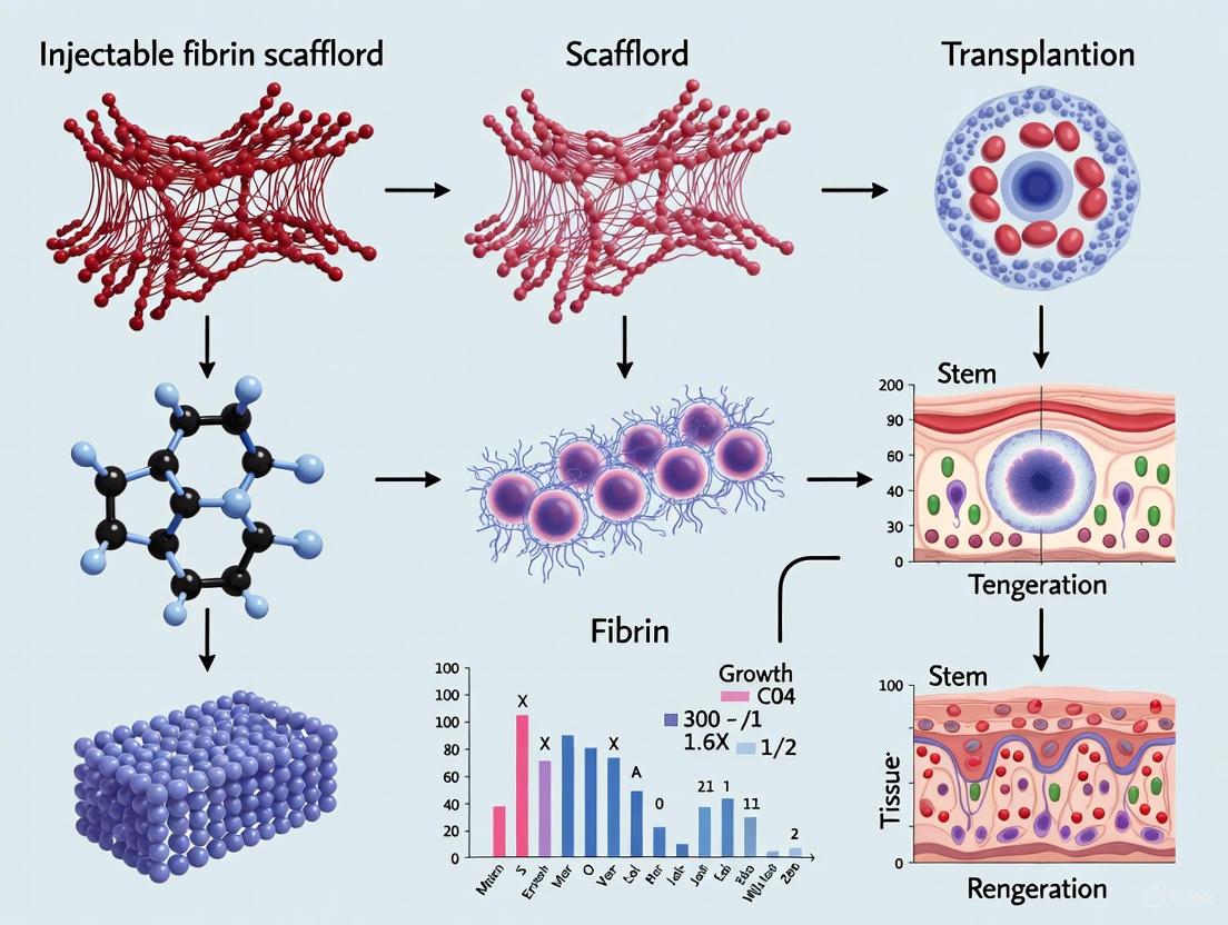
| Platelet-Rich Fibrin (PRF) / Injectable Fibrin Scaffold (IFS) | Autologous platelet concentrate serving as a source of innate growth factors and a biodegradable scaffold [36]. | Prepared via one-step centrifugation (e.g., 3000 rpm for 10 min) without anticoagulants [36]. |
| Mesenchymal Stromal Cells (MSCs) | Therapeutic cell population with multipotent differentiation capacity and potent immunomodulatory/paracrine activity [33]. | Isolated from bone marrow or other sources; cultured in DMEM with 10% FBS [36]. |
Protocol: Fabrication of Fibrin and Fibrin/PVA Composite Scaffolds
Preparation of an Injectable Fibrin Scaffold (IFS) from Whole Blood
This protocol yields a liquid, growth-factor-rich fibrin scaffold suitable for cell mixing and injection [36].
- Blood Collection and Anticoagulation: Draw whole blood into a tube containing an anticoagulant (e.g., heparin lithium) [36].
- Centrifugation: Centrifuge the blood at 3000 rpm for 10 minutes [36].
- IFS Harvest: Carefully collect approximately 1 mL of the transparent liquid (IFS) from approximately 3 mm above the junction point of the erythrocyte aggregation at the bottom of the tube [36].
- Characterization: The resulting IFS contains a loose fibrin network, platelets, white blood cells, and growth factors and can be used directly for cell suspension [36].
Fabrication of Cross-linked Fibrin/PVA Porous Scaffolds
This method utilizes emulsion templating to create macro-porous scaffolds with enhanced mechanical properties [35].
- Solution Preparation:
- Emulsion Formation:
- In a tube, mix PVA solution, decane (as a porogen), and a surfactant (e.g., Triton CG110) using a pulsatile method until a stable oil-in-water (O/W) emulsion is formed, indicated by increased viscosity [35].
- Coagulation and Casting:
- In a separate tube, combine the FNG solution, 1M CaCl₂, and thrombin, swirling gently for 10 seconds [35].
- Add the prepared emulsion to this coagulation mixture and blend for a further 30 seconds [35].
- Pour the mixture into a casting tray and incubate at 37°C for 1 hour to allow fibrin polymerization and clot formation [35].
- Cross-linking:
- Immerse the formed scaffold in a cross-linking solution (e.g., 0.2% v/v glutaraldehyde in 20% MES/80% Ethanol) for 4 hours [35].
- To investigate the effects of cross-linking degree, vary the glutaraldehyde concentration (e.g., 0.05%, 0.2%, 0.5%, or 1%) or use an alternative like EDC/NHS [35].
- Stabilize the cross-linked structure by adding 0.1% NaBH₄ as a reducing agent [35].
- Washing and Freeze-Drying:
Protocol: Cell Seeding and Construct Preparation for Injection
Cell Culture and Harvest
- Isolation and Expansion: Isolate MSCs from relevant tissue (e.g., rabbit bone marrow or skin) and culture in Dulbecco's modified Eagle's medium (DMEM) supplemented with 10% FBS, 100 U/mL penicillin, and 100 U/mL streptomycin at 37°C in a 5% CO₂ incubator. Passage cells at 80% confluence [36].
- Harvesting for Encapsulation: Upon reaching the desired confluence, harvest cells using standard trypsinization. Terminate the reaction with serum-containing medium, centrifuge the cell suspension, and resuspend the pellet in an appropriate carrier (e.g., sterile saline, culture medium, or the prepared IFS) to achieve a high cell density suitable for injection [33].
Incorporating Cells into Fibrin Constructs
- For IFS-based Constructs: Gently mix the concentrated cell suspension directly with the prepared IFS. The liquid scaffold can be drawn into a syringe immediately for injection [36].
- For Pre-formed Porous Scaffolds: Seed the concentrated cell suspension directly onto the freeze-dried scaffold. Allow time for cell attachment and infiltration under static culture conditions or use dynamic seeding methods before implantation [35].
Characterization and Quality Control of Constructs
Structural and Mechanical Analysis
- Scanning Electron Microscopy (SEM): To evaluate the pore morphology and microstructure. Scaffolds are fixed, dehydrated, freeze-dried, cross-sectioned, and sputter-coated with gold before imaging. Fibrin/PVA scaffolds fabricated via emulsion templating show interconnected porous structures with an average pore size of ~330 µm [36] [35].
- Porosity Measurement: Use techniques like the BET method with nitrogen adsorption to determine specific surface area and pore volume [36].
- Mechanical Testing: Perform uniaxial tensile testing to determine ultimate tensile strength and elongation. For example, cross-linked fibrin/PVA scaffolds can achieve an ultimate tensile strength of ~0.12 MPa with ~50% elongation [35].
- Degradation Profiling: Incubate scaffolds in a proteolytic solution (e.g., trypsin) and measure mass loss over time. The degradation rate can be controlled by varying the cross-linking type and degree [35].
Biological Activity and Cell-Scaffold Interaction
- Sustained Release of Growth Factors: Quantify the release kinetics of key growth factors (PDGF, VEGF, TGF-β1, IGF, FGF, EGF) from the scaffold using ELISA assay over a period of at least 15 days. IFS has been shown to provide sustained release for over two weeks [36].
- Cell Proliferation Assay (MTT Assay): Seed cells (e.g., BMSCs, fibroblasts) in the presence of the scaffold or its extracts and measure metabolic activity at 24-hour intervals to assess cytocompatibility and proliferative effects [36].
- Cell Morphology and Integration: Use SEM or fluorescence microscopy to confirm that cells (e.g., MSCs) can attach, penetrate, and proliferate within the scaffold with an elongated and stretched morphology [35].
Table 2: Quantitative Characterization of Fibrin-Based Scaffolds
| Parameter | Method | Exemplary Results from Literature |
|---|---|---|
| Average Pore Size | SEM Analysis | ~330 µm (Fibrin/PVA scaffold) [35] |
| Ultimate Tensile Strength | Mechanical Testing | ~0.12 MPa (Fibrin/PVA scaffold) [35] |
| Elongation at Break | Mechanical Testing | ~50% (Fibrin/PVA scaffold) [35] |
| Growth Factor Release | ELISA | Sustained release of PDGF, VEGF, TGF-β1, etc., for over 15 days (IFS) [36] |
| Cell Proliferation | MTT Assay | Promoted proliferation of BMSCs and skin fibroblasts (IFS) [36] |
In Vivo Application: Injection and Efficacy Assessment
Preclinical Injection Model
The efficacy of cell-laden fibrin constructs is typically evaluated in defect models, such as a murine full-thickness skin excision model [35].
- Animal Preparation: Anesthetize the animal and create a full-thickness skin wound.
- Construct Application:
- Post-Op Monitoring: Monitor animals for signs of infection and administer analgesics and antibiotics as needed (e.g., penicillin) [36].
Assessment of Therapeutic Efficacy
- Histological Analysis: Harvest tissue at defined endpoints. Process sections and stain with Masson's trichrome to evaluate collagen deposition, tissue architecture, and scaffold integration [36] [35].
- Evaluation of Healing Metrics: Compare test and control groups for:
- Wound Closure Rate: Acceleration of epithelial closure [35].
- Neodermal Formation: Promotion of deeper, well-structured tissue regeneration [35].
- Angiogenesis: Enhanced formation of new blood vessels, a key pro-angiogenic effect of fibrin [35].
- Collagen Deposition: Greater and more organized collagen fiber deposition [36] [35].
- Regulation of MMPs/TIMPs: Assess mechanisms by analyzing markers like MMP-1, MMP-9, TIMP-1, and TIMP-2, which indicate balanced tissue remodeling [36].
Application Notes
Rationale and Therapeutic Principle
The management of glioblastoma (GBM) remains a formidable challenge in neuro-oncology. Despite a standard of care that includes maximal safe surgical resection, radiotherapy, and temozolomide chemotherapy, the prognosis remains dismal with a median survival of only 12–15 months [37] [38]. A significant factor in therapeutic failure is the tumor's highly infiltrative nature, which prevents complete surgical removal, and the presence of a protective blood-brain barrier that limits effective drug delivery [37]. Furthermore, the glioblastoma microenvironment is profoundly immunosuppressive, characterized by tumor-associated macrophages, myeloid-derived suppressor cells, and regulatory T cells that create a niche conducive to tumor growth and immune evasion [38].
Neural stem cell (NSC)-based therapies represent a paradigm shift in addressing these challenges. NSCs possess an innate tumor-homing capability, enabling them to selectively migrate toward and infiltrate disseminated glioblastoma cells that remain after surgery [37]. This remarkable biological property allows NSCs to be engineered as targeted delivery vehicles for cytotoxic agents, effectively turning them into precision-guided weapons against residual tumor deposits. When combined with injectable fibrin-based scaffolds, this approach addresses two critical limitations in GBM therapy: ensuring the localized retention of therapeutic cells at the resection site and providing a supportive matrix that enhances cell survival and functionality [39] [40] [3].
The conceptual framework for this therapy involves creating an implantable "therapeutic depot" within the post-surgical cavity. This depot consists of engineered cytotoxic NSCs suspended within a fibrin scaffold that serves both as a physical matrix to prevent cell leakage and as a bioactive environment that supports NSC viability and function [3] [41]. The scaffold gradually degrades as the NSCs migrate toward infiltrative tumor cells, delivering their cytotoxic payload directly to the disease sites while sparing healthy brain tissue.
Key Molecular Targets and Mechanisms
The efficacy of cytotoxic NSC therapy depends on targeting key molecular pathways that drive glioblastoma pathogenesis and treatment resistance:
Oncogenic Signaling Pathways: EGFR amplification occurs in approximately 60% of GBM cases, making it a prime target for therapeutic intervention. The PI3K/AKT/mTOR pathway is another critical signaling cascade frequently dysregulated in GBM, promoting tumor growth, survival, and therapeutic resistance [38]. Engineered NSCs can be designed to deliver inhibitors specifically targeting these pathways.
Glioma Stem Cells (GSCs): GSCs represent a subpopulation of treatment-resistant cells with self-renewal capabilities that drive tumor recurrence [38]. These cells often exhibit enhanced survival mechanisms and contribute to the heterogeneous nature of GBM. Targeting GSCs is essential for achieving durable treatment responses.
Tumor Microenvironment (TME): The GBM TME is characterized by immunosuppressive elements including tumor-associated macrophages (TAMs), myeloid-derived suppressor cells (MDSCs), and regulatory T cells [38]. Modulating this microenvironment to overcome immune evasion represents a promising therapeutic strategy.
Angiogenic Pathways: Microvascular proliferation is a histological hallmark of GBM, driven largely by vascular endothelial growth factor (VEGF) signaling [37]. Targeting this neovascularization can disrupt tumor nutrient supply and limit growth.
Table 1: Key Molecular Targets in Glioblastoma Therapy
| Target Category | Specific Targets | Therapeutic Approach | Challenge |
|---|---|---|---|
| Receptor Tyrosine Kinases | EGFR, PDGFR | Enzyme/prodrug systems (e.g., CD::UPRT), targeted inhibitors | Heterogeneous expression, resistance mutations |
| Intracellular Signaling | PI3K/AKT/mTOR, RAS/MAPK | Pathway inhibitors, RNA interference | Pathway redundancy, feedback loops |
| Tumor Microenvironment | VEGF, TAMs, MDSCs | Anti-angiogenics, immunomodulators | Immunosuppressive niche, TME plasticity |
| Glioma Stem Cells | CD133, CD44, ALDH | Stem cell-directed cytotoxins | Cellular plasticity, dormancy |
Comparative Therapeutic Platforms
The exploration of NSC-based therapies occurs within a broader landscape of innovative GBM treatments. Several emerging modalities show promise in addressing the limitations of conventional therapies:
Electric Field Therapy: Tumor Treating Fields (TTFields) deliver low-intensity, intermediate-frequency alternating electric fields via scalp-mounted arrays to disrupt mitotic spindle formation in dividing cells [37]. While clinically validated, adoption remains limited due to the need for ≥18 hours daily wear and associated impacts on quality of life.
Radiofrequency Electromagnetic Fields: A novel approach uses amplitude-modulated 27.12 MHz radiofrequency electromagnetic fields (AM RF EMF) tuned to glioblastoma-specific frequencies [42]. This therapy has demonstrated particular effectiveness against tumor stem cells and depends on calcium channel Cav3.2 (CACNA1H) for its activity.
Immunotherapies: Checkpoint inhibitors, chimeric antigen receptor (CAR) T-cell therapies, dendritic cell vaccines, and oncolytic viruses represent promising avenues to modulate immune responses against GBM [37] [43]. However, clinical success remains limited due to tumor heterogeneity and the immunosuppressive TME.
Biomaterial-Based Strategies: Beyond fibrin scaffolds, other natural and synthetic biomaterials are being investigated as platforms for localized therapy delivery. These include hyaluronic acid-based hydrogels, nanoparticle systems, and polymer-based slow-release formulations [39] [3].
Table 2: Comparison of Emerging Glioblastoma Therapeutic Platforms
| Therapeutic Platform | Mechanism of Action | Advantages | Limitations |
|---|---|---|---|
| Cytotoxic NSCs in Fibrin Scaffold | Tumor-homing delivery of therapeutic agents | Targeted delivery, bypasses BBB, fills resection cavity | Cell sourcing, genetic stability, safety monitoring |
| TTFields | Disrupts mitotic spindle via alternating electric fields | Non-invasive, proven efficacy in trials | Require extended daily use (≥18h), dermatological effects |
| AM RF EMF | Tumor-specific frequency disruption of cell division | Systemic delivery, targets cancer stem cells | Early development, mechanism not fully elucidated |
| Immunotherapies | Enhances anti-tumor immune responses | Potential for durable response, immune memory | Limited efficacy in immunosuppressive TME |
| Local Chemotherapy | Direct cytotoxic drug delivery to resection cavity | Bypasses BBB, high local concentration | Limited penetration into brain parenchyma |
Experimental Protocols
Fabrication of Aligned Fibrin Nanofiber Hydrogels
The structural alignment of fibrin scaffolds is critical for directing NSC migration and enhancing their tumor-homing capabilities. The following protocol adapts established methods for creating hierarchically aligned fibrin nanofiber hydrogel scaffolds (AFGs) [39]:
Materials:
- Fibrinogen from human plasma (Sigma-Aldrich)
- Thrombin from human plasma (Sigma-Aldrich)
- Polyethylene oxide (PEO, MW ~900,000; Sigma-Aldrich)
- Calcium chloride (CaCl₂)
- Electrospinning apparatus with rotating collector
- Sterile phosphate-buffered saline (PBS)
- 70% ethanol for sterilization
Procedure:
- Prepare electrospinning solution by dissolving fibrinogen in distilled water at a concentration of 80-100 mg/mL.
- Add polyethylene oxide (PEO) to the fibrinogen solution at a concentration of 8 mg/mL to enhance fiber formation [39].
- Load the solution into a syringe with a metallic needle (21-25 gauge) and place it in the electrospinning apparatus.
- Prepare a coagulation bath containing 4 U/mL thrombin and 50 mM CaCl₂ in a container with a rotating mandrel collector.
- Set the electrospinning parameters: voltage = 5 kV, flow rate = 3 mL/min, needle-to-collector distance = 10-15 cm.
- Rotate the collector at 50 rpm to ensure alignment of the fibrin fibers during collection [39].
- Collect the electrospun fibrin fibers as they form aligned bundles in the coagulation bath.
- Sterilize the fibrin fiber bundles by immersion in 75% ethanol under ultraviolet light for 30 minutes, followed by washing with sterile distilled water.
- Characterize the scaffold properties: fiber diameter (~100 nm), Young's modulus (~1.5 kPa), and degradation profile.
Quality Control:
- Verify fiber alignment using scanning electron microscopy (SEM)
- Confirm sterility through microbiological testing
- Assess mechanical properties using rheometry
- Validate biocompatibility through in vitro cell viability assays
Engineering Cytotoxic Neural Stem Cells
The creation of therapeutic NSCs involves genetic modification to express cytotoxic agents specifically activated within the tumor environment:
Materials:
- Human neural stem cells (e.g., from fetal tissue or induced pluripotent stem cells)
- Lentiviral vectors encoding therapeutic transgenes
- Cytotoxic payload constructs (e.g., cytosine deaminase::uracil phosphoribosyltransferase fusion protein)
- Polybrene transduction enhancer
- Puromycin for selection
- NSC culture medium: DMEM/F-12 with B27 supplement, EGF (20 ng/mL), FGF-2 (20 ng/mL)
- Differentiation medium: DMEM/F-12 with B27 supplement, 1% FBS
Genetic Engineering Protocol:
- Culture NSCs in complete medium under standard conditions (37°C, 5% CO₂) until 70-80% confluence.
- Design lentiviral vectors encoding:
- A therapeutic transgene (e.g., CD::UPRT for 5-FC conversion to 5-FU)
- A reporter gene (e.g., GFP or luciferase for tracking)
- A selection marker (e.g., puromycin resistance)
- Produce lentiviral particles using HEK293T packaging cells and concentrate to high titer (>10⁸ IU/mL).
- Transduce NSCs at an MOI of 10-20 in the presence of 8 μg/mL Polybrene by spinoculation (centrifugation at 800 × g for 30 minutes at 32°C).
- After 24 hours, replace the transduction medium with fresh complete medium.
- Begin antibiotic selection 48 hours post-transduction using 1-2 μg/mL puromycin for 7-10 days.
- Expand the selected polyclonal population and validate transgene expression through:
- Fluorescence microscopy (for reporter genes)
- Western blotting (for therapeutic transgenes)
- Functional assays (e.g., 5-FC conversion efficiency)
Functional Validation:
- Assess tumor-homing capability using Transwell migration assays toward GBM-conditioned medium.
- Evaluate cytotoxic activity by co-culturing engineered NSCs with patient-derived GBM cells in the presence of prodrug (5-FC).
- Verify safety profile by testing engineered NSC toxicity toward normal human astrocytes.
- Determine in vivo distribution and persistence using bioluminescent imaging in orthotopic models.
In Vivo Evaluation in Orthotopic Glioblastoma Models
The therapeutic efficacy of the NSC-fibrin scaffold construct must be evaluated in clinically relevant animal models:
Materials:
- Immunocompromised mice (e.g., NOD-scid or NSG)
- Patient-derived glioblastoma stem cells (GSCs) or established GBM cell lines (e.g., U87, GL261)
- Stereotactic frame for intracranial injections
- MRI system for in vivo monitoring
- Bioluminescence imaging system (if using luciferase-expressing cells)
Surgical Implantation Procedure:
- Anesthetize mice using ketamine/xylazine or isoflurane inhalation.
- Secure the mouse in a stereotactic frame and expose the skull through a midline incision.
- Create a burr hole at coordinates relative to bregma: +1.0 mm anteroposterior, +2.0 mm mediolateral.
- Inject 2-3 μL of GBM cells (50,000-100,000 cells) suspended in PBS at a depth of 2.5-3.0 mm from the dura to establish orthotopic tumors.
- Allow tumors to establish for 7-10 days, confirmed by MRI or bioluminescence imaging.
- Perform a second surgery to debulk the tumor (simulating human resection) and implant the therapeutic construct:
- Prepare the NSC-fibrin scaffold by mixing 2 × 10⁶ engineered NSCs with 50 μL fibrinogen solution, then adding 50 μL thrombin solution (4 U/mL) with CaCl₂ (50 mM)
- Quickly inject the mixture into the resection cavity before gelation occurs
- Alternatively, implant pre-formed AFG bundles seeded with NSCs [39]
- For prodrug-activated systems, administer 5-fluorocytosine (500 mg/kg) intraperitoneally daily beginning 3 days post-implantation.
Assessment Parameters:
- Survival analysis using Kaplan-Meier curves with log-rank test
- Tumor volume monitoring by weekly MRI
- NSC distribution tracking via bioluminescence imaging
- Histopathological analysis at endpoint:
- H&E staining for general morphology
- Immunofluorescence for NSC markers (Nestin, SOX2), tumor markers (GFAP, Ki-67), and apoptosis (TUNEL)
- Analysis of tumor burden, proliferation index, and apoptosis rates
Biosafety and Biocompatibility Assessment
Rigorous safety evaluation is essential before clinical translation:
Comprehensive Safety Protocol:
- Ectopic growth assay: Subcutaneous implantation of engineered NSCs in immunocompromised mice to assess tumorigenic potential.
- Systemic distribution analysis: Quantitative PCR for human Alu sequences in distant organs to detect unintended NSC migration.
- Immune response profiling: Flow cytometry analysis of T-cell infiltration and cytokine levels in treated brains.
- Off-target toxicity assessment: Histopathological examination of major organs (liver, kidney, heart, lungs) for damage.
- Scaffold degradation monitoring: MRI tracking of fibrin scaffold resolution over time.
The Scientist's Toolkit
Table 3: Essential Research Reagents and Materials
| Category/Item | Specifications | Research Function | Notes for Application |
|---|---|---|---|
| Fibrinogen | Human plasma-derived, >80% clottable | Scaffold base material | Adjust concentration to control pore size and mechanical properties |
| Thrombin | Human plasma-derived, 1000 NIH units/mg | Fibrin cross-linking | Concentration controls gelation kinetics |
| Neural Stem Cells | Fetal-derived or iPSC-derived, nestin+/SOX2+ | Therapeutic cell vehicle | Pre-validate tumor-homing capacity before engineering |
| Lentiviral Vectors | VSV-G pseudotyped, third-generation | Genetic modification | Use inducible systems for safety control |
| 5-Fluorocytosine | >99% purity, pharmaceutical grade | Prodrug for CD::UPRT system | Optimize dosing schedule for maximal efficacy/minimal toxicity |
| Matrigel | Growth factor reduced | Tumor cell implantation | Provides initial support for tumor engraftment |
| Temozolomide | Pharmaceutical grade | Standard care comparator | Essential for clinically relevant study design |
| GBM Cell Lines | Patient-derived spheres or established lines (U87, GL261) | Disease modeling | Use multiple lines to capture heterogeneity |
Signaling Pathways and Workflow Diagrams
The integration of engineered neural stem cells with fibrin scaffold technology represents a promising multimodal approach for addressing the formidable challenge of glioblastoma recurrence. This platform combines the tumor-homing capability of NSCs with the sustained local delivery advantages of biomaterial scaffolds, creating a therapeutic reservoir within the post-surgical cavity that actively targets residual infiltrative disease.
Future developments in this field will likely focus on several key areas:
- Personalization approaches that match therapeutic NSCs to individual patient tumor profiles
- Multi-mechanistic engineering of NSCs to simultaneously target multiple oncogenic pathways
- Advanced scaffold design incorporating controlled release of additional therapeutic agents
- Non-invasive monitoring systems using reporter genes for real-time tracking of NSC distribution
- Combination strategies with immunotherapies to overcome the immunosuppressive tumor microenvironment
As research progresses, this technology platform holds significant potential to transform the management of glioblastoma by addressing the critical challenge of post-surgical recurrence that currently limits patient survival. The continued refinement of both cellular and biomaterial components will be essential for successful clinical translation.
Enhancing Liver Regeneration with Adipose-Derived Stem Cells (ADSCs)
Application Notes
The use of Adipose-Derived Stem Cells (ADSCs) delivered via injectable fibrin scaffolds represents a significant advancement in regenerative strategies for liver repair. This approach directly addresses the critical clinical challenge of post-hepatectomy liver failure (PHLF), a major cause of morbidity and mortality following liver resection [44]. By combining the regenerative paracrine signaling of ADSCs with the structural and protective benefits of a fibrin matrix, this therapy enhances the liver's innate regenerative capacity, offering a promising solution for patients with compromised liver function.
The therapeutic efficacy of this approach stems from a synergistic mechanism of action. Fibrin scaffolds act as a protective, three-dimensional microenvironment that significantly improves the retention and survival of transplanted ADSCs at the injury site [45]. Research demonstrates that suspending human mesenchymal stem cells in a fibrin matrix increased initial retention in a surgical resection cavity by 2-fold and prolonged cell persistence by 3-fold compared to conventional delivery methods [45]. This scaffold facilitates a sustained and localized release of crucial hepatotropic factors from the encapsulated ADSCs.
The encapsulated ADSCs exert their primary effects through a potent paracrine mechanism. In vitro studies show that ADSCs in a fibrin gel (gelADSC) sustain the release of key growth factors—hepatocyte growth factor (HGF), vascular endothelial growth factor (VEGF), and stromal cell-derived factor-1 (SDF-1)—for at least 7 days [44]. This sustained secretion is critical for activating downstream regenerative pathways in the injured liver. Furthermore, the use of a fibrin scaffold significantly enhances the secretion of extracellular vesicles (EVs) from ADSCs, which are now recognized as essential mediators of intercellular communication that promote tissue repair [44] [46].
The combined action of this system orchestrates liver regeneration through multiple interconnected pathways:
- Upregulation of Cell Cycle Proliferation: Directly stimulating hepatocyte proliferation to replenish lost parenchymal mass.
- Promotion of Fatty Acid Oxidation: Enhancing metabolic function and energy production in the regenerating liver.
- Activation of Mitochondrial Biogenesis and Mitophagy: Recent research confirms that ADSCs modulate mitochondrial function via the DDIT4/PGC-1α axis, restoring mitochondrial mass and function after injury, which is crucial for cellular recovery [47].
- Angiogenesis Induction: Supporting the formation of new blood vessels to restore adequate vascularization and nutrient delivery to the regenerating tissue [44].
The therapeutic potential of gelADSC has demonstrated particular promise in compromised liver conditions. Experimental data reveals these effects are significantly favorable compared to intravenously administered ADSCs, especially in models of chronic hepatitis, highlighting its enhanced efficacy in diseased livers with reduced regenerative capacity [44].
Experimental Protocols
Protocol: Preparation of ADSC-Loaded Fibrin Gel (gelADSC)
Objective: To create a standardized, injectable fibrin scaffold encapsulating ADSCs for transplantation in liver regeneration studies [44] [45].
Materials:
- Adipose-Derived Stem Cells (ADSCs): Isolated from human adipose tissue (e.g., lipoaspirates), culture-expanded, and characterized by flow cytometry for positive markers (CD44, CD73, CD90, CD105) and negative markers (CD31, CD45) [48] [49].
- Fibrin Sealant Kit: Clinically approved, such as TISSEEL (Baxter Healthcare Corp.), containing sealer (fibrinogen) and thrombin solutions [45].
- Cell Culture Medium: DMEM supplemented with 10% FBS and 1% penicillin/streptomycin.
- Sterile Surgical Instruments: for in vivo implantation.
Procedure:
- ADSC Preparation: Harvest ADSCs at 80-90% confluence using 0.25% trypsin/EDTA. Centrifuge, resuspend in culture medium, and perform a cell count. Adjust concentration to 1.25–6.25 x 10^6 cells in an 8 µL volume [45].
- Scaffold Formation: Under sterile conditions, mix the 8 µL ADSC suspension thoroughly with 8 µL of the fibrinogen sealer solution.
- Gelation Initiation: Add 8 µL of the thrombin solution (concentration: 400–625 U/mL) to the ADSC-fibrinogen mixture. Pipette gently for approximately 30 seconds to initiate polymerization and ensure uniform cell distribution [45].
- Incubation: Allow the mixture to solidify completely in a humidified incubator (37°C, 5% CO2) for 30-60 minutes. For in vivo application, the gel can be formed ex vivo for implantation or injected in a liquid state to polymerize in situ.
- In Vivo Implantation: In rodent hepatectomy models, the gelADSC construct is typically applied directly to the resection surface or injected into the remaining liver lobe [44].
Protocol: In Vivo Evaluation in a Murine Hepatectomy Model
Objective: To assess the efficacy of gelADSC in enhancing liver regeneration post-partial hepatectomy [44].
Materials:
- Experimental Animals: C57BL/6 mice (e.g., 8-12 weeks old), including healthy and chronic hepatitis models.
- gelADSC Construct: Prepared as per Protocol 2.1.
- Control Groups: Untreated, fibrin scaffold only, and intravenous ADSC injection.
- Analytical Reagents: Reagents for RNA/protein extraction, histology (formalin, paraffin), and blood serum biochemistry.
Procedure:
- Surgical Procedure: Perform a 70% partial hepatectomy under general anesthesia using standard surgical techniques.
- Treatment Application: Randomly assign animals to treatment groups. Apply the pre-formed gelADSC construct to the resection site or remnant liver.
- Post-operative Monitoring: Monitor animals for recovery and survival. Collect tissue and serum samples at predetermined endpoints (e.g., 24, 48, 72 hours, 7 days post-surgery).
- Outcome Analysis:
- Liver Regeneration: Calculate the liver-to-body weight ratio.
- Gene Expression: Analyze the expression of cell cycle genes (e.g., Cyclins, PCNA) and fatty acid oxidation genes (e.g., PPARα, CPT1A) via qRT-PCR [44].
- Histology: Perform H&E staining to assess morphology and immunohistochemistry for proliferation markers (e.g., Ki-67).
- Functional Assessment: Measure serum albumin, ALT, AST levels to evaluate liver function and injury.
- EV Analysis: Isitate and quantify EVs from liver homogenates to correlate their levels with regenerative outcomes [44].
Data Presentation
Table 1: Key Growth Factors Sustained Released by gelADSC In Vitro [44]
| Growth Factor | Function in Liver Regeneration | Sustained Release Profile |
|---|---|---|
| HGF (Hepatocyte Growth Factor) | Primary mitogen for hepatocytes; stimulates proliferation. | Sustained release for at least 7 days. |
| VEGF (Vascular Endothelial Growth Factor) | Promotes angiogenesis; establishes blood supply. | Sustained release for at least 7 days. |
| SDF-1 (Stromal cell-Derived Factor-1) | Chemoattractant for progenitor cells; supports cell homing. | Sustained release for at least 7 days. |
Table 2: In Vivo Therapeutic Outcomes of gelADSC in Murine Hepatectomy Models [44]
| Experimental Model | Key Findings | Proposed Mechanism |
|---|---|---|
| Normal Mice | Significantly enhanced postoperative liver regeneration. | Upregulation of the cell cycle and fatty acid oxidation pathways. |
| Mice with Chronic Hepatitis | Therapeutic effects potentially superior to intravenous ADSCs. | Enhanced secretion of extracellular vesicles (EVs) promoting fatty acid oxidation. |
Table 3: Research Reagent Solutions for ADSC-Fibrin Scaffold Experiments
| Reagent / Material | Function / Application | Exemplar Product / Composition |
|---|---|---|
| Adipose-Derived Stem Cells (ADSCs) | Cellular source for paracrine factor secretion and differentiation. | Isolated from human lipoaspirate; characterized by CD44+, CD90+, CD105+, CD31-, CD45- [48] [49]. |
| Fibrin Sealant | Forms the injectable, biodegradable scaffold for cell encapsulation. | TISSEEL (Fibrinogen + Thrombin) [45]. |
| Chondrogenic/Osteogenic/Adipogenic Differentiation Kits | Functional validation of ADSC multipotency. | Media containing TGF-β3, BMPs, Ascorbate, Dexamethasone [48]. |
| DMEM Culture Medium | Base medium for cell expansion and scaffold culture. | Dulbecco's Modified Eagle Medium, high glucose, with 10% FBS. |
| Antibodies for Flow Cytometry | Phenotypic characterization of ADSCs. | Anti-human CD44, CD73, CD90, CD105, CD31, CD45 [48] [49]. |
| qRT-PCR Reagents | Analysis of lineage-specific gene expression (e.g., for hepatogenesis). | Primers for Albumin, HNF4α, Cyclins, PPARα [44]. |
Signaling Pathways and Workflow Visualizations
The repair and regeneration of neural tissue represent a significant challenge in regenerative medicine. Injectable fibrin scaffolds have emerged as a pivotal technology in this domain, particularly for cell transplantation research. These scaffolds serve as a temporary, three-dimensional extracellular matrix that mimics the native neural microenvironment. Fibrin, a natural polymer derived from fibrinogen, facilitates critical cellular processes including cell adhesion, proliferation, and differentiation. Its mechanical properties can be finely tuned through cross-linking agents and concentration adjustments, making it exceptionally suitable for supporting delicate neural tissues. When used as an injectable carrier, fibrin enables minimally invasive delivery of therapeutic cells, such as neural progenitors or stem cells, to lesion sites in the central nervous system. This application note details the use of injectable fibrin scaffolds to enhance neuronal viability and promote neurite outgrowth—two fundamental requirements for successful neural tissue engineering.
Application Notes: Key Experimental Findings
Recent studies have demonstrated the efficacy of fibrin scaffolds in supporting neural cells. The data summarized below provide quantitative evidence of their role in enhancing cell survival and inducing neurite extension.
Table 1: Effects of Genipin on hiPSC-Derived Neural Progenitors in 2D Culture
| Parameter | Control (0 mM Genipin) | 0.05 mM Genipin | ≥0.25 mM Genipin |
|---|---|---|---|
| Cell Viability (after 8 days) | Baseline | Significantly increased compared to control [50] | Induced cell death within 24 hours [50] |
| Neurite Length (after 9 days) | Baseline | Substantially greater [50] | Not tested (toxic) |
| Neurite Branching (after 9 days) | Baseline | Substantially greater [50] | Not tested (toxic) |
| Hypothesized Signaling Pathway | Not applicable | NO-cGMP-dependent protein kinase, followed by ERK activation [50] | Not applicable |
Table 2: Properties of 3D Fibrin Scaffolds Cross-linked with Genipin
| Genipin Concentration | Scaffold Degradation Time | Compressive Modulus | Fiber Density | Pore Size | Neurite Outgrowth |
|---|---|---|---|---|---|
| 0 mM (Fibrin only) | ~5 days [50] | Not specified | Baseline | Baseline | Not supported after degradation |
| 1.0 - 2.5 mM | >14 days [50] | Similar to spinal cord tissue (at 2.5 mM) [50] | Increased [50] | Consistent or increased [50] | Induced for 14 days [50] |
| 5.0 - 10.0 mM | >14 days [50] | Increased (after initial decrease) [50] | Significantly increased [50] | Decreased (at 10 mM) [50] | Not the focus of study |
The data from the genipin-crosslinked fibrin scaffolds show a clear advantage over standard fibrin, particularly in terms of scaffold longevity, which is essential for the slow process of neuronal maturation [50]. Furthermore, a separate study on injectable fibrin glue for skeletal myoblast transplantation in ischemic myocardium corroborates the general utility of fibrin, showing it significantly increases transplanted cell survival area and reduces infarct size [40].
Experimental Protocols
Protocol: Fabrication of Genipin-Crosslinked Fibrin Scaffolds for Neural Aggregates
This protocol describes the creation of 3D fibrin scaffolds with enhanced mechanical stability for the culture of human induced pluripotent stem cell (hiPSC)-derived neural aggregates [50].
I. Materials
- Fibrinogen (from human plasma)
- Thrombin (from human plasma)
- Genipin
- Calcium Chloride (CaCl₂)
- Phosphate Buffered Saline (PBS)
- Cell culture media
II. Method
- Preparation of Fibrinogen-Genipin Solution: Dissolve fibrinogen in PBS to a final concentration of 20 mg/mL. Add genipin to this solution to achieve the desired final cross-linking concentration (e.g., 1.5 mM, 2.5 mM). Mix thoroughly.
- Preparation of Thrombin Solution: Dissolve thrombin in a 40 mM CaCl₂ solution.
- Scaffold Polymerization: Combine the fibrinogen-genipin solution with the thrombin solution at a 1:1 ratio. For example, mix 500 µL of each solution in a well of a 24-well plate.
- Gelation: Allow the mixture to polymerize into a gel at 37°C for 30-60 minutes.
- Equilibration: After polymerization, carefully add cell culture media to submerge the scaffold and allow it to equilibrate for at least 1 hour before cell seeding.
Protocol: Seeding hiPSC-Derived Neural Aggregates onto 3D Fibrin Scaffolds
This protocol follows the successful methodology for promoting neurite outgrowth within 3D scaffolds [50].
I. Materials
- hiPSC-derived neural aggregates
- Aggrewell plates
- Complete neural differentiation media
- Genipin-crosslinked fibrin scaffolds (from Protocol 3.1)
II. Method
- Neural Aggregate Formation: Culture hiPSC-derived neural progenitors in Aggrewell plates for 7 days to form aggregates. Change 75% of the media every other day.
- Selection: Visually select neural aggregates that exhibit signs of differentiation (approximately 50% at day 7).
- Seeding: Gently transfer individual neural aggregates onto the surface of the pre-equilibrated fibrin scaffolds.
- Culture: Culture the seeded scaffolds for up to 14 days, with media changes as per standard neural culture protocols. The scaffolds will support continued differentiation and neurite extension into the 3D matrix.
Protocol: 3D Bioprinting of a Fibrin-Based Dermal Scaffold for DRG Neuron Culture
This protocol adapts a advanced bioprinting approach to create an innervated tissue model for studying peripheral nerve regeneration [51].
I. Materials
- Bioink: 2% Fibrinogen, 0.5% Alginate, 10% Gelatin
- Thrombin/CaCl₂ cross-linking solution
- NIHT-3T3 fibroblasts
- Primary murine Dorsal Root Ganglion (DRG) neurons
- Microextrusion bioprinter with temperature-controlled printhead and stage
II. Method
- Bioink Preparation: Mix the fibrinogen, alginate, and gelatin solutions to form the bioink. Keep the bioink at a temperature above its gelation temperature (Tg ≈ 28°C) to maintain viscosity for printing.
- Cell Incorporation: Blend NIHT-3T3 fibroblasts uniformly into the bioink at the desired density.
- Bioprinting Parameters:
- Nozzle Temperature: 29-31°C
- Printing Stage Temperature: Below Tg (e.g., 15-25°C) to facilitate rapid gelation.
- Printing Speed: 800 mm/min
- Z-printing distance: Optimize for strand width (e.g., ~225 µm for a 300 µm nozzle).
- Scaffold Fabrication: Print the cell-laden bioink into a multilayered, dermal-like construct.
- Cross-linking: After printing, immerse the scaffold in a thrombin/CaCl₂ solution to cross-link the fibrinogen to fibrin and the alginate to calcium alginate, creating a stable composite hydrogel.
- DRG Seeding and Culture: Seed primary murine DRG neurons on top of the bioprinted scaffold. Culture the co-cystem to study enhanced neurite outgrowth and complex branching patterns supported by the fibroblasts in the 3D environment.
Signaling Pathways and Experimental Workflows
Genipin-Induced Neurite Outgrowth Pathway
The following diagram illustrates the hypothesized signaling cascade by which genipin promotes neurite extension in neural cells, based on studies of Neuro2A and PC12 cells [50].
Workflow for 3D Bioprinting an Innervated Tissue Construct
This workflow outlines the process for creating a bioprinted dermal-like scaffold co-cultured with sensory neurons to study axonal regeneration [51].
The Scientist's Toolkit: Research Reagent Solutions
Table 3: Essential Materials for Neural Tissue Engineering with Fibrin Scaffolds
| Reagent/Material | Function in the Protocol | Examples & Notes |
|---|---|---|
| Fibrinogen | The primary structural protein component of the scaffold, forms the 3D fibrous network upon polymerization [50] [3]. | Human plasma-derived; typically used at 20 mg/mL [50]. |
| Thrombin | Serine protease that cleaves fibrinogen to initiate fibrin polymerization [50] [3]. | Used in combination with CaCl₂ as a cross-linking solution. |
| Genipin | Natural cross-linker that stabilizes fibrin scaffolds, decreases degradation rate, and possesses neuritogenic and neuroprotective properties [50]. | Concentrations of 1.5-2.5 mM are effective for neural cultures without toxicity [50]. |
| Alginate | A polysaccharide used in composite bioinks to provide immediate structural integrity during and after 3D bioprinting [51]. | Often cross-linked with Ca²⁺ ions. Used at 0.5% in bioinks [51]. |
| Gelatin | A denatured collagen used in bioinks for its thermally reversible gelation properties, improving printability [51]. | Provides cell-adhesion motifs. Key component for temperature-controlled extrusion [51]. |
| hiPSC-Derived Neural Progenitors | A clinically relevant cell source for generating human neurons in vitro [50]. | Can be pre-differentiated into neural aggregates before seeding on scaffolds [50]. |
| Dorsal Root Ganglion (DRG) Neurons | Primary sensory neurons used for modeling peripheral nerve regeneration and neurite outgrowth [51]. | Often used in co-culture with supportive cells like fibroblasts. |
| NIHT-3T3 Fibroblasts | A supportive feeder cell line that secretes factors promoting neuronal health and axonal regeneration in 3D co-culture systems [51]. | Can be embedded within the bioprinted scaffold. |
The field of bone tissue engineering increasingly focuses on combining fibrin-based scaffolds with osteoconductive materials such as hydroxyapatite (HA) and β-tricalcium phosphate (β-TCP) to develop advanced regenerative solutions. This approach is particularly pivotal in the context of injectable fibrin scaffolds for cell transplantation, as it synergizes the biological benefits of fibrin with the structural and osteoconductive properties of bone graft substitutes. Fibrin, a natural polymer derived from blood, serves as an excellent cell delivery vehicle and provides a native environment for cellular attachment, proliferation, and the sustained release of growth factors [52] [53]. When combined with osteoconductive materials, these composites address a critical challenge in bone regeneration: creating a scaffold that is both biologically active and mechanically supportive for osteoprogenitor cell migration and new bone formation.
The rationale for this combinatorial strategy stems from the limitations of using these materials in isolation. While synthetic biomaterials like HA and β-TCP provide excellent osteoconductive scaffolds and structural support, they often lack the inherent bioactivity to significantly accelerate the regeneration process [30]. Conversely, fibrin alone, despite its excellent cellular interactions and growth factor retention capabilities, does not provide the optimal mechanical stability or osteoconductive guidance for critical-sized bone defects [54]. The integration of these materials creates a composite matrix that leverages the advantages of each component, resulting in a microenvironment that actively supports the entire bone regeneration cascade, from initial cell recruitment to final matrix mineralization and remodeling [55] [30].
Fibrin-Based Biomaterials: Properties and Versatility
Fibrin-based biomaterials represent a class of autologous-derived bioactive matrices that have evolved significantly since the first generation of platelet concentrates. Second-generation platelet-rich fibrin (PRF) forms a dense, natural fibrin matrix enriched with platelets, leukocytes, and a multitude of growth factors through a simple centrifugation process without anticoagulants [53]. This preparation method results in a scaffold that gradually releases physiological doses of growth factors such as platelet-derived growth factor (PDGF), transforming growth factor-β (TGF-β), and vascular endothelial growth factor (VEGF) over an extended period, making it ideal for long-term tissue regeneration processes [53]. The fibrin architecture provides a natural substrate for cell migration and attachment, while the incorporated growth factors stimulate angiogenesis and osteogenesis, creating a conducive microenvironment for bone healing.
The versatility of fibrin scaffolds is evidenced by their various formulations, each designed for specific clinical applications. Injectable PRF (i-PRF), prepared at low centrifugation speeds, remains in liquid form and can be mixed with bone grafts or cells for minimally invasive applications [53]. This format is particularly valuable for creating injectable tissue-engineered bone constructs, as demonstrated in studies where i-PRF combined with osteoblastic bone marrow mesenchymal stem cell (BMSC) sheets was used to construct injectable bone grafts that significantly enhanced ectopic bone formation in nude mice [56]. Other variants include leukocyte-rich L-PRF, which forms a dense fibrin membrane suitable as a barrier membrane; advanced PRF (A-PRF), processed at lower speeds to enhance leukocyte and cytokine retention; and T-PRF, prepared in titanium tubes to avoid silica-induced clotting [54] [53]. This diversity in formulation allows researchers to select the most appropriate fibrin derivative based on the specific requirements of their bone regeneration strategy.
Osteoconductive Materials: HA and β-TCP Properties
Hydroxyapatite (HA) and beta-tricalcium phosphate (β-TCP) represent two of the most widely used osteoconductive biomaterials in bone tissue engineering. HA is a chemical analog of bone mineral composition, making it highly biocompatible and bioactive [30]. Its interconnected porous structure, high tensile strength, and resorption capability make it an effective scaffold for bone regeneration. Modern advancements have led to the development of nano-structured HA, which demonstrates enhanced protein adsorption, cell adhesion, and surface roughness, thereby improving mechanical and biological properties for tissue healing [30]. However, conventional synthetic HA has coarser crystals than natural bone apatite, which can potentially limit its bioactivity in certain applications.
β-TCP is another promising calcium phosphate ceramic known for its biocompatibility and resorbability. Unlike HA, β-TCP typically degrades more rapidly in physiological environments through chemical dissolution and osteoclastic activity, creating space for new bone formation [30]. This material dissolves in acids released by osteoclasts or macrophages, and its degradation products can be assimilated by the human body without adverse effects [30]. β-TCP is often used in composite grafts for periodontal and alveolar bone regeneration, where its combination with HA creates a biomimetic material that balances stability with resorption kinetics [30]. Both materials serve as excellent osteoconductive matrices, providing a three-dimensional scaffold that supports the migration, proliferation, and differentiation of osteoprogenitor cells, while also facilitating vascular invasion, which is crucial for successful bone regeneration.
Table 1: Key Properties of Osteoconductive Materials Used with Fibrin
| Material | Crystal Structure | Degradation Rate | Osteoconductivity | Key Advantages |
|---|---|---|---|---|
| Hydroxyapatite (HA) | Analogous to bone mineral | Slow degradation (<1 year) | Excellent | High biocompatibility, promotes direct bone bonding |
| β-Tricalcium Phosphate (β-TCP) | More soluble than HA | Moderate degradation (6-18 months) | Excellent | Balanced resorption rate, space-making properties |
| HA/β-TCP Composites | Biphasic calcium phosphate | Tunable degradation | Enhanced | Optimized resorption profile, improved bioactivity |
Mechanisms of Bone Regeneration with Fibrin Composites
The combination of fibrin with osteoconductive materials creates a synergistic environment that enhances multiple aspects of the bone regeneration process. The fundamental mechanism begins with the fibrin component establishing a provisional matrix that recruits mesenchymal stem cells (MSCs) and osteoprogenitor cells through the gradual release of chemotactic growth factors [53]. Simultaneously, the osteoconductive materials (HA or β-TCP) provide a mechanical scaffold for these cells to adhere, proliferate, and differentiate along the osteoblastic lineage. The interconnected porous structure of these composites facilitates vascular invasion, which is critical for delivering nutrients and oxygen to the developing tissue, while also removing metabolic waste products [30]. This coordinated sequence of events creates an optimal microenvironment for the sequential stages of bone healing: inflammation, repair, and remodeling.
At the molecular level, these composites influence bone regeneration through multiple signaling pathways. The growth factors released from the fibrin matrix, including TGF-β, PDGF, and VEGF, activate intracellular signaling cascades such as SMAD, MAPK, and PI3K/Akt pathways, which regulate cell proliferation, differentiation, and matrix production [53]. The mechanical properties of the osteoconductive materials additionally provide physical cues that influence cellular behavior through mechanotransduction pathways. The degradation products of calcium phosphate materials, particularly calcium and phosphate ions, have been shown to stimulate osteogenic differentiation of MSCs and promote mineralization of the extracellular matrix [30]. This multifaceted mechanism results in enhanced osteogenic differentiation and accelerated bone matrix deposition compared to either component alone.
Diagram 1: Mechanism of bone regeneration with fibrin composites, showing how cellular recruitment, molecular signaling, and scaffold properties interact to produce bone regeneration outcomes.
Application Notes and Protocols
Fibrin with Hydroxyapatite for Periodontal Regeneration
The combination of PRF with hydroxyapatite has demonstrated significant efficacy in periodontal bone regeneration and periapical defect healing. In a clinical case report documenting the treatment of a large periapical lesion, the surgical protocol involved reflecting a mucoperiosteal flap after administration of local anesthesia with adrenaline [55]. Following debridement of the inflammatory tissue and root-end resection, the defect was filled with a mixture of PRF and HA bone graft crystals [55]. The PRF was prepared by collecting 20mL of venous blood without anticoagulant and centrifuging it at 3,000 rpm for 10 minutes, yielding a structured fibrin clot that was separated from the red corpuscle base using sterile tweezers [55]. The HA crystals were sprinkled over the PRF gel, and the combination was placed into the defect site, followed by coverage with a PRF membrane. Clinical follow-up revealed uneventful wound healing, and radiographic examination showed complete replacement of HA crystals with new bone at the 2-year follow-up [55]. This protocol highlights the potential of PRF-HA composites to accelerate graft resorption and induce rapid bone formation in contained defects.
Injectable PRF with Stem Cell Sheets for Tissue-Engineered Bone
A sophisticated approach to constructing injectable tissue-engineered bone utilizes i-PRF combined with osteoblastic bone marrow mesenchymal stem cell (BMSC) sheets [56]. The protocol begins with preparing i-PRF from fresh blood using short-time, low-speed centrifugation [56]. BMSCs are subjected to continuous osteogenic induction for 2 weeks, during which they form membrane-like structures containing calcium nodules. The i-PRF is then mixed with the osteoblastic BMSC sheets and transplanted subcutaneously in nude mice via injection [56]. After 8 weeks of transplantation, evaluation through micro-CT scanning and histological analysis revealed that the group with i-PRF and BMSC sheets had larger tissue volumes, higher bone density, more mineralized deposits, and more woven bone structures compared to BMSC sheets alone [56]. This approach demonstrates that i-PRF can significantly enhance the osteogenic capacity of stem cell sheets, creating a promising method for constructing injectable tissue-engineered bone with applications in minimally invasive regenerative procedures.
Fibrin with β-TCP for Periapical Bone Augmentation
The combination of PRF with β-TCP has been successfully implemented in periapical bone augmentation procedures. In a documented case, a patient with a large periapical cyst underwent apical surgery after conventional endodontic treatment proved unsuccessful [57]. Following reflection of a submarginal mucoperiosteal flap and defect debridement, PRF was prepared according to the protocol developed by Freymiller and Aghaloo, which involved collecting intravenous blood without anticoagulant and immediately centrifuging at 3,000 rpm for 10 minutes [57]. The resulting structured fibrin clot was separated from the red corpuscles and mixed with β-TCP granules before being augmented into the intrabony defect. Regular follow-up examinations at 3, 6, 9, and 12 months demonstrated progressive bone regeneration without clinical symptoms [57]. The combination was found to promote faster healing compared to using these biomaterials alone, suggesting a synergistic effect between the osteoconductive properties of β-TCP and the growth factor release from PRF.
Table 2: Performance Comparison of Fibrin Composites in Bone Regeneration Models
| Fibrin Composite | Application Model | Key Outcomes | Time to Significant Results |
|---|---|---|---|
| i-PRF + BMSC Sheets | Subcutaneous implantation in nude mice | Larger tissue volumes, higher bone density, more mineralized deposits | 8 weeks [56] |
| PRF + Hydroxyapatite | Human periapical defect | Complete replacement of graft with new bone, uneventful healing | 2 years [55] |
| PRF + β-TCP | Human periapical cyst | Progressive bone regeneration, resolution of clinical symptoms | 3-12 months [57] |
| Fibrin + Hyaluronic Acid | Mouse implantation model | Enhanced vascularization, cell infiltration, extracellular matrix formation | 4-12 weeks [52] |
Diagram 2: Experimental workflow for preparing and evaluating fibrin-osteoconductive material composites, showing the key steps from blood collection to outcome evaluation.
The Scientist's Toolkit: Essential Research Reagents and Materials
Table 3: Essential Research Reagents for Fibrin-Based Bone Tissue Engineering
| Reagent/Material | Function/Purpose | Example Application |
|---|---|---|
| Sodium Hyaluronate | Base material for hybrid hydrogels | Creating crosslinked HA-fibrin composites [52] |
| Calcium Gluconate | Fibrin crosslinking activator | Initiating fibrin polymerization in cryoprecipitate [52] |
| Butanediol Diglycidyl Ether (BDDE) | HA crosslinker | Creating hydrogels with controlled degradation [52] |
| Divinyl Sulfone (DVS) | HA crosslinker | Producing more degradation-resistant hydrogels [52] |
| Cryoprecipitate | Source of concentrated fibrinogen | Enhancing fibrinogen content in composite scaffolds [52] |
| β-TCP Granules | Osteoconductive filler | Providing scaffold for bone ingrowth in periapical defects [57] |
| Hydroxyapatite Crystals | Osteoconductive material | Enhancing bone regeneration in periodontal defects [55] |
| Cell Culture Media | Osteogenic differentiation | Inducing BMSC differentiation into osteoblastic lineage [56] |
Practical Considerations and Regulatory Framework
When implementing fibrin-based composites in research settings, several practical considerations must be addressed to ensure successful outcomes. The preparation methodology significantly influences the structural and biological properties of the final composite. For instance, centrifugation speed and time during PRF preparation affect the fibrin architecture, cell content, and growth factor release profile [54]. Lower speeds (e.g., 700 rpm) produce i-PRF suitable for injectable applications, while higher speeds (e.g., 3000 rpm) generate more solid PRF forms appropriate for membrane applications [53]. Similarly, the particle size and porosity of osteoconductive materials impact their resorption kinetics and bone-forming capacity. Researchers should also consider the mechanical properties of the composite, as the addition of HA or β-TCP to fibrin enhances the structural stability of the scaffold, preventing early collapse in load-bearing applications.
From a regulatory perspective, autologous PRP, PRF, and conditioned serum products are subject to specific guidelines that researchers must consider. According to regulatory agencies, these products are typically exempt from certain regulations when specific criteria are met: the blood is collected from a patient under the clinical care of a registered medical or dental practitioner; the product is manufactured and used by the practitioner with primary responsibility for clinical care; it is for a single indication in a single clinical procedure; and it is minimally manipulated and for homologous use [58]. However, substantial variations in preparation methods and level of processing can influence the regulatory classification, with products subjected to more than minimal manipulation (e.g., addition of activating compounds or culture with other cells) potentially facing higher regulatory scrutiny [58]. Researchers developing these technologies for eventual clinical translation should carefully consider these regulatory parameters during experimental design.
The strategic combination of fibrin with osteoconductive materials like hydroxyapatite and β-TCP represents a significant advancement in bone tissue engineering, particularly for injectable applications in cell transplantation research. These composites successfully integrate the biological benefits of fibrin—including excellent cell compatibility, growth factor delivery, and injectability—with the structural advantages of osteoconductive ceramics, resulting in enhanced bone regeneration outcomes across various experimental and clinical models. The protocols and application notes detailed in this document provide researchers with practical methodologies for implementing these advanced constructs in their investigations, while the mechanistic insights offer a scientific foundation for further innovation. As research in this field progresses, optimization of material ratios, crosslinking methods, and delivery techniques will continue to enhance the efficacy and clinical translatability of these promising regenerative solutions.
Solving Practical Challenges: Enhancing Fibrin Scaffold Performance and Stability
Injectable fibrin scaffolds are a cornerstone of modern cell transplantation research, prized for their excellent biocompatibility, innate cell-binding motifs, and ability to cure in situ, creating a biomimetic environment for transplanted cells [59] [7]. However, a significant limitation impedes their broader application: rapid and uncontrolled degradation in vivo [60] [61]. The native fibrin scaffold undergoes enzymatic cleavage by plasmin and cell-secreted proteases, often leading to structural collapse before the newly formed tissue has sufficiently matured [60] [62]. This rapid degradation compromises mechanical support for the transplanted cells, jeopardizes the structural integrity of the nascent tissue, and can ultimately lead to graft failure. Therefore, developing robust strategies to modulate and prolong the lifespan of these scaffolds is a critical research objective. This Application Note details practical methodologies to control fibrin scaffold degradation, providing researchers with validated protocols to enhance the efficacy of their cell transplantation studies.
Core Strategies and Quantitative Data
The following table summarizes the primary strategies for prolonging fibrin scaffold lifespan, their mechanisms of action, and key quantitative outcomes from foundational and contemporary studies.
Table 1: Strategies for Prolonging Fibrin Scaffold Lifespan
| Strategy | Mechanism of Action | Key Reagents & Concentrations | Reported Outcomes |
|---|---|---|---|
| Pharmacological Inhibition [60] | Slows fibrinolysis by inhibiting plasmin activity. | Aprotinin (a serine protease inhibitor); specific concentration must be optimized for the system. | Significant slowdown of fibrin gel degradation; maintained scaffold integrity for several weeks in culture [60]. |
| Enzymatic Cross-linking [7] [62] | Strengthens fibrin network by introducing covalent bonds between fibrin monomers and ECM proteins via transglutaminases. | Factor XIII (FXIII) [7], Transglutaminase 2 (TG2) [62]. | Enhanced deposition of collagen and fibronectin; increased scaffold stability and reduced contraction; improved mineralization in bone tissue models [62]. |
| Biomaterial Composition [63] | Modifies the intrinsic physical density and polymerization kinetics of the fibrin network. | High Fibrinogen (18 mg/mL) & High Thrombin (100 IU/mL) [63]. | Optimal for human mesenchymal stem cell (hMSC) spreading and proliferation; scaffold permitted cell migration in vivo [63]. |
| Composite/Hybrid Scaffolds [60] [61] | Combines fibrin with other natural or synthetic polymers to create a reinforced network. | Alginate [60], Poly(lactide-co-glycolide) (PLGA) fibers [61]. | Improved mechanical strength and burst pressure in heart valve constructs; reduced cell-mediated scaffold shrinkage and contraction [61]. |
A crucial relationship exists between the initial composition of the fibrinogen and thrombin solutions and the resulting scaffold's properties. The table below provides a guideline for optimizing these parameters for different applications.
Table 2: Optimizing Fibrin Scaffold Formulation for Desired Properties
| Target Application | Fibrinogen Concentration | Thrombin Activity | Resulting Scaffold Characteristics |
|---|---|---|---|
| Cell Proliferation & Migration [63] | 18 mg/mL | 100 IU/mL | Allows good cell spreading and proliferation; suitable as a cell delivery vehicle. |
| Enhanced Mechanical Stability | High (e.g., 50 mg/mL [62]) | High | Increased matrix density and resistance to proteolytic degradation. |
| Controlled Growth Factor Release [60] | Customized with immobilized inhibitors | Customized | Enables stabilization and sustained release of bioactive factors (e.g., VEGF, BMP-2). |
Experimental Protocols
Protocol: Fabricating a Stabilized Fibrin Scaffold with Aprotinin and TG2-Modified Cells
This protocol describes creating a fibrin scaffold for bone defect repair, incorporating aprotinin to retard fibrinolysis and using TG2 gene-modified Ectomesenchymal Stem Cells (EMSCs) to enhance extracellular matrix (ECM) deposition and calcification [62].
Research Reagent Solutions
| Reagent | Function / Explanation |
|---|---|
| Rat Fibrinogen | The primary structural polymer of the scaffold. |
| Rat Thrombin | Enzyme that cleaves fibrinogen to initiate fibrin polymerization. |
| Aprotinin | Serine protease inhibitor that slows scaffold degradation by inhibiting plasmin. |
| TG2 gene-modified EMSCs | Seed cells that overexpress Transglutaminase 2, enhancing ECM protein cross-linking and scaffold stabilization. |
| Osteogenic Medium | Contains ascorbic acid, β-glycerophosphate, and dexamethasone to induce osteogenic differentiation. |
Workflow Diagram:
Step-by-Step Methodology:
Cell Preparation:
- Isolate EMSCs from the nasal respiratory mucosa of Sprague-Dawley rats using a tissue-adherent culture method in DMEM/F12 medium supplemented with 10% FBS [62].
- Culture cells until passage 7. Verify stem cell character via immunofluorescence for markers like vimentin, nestin, and S100.
- Transduce EMSCs at ~70% confluence with a TG2-GFP recombinant adenovirus (Ad-TG2-GFP) to generate TG2-overexpressing cells. Use an Ad-GFP virus as a control.
Fibrin Scaffold Polymerization:
- Prepare a fibrinogen solution at a concentration of 18 mg/mL (for cell delivery) [63] or up to 50 mg/mL (for enhanced stability) [62] in a sterile physiological buffer (e.g., PBS).
- Prepare a thrombin solution at 100 IU/mL in a 40mM CaCl₂ solution. To this, add aprotinin at a concentration optimized for your system (e.g., 1000 KIU/mL) to inhibit fibrinolysis [60].
- Critical Step: Gently mix the cell suspension (TG2-EMSCs in culture medium), fibrinogen solution, and thrombin solution in a pre-determined ratio (e.g., 2:1:1 v/v) quickly but thoroughly.
- Immediately transfer the mixture to the desired mold or directly inject it. Incubate at 37°C for 20-40 minutes to allow complete gelation.
In Vitro Culture and Analysis:
- After polymerization, carefully overlay the scaffold with osteogenic medium (DMEM/F12 with 10% FBS, 50 µg/mL ascorbic acid, 10mM β-glycerophosphate, and 100nM dexamethasone).
- Culture for 3-5 weeks, refreshing the medium every 2-3 days.
- Assessment: Monitor scaffold degradation visually and by mass loss. Analyze osteogenic differentiation by measuring Alkaline Phosphatase (ALP) activity and performing Alizarin Red S staining for mineralization. Assess ECM deposition and cross-linking via immunohistochemistry for Collagen I, Osteocalcin, and Fibronectin.
Protocol: Evaluating Degradation Kinetics In Vitro
This protocol provides a standardized method to quantitatively compare the degradation resistance of different fibrin scaffold formulations.
Workflow Diagram:
Step-by-Step Methodology:
- Scaffold Fabrication: Prepare fibrin scaffolds according to the protocol in 3.1, creating multiple groups (e.g., Standard Fibrin, Fibrin + Aprotinin, Fibrin + High Cross-linker).
- Initial Weight (W₀): For each scaffold, carefully remove after polymerization, gently blot excess surface liquid, and record the initial wet weight. For higher accuracy, lyophilize the scaffolds and record the initial dry weight (W₀).
- Incubation: Place each scaffold in a separate tube containing a sufficient volume of degradation buffer. This can be PBS (for baseline stability), PBS containing plasmin (e.g., 0.1 U/mL) to simulate enzymatic degradation, or standard cell culture medium (with serum proteases).
- Time-Course Monitoring: Incubate the tubes at 37°C on an orbital shaker. At predetermined time points (e.g., days 1, 3, 7, 14), remove scaffolds from their tubes, gently rinse, and lyophilize to determine the dry weight at time t (W_t).
- Data Analysis: Calculate the percentage of mass remaining at each time point:
(W_t / W₀) × 100%. Plot these values over time to generate degradation profiles for each experimental group. Statistical analysis (e.g., two-way ANOVA) will reveal significant differences in degradation rates.
Pathway Visualization: Molecular Mechanisms of Fibrin Stabilization
The following diagram illustrates the key molecular interactions and pathways involved in stabilizing a fibrin scaffold against rapid degradation, integrating cross-linking and inhibitory mechanisms.
Diagram Title: Molecular stabilization pathways in fibrin scaffolds.
Pathway Explanation: The diagram contrasts the default degradation pathway (red) with active stabilization strategies (blue/green). The native fibrin network is susceptible to cleavage by Plasmin, leading to degradation. This can be counteracted by the addition of Aprotinin, a plasmin inhibitor. Concurrently, Thrombin activates endogenous Factor XIII or engineered cell-secreted TG2, which cross-links fibrin monomers and incorporates other ECM proteins (e.g., Fibronectin, Collagen) into a stable, hybrid network, significantly enhancing scaffold longevity and function [7] [60] [62].
Injectable fibrin scaffolds are a cornerstone of modern tissue engineering strategies for cell transplantation, offering inherent biocompatibility and excellent cell delivery capabilities. A significant challenge, however, lies in their rapid degradation and insufficient mechanical strength for many target environments, such as the intervertebral disc or articular cartilage. Genipin (GNP), a natural crosslinking agent derived from Gardenia jasminoides fruits, presents a powerful solution to these limitations. This document provides detailed Application Notes and Protocols for using genipin to strategically modulate the stiffness and degradation kinetics of fibrin-based scaffolds, thereby enhancing their utility and performance in demanding regenerative medicine applications. Compared to synthetic crosslinkers like glutaraldehyde, genipin boasts significantly lower cytotoxicity—reportedly 10,000 times less toxic to 3T3 fibroblasts—and possesses inherent anti-inflammatory and antioxidant biological activities, making it particularly suitable for cell transplantation research [64] [65].
Key Properties of Genipin-Crosslinked Fibrin
Quantitative Effects on Scaffold Properties
Crosslinking fibrin with genipin induces significant, quantifiable changes in the scaffold's physical and biological characteristics. The tables below summarize key data from established research to guide experimental planning.
Table 1: Mechanical and Physical Properties of Genipin-Crosslinked Fibrin
| Genipin Concentration | Fibrinogen Concentration | Elastic/Compressive Modulus | Key Outcome |
|---|---|---|---|
| Not specified (0.25:1 w/w ratio or less) [64] | Not specified | Matched native annulus fibrosus tissue [64] | Achieved target mechanical properties for disc repair. |
| 0.02% (w/v) [66] | Decellularized NP hydrogel | ~5 kPa (similar to human NP) [66] | Optimal biocompatibility and induction of NP-related genes. |
| 0.04% (w/v) [66] | Decellularized NP hydrogel | Increased vs. 0.02% group [66] | Higher crosslinking density; potential reduction in biocompatibility. |
| 22 µM - 220 µM [67] | Cell-seeded agarose (supplemented) | Increased dynamic modulus (G*) [67] | Enhanced mechanical properties of de novo ECM in culture. |
Table 2: Biological and Degradation Properties of Genipin-Crosslinked Fibrin
| Property | Effect of Genipin Crosslinking | Research Context |
|---|---|---|
| Biocompatibility/Cell Viability | Maintained with ratios ≤ 0.25:1 (w/w genipin:fibrin); slower proliferation & rounded morphology [64]. | Human disc cells in vitro [64]. |
| Anti-inflammatory Activity | Demonstrated anti-inflammatory properties in vivo [64] [65]. | Crosslinked materials implanted in rats [64]. |
| Enzymatic Degradation | Increased resistance to enzymatic degradation [67]. | Protection against catabolic degradation in culture [67]. |
| Adhesion to Native Tissue | Superior adhesion, withstanding strains exceeding physiological levels (failure at 115-130% strain) [64]. | Lap tests with annulus fibrosus tissue [64]. |
Mechanism of Crosslinking
Genipin functions by forming covalent bridges between primary amine groups present on protein chains. In fibrin, this primarily involves the ε-(γ-glutamyl)lysine bonds and other amine-containing residues [11]. This reaction proceeds through a nucleophilic attack by the primary amine on the genipin olefinic carbon atom, resulting in the formation of a heterocyclic compound and creating a stable, irreversible covalent bond between two polymer chains [68]. This crosslinking network is responsible for the enhanced mechanical strength, reduced swelling ratio, and decreased degradation rate. A notable characteristic of the reaction is the formation of a dark blue pigment upon reaction with amine groups, which can be used as a visual indicator of the crosslinking process [68].
Diagram 1: The chemical mechanism of genipin crosslinking with fibrin, leading to a reinforced network and improved material properties.
Experimental Protocols
Protocol 1: Formulating Genipin-Crosslinked Fibrin Hydrogels
This protocol describes the fabrication of genipin-crosslinked fibrin gels for use as a biomaterial, based on established methods [64].
Research Reagent Solutions
Table 3: Essential Reagents for Genipin-Crosslinked Fibrin Hydrogels
| Reagent | Function | Typical Working Concentration / Note |
|---|---|---|
| Fibrinogen (bovine plasma) | Structural polymer for hydrogel formation. | 200 - 300 mg/mL in PBS [64]. |
| Thrombin (bovine plasma) | Enzyme that cleaves fibrinogen to initiate fibrin polymerization. | 100 U/mL in PBS [64]. |
| Genipin | Natural crosslinker; forms covalent bonds between amine groups. | 400 mg/mL stock in DMSO; use to achieve desired final w/w ratio (e.g., 0.25:1 or less genipin:fibrin) [64]. |
| Phosphate Buffered Saline (PBS) | Solvent and physiological buffer. | For dissolving fibrinogen and thrombin. |
| Dimethyl Sulfoxide (DMSO) | Solvent for preparing genipin stock solution. | High-purity, sterile. |
Step-by-Step Workflow
Diagram 2: Experimental workflow for fabricating genipin-crosslinked fibrin hydrogels.
- Preparation of Fibrinogen Solution: Dissolve bovine plasma fibrinogen in PBS at a high concentration (e.g., 200, 250, or 300 mg/mL). Gently agitate until fully dissolved. This solution can be kept on ice to prevent premature gelation.
- Preparation of Thrombin-Genipin Mixture: Combine thrombin (100 U/mL in PBS) with the appropriate volume of genipin stock solution (400 mg/mL in DMSO) to achieve the desired final genipin-to-fibrin weight ratio. For initial screening, ratios of 0.25:1 or less (genipin:fibrin) are recommended for maintaining cell compatibility [64].
- Gel Fabrication: Pipette the desired volume of fibrinogen solution into the mold (e.g., a well plate or custom silicone mold). Immediately add the thrombin-genipin mixture to the fibrinogen and mix thoroughly but gently to ensure homogeneity. The final concentration of fibrinogen in the gel will be diluted by the added mixture.
- Gelation and Crosslinking: Transfer the mold to a 37°C incubator. Allow the gel to polymerize and crosslink for a minimum of 48 hours [66]. The progression of crosslinking can be visually monitored by the development of a blue color.
- Post-Fabrication Handling: After crosslinking, rinse the gels with sterile PBS or the intended culture medium to remove any unreacted genipin prior to cell seeding or in vitro testing.
Protocol 2: Characterizing Crosslinking Degree and Mechanical Properties
Ninhydrin Assay for Crosslinking Degree
The ninhydrin assay quantifies the consumption of free amine groups during crosslinking, providing an indirect measure of the crosslinking density [66].
- Sample Preparation: Lyophilize known weights of uncrosslinked and genipin-crosslinked fibrin hydrogels.
- Reaction: Incubate each sample with a ninhydrin solution (2% w/v) at a specific temperature (e.g., 95°C) for a set period (e.g., 20 minutes). Ninhydrin reacts with free amines to produce a purple chromophore.
- Quantification: Measure the absorbance of the solution at a wavelength of 570 nm. The crosslinking degree (CD) can be calculated using the formula: ( CD (\%) = \frac{(A{uncrosslinked} - A{crosslinked})}{A_{uncrosslinked}} \times 100 ) where ( A ) is the absorbance at 570 nm.
Unconfined Compression Testing
This test measures the elastic modulus of the hydrogel, which is critical for matching the mechanical properties of the target tissue [67] [66].
- Sample Preparation: Fabricate cylindrical hydrogel constructs (e.g., 4-8 mm diameter, 2-3 mm height).
- Equilibrium Modulus (E_Y):
- Apply a small tare load (e.g., 0.02 N) to ensure contact.
- Compress the sample to a defined strain (e.g., 10-15%) at a slow strain rate.
- Hold the position and allow the stress to relax until an equilibrium stress is reached.
- Calculate the equilibrium modulus as the equilibrium stress divided by the applied strain.
- Dynamic Modulus (G*):
- After stress relaxation, superimpose a small sinusoidal strain (e.g., 1-2% amplitude) at a physiological frequency (e.g., 0.1-1 Hz).
- The dynamic modulus is calculated from the ratio of the stress amplitude to the strain amplitude.
Application Notes for Cell Transplantation Research
Integrating Crosslinked Scaffolds with Cells
When using these scaffolds for cell delivery, the integration strategy is paramount for success. The diagram below outlines two primary approaches.
Diagram 3: Strategic approaches for integrating cells with genipin-crosslinked fibrin scaffolds.
- Cell Encapsulation: For a truly 3D culture environment, cells can be suspended in the fibrinogen solution before adding the thrombin-genipin mixture. This encapsulates the cells directly within the forming matrix. Note: It is critical to use genipin concentrations that are cytocompatible (e.g., ≤ 0.25:1 w/w ratio or 0.02% w/v) for this approach, as cells will be present during the crosslinking reaction [64] [66]. Always conduct viability assays (e.g., Live/Dead staining) post-encapsulation.
- Seeding onto Pre-formed Scaffolds: Alternatively, genipin-crosslinked fibrin scaffolds can be fabricated, washed, and then seeded with cells on their surface. This avoids exposing cells to the crosslinking chemistry but may result in less uniform 3D cell distribution if the crosslinked matrix has small pore sizes that limit infiltration.
Troubleshooting and Optimization
- Poor Gelation or Weak Mechanics: Ensure fibrinogen concentration is sufficiently high (≥ 200 mg/mL). Verify the activity of the thrombin solution and that the genipin stock is fresh and properly dissolved.
- Excessive Cytotoxicity: Reduce the genipin concentration or the crosslinking time. For encapsulation studies, ensure the genipin is thoroughly mixed to avoid localized high-concentration zones.
- Insufficient Degradation Resistance: Increase the genipin concentration or extend the crosslinking time. Characterize the degradation profile in the presence of the relevant enzymes (e.g., collagenase, matrix metalloproteinases) that mimic the target in vivo environment [67].
The strategic crosslinking of injectable fibrin scaffolds with genipin provides researchers with a robust and tunable methodology to enhance the mechanical properties and control the degradation profile of their cell delivery systems. The protocols and data outlined herein offer a foundation for integrating this technology into a tissue engineering workflow for applications ranging from intervertebral disc repair to cartilage regeneration. By carefully optimizing genipin concentration and fabrication parameters, it is possible to create a scaffold that not only provides critical mechanical support but also fosters a conducive microenvironment for transplanted cell survival, integration, and function.
The success of cell transplantation in regenerative medicine critically depends on the rapid establishment of a functional vascular network within engineered tissues. Injectable fibrin scaffolds have emerged as a promising platform for this purpose, offering excellent biocompatibility, biodegradability, and inherent bioactivity [3]. These scaffolds provide an initial matrix that facilitates critical cellular processes including migration, proliferation, differentiation, and adhesion [3]. However, the native regenerative capacity of fibrin can be substantially enhanced through strategic biofunctionalization – the incorporation of specific growth factors to guide cellular behavior. This application note details protocols for incorporating Vascular Endothelial Growth Factor (VEGF), basic Fibroblast Growth Factor (bFGF), and Bone Morphogenetic Protein 2 (BMP2) into fibrin-based scaffolds to synergistically promote angiogenesis and osteogenic differentiation, with specific emphasis on their application in cell transplantation research.
Growth Factor Bioactivity and Synergistic Effects
Table 1: Summary of Growth Factor Bioactivity and Synergistic Effects
| Growth Factor | Primary Cellular Targets | Key Functions | Effective Concentrations (In Vitro) | Synergistic Interactions |
|---|---|---|---|---|
| VEGF | Human Umbilical Vein Endothelial Cells (HUVECs) | Stimulates proliferation, migration, and tube formation of endothelial cells; promotes angiogenesis [69]. | Varies by application (see specific protocols) | Primarily angiogenic; does not significantly influence BMP2-induced osteogenic differentiation of MSCs [69]. |
| bFGF | Mesenchymal Stem Cells (MSCs) | Powerful mitogenic factor; promotes MSC proliferation; pre-treatment enhances BMP2-induced osteogenesis [69] [70]. | Low concentration pre-treatment (e.g., 50 ng/mL [70]) for 1 day prior to BMP2 stimulation [69]. | Enhances BMP2-induced osteogenic differentiation via PI3K/Akt-GSK3β-β-catenin pathway regulation [69]. |
| BMP2 | Mesenchymal Stem Cells (MSCs) | Strong osteoinductive factor; induces osteogenic differentiation of MSCs; essential for bone fracture repair [69] [70]. | Common concentration: 100 ng/mL [70]. | Synergistic with bFGF pre-treatment; combined application significantly enhances bone formation and vascularization compared to either factor alone [69] [70]. |
Release Kinetics and Scaffold Performance Metrics
Table 2: Scaffold Performance and Release Kinetics of Biofunctionalized Systems
| Parameter | System/Model | Key Findings | Quantitative Results |
|---|---|---|---|
| Hierarchical Release Profile | GelMA/silk fibroin scaffold with VEGF, bFGF, BMP2 [69] | Early release of VEGF and bFGF; sustained release of BMP2 mimics natural bone healing cascade. | Enhanced angiogenesis, osteogenesis, and bone repair capacity demonstrated in vivo [69]. |
| Sustained Release System | Sodium alginate hydrogel (model protein: BSA) [70] | Initial burst release followed by sustained release profile suitable for growth factor delivery. | ~19.1% burst release within 2 hours; ~42.3% cumulative release at 24 hours; ~67.7% cumulative release at 120 hours [70]. |
| Degradation Profile | Sodium alginate hydrogel (3% w/v) [70] | Rapid initial degradation followed by slower, sustained degradation. | ~54.7% degradation at 7 days; ~72% degradation at 14 days; ~90% degradation at 21 days [70]. |
| Cell Proliferation | BMSCs with bFGF (50 ng/mL) and BMP2 (100 ng/mL) in PLGA/PCL/nHA scaffold [70] | Combined growth factor application significantly enhances cell proliferation. | Significantly higher cell proliferation (p < 0.05 or p < 0.01) with bFGF/BMP2 combination vs. either factor alone at 7 days [70]. |
| In Vivo Bone Formation | Dual delivery of bFGF/BMP2 vs. single factors in rabbit mandible defect [70] | Combined growth factors enhance both bone formation and vascularization. | Significantly higher bone formation and vascularization in dual-release group at 4 and 12 weeks post-implantation [70]. |
Experimental Protocols
Protocol: Fabrication of Growth Factor-Loaded Fibrin Scaffolds
Objective: To prepare sterile, injectable fibrin scaffolds incorporating VEGF, bFGF, and BMP2 with controlled release characteristics.
Materials:
- Fibrinogen (from human plasma, ≥80% clottable)
- Thrombin (from human plasma, ≥1000 NIH units/mg)
- Aprotinin solution (to inhibit fibrinolysis)
- Recombinant Human VEGF₁₆₅, bFGF, BMP2
- Sodium alginate (high G-content, for sustained release)
- Calcium chloride (CaCl₂), anhydrous
- Sterile phosphate-buffered saline (PBS), pH 7.4
- Sterile syringes and mixing vessels
Procedure:
- Growth Factor Pre-complexation:
- Prepare a 3% (w/v) sodium alginate solution in sterile PBS.
- Gently mix each growth factor into separate aliquots of the sodium alginate solution to achieve desired final concentrations (e.g., VEGF: 50 ng/mL, bFGF: 50 ng/mL, BMP2: 100 ng/mL) [70].
- Allow the growth factor-alginate mixtures to equilibrate for 30 minutes at 4°C.
- Fibrin Scaffold Preparation:
- Prepare fibrinogen solution at 20 mg/mL in sterile PBS containing aprotinin (100 KIU/mL) to prevent premature degradation.
- Incorporate the growth factor-loaded alginate solutions into the fibrinogen solution with gentle mixing.
- Prepare thrombin solution at 10 IU/mL in 40 mM CaCl₂ solution. The CaCl₂ serves both to crosslink the fibrin and to gel the alginate.
- For injection, load the fibrinogen-growth factor mixture and thrombin-CaCl₂ solution into separate syringes connected via a double-barrel syringe system.
- Mix components at the point of delivery by passing through a static mixer tip, directly injecting into the target site. Gelation typically occurs within 5-15 minutes [3] [70].
Quality Control:
- Confirm sterility through microbiological testing.
- Validate growth factor incorporation efficiency using ELISA.
- Characterize release kinetics in vitro using the protocol in Section 3.3.
Protocol: In Vitro Assessment of Angiogenic Potential
Objective: To evaluate the bioactivity of released growth factors using human umbilical vein endothelial cell (HUVEC) cultures.
Materials:
- HUVECs (passage 3-6)
- Endothelial Cell Growth Medium-2 (EGM-2)
- Matrigel Basement Membrane Matrix
- 24-well and 96-well tissue culture plates
- Calcein AM staining solution
Tube Formation Assay:
- Conditioned Media Collection:
- Prepare fibrin scaffolds with and without growth factors in transwell inserts.
- Culture scaffolds in EGM-2 basal medium for 24-48 hours.
- Collect conditioned media and centrifuge to remove particulates.
Assay Setup:
- Thaw Matrigel on ice and coat 96-well plates (50 μL/well). Polymerize for 30 minutes at 37°C.
- Seed HUVECs (1.5×10⁴ cells/well) in the conditioned media.
- Incubate at 37°C, 5% CO₂ for 6-16 hours.
Analysis:
- Image tube networks using phase-contrast microscopy (4-5 random fields/well).
- Quantify total tube length, number of branch points, and meshed area using ImageJ with Angiogenesis Analyzer plugin.
- Compare scaffolds with VEGF/bFGF to negative controls (scaffold only) and positive controls (full EGM-2) [69].
Protocol: Analysis of Growth Factor Release Kinetics
Objective: To quantify the release profile of growth factors from fibrin scaffolds over time.
Materials:
- ELISA kits for Human VEGF, bFGF, and BMP2
- Protein release buffer (PBS with 0.1% BSA and 0.01% Tween-20)
- Orbital shaker set at 37°C
- Microcentrifuge tubes
Procedure:
- Sample Preparation:
- Prepare fibrin scaffolds (100 μL volume) with known quantities of each growth factor in microcentrifuge tubes (n=5 per time point).
- Add 1 mL of release buffer to each tube and incubate at 37°C with gentle shaking.
Sample Collection:
- At predetermined time points (2 h, 6 h, 12 h, 24 h, 48 h, 72 h, 96 h, 120 h, 168 h), completely remove and replace the release buffer.
- Store samples at -80°C until analysis.
Quantification:
- Use specific ELISA kits according to manufacturer instructions to measure growth factor concentrations in collected samples.
- Calculate cumulative release percentages at each time point and plot release curves.
- Model release kinetics to determine whether release follows diffusion-controlled, swelling-controlled, or degradation-controlled mechanisms [70].
Signaling Pathways
bFGF Priming Enhances BMP2-Induced Osteogenesis
The molecular mechanism by which bFGF pretreatment enhances BMP2-induced osteogenesis involves the PI3K/Akt-GSK3β-β-catenin signaling pathway [69]. This crosstalk between growth factor signaling pathways significantly amplifies the osteogenic response.
Experimental Workflow for Scaffold Biofunctionalization and Testing
The following diagram outlines the comprehensive workflow for creating and validating biofunctionalized fibrin scaffolds, from preparation through in vitro and in vivo assessment.
The Scientist's Toolkit: Research Reagent Solutions
Table 3: Essential Research Reagents for Biofunctionalization Studies
| Reagent / Material | Function / Application | Key Characteristics & Notes |
|---|---|---|
| Fibrinogen from human plasma | Main scaffold component; forms fibrin network upon thrombin activation [3]. | ≥80% clottable protein; concentration typically 10-30 mg/mL for injectable scaffolds. |
| Thrombin from human plasma | Serine protease that converts fibrinogen to fibrin [3]. | Typically used at 2-20 IU/mL; concentration affects gelation time and scaffold structure. |
| Sodium Alginate (high G-content) | Natural polysaccharide for sustained growth factor delivery [70]. | Forms hydrogel with divalent cations (e.g., Ca²⁺); 3% (w/v) solution common for growth factor delivery. |
| Recombinant Human VEGF₁₆₅ | Induces endothelial cell proliferation, migration, and angiogenesis [69]. | Primary angiogenic factor; often used in combination with other factors for vascularized tissues. |
| Recombinant Human bFGF | Promotes MSC proliferation; enhances BMP2-induced osteogenesis via PI3K/Akt pathway [69] [70]. | Pre-treatment strategy (1 day before BMP2) significantly enhances osteogenic differentiation. |
| Recombinant Human BMP2 | Potent osteoinductive factor; induces osteogenic differentiation of MSCs [69] [70]. | Strongest osteoinductive BMP; typically used at 100 ng/mL in combination with bFGF (50 ng/mL). |
| Aprotinin | Serine protease inhibitor; prevents fibrin scaffold degradation [3]. | Extends scaffold residence time; typically used at 100 KIU/mL in fibrinogen solution. |
| PLGA/PCL/nHA Composite Scaffold | Synthetic alternative for bone tissue engineering [70]. | Provides mechanical support; nano-hydroxyapatite (nHA) enhances osteoconductivity. |
The field of tissue engineering and regenerative medicine is increasingly focused on developing advanced materials that can overcome the limitations of single-component systems. While injectable fibrin scaffolds have emerged as a promising platform for cell transplantation due to their excellent biocompatibility, inherent bioactivity, and ability to create a permissive environment for cell survival and integration, they often lack the mechanical robustness and structural stability required for many clinical applications, particularly in load-bearing contexts [3] [71] [11]. This creates a compelling rationale for developing synergistic composites that combine the biological advantages of fibrin with the enhanced mechanical and functional properties of synthetic polymers and ceramics.
Fibrin, a natural polymer derived from fibrinogen, plays a crucial role in the body's natural healing process. It promotes cell proliferation, migration, differentiation, and adhesion, making it an ideal base material for cell delivery [3]. However, its relatively rapid degradation rate and suboptimal mechanical strength can limit its application scope [72] [11]. By integrating synthetic polymers such as poly(lactic-co-glycolic acid) (PLGA) and polycaprolactone (PCL), researchers can significantly enhance the scaffold's mechanical integrity and provide tunable degradation profiles [73] [11]. Furthermore, the incorporation of ceramic components such as β-tricalcium phosphate (β-TCP) introduces osteoconductive properties essential for bone regeneration, while also modifying the scaffold's mechanical properties to better match those of native tissues [74].
The creation of injectable hybrid scaffolds represents a strategic approach to addressing multiple challenges simultaneously: protecting cells during the injection process, providing immediate mechanical support, facilitating long-term functional integration with host tissues, and enabling controlled delivery of therapeutic agents [71] [32]. These advanced composites thus create a synergistic environment where each component contributes specific functionalities, resulting in a system whose overall performance exceeds the sum of its individual parts.
Key Component Properties and Selection Criteria
Material Properties and Functions
Table 1: Properties and Functions of Scaffold Components
| Component Category | Example Materials | Key Properties | Primary Functions | Considerations |
|---|---|---|---|---|
| Natural Polymer (Base) | Fibrin | Excellent biocompatibility, promotes cell adhesion/migration, inherent bioactivity, controllable biodegradability [3] [11] | Cell delivery vehicle, provides bioactive cues for tissue regeneration, hemostasis | Rapid degradation, low mechanical strength, potential immunogenicity [75] [11] |
| Synthetic Polymer | PLGA, PCL, PLA | Tunable mechanical properties, controllable degradation rates, processability [73] [75] [11] | Enhances mechanical strength, provides structural stability, enables controlled drug delivery | Limited bioactivity, potential acidic degradation products [73] |
| Ceramic | β-TCP, Hydroxyapatite (HA) | Osteoconductivity, biocompatibility, compression resistance [74] | Enhances bone regeneration, improves mechanical properties (compression), modifies degradation | Brittleness, difficult to process alone, variable resorption rates [74] |
Quantitative Enhancement Through Material Integration
The strategic combination of these materials leads to measurable improvements in scaffold performance. Research demonstrates that combining fibrin with PLGA can increase mechanical strength approximately 60-fold compared to fibrin alone, potentially approaching the compressive modulus of articular cartilage (0.24–0.85 MPa) [11]. The integration of ceramic materials allows for the creation of scaffolds with hierarchical porosity, where macropores (>100 µm) facilitate cell infiltration and vascularization, while micropores (<50 µm) enhance protein adsorption and cell adhesion [74]. Furthermore, modifying fibrin with cross-linking agents such as tannic acid has been shown to significantly prolong degradation time while simultaneously introducing antibacterial properties, thereby addressing multiple limitations simultaneously [72].
Table 2: Performance Enhancement Through Material Composition
| Performance Parameter | Fibrin Alone | Hybrid Composite (Example) | Clinical Significance |
|---|---|---|---|
| Mechanical Strength | Low (varies with concentration) [11] | ~60x increase (e.g., Fibrin-PLGA) [11] | Withstands physiological loads in bone/osteochondral defects |
| Degradation Profile | Relatively rapid [72] [11] | Tunable and extended (e.g., TA-crosslinked Fibrin) [72] | Matches tissue ingrowth rate, provides longer-term support |
| Biological Function | Native bioactivity, cell support [3] | Enhanced osteoconductivity (with ceramics), antibacterial properties (with crosslinkers) [72] [74] | Directs specific tissue regeneration, reduces infection risk |
Experimental Protocols for Hybrid Scaffold Fabrication and Evaluation
Protocol 1: Fabrication of an Injectable Fibrin-Ceramic Composite Scaffold
This protocol describes the incorporation of β-Tricalcium Phosphate (β-TCP) microparticles into an injectable fibrin hydrogel to enhance osteoconductivity and mechanical properties for bone regeneration applications.
Research Reagent Solutions:
- Fibrinogen Solution: 20-40 mg/mL in physiological buffer (e.g., PBS).
- Thrombin Solution: 5-20 IU/mL in 40 mM Calcium Chloride (CaCl₂).
- β-TCP Microparticles: Sterile, 10-50 µm particle size.
- Cell Suspension: Human Umbilical Cord Mesenchymal Stem Cells (hUC-MSCs) or other relevant cell type, prepared at 1-5 x 10^6 cells/mL in culture medium.
Procedure:
- Pre-mixing: In one syringe of a dual-barrel system, combine the Fibrinogen Solution with the β-TCP Microparticles at a ratio of 9:1 (v/w). Mix gently but thoroughly until the ceramic particles are uniformly suspended.
- Cell Incorporation: To the same syringe, add the desired volume of Cell Suspension. Gently mix to create a homogeneous cell-loaded fibrinogen-ceramic composite. Avoid introducing air bubbles.
- Dual-Barrel Loading: Load the Fibrinogen-β-TCP-Cell mixture into one chamber of a dual-barrel syringe. Load the Thrombin Solution into the second chamber.
- Injection and Gelation: Attach a mixing tip or applicator to the dual-barrel syringe. Express the contents steadily into the target defect site. The fibrinogen and thrombin solutions will mix during passage through the tip, initiating polymerization upon deposition into the defect. Gelation typically occurs within 1-5 minutes.
- Post-Injection Incubation: Allow the injected construct to stabilize for 15-30 minutes at 37°C before closing the surgical site to ensure complete gelation.
Protocol 2: Mechanical and Degradation Characterization of Hybrid Scaffolds
This protocol outlines the quantitative assessment of the mechanical properties and enzymatic degradation profile of the developed hybrid scaffolds.
Research Reagent Solutions:
- Phosphate Buffered Saline (PBS), pH 7.4.
- Plasmin Solution: 2 µg/mL in PBS, prepared fresh.
- Compression Test Equipment: Standard mechanical tester with a 5-50 N load cell.
Procedure: A. Compression Testing:
- Sample Preparation: Fabricate cylindrical scaffold samples (e.g., 8 mm diameter x 4 mm height) of both the pure fibrin control and the fibrin-ceramic hybrid.
- Hydration: Incubate all samples in PBS at 37°C for 1 hour before testing.
- Testing: Place each sample between parallel plates of the mechanical tester. Apply a uniaxial compressive load at a constant strain rate (e.g., 1 mm/min).
- Data Analysis: Record the force and displacement data. Calculate the Compressive Modulus from the linear region of the resulting stress-strain curve (typically between 10-15% strain).
B. In Vitro Degradation Assay:
- Initial Weighing (W₀): Fabricate and accurately weigh initial mass of sterile scaffold samples.
- Incubation: Immerse each sample in 1 mL of Plasmin Solution in a separate microcentrifuge tube. Incubate at 37°C.
- Monitoring: At predetermined time points (e.g., 1, 3, 7, 14 days), remove samples from incubation.
- Measurement: Gently rinse samples with deionized water, lyophilize, and record the dry mass (Wₜ).
- Calculation: Calculate the percentage of remaining mass at each time point using the formula: Remaining Mass (%) = (Wₜ / W₀) × 100.
Protocol 3:In VivoEvaluation in a Bone Defect Model
This protocol describes the implantation of the hybrid scaffold into a critical-sized femoral defect in a rodent model to assess bone regeneration efficacy.
Research Reagent Solutions:
- Injectable Hybrid Scaffold: Prepared as in Protocol 1, sterile.
- Control Scaffolds: Fibrin-only scaffold and/or an empty defect group.
- Anesthesia and Analgesia: Appropriate reagents approved by the Institutional Animal Care and Use Committee.
Procedure:
- Surgical Creation of Defect: Anesthetize the rat. Create a critical-sized segmental defect (e.g., 4-6 mm) in the femur using a sterile surgical oscillating saw under continuous irrigation.
- Scaffold Implantation: Randomize animals into experimental groups. Inject the prepared hybrid scaffold or control materials directly into the defect site to fill the cavity completely.
- Post-Op Care: Administer post-operative analgesics and monitor animals according to approved ethical guidelines.
- Analysis Time Points: Euthanize animals at predefined endpoints (e.g., 4, 8, and 12 weeks post-implantation).
- Outcome Assessment:
- Micro-Computed Tomography (µCT): Quantify new bone volume (BV), tissue mineral density (TMD), and trabecular number/thickness within the defect site.
- Histology: Process explanted femurs for sectioning and staining (e.g., H&E, Masson's Trichrome). Score for bone formation, scaffold degradation, and cellular response.
The Scientist's Toolkit: Essential Research Reagents and Materials
Table 3: Essential Research Reagents for Injectable Hybrid Scaffold Development
| Reagent/Material | Function/Application | Example Specification | Key Considerations |
|---|---|---|---|
| Fibrinogen | Base natural polymer for cell encapsulation and bioactive scaffold formation [3] [11] | Lyophilized powder, ≥80% clottable protein | Concentration determines final gel stiffness and pore size [11] |
| Thrombin | Enzyme for initiating fibrin polymerization [11] | Lyophilized powder, 1000-5000 IU/vial | Concentration controls gelation kinetics; used with CaCl₂ |
| β-TCP Microparticles | Ceramic component for osteoconduction and mechanical enhancement [74] | Porous, 10-50 µm particle size, >95% purity | Particle size influences injectability and cell response |
| PLGA Microspheres | Synthetic polymer for controlled drug/growth factor delivery and mechanical reinforcement [32] [11] | 50:50 lactide:glycolide, 10-100 µm diameter | Copolymer ratio and MW control degradation rate |
| Tannic Acid | Natural crosslinker for improving mechanical strength and degradation resistance [72] | Powder, >95% purity | Crosslinking time and concentration must be optimized for biocompatibility |
| Dual-Barrel Syringe | Delivery system for in-situ polymerization of injectable scaffolds [71] | Sterile, disposable, with mixing tips | Essential for cell viability by minimizing pre-injection crosslinker exposure [71] |
| Mesenchymal Stem Cells (MSCs) | Primary cellular component for therapeutic regeneration [32] | Human Umbilical Cord (hUC-MSCs) or Bone Marrow-derived (BMSCs) | Cell density and viability critical for therapeutic efficacy [71] |
The development of synergistic composites combining fibrin with synthetic polymers and ceramics represents a significant advancement in the design of injectable scaffolds for cell transplantation. By leveraging the unique properties of each component material, researchers can create systems that not only support cell viability during the critical transplantation phase but also provide the appropriate mechanical and biological cues to guide functional tissue regeneration. The protocols outlined herein provide a foundational framework for the fabrication, characterization, and evaluation of such hybrid systems.
Future directions in this field are likely to focus on increasing the complexity and intelligence of these scaffolds. The emergence of 4D printing and stimuli-responsive biomaterials promises the development of dynamic scaffolds that can adapt their properties or configuration in response to specific physiological stimuli, further enhancing tissue integration and regeneration [76]. Furthermore, the integration of advanced manufacturing technologies such as 3D bioprinting will enable the creation of patient-specific scaffolds with spatially organized biochemical and mechanical cues, ultimately leading to more precise and effective regenerative therapies [74]. As these technologies mature, the synergistic combination of materials will continue to be a cornerstone strategy for addressing the complex challenges of tissue engineering and regenerative medicine.
The success of cell transplantation therapies in regenerative medicine is fundamentally constrained by the low survival rate of transplanted cells, with typically only 1-20% of cells surviving the process [71]. A critical factor influencing this outcome is the microenvironment provided by the scaffold. Injectable fibrin scaffolds have emerged as a promising platform due to their high biocompatibility, biodegradability, and ability to mimic the natural extracellular matrix [3]. Within this context, the mechanical properties and architectural features, particularly porosity, of the scaffold are not merely passive structural elements but active determinants of cell viability, function, and eventual therapeutic efficacy. This Application Note details the key parameters, experimental protocols, and reagent solutions for researchers to systematically optimize these physical properties in fibrin-based scaffolds to maximize the survival and function of transplanted cells.
The Impact of Scaffold Properties on Cell Viability
The physical properties of a scaffold directly address distinct challenges faced by cells throughout the transplantation process, from injection to long-term integration [71].
Scaffold Porosity and Architecture
Porosity governs cell migration, nutrient diffusion, and waste removal. Cell-scale porosity has been shown to minimize the foreign body reaction—a chronic inflammatory response that leads to fibrotic encapsulation of implants—and promote integration with host tissue [77]. Furthermore, scaffolds with uniform, interconnected pores facilitate superior cell infiltration and the formation of complex structures, such as innervated myofibers in volumetric muscle loss models [77].
Mechanical Properties
The mechanical stiffness of a scaffold should be tailored to the target tissue. A substrate stiffness of approximately 12 kPa, for instance, is optimal for myotube differentiation [77]. Mechanically stable scaffolds prevent anoikis (apoptosis due to lack of cell adhesion) by providing essential mechanical signaling and protect cells from the compressive forces of surrounding tissues [50]. For neural tissue engineering, fibrin scaffolds crosslinked with genipin exhibited compressive moduli similar to spinal cord tissue, which supported the viability and induced neurite outgrowth from human induced pluripotent stem cell (hiPSC)-derived neural progenitors [50].
Table 1: Key Scaffold Parameters and Their Impact on Cell Viability
| Parameter | Optimal Range / Value | Biological Impact | Supporting Evidence |
|---|---|---|---|
| Pore Size | 86 µm [77], 150-250 µm (chondrocytes) [11], 200-300 µm (MSCs) [11] | Prevents foreign body reaction, enables cell migration & angiogenesis, supports nutrient diffusion. | MAP scaffolds with 86 µm pores promoted vascularization and myofiber formation [77]. |
| Mechanical Stiffness | ~12 kPa (myogenic differentiation) [77], Spinal cord-like (neural tissue) [50] | Directs stem cell differentiation, provides mechanical support, prevents anoikis. | PEG hydrogels tuned to 11.35 kPa matched optimal stiffness for myotube differentiation [77]. |
| Degradation Rate | Tunable via crosslinkers (e.g., genipin, aprotinin) | Must match rate of new tissue formation; slow degradation supports long-term culture. | Genipin-crosslinked fibrin scaffolds remained intact for 14+ days vs. 5 days for untreated fibrin [50]. |
Experimental Protocols for Scaffold Characterization and Cell Viability Assessment
Protocol: Fabrication of Tunable Fibrin Scaffolds
This protocol outlines the creation of fibrin scaffolds with controllable mechanical and physical properties through genipin crosslinking, adapted from methods validated with hiPSC-derived neural aggregates [50].
1. Reagent Preparation:
- Fibrinogen Solution: Prepare a sterile, purified fibrinogen solution in Tris-buffered saline (TBS). Concentrations between 10 mg/mL and 50 mg/mL have been employed for tissue engineering [13] [14].
- Thrombin Solution: Prepare a solution of 2 NIH units/mL of thrombin in a buffer containing 2.5 mM CaCl₂ to enable polymerization [13].
- Genipin Stock Solution: Prepare a concentrated genipin solution (e.g., 100 mM) in a suitable solvent like DMSO.
2. Scaffold Polymerization and Crosslinking:
- In a well of a 24-well plate, combine the fibrinogen solution with the genipin stock solution to achieve the desired final crosslinking concentration (e.g., 1.0 mM to 2.5 mM for neural applications [50]).
- Initiate polymerization by adding the thrombin/CaCl₂ solution and mix thoroughly but gently. A typical total scaffold volume is 400 µL [13].
- Allow the scaffold to polymerize and crosslink for at least 1 hour at 37°C. Genipin-crosslinked scaffolds will develop a characteristic blue color.
3. Pre-conditioning:
- Before cell seeding, equilibrate the scaffolds in an appropriate cell culture medium. If aiming to slow degradation, include a protease inhibitor like aprotinin (optimal concentration must be determined for the cell type, e.g., 50 µg/mL for dissociated embryoid bodies) [13].
Protocol: Assessing Scaffold Mechanical Properties
1. Compression Testing:
- Sample Preparation: Fabricate fibrin scaffolds in a standardized cylindrical shape.
- Measurement: Use a uniaxial mechanical tester with a load cell. Apply a constant strain rate to the scaffold and record the resulting force.
- Analysis: Calculate the Compressive (Young's) Modulus from the linear-elastic (slope) region of the resulting stress-strain curve. Compare this value to the modulus of the native target tissue [50].
Protocol: Evaluating Cell Viability and Function within Scaffolds
1. Cell Seeding:
- Seed cells onto or into the pre-formed scaffold. For 3D culture within the scaffold, a cell suspension can be carefully mixed with the fibrinogen solution before thrombin addition [13].
2. Viability and Proliferation Assay (XTT):
- At predetermined time points (e.g., 24h and 168h post-seeding), incubate the cell-scaffold constructs with the XTT reagent.
- Measure the absorbance of the medium at 460 nm. The metabolic activity, as indicated by absorbance, correlates with the number of viable cells and can be tracked over time to assess proliferation [78].
3. Immunohistochemical (IHC) Analysis:
- Fix the cell-scaffold constructs, typically with 4% paraformaldehyde.
- Permeabilize cells and block non-specific binding sites.
- Incubate with primary antibodies against cell-specific markers (e.g., β-III tubulin/TUJ1 for neurons [50], CK14 and p63 for limbal epithelial cells [79]).
- Incubate with fluorescently-labeled secondary antibodies and a nuclear stain (e.g., DAPI).
- Image using confocal or fluorescence microscopy to assess cell survival, differentiation, and morphology in 3D.
Diagram 1: A workflow for the iterative optimization of fibrin scaffolds, integrating physical characterization and biological assessment.
The Scientist's Toolkit: Essential Research Reagents
Table 2: Key Research Reagent Solutions for Fibrin Scaffold Engineering
| Reagent / Material | Function in Scaffold Optimization | Example Application & Rationale |
|---|---|---|
| Fibrinogen (Purified) | The primary structural polymer forming the scaffold's 3D fibrous network. | Sourced from human plasma; concentration (e.g., 10-50 mg/mL) controls initial scaffold density and mechanical strength [13] [14]. |
| Thrombin | Serine protease that cleaves fibrinogen to initiate polymerization into a fibrin hydrogel. | Used at 2 NIH U/mL with CaCl₂; concentration affects polymerization speed and fiber architecture [13]. |
| Genipin | Natural crosslinking agent that forms stable covalent bonds between fibrin strands. | Enhances mechanical stability and slows degradation (0.05 mM soluble is neuritogenic; 1-5 mM for scaffold crosslinking) [50]. |
| Aprotinin | Protease inhibitor that slows fibrin degradation by plasmin. | Extends scaffold longevity in culture; optimal concentration is cell-type dependent (e.g., 5-50 µg/mL) [13]. |
| RGD Peptide | Synthetic cell-adhesion ligand incorporated into synthetic scaffolds to promote integrin-mediated cell attachment. | Prevents anoikis by providing essential survival signals from the matrix [77] [71]. |
| XTT Cell Viability Kit | Colorimetric assay to quantify metabolic activity of cells within the scaffold, serving as a proxy for viability and proliferation. | Allows for non-destructive, longitudinal tracking of cell health in 3D culture over time [78]. |
The optimization of injectable fibrin scaffolds for cell transplantation is a multifaceted endeavor requiring careful consideration of physical and mechanical cues. As outlined in this Application Note, parameters such as pore size, mechanical stiffness, and degradation rate are not fixed properties but can be precisely tuned through fibrinogen concentration, crosslinking strategies, and the use of protease inhibitors. The provided protocols for scaffold fabrication, mechanical characterization, and biological assessment, along with the essential reagent toolkit, offer a structured pathway for researchers to develop advanced, clinically relevant cell delivery systems. By systematically applying these principles, scientists can significantly enhance transplanted cell viability and function, thereby accelerating the translation of cell-based therapies from the bench to the bedside.
Evidence and Evaluation: Benchmarking Fibrin Against Alternative Biomaterials
The pursuit of optimal scaffolds for bone tissue engineering is a central focus in regenerative medicine. Within the context of injectable fibrin scaffolds for cell transplantation research, a critical question arises: how do sophisticated, commercially prepared fibrin sealants compare to the body's natural scaffold, the autologous blood clot? This application note addresses this question directly by synthesizing findings from a head-to-head comparative study, providing researchers and drug development professionals with structured data and detailed protocols to inform their model system selection.
A direct comparative study utilizing a murine critical-sized calvarial defect model demonstrated that fibrin sealant (FS) scaffolds are significantly more efficacious than autologous blood clot (BC) scaffolds in promoting human muscle-derived stem cell (hMDSC)-mediated bone regeneration. While both scaffolds supported cell survival and migration, quantitative micro-CT analysis revealed that the volume of newly formed bone was significantly greater in the FS group. This indicates that for applications requiring robust osteogenesis, fibrin sealant is the superior choice, despite the economic and autologous appeal of simple blood clots [80] [81].
Quantitative Data Comparison
The following tables summarize the key quantitative findings from the direct comparative study.
Table 1: In Vitro Performance Metrics
| Parameter | Autologous Blood Clot (BC) | Fibrin Sealant (FS) | Notes |
|---|---|---|---|
| Cell Migration | Supported; hMDSCs migrated out of clot [80] | Not explicitly tested in this context | Demonstrated scaffold permeability |
| Osteogenic Differentiation | Positive; migrated cells differentiated into ALP+ osteoblasts [80] | Positive; supports osteogenic differentiation [80] | Both scaffolds provide a conducive environment |
| Pellet Mineralization (with BMP2-transduced hMDSCs) | Not Applicable | Significantly larger mineralized pellets than non-transduced cells [80] | Confirms cell functionality in 3D culture |
Table 2: In Vivo Bone Regeneration Outcomes (Murine Calvarial Defect Model)
| Outcome Measure | Autologous Blood Clot (BC) | Fibrin Sealant (FS) | Statistical Significance |
|---|---|---|---|
| New Bone Formation | Significantly less | Significantly more | ( p < 0.05 ) [80] |
| GFP+ Donor Cell Presence | No significant difference | No significant difference | Comparable cell retention [80] |
| Osteoblast/Osteoclast Presence in New Bone | No significant difference | No significant difference | Similar remodeling activity [80] |
Detailed Experimental Protocols
Protocol 1: In Vitro Cell Survival and Osteogenesis in a Human Blood Clot Scaffold
This protocol assesses the capacity of a blood clot to maintain stem cell viability and support their osteogenic differentiation [80].
- Step 1: Blood Collection and Clot Formation. Obtain 10 mL of fresh human whole blood under approved ethical guidelines. Mix the blood with ( 5 \times 10^5 ) lenti-GFP-transduced hMDSCs resuspended in 1 mL of phosphate-buffered saline (PBS).
- Step 2: Clot Conditioning. Allow the mixture to form a solid clot at room temperature. Wrap the clot in a sterile woven cotton sponge to absorb residual serum.
- Step 3: Culture. Cut the clot into six segments and place them in a 12-well culture plate. Maintain the clots in culture at 37°C and 5% CO(_2) for 7 days.
- Step 4: Analysis of Cell Migration and Differentiation. After 7 days, remove the clots. The cells that have migrated out will be adherent to the plate. Replace the medium and culture these migrated cells for an additional 25 days in either proliferation or osteogenic medium. Finally, fix the cells and stain for Alkaline Phosphatase (ALP) to confirm osteoblast differentiation using a commercial kit (e.g., Sigma-Aldrich 86C Kit).
Protocol 2: Murine Critical-Sized Calvarial Defect Model for Scaffold Comparison
This in vivo protocol provides a direct method for comparing the bone regenerative efficacy of two scaffolds [80].
- Step 1: Animal Model and Defect Creation. Use male ICR-SCID mice. Create a 5-mm critical-sized calvarial defect using a trephine drill under approved animal care protocols.
- Step 2: Scaffold and Cell Preparation.
- Fibrin Sealant (FS) Group: Mix lenti-BMP2/GFP-transduced hMDSCs with the fibrin sealant (e.g., Tisseel, Baxter) according to the manufacturer's instructions immediately prior to implantation.
- Autologous Blood Clot (BC) Group: Draw ~50 µL of blood from the murine retro-orbital sinus. Rapidly mix the blood with the same number of transduced hMDSCs (in a small volume of PBS, e.g., 10 µL) and immediately deliver to the defect. Preliminary optimization is required to ensure clot formation time is suitable for the surgical setting.
- Step 3: Implantation. Carefully inject or place the prepared cell-scaffold construct into the critical-sized defect.
- Step 4: Outcome Analysis. After a suitable healing period (e.g., 5-8 weeks):
- Micro-Computed Tomography (micro-CT): Quantify the volume of new mineralized bone formation within the defect using a system like Viva CT 40 (Scanco Medical AG).
- Histology: Process explained calvaria for histological sectioning. Perform staining such as Von Kossa (for mineralization) and H&E to assess tissue morphology and the presence of donor cells via GFP fluorescence.
Visual Experimental Workflows
Diagram 1: Overall experimental workflow for the direct comparison of fibrin sealant and autologous blood clot scaffolds, from in vitro characterization to in vivo evaluation.
Diagram 2: Mechanism of action for an injectable cell-seeded scaffold in a bone defect environment, leading to divergent regenerative outcomes between the two scaffold types.
The Scientist's Toolkit: Key Research Reagents & Materials
Table 3: Essential Materials for Recreating the Comparative Study
| Item | Function/Description | Example Product/Citation |
|---|---|---|
| Human Muscle-Derived Stem Cells (hMDSCs) | Primary cell source with osteogenic differentiation potential. Isolated via preplate technique [80]. | Preplate technique (PP6 cells) [80] |
| Lenti-BMP2/GFP Viral Vector | Genetically modifies hMDSCs to overexpress Bone Morphogenetic Protein 2 (BMP2), a potent osteoinductive factor [80]. | Constructed in-house [80] |
| Fibrin Sealant | Commercial allogeneic scaffold; a controlled mixture of fibrinogen and thrombin that mimics the final stage of coagulation [80] [82]. | Tisseel (Baxter) [80], Evicel (Ethicon) [82] |
| Micro-Computed Tomography (micro-CT) System | Non-destructive, high-resolution 3D imaging for quantitative analysis of mineralized bone volume in defects and pellets [80]. | Viva CT 40 (Scanco Medical AG) [80] |
| Osteogenic Medium Supplements | Induces and supports osteogenic differentiation in culture. Typically includes dexamethasone, ascorbic acid, and β-glycerophosphate [80]. | Sigma-Aldrich [80] |
| Critical-Sized Calvarial Defect Model | A standardized in vivo model for evaluating bone regeneration in a defect that will not heal spontaneously [80]. | 5-mm defect in SCID mice [80] |
Injectable fibrin scaffolds have emerged as a cornerstone technology in regenerative medicine, serving as a bioactive carrier for cell transplantation to repair damaged tissues. These scaffolds provide a three-dimensional microenvironment that mimics the native extracellular matrix, facilitating critical cellular processes such as proliferation, migration, and differentiation [3]. The fibrin network is particularly advantageous due to its high biocompatibility, good degradability, and inherent hemostatic properties [3]. Furthermore, its structure can be modified and biofunctionalized to enhance both its biochemical and mechanical properties, broadening its application scope in tissue engineering [3].
Acknowledgment: This research was supported by the Institute for Regenerative Medicine. The authors thank the members of the Biomaterials Characterization Facility for their technical assistance. Conflict of Interest: The authors declare no conflicts of interest.
Quantitative Efficacy Metrics for Cell-Based Therapies
The therapeutic success of cell-laden fibrin scaffolds is contingent upon a triad of critical outcomes: the ability to retain cells at the implantation site, the persistence and survival of these cells, and their ultimate functional integration into host tissue. The following tables summarize key quantitative metrics used to evaluate these parameters across different tissue systems, as demonstrated in recent preclinical and clinical studies.
Table 1: Metrics for Cell Retention, Viability, and Engraftment
| Metric Category | Specific Metric | Experimental Model | Reported Outcome | Citation |
|---|---|---|---|---|
| Cell Retention & Survival | Viable ADSCs within fibrin gel (in vivo) | Mouse liver resection model | Majority of ADSCs remained viable within the fibrin scaffold 7 days post-implantation. | [10] |
| HIF-1α expression (in vitro) | ADSCs in Fibrin Gel (gelADSC) | Hypoxic condition upregulated HIF-1α expression on days 3, 5, and 7, enhancing cytokine secretion. | [10] | |
| Cell Integration & Phenotype | Expression of RPE signature genes | RPESC-RPE cell product | Transplant-effective RPE clusters expressed 136-163 out of 171 RPE signature genes (79-95%). | [83] |
| Predictive biomarker (lncRNA TREX) | RCS rat retinal degeneration model | TREX identified as a predictive marker for in vivo efficacy; overexpression increased cell integration. | [83] |
Table 2: Metrics for Functional and Structural Outcomes
| Metric Category | Specific Metric | Experimental Model | Reported Outcome | Citation |
|---|---|---|---|---|
| Functional Recovery (Clinical) | WOMAC Total Score | Human patients with chondral defects | Significant improvement with delayed i-PRF injection (20.1 ± 4.3) vs. control (23.2 ± 3.4) at 24 months. | [84] |
| MOCART 2.0 Score (MRI) | Human patients with chondral defects | Significantly improved cartilage repair in i-PRF group (57.1) vs. control (50.0) at ≥12 months. | [84] | |
| Biomechanical Restoration | Tensile Strength | Rabbit chronic rotator cuff tear model | CGF+SVF–enriched fibrin scaffold showed highest tensile strength (116.14 ± 8.49 N). | [85] |
| Failure Mode (Midsubstance tears) | Rabbit chronic rotator cuff tear model | 85.7% of tendons in CGF+SVF group failed via robust midsubstance tears vs. 28.6% in control. | [85] | |
| Histological & Tissue-Level | Vascularization Score | Rabbit chronic rotator cuff tear model | CGF+SVF–enriched fibrin scaffold had highest score (3.7 ± 0.5), indicating improved angiogenesis. | [85] |
| Collagen Fiber Regularity | Rabbit chronic rotator cuff tear model | CGF+SVF–enriched fibrin scaffold had significantly better score (3.6 ± 0.5) vs. control. | [85] | |
| Proliferating Cell Nuclear Antigen (PCNA) | Mouse liver resection model | Significant increase in PCNA-positive cells in gelADSC group, indicating enhanced cell cycle progression. | [10] |
Detailed Experimental Protocols
Protocol: Preparation and Application of a CGF+SVF-Enriched Fibrin Scaffold in a Tendon Repair Model
This protocol details the methodology for enhancing tendon healing in a chronic rotator cuff tear model using a composite biological scaffold, based on the work of Kara et al. [85].
I. Materials
- Animals: 28 male New Zealand rabbits (24 weeks old, 3-4 kg).
- Anesthesia: Ketamine (35-40 mg/kg) and Xylazine (3-5 mg/kg).
- Antibiotic: Gentamicin (3 mg/kg).
- Surgical Materials: Silicone Penrose drain (8 mm diameter), sutures.
- Blood Collection: 9 mL Vacuette tubes.
- Centrifuge: Capable of variable relative centrifugal force (RCF).
II. Methods Phase 1: Creation of Chronic Tendon Injury Model (Week 0)
- Anesthetize the rabbit and position it laterally.
- Shave the shoulder and make a 3 cm longitudinal skin incision using a deltopectoral approach to expose the subscapularis tendon.
- Perform a full-thickness cut at the tendon's insertion site on the humerus.
- Wrap a silicone Penrose drain around the ruptured tendon tip to prevent adhesion to surrounding soft tissues.
- Suture the fascia and skin.
- Allow 6 weeks for chronic degenerative changes to develop.
Phase 2: Preparation of Biological Components (Week 6)
- Concentrated Growth Factor (CGF) Preparation:
- Collect 9 mL of blood via venipuncture into a sterile Vacuette tube without anticoagulant.
- Centrifuge the tube using the following sequence: 69 RCF for 2 min, 54 RCF for 4 min, 69 RCF for 4 min, and 85 RCF for 3 min, with gradual deceleration.
- After centrifugation, three blood fractions will be visible. Harvest the intermediate phase, a dense fibrin block identified as CGF.
- Stromal Vascular Fraction (SVF) Isolation:
- Make a 3 cm transverse incision in the midline suprapubic region to expose the inguinal fat pad.
- Excise the fat pad and isolate the SVF using standard enzymatic digestion and centrifugation protocols.
Phase 3: Surgical Repair and Scaffold Implantation (Week 6)
- Re-anesthetize the animal and re-enter the original surgical site.
- Perform a transosseous repair of the subscapularis tendon.
- For the experimental group (CGF+SVF-enriched fibrin scaffold), apply the prepared CGF and SVF mixture directly into the bone tunnel during repair.
- Close the surgical site.
III. Endpoint Analysis (Week 12)
- Euthanize animals and collect tissue samples.
- Perform macroscopic, histological (e.g., vascularization, cellularity, collagen organization), immunohistochemical, and biomechanical (e.g., tensile strength) analyses.
Protocol: Delayed Intra-articular i-PRF Injection for Cartilage Repair
This protocol describes the delayed application of injectable platelet-rich fibrin (i-PRF) following nanofracture to enhance cartilage repair, as validated in a human clinical study [84].
I. Patient Selection and Preparation
- Inclusion Criteria: Symptomatic patients (aged 18-60) with unilateral femoral condyle or trochlear chondral defects (ICRS Grade III–IV) larger than 2 cm² who have failed at least six months of conservative treatment.
- Exclusion Criteria: Platelet counts <120,000, hemoglobin <10 g/dL, diffuse osteoarthritis (Kellgren-Lawrence grade >2), inflammatory arthritis, prior cartilage surgery, ligamentous instability, or malalignment.
- Pre-procedure: Patients discontinue anti-inflammatory medications at least three days prior to i-PRF injection.
II. Surgical Procedure: Nanofracture
- Perform diagnostic arthroscopy to verify the location and characteristics of the chondral lesion.
- Debride the unstable cartilage and calcified cartilage layer using a ring curette or shaver to create stable vertical margins.
- Use a nano-fracture awl system (approx. 1 mm tip) to create perforations in the subchondral bone, spaced 3-4 mm apart across the entire lesion bed at a depth of 5-10 mm.
- Achieve heostasis, irrigate the joint thoroughly, and close the portal.
III. i-PRF Preparation and Delayed Injection (Week 3 Post-op)
- i-PRF Preparation:
- Collect venous blood (typically 10-20 mL) in a dry glass or plastic tube without anticoagulant.
- Centrifuge at 60 RCF (approx. 700 rpm) for 5-8 minutes.
- Collect the upper liquid layer, which is the i-PRF concentrate.
- Delayed Injection:
- At three weeks post-nanofracture, perform an intra-articular injection of the prepared i-PRF into the affected knee joint under aseptic conditions.
- No postoperative restrictions are required beyond standard post-injection care.
IV. Outcome Assessment
- Evaluate functional outcomes using WOMAC and IKDC scores at baseline, 6, 12, and 24 months postoperatively.
- Assess cartilage repair via MOCART 2.0 scoring on MRI at 12 months or later.
Signaling Pathways and Workflow Visualizations
Mechanism of i-PRF in Cartilage Repair
Timeline for CGF+SVF Tendon Repair Study
The Scientist's Toolkit: Research Reagent Solutions
Table 3: Essential Reagents and Materials for Fibrin Scaffold Research
| Reagent/Material | Function/Application | Example Usage in Protocol |
|---|---|---|
| Fibrinogen & Thrombin | Core components forming the fibrin polymer network upon mixing. | Base material for creating autologous fibrin scaffolds [3] [29]. |
| Silk Fibroin (SF) | Natural polymer blended with fibrin to enhance mechanical strength of scaffolds. | Used in SF/fibrin composite vascular scaffolds (e.g., 25:75 ratio) [29]. |
| Concentrated Growth Factor (CGF) | 3rd-generation platelet concentrate providing a dense fibrin scaffold with growth factors and stem cells. | Applied in a fibrin gel to enhance tendon-bone healing in rotator cuff repairs [85]. |
| Stromal Vascular Fraction (SVF) | Heterogeneous cell population from adipose tissue containing mesenchymal stem cells (MSCs), endothelial progenitors, and immunomodulatory factors. | Combined with CGF in a fibrin scaffold to synergistically improve tendon healing outcomes [85]. |
| Adipose-Derived Stem Cells (ADSCs) | Mesenchymal stem cells with multipotent differentiation potential and paracrine signaling capacity. | Embedded in fibrin gel (gelADSC) and applied to liver surface to promote regeneration [10]. |
| Injectable PRF (i-PRF) | Liquid platelet-rich fibrin formulation for minimally invasive delivery, providing sustained growth factor release. | Used for delayed intra-articular injection post-nanofracture to improve cartilage repair [84] [86]. |
| Nanofracture Awl System | Surgical instrument for creating micro-perforations in subchondral bone to recruit progenitor cells. | Used to create narrow, deep channels in the bone underlying cartilage defects [84]. |
| Formic Acid | Solvent for dissolving silk fibroin and fibrin for electrospinning of composite scaffolds. | Used to prepare homogeneous 10 wt% solutions for fabricating SF/fibrin vascular materials [29]. |
The pursuit of optimal biomaterials for cell transplantation represents a cornerstone of modern regenerative medicine. Among the various candidates, injectable fibrin scaffolds have emerged as a leading platform, distinguished by their innate bioactivity and clinical translatability. This application note provides a structured, evidence-based comparison between fibrin, collagen, and prominent synthetic hydrogels, focusing on their performance as injectable matrices for cell delivery. Fibrin, a natural polymer central to the body's own wound healing process, exhibits a unique combination of exceptional biocompatibility, natural cell adhesion motifs, and in-situ gelation capabilities [87]. However, its relatively rapid degradation and initial mechanical softness have historically prompted comparisons with other natural and synthetic alternatives. By systematically evaluating these materials across key performance parameters—from mechanical tunability and biochemical signaling to practical handling in a research setting—this analysis aims to equip researchers with the data necessary to select the most appropriate scaffold for their specific cell transplantation objectives. The following sections present quantitative comparisons, detailed protocols for assessing scaffold performance, and visual guides to the underlying biology, framing fibrin not merely as a standalone material but as a versatile foundation for next-generation biohybrid scaffold design.
Comparative Analysis of Hydrogel Properties
The selection of an appropriate hydrogel for cell transplantation requires a multi-faceted evaluation. The table below provides a quantitative and qualitative comparison of fibrin against other common natural and synthetic hydrogels, highlighting its distinctive profile for injectable cell delivery applications.
Table 1: Head-to-Head Comparison of Hydrogel Properties for Cell Transplantation
| Property | Fibrin | Collagen | Alginate | Synthetic (e.g., PEG, PA) |
|---|---|---|---|---|
| Biocompatibility & Cell Adhesion | Excellent (native RGD sequences) [87] | Excellent (native RGD sequences) [88] | Poor (requires modification with RGD) [88] | Variable (requires biofunctionalization) [89] [90] |
| Gelation Mechanism | Enzymatic (thrombin); Fast, occurs under physiological conditions [87] | pH/temperature-dependent self-assembly; Slower [87] | Ionic crosslinking (e.g., Ca²⁺); Instantaneous [88] | Variable (photo, chemical); Highly controllable [89] |
| Typical Stiffness Range | 0.1 - 5 kPa [91] | 0.5 - 5 kPa [87] | 1 - 100 kPa [88] | 0.1 - 100 kPa (highly tunable) [92] [89] |
| Degradation Profile | Rapid, enzyme-mediated (plasmin); tunable with crosslinkers [87] [91] | Moderate, enzyme-mediated (MMPs) [90] | Slow, ion exchange; not enzymatic [88] | Controlled via chemistry; can be highly stable or degradable [89] |
| Angiogenic Potential | High (natural role in wound healing) [87] | Moderate [90] | Low | None (unless functionalized) [90] |
| Immunogenicity Risk | Low (autologous source possible) | Low (but batch variability) | Low | Very Low |
| Key Advantages | Clinically approved, inherent bioactivity, injectable | Inherent bioactivity, mimics major ECM component | Excellent mechanical tunability, low cost | Maximum control over properties, high reproducibility |
| Primary Limitations | Rapid degradation, weak mechanics, batch variability | Weak mechanics, shrinkage, opacity | Lack of cell adhesion, non-physiological degradation | Often requires complex modification for bioactivity |
The Scientist's Toolkit: Essential Research Reagents
To conduct rigorous research on injectable fibrin scaffolds, a standardized set of reagents and materials is required. The following table outlines key solutions and their critical functions in formulating and testing fibrin-based cell delivery systems.
Table 2: Key Research Reagent Solutions for Fibrin Scaffold Work
| Reagent/Material | Function/Description | Research Application Note |
|---|---|---|
| Fibrinogen | Precursor protein; concentration dictates final fibrin network density and mechanical properties. | Sourced from human or bovine plasma; must be purified and sterilized. Concentration typically ranges from 2.5 to 20 mg/mL for cell encapsulation [87]. |
| Thrombin | Serine protease that cleaves fibrinogen to initiate polymerization. | Concentration controls gelation kinetics. High thrombin (>5 IU/mL) leads to fast setting and finer, more opaque networks; low thrombin (<1 IU/mL) creates coarser, clearer gels [87]. |
| Factor XIII | Crosslinking transglutaminase that stabilizes the fibrin clot. | Enhances mechanical strength and resistance to fibrinolysis. Often included in commercial fibrin sealant kits [87]. |
| Aprotinin | Serine protease inhibitor that slows fibrin degradation. | Used to tune the scaffold's residence time by inhibiting plasmin. Critical for extending scaffold lifetime in vivo from days to weeks [30] [87]. |
| RGD Peptide | Cell-adhesive peptide (Arg-Gly-Asp). | A key modifier for synthetic hydrogels like PEG and alginate to confer cell adhesiveness, a property inherent to fibrin and collagen [88] [90]. |
| GelMA (Gelatin Methacryloyl) | A widely used modified natural polymer. | Serves as a common comparator in hydrogel studies. GelMA is photocrosslinkable, offering more control than pure collagen or fibrin [88]. |
Fibrin's Polymerization and Cellular Integration Pathway
The efficacy of fibrin as a cell delivery scaffold is rooted in its natural biochemical and mechanical signaling pathways. The following diagram illustrates the key processes from gelation triggered by thrombin to integrin-mediated cell signaling that directs transplanted cell fate.
Fibrin Polymerization and Signaling Cascade. This diagram visualizes the enzymatic conversion of fibrinogen to a crosslinked fibrin matrix and the subsequent activation of key intracellular signaling pathways (FAK, MAPK/ERK, PI3K/Akt) via integrin binding, which collectively promote the survival and proliferation of transplanted cells [87] [90].
Experimental Protocol: Evaluating an Injectable Fibrin-Cell Construct
This protocol details the preparation, injection, and in-vitro assessment of a fibrin-based scaffold encapsulating cells, providing a standardized method to evaluate key performance metrics.
Objective: To formulate and characterize an injectable fibrin hydrogel for mesenchymal stem cell (MSC) delivery, assessing its gelation kinetics, cell viability, and angiogenic potential.
Materials:
- Fibrinogen from human plasma (e.g., Sigma-Aldrich, F3879)
- Thrombin from bovine plasma (e.g., Sigma-Aldrich, T7513)
- Aprotinin (e.g., Sigma-Aldrich, A1153)
- Phosphate Buffered Saline (PBS)
- Human MSCs (e.g., Lonza)
- Cell culture medium (α-MEM, 10% FBS)
- Calcein-AM / Propidium Iodide live/dead stain
- Equipment: 1 mL syringes, dual-barrel syringe system or separate syringes, 37°C incubator, confocal microscope.
Procedure:
- Solution Preparation:
- Fibrinogen/Cell Suspension: Dissolve fibrinogen in PBS at a final concentration of 10 mg/mL. Add aprotinin to a concentration of 100 KIU/mL to inhibit degradation. Gently mix with a pellet of 2 x 10^6 MSCs and keep on ice.
- Thrombin Solution: Prepare thrombin at 2 IU/mL in a 2 mM CaCl₂ solution (calcium is required for thrombin activity and Factor XIII crosslinking).
Hydrogel Casting and Injection (for in-vitro analysis):
- Standard Gelation (Casting): For control gels, combine the fibrinogen-cell suspension and thrombin solution at a 1:1 ratio in a mold (e.g., a 48-well plate). Mix gently and incubate at 37°C for 15 minutes to form a gel.
- Simulated Injection: Load the fibrinogen-cell suspension into one 1 mL syringe and the thrombin solution into another. Connect them via a luer lock connector or use a dual-barrel syringe. Express the contents through a 27-gauge needle into a well plate to simulate the shear forces of injection. Incubate at 37°C for 15 minutes.
Analysis and Characterization:
- Gelation Time: Determine vial tilt method or rheometry.
- Cell Viability (Live/Dead Assay): At 24 and 72 hours post-gelation, incubate hydrogels with Calcein-AM (2 µM) and Propidium Iodide (4 µM) in PBS for 45 minutes. Image using a confocal microscope. Viability should exceed 80% in cast gels and 75% in injected gels.
- Angiogenic Gene Expression (RT-qPCR): After 3 days in culture, extract RNA from hydrolyzed gels and analyze expression of key angiogenic markers like VEGF and Vimentin (a stromal cell marker), comparing against cells cultured in collagen (Col/Alg) scaffolds [93]. GAPDH serves as a housekeeping gene.
The direct, head-to-head comparison underscores that fibrin's primary advantage lies not in superior mechanical strength, but in its unmatched bioactivity and clinical readiness. Its innate capacity to promote robust cell adhesion and drive angiogenesis through natural signaling pathways makes it a superior choice for applications where rapid host integration and vascularization are critical for transplanted cell survival [87] [90]. Furthermore, its injectability and enzymatic gelation mechanism offer practical benefits for minimally invasive delivery.
The future of injectable fibrin scaffolds is not merely in their use as pure materials, but as versatile platforms for creating biohybrid systems. The most promising strategy involves augmenting fibrin with other materials to overcome its limitations while preserving its bioactive core. Researchers are increasingly focusing on combining fibrin with synthetic polymers like polyacrylamide (PA) to create interpenetrating networks that confer independent control over mechanics and biochemistry, as demonstrated in DECIPHER scaffolds [92]. Other strategies include blending with alginate for improved structural integrity or incorporating nanoparticles for controlled growth factor release [89]. This biohybrid approach, which leverages the strengths of multiple material classes, is poised to generate the next generation of intelligent, effective, and clinically viable scaffolds for cell transplantation.
Fibrin sealants are a class of biological adhesives that mimic the final stage of the natural coagulation cascade. These two-component agents, comprising fibrinogen and thrombin, rapidly form a cross-linked fibrin clot upon application to achieve hemostasis, tissue sealing, and adhesion [94]. Beyond their established surgical roles, fibrin sealants have emerged as a critical enabling technology in regenerative medicine, serving as injectable scaffolds for cell transplantation. Their unique capacity to provide a three-dimensional, biocompatible, and biodegradable matrix makes them an ideal candidate for delivering therapeutic cells to target tissues, a context of significant relevance to ongoing research in injectable fibrin scaffolds for cell transplantation [45] [3].
The FDA-approved fibrin sealants, such as TISSEEL, represent a state of clinical translation readiness that is directly applicable to this research field. They offer a pre-qualified, pathogen-safe, and surgically compatible platform that can be leveraged to bridge the gap between laboratory-scale cell delivery concepts and clinical applications. This review synthesizes the current surgical applications of these sealants and details the experimental protocols that utilize them as scaffolds, providing a direct pathway for their adoption in advanced cell therapy research.
FDA-Approved Fibrin Sealants: A Comparative Analysis
Several fibrin sealants have received approval from the U.S. Food and Drug Administration (FDA) for specific surgical indications. The following table summarizes key products, their manufacturers, and approved uses.
Table 1: Overview of FDA-Approved Fibrin Sealants
| Product Name | Manufacturer | STN Number | FDA-Approved Indications | Approval Details |
|---|---|---|---|---|
| TISSEEL [95] | Baxter Healthcare Corporation | 103980 | Adjunct to hemostasis in adult and pediatric patients (≥1 month) undergoing surgery when control of bleeding by conventional techniques is ineffective or impractical. Also indicated for sealing colonic anastomoses [94]. | First approved in the US in 1998 [94]. |
| VISTASEAL [96] | Grifols | Not specified in sources | Adjunct to hemostasis for mild to moderate bleeding in adults undergoing surgery when control of bleeding by standard surgical techniques is ineffective or impractical [94]. Approved for pediatric patients as of October 2024 [96]. | Approved in the US in 2017 [94]. |
| TACHOSIL [97] | Corza Medical GmbH | BL 125351 | Fibrin sealant patch indicated for use with manual compression in adult and pediatric patients as an adjunct to hemostasis in cardiovascular and hepatic surgery. | Approved as a patch format for adjunctive hemostasis. |
Key Differentiators in Surgical Practice
While all fibrin sealants function by forming a fibrin clot, critical differences exist in their composition, supporting evidence, and specific applications, which inform both surgical and research selection.
- Composition and Pathogen Safety: TISSEEL is derived from pooled human plasma and includes synthetic aprotinin, a fibrinolysis inhibitor that increases the clot's resistance to degradation in a fibrinolytic environment [94]. The manufacturing process for all commercial sealants incorporates rigorous pathogen inactivation steps, including vapor heating and solvent/detergent treatment, to minimize the risk of viral transmission [94].
- Clinical Evidence Base: TISSEEL boasts one of the most extensive portfolios of clinical research, with studies spanning cardiothoracic, general, orthopedic, and other surgical specialties, supporting its use in a wide range of bleeding scenarios [94]. VISTASEAL's approval was bolstered by a prospective, randomized, multicenter clinical trial in vascular surgery [94], and its recent pediatric approval was based on a phase 3b study demonstrating a greater than 95% efficacy rate in achieving hemostasis within four minutes [96].
- Formulation Versatility: The existence of both liquid (TISSEEL, VISTASEAL) and patch (TACHOSIL) forms significantly expands their utility. Liquid sealants are ideal for irregular surfaces and injectable applications, while patches are suited for targeted application and easier handling in specific open surgical scenarios [97].
Fibrin Sealants as Injectable Scaffolds: From Mechanism to Application
The Dual Role: Hemostasis and 3D Microenvironment
In the context of cell transplantation, fibrin sealants transition from a passive hemostatic agent to an active, functional scaffold. Their mechanism of action provides the foundational requirements for successful cell engraftment:
- Biomimetic Clot Formation: The combination of fibrinogen and thrombin replicates the body's natural clotting cascade, resulting in a fibrin polymer network that is inherently biocompatible and recognized by cellular integrins [3] [94]. This network is structurally analogous to the provisional extracellular matrix (ECM) present in early wound healing.
- Provisional Scaffold for Cells: The resulting 3D fibrin matrix acts as a temporary ECM, facilitating cell adhesion, proliferation, and migration through native binding sites [3]. It provides mechanical support that protects transplanted cells from sheer forces and anoikis (detachment-induced cell death).
- Controlled Biodegradation: Fibrin is naturally degraded by the body's fibrinolytic system over a period of days to weeks, coinciding with the timeline of tissue repair and regeneration. This controlled resorption prevents long-term foreign body reactions [94].
Table 2: Advantages of Fibrin-Based Scaffolds in Cell Transplantation
| Advantage | Explanation | Relevance to Cell Therapy |
|---|---|---|
| Excellent Biocompatibility | Derived from natural clotting components; biodegradable and non-toxic [3] [94]. | Minimizes inflammatory response, creating a favorable niche for transplanted cells. |
| Inherent Bioactivity | Contains native binding domains (e.g., RGD sequences) for cell adhesion [3]. | Promotes cell-scaffold interaction, enhancing cell survival and function. |
| Injectable & Moldable | Can be applied as a liquid that gels in situ [3]. | Enables minimally invasive delivery and conforms to complex-shaped tissue defects. |
| Clinical Translation Readiness | FDA-approved formulations are already used in human surgeries [95] [45]. | Significantly reduces regulatory hurdles for clinical adoption in cell therapy protocols. |
Evidence of Efficacy in Preclinical Models
Research has demonstrated the superior performance of fibrin scaffolds over traditional cell suspension injections. A pivotal study in a murine model of glioblastoma (GBM) resection revealed that suspending human mesenchymal stem cells (hMSCs) in TISSEEL increased initial cell retention in the surgical cavity by 2-fold and prolonged cell persistence by 3-fold compared to cells delivered in suspension [45]. This enhanced retention directly translated to therapeutic benefits: intracavity seeding of hMSCs engineered to release the cytotoxic agent TRAIL (hMSC-sTR) encapsulated in fibrin reduced post-surgical GBM volumes 6-fold and increased median survival from 15 to 36 days [45].
Similarly, in a model of post-hepatectomy liver regeneration, applying adipose-derived stem cells (ADSCs) in a fibrin gel (gelADSC) to the liver surface significantly enhanced regeneration. The gelADSC group showed sustained cytokine release and upregulated pathways related to the cell cycle and fatty acid oxidation, leading to a significantly higher liver-to-body weight ratio and improved survival after a 90% hepatectomy [10]. These studies underscore the critical role of the fibrin scaffold in enhancing cell delivery and therapeutic efficacy.
Experimental Protocols for Utilizing FDA-Approved Fibrin Sealants in Research
The following protocols provide detailed methodologies for using commercial fibrin sealants, specifically TISSEEL, as scaffolds for cell transplantation in preclinical research.
Protocol 1: Creating a Fibrin-Based Stem Cell Patch for Oncology Research
This protocol is adapted from studies investigating cytotoxic stem cell therapy for glioblastoma [45].
Research Reagent Solutions Table 3: Essential Materials for Fibrin-Based Stem Cell Patch
| Item | Function/Description | Example/Supplier |
|---|---|---|
| TISSEEL Fibrin Sealant | Provides the fibrinogen and thrombin components for scaffold formation. | Baxter Healthcare Corp. [45]. |
| Therapeutic Stem Cells | The vehicle for cytotoxic agent delivery (e.g., MSCs engineered to express TRAIL). | Prepared in-house. |
| Cell Culture Medium | Maintains cell viability during the patch preparation process. | e.g., DMEM with 10% FBS [45]. |
| Sterile Micropipettes and Tips | For precise handling and mixing of small liquid volumes. | Standard laboratory supplier. |
Step-by-Step Methodology:
- Preparation of Cells: Harvest and count the therapeutic stem cells (e.g., hMSC-sTR). Keep them on ice in a suspension medium at a high concentration (e.g., 100,000 - 500,000 cells in a small volume).
- Mixing with Fibrinogen Component: Under sterile conditions, transfer 8 µL of the TISSEEL "Sealer Solution" (fibrinogen component) to a sterile microtube. Gently mix the cell suspension with this Sealer Solution using a pipette tip. Avoid introducing air bubbles.
- Initiation of Gelation: Add 8 µL of the TISSEEL "Thrombin" preparation to the cell-fibrinogen mixture. Physically mix the combined solutions using a micropipette tip for approximately 30 seconds to ensure homogeneity.
- Patch Formation: Immediately after mixing, deposit the droplet onto a sterile surface. Gently flatten the droplet with a sterile implement to create a patch of desired thickness (e.g., ~1 mm).
- Curing and Culture: Allow the patch to polymerize fully for 5-10 minutes in a humidified environment at 37°C. Once set, transfer the patch to a culture dish and cover with standard cell culture medium for pre-implantation culture or implant immediately in vivo.
Experimental Workflow Diagram:
Protocol 2: Injectable Fibrin-Cell Formulation for Regenerative Therapy
This protocol details the creation of an injectable formulation for delivering cells to ischemic or injured tissue, adapted from cardiac and liver regeneration studies [40] [10].
Step-by-Step Methodology:
- Cell Preparation: Harvest and concentrate the therapeutic cells (e.g., skeletal myoblasts, ADSCs) in a small volume of buffer or serum-free medium.
- Scaffold Component Mixing: In a sterile syringe, combine the concentrated cell suspension with the fibrinogen component of the sealant. Draw the mixture in and out of the syringe gently to mix.
- Loading the Dual-Chamber System: If using a commercial dual-syringe system provided with the sealant, one component can be replaced with the cell suspension. Alternatively, use a custom dual-barrel syringe that allows for separate loading of the cell-fibrinogen mixture and the thrombin component.
- Injection: Equip the syringe with an appropriate needle. As the contents of the two syringes are expelled simultaneously through a common applicator tip, the fibrinogen (with cells) and thrombin mix during delivery, initiating gelation upon deposition into the target tissue.
- In-Situ Polymerization: The formulation will gel within seconds to minutes after injection, entrapping the cells within the fibrin matrix at the site of injury.
Key Considerations:
- Gelation Time: The speed of gelation is influenced by the concentration of thrombin. Higher thrombin concentrations lead to faster set times.
- Cell Viability: Researchers must validate that the mixing and injection process, along with the acidic pH of some sealants, does not adversely affect the viability and function of their specific cell type [98].
- Volume Control: The total injectable volume must be carefully considered based on the target tissue's capacity to avoid compromising local blood flow.
The Scientist's Toolkit: Essential Research Reagents
Table 4: Key Reagent Solutions for Fibrin Scaffold Cell Transplantation Research
| Research Reagent | Function in Experiment | Specific Examples & Notes |
|---|---|---|
| FDA-Approved Fibrin Sealant | Serves as the clinically translatable scaffold matrix. | TISSEEL [45], VISTASEAL [96]. The choice may depend on aprotinin content or specific licensing. |
| Therapeutic Cell Lines | The primary agent for regenerative or cytotoxic therapy. | Human Mesenchymal Stem Cells (hMSCs) [45], Adipose-Derived Stem Cells (ADSCs) [10]. |
| Lentiviral Vectors | For genetic engineering of cells to express reporter or therapeutic genes. | Vectors for GFP-luciferase fusion proteins [45] or secretable TRAIL (sTR) [45]. |
| In Vivo Imaging System | To track cell retention, persistence, and distribution longitudinally. | Bioluminescence imaging (BLI) for luciferase-expressing cells [45]; MRI for iron-labeled cells [10]. |
| Histology & IHC Reagents | For post-mortem analysis of cell survival, integration, and therapeutic effect. | Antibodies for GFAP (astrocytes), IBA1 (microglia) [98], PCNA (proliferation) [10]. |
FDA-approved fibrin sealants like TISSEEL and VISTASEAL represent a platform of immediate clinical translation readiness for the field of injectable scaffolds in cell transplantation research. Their well-defined composition, established safety profile, and versatility as both hemostats and 3D microenvironments provide a robust foundation for developing advanced cell therapies. The experimental protocols detailed herein, supported by compelling preclinical evidence in oncology and regenerative medicine, offer a clear roadmap for scientists to leverage these powerful tools. Integrating these clinically validated scaffolds into research pipelines can significantly accelerate the journey from laboratory discovery to viable clinical therapies for a range of debilitating diseases.
This document provides a comprehensive safety and immunogenicity profile for injectable fibrin-based scaffolds, a central component of a broader thesis on advanced platforms for cell transplantation. Fibrin, a natural biodegradable polymer, serves as an ideal scaffold due to its excellent biocompatibility, inherent bioactivity, and regulatory approval for human use by the U.S. Food and Drug Administration [39] [7] [99]. Data synthesized from recent preclinical studies in rodents, canines, and non-human primates, as well as clinical applications, consistently demonstrate that these scaffolds exhibit a favorable safety profile, supporting cell survival and integration without significant adverse reactions. The following sections detail quantitative safety data, standardized experimental protocols for biocompatibility assessment, and visual workflows to guide researchers and drug development professionals in the evaluation of these promising biomaterials.
Quantitative Safety and Biocompatibility Data
The safety of injectable fibrin scaffolds is evaluated across multiple dimensions, including in vitro cytotoxicity, in vivo host response, and scaffold degradation kinetics. The data below, consolidated from recent studies, provides a benchmark for expected performance.
Table 1: Comprehensive Biocompatibility and Safety Profile of Fibrin Scaffolds
| Evaluation Parameter | Test System / Model | Key Findings | Source/Reference |
|---|---|---|---|
| Cytotoxicity | In vitro (Cell Counting Kit-8 assay) | No cytotoxic effects on human umbilical cord mesenchymal stem cells (hUC-MSCs); high cell viability maintained [100]. | |
| In Vivo Biocompatibility | Non-human primate (Macaca fascicularis) | No significant adverse reactions observed over a 24-week post-implantation period [39]. | |
| Inflammation & Immune Response | Mouse full-thickness skin wound model | No significant immunogenicity reported; scaffold supported healing without exacerbating inflammation [100]. | |
| Degradation Timeline | Porcine sub-retinal implantation | Complete degradation observed by postoperative week 8 [99]. | |
| Degradation Timeline (Accelerated) | Porcine sub-retinal implantation with RPE debridement | Complete degradation observed by postoperative week 4 [99]. | |
| Mechanical Stability | High-fibrinogen BPCP scaffold | Increased stiffness and stable adhesion, with almost no scaffold retraction at 24 hours [9]. |
Experimental Protocols for Safety and Efficacy Evaluation
To ensure reproducible assessment of fibrin scaffolds, the following detailed protocols are provided.
Protocol: In Vitro Cytotoxicity Assessment via CCK-8 Assay
This protocol evaluates the potential cytotoxic effects of the fibrin hydrogel scaffold extract on cells in culture [100].
Materials:
- Fibrinogen and thrombin solutions
- Cell culture media
- Cell Counting Kit-8 (CCK-8)
- 96-well culture plate
- Microplate reader
Methodology:
- Hydrogel Extract Preparation: Synthesize fibrin hydrogel as per Section 2.2. Incubate the hydrogel in culture media at 37°C and 5% CO₂ for 24 hours. Collect the supernatant and filter-sterilize it to obtain a 100% hydrogel extract.
- Cell Seeding and Treatment: Seed target cells (e.g., hUC-MSCs) in a 96-well plate. After cell attachment, replace the standard culture medium with the 100% hydrogel extract.
- Incubation and Assaying: Incubate the cells for the desired time points (e.g., 24 and 48 hours). Add 10 µL of CCK-8 solution to each well and incubate for 4 hours.
- Data Acquisition and Analysis: Measure the absorbance at 450 nm using a microplate reader. Compare the absorbance of treated cells to untreated controls (cultured in standard media) to calculate relative cell viability.
Protocol: In Vivo Biocompatibility and Degradation in Large Animal Models
This protocol outlines the procedure for assessing the safety and degradation of AFG in a non-human primate spinal cord injury model, as a critical step toward clinical translation [39].
Materials:
- Aligned fibrin nanofibre hydrogel (AFG)
- Non-human primates (e.g., Macaca fascicularis)
- Standard surgical suite and instruments
- Isoflurane anesthesia system
- Post-operative care equipment
Methodology:
- Preoperative Preparation: House animals under appropriate conditions with a 12-hour light/dark cycle. Deprive of food and water for 12 hours prior to surgery. Anesthetize using intramuscular Zoletil followed by maintained isoflurane inhalation. Monitor vital signs throughout the procedure.
- Surgical Implantation: Place the animal in a prone position. Make a skin incision at the T9-T10 level, strip tissue to expose the lamina, and perform a laminectomy. Cut the dura mater to expose the spinal cord. Carefully transect a 1-cm lateral right half of the spinal cord. Implant 12 bundles of AFG to fill the surgical gap.
- Post-operative Care: Administer penicillin to prevent infection. Manually empty the bladder twice daily until the micturition reflex recovers. Clean the wound site daily and provide passive limb flexion to prevent atrophy.
- Safety and Degradation Monitoring: Observe animals for 24 weeks for any signs of adverse reactions. Use serial in vivo imaging (e.g., OCT for ocular models [99]) and terminal histology to evaluate scaffold degradation, cavity formation, and host tissue integration.
Visualizing the Safety Evaluation Workflow
The following diagram illustrates the logical progression of experiments to establish a comprehensive safety and immunogenicity profile for injectable fibrin scaffolds.
Safety and Immunogenicity Evaluation Workflow
The Scientist's Toolkit: Essential Research Reagents and Materials
Table 2: Key Reagents for Fibrin Scaffold Development and Testing
| Reagent/Material | Function/Application | Example Details |
|---|---|---|
| Fibrinogen | Core structural protein for scaffold formation. | Often dissolved in sterile water; concentration can be adjusted (e.g., 10 mg/mL) or enriched to modify mechanical properties [9] [100]. |
| Thrombin | Enzyme that catalyzes the polymerization of fibrinogen to form the fibrin hydrogel. | Dissolved in a calcium chloride solution (e.g., 25 U/mL in 40 mM CaCl₂) to activate coagulation factor XIII for a stable clot [9] [100]. |
| Calcium Chloride (CaCl₂) | Essential cofactor for thrombin activity and factor XIII activation. | Used in thrombin solution, typically at concentrations of 40-50 mM [39] [100]. |
| Cell Counting Kit-8 (CCK-8) | Colorimetric assay for quantifying cell viability and proliferation in cytotoxicity tests. | A tetrazolium salt is reduced by cellular dehydrogenases to a colored formazan product, measured at 450 nm [100]. |
| Polyethylene Oxide (PEO) | Used in electrospinning to create aligned fibrin nanofibre (AFG) scaffolds. | Added to the fibrinogen solution to facilitate the electrospinning process [39]. |
| Balanced Protein-Concentrate Plasma (BPCP) | An autologous platelet-rich plasma formulation providing a balanced mix of platelets and plasma proteins for enhanced regenerative potential [9]. | Serves as a base for creating advanced, fully autologous fibrin scaffolds. |
Conclusion
Injectable fibrin scaffolds represent a versatile and powerful platform for advancing cell transplantation therapies. The synthesis of evidence confirms that fibrin's innate biological properties—excellent biocompatibility, ability to promote crucial cell-scaffold interactions, and surgical convenience—make it a superior choice for a wide range of applications, from cancer therapy to organ regeneration. Key innovations, particularly in cross-linking and biofunctionalization, have successfully addressed initial limitations related to mechanical stability and degradation rate, thereby enhancing its therapeutic window. Furthermore, direct comparative studies validate its efficacy over alternatives like autologous blood clots. The future of injectable fibrin scaffolds lies in the development of next-generation, smart composites that offer spatiotemporal control over growth factor release and cell differentiation, paving the way for more complex tissue engineering and personalized regenerative medicine solutions. Continued research into these optimized formulations is essential for achieving widespread clinical adoption and fulfilling the immense potential of cell-based therapies.
References
- 1. Fibrinogen Oxidation and Thrombosis: Shaping Structure and ... [https://pmc.ncbi.nlm.nih.gov/articles/PMC12024030/]
- 2. Fibrinogen and fibrin structure and functions [https://www.sciencedirect.com/science/article/pii/S1538783622162951]
- 3. Developing fibrin-based biomaterials/scaffolds in tissue ... [https://www.sciencedirect.com/science/article/pii/S2452199X24003438]
- 4. Fabrication of Fibrin/Polyvinyl Alcohol Scaffolds for Skin ... [https://www.mdpi.com/2073-4360/15/5/1151]
- 5. Tannic acid modified horizontal platelet-rich fibrin ... [https://www.nature.com/articles/s41598-025-22012-3]
- 6. Laminin-511-functionalized fibrin gel enables in ... [https://www.sciencedirect.com/science/article/pii/S0945053X25001039]
- 7. Developing fibrin-based biomaterials/scaffolds in tissue ... [https://pmc.ncbi.nlm.nih.gov/articles/PMC11375146/]
- 8. Safety of autologous nucleus pulposus cell transplantation ... [https://pmc.ncbi.nlm.nih.gov/articles/PMC12487140/]
- 9. Engineering Regenerative Fibrin Scaffold from Balanced ... [https://pmc.ncbi.nlm.nih.gov/articles/PMC12655132/]
- 10. Adipose-derived stem cells using fibrin gel as a scaffold ... [https://www.nature.com/articles/s41598-025-90805-7]
- 11. Physical, Mechanical, and Biological Properties of Fibrin ... [https://www.mdpi.com/1422-0067/23/17/9879]
- 12. Recent Advances in the Development and Application of Cell ... [https://pmc.ncbi.nlm.nih.gov/articles/PMC12071569/]
- 13. Optimization of Fibrin Scaffolds for Differentiation of Murine ... [https://pmc.ncbi.nlm.nih.gov/articles/PMC1794024/]
- 14. Fibrin Scaffolds Perfused with Transforming Growth Factor ... [https://www.mdpi.com/1422-0067/25/17/9563]
- 15. Optimization of Three Dimensional Culturing of the HepG2 ... [https://brieflands.com/journals/hepatmon/articles/15435]
- 16. First step in developing a 3D biodegradable fibrin scaffold for ... [https://ovarianresearch.biomedcentral.com/articles/10.1186/1757-2215-6-83]
- 17. Injectable Fibrin Scaffold Improves Cell Transplant Survival ... [https://www.sciencedirect.com/science/article/pii/S0735109704009945]
- 18. Quantitative analysis of cells encapsulated in a scaffold [https://www.sciencedirect.com/science/article/pii/S2215016120303666]
- 19. Autologous peripheral blood mononuclear cell-loaded ... [https://pmc.ncbi.nlm.nih.gov/articles/PMC12551541/]
- 20. Mechanisms of fibrin polymerization and clinical implications [https://pmc.ncbi.nlm.nih.gov/articles/PMC3591795/]
- 21. Fibrin Polymerization - an overview [https://www.sciencedirect.com/topics/neuroscience/fibrin-polymerization]
- 22. Semi-synthetic fibrous fibrin composites promote 3D ... [https://pmc.ncbi.nlm.nih.gov/articles/PMC11999628/]
- 23. Exploring the Impact of Extraplatelet Content on Fibrin ... [https://www.mdpi.com/1422-0067/26/13/5967]
- 24. Fibrin-based 3D Matrices Induce Angiogenic Behavior of ... [https://pmc.ncbi.nlm.nih.gov/articles/PMC4380681/]
- 25. Top 10 Companies in the Medical Fibrin Glue Market (2025) [https://liveindustryinsights.com/2025/09/top-10-companies-in-the-medical-fibrin-glue-market-2025-innovators-leading-hemostasis-and-tissue-sealing-advancements/]
- 26. Top Fibrin Glue Companies & How to Compare Them (2025) [https://www.linkedin.com/pulse/top-fibrin-glue-companies-how-compare-them-2025-wealth-frontier-fo0sf]
- 27. Biocompatible Glues: Recent Progress and Emerging ... [https://pmc.ncbi.nlm.nih.gov/articles/PMC12252461/]
- 28. Recent advances in bionic scaffolds for cartilage tissue ... [https://pmc.ncbi.nlm.nih.gov/articles/PMC12289700/]
- 29. Electrospun silk fibroin/fibrin vascular scaffold with superior ... [https://www.nature.com/articles/s41598-024-54638-0]
- 30. Update on synthetic biomaterials combined with fibrin ... [https://pmc.ncbi.nlm.nih.gov/articles/PMC12146973/]
- 31. The Optimization of Culture Conditions for Injectable ... [https://www.mdpi.com/2310-2861/11/2/141]
- 32. Integration of stem cells, hydrogels, and microspheres [https://www.sciencedirect.com/science/article/pii/S1385894725014147]
- 33. Mesenchymal stromal cell-laden hydrogels in tissue ... [https://www.frontiersin.org/journals/pharmacology/articles/10.3389/fphar.2025.1670649/full]
- 34. Mesenchymal stromal cell-laden hydrogels in tissue ... [https://pmc.ncbi.nlm.nih.gov/articles/PMC12464487/]
- 35. Fabrication of Fibrin/Polyvinyl Alcohol Scaffolds for Skin ... [https://pmc.ncbi.nlm.nih.gov/articles/PMC10006947/]
- 36. An Injectable Fibrin Scaffold Rich in Growth Factors for Skin ... [https://pmc.ncbi.nlm.nih.gov/articles/PMC7884109/]
- 37. Exploring Neural Stem Cell Therapies as Innovative ... [https://pmc.ncbi.nlm.nih.gov/articles/PMC12634926/]
- 38. Glioblastoma at the crossroads: current understanding and ... [https://www.nature.com/articles/s41392-025-02299-4]
- 39. Preclinical evaluation of aligned fibrin nanofibre hydrogels in ... [https://pmc.ncbi.nlm.nih.gov/articles/PMC12582039/]
- 40. Injectable fibrin scaffold improves cell transplant survival, ... [https://pubmed.ncbi.nlm.nih.gov/15358036/]
- 41. Safety of autologous nucleus pulposus cell transplantation ... [https://bmcmusculoskeletdisord.biomedcentral.com/articles/10.1186/s12891-025-09186-7]
- 42. New Radiofrequency Therapy Slows Glioblastoma Growth ... [https://www.oncotarget.com/news/pr/new-radiofrequency-therapy-slows-glioblastoma-growth-and-targets-tumor-stem-cells/]
- 43. Recurrent Glioblastoma Treatment: State of the Art and Future ... [https://pmc.ncbi.nlm.nih.gov/articles/PMC9405902/]
- 44. Adipose-derived stem cells using fibrin gel as a scaffold ... [https://pubmed.ncbi.nlm.nih.gov/39984656/]
- 45. Fibrin matrices enhance the transplant and efficacy of ... [https://pmc.ncbi.nlm.nih.gov/articles/PMC4790749/]
- 46. Adipose-derived stem cell exosomes: multifaceted therapeutic ... [https://pmc.ncbi.nlm.nih.gov/articles/PMC12527763/]
- 47. Adipose-derived mesenchymal stem cell therapy modulates ... [https://stemcellres.biomedcentral.com/articles/10.1186/s13287-025-04815-3]
- 48. Fibrin Scaffolds Designing in order to Human Adipose-derived ... [https://pmc.ncbi.nlm.nih.gov/articles/PMC4651286/]
- 49. Adipose-derived stem cell-based optimization strategies for ... [https://stemcellres.biomedcentral.com/articles/10.1186/s13287-024-03703-6]
- 50. Mechanically stable fibrin scaffolds promote viability and ... [https://www.nature.com/articles/s41598-017-06570-9]
- 51. A 3D-bioprinted dermal-like scaffold incorporating fibroblasts ... [https://pubs.rsc.org/en/content/articlehtml/2025/tb/d4tb02823f]
- 52. Application of Injectable, Crosslinked, Fibrin-Containing ... [https://pmc.ncbi.nlm.nih.gov/articles/PMC9396986/]
- 53. Platelet-rich fibrin as an autologous biomaterial for bone ... [https://www.frontiersin.org/journals/bioengineering-and-biotechnology/articles/10.3389/fbioe.2024.1286035/full]
- 54. Platelet-Rich Fibrin as a True Bone Graft Substitute [https://pmc.ncbi.nlm.nih.gov/articles/PMC12396928/]
- 55. Combination of platelet rich fibrin, hydroxyapatite and PRF ... [https://pmc.ncbi.nlm.nih.gov/articles/PMC3698593/]
- 56. The study on the construction of injectable tissue ... [https://www.sciencedirect.com/science/article/pii/S2667147625000457]
- 57. Platelet-Rich Fibrin with β-Tricalcium Phosphate—A Noval ... [https://pmc.ncbi.nlm.nih.gov/articles/PMC3483708/]
- 58. Regulating platelet-rich plasma (PRP) ... [https://www.tga.gov.au/resources/guidance/regulating-platelet-rich-plasma-prp-platelet-rich-fibrin-prf-and-conditioned-serum]
- 59. Fibrin gel as an injectable biodegradable scaffold and cell carrier for tissue engineering - PubMed [https://pubmed.ncbi.nlm.nih.gov/25853146/]
- 60. Fibrin Scaffold - an overview [https://www.sciencedirect.com/topics/medicine-and-dentistry/fibrin-scaffold]
- 61. Fibrin Scaffold - an overview | ScienceDirect Topics [https://www.sciencedirect.com/topics/agricultural-and-biological-sciences/fibrin-scaffold]
- 62. Functional tissue-engineered bone-like graft made of a ... [https://www.nature.com/articles/s41427-021-00297-w]
- 63. A biodegradable fibrin scaffold for mesenchymal stem cell transplantation - PubMed [https://pubmed.ncbi.nlm.nih.gov/12695076/]
- 64. GENIPIN-CROSSLINKED FIBRIN HYDROGELS AS A ... [https://pmc.ncbi.nlm.nih.gov/articles/PMC3215264/]
- 65. Genipin-Crosslinking Effects on Biomatrix Development for ... [https://www.frontiersin.org/journals/bioengineering-and-biotechnology/articles/10.3389/fbioe.2022.865014/full]
- 66. Genipin Cross-Linked Decellularized Nucleus Pulposus ... [https://www.frontiersin.org/journals/bioengineering-and-biotechnology/articles/10.3389/fbioe.2021.807883/full]
- 67. Genipin Enhances the Mechanical Properties of Tissue ... [https://pmc.ncbi.nlm.nih.gov/articles/PMC2767416/]
- 68. Genipin-Crosslinked Chitosan Gels and Scaffolds for ... [https://www.mdpi.com/1660-3397/13/12/7068]
- 69. Hierarchically released biomimetic scaffold incorporating ... [https://www.sciencedirect.com/science/article/pii/S0264127525010299]
- 70. Dual Delivery of BMP-2 and bFGF from a New Nano ... [https://pmc.ncbi.nlm.nih.gov/articles/PMC3709809/]
- 71. Design of Injectable Materials to Improve Stem Cell ... [https://pmc.ncbi.nlm.nih.gov/articles/PMC5576562/]
- 72. Tannic acid modified horizontal platelet-rich fibrin ... [https://pubmed.ncbi.nlm.nih.gov/41173918/?utm_source=FeedFetcher&utm_medium=rss&utm_campaign=None&utm_content=1vwc2FOtmnQX3oT6JH0fnM9RDvQrjTbjXuw8JBye09oWg0uk8K&fc=None&ff=20251101204916&v=2.18.0.post22+67771e2]
- 73. Multifunctional Scaffolds and Synergistic Strategies in Tissue ... [https://pmc.ncbi.nlm.nih.gov/articles/PMC8230126/]
- 74. Advances in 3D-printed scaffold technologies for bone defect ... [https://biomedical-engineering-online.biomedcentral.com/articles/10.1186/s12938-025-01381-w]
- 75. Polymer-Based Scaffolds as an Implantable Material in ... [https://pmc.ncbi.nlm.nih.gov/articles/PMC11943271/]
- 76. Advances in smart hybrid scaffolds: A strategic approach ... [https://www.sciencedirect.com/science/article/pii/S2666138125000039]
- 77. Cell-scale porosity minimizes foreign body reaction and ... [https://www.nature.com/articles/s41536-025-00395-1]
- 78. Optimization of Gelatin-Based Scaffolds for Soft Tissue ... [https://pmc.ncbi.nlm.nih.gov/articles/PMC12470226/]
- 79. Bio-printing method as a novel approach to obtain a fibrin ... [https://www.nature.com/articles/s41598-024-73383-y]
- 80. Comparison of Autologous Blood Clots with Fibrin Sealant ... [https://pmc.ncbi.nlm.nih.gov/articles/PMC8391974/]
- 81. Comparison of Autologous Blood Clots with Fibrin Sealant as ... [https://pubmed.ncbi.nlm.nih.gov/34440188/]
- 82. A comparison of the mechanical, kinetic, and biochemical ... [https://pubmed.ncbi.nlm.nih.gov/21150581/]
- 83. Identifying biomarkers of heterogeneity and transplantation ... [https://pmc.ncbi.nlm.nih.gov/articles/PMC10510736/]
- 84. Comparative Outcomes of Delayed i-PRF Combination ... [https://pmc.ncbi.nlm.nih.gov/articles/PMC12565849/]
- 85. Fibrin Scaffold With Concentrated Growth Factor and ... [https://pmc.ncbi.nlm.nih.gov/articles/PMC12495194/]
- 86. The Role of Injectable Platelet-Rich Fibrin in Orthopedics [https://pmc.ncbi.nlm.nih.gov/articles/PMC12025550/]
- 87. based on Hydrogels and collagen – frontiers and applications fibrin [https://www.degruyterbrill.com/document/doi/10.1515/bnm-2015-0025/html]
- 88. Frontiers | Hydrogel based scaffolds in cardiovascular disease... [https://www.frontiersin.org/journals/cell-and-developmental-biology/articles/10.3389/fcell.2025.1678848/full]
- 89. Advancements in Hydrogels: A Comprehensive Review of ... [https://www.mdpi.com/2073-4360/17/15/2026]
- 90. Biomaterials in tissue repair and regeneration: key insights ... [https://www.frontiersin.org/journals/medical-technology/articles/10.3389/fmedt.2025.1565810/full]
- 91. Extracellular matrix-mimetic hydrogels for treating neural tissue injury... [https://pmc.ncbi.nlm.nih.gov/articles/PMC8599642/]
- 92. Hybrid hydrogel–extracellular matrix scaffolds identify ... [https://www.nature.com/articles/s41563-025-02234-6]
- 93. between wharton’s jelly and Comparison natural collagen ... hydrogels [https://journals.plos.org/plosone/article?id=10.1371/journal.pone.0329690]
- 94. Choosing a Fibrin Sealant: Tisseel vs Vistaseal [https://advancedsurgery.baxter.com/choosing-fibrin-sealant]
- 95. TISSEEL [https://www.fda.gov/vaccines-blood-biologics/approved-blood-products/tisseel]
- 96. Grifols receives FDA approval to treat surgical bleeding in ... [https://www.grifols.com/en/view-news/-/news/grifols-receives-fda-approval-to-treat-surgical-bleeding-in-pediatric-patients-with-its-fibrin-sealant-solution]
- 97. TACHOSIL [https://www.fda.gov/vaccines-blood-biologics/approved-blood-products/tachosil]
- 98. Comparison between self‐assembling peptide nanofiber ... [https://pmc.ncbi.nlm.nih.gov/articles/PMC5351096/]
- 99. Fibrin hydrogels are safe, degradable scaffolds for sub-retinal ... [https://journals.plos.org/plosone/article?id=10.1371/journal.pone.0227641]
- 100. In Situ-Formed Fibrin Hydrogel Scaffold Loaded With ... [https://pmc.ncbi.nlm.nih.gov/articles/PMC9969468/]