Navigating ISCT Standards for Stem Cell Product Release: A Comprehensive Guide to Identity, Potency, and Quality Control
This article provides researchers, scientists, and drug development professionals with a detailed overview of the International Society for Cell & Gene Therapy (ISCT) standards for stem cell product release criteria.
Navigating ISCT Standards for Stem Cell Product Release: A Comprehensive Guide to Identity, Potency, and Quality Control
Abstract
This article provides researchers, scientists, and drug development professionals with a detailed overview of the International Society for Cell & Gene Therapy (ISCT) standards for stem cell product release criteria. It covers foundational principles, from minimal identity criteria to evolving nomenclature, and delves into methodological applications, including the critical matrix approach for potency assays. The content addresses common troubleshooting challenges such as donor variability and biological complexity, while also exploring validation strategies and the integration of international standards like ISO/TS22859:2022. By synthesizing current ISCT guidelines and regulatory expectations, this guide aims to support the development of safe, efficacious, and consistent stem cell-based therapeutics.
Foundations of ISCT Standards: Defining Identity and Navigating the Evolving Regulatory Landscape for Stem Cell Products
The ISCT Minimal Criteria for Defining Multipotent Mesenchymal Stromal Cells
The field of multipotent mesenchymal stromal cell (MSC) research has experienced exponential growth since the initial isolation of these cells several decades ago. As investigative interest expanded from basic science to clinical applications, the lack of a unified definition for what constitutes an MSC emerged as a significant challenge. The absence of standardized characterization made comparisons between studies difficult and hindered the progress of clinical translation [1]. In response to this critical need, the Mesenchymal and Tissue Stem Cell Committee of the International Society for Cellular Therapy (ISCT) introduced a landmark position statement in 2006, establishing the minimal criteria for defining human MSCs [2]. These criteria, which have become the cornerstone of MSC research, were designed to foster a more uniform characterization of MSCs and facilitate the exchange of data among investigators worldwide [2]. This guide provides a comprehensive analysis of these defining standards, their experimental validation, and their evolving role in the context of stem cell product release criteria.
The ISCT Minimal Criteria: A Three-Pillar Framework
The ISCT committee established three fundamental criteria that must be simultaneously satisfied for a cell population to be defined as multipotent mesenchymal stromal cells. These criteria serve as the foundation for MSC characterization across diverse tissue sources and experimental applications. [2]
Table 1: The Core ISCT Minimal Criteria for Defining MSCs
| Criterion Number | Description | Key Details |
|---|---|---|
| 1. Plastic Adherence | Cells must adhere to tissue culture plastic under standard culture conditions. | This is a basic functional property of MSCs, first observed in their initial isolations. |
| 2. Specific Surface Marker Expression | ≥95% of cells must express CD105, CD73, and CD90, while ≤2% of cells must lack expression of CD45, CD34, CD14 or CD11b, CD79α or CD19, and HLA-DR. | This immunophenotype distinguishes MSCs from hematopoietic cells. |
| 3. Multilineage Differentiation Potential | Cells must demonstrate an ability to differentiate into osteoblasts, adipocytes, and chondroblasts in vitro. | Confirmation of tri-lineage mesenchymal differentiation is required. |
The following diagram illustrates the logical workflow for characterizing MSCs according to the ISCT minimal criteria:
Detailed Experimental Protocols for MSC Characterization
Protocol for Surface Marker Analysis via Flow Cytometry
The immunophenotypic profile is a critical component of MSC identity, serving to distinguish MSCs from hematopoietic cell populations. The following protocol details the standard methodology for confirming surface marker expression. [2] [3]
Key Reagent Solutions:
- Antibody Panel: Fluorescently-conjugated monoclonal antibodies against CD105, CD73, CD90, CD45, CD34, CD14/CD11b, CD79α/CD19, and HLA-DR.
- Staining Buffer: Phosphate-buffered saline (PBS) supplemented with 2 mM EDTA and 0.5% bovine serum albumin (BSA), pH 7.2.
- Viability Stain: Annexin V or a similar dye to gate out non-viable cells.
- Isotype Controls: Appropriate isotype-matched antibodies for setting negative populations.
Methodology:
- Cell Preparation: Harvest MSC cultures and create a single-cell suspension. Wash cells and resuspend in staining buffer at a concentration of 1x10^7 cells/mL.
- Antibody Staining: Aliquot cells into staining tubes. Add fluorochrome-conjugated antibodies or isotype controls to the cell pellets. Incubate for 30-60 minutes in the dark at 4°C.
- Washing and Resuspension: Wash cells twice with cold staining buffer to remove unbound antibody. Resuspend the final cell pellet in an appropriate volume of buffer for acquisition.
- Flow Cytometry Analysis: Acquire a minimum of 20,000 events per sample on a flow cytometer. Analyze data using flow cytometry software, gating on viable, single cells. A population is defined as positive if ≥95% of cells express CD105, CD73, and CD90, and ≤2% express the hematopoietic markers. [3]
Protocol for Trilineage Differentiation Assay
The in vitro differentiation capacity is the functional hallmark of MSCs. The standard assay involves culturing MSCs in specific inductive media to promote differentiation down osteogenic, adipogenic, and chondrogenic lineages. [2]
Key Reagent Solutions:
- Basal Media: Dulbecco's Modified Eagle Medium (DMEM), low glucose, supplemented with 10% Fetal Bovine Serum (FBS), L-glutamine, and penicillin/streptomycin.
- Osteogenic Induction Supplements: 100 nM Dexamethasone, 10 mM β-glycerophosphate, 0.05 mM Ascorbic acid-2-phosphate.
- Adipogenic Induction Supplements: 1 μM Dexamethasone, 0.5 mM Isobutylmethylxanthine (IBMX), 0.1 mM Indomethacin, 10 μg/mL Insulin.
- Chondrogenic Induction Supplements: 100 nM Dexamethasone, 0.17 mM Ascorbic acid-2-phosphate, 1 mM Sodium Pyruvate, 0.35 mM Proline, 1% ITS+ Premix (Insulin-Transferrin-Selenium), and 10 ng/mL Transforming Growth Factor-beta (TGF-β).
Methodology:
- Osteogenic Differentiation:
- Seed MSCs at a high density (e.g., 3.0x10^4 cells/cm²) in basal media.
- After 24 hours, replace media with osteogenic induction media.
- Culture for 2-3 weeks, changing the media twice weekly.
- Staining: Fix cells with 4% formaldehyde and stain with 2% Alizarin Red S solution to detect calcium deposits.
Adipogenic Differentiation:
- Seed MSCs at a high density (e.g., 2.0x10^4 cells/cm²) in basal media.
- After 24 hours, replace media with adipogenic induction media.
- Culture for 1-3 weeks, changing the media twice weekly.
- Staining: Fix cells with 4% formaldehyde and stain with fresh, filtered Oil Red O working solution to visualize lipid vacuoles.
Chondrogenic Differentiation:
- Pellet 2.5x10^5 MSCs in a conical tube via gentle centrifugation.
- Culture the pellet in chondrogenic induction media for 3-4 weeks, without disturbing the pellet.
- Staining: Fix the micromass with 4% formaldehyde, embed in paraffin, section, and stain with 1% Alcian Blue solution at pH 1.0 to detect sulfated proteoglycans.
Table 2: Summary of Trilineage Differentiation Assay Conditions
| Lineage | Culture Format | Key Inductive Factors | Differentiation Time | Detection Method |
|---|---|---|---|---|
| Osteoblast | 2D Monolayer | Dexamethasone, β-glycerophosphate, Ascorbic acid | 2-3 weeks | Alizarin Red S (Mineralization) |
| Adipocyte | 2D Monolayer | Dexamethasone, IBMX, Indomethacin, Insulin | 1-3 weeks | Oil Red O (Lipid Vacuoles) |
| Chondroblast | 3D Pellet / Micromass | TGF-β, Dexamethasone, Ascorbic acid, ITS | 3-4 weeks | Alcian Blue / Toluidine Blue (Proteoglycans) |
Evolution and Refinements Beyond the Minimal Criteria
While the 2006 minimal criteria remain a foundational standard, the ISCT and the broader scientific community have recognized the need for refinements to address emerging challenges and incorporate new knowledge. The minimal criteria are necessary but not always sufficient to predict the functional potency of MSC preparations for specific therapeutic applications. [3] [1]
Clarifications on Nomenclature and Tissue Source
A significant update came in 2019, when the ISCT MSC committee released a new position statement to clarify the nomenclature. [4]
- Stromal vs. Stem: The committee recommends using "Mesenchymal Stromal Cells" to describe the bulk, heterogeneous population. The term "Mesenchymal Stem Cells" should be reserved only for populations where rigorous in vitro and in vivo evidence of stemness (self-renewal and differentiation) exists. [4]
- Tissue of Origin: The tissue source of the cells should be specified (e.g., BM-MSC for bone marrow, AD-MSC for adipose tissue, UC-MSC for umbilical cord), as MSCs from different sources exhibit varied phenotypic and functional properties. [4] [5]
The Push for Functional Potency Assays
Research has demonstrated that MSCs meeting the minimal criteria can have vastly different therapeutic potentials. One study showed that MSCs with high-growth capacity, while meeting all ISCT criteria, produced approximately double the volume of mineralized tissue in vivo compared to low-growth capacity MSCs. [3] This highlights that adherence to plastic, specific marker expression, and in vitro trilineage capacity are not, in themselves, predictive of stem cell potency for specific clinical outcomes. [3]
Consequently, the field is moving towards incorporating functional potency assays tailored to the intended therapeutic mechanism of action (MOA). The ISCT suggests using a "matrix of assays" to capture the multimodal properties of MSCs, which may include: [4]
- Immunomodulatory Assays: Measuring the suppression of T-cell or other immune cell proliferation.
- Secretome Analysis: Quantifying the production of key trophic factors (e.g., VEGF, HGF, PGE2) or analyzing extracellular vesicles.
- Molecular Profiles: Assessing gene expression of transcripts like TWIST-1 and DERMO-1, which have been correlated with higher growth capacity and bone-forming ability. [3]
The following diagram illustrates this expanded, mechanism-of-action-driven characterization strategy:
The Scientist's Toolkit: Essential Reagents for MSC Characterization
Table 3: Key Research Reagent Solutions for MSC Characterization
| Reagent / Solution | Primary Function | Examples & Notes |
|---|---|---|
| Fetal Bovine Serum (FBS) | Standard supplement for MSC basal growth media. Provides essential growth factors and nutrients. | Subject to batch-to-batch variability. Risk of xenogeneic immune reactions. [6] |
| Human Platelet Lysate (hPL) | Serum alternative for clinical-grade expansion. Xeno-free, promotes robust MSC proliferation. | Reduces immunogenicity risks. Can be derived from pooled human platelets. [7] |
| Flow Cytometry Antibody Panel | Immunophenotypic characterization. Confirms expression of positive and negative marker profiles. | Must include CD105, CD73, CD90 (positive) and CD45, CD34, etc. (negative). [2] [3] |
| Tri-lineage Differentiation Kits | Induction of osteogenic, adipogenic, and chondrogenic lineages. Validates multipotency. | Available as pre-mixed media supplements from various suppliers. Requires specific culture conditions for each lineage. [2] |
| Recombinant Human FGF-2 | Culture supplement to enhance MSC proliferation and maintain differentiation potential. | Significantly reduces time to reach target cell doses in expansion protocols. [6] |
| Interferon-gamma (IFN-γ) | Used for "licensing" or pre-conditioning MSCs. Enhances immunomodulatory potency. | Mimics inflammatory environment, upregulating IDO and other immunomodulatory factors. [4] |
The ISCT Minimal Criteria have provided an indispensable framework for standardizing MSC research, enabling meaningful comparisons across thousands of studies and laying the groundwork for clinical development. The three pillars of plastic adherence, specific immunophenotype, and trilineage differentiation capacity remain the mandatory starting point for defining these cells. However, as the field advances towards more targeted therapeutic applications, the foundational criteria are being supplemented with more sophisticated functional potency assays. The evolution of the ISCT guidelines—emphasizing precise nomenclature, tissue source specification, and mechanism-of-action-driven characterization—reflects the growing maturity of the field. For researchers and drug development professionals, adhering to the minimal criteria while embracing these refined characterization strategies is paramount for developing reproducible, safe, and efficacious MSC-based therapies that can successfully transition from the laboratory to the clinic.
The evolution from "Mesenchymal Stem Cells" to "Mesenchymal Stromal Cells" represents far more than semantic preference; it reflects a fundamental shift in scientific understanding that aligns nomenclature with biological function and therapeutic mechanism. This transition, championed by leading international organizations including the International Society for Cell & Gene Therapy (ISCT), addresses the critical need for accurate terminology in scientific communication, regulatory frameworks, and public understanding [8]. The clarification emerged from accumulating evidence that these cells exert their primary therapeutic effects through paracrine signaling and immunomodulation rather than lineage-driven tissue regeneration [8]. This position statement traces the historical context, driving evidence, and practical implications of this nomenclature evolution within the broader context of stem cell product release criteria and ISCT standards.
The distinction carries substantial consequences for the field. Persistence of the "stem cell" label has been susceptible to misuse by unregulated providers and can foster regeneration-centric public expectations that do not align with the predominant mechanism of action [8]. Mechanism-aligned terminology serves as a corrective measure that enhances informed consent, improves regulatory clarity, and strengthens scientific accuracy across research, clinical practice, and public communication [8].
Historical Context and Evolution of Terminology
The journey of MSC nomenclature reveals a dynamic interplay between initial discoveries and refining understandings of cellular biology. The historical timeline and key terminological milestones illustrate how the field's conceptualization of these cells has matured over decades.
Table: Historical Evolution of MSC Nomenclature
| Time Period | Dominant Terminology | Key Proponents/Events | Rationale and Significance |
|---|---|---|---|
| 1970s | Colony-Forming Unit-Fibroblasts (CFU-F) | Friedenstein and colleagues [9] | Initial identification based on plastic-adherence and colony-forming ability of bone marrow cells. |
| 1988 | Stromal Stem Cells | Owen and colleagues [9] | Emphasized the residence of these cells in stromal rather than hematopoietic compartments. |
| 1991 | Mesenchymal Stem Cells | Arnold Caplan [9] [10] | Highlighted perceived self-renewal and differentiation capabilities into mesenchymal tissues. |
| 2005/2006 | Multipotent Mesenchymal Stromal Cells | ISCT Position Statement [11] | Recommended "stromal" as the scientifically accurate term for the heterogeneous plastic-adherent population, while retaining the "MSC" acronym. |
| 2019-Present | Mesenchymal Stromal Cells (with tissue source specified) | ISCT Updated Position Statement [12] [13] | Reinforced "stromal" and advised supplementing the acronym with tissue-source origin (e.g., MSC(M) for bone marrow). |
The Driving Forces Behind Change
The initial term "mesenchymal stem cells" gained widespread adoption following its coinage by Arnold Caplan in 1991, based on the capacity of these cells to differentiate into bone, cartilage, and fat in vitro [9] [10]. However, as research progressed, a critical inconsistency became apparent: the recognized biological properties of the typical unfractionated population of plastic-adherent cells did not meet the rigorous, generally accepted criteria for stem cell activity in vivo [11]. This discrepancy rendered the original name "scientifically inaccurate and potentially misleading to the lay public" [11].
Consequently, the ISCT initiated a nomenclature clarification in 2005, suggesting that "multipotent mesenchymal stromal cells" be used for the fibroblast-like plastic-adherent population, while reserving "mesenchymal stem cells" only for subpopulations that definitively demonstrate stem cell properties with both in vitro and in vivo evidence [11] [13]. This established the crucial principle that the "MSC" acronym could be retained for both populations, but that investigators must clearly define the more scientifically correct designation in their reports [11].
The ISCT Position: Clarifying Standards and Nomenclature
The ISCT Mesenchymal Stromal Cell committee has been instrumental in providing ongoing guidance to standardize the field. Their 2019 position statement further solidified the nomenclature framework, offering three specific recommendations for using the "MSCs" acronym [12] [13]:
- Supplementation with tissue-source origin: The acronym should be supplemented by the tissue-source origin of the cells (e.g., BM-MSC for bone marrow, WJ-MSC for Wharton's jelly). This practice highlights tissue-specific properties and biological variability [12] [5].
- Default to "stromal": The term should be intended to mean "Mesenchymal Stromal Cells" unless rigorous evidence for "stemness" exists, supported by both in vitro and in vivo data [12].
- Association with functional assays: MSC identity should be associated with a robust matrix of functional assays informed by the intended therapeutic mode of action, moving beyond minimal criteria to mechanism-relevant characterization [12].
This refined position has been integrated into international biobanking standards, such as ISO/TS22859:2022 for Wharton's jelly-derived MSCs and ISO24651:2022 for bone marrow-derived MSCs, which explicitly demarcate the differences between "Mesenchymal Stromal Cells" and "Mesenchymal Stem cells" [5]. These standards acknowledge the ISCT recommendation for suffix abbreviations, promoting global harmonization in research and development [5].
Mechanisms of Action: The Science Behind the Name Change
The transition in terminology is fundamentally underpinned by a paradigm shift in the understanding of how MSCs mediate their therapeutic effects. Rather than functioning primarily through differentiation and direct tissue replacement—a hallmark of true stem cell activity—converging evidence from both preclinical models and clinical trials demonstrates that their benefits are mediated predominantly through paracrine signaling and immunomodulation [8].
Immunomodulatory and Paracrine Mechanisms
Mechanistic studies reveal that MSCs achieve their clinical effects through a complex repertoire of secreted factors and extracellular vesicles that modulate the immune environment [8] [10]. Key mechanisms include:
- Suppression of effector T-cell activation and expansion of regulatory T cells (Tregs) [8].
- Downregulation of proinflammatory cytokines including IL-1β, TNF-α, IL-6, and IL-17A, while promoting anti-inflammatory mediators like IL-10 and TGF-β [8].
- Inhibition of dendritic cell maturation and modulation of B-cell activation, thereby reducing autoreactivity [8].
- Reprogramming of myeloid cells toward inflammation-resolving phenotypes [8].
- Secretion of trophic factors that promote tissue repair, angiogenesis, and cell survival via the release of biomolecules and extracellular vesicles (MSC-EVs) [10].
This understanding of the primary mechanism of action has direct implications for the design of potency assays and release criteria, shifting the focus from differentiation potential to immunomodulatory and secretory capacity.
Diagram: Primary Therapeutic Mechanisms of MSCs. The diagram illustrates how activated MSCs exert effects primarily through paracrine signaling and immunomodulation, leading to therapeutic outcomes.
Experimental Characterization and Functional Assays
The redefinition of MSCs necessitates updated approaches to characterization. The classic triad of differentiation (osteogenic, adipogenic, chondrogenic) and surface marker expression (CD105+, CD73+, CD90+, CD45-, CD34-, etc.), while foundational, are now considered insufficient for predicting therapeutic potency [10]. The ISCT now recommends a "robust matrix of functional assays" that are "informed by the intended therapeutic mode of actions" [12].
Key Experimental Protocols for Characterization
The following experimental methodologies are critical for characterizing MSCs according to contemporary standards that align with their nomenclature and mechanism of action:
Immunomodulatory Potency Assay (T-cell Suppression): This functional co-culture assay measures the capacity of MSCs to suppress the proliferation of activated T-cells. responder T-cells are activated using anti-CD3/CD28 antibodies or mitogens like PHA and co-cultured with MSCs at varying ratios. Proliferation is quantified via 3H-thymidine incorporation, CFSE dilution, or other cell viability dyes. The assay should include measurement of induced IDO activity in MSCs following stimulation with IFN-γ, a key immunomodulatory mechanism [10].
Secretome Profiling (Multiplex ELISA/Luminex): This protocol characterizes the paracrine activity of MSCs by quantifying their secretion profile of bioactive molecules. Conditioned media is collected from MSCs under baseline and inflammatory priming conditions (e.g., with IFN-γ and TNF-α). The media is then analyzed using multiplex immunoassays (e.g., Luminex) or ELISA arrays to quantify key factors such as VEGF (angiogenesis), PGE2 (immunomodulation), TGF-β, IL-6, and other cytokines and trophic factors relevant to the intended therapeutic application [10].
Extracellular Vesicle (EV) Characterization (NTA & Western Blot): This procedure analyzes MSC-derived extracellular vesicles, which are critical mediators of paracrine effects. EVs are isolated from conditioned media via ultracentrifugation or size-exclusion chromatography. Particle size distribution and concentration are determined using Nanoparticle Tracking Analysis (NTA). Western blot analysis is used to confirm the presence of EV marker proteins (e.g., CD63, CD81, TSG101) and the absence of negative markers (e.g., calnexin). Functional transfer of EV cargo can be assessed in target cell cultures [10].
Table: Matrix of Functional Assays for MSC Characterization
| Assay Category | Specific Assay | Measured Parameters | Relevance to Mechanism |
|---|---|---|---|
| Immunomodulation | T-cell Suppression Assay | % Inhibition of T-cell proliferation; IDO activity | Directly tests primary therapeutic mechanism for immune applications [10]. |
| Paracrine Activity | Secretome Profiling (Multiplex) | Concentration of VEGF, PGE2, IL-10, TGF-β, etc. | Quantifies trophic factor and cytokine secretion capacity [10]. |
| Vesicle Communication | Extracellular Vesicle Analysis | Particle concentration (NTA), EV markers (WB) | Evaluates vesicle-mediated paracrine signaling [10]. |
| Classical Criteria | Trilineage Differentiation | Oil Red O (fat), Alizarin Red (bone), Alcian Blue (cartilage) | Confirms multipotency, though relevance is application-dependent [10]. |
| Phenotype | Flow Cytometry | CD105, CD73, CD90 (≥95%+); CD45, CD34 (≤2%+) | Confirms basic surface phenotype per minimal criteria [10]. |
The Scientist's Toolkit: Essential Reagents and Materials
Successful characterization of MSCs according to the updated standards requires specific research reagents and materials. The following table details key solutions essential for the featured experiments.
Table: Key Research Reagent Solutions for MSC Characterization
| Reagent/Material | Function and Application | Experimental Context |
|---|---|---|
| Defined MSC Culture Media | Provides a consistent, xeno-free or serum-free environment for MSC expansion, minimizing batch variability and supporting clinical translation. | Essential for all cell culture work, ensuring reproducible growth and maintenance of MSC properties [10]. |
| Pro-inflammatory Cytokines (IFN-γ, TNF-α) | Used to "prime" or activate MSCs in vitro to enhance their immunomodulatory properties, such as inducing IDO expression. | Critical for immunomodulatory potency assays and secretome profiling under inflammatory conditions [10]. |
| CD3/CD28 Activator Beads | Used to polyclonally activate T-cells in a controlled manner for T-cell suppression assays. | Key reagent for functional immunomodulation assays [10]. |
| Flow Cytometry Antibody Panels | Fluorescently-labeled antibodies against CD105, CD73, CD90, CD45, CD34, HLA-DR, etc., for phenotypic characterization. | Required for verifying MSC identity according to ISCT minimal criteria [10]. |
| Differentiation Induction Kits | Pre-mixed media supplements for inducing osteogenic, adipogenic, and chondrogenic differentiation. | Standardized tools for demonstrating multilineage differentiation potential [10]. |
| EV Isolation Kits & NTA Standards | Reagents for isolating extracellular vesicles from conditioned media and standardized particles for calibrating nanoparticle tracking instruments. | Essential for the isolation and quantitative analysis of MSC-derived extracellular vesicles [10]. |
The evolution from "mesenchymal stem cells" to "mesenchymal stromal cells" signifies a critical maturation of the scientific field. This transition is not merely semantic but is fundamental to the responsible translation of MSC therapies, aligning terminology with biological function and predominant clinical mechanisms of action [8]. The ongoing leadership of the ISCT Mesenchymal Stromal Cell committee in refining nomenclature and characterization standards provides a essential framework for researchers, clinicians, and regulators [12] [5] [14].
By adopting mechanism-explicit terminology and corresponding functional assays, the field can more accurately represent MSC therapies as powerful immunomodulatory and paracrine agents, setting realistic expectations and providing a robust framework for product development and evaluation. This refinement clarifies the identity of these cells for clinicians, aligns trial design with biological mechanism, and differentiates evidence-based therapies from unsubstantiated "stem cell" narratives, ultimately strengthening scientific communication and accelerating the development of effective cellular therapeutics [8].
The transition of stem cell therapies from research to clinical application hinges on the establishment of robust and standardized release criteria. These criteria ensure that cellular products administered to patients meet stringent benchmarks for safety, identity, and quality. Among the essential requirements for product release, three criteria stand as fundamental pillars: identity (verification of the correct cell type), viability (confirmation of cell survival and fitness), and sterility (assurance of freedom from contamination). The International Society for Cell & Gene Therapy (ISCT) has been instrumental in developing consensus standards that provide the framework for these critical quality assessments, particularly for mesenchymal stromal cells (MSCs) [5]. These standards create a shared language and methodological approach that enables consistency across laboratories and manufacturing facilities worldwide, forming the foundation for reliable clinical trial outcomes and eventual regulatory approvals.
The biological complexity of stem cell products presents unique challenges in quality control that conventional pharmaceuticals do not face. Living cells are dynamic, sensitive to handling conditions, and exhibit inherent biological variability. Furthermore, unlike traditional drugs, cell therapies cannot be terminally sterilized, making aseptic processing and rigorous testing paramount. Within this context, the core release criteria of identity, viability, and sterility serve as non-negotiable checkpoints that must be met before clinical use. The ongoing development of International Organization for Standardization (ISO) documents, such as ISO/TS22859:2022 for Wharton's jelly-derived MSCs and ISO24651:2022 for bone marrow-derived MSCs, reflects the global effort to harmonize these standards, with ISCT providing extensive input to ensure their practical utility and scientific validity [5].
Identity Assays: Confirming Cellular Phenotype
Identity testing confirms that the cellular product contains the intended cell type with the expected characteristics. For mesenchymal stromal cells, ISCT has established minimal defining criteria that include plastic adherence, specific surface marker expression, and multipotent differentiation potential [5]. The ISCT MSC Committee advocates for a matrix of assays rather than reliance on a single test to comprehensively establish cellular identity. This approach acknowledges the biological complexity of MSCs and the limitations of any individual methodology.
Surface Marker Analysis by Flow Cytometry
Flow cytometry represents the gold standard for quantifying expression of characteristic surface antigens that define a cellular identity. The ISCT standards specify that for human MSCs, ≥95% of the population must express CD105, CD73, and CD90, while ≤2% must lack expression of CD45, CD34, CD14/CD11b, CD79α/CD19, and HLA-DR [5]. This phenotypic profile distinguishes MSCs from hematopoietic and other contaminating cell types. The technical standards ISO/TS22859 and ISO24651 reinforce these criteria and further recommend using standardized nomenclature suffixes to denote tissue of origin, such as MSC(WJ) for Wharton's jelly-derived and MSC(M) for bone marrow-derived cells, providing crucial specificity in product characterization [5].
Table 1: Core Surface Markers for MSC Identity Confirmation
| Marker Category | Specific Markers | Acceptance Criterion | Methodological Standard |
|---|---|---|---|
| Positive Markers | CD105, CD73, CD90 | ≥95% positive | Flow cytometry with isotype controls |
| Negative Markers | CD45, CD34, CD14/CD11b, CD79α/CD19 | ≤2% positive | Flow cytometry with viability dyes |
| Additional Characterization | HLA-DR | ≤2% positive (unless stimulated) | Context-dependent analysis |
Experimental Protocol: Flow Cytometric Analysis
Methodology:
- Cell Preparation: Harvest cells using standard methods (e.g., enzymatic detachment with trypsin/EDTA for adherent MSCs). Wash cells twice with cold phosphate-buffered saline (PBS) containing 1% bovine serum albumin (BSA) to preserve cell surface epitopes and prevent non-specific binding.
- Antibody Staining: Aliquot approximately 1×10^5 to 5×10^5 cells per test tube. Add fluorochrome-conjugated antibodies according to manufacturer-recommended concentrations. Include isotype-matched control antibodies to establish background fluorescence and fluorescence-minus-one (FMO) controls for accurate gating in multicolor panels.
- Incubation: Protect stained cells from light and incubate at 2-8°C for 30 minutes.
- Washing and Fixation: Wash cells twice with cold PBS/BSA to remove unbound antibody. Resuspend in 200-500µL of PBS/BSA, optionally adding a viability dye (e.g., 7-AAD or propidium iodide) to exclude dead cells from analysis. For delayed analysis, cells may be fixed with 1-4% paraformaldehyde.
- Data Acquisition: Analyze samples using a flow cytometer calibrated with appropriate compensation controls. Acquire a minimum of 10,000 events per sample, gating on viable, single cells based on forward and side scatter properties.
- Data Analysis: Determine the percentage of positive cells using histogram or dot plot analysis, with thresholds set using isotype control samples.
Viability Assessment: Measuring Cellular Fitness
Viability testing serves as a critical indicator of product quality and potency, confirming that cells have survived the manufacturing and preservation processes. While basic viability measures the percentage of live cells, a comprehensive assessment also evaluates metabolic activity and cellular fitness, which may better predict in vivo performance. The ISCT MSC Committee emphasizes that viability alone is insufficient to determine clinical potency, recommending additional assessments of cellular "fitness" that reflect the metabolic and functional state of the cells [5].
Comparative Viability Assessment Methods
Multiple complementary methods exist for evaluating cell viability, each with distinct advantages, limitations, and appropriate applications. No single method provides a complete picture of cellular health, which is why a combination approach is often employed throughout product manufacturing.
Table 2: Viability and Fitness Assessment Methods
| Method | Principle | Typical Acceptance | Advantages | Limitations |
|---|---|---|---|---|
| Trypan Blue Exclusion | Membrane integrity | ≥70-80% viable | Rapid, inexpensive | Does not detect early apoptosis |
| Flow Cytometry with Viability Dyes | Membrane integrity/ enzymatic activity | ≥80% viable | Distinguishes necrotic/apoptotic cells | Requires specialized equipment |
| Metabolic Assays (MTT/XTT) | Mitochondrial reductase activity | Relative to controls | Measures metabolic function | Indirect measure, affected by culture conditions |
Experimental Protocol: Trypan Blue Exclusion Assay
Methodology:
- Cell Preparation: Obtain a homogeneous single-cell suspension using appropriate dissociation methods. For adherent MSCs, this typically involves trypsinization followed by neutralization with serum-containing medium.
- Dye Mixing: Combine 10µL of cell suspension with 10µL of 0.4% trypan blue solution (1:1 dilution) in a microcentrifuge tube. Mix gently by pipetting. Note: Trypan blue is toxic to cells, so the mixture should be analyzed within 1-5 minutes of mixing.
- Cell Counting: Transfer approximately 10-15µL of the cell-dye mixture to a hemocytometer chamber. Using a light microscope at 100-200x magnification, count the number of unstained (viable) and blue-stained (non-viable) cells in the four corner squares of the hemocytometer.
- Calculation: Calculate viability using the formula: % Viability = [Number of viable cells / (Number of viable + non-viable cells)] × 100. A minimum of 200 cells should be counted for statistical reliability.
- Additional Considerations: Cell concentration can be simultaneously determined using the hemocytometer count. Results should be interpreted in context with other fitness assays, as membrane integrity alone may not fully reflect functional capacity.
Sterility Testing: Ensuring Microbial Safety
Sterility testing represents a non-negotiable release criterion to ensure patient safety by detecting bacterial, fungal, and mycobacterial contamination. Regulatory authorities worldwide require rigorous sterility assurance throughout the manufacturing process, not just as a final product test. The FDA's guidance documents emphasize comprehensive testing strategies that include in-process testing, environmental monitoring, and final product assessment [15]. For cellular products, which cannot be terminally sterilized, the emphasis shifts to process control and aseptic processing validation.
Comprehensive Sterility Testing Framework
Sterility testing encompasses multiple methodologies designed to detect diverse contaminating microorganisms with varying growth requirements and incubation characteristics. The integration of rapid microbiological methods (RMM) represents a significant advancement, offering faster results compared to traditional culture-based approaches.
Table 3: Sterility Testing Methodologies
| Test Category | Specific Methods | Detection Capability | Time to Result | Regulatory Status |
|---|---|---|---|---|
| Culture-Based | BacT/ALERT, BACTEC | Aerobic/anaerobic bacteria, yeast | 5-14 days | USP <71>, Ph. Eur. 2.6.1 |
| Rapid Methods | PCR-based assays, Gram staining | Broad microbial detection, bacteria/fungi | 24-48 hours | Supplementary test |
| Mycoplasma | PCR, culture, indicator cell culture | Mycoplasma species | 1-28 days (method-dependent) | Required for cell therapies |
Experimental Protocol: BacT/ALERT Sterility Testing
Methodology:
- Sample Collection: Aseptically collect 5-10mL of the final cell product suspension. For smaller volume products, a minimum of 1mL is typically tested. Include appropriate controls: negative control (sterile culture medium) and positive controls (inoculated with low levels of reference strains like Staphylococcus aureus and Candida albicans).
- Sample Inoculation: Under aseptic conditions, inject the test sample into both BacT/ALERT iAST (aerobic) and iNST (anaerobic) culture bottles using a sterile syringe. The divided sample volume should follow manufacturer recommendations, typically 1-5mL per bottle.
- Incubation and Monitoring: Load the inoculated bottles into the BacT/ALERT microbial detection system. The system automatically incubates the bottles at 35±1.5°C and continuously monitors for microbial growth by measuring CO₂ production every 10 minutes. The standard incubation period is 5-7 days for the aerobic bottle and 7-14 days for the anaerobic bottle.
- Result Interpretation: A positive result is indicated by a color change in the sensor and a corresponding automated signal. Any positive bottles must be subcultured onto appropriate solid media for microorganism identification. The test is considered valid only if the positive controls show growth and negative controls remain negative.
- Rapid Method Supplementation: To address the need for faster results, many facilities implement polymerase chain reaction (PCR)-based screening of in-process samples, which can provide results within 24-48 hours while culture methods are ongoing.
Integrated Testing Workflow
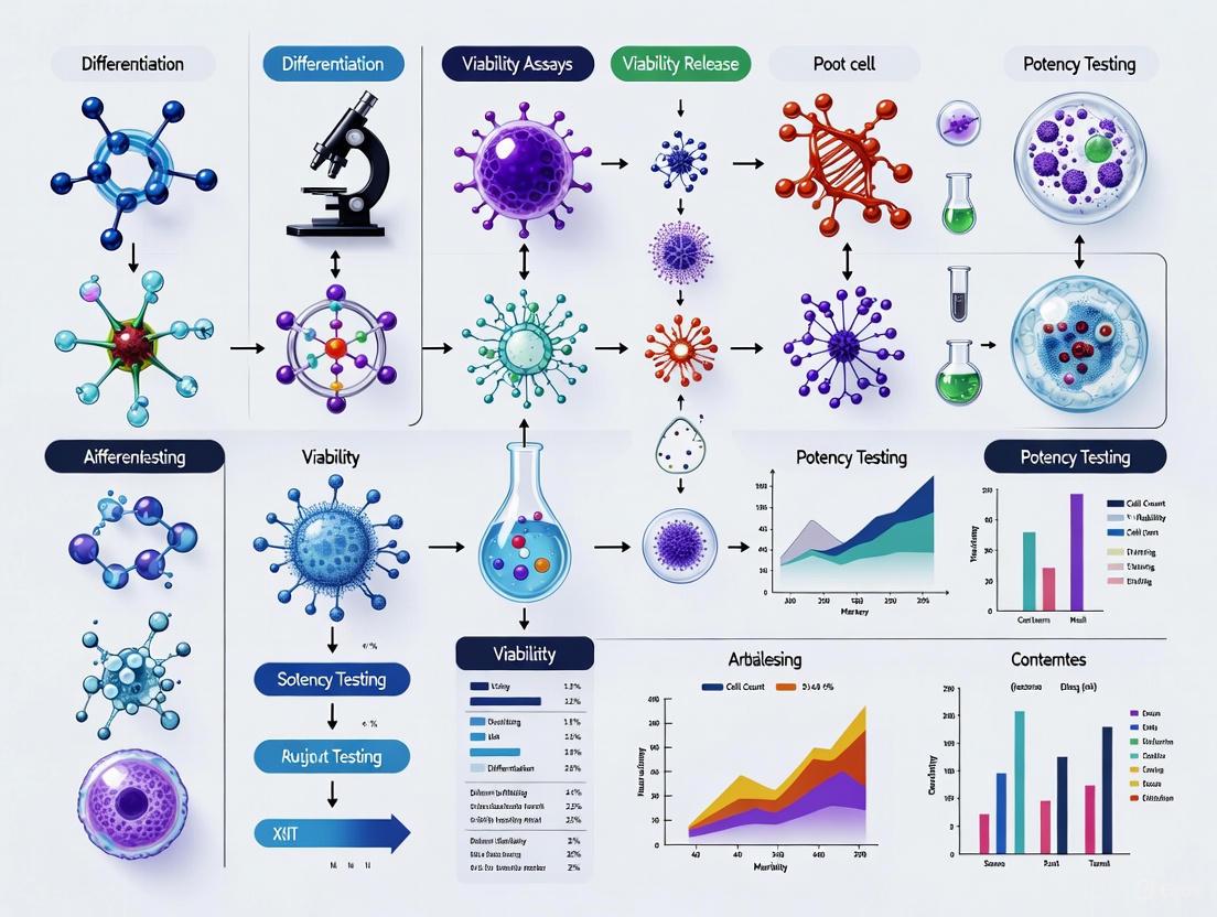
The pathway from cell manufacturing to product release involves a coordinated series of quality control checks, with identity, viability, and sterility testing operating as interconnected rather than isolated assessments. The following workflow visualizes this integrated testing approach:
Integrated Testing Workflow for Stem Cell Products
This integrated testing model demonstrates how identity, viability, and sterility assessments converge to inform the final release decision. The process begins with sample preparation from the manufactured cell product, followed by parallel testing streams. Critical to this model is the integration of results from all three testing domains, which provides a comprehensive product quality profile. The quality control review represents the decision point where all data is evaluated against predetermined specifications, leading to either product release or rejection. This systematic approach ensures that only products meeting all quality attributes proceed to patient administration.
The Scientist's Toolkit: Essential Research Reagents
Implementing robust identity, viability, and sterility testing requires specific reagents, equipment, and materials. The following toolkit outlines essential solutions utilized in the experimental protocols described in this guide, with an emphasis on standardized reagents that support reproducibility across laboratories.
Table 4: Essential Research Reagents for Core Release Testing
| Reagent/Material | Specific Examples | Primary Function | Application Notes |
|---|---|---|---|
| Flow Cytometry Antibodies | Anti-human CD105, CD73, CD90, CD45, CD34 | Immunophenotypic characterization | Use validated clones; titrate for optimal signal:noise |
| Viability Stains | Trypan blue, 7-AAD, propidium iodide | Membrane integrity assessment | Distinguish between apoptotic/necrotic cells |
| Sterility Culture Media | BacT/ALERT iAST/iNST, Thioglycollate broth | Microbial growth detection | Validate for cell therapy products with possible residues |
| Cell Dissociation Reagents | Trypsin/EDTA, enzyme-free alternatives | Generate single-cell suspensions | Optimize for cell type to maintain surface epitopes |
| PCR Master Mixes | Mycoplasma detection kits, 16S rRNA primers | Rapid microbial detection | Validate sensitivity/specificity against compendial methods |
The establishment of consensus standards for identity, viability, and sterility testing represents a pivotal achievement in the maturation of the stem cell therapy field. The ongoing efforts by ISCT, ISO, and regulatory agencies worldwide are creating a harmonized framework that supports both innovation and patient safety. As the field advances, particularly with the emergence of new technologies like induced pluripotent stem cell (iPSC)-derived therapies, these core release criteria will continue to serve as the foundation for quality assurance [16]. The ISCT's active role in developing international standards, such as the ISO documents for MSC characterization, provides a model for how professional societies can bridge the gap between research and clinical translation [5].
Looking forward, the integration of advanced analytical methods and the development of potency assays linked to clinical mechanisms of action will further strengthen the quality framework for stem cell products. As noted in the 2025 regulatory landscape, authorities are increasingly emphasizing post-approval monitoring and real-world data collection to complement pre-market quality assessment [17]. For researchers and product developers, adherence to these evolving but stabilizing standards for identity, viability, and sterility is no longer optional—it is an essential requirement for successful clinical translation and the eventual delivery of safe, effective stem cell therapies to patients in need.
The development and approval of stem cell and advanced therapy medicinal products (ATMPs) are governed by a complex framework of international standards and regional regulatory requirements. The International Society for Cell & Gene Therapy (ISCT), a global society of clinicians, regulators, researchers, and industry partners, plays a pivotal role in establishing scientific and technical standards that often inform regulatory thinking worldwide [18]. While ISCT develops voluntary consensus standards focused on scientific rigor and characterization, regulatory agencies like the US Food and Drug Administration (FDA) and European Medicines Agency (EMA) establish legally binding requirements for product approval and commercialization [19] [5]. This guide objectively compares these frameworks, particularly focusing on how ISCT guidelines complement and interact with formal regulatory pathways for stem cell product release criteria.
The regulatory environment for cell and gene therapies (CGT) is rapidly evolving. In 2025, regulators have taken significant steps to balance innovation with safety, scalability, and equitable access. The FDA has released several new draft guidance documents addressing expedited programs, post-approval data collection, and innovative trial designs for small populations [17] [20]. Simultaneously, ISCT continues to advance the field through its committee work on mesenchymal stromal cells (MSCs), global regulatory summits, and contributions to international standards [5] [20]. Understanding the alignment and distinctions between these frameworks is essential for researchers and drug development professionals navigating the pathway from discovery to approved therapies.
Foundational Standards and Guidelines
ISCT's Role in Standardization
ISCT provides critical scientific foundations for the field through position papers, committee perspectives, and contributions to international standards. A key achievement is the minimal criteria for defining multipotent mesenchymal stromal cells, initially published in 2006 and cited over 11,000 times, which has created a foundational consensus for MSC characterization [5]. More recently, the ISCT MSC Committee has contributed extensively to International Standards Organization (ISO) technical specifications ISO/TS 22859:2022 for Wharton's jelly-derived MSCs and ISO 24651:2022 for bone marrow-derived MSCs [5]. These documents provide consensus-based recommendations for tissue collection, cell isolation, characterization, and quality control assays at the research and development stage.
ISCT advocates for a "matrix model" of assays for comprehensive MSC characterization, recommending assessment of cell identity, gene expression, soluble factor expression, and functional immunomodulatory assays rather than relying on a single potency test [21] [5]. The Society has also led important nomenclature standardization, distinguishing between "Mesenchymal Stromal Cells" and "Mesenchymal Stem Cells" and recommending tissue-specific abbreviations such as MSC(WJ) for Wharton's Jelly-derived cells and MSC(M) for bone marrow-derived cells [5]. These scientific standards, while voluntary, provide essential guidance for maintaining rigor and reproducibility in stem cell research and early development.
FDA Regulatory Framework
The FDA regulates cell and gene therapies under the Public Health Service Act and the Federal Food, Drug, and Cosmetic Act [21]. For cell therapy products, including mesenchymal stromal cells, the FDA requires an Investigational New Drug Application (IND) before conducting clinical trials in the United States [21]. The Center for Biologics Evaluation and Research (CBER) oversees this process, with recent developments including three significant draft guidance documents issued in September 2025:
- Expedited Programs for Regenerative Medicine Therapies: Details leveraging RMAT designation, Fast Track, and Breakthrough Therapy pathways [17] [22]
- Postapproval Methods to Capture Safety and Efficacy Data: Emphasizes real-world data collection for long-term safety without delaying initial approvals [17] [22]
- Innovative Designs for Clinical Trials in Small Populations: Encourages adaptive, Bayesian, and externally controlled designs for rare disease trials [17] [22]
A fundamental FDA requirement is the development of potency assays as part of release criteria for advanced clinical trials aimed at marketing approval [21]. The FDA defines potency as "the therapeutic activity of the drug product as indicated by appropriate laboratory tests or by adequately developed and controlled clinical data" [21]. The agency recognizes the challenges in defining potency assays for complex cell therapies and evaluates adequacy on a case-by-case basis [21].
EMA Regulatory Framework
In the European Union, cell-based therapies are regulated as Advanced Therapy Medicinal Products (ATMPs) under Regulation (EC) No. 1394/2007 [21]. The EMA's Committee for Advanced Therapies (CAT) provides specialized assessment of ATMPs, with recent activities including workshops on gene editing and updates to Good Manufacturing Practice (GMP) guidelines specific to ATMPs [20] [23]. Similar to the FDA, the EMA requires demonstration of potency (biological activity) based on the product's attributes and linked to relevant biological properties [21].
The EMA is currently developing a concept paper on the revision of Part IV of Eudralex Volume 4, which contains GMP guidelines specific to ATMPs [20]. The consultation period for this important update ran from May to July 2025, reflecting the evolving nature of these regulations. The EMA also collaborates with international partners through initiatives like the Gene Therapies Global Pilot Program (CoGenT), which explores concurrent, collaborative regulatory reviews with the FDA and other international regulators to increase harmonization and improve review efficiency [17].
Comparative Analysis of Key Regulatory Elements
Product Characterization and Release Criteria
Table 1: Comparison of Product Release Criteria and Characterization Requirements
| Requirement | ISCT Guidelines | FDA Requirements | EMA Requirements |
|---|---|---|---|
| Potency Testing | Matrix of assays approach (quantitative RNA, flow cytometry, secretome analysis) [21] | Bioassay or surrogate measuring biological activity; case-by-case evaluation [21] | Bioassay based on intended biological effect; should relate to clinical response [21] |
| Identity Testing | Minimal criteria (plastic adherence, specific surface antigen expression, differentiation capacity) [5] | Identity and strength of all active ingredients [21] | Similar to FDA; based on product characteristics [23] |
| Starting Materials | Recommendations for tissue collection and cell isolation [5] | Critical raw materials approach; enhanced control based on risk and development stage [23] | Defined "starting materials" (become part of drug substance); GMP principles apply [23] |
| Viral Vector Testing | Not specifically addressed in ISCT MSC guidelines | Classified as drug substance; requires functional potency assays and RCV testing of cell-based drug product [23] | Considered starting materials; RCV testing once on vector may be sufficient [23] |
Manufacturing and Quality Control
Table 2: Comparison of Manufacturing and Quality Control Requirements
| Aspect | ISCT Guidelines | FDA Requirements | EMA Requirements |
|---|---|---|---|
| Manufacturing Standards | Recommendations for cryopreservation, storage, thawing, and transport [5] | cGMP compliance (21 CFR 210-211); risk-based approach [23] | GMP compliance (Eudralex Vol 4); specific ATMP guidelines in development [20] [23] |
| Donor Testing | Not specifically addressed in core guidelines | Governed by 21 CFR 1271 subpart C; tested in CLIA-accredited labs [23] | Governed by EUTCD; handled in licensed premises and accredited centres [23] |
| Process Validation | Not addressed in ISCT MSC guidelines | Statistically adequate number of batches based on variability [23] | Generally three consecutive batches; some flexibility allowed [23] |
| Comparability | Not directly addressed | Draft guidance (July 2023) on CGT comparability; risk-based approach [23] | Q&A document on comparability; specific attributes for GM cells [23] |
Clinical Development and Regulatory Pathways
Table 3: Comparison of Clinical Development and Regulatory Pathways
| Pathway | ISCT Role | FDA Framework | EMA Framework |
|---|---|---|---|
| Expedited Pathways | Advocacy and educational programs | RMAT designation, Fast Track, Breakthrough Therapy [17] [22] | PRIME scheme; adaptive pathways [17] |
| Trial Designs | Educational sessions on innovative designs | Encourages adaptive, Bayesian, externally controlled designs for small populations [17] [22] | Similar acceptance of innovative designs; reflection paper on external controls in development [20] |
| Post-Approval Evidence | Not directly addressed | Draft guidance on post-approval safety and efficacy data collection [17] [22] | Similar requirements for long-term follow-up; good pharmacovigilance practices [22] |
| Global Alignment | Global Regulators Summit (May 2025) [20] | Participant in CoGenT global collaborative review pilot [17] | Participant in CoGenT; collaboration with international partners [17] |
Experimental Protocols for Potency Assessment
ISCT-Recommended Matrix of Assays for MSC Potency
The ISCT perspective on immune functional assays for mesenchymal stromal cells as potency release criterion outlines three preferred analytical methods that form a comprehensive matrix approach [21]:
4.1.1 Quantitative RNA Analysis of Selected Gene Products
- Methodology: Quantitative reverse transcription polymerase chain reaction (qRT-PCR) for genes associated with immunomodulatory functions
- Key Targets: IDO1, PTGS2, TGF-β, IL-10, TSG-6, and other relevant genes based on mechanism of action
- Standardization: Normalization to housekeeping genes; establishment of reference ranges
- Validation Parameters: Accuracy, precision, specificity, linearity, range, and robustness
4.1.2 Flow Cytometry Analysis of Functionally Relevant Surface Markers
- Methodology: Multiparametric flow cytometry with validated antibodies
- Key Markers: PD-L1, ICAM-1, VCAM-1, HLA-DR, and other immunomodulatory surface proteins
- Standardization: Instrument calibration, compensation controls, and quantitative fluorescence standards
- Validation: Precision, specificity, stability, and reproducibility across operators and sites
4.1.3 Protein-Based Assay of Secretome
- Methodology: Multiplex immunoassays (Luminex, ELISA) for soluble factors
- Key Analytes: PGE2, IDO, TGF-β, IL-10, IL-6, and other relevant cytokines/mediators
- Stimulation Conditions: Standardized licensing conditions (e.g., IFN-γ, TNF-α stimulation)
- Validation: Reference standards, quantification limits, and precision across the dynamic range
FDA-Recommended Potency Assay Development
The FDA recommends a systematic approach to potency assay development that addresses several key parameters [21]:
4.2.1 Bioassay Development
- Principle: Measure biological activity in a living system (cell-based, tissue-based, or animal model)
- Design: Quantitative dose-response relationship with appropriate controls
- Endpoint: Relevant to proposed mechanism of action and clinical effect
- Validation: Accuracy, precision, specificity, linearity, range, and robustness
4.2.2 Surrogate Assay Development
- Application: When suitable bioassay is not feasible
- Types: Immunochemical (flow cytometry, ELISA), molecular (qPCR, microarray), or biochemical (protein binding, enzymatic) methods
- Justification: Demonstration that surrogate marker correlates with biological activity
- Validation: Same rigorous validation as bioassays
Visualizing the Regulatory Alignment Strategy
The following diagram illustrates the strategic alignment between ISCT guidelines and regulatory requirements for stem cell product development:
Strategic Alignment Between ISCT Guidelines and Regulatory Requirements
The Scientist's Toolkit: Essential Research Reagent Solutions
Table 4: Essential Research Reagents for Stem Cell Characterization and Potency Assessment
| Reagent Category | Specific Examples | Function in Regulatory Compliance | ISCT/FDA/EMA Context |
|---|---|---|---|
| Flow Cytometry Antibodies | CD73, CD90, CD105, CD45, CD34, HLA-DR, PD-L1 | Identity and purity assessment; immunomodulatory marker expression | ISCT minimal criteria; FDA identity testing [21] [5] |
| qPCR Assays | IDO1, PTGS2, TGF-β, IL-10, TSG-6 reference genes | Quantitative potency assessment at RNA level | ISCT-recommended matrix approach [21] |
| Multiplex Cytokine Arrays | PGE2, IDO, TGF-β, IL-10, IL-6 detection | Secretome analysis for functional potency | ISCT-recommended matrix approach; FDA potency assessment [21] |
| Cell Culture Supplements | Defined FBS, xeno-free media, cytokines (IFN-γ, TNF-α) | Standardized licensing conditions for potency assays | Manufacturing consistency (FDA cGMP, EMA GMP) [23] |
| Reference Standards | Characterized MSC lines, quantitative fluorescence standards | Assay calibration and comparability | FDA reference materials requirement; ISCT advocacy for public standards [21] |
The regulatory framework for stem cell therapies requires careful navigation of both international scientific standards and regional regulatory requirements. ISCT guidelines provide essential scientific foundations for product characterization, particularly through its minimal criteria for MSCs and advocacy for a matrix approach to potency assessment. Meanwhile, the FDA and EMA establish legally binding requirements for product development, manufacturing, and clinical evidence generation that must be met for market authorization.
The most successful development strategies will integrate ISCT's scientific guidance with regulatory requirements from the earliest stages of product development. This includes adopting ISCT-recommended characterization methods that can be validated to meet FDA and EMA expectations for potency testing, implementing manufacturing processes that align with both ISCT standards and GMP requirements, and designing clinical development plans that leverage expedited pathways available through both agencies.
Recent developments in 2025, including new FDA draft guidance documents and ongoing EMA guideline updates, demonstrate continued evolution toward more flexible, risk-based frameworks for cell and gene therapies. Simultaneously, ISCT's ongoing work on global standards and educational initiatives helps bridge the gap between scientific innovation and regulatory compliance. By understanding and strategically applying both sets of guidelines, researchers and drug development professionals can advance promising stem cell therapies more efficiently while maintaining the rigorous standards required for regulatory approval and patient safety.
The Critical Role of Oversight and Ethical Principles in Stem Cell Product Development
The transition of stem cell research from laboratory discovery to clinical therapy represents one of the most promising yet challenging frontiers in modern medicine. The unique properties of stem cells—including their capacity for self-renewal and differentiation into specialized cell types—make them indispensable for regenerative medicine applications aimed at treating a wide range of debilitating diseases and injuries [24]. However, these same properties introduce significant safety concerns, including the potential for tumorigenesis, immunological rejection, and unintended tissue formation [24] [25]. The field of stem cell therapy has evolved dramatically since the first successful bone marrow transplantation in 1968, with recent advancements in human pluripotent stem cells (hPSCs) and multipotent mesenchymal stem cells (MSCs) opening new doors for patients suffering from diseases and disorders that have yet to be treated [25].
Within this context, oversight and ethical principles form the critical foundation for ensuring that stem cell-based products are both safe and effective. The international diversity of cultural, political, legal, and ethical issues associated with stem cell research necessitates rigorously shared principles in science that call for rigor, oversight, and transparency in all areas of practice [19]. Adherence to these principles provides assurance that stem cell research is conducted with scientific and ethical integrity and that new therapies are evidence-based [19]. This comprehensive review examines the current oversight landscape, ethical frameworks, and experimental approaches that together ensure the responsible development of stem cell-based therapies, with particular attention to the latest standards and their practical implementation in product development.
The Evolving Regulatory and Standards Landscape
International Guidelines and Ethical Frameworks
The International Society for Stem Cell Research (ISSCR) has established comprehensive guidelines that serve as an international benchmark for stem cell research and clinical translation. These guidelines maintain and underscore widely shared ethical principles while addressing the rapid scientific advances in the field. The most recent 2025 update to the ISSCR guidelines specifically refines recommendations for stem cell-based embryo models (SCBEMs) in response to scientific and oversight developments in this rapidly evolving area of research [19]. These guidelines promote an "ethical, practical, and sustainable approach to stem cell research and the development of cell therapies that can improve human health and be made available to patients in need" [19].
Fundamental ethical principles articulated in these guidelines include:
- Integrity of the Research Enterprise: Independent peer review, oversight, replication, institutional oversight, and accountability at each stage of research are essential processes for maintaining trustworthiness and reliability [19].
- Primacy of Patient/Participant Welfare: Physicians and researcher-clinicians must never excessively place vulnerable patients or research subjects at risk, and clinical testing should never allow promise for future patients to override the welfare of current research subjects [19].
- Respect for Patients and Research Subjects: Researchers must empower potential human research participants to exercise valid informed consent and provide accurate information about risks and the current state of evidence for novel stem cell-based interventions [19].
- Transparency: Timely exchange of accurate scientific information to other interested parties, including various public groups, is essential. Researchers and sponsors should promote open and prompt sharing of ideas, methods, data, and materials by publishing both positive and negative results in a timely manner [19].
- Social and Distributive Justice: The benefits of clinical translation efforts should be distributed justly and globally, with particular emphasis on addressing unmet medical and public health needs. Risks and burdens associated with clinical translation should not be borne by populations that are unlikely to benefit from the knowledge produced in these efforts [19].
The ISCT 2025 MSC Standards: A Paradigm Shift in Characterization
A significant development in the field occurred in May 2025 when the International Society for Cell & Gene Therapy (ISCT) released new identification criteria for Mesenchymal Stromal Cells (MSCs), bringing an end to nearly two decades of academic debate over their identity and function [26]. This represents a substantial shift from the previous 2006 standards and establishes a new framework for the development and quality control of cell therapy products.
The most striking change in the new standard is the formal definition of MSCs as "Mesenchymal Stromal Cells" instead of the widely used term "Mesenchymal Stem Cells." This is not merely a semantic adjustment but a fundamental reevaluation based on extensive scientific evidence. According to the new standard, researchers who wish to continue using the term "Mesenchymal Stem Cells" must provide experimental evidence that the cells possess actual stem cell properties—such as self-renewal and multi-lineage differentiation potential [26].
Table 1: Comparison of ISCT 2006 vs. 2025 MSC Identification Standards
| Standard Element | 2006 Standard | 2025 Standard |
|---|---|---|
| Cell Definition | Mesenchymal Stem Cells (MSCs) | Mesenchymal Stromal Cells (MSCs) |
| Stemness Requirement | Must demonstrate trilineage differentiation | Must provide evidence to use the term "stem" |
| Marker Detection | Qualitative (positive/negative) | Quantitative (thresholds and percentages) |
| Tissue Origin | Not emphasized | Must be specified and considered |
| Critical Quality Attributes | Not required | Must assess efficacy and functional properties |
| Culture Conditions | No standard reporting requirement | Detailed parameter reporting required |
The updated standards introduce several critical changes that impact product development and characterization:
Optimized Identification Criteria: The new standards comprehensively upgrade identification criteria, particularly in surface marker detection. Positive markers (CD73, CD90, and CD105) are still recognized as basic positive markers, but researchers must now specify the threshold percentage for positive identification via flow cytometry. CD45 (a hematopoietic marker) must be included as a negative marker to ensure the cell population is not contaminated by hematopoietic lineages. Complete results for each marker, including the percentage of positive cells, must be reported to improve data transparency and comparability [26].
Emphasis on Tissue Origin and Quality Attributes: The new standards require specification of the tissue origin of MSCs, acknowledging that cells from different sources may have distinct phenotypic and functional properties. Furthermore, the standards incorporate efficacy and functional characterization into Critical Quality Attributes (CQAs), emphasizing the need to describe these attributes to define the clinical functionality of MSCs [26].
Revised Differentiation Requirements: Notably, the two key identification criteria from the 2006 standard—"trilineage differentiation in vitro" (osteogenesis, adipogenesis, and chondrogenesis) and "adherence to plastic under standard conditions"—are no longer mandatory. This adjustment acknowledges the limitations of traditional "stemness" assays in distinguishing true stem cells from more specialized stromal cell populations [26].
Experimental Design and Methodological Approaches for Stem Cell Product Characterization
Design of Experiments (DOE) for Process Optimization
The complexity of stem cell bioprocessing requires the examination of multiple components that must be controlled to arrive at the correct state of the cell at the end of the process. Traditional experimentation in stem cell biology has typically been conducted using a one-factor-at-a-time (OFAT) approach, but this method has significant limitations for optimizing the multiple interacting variables in stem cell culture systems [27].
Design of Experiments (DOE) provides a statistical framework for efficiently searching through the multi-dimensional problem space of possible protocols in a timely and cost-effective manner. DOE methods enable researchers to:
- Maximize Information Yield: Carefully select experimental points to maximize information content relevant to the research questions of interest [27].
- Understand Interactions: Identify interactions between factors that OFAT approaches miss, which is particularly important given the complexity of stem cell bioprocessing involving multiple inputs (e.g., signaling pathways, oxygenation, duration of process steps, shear effects) [27].
- Optimize Yield and Sensitivity: Address two critical but often-overlooked metrics for stem cell bioprocesses: yield (the quantity of output cells of the desired type produced) and sensitivity (the robustness of the process in the face of minor variations in input variables) [27].
Response Surface Methodology (RSM) provides a structured approach for sequential experimentation, beginning with screening experiments to identify influential factors, followed by optimization experiments to refine process conditions, and finally verification experiments to confirm optimal settings [27]. This approach is particularly valuable for stem cell bioprocessing optimization, where the quantities of material required for therapies can exceed 10^9 cells per patient per treatment [27].
The following diagram illustrates the experimental workflow integrating DOE principles with stem cell product development:
Advanced Characterization Techniques
Robust characterization of stem cell products requires multiple complementary approaches to assess identity, potency, purity, and safety. The U.S. Food and Drug Administration (FDA) scientists are developing laboratory techniques to enable careful evaluation and characterization of these products to reliably predict whether they will be safe and effective [28].
Key characterization strategies include:
Molecular Marker Identification: Research focuses on identifying molecules that exert critical influence on the growth and differentiation of stem cells. Such molecules can be used in tests that evaluate and characterize cells during the manufacturing process and as lot-release measurements for cell-therapy products. Technologies employed include microarrays (to study the state of activity of tens of thousands of genes), RT-PCR (to amplify pieces of DNA), and flow cytometry (to automatically identify, count and examine large numbers of cells) [28].
Morphological Profiling: Advanced morphological profiling using machine learning can reveal emergent subpopulations of cells that predict functional characteristics. For example, research has shown that morphological features of interferon-gamma-stimulated mesenchymal stromal cells can predict overall immunosuppressive capacity [28]. This approach allows for non-invasive assessment of cell quality and functionality.
Functional Assays: The development of quantitative biological assays is essential for measuring critical activities such as differentiation potential, immunosuppressive capacity, and other functional properties. FDA researchers have found that several biological activities decrease the longer MSCs are cultured, and that MSCs derived from different donors vary in the amount of these activities, highlighting the need for robust functional characterization [28].
Analytical Framework: Comparison of Regulatory Approaches and Clinical Applications
International Regulatory Frameworks for Stem Cell Therapies
The regulatory framework for stem cell therapy is structured in three tiers: legislation enacted by legislatures, regulations adopted by the executive branch, and guidelines published by regulatory entities [29]. Different regions have developed distinct approaches to balancing ethical considerations, safety concerns, and innovation facilitation:
Table 2: Comparative Analysis of Regulatory Frameworks for Stem Cell Therapies
| Region | Regulatory Approach | Key Guidelines/Directives | Emphasis |
|---|---|---|---|
| European Union | Rigorous guidelines prioritizing safety and ethical considerations | Directive 2001/83/EC, Regulation 1394/2007 on Advanced Therapy Medicinal Products (ATMPs) | Ethical concerns, especially around use of human embryos; centralized authorization through EMA |
| United States | Flexible regulatory stance facilitating rapid development | 21 CFR 1271 (HCT/Ps), Guidance on Regulatory Considerations for Human Cells, Tissues, and Cellular and Tissue-Based Products | Risk-based approach; Center for Biologics Evaluation and Research (CBER) oversight; IND requirement for manipulated cells |
| Japan & South Korea | Balanced approach incorporating practices from both EU and US | Japan: Guidelines on clinical research using human stem cells (2006), Technical guidance for regenerative medical products (2016) | Progressive legislation with accelerated approval pathways for regenerative medicine products |
| International Standards | Voluntary guidelines promoting global consistency | ISSCR Guidelines (2025 update), ISCT MSC Standards (2025) | Ethical principles, scientific rigor, transparency, and harmonization of standards |
The regulatory differences significantly impact the pace and scope of stem cell therapy development. Countries with more flexible regulatory guidelines, such as the United States and Japan, tend to be in a leading position in terms of clinical trial activity, while countries in the EU fall behind due to more rigorous regulations [29].
Clinical Trial Trends and Therapeutic Applications
Comprehensive analysis of clinical trial data reveals significant growth in the number of stem cell clinical trials since 2008, particularly those involving induced pluripotent stem cells (iPSCs) [29]. The distribution of these trials varies substantially by country, reflecting the impact of different regulatory environments.
Therapeutic studies involving iPSCs predominantly target conditions affecting the cardiovascular and nervous systems, which are considered vital and often have limited treatment options [29]. The safety profile of stem cell-based therapies is supported by a large body of preclinical and clinical studies, especially adult stem cell therapy such as MSC-based products. However, clinical trials have not yet yielded consistent data supporting the efficacy of the treatments, as numerous studies have shown paradoxical results and no statistically significant differences in outcomes, even in phase III trials [25].
The mechanisms underlying these therapeutic effects appear to involve immune modulation rather than direct regenerative function in many cases. This understanding has led to increased emphasis on characterizing the secretory profile and immunomodulatory properties of stem cell products as critical quality attributes [25].
Implementation Framework: Research Reagents and Experimental Toolkit
The successful implementation of stem cell product development protocols requires specialized reagents and materials that ensure consistency, safety, and efficacy. The following table details key research reagent solutions essential for stem cell product development and characterization:
Table 3: Essential Research Reagent Solutions for Stem Cell Product Development
| Reagent Category | Specific Examples | Function & Importance | Quality Considerations |
|---|---|---|---|
| Serum-Free Media | YOCON MSC Serum-Free Media, Corning Ascent System | Provides defined, xeno-free culture environment; reduces batch variability and contamination risk | GMP-grade composition; compliance with regulatory requirements; support for clinical-scale expansion |
| Cell Culture Systems | Corning CellSTACK, HYPERStack, CellCube, Ascent Fixed Bed Bioreactor | Enable scalable expansion of adherent stem cells; support process standardization and closed-system manufacturing | Scalability from research to production; compatibility with GMP environments; validation data provided |
| Characterization Tools | Flow cytometry panels (CD73, CD90, CD105, CD45), Videodrop for exosomes | Quantitative assessment of cell identity, purity, and potency; characterization of secreted factors | Standardized protocols; reference materials; validation for regulatory submissions |
| Differentiation Assays | Trilineage differentiation kits (osteogenic, adipogenic, chondrogenic) | Functional assessment of differentiation potential; historical standard for stemness (now optional under 2025 ISCT) | Standardized protocols; reference standards; quantitative readout methods |
| Cryopreservation Solutions | Defined composition cryomediums | Maintain cell viability and functionality post-thaw; ensure product consistency | Serum-free, animal component-free formulations; validated recovery protocols |
The selection of appropriate research reagents is particularly critical in light of the updated ISCT 2025 standards, which emphasize detailed reporting of culture conditions, medium components, passaging methods, and culture environment parameters [26]. Implementation of closed-system platforms, such as the Corning CellCube system described in ISCT 2025 presentations, can achieve comparable cell density, population doubling time, and marker expression to traditional vessels while offering superior scalability and reduced media consumption [30].
Integration of Oversight into the Stem Cell Product Development Pipeline
The development of safe and effective stem cell products requires the integration of oversight and ethical considerations at every stage of the process, from initial cell line establishment to final product release. The following diagram illustrates how these elements integrate throughout the development pipeline:
This integrated approach ensures that ethical principles and oversight mechanisms are not merely add-on considerations but fundamental components of the product development process. Key integration points include:
Cell Line Establishment: Implementation of rigorous donor screening and informed consent processes that respect patient autonomy and privacy, in accordance with ISSCR guidelines on respect for patients and research subjects [19].
Process Development: Application of Quality by Design (QbD) principles and DOE methodologies to optimize manufacturing processes while ensuring consistency and safety [27].
Preclinical Evaluation: Comprehensive safety assessment including tumorigenicity testing, biodistribution studies, and functional characterization that addresses the unique risks associated with stem cell-based products [28].
Clinical Trials: Ethical trial design that prioritizes patient welfare, ensures valid informed consent, and promotes social justice through appropriate participant selection and fair distribution of risks and benefits [19].
Product Release: Implementation of rigorous lot-release criteria based on Critical Quality Attributes that reliably predict product safety and efficacy [26] [28].
The field of stem cell product development stands at a pivotal moment, with evolving standards, increasing clinical experience, and advancing characterization technologies converging to create new opportunities for therapeutic innovation. The recent updates to international standards, particularly the ISCT 2025 MSC criteria and ISSCR 2025 guidelines, reflect a maturing understanding of stem cell biology and a strengthened commitment to ethical principles and scientific rigor.
The critical role of oversight and ethical principles in stem cell product development cannot be overstated. These elements provide the essential framework that ensures the safety of patients, the integrity of the research enterprise, and the ultimate translation of promising therapies to clinical practice. As the field continues to evolve, several key developments will shape its future trajectory:
First, the harmonization of international standards will be essential for facilitating global collaboration and ensuring consistent product quality. While regulatory approaches may differ across regions, core ethical principles and scientific standards should converge to promote excellence and patient safety worldwide [29].
Second, advances in characterization technologies, particularly those enabling non-invasive assessment of cell quality and potency, will enhance our ability to predict in vivo performance and ensure product consistency. The integration of morphological profiling, molecular analyses, and functional assays provides a comprehensive approach to product characterization that aligns with the updated ISCT standards [28].
Finally, the continued emphasis on ethical principles—including transparency, social justice, and respect for research participants—will maintain public trust and ensure that the benefits of stem cell research are distributed fairly across global communities. By upholding these principles while pursuing scientific innovation, the stem cell research community can fulfill its potential to transform the treatment of debilitating diseases and injuries.
The journey from basic stem cell research to clinically effective therapies is complex and challenging, but through the consistent application of rigorous oversight, ethical principles, and scientific excellence, this promising field can deliver on its potential to revolutionize medicine and improve human health.
Application of ISCT Methodologies: Implementing Potency Assays and a Matrix Approach for Product Characterization
The ISCT-Recommended Matrix Assay Approach for Comprehensive Product Characterization
The development of robust potency assays is a critical requirement for the release of advanced therapy medicinal products (ATMPs), including Mesenchymal Stromal Cells (MSCs), in late-stage clinical trials. Unlike traditional pharmaceuticals with single, well-defined mechanisms of action, MSCs exert their therapeutic effects through multiple synergistic pathways, including immunomodulation, trophic factor secretion, and tissue integration. This complexity challenges conventional single-parameter potency assays. Recognizing this limitation, the International Society for Cell & Gene Therapy (ISCT) recommends an assay matrix approach that collectively captures the multifaceted effector functions of MSC products [31].
This approach acknowledges that no single assay can adequately represent the complete biological functionality of MSCs. Instead, a panel of assays is employed to evaluate critical quality attributes (CQAs) that predict clinical performance. The matrix approach provides a comprehensive profile of the product's functional potency, ensuring batch-to-batch consistency and helping to bridge the gap between in vitro characterization and in vivo mechanism of action [32] [5]. This guide explores the implementation, experimental protocols, and comparative data of the ISCT-recommended matrix assay, providing a framework for researchers and drug development professionals.
Core Principles of the ISCT Matrix Approach
The fundamental principle of the matrix approach is the concurrent use of multiple complementary assays to assess a cell product's functional capacity. For MSCs, the ISCT Mesenchymal Stromal Cell Committee has specifically advocated for this strategy to overcome the limitations of single-parameter potency testing [31]. The matrix is designed to capture several key biological aspects:
- Multiparameter Characterization: The approach moves beyond basic identity criteria (plastic adherence, surface marker expression, and tri-lineage differentiation) to include functional assessments of immunomodulation, secretome analysis, and signaling pathway activation [5].
- Mechanism-of-Action Informed: Assays selected for the matrix should reflect the proposed biological mechanisms through which the cells mediate their therapeutic effect, whether through paracrine signaling, direct cell-cell contact, or a combination of pathways.
- Fitness and Viability Assessment: The matrix includes metrics for cell fitness and viability, recognizing that these parameters directly influence therapeutic potency. Route of administration and host factors are also considered variables that can impact clinical outcomes [5].
Experimental Deep Dive: A Phospho-STAT Matrix Assay for MSC Potency
A prime example of the matrix approach in practice is a potency assay that captures the STAT (Signal Transducer and Activator of Transcription) phosphorylation profile of MSCs in response to a inflammatory secretome. This "phospho matrix" approach evaluates MSCs as both generators and sensors of the immunomodulatory microenvironment.
Experimental Protocol and Workflow
The following protocol outlines the key steps for implementing the phospho-STAT matrix assay [31]:
- MSC and PBMC Preparation: Culture MSCs from sources like bone marrow (BM), adipose tissue (AT), or umbilical cord (UC) under standard conditions. Isolate Peripheral Blood Mononuclear Cells (PBMCs) from donor blood.
- Generation of Conditioned Secretome:
- Co-culture live MSCs or heat-inactivated MSCs (HI-MSCs) with SEB-activated allogeneic PBMCs. Use escalating ratios of MSCs to PBMCs.
- Include control cultures of HI-MSCs alone, live MSCs alone, and activated PBMCs alone.
- Collect supernatants (conditioned secretome) from all cultures after a defined period.
- Stimulation of "Sensor MSCs":
- Expose new MSCs from the same donor ("Sensor MSCs") to the various conditioned secretomes.
- Perform a short-term stimulation (e.g., 15 minutes).
- Intracellular Staining and Phosflow Analysis:
- Fix and permeabilize the sensor MSCs immediately after stimulation.
- Stain cells with fluorescently-labeled antibodies against phosphorylated STAT proteins (pSTAT1, pSTAT3, pSTAT4, pSTAT5, pSTAT6) and a live-dead marker (e.g., 7AAD).
- Analyze stained cells using flow cytometry to measure phosphorylation levels via Mean Fluorescence Intensity (MFI).
The diagram below visualizes this integrated experimental workflow and its core logic.
Key Findings and Signaling Pathway Insights
This loop analytical approach identifies a specific phosphorylation signature that correlates with MSC potency. The secretome generated from co-cultures of live MSCs with activated PBMCs uniquely reduces phosphorylation of STAT1 and STAT3 in the sensor MSCs. This effect is not observed with the secretome from HI-MSCs co-cultured with PBMCs, which behaves similarly to the secretome from PBMCs alone [31]. This specific modulation of the JAK-STAT signaling pathway serves as a functional biomarker for MSC immunomodulatory capacity.
The diagram below illustrates the core signaling logic discovered through this assay.
Comparative Performance Data
The phospho-STAT matrix assay allows for quantitative comparison of MSC potency across different tissue sources and donor lots. The following tables summarize key experimental data from the assay, demonstrating its application in product characterization.
Table 1: Differential STAT Phosphorylation in Bone Marrow-Derived MSCs [31] This table shows the Area Under the Curve (AUC) values for phospho-STAT signals from two different bone marrow MSC donors, highlighting donor-specific variations in the potency signature (pSTAT1 and pSTAT3 modulation).
| MSC Donor | Culture Condition | pSTAT1 AUC | pSTAT3 AUC | pSTAT4 AUC | pSTAT5 AUC |
|---|---|---|---|---|---|
| Donor #1 | HI-MSCs + PBMCs | 25,143 | 8,452 | 2,111 | 4,749 |
| Live MSCs + PBMCs | 18,388 | 4,396 | 2,374 | 3,993 | |
| Difference (ΔAUC) | 6,755 | 4,056 | -263 | 756 | |
| Donor #2 | HI-MSCs + PBMCs | 18,349 | 5,929 | 2,012 | 1,263 |
| Live MSCs + PBMCs | 6,778 | 3,666 | 2,026 | 911 | |
| Difference (ΔAUC) | 11,571 | 2,263 | -14 | 352 |
Table 2: Inter-Source Comparison of Functional MSC Properties [33] [31] This table compares critical quality attributes and functional outcomes for MSCs derived from different tissue sources, as evaluated by various matrix assays.
| Property / Assay | Bone Marrow MSCs | Adipose Tissue MSCs | Umbilical Cord MSCs |
|---|---|---|---|
| Source Abundance | Low | High | High |
| Standard Isolation Method | Density Gradient Centrifugation [33] | Enzymatic Digestion [33] | Enzymatic Digestion or Explant [33] |
| Immunophenotype (ISCT) | CD73+, CD90+, CD105+, CD45- [31] | CD73+, CD90+, CD105+, CD45- [31] | CD73+, CD90+, CD105+, CD45- [31] |
| T-cell Suppression | Dose-dependent inhibition [31] | Dose-dependent inhibition [31] | Dose-dependent inhibition [31] |
| pSTAT1/3 Modulation | Robust downregulation [31] | Robust downregulation [31] | Robust downregulation [31] |
| Key Advantage | Most historical data, gold standard | High cell yield, easy access | Proliferative capacity, less invasive |
The Scientist's Toolkit: Essential Reagents and Materials
Successfully implementing the ISCT-recommended matrix assay requires specific reagents and tools. The following table details essential materials and their functions based on the cited experimental protocols.
Table 3: Key Research Reagent Solutions for Matrix Assay Implementation
| Reagent / Material | Function in the Assay | Specific Examples / Notes |
|---|---|---|
| MSC Culture Media | Expansion and maintenance of MSCs. | Animal-origin free (AOF) media (e.g., TeSR-AOF 3D) enhance traceability and viral safety [34]. |
| Dissociation Reagents | Passaging and harvesting MSCs. | Gentle Cell Dissociation Reagent (GCDR) or other enzymatic reagents (e.g., Trypsin) [34]. |
| Activation Agent | Stimulating immune cells in co-culture. | Staphylococcus Enterotoxin B (SEB) for activating PBMCs [31]. |
| Flow Cytometry Antibodies | Cell phenotyping and intracellular signaling analysis. | Antibodies against CD73, CD90, CD105, CD45; and phospho-specific antibodies for pSTAT1, pSTAT3, etc. [31]. |
| Viability Stain | Distinguishing live from dead cells. | 7-AAD (7-aminoactinomycin D) for flow cytometry [31]. |
| Digital PCR Systems | Vector genome integrity analysis in gene-modified MSCs. | QIAcuity dPCR system for sensitive, multiplexed analysis of genome integrity [35]. |
| Bioreactor Systems | Scaling up MSC production for clinical applications. | PBS-MINI Bioreactors for scalable 3D suspension culture [34]. |
The ISCT-recommended matrix assay approach represents a paradigm shift in cell-based product characterization, moving from simplistic quality control to a comprehensive, mechanism-based understanding of product potency. The phospho-STAT assay exemplifies how a well-designed matrix can capture critical functional attributes—such as the capacity to modulate an inflammatory environment—that directly predict biological activity. As the field advances, adopting these robust, multivariate assays is no longer optional but essential for ensuring the consistent quality, efficacy, and safety of cellular therapeutics throughout clinical development and eventual market approval. This rigorous framework for product characterization is fundamental to the successful translation of stem cell therapies from the laboratory to the clinic.
Developing Potency Assays Linked to Mechanism of Action (MOA)
For developers of cell and gene therapies, demonstrating product potency—the quantitative measure of a product's specific biological activity—is a fundamental regulatory requirement. A potency assay is not merely a quality control test; it is a direct link between a product's proposed mechanism of action (MOA) and its demonstrated biological function, providing assurance of lot-to-lot consistency and product quality [36] [37]. The development of these assays, particularly those that are MOA-based, remains one of the most significant challenges in the field, essential for advancing products from research through clinical trials and to commercial approval [38] [39].
The American Society of Gene & Cell Therapy (ASGCT) emphasizes that while potency assays are a cornerstone of the control strategy, the link between product characteristics and clinical performance is often product-specific and can be particularly difficult to establish early in development [38]. This guide objectively compares the current approaches, requirements, and practical solutions for developing robust, MOA-linked potency assays, providing a structured framework for researchers and drug development professionals.
Regulatory Framework and Current Landscape
Regulatory Expectations and Phase-Appropriate Approach
Global regulatory agencies, including the FDA and EMA, recognize potency as a critical quality attribute (CQA) and require potency assays for the lot release of biologics [36] [40]. The primary regulatory expectation is that the potency assay should reflect the product's biological activity and be based on its MOA, whenever scientifically feasible [41]. However, the ASGCT advocates for a risk-based, phase-appropriate approach, acknowledging that a fully validated, MOA-based potency assay may not be possible early in clinical development due to assay complexity, variability, and an evolving understanding of the product's mechanism [38]. The guidance suggests that potency assays critical for late-stage and pivotal trials may begin as characterization assays in early phases, with acceptance criteria tightening as product and process knowledge increases [38].
Analysis of Potency Assays in Approved Products
An analysis of the 31 U.S. FDA-approved cell therapy products (CTPs) as of 2024 provides a revealing snapshot of current industry practices. A total of 104 potency tests were reported across these products, averaging 3.4 tests per CTP [39]. The types of measurements used are categorized in the table below.
Table 1: Categorization of Potency Tests Used in 31 FDA-Approved Cell Therapy Products
| Category of Potency Test | Number of Tests | Percentage of Non-Redacted Tests | Example Measurements |
|---|---|---|---|
| Viability and Count | 37 | 52% | Cell viability, total nucleated cell count [39] |
| Expression | 19 | 27% | CAR expression (e.g., via flow cytometry) [39] |
| Bioassays | 7 | 7% | IFN-γ secretion upon target cell recognition [39] [41] |
| Genetic Modification | 6 | 9% | Vector copy number, transduction efficiency [39] |
| Histology | 2 | 3% | Tissue structure and morphology assessment [39] |
This data shows a strong reliance on physicochemical tests like "Viability and Count" and "Expression," which are often more reproducible and easier to validate. The relatively low number of formal "Bioassays" may be surprising; however, this must be interpreted with caution as 32% of potency tests in the analysis were redacted for proprietary reasons [39]. Furthermore, a single, complex bioassay is often supplemented with other tests to form a potency assay matrix that collectively ensures product quality [36] [5].
A Practical Framework for MOA and Potency
Distinguishing Key Concepts
A significant challenge in assay development is the confusion surrounding core concepts. The following definitions, adapted from regulatory guidance and metrological principles, provide essential clarity [41]:
- Mechanism of Action (MOA): The specific process through which a product produces its intended effect.
- Potency: The product attribute that enables it to achieve its intended MOA.
- Potency Test: A test that measures the potency attribute.
- Efficacy: The ability of the product to have the desired effect in patients.
It is a common misconception to equate potency with efficacy. Potency is a laboratory-measured attribute, while efficacy is a clinical outcome; the two are linked, but not directly, through the proposed MOA [41].
The Potency and Efficacy Relationship
The following diagram illustrates the logical relationship between a product's mechanism of action, its measurable potency, and its ultimate clinical efficacy.
This conceptual separation is critical. A product can be "potent" in the lab (showing a strong result in its potency test) but not efficacious in patients if the MOA is incorrect or does not translate to the human system. Conversely, a product might be efficacious even if a specific potency test does not correlate with clinical outcomes, as long as the risk-benefit profile is acceptable and an alternative control strategy is in place [41].
Experimental Protocols for Potency Assay Development
The Tiered Development Lifecycle
Developing a robust potency assay is a multi-stage process that parallels drug development. A tiered approach allows for phase-appropriate refinement, aligning with regulatory expectations [36] [40]. The following workflow outlines the key stages from early development through commercial validation.
Stage 1: Assay Development. This initial, bespoke phase involves designing an assay that reflects the product's MOA. Key activities include [36]:
- Cell Line Selection: Choosing a cell line based on vector tropism and compatibility with the therapy's promoters and MOA.
- Readout Optimization: Designing protocols to reliably detect the relevant biological response (e.g., vector-derived RNA, protein expression, enzymatic activity, or a downstream cellular response).
- Analysis Method Definition: Selecting an appropriate statistical model (e.g., four-parameter logistic regression for dose-response curves) for calculating relative potency.
Stage 2: Assay Qualification. This provides the first formal assessment of assay performance and variability, supporting initial clinical trials (IND/CTA). It establishes preliminary parameters for accuracy, precision, and range [40].
Stage 3: Assay Validation. This is a full GMP study that formally verifies the assay's performance characteristics—including specificity, accuracy, precision, and robustness—for its intended use in commercial lot release and stability testing [36] [40].
Key Research Reagents and Materials
The table below details essential reagents and their critical functions in establishing a robust potency assay, particularly for cell-based systems.
Table 2: Essential Research Reagents for Potency Assay Development
| Reagent / Material | Function in Potency Assay | Key Considerations |
|---|---|---|
| Cell Line / Cellular Model | Serves as the biological system to measure the product's functional activity [36]. | Must be relevant to the MOA; requires careful banking and characterization to ensure reproducibility [36]. |
| Reference Standard (RS) | A well-characterized drug lot against which test sample potency is measured relative (%RP) [40]. | Critical for controlling inter- and intra-assay variability; must be stable and representative of the clinical product [40]. |
| Assay Controls | Materials with known potency used to ensure each assay run is valid (system suitability) [40]. | Includes positive, negative, and quality controls to monitor assay performance and sensitivity [42]. |
| Critical Assay Reagents | Antibodies, detection substrates, culture media, and other components essential for the test [43]. | Require strict qualification and sourcing strategy to prevent variability and supply shortages [42] [43]. |
Comparative Analysis of Potency Assay Strategies
Addressing Variability and Statistical Considerations
A major technical challenge in potency testing is managing assay variability. Bioassays, especially functional cell-based assays, inherently have higher variability than physicochemical methods [40]. This variability arises from multiple biological and operational factors, including the cell line passage number, reagent lots, and analyst technique [36] [40].
The statistical framework for potency assays relies on measuring Relative Potency (%RP) against a reference standard, rather than absolute quantification. The most common model for analyzing dose-response data is the four-parameter logistic (4PL) fit, which estimates the horizontal shift (e.g., EC50) between the standard and test sample curves [40]. To control variability, sponsors must implement a replication strategy, often using multiple dilution series within a run, and may average results over several independent assay runs to generate a final reportable value [40].
Case Study: Kymriah and the Correlation Challenge
The approval of Kymriah (tisagenlecleucel) provides an instructive case study. Its potency was defined by the ability of CAR T-cells to secrete interferon-γ (IFN-γ) upon exposure to CD19+ target cells [41]. While this assay measures a key T-cell effector function, the FDA noted that "IFN-γ production varied greatly from lot-to-lot, making it difficult to correlate IFN-γ production in vitro to tisagenlecleucel safety or efficacy" [41]. Publicly available data shows an overlap in potency values between clinical responders and non-responders, highlighting that a single in vitro bioassay may not fully capture the complex, in vivo MOA of a cell therapy product [41]. This underscores the importance of the potency assay matrix and the reality that a perfect correlation with clinical outcome is not always achievable for licensure.
Developing potency assays linked to MOA is a complex but essential endeavor. The current regulatory landscape encourages a phase-appropriate, risk-based strategy that evolves with product knowledge [38]. The analysis of approved products reveals a pragmatic combination of simpler, robust tests (viability, expression) with more complex, MOA-reflective bioassays to form a comprehensive potency assurance strategy [39].
The field continues to advance with emerging technologies like high-throughput assay designs and automation promising to reduce variability, enhance reproducibility, and accelerate development timelines [36]. Furthermore, international standards organizations like ISO are working to harmonize practices for specific cell types, such as mesenchymal stromal cells, which will provide greater clarity and consistency for the industry [5]. Ultimately, a strong collaboration between developers, regulators, and contract organizations throughout the product lifecycle is key to successfully implementing potency assays that ensure the consistent quality and therapeutic promise of cell and gene therapies.
Within the framework of the International Society for Cellular Therapy (ISCT) standards, defining robust release criteria for stem cell products, particularly potency assays, remains a significant challenge for researchers and drug development professionals. The ISCT identifies quantitative RNA analysis, flow cytometry, and protein-based secretome analysis as three preferred analytical methods that can inform a matrix assay approach for immunomodulatory mesenchymal stromal cell (MSC)-like products [21]. These assays are crucial for measuring the biological activity of cellular products, a requirement from Regulatory Authorities for advanced clinical trials and eventual product registration [21]. This guide objectively compares the performance, applications, and experimental protocols for these three key assay types, providing a foundation for their use in stem cell product characterization and release testing.
Performance Comparison of Analytical Assays
The following table summarizes the core characteristics, performance metrics, and primary applications of quantitative RNA analysis, flow cytometry, and protein-based secretome analysis, enabling direct comparison for informed method selection.
Table 1: Performance and Application Comparison of Key Analytical Assays
| Feature | Quantitative RNA Analysis | Flow Cytometry | Protein-Based Secretome Analysis |
|---|---|---|---|
| Measured Target | Gene expression levels of specific RNA transcripts (e.g., cytokines, immunomodulators) [21] | Protein abundance and characterization on cell surfaces or intracellularly [21] | Proteins actively released or shed into the extracellular environment (e.g., cytokines, growth factors) [44] [21] |
| Technology Principle | Reverse transcription polymerase chain reaction (RT-PCR, quantitative PCR) [21] | Laser-based detection of fluorescently-labeled antibodies bound to cell targets [45] | Mass spectrometry (LC-MS) often coupled with enrichment or labeling techniques (e.g., SILAC, AHA) [44] [46] |
| Key Performance Metrics | High sensitivity and specificity; wide dynamic range for quantification [21] | High-throughput, multi-parameter data from single cells; high specificity [45] [21] | Unbiased, comprehensive profiling; detects thousands of proteins simultaneously [44] |
| Primary Applications in ISCT Context | Potency assays via analysis of licensed MSCs; measurement of immunomodulatory gene products [21] | Product identity; potency via surface marker quantification (e.g., activation markers) [21] | Potency assays via analysis of paracrine factors; understanding mechanism of action [21] |
| Key Advantages | High sensitivity for low-abundance transcripts; quantitative precision; relatively simple data output [21] | Single-cell resolution; ability to detect heterogeneous cell populations; can analyze complex samples [45] | Unbiased discovery tool; provides direct measurement of secreted effector molecules [44] [46] |
| Key Limitations/Challenges | Provides indirect protein measurement; does not capture post-translational modifications | Limited number of parameters per run (despite multiplexing); requires specific antibodies [21] | Dynamic range challenges from serum proteins; requires specialized instrumentation and expertise [44] |
Experimental Protocols and Methodologies
Quantitative RNA Analysis for Potency Assays
This protocol outlines the steps for using qRT-PCR to measure the expression of immunomodulatory genes in MSC products, a method endorsed by the ISCT for potency assay development [21].
Workflow Diagram: Quantitative RNA Analysis
Detailed Protocol:
- Sample Preparation: Culture and license MSCs in vitro with appropriate stimuli (e.g., pro-inflammatory cytokines like IFN-γ) to induce relevant biological responses [21].
- RNA Isolation: Lyse cells and isolate total RNA using commercial kits (e.g., phenol-guanidine isothiocyanate-based methods). Ensure RNA integrity and concentration are measured using spectrophotometry or microfluidics.
- Reverse Transcription (cDNA Synthesis): Convert equal amounts of high-quality RNA (e.g., 1 µg) into complementary DNA (cDNA) using a reverse transcriptase enzyme and oligo(dT) or random hexamer primers.
- Quantitative PCR (qPCR): Prepare reactions containing cDNA template, gene-specific forward and reverse primers, and a fluorescent DNA-binding dye (e.g., SYBR Green) or probe (e.g., TaqMan). Run the reaction in a real-time PCR instrument. The cycle threshold (Ct) value, the cycle number at which fluorescence exceeds a background threshold, is used for quantification.
- Data Analysis: Normalize the Ct values of target genes to housekeeping genes (e.g., GAPDH, ACTB) to calculate ∆Ct. For licensed vs. unlicensed comparisons, calculate the fold change in gene expression using the 2^–(∆∆Ct) method. The resulting data on genes like IDO1, PTGS2, or TSG6 can serve as a quantitative potency readout [21].
Flow Cytometry for Surface Marker Analysis
This protocol describes the use of flow cytometry for immunophenotyping MSC products or quantifying functionally relevant surface markers post-licensing, which can be part of a potency assay matrix [21].
Workflow Diagram: Flow Cytometry Analysis
Detailed Protocol:
- Sample Preparation: Harvest MSC cultures using a cell dissociation reagent. Wash cells with a cold flow cytometry staining buffer (e.g., PBS containing 1-2% fetal bovine serum) to remove culture media.
- Antibody Staining: Resuspend cell pellets in staining buffer and incubate with fluorophore-conjugated antibodies against target surface markers (e.g., immunomodulatory markers like PD-L1, HLA-DR, or standard identity markers like CD73, CD90, CD105) [21]. Include isotype-matched control antibodies to set background fluorescence.
- Fixation: After incubation, wash cells to remove unbound antibody. Fix cells with a mild paraformaldehyde solution (e.g., 1-4%) if analysis is not immediate, ensuring fixation does not affect the fluorescent signals.
- Data Acquisition: Resuspend stained cells in an appropriate sheath fluid and run on a flow cytometer. Adjust instrument settings (voltage, compensation) using single-stained controls and compensation beads to account for spectral overlap between fluorophores.
- Data Analysis: Use flow cytometry analysis software. Create a sequential gating strategy to select the live, single-cell population. Analyze the median fluorescence intensity (MFI) or the percentage of positive cells for each marker. Changes in MFI for specific functional markers can serve as a quantitative potency measure [21].
Protein-Based Secretome Analysis
This protocol outlines the steps for characterizing the secretome of MSCs using mass spectrometry, which can identify critical paracrine factors contributing to their mechanism of action [44] [21].
Workflow Diagram: Secretome Analysis by Mass Spectrometry
Detailed Protocol:
- Serum-Free Conditioning: To avoid interference from high-abundance serum proteins, culture MSCs in serum-free medium for a defined period (e.g., 6-24 hours) [44]. Cell viability must be monitored closely (e.g., via lactate dehydrogenase release assay) to ensure secreted proteins are not contaminated by intracellular proteins from dead cells [44].
- Sample Collection and Pre-Clearing: Collect the conditioned medium and centrifuge it to remove any cellular debris or floating cells.
- Protein Preparation and Digestion: Concentrate the proteins in the conditioned medium using ultrafiltration or protein precipitation (e.g., with acetone or TCA). Redissolve the protein pellet and digest it into peptides using a protease, most commonly trypsin.
- LC-MS/MS Analysis: Separate the resulting peptides using liquid chromatography (LC) and analyze them with a tandem mass spectrometer (MS/MS). The instrument can be operated in Data-Dependent Acquisition (DDA) or Data-Independent Acquisition (DIA/SWATH-MS) mode, with the latter providing higher reproducibility and depth [44].
- Data Analysis: Search the acquired MS/MS spectra against a protein sequence database for identification. Use label-free or isotope-labeling-based quantitative methods to compare protein abundance across different conditions (e.g., licensed vs. unlicensed MSCs). The identification and quantification of key secreted proteins (e.g., TGF-β, IDO1, PGE2) can form the basis of a secretome-based potency assay [21].
Research Reagent Solutions
The following table details essential materials and reagents required for the execution of the featured assays.
Table 2: Key Research Reagents and Their Functions
| Reagent/Material | Function in Assay |
|---|---|
| Fluorophore-Conjugated Antibodies | Bind specifically to target cell surface or intracellular proteins for detection by flow cytometry [21]. |
| Gene-Specific Primers & Probes | Amplify and detect specific RNA/DNA sequences in quantitative PCR assays [21]. |
| Stable Isotope-Labeled Amino Acids (SILAC) | Metabolically label the cellular proteome to distinguish cell-derived secreted proteins from serum contaminants in secretomics [44]. |
| Azidohomoalanine (AHA) | A methionine analog incorporated into newly synthesized proteins; enables click-chemistry-based enrichment of the newly secreted proteome under serum-containing conditions [44] [46]. |
| Submicron Fluorescent Beads | Used for calibration, setting thresholds, and performance comparison of different flow cytometer models [45]. |
| Trypsin, Mass Spectrometry Grade | Protease enzyme used to digest proteins into peptides for downstream LC-MS/MS analysis [44]. |
The field of Mesenchymal Stromal Cell (MSC) research has reached scientific maturity, with approximately 1,616 clinical trials and approved MSC products in regions including Canada, Europe, and Oceania [5]. Despite this progress, a significant challenge has persisted: the lack of consensus on isolation and characterization protocols for these cells [5]. This absence of harmonization has hampered reproducibility and comparability of research findings across different laboratories worldwide. In response to this critical need for standardized documentation that provides specific recommendations on nomenclature and characterization of MSCs at the research and development (R&D) stage, the International Standards Organization (ISO) published two foundational documents: ISO/TS 22859:2022 for Wharton's jelly-derived MSCs and ISO 24651:2022 for bone marrow-derived MSCs [5] [47].
These international consensus standards were developed with extensive input from the International Society for Cell and Gene Therapy (ISCT) MSC Committee, which provided extensive written input during all drafting stages over several years [5]. The standards represent a progressive advancement in the standardization and characterization of MSCs used at an R&D level, offering consensus-based recommendations for tissue collection processes, cell isolation, cell characterization, and quality control assays [5]. Both documents carefully limit their scope to culture-expanded MSCs used for research and development purposes, explicitly excluding guidance on MSC manufacturing protocols [5] [47]. This focused comparative guide provides researchers, scientists, and drug development professionals with a detailed analysis of both standards, contextualized within the broader framework of stem cell product release criteria and ISCT standards research.
Comparative Analysis of ISO/TS22859:2022 and ISO24651:2022
Scope, Origin, and Development
ISO/TS 22859:2022 and ISO 24651:2022 were developed through ISO's Technical Committee (TC) 276 on Biotechnology, which includes voting experts from 31 countries and follows a rigorous, multi-stage consensus process [47]. The development involved several drafting stages: Working Draft (WD), Committee Draft (CD), Draft International Standard (DIS), and Final Draft International Standard [47]. The ISCT MSC Committee contributed significantly through an official, elected liaison, ensuring alignment with established scientific consensus and existing ISCT position papers [47].
Table 1: Fundamental Characteristics of ISO MSC Biobanking Standards
| Characteristic | ISO/TS 22859:2022 | ISO 24651:2022 |
|---|---|---|
| Full Title | Biotechnology. Biobanking. Requirements for human mesenchymal stromal cells derived from umbilical cord tissue [48] | Biotechnology. Biobanking. Requirements for human mesenchymal stromal cells derived from bone marrow [5] |
| Tissue Source | Wharton's Jelly (Umbilical Cord Tissue) [5] | Bone Marrow [5] |
| Standard Type | Technical Specification (TS) [47] | Full International Standard [47] |
| Revision Cycle | Review every 3 years [5] | Review every 5 years [5] |
| Suffix Abbreviation | MSC(WJ) [5] | MSC(M) [5] |
| Primary Focus | Biobanking for R&D purposes [47] | Biobanking for R&D purposes [47] |
Key Similarities and Harmonized Approaches
Both standards share several fundamental commonalities that establish consistent international frameworks for MSC biobanking:
- Nomenclature: Both standards clearly define and demarcate the differences between Mesenchymal Stromal Cells and Mesenchymal Stem Cells, adopting the ISCT-recommended suffix abbreviations MSC(WJ) for Wharton's Jelly-derived cells and MSC(M) for bone marrow-derived cells [5].
- Characterization Philosophy: Each standard contains well-defined characterization sections based on a matrix model of assays, reflecting the ISCT MSC Committee's position that MSC characterization should be assessed using multiple complementary methods rather than relying on a single assay [5] [47].
- Process Coverage: Both documents provide comprehensive recommendations for the complete biobanking workflow, including tissue collection processes, cell isolation, cryopreservation, storage, thawing, and transportation of isolated MSCs [5].
- Voluntary Adoption: As with most ISO standards, implementation remains voluntary rather than a formal regulatory requirement, though ISCT encourages researchers worldwide to adopt these practices to ensure the highest standards are consistently applied to MSC therapeutics at R&D stages [5].
Critical Differences and Source-Specific Adaptations
Despite their shared foundational principles, key differences exist between the standards, primarily reflecting adaptations to the biological specificities of their respective tissue sources:
Table 2: Comparative Analysis of Standard Requirements and Specifications
| Parameter | ISO/TS 22859:2022 (MSC(WJ)) | ISO 24651:2022 (MSC(M)) |
|---|---|---|
| Tissue Collection | Specific protocols for umbilical cord tissue collection, timing, and processing [5] | Specific protocols for bone marrow aspiration, anticoagulation, and transport [5] |
| Cell Isolation | Enzymatic digestion protocols optimized for Wharton's jelly matrix [47] | Density gradient centrifugation and plastic adherence protocols [47] |
| Characterization Emphasis | May include specific markers relevant to perinatal tissue origin [5] | May include markers more relevant to adult tissue origin [5] |
| Document Status | Technical Specification - often an interim step before full standard [47] | Full ISO Standard - represents higher maturity level of consensus [47] |
| Revision Timeline | More frequent review cycle (3 years) for emerging evidence [5] | Standard review cycle (5 years) for more established field [5] |
Experimental Characterization Methodologies
Matrix Approach to Potency Assessment
The ISO standards advocate for a matrix of assays approach to MSC characterization, reflecting the complex, multifunctional nature of MSC biological activities [5] [47]. This methodology aligns with earlier ISCT perspectives on immune functional assays for mesenchymal stromal cells as potency release criteria [21]. The matrix approach addresses the fundamental challenge that no single test can adequately measure all product attributes that predict clinical efficacy, particularly for MSC-like cells with complex and not fully characterized mechanisms of action [21].
Core Matrix Assay Components:
- Quantitative RNA analysis of selected gene products related to MSC function and identity
- Flow cytometry analysis of functionally relevant surface markers beyond minimal criteria
- Protein-based analysis of secretome including immunomodulatory factors and paracrine mediators [21]
The standards emphasize that these characterization sections, which detail recommended matrices of cell identity, gene expression, soluble factor expression, and functional immunomodulatory assays, represent comprehensive contributions from the ISCT MSC Committee [5]. This multidimensional characterization strategy can be used to evaluate different manufacturing strategies, even though the standards themselves do not provide specific manufacturing guidance [5].
Standards Development and Revision Framework
The development of both ISO standards followed a rigorous international consensus process with built-in mechanisms for future revisions, acknowledging that as understanding of MSC biology deepens, technical revisions will be required to specify sections of the standards [5]. The formal revision process takes place every 3 years following publication for ISO/TS documents and every 5 years for full ISO standards [5].
Essential Research Reagent Solutions for Implementation
Successfully implementing the characterization methodologies outlined in ISO/TS 22859:2022 and ISO 24651:2022 requires specific research reagents and materials. The following table details key solutions essential for compliance with the standards' recommended practices:
Table 3: Essential Research Reagent Solutions for MSC Characterization
| Reagent/Material | Primary Function | Application in Standards |
|---|---|---|
| Flow Cytometry Antibody Panels | Detection of surface markers beyond minimal criteria (CD73, CD90, CD105) | Identity assessment through immunophenotyping [21] |
| qPCR Reagents & Probes | Quantitative RNA analysis of selected gene products | Potency assessment through gene expression profiling [21] |
| ELISA/Multiplex Immunoassay Kits | Protein-based analysis of secretome components | Measurement of immunomodulatory factors (PGE2, IDO, TSG-6) [21] |
| Cell Culture Media & Supplements | Maintenance of MSC phenotype during expansion | Ensuring consistent cell fitness and viability [5] |
| Cryopreservation Solutions | Long-term storage of MSC stocks | Maintaining cell viability and functionality post-thaw [5] |
| Functional Assay Components | In vitro immunomodulation testing (e.g., lymphocyte proliferation) | Potency assessment through biological activity measurement [21] |
Integration with Broader Regulatory and Standards Framework
Relationship with ISCT Standards and Position Papers
The ISO/TS 22859:2022 and ISO 24651:2022 standards demonstrate strong alignment with established ISCT position statements, particularly regarding MSC nomenclature and characterization principles [5] [47]. This harmonization is the direct result of formal liaison relationships between ISO TC 276 and the ISCT MSC Committee, ensuring that international standards reflect scientific consensus within the field [47]. Key areas of alignment include:
- Nomenclature Standards: Both ISO standards adopt the precise terminology and suffix abbreviations (MSC(WJ) and MSC(M)) established in ISCT's consensus statements on MSC nomenclature [5] [47].
- Characterization Principles: The matrix of assays approach for MSC characterization in the ISO standards reflects the ISCT MSC Committee's position that such characterization should be assessed using multiple complementary methods rather than relying on single assays [5] [47].
- Minimum Criteria Foundation: The standards build upon the foundational ISCT minimal criteria for defining multipotent mesenchymal stromal cells established in 2006, which has received more than 11,000 citations since its publication [5].
Positioning within Global Regulatory Expectations
While the ISO biobanking standards are specifically scoped for research and development purposes, they create an important foundation for future regulatory submissions by establishing standardized characterization approaches that can inform potency assay development [21] [47]. Regulatory authorities including the U.S. Food and Drug Administration (FDA) and European Medicines Agency (EMA) require potency assays for advanced clinical trials, and the standardized characterization approaches in these ISO documents provide a scientific basis for developing such assays [21].
The implementation of these standards at the R&D stage represents a proactive step toward addressing regulatory expectations for advanced therapy medicinal products (ATMPs), as characterized cells with well-defined properties are more likely to generate reproducible and reliable preclinical data that can support eventual regulatory submissions [21] [47]. The standards acknowledge their current limitation to research use while creating a pathway toward future clinical application through their formal revision processes [47].
ISO/TS 22859:2022 and ISO 24651:2022 represent significant milestones in the international standardization of MSC biobanking for research purposes. While voluntary in nature, their adoption addresses a critical need for harmonization in MSC research practices across different laboratories and organizations worldwide [5]. The ISCT MSC Committee envisions that the uptake and adoption of the suggested practices detailed in these standards will be an organic process, similar to what occurred previously with the widely adopted ISCT MSC Committee Position Statement on minimal criteria for defining MSCs [5].
The future impact of these standards will likely extend beyond their immediate R&D scope, as they establish characterization frameworks that can inform the development of potency assays required for advanced clinical trials [21]. As living documents with formal revision mechanisms, both standards are positioned to evolve alongside scientific advances in understanding MSC biology, with the ISO/TS 22859:2022 scheduled for review within 3 years and ISO 24651:2022 within 5 years of publication [5]. Researchers and biobanks implementing these standards contribute to a more robust, reproducible, and internationally harmonized foundation for MSC research and therapeutic development.
The International Society for Cell & Gene Therapy (ISCT) has established itself as a pivotal organization in advancing standardized practices for Mesenchymal Stromal Cell (MSC) therapies. The journey from basic research to clinically applicable therapies demands rigorous standardization to ensure product quality, safety, and efficacy. ISCT's standards provide a critical framework for this transition, addressing the inherent challenges of cellular heterogeneity and manufacturing consistency that have long complicated MSC therapeutic development [49] [47]. These guidelines serve as a bridge between research use and clinical application, creating a pathway for translating promising preclinical findings into regulated clinical trial materials.
The ISCT MSC Committee has been instrumental in developing consensus positions on MSC nomenclature, characterization, and functional assessment [5]. Their work directly informs international standards, including those published by the International Standards Organization (ISO), creating a cohesive ecosystem for quality assurance from bench to bedside. This case study exploration examines how these standards are practically implemented across the therapeutic development continuum, highlighting the technical requirements, analytical methods, and quality systems essential for successful clinical translation.
Fundamental ISCT Standards Framework for MSC Characterization
Core Characterization Standards
ISCT's foundational standards establish the minimal criteria for defining MSCs, creating a baseline for quality assessment throughout development. These standards have evolved to address the complex biological characteristics of MSCs and their diverse mechanisms of action, which range from immunomodulation to trophic support [21] [49].
Table 1: Evolution of ISCT Characterization Standards for MSCs
| Standard Component | Initial ISCT Position (2006) | Updated ISCT Framework |
|---|---|---|
| Nomenclature | "Mesenchymal stem cell" | "Mesenchymal stromal cell" with tissue-specific suffixes: MSC(M) for bone marrow, MSC(WJ) for Wharton's Jelly [5] [47] |
| Minimum Criteria | Plastic adherenceSpecific surface antigen expression (CD73+, CD90+, CD105+; CD34-, CD45-, CD11b-, CD19-, HLA-DR-)Tri-lineage differentiation potential (osteogenic, adipogenic, chondrogenic) | Matrix of assays including gene expression, secretome analysis, and functional immunomodulation assessments [5] [21] |
| Primary Focus | Cell identity and basic potency | Comprehensive characterization linking critical quality attributes to mechanism of action and clinical potency |
The implementation of these standards begins at the earliest research stages and extends through clinical development. The standards emphasize that characterization should employ a multivariate approach rather than relying on single-parameter assessments, acknowledging the complex biological nature of MSC products [47].
Integration with International Standards
ISCT has actively collaborated with the International Standards Organization (ISO) to develop globally recognized biobanking standards for research-grade MSCs. The ISO/TS 22859:2022 for Wharton's Jelly-derived MSCs and ISO 24651:2022 for bone marrow-derived MSCs incorporate ISCT's recommendations on nomenclature and characterization, creating an international consensus framework for research and development [5] [47]. These standards provide specific recommendations for:
- Tissue collection processes and cell isolation methods
- Comprehensive cell characterization and quality control assays
- Cryopreservation, storage, thawing, and transport protocols
- Functional characterization using a matrix of assays
This integration between professional society guidelines and international standards creates a robust foundation for translating research findings into clinically applicable therapies while maintaining scientific rigor and reproducibility.
Preclinical R&D Phase: Standards Implementation
Establishing Foundational Characterization
The application of ISCT standards begins in preclinical research, where fundamental cell characteristics are established and optimized. During this phase, researchers implement comprehensive characterization protocols that align with ISCT recommendations and form the basis for later quality control systems.
Diagram 1: Preclinical R&D Workflow. This flowchart outlines the key stages in the preclinical development of MSC therapies, where initial characterization and standardization begin.
The preclinical phase emphasizes rigorous characterization using a matrix of assays as recommended by ISCT, including:
- Identity Testing: Flow cytometry for surface markers (CD73, CD90, CD105) with appropriate negative markers to confirm MSC phenotype according to ISCT minimal criteria [21] [47]
- Functional Potency Assays: Quantitative RNA analysis of immunomodulatory genes (IDO, PD-L1), flow cytometry for functionally relevant surface markers, and protein-based analysis of secretome components [21]
- Stability Assessment: Evaluation of cellular properties throughout culture expansion to identify potential drift in critical quality attributes
Addressing MSC Heterogeneity
A significant challenge in preclinical development is the inherent biological heterogeneity of MSCs, which varies based on tissue source, donor characteristics, and culture conditions [49]. ISCT standards help researchers manage this variability by establishing clear characterization benchmarks that enable meaningful comparisons across different cell sources and manufacturing processes.
Stakeholder interviews with MSC experts reveal that 85% consider standardized assays that enable benchmarking across manufacturers and processes as "critical" or "very important" for advancing the field [49]. This consensus highlights the practical value of ISCT standards in addressing biological variability during preclinical development.
Table 2: ISCT-Aligned Characterization Methods for Preclinical MSC Development
| Characterization Category | Specific Methods | ISCT/ISO Standard Alignment |
|---|---|---|
| Identity Markers | Flow cytometry for CD73, CD90, CD105 positive markers; CD34, CD45, HLA-DR negative markers | Direct implementation of minimal criteria [5] [47] |
| Functional Potency | Quantitative PCR for immunomodulatory genes (IDO, PGE2); Mixed lymphocyte reaction; T cell suppression assays | Matrix approach for immunomodulatory assessment [21] |
| Source-Specific Characterization | Tissue-specific biomarkers; Differentiation potential assays; Secretome analysis | ISO/TS 22859:2022 for MSC(WJ); ISO 24651:2022 for MSC(M) [5] |
| Standardization Tools | Reference materials; Assay protocols; Data reporting standards | ISCT position papers on nomenclature and characterization [47] |
Transition to Clinical Manufacturing: Quality Systems
Implementing Phase-Appropriate Quality Management
The transition from preclinical research to clinical trial material manufacturing requires establishing robust quality systems that incorporate ISCT standards while meeting regulatory requirements. For early-stage companies, phase-appropriate Quality Management Systems (QMS) provide a practical framework for this transition without overburdening limited resources [50].
QMS in a Box represents one approach to implementing phase-appropriate quality systems specifically designed for virtual biopharma companies that outsource manufacturing and testing. This system provides:
- Standard Operating Procedures (SOPs) covering document management, training programs, vendor qualification, issues and CAPAs, change control, and specifications generation
- Electronic Document Management System (EDMS) integration compatible with systems like Veeva, MasterControl, or ZenQMS
- 21 CFR Part 11 Compliance for electronic signatures, essential for virtual companies with geographically distributed teams [50]
These systems help ensure that ISCT characterization standards are maintained as cells transition from research-scale to clinical-scale manufacturing, preserving critical quality attributes established during preclinical development.
Manufacturing Process Controls
Implementing controlled manufacturing processes represents a crucial step in applying ISCT standards to clinical trial materials. The ISSCR Guidelines emphasize that "cellular derivatives generated from stem cells and tissues are considered manufactured products" subject to regulations ensuring quality, consistency, purity, and safety [51]. Key manufacturing considerations include:
- Quality Control Systems: All reagents and processes should be subject to quality control systems and standard operating procedures to ensure consistency [51]
- Process Monitoring: Comprehensive in-process testing and controls to maintain ISCT-defined characteristics throughout expansion and manufacturing
- Donor Screening and Testing: For allogeneic products, thorough donor screening and testing in compliance with regulatory guidelines to mitigate risks of adventitious agent transmission [51]
The manufacturing process must be designed to preserve the critical quality attributes identified during preclinical characterization, ensuring that cells administered to clinical trial participants maintain the therapeutic properties demonstrated in preclinical models.
Clinical Trial Material Release: Applying Potency Assays
ISCT Perspective on Potency Testing
The application of ISCT standards culminates in the release testing of clinical trial materials, where potency assays serve as a critical bridge between characterized products and their proposed mechanism of action. ISCT recognizes that potency measurements should reflect the product's relevant biological properties and serve as a measure of comparability between production lots [21].
ISCT advocates for a matrix approach to potency testing that employs multiple analytical methods to comprehensively assess MSC function [21]. This approach acknowledges the complex, multifunctional nature of MSCs and the challenges of correlating in vitro measures with in vivo clinical effects.
Diagram 2: Potency Assay Development. This diagram illustrates the key stages in developing potency assays for MSC clinical trial material release, from defining mechanism of action to establishing validated release criteria.
Potency Assay Implementation
Implementing potency assays for clinical trial material release involves practical considerations that balance scientific rigor with regulatory requirements. The FDA guidance on potency tests for cellular therapy products allows flexibility in determining appropriate measurements of potency for each product, while requiring that these assays demonstrate accuracy, precision, specificity, and robustness [21].
Table 3: Potency Assay Implementation for MSC Clinical Trial Release
| Assay Category | Specific Examples | ISCT Recommendations | Regulatory Considerations |
|---|---|---|---|
| Quantitative RNA Analysis | qRT-PCR for IDO, IL-6, TGF-β, PGE2 pathway genes | Analysis of selected gene products linked to immunomodulation [21] | Validation for accuracy, precision, specificity, linearity, range, and robustness [21] |
| Flow Cytometry | Surface expression of IFN-γR1, PD-L1, CD54 | Analysis of functionally relevant surface markers induced by cytokine licensing [21] | Quantitative data with predefined acceptance/rejection criteria [21] |
| Protein-Based Secretome Analysis | ELISA/MSD for PGE2, IDO, TGF-β1, HLA-G | Protein-based assay of secretome components [21] | Use of appropriate reference materials, standards, and controls [21] |
| Functional Bioassays | T cell suppression; Mixed lymphocyte reaction; Monocyte function modulation | Immune functional assays as potency release criterion for advanced phase trials [21] | Bioassays should provide quantitative data that meet predefined acceptance criteria [21] |
The implementation of these assays requires careful consideration of the limited stability of cellular products and the limited lot size that restricts the material available for testing [21]. ISCT recommends that potency assays acceptable to Regulatory Authorities be rendered publicly accessible to advance the field collectively [21].
Case Studies: Standardization in Practice
ISO/ISCT Biobanking Standards Implementation
A practical case study in applying ISCT standards from research to clinical application can be found in the implementation of ISO/TS 22859:2022 and ISO 24651:2022 for MSC biobanking. These international standards, developed with extensive ISCT input, provide specific requirements for biobanking MSCs from Wharton's Jelly and bone marrow for research and development purposes [5] [47].
The development process for these standards involved:
- Iterative Input: Extensive written input from ISCT MSC Committee during all drafting stages over several years
- Consensus Building: International expert review and voting through ISO's standardized process
- Alignment with ISCT Positions: Incorporation of ISCT recommendations on nomenclature (MSC(WJ) and MSC(M)) and characterization approaches [47]
These standards represent a practical implementation of ISCT's position on MSC characterization, requiring multivariate assessment rather than reliance on single parameters. They demonstrate how ISCT standards can be operationalized in international consensus documents that support both research and clinical translation.
Clinical Trial Reporting Standards
Recent ISCT initiatives have addressed standardization in clinical trial design and reporting for MSC therapies in autoimmune diseases. A 2024 ISCT workshop brought together key opinion leaders in MSC biology, clinical applications, and regulatory affairs to establish minimal criteria for peer-reviewed reporting of MSC clinical trials [32].
The resulting recommendations provide a framework for:
- Product Characterization: Standardized reporting of critical manufacturing parameters and quality attributes
- Clinical Trial Design: Consistent structure for protocols and endpoints to enable cross-trial comparisons
- Data Reporting: Minimum required data elements to ensure transparency and reproducibility
This initiative addresses the challenge of interpreting clinical trial results across different studies, ultimately supporting the development of more effective MSC therapies by enabling meaningful meta-analyses and cross-study comparisons [32].
Essential Research Tools and Reagents
Implementing ISCT standards requires specific research tools and reagents that enable standardized characterization and quality assessment. The following table details key solutions essential for applying these standards across the development continuum.
Table 4: Essential Research Reagent Solutions for ISCT Standards Implementation
| Reagent/Tool Category | Specific Examples | Application in ISCT Standards | Critical Function |
|---|---|---|---|
| Characterization Antibodies | CD73, CD90, CD105 positive markers; CD34, CD45, HLA-DR negative markers | Identity testing per ISCT minimal criteria [21] [47] | Confirmation of MSC phenotype and exclusion of hematopoietic contaminants |
| Differentiation Kits | Osteogenic, adipogenic, chondrogenic differentiation media | Tri-lineage differentiation potential assessment [21] [47] | Demonstration of multipotent differentiation capacity |
| qPCR Reagents | Primers/probes for IDO, PGE2, TGF-β pathway genes | Quantitative RNA analysis for potency assessment [21] | Measurement of immunomodulatory gene expression |
| ELISA/MSD Kits | PGE2, IDO, TGF-β1, HLA-G detection kits | Protein-based secretome analysis [21] | Quantification of immunomodulatory factor secretion |
| Cell Culture Media | Xeno-free, serum-free media formulations | Culture expansion under defined conditions [51] | Maintenance of consistent cell characteristics during expansion |
| Reference Materials | Characterized MSC lines; Assay controls | Assay standardization and qualification [21] [49] | Benchmarking across laboratories and manufacturing sites |
The application of ISCT standards from preclinical R&D through clinical trial material release provides an essential framework for advancing MSC therapies from research concepts to clinically viable products. These standards create a continuum of quality assessment that begins with basic characterization and extends through lot release testing for clinical trials. The case studies examined demonstrate how ISCT's consensus positions on nomenclature, characterization, and potency assessment are operationalized in practice through international standards, quality systems, and analytical methods.
The successful implementation of these standards requires commitment across the development continuum—from academic researchers establishing fundamental cell characteristics to manufacturers maintaining critical quality attributes at clinical scale. As the field continues to evolve, ISCT standards provide a stable foundation while accommodating scientific advances through regular review and revision processes. This balance between consistency and flexibility enables the field to advance while maintaining the rigor necessary for developing safe and effective MSC therapies.
For researchers and developers, adherence to these standards not only supports regulatory compliance but also enhances scientific reproducibility and accelerates therapeutic development. By providing clear benchmarks for quality assessment, ISCT standards help ensure that promising MSC therapies can successfully navigate the challenging path from preclinical research to clinical application.
Troubleshooting Common Challenges: Addressing Variability, Standardization, and Regulatory Hurdles in Stem Cell Product Release
Managing Donor and Tissue Source Variability in MSC Products
The therapeutic promise of Mesenchymal Stromal Cells (MSCs) is significantly challenged by inherent biological variability, which remains a substantial hurdle in clinical translation and commercialization. Donor-specific factors and tissue source differences create profound inconsistencies in cell potency, secretory profiles, and ultimately, therapeutic efficacy [1] [52]. This variability persists despite adherence to minimal defining criteria established by the International Society for Cell & Gene Therapy (ISCT), revealing the insufficiency of current standards for predicting in vivo performance [3].
Recognizing this critical gap, the field is undergoing a significant transformation. The ISCT released updated identification criteria in 2025, fundamentally redefining MSCs as "Mesenchymal Stromal Cells" to better reflect their heterogeneous nature and emphasizing the need for rigorous functional characterization over traditional "stemness" assays [26]. This evolution from the 2006 standards marks a pivotal shift toward quality control paradigms that prioritize Critical Quality Attributes (CQAs) linked to clinical functionality, moving beyond mere phenotypic markers to ensure consistent therapeutic performance across diverse donor and tissue sources [26] [52].
MSCs can be isolated from multiple tissues, each with distinct advantages, limitations, and functional characteristics. Understanding these source-specific differences is essential for selecting appropriate cells for particular therapeutic applications and for designing strategies to mitigate variability.
Table: Comparative Analysis of Primary MSC Tissue Sources
| Tissue Source | Key Advantages | Major Limitations | Reported Potency and Functional Characteristics |
|---|---|---|---|
| Bone Marrow (BM-MSCs) | - Most extensively studied source [53]- High differentiation potential [53]- Strong immunomodulatory effects [53] | - Invasive, painful harvest procedure [53]- Declining cell yield and proliferative capacity with donor age [53] | - Gold standard for osteogenic potential [53]- High levels of immunomodulatory factor secretion [53] |
| Adipose Tissue (AD-MSCs) | - Abundant tissue source, minimally invasive harvest [53]- High cell yield per gram of tissue [53] [54]- Superior proliferative capacity [54] | - Donor metabolic health (e.g., diabetes, obesity) can impact cell function [52] | - Strong angiogenic and wound-healing properties [53]- Comparable immunomodulation to BM-MSCs [53] |
| Umbilical Cord (UC-MSCs) | - Non-invasive collection, no donor risk [53]- Enhanced proliferation and expansion potential [53]- Lower immunogenicity, suitable for allogeneic use [53] | - Finite source dependent on birth rates and donor consent- Potential batch-to-batch variability [55] | - Robust proliferative and immunomodulatory capacity [53]- Distinct secretome profile compared to adult sources [55] |
Quantifying Donor and Source-Related Variability: Experimental Data
The heterogeneity outlined in the comparative analysis manifests in quantifiable differences in critical cellular functions. Key experimental approaches have been developed to measure this variability and link it to therapeutic outcomes.
Experimental Protocol for Assessing Growth and Potency
A foundational study established a methodology to categorize donor MSCs based on growth capacity and link this to a clinically relevant potency endpoint—ectopic bone formation [3].
- Cell Isolation and Culture: Human MSCs are isolated from bone marrow mononuclear cells via plastic adherence and cultured in standard maintenance media (e.g., DMEM with 10% fetal calf serum) [3].
- Group Stratification: Donor-derived MSC populations are grouped into "high-growth capacity" or "low-growth capacity" based on their long-term expansion potential in vitro [3].
- In Vitro Analysis: Groups are compared using the following assays:
- Cell-Size Analysis: Using flow cytometry (FACSArray Bioanalyzer) to determine the fraction of small-sized cells [3].
- Colony-Forming Unit (CFU-F) Assay: Quantifying the number of colonies formed per 10^5 cells plated after 14 days of culture [3].
- Telomere Length Analysis: Performing telomere length measurement on genomic DNA from early and late passages [3].
- Surface Marker Profiling: Extending beyond standard ISCT markers to include STRO-1 and PDGFR-α via flow cytometry [3].
- Gene Expression: Assessing transcripts like TWIST-1 and DERMO-1 via RT-qPCR [3].
- In Vivo Potency Assay: Implanting donor-matched MSCs into an ectopic (e.g., subcutaneous) bone formation model in immunocompromised mice and quantitatively measuring the volume of mineralized tissue formed [3].
Key Experimental Findings on Donor Variability
Application of the above protocol revealed that the standard ISCT criteria were insufficient to distinguish between high- and low-potency MSC populations [3]. The high-growth capacity MSCs, which produced approximately double the volume of mineralized tissue in vivo, were characterized by a distinct set of attributes [3]:
- Smaller cell size
- Greater colony-forming efficiency
- Longer telomeres
- Higher expression of surface markers STRO-1 and PDGFR-α
This work underscores that adherence to plastic and trilineage differentiation—cornerstones of the 2006 ISCT criteria—are poor predictors of therapeutic efficacy for specific applications like bone formation [3].
Standardization and Enhancement Strategies
To overcome the challenge of variability, the field is moving toward more comprehensive standardization and developing strategies to enhance MSC functionality.
Evolving Standards: From ISCT 2006 to ISCT 2025
The recently updated ISCT standards directly address the need to manage variability through more rigorous characterization [26].
Table: Evolution of ISCT MSC Identification Standards
| Standard Element | ISCT 2006 Standard | ISCT 2025 Standard |
|---|---|---|
| Cell Definition | Mesenchymal Stem Cells | Mesenchymal Stromal Cells (must provide evidence to use "stem") |
| Stemness Requirement | Must demonstrate trilineage differentiation | No longer mandatory; requires evidence for "stem cell" claims |
| Marker Detection | Qualitative (positive/negative) | Quantitative (must specify thresholds and percentages) |
| Tissue Origin | Not emphasized | Must be specified and considered in characterization |
| Critical Quality Attributes | Not required | Must assess efficacy and functional properties |
| Culture Conditions | No standard reporting requirement | Detailed parameter reporting required |
Strategic Workflow for Managing MSC Variability
The following diagram synthesizes the key steps and decision points in a robust strategy for managing donor and tissue source variability, from initial characterization to final product release.
The Scientist's Toolkit: Key Reagents and Materials
Successful management of MSC variability depends on a suite of critical research reagents and tools.
Table: Essential Research Reagent Solutions for MSC Characterization
| Reagent / Material | Critical Function | Application in Variability Management |
|---|---|---|
| Defined Serum-Free Media | Provides consistent, xeno-free culture conditions to eliminate lot-to-loat variability introduced by fetal bovine serum [26]. | Standardizes expansion across different donor cells, ensuring phenotypic and functional stability [26]. |
| Flow Cytometry Panels | Enables quantitative analysis of surface markers beyond CD73/90/105 (e.g., STRO-1, PDGFR-α) [3]. | Identifies subpopulations and potency-related markers that correlate with therapeutic efficacy [3]. |
| Potency Assay Kits | Measures specific secretory (e.g., VEGF, HGF, PGE2) or immunomodulatory functions [3] [1]. | Provides quantitative, product-specific CQAs that predict in vivo performance, moving beyond trilineage differentiation [3]. |
| cGMP-Grade Enzymes | Standardizes cell dissociation during passaging with high purity and activity [26]. | Maintains cell viability and surface marker integrity, reducing processing-induced variability [26]. |
| Cryopreservation Media | Ensures high post-thaw viability and functional recovery with defined composition [26]. | Maintains consistent cell quality from manufacturing to administration, crucial for clinical translation [26]. |
Managing donor and tissue source variability is not merely a technical obstacle but a fundamental requirement for the successful clinical translation of MSC-based therapies. The historical reliance on minimal phenotypic criteria has proven inadequate for predicting therapeutic efficacy. The path forward, as charted by the latest ISCT standards and contemporary research, requires a commitment to quantitative, function-based characterization and the implementation of product-specific Critical Quality Attributes. By systematically addressing variability through enhanced release criteria, standardized potency assays, and strategic enhancement protocols, the field can progress toward delivering MSC products with the consistency, quality, and predictable efficacy demanded by regulators, clinicians, and, most importantly, patients.
The journey of a stem cell therapy from the controlled environment of the laboratory to the complex biological system of a patient represents one of the most significant challenges in regenerative medicine. In vitro studies, conducted in artificial environments like petri dishes, provide essential foundational data on cell behavior, potency, and mechanism of action [56]. However, these systems cannot fully replicate the physiological conditions, systemic interactions, and immune responses present in living organisms [56]. In vivo studies, performed in whole living organisms including animal models and human clinical trials, are indispensable for understanding how stem cell therapies function in realistic biological contexts [56]. The transition between these domains—often called the "valley of death" in therapeutic development—requires meticulous planning, robust characterization, and hierarchical testing strategies to ensure that promising in vitro results translate to genuine clinical benefits.
This challenge is particularly acute for stem cell-based products (SCBPs), where biological complexity manifests through multiple dimensions: heterogeneous cell populations, dynamic differentiation states, paracrine signaling mechanisms, and complex host-tissue interactions. The International Society for Cell & Gene Therapy (ISCT) has emphasized that comprehensive characterization strategies must address this complexity through tiered testing approaches that bridge molecular analyses, functional assays, and in vivo validation [47]. With approximately 30% of drug candidates failing in human clinical trials due to adverse effects not detected in preclinical studies, and another 60% failing due to lack of efficacy, the limitations of relying solely on either in vitro or in vivo models become starkly apparent [56].
Comparative Analysis of Experimental Approaches
The following analysis examines the distinct advantages and limitations of in vitro and in vivo methodologies throughout the therapeutic development pipeline. Understanding these complementary roles is essential for designing efficient and predictive testing strategies.
Table 1: Strategic Comparison of In Vitro and In Vivo Methodologies
| Development Aspect | In Vitro Approaches | In Vivo Approaches |
|---|---|---|
| Primary Utility | High-throughput screening, mechanism of action studies, initial safety assessment [56] [57] | Efficacy validation, systemic toxicity assessment, biodistribution studies [56] |
| Environmental Control | High - Precise manipulation of specific variables [56] | Limited - Complex, interacting biological systems [56] |
| Throughput & Cost | High throughput, generally lower cost per data point [57] | Lower throughput, significantly higher costs [57] |
| Biological Complexity | Reductionist - isolates specific cellular mechanisms [56] | Holistic - maintains physiological complexity and systems biology [56] |
| Predictive Value for Clinical Outcomes | Variable - essential but insufficient alone [56] | Higher - but species differences can limit translatability [56] |
| Regulatory Role | Early product characterization, lot-release testing, potency assays [28] [58] | Preclinical safety and efficacy data supporting clinical trial initiation [59] |
The integration of these approaches creates a synergistic framework for addressing biological complexity. In vitro systems excel at deconstructing complex biological phenomena into manageable, interpretable components, allowing researchers to isolate specific mechanisms of action and establish preliminary dose-response relationships. Conversely, in vivo models reconstruct this complexity, revealing emergent properties that cannot be predicted from reduced systems alone. The ISCT Mesenchymal Stromal Cell Committee has advocated for a matrix-of-evidence approach where in vitro and in vivo data are weighted according to their relevance to the intended clinical application and the specific biological questions being addressed [47].
Essential Research Reagents and Methodologies
Advanced stem cell characterization relies on a sophisticated toolkit of reagents and technologies that enable researchers to navigate biological complexity. The following table catalogizes essential solutions that support robust experimental outcomes across both in vitro and in vivo domains.
Table 2: Essential Research Reagent Solutions for Stem Cell Characterization
| Reagent Category | Specific Examples | Primary Function | Application Context |
|---|---|---|---|
| Cell Culture Media | Serum-free defined media, specialized differentiation kits | Maintain stemness or direct differentiation along specific lineages [58] | In vitro expansion and differentiation protocols |
| Characterization Antibodies | CD markers, pluripotency factors (OCT4, SOX2, NANOG), lineage-specific markers | Identification and quantification of cell populations via flow cytometry and ICC [47] | In vitro phenotyping and purity assessment |
| Functional Assay Kits | Suppression of lymphocyte proliferation, adipogenic/osteogenic differentiation kits | Potency assessment through functional readouts [28] [47] | In vitro potency and biological activity testing |
| Molecular Analysis Tools | Microarrays, RT-PCR panels, genomic sequencing kits | Genetic and epigenetic stability assessment [28] [58] | In vitro safety and characterization profiling |
| Animal Imaging Agents | Luciferase reporters, MRI contrast agents, fluorescent tags | In vivo cell tracking, biodistribution, and persistence monitoring [59] | In vivo fate and migration studies |
| In Vivo Disease Models | Immunodeficient mice, genetically engineered models, disease-specific models | Preclinical efficacy and safety assessment [59] [57] | In vivo therapeutic effect evaluation |
The strategic selection and quality control of these reagents is paramount for generating reliable, reproducible data. International standards developed through organizations like ISO/TC 276 in collaboration with ISCT provide critical guidance for establishing consistent reagent qualification practices [47]. Furthermore, the U.S. FDA emphasizes that potency assays—which may incorporate multiple reagent types—must be developed early in the product lifecycle and refined throughout clinical development to ensure they measure biological activity relevant to the intended mechanism of action [28].
Experimental Workflows for Tiered Product Characterization
A systematic, tiered approach to stem cell product characterization integrates progressively complex models to build confidence in product quality, safety, and efficacy. The following workflow illustrates how in vitro and in vivo methods complement each other throughout the development process.
Phase 1: Comprehensive In Vitro Characterization
Identity and Purity Assessment begins the characterization cascade with definitive marker expression profiling. According to ISCT standards, mesenchymal stromal cells (MSCs) must demonstrate expression of CD73, CD90, and CD105 in ≥95% of the population while lacking expression of hematopoietic markers (CD34, CD45, CD14, CD19, HLA-DR) in ≤2% of cells [47]. Methodology: Cells are harvested, stained with fluorochrome-conjugated antibodies against relevant markers, and analyzed by flow cytometry. Isotype controls establish background staining levels, and compensation controls address spectral overlap between fluorochromes [47].
Potency Assay Development represents a critical bridge between product characterization and biological effect. The FDA emphasizes that potency assays should measure "the specific ability or capacity of the product... to effect a given result" and should be biologically relevant to the proposed mechanism of action [28]. Methodology: For immunosuppressive MSCs, a common potency assay involves co-culturing MSCs with activated peripheral blood mononuclear cells (PBMCs) and measuring suppression of T-cell proliferation via 3H-thymidine incorporation or CFSE dilution. Alternative approaches may quantify secretion of immunomodulatory factors (PGE2, IDO) or tri-lineage differentiation potential (osteogenic, adipogenic, chondrogenic) using specialized induction media and subsequent staining with lineage-specific dyes [28].
Safety and Tumorigenicity Assessment in vitro provides early warning signals of potential risks. Methodology: Karyotype analysis performed at various population doublings detects gross chromosomal abnormalities. More sensitive genomic approaches include whole-genome sequencing to identify point mutations and copy number variations. For pluripotent stem cell-derived products, sensitive PCR-based methods detect residual undifferentiated cells that could pose teratoma risks [58]. The Chinese FDA guidelines specifically recommend evaluating genetic and epigenetic stability "at appropriate stages" of product development, acknowledging that analytical methods for this assessment are continually evolving [58].
Phase 2: In Vivo Validation and Bridging Studies
Biodistribution and Engraftment Studies determine the fate of administered cells and their integration into host tissues. Methodology: Cells are labeled with luciferase reporters for bioluminescent imaging, superparamagnetic iron oxide nanoparticles for MRI tracking, or radioactive isotopes for PET imaging. Animals are serially imaged over time to monitor cell migration, persistence, and clearance. The FDA recommends that "the presence of the administered stem cells in places other than those intended should be investigated" to assess potential off-target effects [59].
Efficacy Evaluation in Disease Models provides critical proof-of-concept data before human trials. Methodology: Species- and disease-appropriate models are selected to best recapitulate human pathology. For immune-mediated diseases, humanized mouse models may be necessary to account for species-specific mechanisms. Dose-ranging studies establish the effective cell number, with careful attention to scaling factors between animal models and humans. Functional outcomes relevant to the clinical endpoint should be prioritized over surrogate markers [59].
Tumorigenicity and Toxicity Assessment in vivo remains essential despite extensive in vitro screening. Methodology: Immunocompromised rodents (e.g., NOD-scid gamma mice) receive high doses of cells and are monitored long-term (often 6-12 months) for tumor formation. Comprehensive necropsy and histopathological examination of major organs identify abnormal growths or tissue damage. The ISO standards for MSC biobanking recommend that in vivo tumorigenicity testing be considered based on specific risk factors including in vitro manipulation and population doublings [47].
Regulatory Frameworks and Standardization Efforts
The regulatory landscape for stem cell products continues to evolve as scientific understanding advances. Major regulatory bodies have established pathways that acknowledge the unique challenges of transitioning from in vitro characterization to in vivo efficacy.
International Standards Organization (ISO) has developed specific standards for biobanking of mesenchymal stromal cells in collaboration with ISCT. ISO 24651 (for bone marrow-derived MSCs) and ISO/TS 22859 (for Wharton's Jelly-derived MSCs) provide internationally recognized requirements for characterization, but notably limit their scope to research use, acknowledging that additional standards are needed for therapeutic applications [47]. These standards represent a "first step in standardization of MSC biobanking and characterization" and are designed to be revised every 3-5 years as scientific knowledge advances [47].
U.S. Food and Drug Administration (FDA) approaches stem cell products as biologics regulated under the Public Health Service Act. The Center for Biologics Evaluation and Research (CBER) oversees this portfolio, requiring Investigational New Drug (IND) applications before clinical testing of most stem cell-based therapies [59]. Through the Office of Tissues and Advanced Therapies (OTAT), the FDA has emphasized the importance of developing "practical and applicable" tests for stem cell products throughout the manufacturing process [28]. The agency's research initiatives focus on identifying "informative molecular markers--molecules whose presence reflects specific states of activity, disease, response to drugs, potency, and other characteristics of cells and tissues" [28].
International Harmonization efforts are increasingly important as cell therapies enter global development pathways. The 2025 FDA draft guidance on "Expedited Programs for Regenerative Medicine Therapies" and "Innovative Designs for Clinical Trials of Cellular and Gene Therapy Products in Small Populations" reflects ongoing regulatory adaptation to the unique challenges of stem cell product development [17]. Similarly, China's Center for Drug Evaluation issued updated guidance in 2023 that specifically addresses chemistry, manufacturing, and control requirements for human stem cell products, emphasizing comprehensive characterization throughout the product lifecycle [58].
Overcoming biological complexity in stem cell product development requires a deliberate, iterative process that acknowledges both the essential role of reductionist in vitro models and the indispensable nature of holistic in vivo validation. The evolving regulatory landscape reflects this balanced approach, with standards and guidelines increasingly focused on establishing meaningful correlations between in vitro potency measures and in vivo biological effects. As the field advances, emerging technologies including artificial intelligence for morphological profiling, multi-omics integration, and microphysiological systems (organ-on-a-chip platforms) offer promising avenues for enhancing the predictive power of preclinical testing strategies. By maintaining scientific rigor while embracing innovation, the stem cell field can continue to narrow the translational gap between promising in vitro results and transformative clinical therapies.
Navigating Lot-to-Lot Variation and Ensuring Product Consistency
For researchers and drug development professionals in the field of stem cell therapeutics, product consistency across manufacturing lots represents one of the most significant translational challenges. The inherent biological variability of starting materials, combined with complex manufacturing processes, creates substantial obstacles to producing standardized cell therapy products. This variation manifests in differences in critical quality attributes (CQAs) including viability, potency, purity, and biological function, potentially compromising product safety and efficacy [60] [21].
The International Society for Cellular Therapy (ISCT) has highlighted that inherent variability for starting materials, including donor variability and tissue source differences (adipose, marrow, puerperal products), presents fundamental challenges for cell therapy products [21]. Similarly, the limited lot sizes and limited material available for testing, particularly for autologous therapies, further complicate consistency efforts. This variability impedes efficient translation by obscuring the evaluation of clinical safety and efficacy, and can render data from in vitro studies unreliable and irreproducible [60].
This guide objectively compares the performance of different stem cell manufacturing approaches in managing lot-to-lot variation, with specific emphasis on mesenchymal stem cell (MSC) platforms. We provide experimental data and methodologies that support the development of robust product release criteria aligned with ISCT perspectives on addressing these critical manufacturing challenges.
Comparative Analysis of MSC Manufacturing Platforms
The table below compares traditional and emerging MSC manufacturing approaches based on their capacity to minimize product variability, with supporting experimental data quantifying key consistency metrics.
Table 1: Performance Comparison of MSC Manufacturing Platforms in Managing Lot-to-Lot Variation
| Manufacturing Platform | Donor Variability Impact | Reported Viability Range | Potency Assay Consistency | Experimental Evidence |
|---|---|---|---|---|
| Donor-Derived MSCs (Bone Marrow) | High | 70-90% [61] | High variability (30-60% CV in immunomodulatory assays) [21] | Functional heterogeneity in differentiation capacity and biomarker expression [21] [62] |
| Donor-Derived MSCs (Adipose) | High | 65-85% [61] | Moderate variability (25-50% CV) [21] | Inconsistent secretion of paracrine factors across donors [21] |
| iPSC-Derived MSCs (iMSCs) | Low (clonal origin) | 85-95% [62] | Improved consistency (10-15% CV) [62] | Uniform transcriptional profiles and predictable differentiation kinetics [62] |
| Engineered iMSCs (Genetic modifications) | Lowest | 88-96% [62] | Highest consistency (5-12% CV) [62] | Enhanced functional stability through controlled expression of therapeutic factors [62] |
The comparative data reveals that iPSC-derived MSCs demonstrate superior consistency metrics across critical quality attributes compared to donor-derived sources. This improvement stems primarily from the clonal origin of iMSCs, which originate from a single defined iPSC line that can self-renew indefinitely, enabling the creation of a stable master bank [62]. This biological starting point fundamentally reduces the intrinsic variability introduced by multiple donor sources in conventional MSC manufacturing.
In contrast, donor-derived MSCs exhibit significant functional heterogeneity, which reflects biological diversity from donor sources and introduces variability in growth characteristics, potency, and therapeutic activity [62]. This variability presents substantial challenges for manufacturing processes and predictable clinical performance. As noted in ISCT perspectives, this inherent variability complicates the development of potency assays that can reliably predict clinical efficacy across multiple manufacturing lots [21].
Experimental Approaches for Assessing and Controlling Variation
Design of Experiments (DOE) for Process Optimization
Statistical design of experiments (DOE) approaches provide powerful methodologies for systematically evaluating and controlling sources of variation in stem cell manufacturing processes. Unlike inefficient one-factor-at-a-time (OFAT) approaches, DOE enables strategic screening of multiple culture conditions through multifactorial screening and quantitative modeling [63]. Key DOE methodologies applicable to stem cell manufacturing include:
- Fractional factorial designs: These resolve main effects and interactions with reduced experimental runs, useful for initial screening of critical process parameters [63].
- Response surface methodology (RSM): This models main effects, interactions, and quadratic effects to identify optimal process parameter settings for consistent output [63].
- Definitive screening design (DSD): An emerging DOE approach that assesses multiple factors with small experimental runs while maintaining ability to estimate quadratic effects [63].
International regulatory authorities including the FDA and EMA recommend using DOE for pharmaceutical product development, particularly within the Quality by Design (QbD) framework for biological products [63]. The implementation of QbD principles facilitates a systematic approach to development that begins with predefined objectives and emphasizes product and process understanding and process control based on sound science and quality risk management.
Kinetic Modeling and Design Space Determination
For MSC cultivation processes, kinetic modeling combined with Monte Carlo simulation can determine a probabilistic design space (DS) – the multidimensional combination of critical process parameters (CPPs) and critical material attributes (CMAs) that ensure critical quality attributes (CQAs) remain within specified ranges [64]. The following methodology has been experimentally validated for MSC cultivation:
Table 2: Experimental Protocol for Design Space Determination in MSC Cultivation
| Experimental Step | Methodology Details | Quality Attributes Measured |
|---|---|---|
| Kinetic Model Development | Set of ODEs simulating cell growth using Monod kinetics with substrate limitation and contact inhibition [64] | Number of adhesion cells (N) and confluency level (P) [64] |
| Parameter Estimation | Re-estimation of maximum specific growth rate (μₘ) using experimental data; calculation of prediction intervals [64] | Growth dynamics and variability quantification [64] |
| Design Space Calculation | Regions of seeding density (Xseed) and harvesting time (th) where prediction intervals meet cell number and confluency specifications [64] | Feasible operating parameters with defined risk levels [64] |
| Experimental Validation | Comparison of model predictions with independent experimental data using classification metrics [64] | Process robustness and reliability [64] |
This model-based approach establishes a dynamic and probabilistic design space that accounts for both growth kinetics and variability in MSC cultivation, enabling researchers to identify robust operating conditions that mitigate lot-to-lot variation [64]. The methodology successfully addresses intrinsic growth variability and enhances the reliability of cultivation processes, with experimental validation demonstrating few false positive results when classifying process conditions [64].
Research Reagent Solutions for Consistency Management
The table below details essential research reagents and their specific functions in monitoring and controlling product consistency throughout stem cell manufacturing processes.
Table 3: Essential Research Reagents for Monitoring Stem Cell Product Consistency
| Reagent Category | Specific Examples | Function in Consistency Management |
|---|---|---|
| Reference Materials | Characterized cell banks, qualified media components [60] | Enable comparability across batches and laboratories; serve as benchmarks for quality attributes [60] |
| Potency Assay Components | ELISA kits for immunomodulatory factors (PGE2, IDO), flow cytometry antibodies for surface markers [21] | Quantify biologically relevant activities; measure critical quality attributes linked to mechanism of action [21] |
| Cell Characterization Tools | Antibody panels for surface markers (CD73, CD90, CD105), differentiation kits [21] | Verify identity and purity; detect unwanted cell populations [21] |
| Genetic Stability Assays | Karyotyping kits, PCR-based assays for pluripotency markers (OCT4, NANOG) [62] | Monitor genetic integrity; detect residual undifferentiated cells in iMSC products [62] |
These reagent solutions enable researchers to implement a comprehensive quality control strategy that addresses ISCT-identified challenges in cell therapy development, particularly the need for potency assays that reflect the product's mechanism of action and can serve as a measure of comparability between production lots [21]. The selection of appropriate reagents should be guided by their ability to measure critical quality attributes that predict clinical performance and their compliance with regulatory expectations for assay validation, including accuracy, precision, specificity, and robustness [21].
Visualizing Experimental Workflows and Logical Relationships
Design Space Determination Workflow
Design Space Determination Workflow
iMSC Manufacturing Process
iMSC Manufacturing Process
The comparative analysis presented demonstrates that iPSC-derived MSCs offer significant advantages in managing lot-to-lot variation compared to traditional donor-derived MSCs, primarily through clonal origin and standardized differentiation processes. However, rigorous process control strategies incorporating DOE principles and design space determination are essential for all manufacturing platforms to ensure consistent product quality.
From a regulatory perspective, ISCT emphasizes that potency assays should represent the product's mechanism of action and serve as a measure of comparability between production lots [21]. As cell therapy advances, regulatory bodies are increasingly emphasizing quality by design principles and the use of advanced analytical approaches, including kinetic modeling and design of experiments, to ensure product consistency [64] [63]. The development of standardized approaches to manage lot-to-lot variation remains critical for the successful clinical translation of stem cell therapies, requiring ongoing collaboration between researchers, manufacturers, and regulatory authorities to establish meaningful product release criteria that ensure both safety and efficacy.
Addressing Regulatory Gaps and Harmonizing Global Standards
The rapid advancement of stem cell therapies has exposed significant disparities in global regulatory frameworks, creating challenges for researchers, developers, and patients seeking access to transformative treatments. This guide examines the current regulatory landscape, identifies critical gaps in product release criteria, and explores ongoing harmonization efforts led by organizations like the International Society for Cell & Gene Therapy (ISCT).
Global Regulatory Landscape Comparison
Regulatory approaches to stem cell therapies vary substantially across key regions, reflecting different balances between safety concerns and innovation promotion.
Table 1: Comparative Analysis of Stem Cell Therapy Regulations in Key Regions (2024-2025)
| Region | Regulatory Approach | Key Legislation/Guidelines | Clinical Trial Approval Model | Stem Cell-Specific Framework |
|---|---|---|---|---|
| European Union | Rigorous, precautionary | EU Directives, Oviedo Convention (ratified by some members) | Prior authorization model | Advanced Therapy Medicinal Products (ATMP) regulation |
| United States | Flexible, innovation-focused | FD&C Act, 21st Century Cures Act, RMAT designation | Prior notification model (IND) | Risk-based approach under CBER |
| Japan | Adaptive, progressive | PMD Act, Conditional Approval System | Prior consultation (PMDA) | Specific regenerative medicine laws |
| South Korea | Balanced, supportive | Pharmaceutical Affairs Act, RMAT-like pathways | Prior authorization with expedited pathways | Specific guidelines for cell therapies |
| Mexico | Developing, fragmented | General Health Law, Draft NOM-260-SSA1-2017 | Case-by-case (COFEPRIS) | Limited specific regulation (in development) |
The European Union and Switzerland maintain particularly rigorous regulations that prioritize safety and ethical considerations, which can potentially hinder innovation pace [65]. In contrast, the United States adopts a more flexible stance that has facilitated rapid development of stem cell therapies, exemplified by the Regenerative Medicine Advanced Therapy (RMAT) designation program that provides increased FDA interaction and possible accelerated approval [17] [66].
Critical Gaps in Stem Cell Product Release Criteria
Current stem cell therapy development faces significant challenges in standardization and quality control that undermine clinical trial consistency and therapeutic reliability.
Manufacturing Inconsistencies
Research from Ireland's Regenerative Medicine Institute (REMEDI) has identified substantial vulnerabilities in stem cell manufacturing processes [67]. Analysis of mesenchymal stem cell (MSC) cultivation revealed multiple sources of variation:
- Source tissue variability: Biological differences in donor tissues
- Isolation techniques: Different methods producing cells all labeled "MSC"
- Media formulation impact: Dramatically different biological properties from different media
- Culture conditions: Environmental factors affecting cell characteristics
A systematic evaluation demonstrated that varying only media composition while keeping all other conditions constant produced cells with "dramatically different biological properties," including differentiation ability, surface phenotype expression, and immunomodulatory activity [67].
Inadequate Release Criteria
Current standard release tests for mesenchymal stem cells (plastic adherence, expression of CD-105, CD-73, and CD-90, and negativity for hematopoietic markers) fail to distinguish between biologically different products [67]. REMEDI's research showed that cells with substantially different therapeutic profiles passed identical release criteria, indicating that "the markers are ineffective" for predicting clinical performance [67].
Figure 1: Impact of Manufacturing Variability on Stem Cell Product Quality and Clinical Outcomes
Experimental Approaches for Standardization
Advanced Donor Screening and Characterization
Session 2 of the 2025 ISCT Global Regulatory Perspectives Roundtable addressed critical challenges in donor selection and characterization [43]:
- Genetic testing limitations: Uncertain predictive power and obligation to inform donors of significant findings
- Machine learning/AI applications: Requirement for large datasets to achieve regulatory acceptance
- Impact of starting material: Critical need to understand donor-to-donor variability effects on final products
- Pooling restrictions: Regulatory prohibition on pooling batches of product, requiring early regulator consultation
Enhanced Potency Assay Development
The ISCT Roundtable emphasized that potency assays "must relate to the disease curative function and mechanism of action (MoA) of the therapy in the final product" [43]. Comprehensive characterization of both Master Cell Banks (MCBs) and Working Cell Banks (WCBs) is essential, with particular attention to how manufacturing process changes affect product consistency and quality.
Table 2: Key Research Reagent Solutions for Stem Cell Standardization
| Reagent/Category | Function/Application | Standardization Importance |
|---|---|---|
| StemRNA Clinical iPSC Seed Clones | GMP-compliant starting material for iPSC-derived therapies | Provides standardized, well-characterized starting material with comprehensive regulatory documentation [16] |
| Defined Culture Media Formulations | Maintenance and expansion of stem cell populations | Reduces biological variability caused by media composition differences [67] |
| Characterized Antibody Panels | Standardized surface marker profiling for release criteria | Enables consistent product characterization across manufacturing sites |
| Reference Standard Cell Lines | Inter-laboratory comparison and assay validation | Facilitates calibration of analytical methods and proficiency testing |
| Genetic Stability Assays | Detection of genetic and epigenetic abnormalities | Critical for iPSC-based products to ensure long-term safety [65] |
Regulatory Harmonization Initiatives
International Collaboration Programs
Recent initiatives aim to address regulatory fragmentation through enhanced global cooperation:
- FDA's Gene Therapies Global Pilot Program (CoGenT): Explores concurrent, collaborative regulatory reviews with international partners like the European Medicines Agency to increase harmonization, improve review efficiency, and accelerate global patient access [17].
- Artificial Intelligence Integration: Regulatory bodies are adopting AI and data analytics to manage CGT manufacturing complexity, with the FDA releasing draft guidance in January 2025 on using AI to support regulatory decision-making [17].
- ISBT 128 Labeling Standards: Global movement toward standardized labeling for international consistency in cell therapy products [43].
Judicial Reinforcement of Regulatory Authority
The Ninth Circuit Court's 2024 decision in U.S. v. California Stem Cell Treatment Center, Inc. reaffirmed the FDA's authority to regulate cell therapies, confirming that stromal vascular fraction (SVF) mixtures qualify as 'drugs' under federal law [68]. This ruling strengthens regulatory oversight against unproven and potentially harmful treatments.
Pathway Toward Global Standards Harmonization
The path forward requires coordinated efforts across multiple stakeholders:
- Redefined Release Criteria: Moving beyond current minimal markers to mechanisms-based potency assays that better predict clinical efficacy [67].
- Standardized Manufacturing Protocols: Development of closed, automated systems to reduce process variability, such as REMEDI's robotic system developed with the Fraunhofer Institute [67].
- Enhanced Regulatory Agility: Balancing accelerated pathways with robust evidence generation, as exemplified by adaptive trial designs and master IND structures discussed in ISCT forums [43].
- International Convergence: Leveraging initiatives like CoGenT to harmonize technical requirements and approval processes across regions [17].
The future of stem cell therapies depends not only on scientific innovation but on developing regulatory systems that can ensure transformative treatments reach patients safely, swiftly, and equitably across global markets [17]. Through continued collaboration between researchers, regulators, and standards organizations like ISCT, the field can address existing gaps while maintaining rigorous scientific and ethical standards.
Optimizing Manufacturing and Scalability While Maintaining Quality
The transition of stem cell therapies from laboratory research to widespread clinical application hinges on overcoming a critical hurdle: optimizing manufacturing and scalability while maintaining stringent quality. The field of regenerative medicine is at a pivotal juncture, where scientific innovation must be matched by advances in production robustness and consistency. Inconsistent clinical trial outcomes, often driven by variable manufacturing protocols and ill-defined release criteria, present a significant barrier to commercialization and patient access [67]. This guide objectively compares current manufacturing approaches within the framework of evolving global standards, providing researchers and developers with the experimental data and methodologies needed to navigate this complex landscape. The International Society for Stem Cell Research (ISSCR) emphasizes that adherence to principles of rigor, oversight, and transparency in all areas of practice provides assurance that stem cell research is conducted with scientific and ethical integrity [19]. By examining the sources of manufacturing variability and the solutions emerging from leading institutions and industry, this analysis provides a roadmap for developing scalable, high-quality stem cell production systems.
Comparative Analysis of Manufacturing Platforms and Outcomes
The quest for scalable manufacturing has led to the development of various platforms, each with distinct advantages and challenges. The core challenge was succinctly identified by researchers from Ireland’s Regenerative Medicine Institute (REMEDI): "The problem relates to poor manufacturing standards, inconsistent protocols, and the use of individualized, rather than internationally standardized, methods" [67]. The following analysis compares key approaches based on experimental data and industry implementation.
Manufacturing Process Variability and Impact
Table 1: Sources of Variability in Stem Cell Manufacturing
| Variable Factor | Impact on Final Product | Supporting Evidence |
|---|---|---|
| Source Tissue Variability | Differences in donor biological characteristics affect cell potency and differentiation capacity. | REMEDI analysis identified this as a primary source of variation across studies [67]. |
| Culture Media Formulation | Dramatically changes biological properties, immunomodulatory activity, and therapeutic profile. | REMEDI study testing 5 commercial media showed significant functional differences despite identical isolation/expansion [67]. |
| Isolation Techniques | Varying methods produce cells with different characteristics, though all may be called "MSCs." | Inconsistent techniques lead to claims of identical products from fundamentally different processes [67]. |
| Degree of Automation | Impacts sterility, reproducibility, operational efficiency, and scalability. | Automated, closed systems improve consistency; market shifting toward advanced automated devices [69]. |
Scalability Solutions Comparison
Table 2: Platform Approaches to Scalable Manufacturing
| Manufacturing Approach | Scalability Potential | Quality Consistency | Representative Implementation |
|---|---|---|---|
| Manual 2D Culture (Traditional) | Low - Labor-intensive, limited expansion capacity | Low - High operator-dependent variability | Academic laboratories, early-stage research [67] |
| Automated Bioreactors | High - Controlled, monitored expansion in 3D environment | Medium-High - Reduced manual intervention, but media/setup variables remain | REMEDI/Fraunhofer fully closed robotic system [67] |
| Allogeneic ("Off-the-Shelf") | Commercially High - Large-scale batches from master cell banks | High - Centralized production with standardized testing | Allogeneic MSC products (e.g., Ryoncil), iPSC-derived therapies [70] [16] |
| Autologous (Patient-Specific) | Logistically Complex - Scale-out rather than scale-up | Medium - Consistent process across multiple parallel productions | Autologous iPSC-derived dopaminergic progenitors for Parkinson's [16] |
The integration of artificial intelligence (AI) and machine learning represents a transformative trend across all platforms. AI is increasingly deployed for real-time monitoring and predictive analytics during manufacturing, ensuring consistency and quality through data-driven decision-making [70] [69]. The U.S. Food and Drug Administration (FDA) released draft guidance in January 2025 on using AI to support regulatory decision-making for drug and biological products, outlining a risk-based credibility assessment framework to ensure AI models are trustworthy and fit for purpose [17].
Experimental Analysis of Critical Process Parameters
Rigorous experimental studies have been essential to quantify the impact of specific process parameters on stem cell quality and functionality. These investigations provide the evidence base for optimizing manufacturing protocols.
REMEDI Media Composition Experiment
Objective: To isolate and quantify the specific impact of culture media formulation on the biological properties and therapeutic profile of mesenchymal stem cells (MSCs), independent of other process variables.
Methodology:
- Cell Source: Bone marrow-derived MSCs were harvested from three different donors.
- Process Control: All cells were expanded under identical conditions using the same bioreactor configuration in the same cell-culture suite, with all procedures performed by a single operator.
- Independent Variable: Five commercially available, serum-free media formulations were evaluated.
- Analysis: The resulting cell populations were characterized for critical quality attributes including:
- Surface phenotype expression (CD markers)
- Differentiation capacity (osteogenic, chondrogenic, adipogenic)
- Immunomodulatory activity
- Compliance with standard MSC release criteria (plastic adherence, expression of CD105, CD73, CD90, lack of hematopoietic markers) [67].
Results and Significance: The study demonstrated that "if you vary the media composition and keep everything else constant, the biological properties of the cells that emerge are dramatically different." Furthermore, cells manufactured using the five different media formulations all met the standard release criteria despite having significant functional differences in their differentiation ability and immunomodulatory activity [67]. This finding is critical for the field, as it reveals that current standard release tests are insufficient for predicting the therapeutic efficacy of a cell product and that media selection is a critical process parameter that must be carefully defined and controlled.
Media Composition Experimental Workflow
iPSC-based Platform Consistency Studies
Objective: To assess the scalability and consistency of induced pluripotent stem cell (iPSC) platforms as a source for differentiated cell therapies.
Methodology:
- Master Cell Banks: Create clonal master iPSC lines using reprogrammed somatic cells.
- Directed Differentiation: Apply standardized differentiation protocols to generate specific cell types (e.g., dopaminergic neurons, retinal cells, cardiomyocytes).
- Scale-Up Production: Utilize bioreactor systems to expand iPSCs and their differentiated progeny.
- Quality Metrics: Monitor genetic stability, purity, potency, and functional performance across multiple batches [16].
Results and Significance: As of December 2024, a major review of the global clinical trial landscape identified 115 trials involving 83 distinct pluripotent stem cell (PSC)-derived products. Over 1,200 patients have been dosed, with no significant class-wide safety concerns reported, indicating that standardized iPSC manufacturing is feasible [16]. iPSC-derived MSCs (iMSCs) are gaining momentum as they offer enhanced consistency and scalability compared to primary MSCs, representing a significant advance toward industrial-scale production [16]. The successful FDA Investigational New Drug (IND) clearance of several iPSC-derived therapies in 2024-2025 for conditions like Parkinson's disease, spinal cord injury, and retinal degeneration further validates this platform's potential for creating standardized, scalable cell therapeutics [16].
Evolving Regulatory Standards and Quality Control
The regulatory landscape is rapidly adapting to the unique challenges of stem cell-based products, with a clear emphasis on quality-by-design and modernized quality control.
The Limitations of Traditional Release Criteria
The standard release criteria for MSCs—plastic adherence, expression of specific surface markers (CD105, CD73, CD90), and absence of hematopoietic markers—have proven inadequate for ensuring consistent therapeutic efficacy. As demonstrated in the REMEDI media experiment, cells can pass these criteria while possessing dramatically different biological properties [67]. This has driven both industry and regulators to seek more predictive and meaningful quality attributes.
Advanced Release Assays and Process Controls
Table 3: Evolving Quality Control and Release Tests
| Quality Attribute | Traditional Method | Advanced/Predictive Method | Regulatory Context |
|---|---|---|---|
| Identity/Purity | Surface marker flow cytometry (CD105, CD73, CD90) | Genomic stability assays, potency assays, 'omics technologies (e.g., transcriptomics) | FDA draft guidance emphasizes genomic testing and cytogenetic analysis [70]. |
| Potency | In vitro differentiation (osteogenic, adipogenic, chondrogenic) | Mechanism-of-action based bioassays (e.g., immunomodulation cytokine secretion) | Critical quality attribute for BLA; RMAT requires preliminary clinical evidence [66] [16]. |
| Viability | Trypan blue exclusion | Automated, imaging-based viability and cell count assessment | Part of Chemistry, Manufacturing, and Controls (CMC) for IND/BLA [66]. |
| Safety | Sterility, mycoplasma, endotoxin | Tumorigenicity assays, in vivo biodistribution studies | Required for all iPSC-based products due to pluripotency concerns [16]. |
The FDA, under initiatives led by Dr. Peter Marks until his recent resignation, had been emphasizing risk-based approaches to ensure product safety and consistency while allowing for flexibility in early-phase clinical trials [66]. The agency has also supported modernizing Chemistry, Manufacturing, and Controls (CMC) requirements, including updated guidance on donor eligibility and potency testing for advanced therapies [66].
Quality Control Criteria Evolution
The Scientist's Toolkit: Essential Research Reagents & Materials
Standardizing research and development workflows requires careful selection of foundational reagents and materials. The following table details key solutions used in advanced stem cell manufacturing research.
Table 4: Essential Reagents and Materials for Stem Cell Manufacturing R&D
| Reagent/Material | Function | Critical Quality Attributes | Application Example |
|---|---|---|---|
| GMP-grade iPSC Seed Clones | Master cell bank for deriving consistent, scalable therapeutic cell types. | Genetic stability, pluripotency, sterility, comprehensive donor screening. | REPROCELL StemRNA Clinical Seed iPSCs submitted to FDA DMF in 2025 [16]. |
| Chemically-defined Media | Support cell growth, maintenance, and differentiation without animal components. | Composition consistency, growth factor concentration, performance qualification. | Variable formulations tested in REMEDI media impact study [67]. |
| Cell Separation/Concentration Systems | Isolate, concentrate, and purify target cell populations from heterogeneous mixtures. | Recovery yield, viability retention, purity, processing time, automation compatibility. | Terumo and Miltenyi Biotec systems for apheresis and MACS-based separation [69]. |
| Automated Bioreactors | Scalable 3D cell expansion in controlled, monitored environments. | Oxygenation, pH control, nutrient delivery, sampling capability, scalability. | REMEDI/Fraunhofer fully closed, robotic system for standardized production [67]. |
| Process Analytical Technology (PAT) | In-line, real-time monitoring of critical process parameters (e.g., cell density, metabolites). | Measurement accuracy, integration capability, reliability. | AI/ML platforms for real-time monitoring and predictive analytics [17] [69]. |
Optimizing the manufacturing and scalability of stem cell therapies while maintaining quality is no longer a secondary consideration but a primary determinant of clinical and commercial success. The experimental evidence clearly demonstrates that inconsistent protocols and inadequate release criteria have been major contributors to variable clinical trial outcomes [67]. The path forward requires a fundamental shift toward standardized, closed-system manufacturing platforms, the adoption of mechanism-based potency assays, and the utilization of clonal, well-characterized cell sources like clinical-grade iPSCs [16]. Regulatory agencies are increasingly emphasizing risk-based approaches and modernized CMC requirements to ensure product consistency without stifling innovation [66] [17]. For researchers and therapy developers, success will depend on implementing quality-by-design principles from the earliest stages of process development, embracing technological advancements in automation and AI, and actively engaging with the evolving regulatory landscape through initiatives like the FDA's pre-IND meeting programs. By addressing these manufacturing and scalability challenges with scientific rigor, the field can fulfill the promise of delivering safe, effective, and accessible stem cell therapies to patients worldwide.
Validation and Comparative Analysis: Ensuring Assay Robustness, Regulatory Compliance, and Global Standard Alignment
For researchers and developers in the cell and gene therapy space, demonstrating a product's biological activity through a validated potency assay is a critical, non-negotiable regulatory requirement. Potency, defined as the quantitative measure of a product's biological activity, is what separates biological medicinal products from those based on small molecules [71]. A well-defined potency assay is not merely a release test; it is a direct link between the product's mechanism of action (MoA) and its clinical response, providing assurance of batch-to-batch consistency, product stability, and ultimately, patient safety and efficacy [71] [72]. This guide examines the core validation parameters—accuracy, precision, and specificity—within the framework of international guidelines and the latest standards from the International Society for Cell & Gene Therapy (ISCT), providing a comparative analysis of methodologies and the experimental data needed for regulatory compliance.
Regulatory Framework and the Imperative for Validated Potency Assays
Global regulatory agencies, including the U.S. Food and Drug Administration (FDA) and the European Medicines Agency (EMA), mandate that potency testing reflects the product's MoA [71]. For Advanced Therapy Medicinal Products (ATMPs), this is particularly challenging due to their complexity and viability. The FDA requires a quantitative functional potency assay for product release, while EMA guidelines acknowledge the challenges of these assays and may allow validated surrogate assays for release, provided a functional assay is available for characterization and correlation is demonstrated [71].
The year 2025 has seen a pivotal shift in the regulatory landscape, with agencies embracing greater flexibility, global collaboration, and technology-driven oversight [17]. The ISCT has also released updated identification criteria for Mesenchymal Stromal Cells (MSCs), moving the focus from traditional "stemness" assays to the inclusion of efficacy and functional characterization within Critical Quality Attributes (CQAs) [26]. This evolution underscores the need for potency assays that not only meet validation criteria but also genuinely represent the therapeutic function of the product.
Core Validation Parameters: Accuracy, Precision, and Specificity
The validation of analytical procedures follows the International Council for Harmonisation (ICH) Q2(R2) guideline, which defines the performance characteristics that must be demonstrated [73]. The following parameters are fundamental.
Accuracy
Accuracy expresses the closeness of agreement between the measured value and a reference value. It demonstrates that an assay correctly measures the analyte it claims to measure.
Experimental Protocol for Assessing Accuracy: A common approach is a spike-and-recovery experiment. A known quantity of the analyte (e.g., recombinant VEGF) is spiked into a sample matrix that is representative of the test article but free of the analyte (e.g., culture medium). The measured concentration is then compared to the theoretical spiked concentration.
Procedure:
- Prepare the blank matrix (e.g., StemFeed culture medium).
- Spike the analyte at a minimum of three concentrations covering the assay's range (e.g., low, mid, and high).
- Analyze each concentration with multiple replicates (n≥3).
- Calculate the percentage recovery for each sample:
(Measured Concentration / Theoretical Concentration) * 100%.
Acceptance Criterion: The mean recovery for each concentration should typically be between 80% and 120%, demonstrating the method is accurate and free from significant matrix interference [73].
Precision
Precision, the closeness of agreement between a series of measurements, is assessed at three levels: repeatability, intermediate precision, and reproducibility.
Experimental Protocol for Assessing Precision: A comprehensive precision study involves multiple analysts and days to capture different sources of variability.
- Procedure:
- Repeatability: One analyst tests multiple aliquots (n≥6) of a homogeneous sample in a single assay run.
- Intermediate Precision: A second analyst performs the same assay on a different day, using a different set of reagents and equipment, if applicable.
Data Analysis: The data from both experiments are analyzed by calculating the Coefficient of Variation (CV%) for each series. The CV is calculated as:
(Standard Deviation / Mean) * 100%.Acceptance Criterion: For cell therapy potency assays, a CV of ≤15-20% is often considered acceptable, with more stringent goals (e.g., ≤10% for repeatability) achievable with automated systems [73].
Specificity
Specificity is the ability to assess the analyte unequivocally in the presence of other components, such as impurities, degradants, or the sample matrix.
Experimental Protocol for Assessing Specificity: Specificity is demonstrated by analyzing the blank matrix and proving the signal is unique to the analyte.
- Procedure:
- Analyze multiple independent lots of the blank matrix (e.g., culture medium, unspiked cell culture supernatant).
- Analyze the sample containing the analyte.
- For cell-based bioassays, specificity can be further demonstrated by using specific inhibitors or neutralising antibodies to block the biological response.
- Acceptance Criterion: The signal in all unspiked matrix samples should be below the assay's Low Limit of Quantification (LLOQ). For example, in a VEGF potency assay, the maximal concentration in unspiked culture medium was 2 pg/mL, which was below the LLOQ of 20 pg/mL, successfully demonstrating specificity [73].
Comparative Analysis of Potency Assay Performance
The following table summarizes validation data from a case study for a VEGF potency assay for a CD34+ cell therapy (ProtheraCytes) and contrasts it with common alternative assay formats, highlighting how methodology impacts validation outcomes [73].
Table 1: Comparative Validation Data for Different Potency Assay Formats
| Validation Parameter | Target Value | Automated Immunoassay (ELLA) [73] | Traditional ELISA [73] | Cell-Based Bioassay (e.g., Cytotoxicity) [74] |
|---|---|---|---|---|
| Linearity (R²) | >0.95 | 0.9972 | Not fully reported | Variable; often >0.90 |
| Working Range | Covers expected sample values | 20 - 2800 pg/mL | Similar range, but higher variability | Limited by dynamic range of cell response |
| Accuracy (% Recovery) | 80-120% | 85-105% | Not fully reported | 70-130% |
| Precision (Repeatability CV%) | ≤10-15% | ≤10% | Up to 30.1% reported | 15-25% |
| Specificity | Signal in blank < LLOQ | Confirmed (2 pg/mL vs 20 pg/mL LLOQ) | Presumed specific, but higher background possible | Must be demonstrated for target-specific response |
Key Insight: Automated, quantitative systems like the ELLA platform can significantly enhance precision by reducing manual handling, a major source of variability in traditional ELISAs [73]. Cell-based bioassays, while functionally relevant, inherently carry higher biological variability.
Experimental Workflow for Potency Assay Validation
The path from development to a fully validated potency assay is multi-staged. The diagram below outlines the key phases and decision points in this critical process.
The Scientist's Toolkit: Essential Reagents and Materials
A robust potency assay relies on high-quality, well-characterized reagents. The following table details key materials and their critical functions in establishing a validated method.
Table 2: Essential Research Reagent Solutions for Potency Assay Development
| Reagent / Material | Function in Potency Assay | Key Considerations |
|---|---|---|
| Reference Standard (RS) | Serves as the benchmark for assigning potency units to test samples; critical for relative potency calculations [75]. | Must be representative of the product. Requires a succession plan (Development → Interim → Primary RS) with rigorous bridging studies [75]. |
| Cell Lines (for Bioassays) | Provides the biological system to measure a functional response (e.g., cytotoxicity, cytokine release) [72]. | Must be relevant to the MoA. Key to select for simplicity, reproducibility, and a wide assay window [72]. |
| Target Antigens / Ligands | Used to stimulate a product-specific response (e.g., CD19 protein for CAR T-cell assays) [74]. | Purity and biological activity are crucial for assay specificity and consistency. |
| Critical Assay Antibodies | Used in ELISA, flow cytometry, or other immunoassays to specifically detect and quantify the analyte (e.g., VEGF) [73]. | Specificity, affinity, and lot-to-lot consistency are paramount. |
| Cell Culture Media | Supports the growth and function of cells used in bioassays or the production of the therapy itself. | Serum-free, defined formulations are preferred to reduce variability and improve regulatory compliance [26]. |
| Digital PCR (dPCR) Reagents | Enables precise quantification of vector copy number (VCN) and analysis of genome integrity for gene therapies [35]. | Offers high sensitivity and is less prone to inhibition than qPCR. Multiplex assays can assess genome integrity [35]. |
Analysis of FDA-Approved CTPs: A Benchmark for Success
An analysis of the 31 FDA-approved Cell Therapy Products (CTPs) up to 2024 provides a real-world benchmark. These products employ an average of 3.4 potency tests per product, with the most common categories being "Viability and count" (52%) and "Expression" (27%) [74]. This demonstrates that while simple assays are necessary, they are often insufficient alone. A combination of methods is typically required to fully capture the product's CQAs. For example, Kymriah (tisagenlecleucel) uses both CAR expression (by flow cytometry) and a functional bioassay (IFN-γ release in response to target cells) to comprehensively assess potency [74].
The successful validation of a potency assay is a cornerstone in the development of any stem cell or advanced therapy product. As regulatory standards evolve, exemplified by ISCT's 2025 MSC criteria focusing on functional CQAs, the bar for demonstrating accuracy, precision, and specificity is higher than ever. The data shows that moving from manual methods to automated, quantitative systems can dramatically improve precision and robustness. By adopting a strategic approach that integrates the MoA early, utilizes a combination of assays, and adheres to rigorous validation protocols, developers can build a compelling CMC package that satisfies global regulators and paves the way for delivering transformative therapies to patients.
Comparative Analysis of ISCT Standards against other International Frameworks
The field of stem cell-based therapy (SCT) has transitioned from small-scale, customized experiments toward routine clinical practice, creating an urgent need for comprehensive standardization to ensure safety, efficacy, and reliability [76]. This evolution presents significant challenges for regulatory authorities and product developers alike, as stem cell therapies involve complex processes including tissue collection, stem cell isolation, purification, expansion, identification testing, preservation, and recovery [76]. The International Society for Cell & Gene Therapy (ISCT), as the global professional society in this field, has emerged as a pivotal force in developing and harmonizing standards for cellular therapies. This comparative analysis examines ISCT's standardization framework against other international systems including the International Organization for Standardization (ISO), regional regulatory bodies, and other professional societies to provide researchers and drug development professionals with a comprehensive understanding of the current standardization landscape.
The need for standardization is particularly acute given the rapid expansion of the SCT market and the technical challenges inherent in working with living cells as therapeutic products [76]. Unlike conventional pharmaceuticals, cell-based therapeutic products (CTPs) derived from pluripotent stem cells exhibit complex properties that require specialized evaluation methods distinct from those used for small-molecule drugs and biopharmaceuticals [77]. This analysis will explore how different standardization frameworks address these unique challenges through specific requirements for characterization, potency assays, and quality control measures essential for stem cell product release criteria.
ISCT Standardization Framework
The International Society for Cell & Gene Therapy operates as a global professional organization with committees dedicated to specific cell types, most notably its Mesenchymal Stromal Cells (MSC) Committee. ISCT's approach to standardization emphasizes consensus-building among international experts and leverages its position as a neutral scientific body to facilitate collaboration between academic researchers, industry developers, and regulatory agencies [47]. Rather than creating legally binding regulations, ISCT develops position statements, guidance documents, and technical recommendations that inform regulatory frameworks and manufacturing practices worldwide.
A key strength of ISCT's approach is its direct collaboration with formal standards development organizations. The society serves as an official liaison to ISO Technical Committee 276 on Biotechnology, ensuring that scientific consensus from the research community directly informs international standardization efforts [47]. This unique positioning allows ISCT to bridge the gap between cutting-edge scientific understanding and practical standardization needs. Furthermore, ISCT maintains active collaborations with other important organizations including the Foundation for the Accreditation of Cellular Therapy (FACT), the Standards Coordinating Body (SCB), and the International Society for Stem Cell Research (ISSCR), creating an interconnected ecosystem of complementary standards [78].
International Organization for Standardization (ISO)
The International Organization for Standardization represents a formal, consensus-based standardization system with documented processes for standard development and adoption. ISO standards undergo rigorous drafting stages including Working Draft (WD), Committee Draft (CD), Draft International Standard (DIS), and Final Draft International Standard (FDIS) before publication [47]. These standards are copyrighted documents available for purchase and are systematically reviewed every 3-5 years to incorporate new scientific insights [47].
ISO's Technical Committee 276 on Biotechnology has published specific standards for biobanking of mesenchymal stromal cells, including ISO/TS 22859 for MSC(Wharton's Jelly) and ISO 24651 for MSC(bone marrow) for research and development purposes [47]. These documents represent international consensus on MSC identity, definition, and characterization, detailing multivariate characterization of MSCs using a matrix of assays. Importantly, these ISO standards were developed with extensive input from and alignment with ISCT's MSC committee recommendations, demonstrating the complementary relationship between these organizations [47].
Regional Regulatory Frameworks
Regional regulatory agencies have established legally binding frameworks for cell and gene therapies, typically employing a risk-based approach to regulation [77]. These frameworks vary significantly by jurisdiction but share common elements focused on ensuring product safety and efficacy.
Table 1: Comparison of Regional Regulatory Frameworks for Cell-Based Therapeutic Products
| Region | Regulatory Category | Governing Agency | Key Characteristics | Expedited Pathways |
|---|---|---|---|---|
| United States | Biological Products (351 HCT/Ps) | FDA/CBER | Risk-based approach, requires BLA approval | RMAT (Regenerative Medicine Advanced Therapy) |
| European Union/UK | Advanced Therapy Medicinal Products (ATMPs) | EMA/CAT | Centralized authorization, case-by-case requirements | PRIME (Priority Medicines) |
| Japan | Regenerative Medical Products | PMDA/MHLW | Three-tiered risk classification, conditional time-limited approval | Conditional Approval System |
| Brazil | Biological Products | ANVISA | Comprehensive framework, regionally advanced | - |
| Argentina | Biological Medicinal Products | ANMAT | Defines stem cell preparations as ATMPs | - |
The U.S. Food and Drug Administration (FDA) regulates cellular therapies as biological products under Section 351 of the Public Health Service Act, requiring Biologics License Application (BLA) approval [77]. The Regenerative Medicine Advanced Therapy (RMAT) designation provides expedited development and review pathways for serious conditions [17]. Similarly, the European Medicines Agency (EMA) categorizes these products as Advanced Therapy Medicinal Products (ATMPs) under Regulation (EC) No 1394/2007, with the Committee for Advanced Therapies (CAT) providing specialized evaluation [77]. Japan's Pharmaceutical and Medical Devices Agency (PMDA) operates under the Regenerative Medicine Products Act, featuring a unique system of conditional, time-limited approvals that require post-market confirmation of efficacy and safety [77].
Other Professional Societies and Accreditation Bodies
The Foundation for the Accreditation of Cellular Therapy (FACT) establishes accreditation standards for cellular therapy processing and administration facilities, with a focus on quality management systems and operational procedures [78]. FACT standards are developed through collaboration with multiple organizations, including ISCT, and have expanded from hematopoietic progenitor cell transplantation to include Immune Effector Cells and cellular therapy product-specific assessments [78].
The International Society for Stem Cell Research (ISSCR) provides comprehensive guidelines for stem cell research and clinical translation, recently updated in 2025 [19]. While ISSCR guidelines are primarily focused on ethical principles and research conduct, they increasingly address clinical translation requirements, creating complementary guidance to ISCT's technically focused standards.
Comparative Analysis of Key Standardization Areas
Scope and Applicability
Each standardization framework operates with distinct scopes and applicability, addressing different aspects of the cellular therapy development pipeline from basic research to clinical application and commercial distribution.
Table 2: Comparison of Scope and Applicability Across Standardization Frameworks
| Framework | Primary Focus | Target Users | Geographic Applicability | Legal Status |
|---|---|---|---|---|
| ISCT | Scientific consensus, nomenclature, characterization assays | Researchers, industry developers, regulators | Global | Voluntary guidance, position statements |
| ISO | Technical specifications, biobanking, quality management | Manufacturers, testing laboratories, biobanks | International (with national adoption) | Voluntary standards (may be referenced in regulations) |
| Regional Regulations | Product approval, safety, efficacy, post-market monitoring | Sponsors, manufacturers, clinical trialists | Jurisdiction-specific (US, EU, Japan, etc.) | Legally binding |
| FACT | Facility accreditation, quality systems, operational procedures | Clinical programs, processing facilities, cord blood banks | International (with specific country adaptations) | Voluntary accreditation |
| ISSCR | Ethical guidelines, research conduct, clinical translation | Researchers, clinicians, institutions, oversight bodies | Global | Voluntary guidelines |
ISCT standards focus primarily on scientific consensus for cell characterization, nomenclature, and critical quality attributes, serving as a foundation for more formal regulatory requirements [47]. In contrast, ISO standards provide technical specifications for specific processes such as biobanking, while regional regulations establish legally enforceable requirements for product approval and marketing [77]. FACT's accreditation standards concentrate on quality systems and operational procedures within clinical programs and processing facilities [78].
Standardization Development Processes
The processes for developing standards vary significantly across organizations, reflecting their different structures, objectives, and stakeholder constituencies.
Figure 1: Standardization Development Processes Across Organizations
ISCT employs a collaborative consensus-building approach through its expert committees, producing position statements and guidance documents that are published in scientific journals such as Cytotherapy [47]. This process emphasizes scientific validity and field acceptance rather than formal regulatory compliance. ISO follows a structured multi-stage process with voting participation from national standards bodies, resulting in copyrighted standards that are commercially available [47]. Regional regulatory agencies typically develop guidelines through public consultation processes that incorporate input from various stakeholders while maintaining legal authority [17] [77].
Notably, these processes are increasingly interconnected rather than operating in isolation. ISCT serves as an official liaison to ISO TC 276, directly feeding scientific consensus into formal standardization processes [47]. Similarly, FACT collaborates with ISCT in standards development and incorporates regulatory requirements into its accreditation standards [78]. This interconnectedness creates an ecosystem where each organization leverages the expertise and authority of others to develop more comprehensive and effective standards.
Technical Requirements for Cell Characterization
A critical area for comparison involves the specific technical requirements for cell characterization across different standardization frameworks, particularly for mesenchymal stromal cells which represent a prominent cell type in cellular therapy development.
Table 3: Comparison of MSC Characterization Requirements Across Frameworks
| Characterization Parameter | ISCT Minimal Criteria | ISO Biobanking Standards | Regional Regulatory Expectations |
|---|---|---|---|
| Viability | Required | Required with specific methods | Required with validated methods |
| Identity (Surface Markers) | ≥95% positive: CD73, CD90, CD105; ≤2% positive: CD34, CD45, CD11b, CD19, HLA-DR | Matrix of assays including surface markers | Extended panel, possibly product-specific |
| Potency | Not specified (varies by application) | Functional assays aligned with intended use | Mechanism-relevant, quantitative potency assays |
| Purity | Not explicitly defined | Microbiological safety, endotoxin testing | Sterility, mycoplasma, endotoxin testing |
| Tumorigenicity | Not required | Recommended for research use | Required for pluripotent stem cell-derived products |
The ISCT Mesenchymal Stromal Cell committee established the minimal defining criteria for human MSCs in 2006, which have become the foundational standard for the field [47]. These criteria include plastic adherence, specific surface marker expression (≥95% positive for CD73, CD90, and CD105; ≤2% positive for CD34, CD45, CD11b, CD19, and HLA-DR), and tri-lineage differentiation potential (osteogenic, adipogenic, and chondrogenic) [47]. ISO biobanking standards build upon these minimal criteria by incorporating a matrix of characterization assays that provide more comprehensive assessment of MSC properties, particularly for research and development applications [47].
Regional regulatory expectations typically exceed these basic requirements, demanding product-specific characterization that includes detailed investigation of critical quality attributes relevant to the specific therapeutic application [77]. For example, regulatory guidelines from the FDA, EMA, and PMDA emphasize the need for mechanism-relevant potency assays that quantitatively measure biological activity related to the product's intended function [77]. Additionally, regulatory frameworks often require more extensive safety testing, particularly for products derived from pluripotent stem cells where tumorigenicity risk assessment is essential [77].
Experimental Methodologies and Analytical Approaches
Cell Counting and Quantification Standards
Accurate cell counting represents a fundamental requirement throughout cell therapy development and manufacturing, with significant implications for dosing, potency, and quality control. The implementation of standardized cell counting methodologies has been addressed through collaborative efforts between ISCT, the Standards Coordinating Body (SCB), and other organizations.
Table 4: Key Components of Cell Counting Standardization
| Component | Description | Standardization Approach |
|---|---|---|
| Terminology | Consistent definitions for cell counting parameters | ISCT-led harmonization of key terms |
| Method Selection | Fit-for-purpose approach based on cell type and application | Decision trees for method selection |
| Experimental Design | Appropriate replication, dilution schemes, controls | Statistical guidance for robust design |
| Quality Metrics | Proportionality index, precision, accuracy | Standardized calculations and acceptance criteria |
| Documentation | Reporting requirements for method description and results | Template development for consistent reporting |
The "Implementation of Cell Counting Standards Course," developed through collaboration between SCB and ISCT, provides comprehensive training on standardized approaches across ten modules covering terminology, method selection, experimental design, statistical analysis, and reporting [79]. This standardized approach emphasizes fit-for-purpose method selection based on understanding the science of different cell counting methods, their specific goals, and the variations in cell types and sample properties [79]. Statistical analysis methodologies include calculation of mean cell counts for each test sample, fit to the proportional model, and determination of the proportionality index (PI) to evaluate the quality of the cell measurement process [79].
MSC Characterization Methodologies
The characterization of mesenchymal stromal cells for product release requires implementation of specific experimental methodologies that align with standardization frameworks. The following section details key methodologies referenced across ISCT, ISO, and regulatory guidelines.
Figure 2: MSC Characterization Workflow Across Standardization Frameworks
Flow Cytometry for Surface Marker Characterization: ISCT's minimal criteria require assessment of specific positive and negative marker profiles using flow cytometry with appropriate instrumentation and standardized antibody panels [47]. The methodology includes staining with fluorochrome-conjugated antibodies against CD73, CD90, CD105 (positive markers) and CD34, CD45, CD11b, CD19, HLA-DR (negative markers), followed by analysis on a flow cytometer calibrated with appropriate compensation controls. ISO standards expand upon these basic requirements to include additional markers relevant to specific tissue sources and applications [47].
Trilineage Differentiation Assays: The assessment of osteogenic, adipogenic, and chondrogenic differentiation potential follows established protocols with defined culture conditions, differentiation inducers, and staining methodologies [47]. Osteogenic differentiation is typically demonstrated through alizarin red S staining of mineralized matrix, adipogenic differentiation through oil red O staining of lipid vacuoles, and chondrogenic differentiation through alcian blue or safranin O staining of proteoglycans. ISO standards provide detailed specifications for differentiation conditions and qualitative/quantitative assessment methods [47].
Potency Assay Development: While ISCT minimal criteria do not specify potency assays, regulatory requirements emphasize the critical importance of mechanism-relevant potency measurements [77]. These assays must quantitatively measure biological activity related to the product's intended mechanism of action, such as immunomodulatory capacity (e.g., T-cell suppression assays for MSCs), paracrine factor secretion, or tissue-specific regenerative potential. The development of these assays requires identification of critical quality attributes (CQAs) that correlate with biological activity and clinical efficacy [77].
Research Reagent Solutions for Standardized Characterization
The implementation of standardized characterization methodologies requires specific research reagents and analytical tools that enable consistent and reproducible results across different laboratories and manufacturing facilities.
Table 5: Essential Research Reagents and Analytical Tools for Stem Cell Characterization
| Reagent/Tool Category | Specific Examples | Function in Standardization | Key Considerations |
|---|---|---|---|
| Flow Cytometry Reagents | REAfinity Antibodies, fluorochrome-conjugated antibodies | Standardized surface marker characterization | Antibody validation, lot consistency, panel design |
| Cell Counting Instruments | MACSQuant Analyzer with Express Mode | Automated, standardized cell quantification | Instrument alignment, reproducibility across sites |
| Differentiation Kits | Trilineage differentiation media with inducers | Standardized differentiation potential assessment | Media composition consistency, differentiation efficiency |
| Automated Cell Processing | CliniMACS Prodigy | Standardized manufacturing processes | Closed system, integration with analytics, scalability |
| Molecular Biology Tools | PCR/qPCR for genetic stability, microbiome tests | Safety and identity testing | Sensitivity, specificity, standardized protocols |
The integration of automated platforms such as the CliniMACS Prodigy for cell processing combined with the MACSQuant Analyzer for analytics represents a trend toward integrated workflow solutions that enhance standardization by reducing manual intervention and associated variability [80]. These systems support inter-instrument alignment through technologies like Smart Gain Technology, facilitating reproducible performance across different manufacturing sites—a critical requirement for global therapy development [80]. Furthermore, the implementation of 21 CFR Part 11 compliant software modules enables automated documentation and reporting that meets regulatory requirements for data integrity [80].
Implementation Challenges and Global Harmonization Efforts
Regional Implementation Disparities
The implementation of standardized approaches varies significantly across different global regions, creating challenges for the development of universally applicable stem cell products. While established regulatory frameworks exist in ICH founding regions (US, EU, Japan), emerging markets in South and Central America face significant hurdles in regulatory development and implementation [81].
Brazil's ANVISA has established the most comprehensive regulatory framework in Latin America, having approved advanced gene therapies including Zolgensma and Luxturna while actively developing standards for MSC therapies [81]. Argentina has defined stem cell preparations as biological medicinal products requiring registration and approval, though challenges remain regarding clarity and harmonization of regulations specific to advanced therapies [81]. In contrast, countries like Mexico and Chile lack fully developed regulatory pathways for ATMPs, though research institutions in these countries are building GMP facilities and conducting clinical trials to support future therapy development [81].
The regulatory fragmentation across global regions creates significant barriers for international development of stem cell therapies. The absence of harmonized standards requires developers to navigate multiple regulatory environments with different requirements, increasing costs and complexity while potentially delaying patient access to innovative treatments [81]. This challenge is particularly acute in the stem cell field where products often target rare diseases with small patient populations that may be insufficient to support region-specific development programs.
Harmonization Initiatives and Future Directions
Multiple international initiatives are actively working to address standardization challenges through harmonization of requirements and collaborative regulatory approaches.
The International Council for Harmonisation of Technical Requirements for Pharmaceuticals for Human Use (ICH) provides a foundational framework for harmonization, though specific guidelines for cell-based therapeutic products are still under development [77]. The FDA's Gene Therapies Global Pilot Program (CoGenT), launched in 2024, explores collaborative regulatory reviews with international partners like the European Medicines Agency to increase harmonization, improve review efficiency, and accelerate global patient access [17]. Modeled after Project Orbis for oncology, this initiative allows foreign regulators to participate in FDA review meetings and share information, potentially reducing duplication and accelerating approvals [17].
ISCT contributes to harmonization through its Global Regulatory Perspectives (GRP) Roundtable series, which brings together regulatory experts, researchers, and industry leaders to address key challenges in cell and gene therapy development [43]. Recent discussions have focused on optimizing cell procurement for multiple geographies, addressing donor variability in allogeneic products, and navigating accelerated approval pathways for multiple indications [43]. These multi-stakeholder discussions help identify common challenges and develop practical solutions that can inform both standardization efforts and regulatory policy.
Emerging technologies, particularly artificial intelligence and machine learning, show significant promise for enhancing standardization and regulatory processes. The FDA's 2025 draft guidance on "Considerations for the Use of Artificial Intelligence To Support Regulatory Decision-Making for Drug and Biological Products" outlines a risk-based credibility assessment framework to ensure AI models used in drug development are trustworthy and fit for purpose [17]. Industry applications already include augmented intelligence systems that scan global regulatory databases, natural language processing tools that analyze inspection reports, and generative AI models that assist with drafting clinical trial protocols and regulatory submissions [17].
The comparative analysis of ISCT standards against other international frameworks reveals a complex, interconnected ecosystem of complementary standardization approaches. ISCT serves as a crucial scientific foundation for the field, developing consensus on nomenclature, characterization criteria, and technical requirements that inform both formal standardization bodies like ISO and regulatory agencies worldwide. The society's collaborative approach, particularly through official liaisons to ISO committees and engagement with regulatory agencies, enables it to bridge the gap between scientific advancement and practical implementation.
For researchers and drug development professionals, understanding the relative roles and authorities of different standardization frameworks is essential for navigating the global cell therapy landscape. ISCT standards provide the scientific bedrock for cell characterization, ISO standards offer technical specifications for specific processes, regional regulations establish legal requirements for product approval, and accreditation bodies like FACT ensure quality systems in clinical implementation. The increasing harmonization between these frameworks, through initiatives like the FDA's CoGenT program and ISCT's Global Regulatory Perspectives, promises to enhance consistency and reduce development barriers.
As the field continues to evolve, standardization efforts must balance the need for scientific rigor with practical implementability across diverse global regions. The ongoing incorporation of novel technologies like artificial intelligence and the expansion of regulatory frameworks in emerging markets will further shape the standardization landscape. By understanding the complementary roles of different standardization frameworks, researchers and developers can more effectively navigate the complex pathway from basic research to clinically implemented stem cell therapies that safely and effectively address unmet patient needs.
In the field of regenerative medicine, ensuring the quality of stem cell products is paramount for both patient safety and therapeutic efficacy. The International Society for Cell & Gene Therapy (ISCT) emphasizes the critical need for robust quality control (QC) systems throughout the biomanufacturing process. Traditional QC methods, which often rely on manual, endpoint assays and periodic sampling, are increasingly inadequate for modern stem cell therapies. These conventional approaches are not only labor-intensive and destructive but also lack the real-time monitoring capabilities necessary for scalable production [82]. The integration of Artificial Intelligence (AI) and data analytics represents a transformative shift in quality control paradigms, enabling dynamic, predictive, and automated monitoring of critical quality attributes (CQAs) throughout the manufacturing process. This evolution aligns with the U.S. Food and Drug Administration's (FDA) 2025 focus on modernizing regulatory frameworks for cell and gene therapies, including draft guidance on using AI to support regulatory decision-making [17]. For researchers and drug development professionals, understanding these technological advancements is crucial for developing stem cell products that meet stringent ISCT standards and regulatory requirements for clinical translation.
Comparative Analysis of Traditional vs. AI-Enhanced Quality Control Methods
Performance Comparison
The transition from traditional QC methods to AI-enhanced approaches represents a significant advancement in capability, reliability, and efficiency. The table below provides a structured comparison of key performance metrics:
Table 1: Performance Comparison of Traditional vs. AI-Enhanced QC Methods
| Quality Attribute | Traditional Methods | AI-Enhanced Methods | Experimental Data Supporting AI Advantage |
|---|---|---|---|
| Cell Morphology & Viability | Manual microscopy; Flow cytometry (static, subjective) | CNN-based image analysis (continuous, objective) | >90% accuracy in predicting iPSC colony formation [82] |
| Differentiation Potential | Endpoint immunostaining; Marker expression analysis | SVM classifiers; Trajectory-based modeling | 88% accuracy in forecasting differentiation outcomes [82] |
| Environmental Monitoring | Offline sampling; Threshold-based control | Predictive modeling from IoT sensor data | 15% improvement in expansion efficiency via RL-guided control [82] |
| Genetic Stability | Karyotyping; Microarrays (low-throughput) | Multi-omics data fusion using deep learning | Early detection of latent instability trajectories [82] |
| Contamination Detection | Visual inspection; Microbial assays | Anomaly detection via sensor data & classifiers | Proactive detection hours before conventional methods [82] |
| Data Integration | Siloed data analysis; Manual correlation | Holistic SysBioAI analysis of multi-omics data | Enables "Iterative Circle of Refined Clinical Translation" [83] |
Workflow Visualization
The following diagram illustrates the fundamental difference between the linear, discrete workflow of traditional quality control and the integrated, continuous workflow enabled by AI:
Figure 1: QC Workflow Comparison. Traditional methods rely on discrete sampling, while AI enables continuous monitoring and automated control.
Detailed Experimental Protocols for AI-Based Quality Control
Protocol 1: Real-Time Morphological Analysis Using Convolutional Neural Networks (CNNs)
Purpose: To non-invasively monitor stem cell morphology, confluence, and colony formation in real-time without destructive sampling [82].
Materials and Equipment:
- Phase-contrast or brightfield microscope with live-cell imaging capabilities
- Computer with GPU acceleration (e.g., NVIDIA Tesla series)
- Python environment with deep learning libraries (TensorFlow, PyTorch, Keras)
- Label-free stem cell cultures in appropriate vessels
Methodology:
- Data Acquisition: Capture time-lapse images of stem cell cultures every 30 minutes over the entire culture period. Maintain constant environmental conditions (37°C, 5% CO₂).
- Image Preprocessing: Apply normalization, background subtraction, and contrast enhancement to raw images. Augment dataset through rotations and flips.
- Model Training:
- Architect a CNN using a U-Net or ResNet framework for segmentation and classification.
- Manually annotate a subset of images for training, labeling features like cell boundaries, colony edges, and differentiation markers.
- Train the model using a supervised learning approach with a cross-entropy loss function and Adam optimizer.
- Validation: Test the trained model on a withheld validation set of images. Assess performance using metrics such as accuracy, precision, recall, and F1-score. Aim for >90% accuracy in colony formation prediction [82].
- Deployment: Integrate the trained model into the live imaging system for real-time analysis. Set thresholds for automated alerts upon detection of abnormal morphological patterns.
Protocol 2: Predictive Monitoring of Environmental Conditions Using Reinforcement Learning
Purpose: To dynamically predict and control critical process parameters (CPPs) like dissolved oxygen and pH to maintain optimal culture conditions [82].
Materials and Equipment:
- Bioreactor system with integrated sensors for pH, dissolved oxygen (DO), and metabolites
- Data acquisition system (e.g., IoT platform)
- Computing environment with reinforcement learning libraries (e.g., OpenAI Gym, Stable-Baselines3)
Methodology:
- Sensor Integration: Calibrate all in-line sensors. Set a high-frequency data logging interval (e.g., every minute) for parameters like DO, pH, and lactate levels.
- Baseline Data Collection: Run multiple culture batches to collect historical data, capturing normal operating ranges and common deviation patterns.
- Model Development:
- Implement a reinforcement learning (RL) algorithm, such as a Deep Q-Network (DQN), where the "agent" is the control system and the "environment" is the bioreactor.
- Define the state space (sensor readings), action space (adjustments to gas flow, stir rate, etc.), and reward function (positive for optimal parameter maintenance, negative for deviations).
- Model Training & Validation: Train the RL model on historical data until it can predict parameter dips (e.g., in oxygen saturation) hours in advance. Validate by showing the model improves culture expansion efficiency by target metrics (e.g., 15%) [82].
- System Implementation: Deploy the model for active control, allowing it to make micro-adjustments to bioreactor parameters. Maintain a supervisory control loop for safety.
Protocol 3: Differentiation Tracking Using Support Vector Machines (SVMs)
Purpose: To non-destructively classify and predict stem cell differentiation stages from brightfield images [82].
Materials and Equipment:
- High-content imaging system
- Fixed cells stained with lineage-specific markers for training data validation
- Computational resources for feature extraction and classification
Methodology:
- Image and Label Collection: Capture brightfield images of stem cells at various stages of a directed differentiation protocol (e.g., towards pancreatic beta cells). In parallel, fix and immunostain counterpart samples for key lineage markers (e.g., PDX1 for pancreatic progenitors) to create a ground-truth dataset.
- Feature Engineering: Extract morphological features (e.g., texture, cell shape, nucleus-to-cytoplasm ratio) from the brightfield images.
- Classifier Training: Train a Support Vector Machine (SVM) classifier using the extracted features and the corresponding differentiation stage labels from the immunostaining data.
- Performance Assessment: Validate the SVM classifier on a separate test set of images. The goal is to achieve high sensitivity (e.g., over 90%) in distinguishing specific lineage commitment stages [82].
- Application: Use the trained model to assess the differentiation status of live cultures in real-time without the need for fixing and staining, allowing for timely intervention in the differentiation process.
The Scientist's Toolkit: Essential Research Reagent Solutions
Implementing AI-driven quality control requires both computational tools and specialized biological reagents. The following table details key solutions and their functions:
Table 2: Essential Research Reagent Solutions for AI-Driven QC
| Tool/Reagent | Function | Application in AI-QC Workflow |
|---|---|---|
| hPSC Genetic Analysis Kit [84] | Detects genetic abnormalities in pluripotent stem cell cultures. | Generates standardized genetic integrity data for AI model training and validation. |
| Live-Cell Imaging Dyes | Labels cellular structures without toxicity. | Provides high-quality, time-lapse image data for CNN-based morphological tracking. |
| Multi-omics Assay Kits (e.g., scRNA-seq, ATAC-seq) | Profiles genomic, transcriptomic, and epigenomic states. | Creates comprehensive data for SysBioAI analysis of genetic stability and mechanisms of action [83]. |
| qPCR Analysis Tool [84] | Performs statistical analysis and visualization of qPCR data. | Validates AI-predicted differentiation outcomes by quantifying marker gene expression. |
| Process Analytical Technology (PAT) Sensors | Monitors CPPs like pH, DO, and metabolites in real-time. | Feeds continuous data streams to predictive environmental monitoring AI models [82]. |
Regulatory and Standards Framework
The adoption of AI in quality control operates within an evolving regulatory landscape. The FDA's 2025 draft guidance, "Considerations for the Use of Artificial Intelligence To Support Regulatory Decision-Making for Drug and Biological Products," outlines a risk-based credibility framework to ensure AI models are trustworthy and fit for purpose [17]. Furthermore, initiatives like the ISCT's webinar series on "Introduction of AI/ML in Cell & Gene Therapy" underscore the industry's drive towards data standardization, advocating for formats like JSON over PDFs to facilitate digital transformation and regulatory review [85]. Adherence to these emerging standards and engagement with regulatory bodies early in the development process is critical for successful clinical translation.
The integration of AI and data analytics into quality control represents a paradigm shift for stem cell manufacturing. The comparative data and detailed protocols presented herein demonstrate that AI-driven methods offer superior performance in monitoring CQAs like morphology, differentiation, and genetic stability compared to traditional assays. These technologies enable a proactive, predictive, and automated QC strategy that is essential for scalable, reproducible, and clinically compliant production of stem cell therapies. As the field advances, the synergy between systems biology and AI—termed SysBioAI—is poised to further refine this approach, creating an iterative loop of product and process optimization [83]. For researchers and drug development professionals, mastering these tools is no longer optional but a fundamental requirement for meeting ISCT standards and delivering safe, effective stem cell products to patients.
The transition of Mesenchymal Stromal Cells (MSCs) from research tools to clinically approved therapeutics represents one of the most significant challenges in regenerative medicine. This journey requires a fundamental shift in quality assessment protocols, from research-grade characterization to clinically-compliant release criteria. The recent landmark FDA approval of Ryoncil (remestemcel-L) for pediatric graft-versus-host disease in December 2024 has brought renewed urgency to understanding these quality benchmarks, validating MSCs as approvable therapeutics after more than 30 years of clinical development [86]. This milestone underscores the critical importance of standardized quality metrics throughout the therapeutic development pipeline.
The International Society for Cell and Gene Therapy (ISCT) Mesenchymal Stromal Cell Committee has been instrumental in driving international consensus on nomenclature, characterization, and standardization to advance cellular therapies utilizing MSCs [14]. Their work, coupled with newly established International Standards Organization (ISO) documents, provides a framework for quality assessment that spans from basic research to commercial manufacturing. Within this context, this guide systematically compares the quality benchmarks for research-grade and clinically-compliant MSCs, providing researchers, scientists, and drug development professionals with experimental protocols and standardized criteria to navigate this complex landscape.
MSC Quality Attributes Across the Development Spectrum
Defining Quality Tiers: From Research to Clinic
MSC-based products exist on a continuum of quality specifications that correlate with their intended use. Research-grade MSCs are typically utilized for early-stage in vitro and preclinical in vivo studies where the primary focus is on biological mechanism and proof-of-concept. In contrast, clinically-compliant MSCs must adhere to Good Manufacturing Practice (GMP) standards as they are classified as Advanced Therapy Medicinal Products (ATMPs) under European regulation 1394/2007 [87]. This regulatory classification imposes stringent requirements on the entire manufacturing process, from donor selection to final product release.
The ISCT MSC Committee has actively contributed to the development of international biobanking standards (ISO/TS22859:2022 for Wharton's jelly-derived MSCs and ISO24651:2022 for bone marrow-derived MSCs) that provide consensus-based recommendations for tissue collection, cell isolation, characterization, and cryopreservation for research and development purposes [5]. These standards represent a critical bridge between research and clinical applications, establishing harmonized practices that facilitate data comparison and technology transfer across organizations and geographical regions.
Comprehensive Quality Benchmarking Table
Table 1: Quality Attribute Comparison Between Research-Grade and Clinically-Compliant MSCs
| Quality Attribute | Research-Grade MSCs | Clinically-Compliant MSCs | Applicable Standards |
|---|---|---|---|
| Regulatory Status | Research Use Only | Advanced Therapy Medicinal Product (ATMP) | EU Regulation 1394/2007 [87] |
| Manufacturing Environment | Basic cell culture facilities | Grade B clean rooms | GMP Standards [88] |
| Donor Screening | Variable, often limited | Rigorous health checks, age restrictions (18-28), viral testing | EMA Directives 2006/17/EC, 2006/86/EC [88] |
| Culture System | May use FBS, xenogeneic components | Xeno-free, defined media (e.g., human platelet lysate) | GMP Standards [88] |
| Identity Testing | Surface marker profile (CD105+, CD73+, CD90+, CD45-, CD34-, CD14-, HLA-DR-) | Surface marker profile + additional identity assays | ISCT Minimal Criteria [5] |
| Viability Assessment | Basic dye exclusion | Validated viability assays with strict release criteria | ISO/TS22859:2022 [5] |
| Sterility Testing | Periodic mycoplasma testing | Comprehensive sterility, endotoxin, mycoplasma testing | Pharmacopoeia standards [88] |
| Potency Assays | Variable, often functional differentiation | Matrix of immunomodulatory and functional assays | ISCT Potency Recommendations [5] |
| Genetic Stability | Rarely assessed | Karyotyping or genetic stability assessment | GMP Requirements [88] |
| Documentation | Laboratory notebooks | Full traceability, electronic batch records | GMP Quality Systems [87] |
The MSC landscape is further diversified by the emergence of induced pluripotent stem cell-derived MSCs (iMSCs), which present distinct quality considerations. Unlike primary MSCs obtained from bone marrow, adipose tissue, or umbilical cord, iMSCs are generated through directed differentiation of iPSCs, offering potential advantages in scalability and consistency [88]. The growing interest in iMSCs is driven by their potential to overcome limitations associated with conventional MSCs, including reduced donor variability, elimination of invasive tissue biopsies, and unlimited supply through indefinite expansion of the parent iPSC line [88].
However, iMSCs introduce unique quality challenges, particularly regarding the stability of the differentiated state and the need for rigorous characterization to ensure complete departure from pluripotency. Reprogrammed MSCs (Repro MSCs) are human iMSC lines differentiated using proprietary protocols with minimal lot-to-lot variability while retaining trilineage differentiation potential [88]. As with primary MSCs, the quality requirements for iMSCs escalate significantly when transitioning from research to clinical applications, necessitating comprehensive characterization of both the parent iPSC master cell bank and the resulting iMSC population.
Experimental Protocols for MSC Quality Assessment
Multipotent Differentiation Capacity Assessment
Purpose: To confirm the trilineage differentiation potential of MSCs, a fundamental functional quality attribute. Principle: MSCs are cultured under specific inductive conditions to promote differentiation into adipocytes, chondrocytes, and osteocytes, followed by histological staining of lineage-specific markers. Materials:
- Mesenchymal Stem Cell Functional Identification Kit (e.g., R&D Systems SC006) or individual differentiation media components
- Adipogenic Induction Medium: DMEM supplemented with dexamethasone, indomethacin, insulin, and IBMX
- Chondrogenic Induction Medium: DMEM supplemented with TGF-β3, dexamethasone, ascorbate-2-phosphate, and ITS+ premix
- Osteogenic Induction Medium: DMEM supplemented with dexamethasone, ascorbate-2-phosphate, and β-glycerophosphate
- Histological Stains: Oil Red O (adipocytes), Alcian Blue (chondrocytes), Alizarin Red S (osteocytes)
Procedure:
- Cell Seeding: Plate MSCs at appropriate densities (adipogenic: 50,000 cells/cm²; chondrogenic: 250,000 cells in micromass; osteogenic: 20,000 cells/cm²) in basal medium.
- Induction: After 24 hours, replace basal medium with appropriate induction medium. Include control wells maintained in basal medium only.
- Medium Refreshment: Change differentiation media every 3-4 days for the duration of the differentiation period (adipogenic: 14 days; chondrogenic: 21 days; osteogenic: 14-21 days).
- Fixation and Staining: At endpoint, rinse cells with PBS, fix in 4% formaldehyde, and perform lineage-specific staining:
- Adipogenic Differentiation: Stain with Oil Red O to visualize lipid vacuoles
- Chondrogenic Differentiation: Stain with Alcian Blue to visualize sulfated proteoglycans
- Osteogenic Differentiation: Stain with Alizarin Red S to visualize calcium deposits
- Quantification: Staining can be quantified through dye extraction and spectrophotometric measurement or image analysis.
Quality Benchmark: Clinically-compliant MSCs should demonstrate robust differentiation capacity across all three lineages, with quantitative metrics established based on intended clinical application.
Immunomodulatory Potency Assay
Purpose: To quantitatively measure the immunosuppressive capacity of MSCs, a critical potency attribute for many clinical applications. Principle: MSCs are co-cultured with activated peripheral blood mononuclear cells (PBMCs), and their ability to suppress T-cell proliferation is measured. Materials:
- Fresh or Cryopreserved PBMCs from healthy donors
- T-cell Activator: CD3/CD28 activation beads or phytohemagglutinin (PHA)
- Flow Cytometry Reagents: CFSE cell proliferation dye, anti-CD3 antibody, propidium iodide
- Culture Medium: RPMI-1640 supplemented with 10% FBS or X-VIVO 15 serum-free medium
Procedure:
- PBMC Preparation: Isolate PBMCs from whole blood using Ficoll density gradient centrifugation and label with CFSE according to manufacturer's protocol.
- MSC Preparation: Plate MSCs at varying densities (e.g., 1:10 to 1:100 MSC:PBMC ratios) in 96-well plates and allow to adhere overnight.
- Activation and Co-culture: Add CFSE-labeled PBMCs (100,000 cells/well) to MSC cultures along with T-cell activator (e.g., CD3/CD28 beads at 1:1 bead:cell ratio).
- Incubation: Culture cells for 3-5 days at 37°C, 5% CO₂.
- Flow Cytometry Analysis: Harvest PBMCs, stain with anti-CD3 antibody, and analyze CFSE dilution on flow cytometer to measure T-cell proliferation.
- Data Analysis: Calculate percentage suppression of T-cell proliferation compared to PBMC-only controls using the formula: % Suppression = [1 - (Proliferation in co-culture / Proliferation in control)] × 100
Quality Benchmark: Clinically-compliant MSCs should demonstrate dose-dependent immunosuppression with established acceptance criteria based on the intended clinical indication. The ISCT MSC Committee recommends using a matrix of potency assays to comprehensively assess immunomodulatory capacity [5].
Visualization of MSC Quality Assessment Workflow
MSC Quality Benchmarking Pathway
Diagram 1: MSC Quality Benchmarking Pathway from research to clinical applications
The Scientist's Toolkit: Essential Reagents for MSC Quality Assessment
Table 2: Essential Research Reagent Solutions for MSC Quality Assessment
| Reagent/Category | Specific Examples | Function in Quality Assessment |
|---|---|---|
| Defined Culture Media | MSC NutriStem XF Medium, DMEM with human platelet lysate | Xeno-free expansion of MSCs for clinical compliance [88] |
| Surface Marker Antibodies | Anti-CD105, CD73, CD90, CD45, CD34, CD14, HLA-DR | Identity confirmation per ISCT minimal criteria [5] [88] |
| Differentiation Kits | Mesenchymal Stem Cell Functional Identification Kit | Standardized assessment of trilineage differentiation potential |
| Immunomodulatory Assay Components | CD3/CD28 activation beads, CFSE proliferation dye | Quantitative measurement of immunosuppressive potency [5] |
| Microbiology Testing Kits | Mycoplasma detection kits, sterility culture bottles | Ensuring product sterility and freedom from adventitious agents [88] |
| Cryopreservation Solutions | Defined cryopreservation media with DMSO | Maintenance of cell viability and functionality post-thaw [5] |
| Flow Cytometry Reagents | Viability dyes, compensation beads, isotype controls | Comprehensive immunophenotyping and functional analysis |
The journey from research-grade to clinically-compliant MSCs demands rigorous attention to quality benchmarks throughout the development process. The recent FDA approval of Ryoncil demonstrates that MSC-based products can successfully navigate this pathway when supported by robust manufacturing processes and comprehensive characterization data [86]. As the field continues to mature, adherence to international standards such as ISO/TS22859:2022 and ISO24651:2022 provides a critical foundation for quality assurance, while ISCT recommendations offer guidance on evolving concepts such as potency assay development and nomenclature standardization [5].
For researchers and drug development professionals, the implementation of systematic quality assessment protocols early in development significantly enhances the likelihood of successful clinical translation. By adopting the benchmarking strategies, experimental protocols, and quality frameworks outlined in this guide, the scientific community can advance the field of MSC therapeutics in a standardized, reproducible manner, ultimately accelerating the delivery of safe and effective cell-based therapies to patients in need.
Establishing Reference Materials and Standards for Comparability Across Labs
The field of stem cell research is transitioning from foundational discovery to clinical application, making the establishment of robust reference materials and standards essential for ensuring comparability and reproducibility across laboratories. For cell therapies to progress from research to regulated medicines, the community must address critical challenges in standardization, especially with the increasing diversity of cell types and manufacturing platforms. The International Society for Cell & Gene Therapy (ISCT) has initiated a significant shift by updating its standards for Mesenchymal Stromal Cells (MSCs), moving from a "stem cell" to a "stromal cell" definition and refining identification criteria to enhance reproducibility [26]. Concurrently, regulatory bodies like the FDA and EMA are refining requirements for clinical-grade cell banks, underscoring the global need for harmonized standards that support the development of safe and efficacious therapies [89]. This guide objectively compares emerging standards and experimental protocols central to establishing cross-laboratory comparability for stem cell products.
Evolving Standards for Cell Characterization
ISCT Standards Update: From Mesenchymal Stem to Stromal Cells
In May 2025, the ISCT released new identification criteria for MSCs, marking a fundamental redefinition of their identity and function. This update, the first since 2006, systematically restructures cell definitions, identification criteria, and quality controls to address long-standing issues of reproducibility and terminological accuracy [26].
Table 1: Comparison of ISCT MSC Identification Standards (2006 vs. 2025)
| Standard Element | 2006 Standard | 2025 Standard |
|---|---|---|
| Cell Definition | Mesenchymal Stem Cells (MSCs) | Mesenchymal Stromal Cells (MSCs) |
| Stemness Requirement | Must demonstrate trilineage differentiation | Must provide evidence to use the term "stem" |
| Marker Detection | Qualitative (positive/negative) | Quantitative (thresholds and percentages) |
| Tissue Origin | Not emphasized | Must be specified and considered |
| Critical Quality Attributes | Not required | Must assess efficacy and functional properties |
| Culture Conditions | No standard reporting requirement | Detailed parameter reporting required |
The most striking change is the formal definition of MSCs as "Mesenchymal Stromal Cells" rather than "Mesenchymal Stem Cells." This is not merely semantic but reflects a scientific consensus based on extensive evidence. Researchers who wish to use the "stem" terminology must now provide experimental evidence of true stem cell properties, such as self-renewal and multi-lineage differentiation potential [26].
The 2025 standard introduces quantitative reporting requirements for surface markers, requiring specification of the threshold percentage for positive identification via flow cytometry for CD73, CD90, and CD105. It also mandates inclusion of CD45 as a negative marker to exclude hematopoietic contamination and requires complete reporting of results for each marker to improve data transparency and comparability [26].
Experimental Protocol: MSC Characterization per 2025 ISCT Standards
Methodology for Comprehensive MSC Characterization:
- Cell Source Documentation: Record tissue origin (e.g., bone marrow, adipose), donor information, and all culture conditions including medium components, passaging methods, and environmental parameters [26].
- Surface Marker Analysis:
- Prepare single-cell suspension and aliquot for flow cytometry
- Stain with antibodies against CD73, CD90, CD105 (positive markers) and CD34, CD45, HLA-DR (negative markers)
- Include appropriate isotype controls
- Analyze using flow cytometry with minimum threshold of 95% positive for positive markers and less than 2% positive for negative markers [26]
- Functional Assays:
- Design specific efficacy tests based on intended clinical application
- Implement potency assays relevant to mechanism of action
- Assess immunomodulatory capacity through co-culture with immune cells
- Evaluate secretory profile through proteomic analysis of conditioned media [26]
- Safety Testing:
- Perform comprehensive microbial testing (bacteria, fungi, mycoplasma)
- Conduct endotoxin testing with critical limits <0.25 EU/mL
- Assess karyotypic stability through chromosomal analysis [26]
MSC Characterization Workflow: Updated 2025 ISCT standards require comprehensive documentation and quantitative analysis.
Standardization in Emerging Therapeutic Platforms
iPSC-Derived Therapies and Master Cell Bank Standards
The emergence of induced pluripotent stem cell (iPSC) platforms presents both opportunities and challenges for standardization. iPSCs offer unlimited expansion potential and differentiation capacity, but require careful standardization to ensure consistent performance across laboratories and manufacturing sites [16] [89].
Table 2: Critical Quality Attributes for Clinical-Grade iPSC Master Cell Banks
| Quality Attribute | Testing Method | Acceptance Criteria | Regulatory Basis |
|---|---|---|---|
| Pluripotency | Flow cytometry (OCT4, SOX2, NANOG), trilineage differentiation | >90% expression of pluripotency markers; differentiation to 3 germ layers | FDA, EMA Guidance |
| Identity | STR profiling, karyotyping | Matches donor; normal karyotype | ICH Q2(R1), Q5A |
| Viability | Trypan blue exclusion | >90% post-thaw viability | FDA CBER Guidance |
| Sterility | Bacteriostasis/fungistasis, mycoplasma testing | No growth observed | Ph. Eur. 2.6.27, USP <71> |
| Purity | Flow cytometry for surface markers, residual vector testing | >95% TRA-1-60+; undetectable reprogramming vectors | EMA/CAT Guideline |
| Potency | Directed differentiation efficiency | Lot-specific based on intended use | 21 CFR 600.3(s) |
According to a 2025 review of EU and US regulatory requirements, manufacturers of clinical-grade iPSC master cell banks must address several key areas for standardization: expression vectors authorized for iPSC generation, minimum identity testing, minimum purity testing (including adventitious agent testing), and stability testing [89]. The establishment of standardized master cell banks enables the development of allogeneic iPSC-derived therapies, which are becoming the preferred manufacturing alternative due to scalability and cost-effectiveness [89].
Experimental Protocol: Clinical-Grade iPSC Generation and Banking
Methodology for GMP-Compliant iPSC Master Cell Bank Establishment:
- Reprogramming Method Selection:
- Use non-integrating methods (e.g., Sendai virus, mRNA, episomal vectors)
- Document vector system and provide evidence of clearance
- Include rigorous testing for residual vectors [89]
- Clone Selection and Expansion:
- Screen multiple clones for genomic integrity and differentiation potential
- Expand selected clones under defined, xeno-free conditions
- Use qualified GMP-compliant reagents throughout process [89]
- Comprehensive Characterization:
- Pluripotency verification through molecular and functional assays
- Genomic stability assessment (karyotyping, CNV analysis)
- Trilineage differentiation potential confirmation
- Identity testing through STR profiling [89]
- Banking and Storage:
- Cryopreserve in controlled-rate freezer with defined cryoprotectant
- Store in vapor phase liquid nitrogen with continuous monitoring
- Establish inventory management system with double verification [89]
Standardization of Cell-Derived Products
Small Extracellular Vesicle (sEV) Production Standards
Small extracellular vesicles (sEVs) have emerged as promising cell-free therapeutic agents, but their manufacturing standardization presents unique challenges. A 2025 study comprehensively evaluated protocols for production of bone marrow MSC-derived sEVs (BM-MSC-sEVs), providing comparative data essential for cross-laboratory comparability [90].
Table 3: Comparison of sEV Production Methods and Culture Conditions
| Production Parameter | Isolation Method | Culture Medium | Particle Yield (Particles/Cell) | Key Characteristics |
|---|---|---|---|---|
| Ultracentrifugation (UC) | Traditional gold standard | DMEM + 10% hPL | 3,751.09 ± 2,058.51 | Cup-shaped morphology, CD9/CD63/TSG101 positive |
| Ultracentrifugation (UC) | Traditional gold standard | α-MEM + 10% hPL | 4,318.72 ± 2,110.22 | Cup-shaped morphology, CD9/CD63/TSG101 positive |
| Tangential Flow Filtration (TFF) | Scalable, GMP-compatible | α-MEM + 10% hPL | Statistically higher than UC | Preserved morphology and marker expression |
| Critical Experimental Finding | Particle yields were significantly higher when isolated by TFF than by UC, making TFF more effective for large-scale GMP-compliant production [90]. |
The study demonstrated that culture conditions significantly impact vesicle production, with α-MEM yielding higher particle concentrations compared to DMEM, though not at statistically significant levels. Importantly, inter-individual variability analysis showed no significant differences in particle size (ranging from 105-131nm) between donors, but significant differences in particle concentrations, highlighting the need for donor screening and standardization [90].
Experimental Protocol: Standardized sEV Production and Characterization
Methodology for Reproducible sEV Production:
- Cell Culture Conditions:
- Culture BM-MSCs in α-MEM supplemented with 10% human platelet lysate (hPL)
- Maintain under GMP-compliant conditions in xeno-free culture medium
- Use consistent passage numbers (P4-P6) for production [90]
- sEV Isolation:
- Collect conditioned medium after 48-72 hours of culture
- Remove cells and debris by centrifugation at 2,000 × g for 30 minutes
- Concentrate using tangential flow filtration with appropriate molecular weight cutoff membranes
- Alternatively, use ultracentrifugation at 100,000 × g for 70 minutes [90]
- sEV Characterization:
- Nanoparticle Tracking Analysis (NTA) for size distribution and concentration
- Transmission Electron Microscopy (TEM) for morphological assessment
- Western blotting for markers (CD9, CD63, TSG101)
- Functional potency assays relevant to intended application [90]
sEV Production Pipeline: TFF isolation method shows superior yield for manufacturing standardization.
The Scientist's Toolkit: Essential Research Reagents
Standardization across laboratories requires carefully selected, well-characterized reagents that ensure experimental reproducibility. The following table details essential research reagents and their functions in stem cell research and manufacturing.
Table 4: Essential Research Reagent Solutions for Standardized Stem Cell Research
| Reagent Category | Specific Examples | Function | Standardization Role |
|---|---|---|---|
| Culture Media | α-MEM, DMEM, defined xeno-free media | Supports cell growth and maintenance | Reduces batch-to-batch variability; enables defined conditions |
| Supplemental Factors | Human platelet lysate (hPL), defined growth factors | Enhances proliferation and maintains cell function | Replaces fetal bovine serum for more consistent performance |
| Dissociation Reagents | Recombinant trypsin alternatives, enzyme-free solutions | Cell passaging and subculture | Prevents proteolytic damage; improves recovery and consistency |
| Characterization Antibodies | CD73, CD90, CD105, CD45, pluripotency markers | Cell phenotype identification | Enables standardized scoring across laboratories |
| Cryopreservation Media | Defined composition cryoprotectants | Long-term cell storage | Maintains post-thaw viability and functionality |
| Quality Control Assays | Mycoplasma detection, endotoxin testing | Safety and quality assessment | Ensures compliance with regulatory standards |
The implementation of standardized reagents is particularly critical for clinical translation. As noted in the 2025 ISCT MSC standards, detailed reporting of culture conditions including medium components, passaging methods, and environment parameters is now mandatory for ensuring reproducibility [26]. Furthermore, manufacturers are increasingly submitting Drug Master Files for clinical-grade iPSC seed clones, providing comprehensive regulatory documentation that includes donor screening, GMP-compliant manufacturing, quality control, and raw material sourcing [16].
Regulatory Framework and International Harmonization
The regulatory landscape for stem cell therapies is rapidly evolving, with increasing emphasis on international harmonization. The FDA and EMA have established parallel pathways for advanced therapy medicinal products (ATMPs), with specific requirements for cell-based therapies [89]. The International Society for Stem Cell Research (ISSCR) has updated its guidelines in 2025 to address stem cell-based embryo models (SCBEMs), retiring the classification of models as "integrated" or "non-integrated" and replacing it with the inclusive term "SCBEMs" [19].
Critical regulatory developments include:
- RMAT Designation: The FDA's Regenerative Medicine Advanced Therapy designation has provided accelerated development pathways, with over 80 designations granted for products targeting serious conditions [66].
- EU Regulatory Framework: The European Medicines Agency has established specific guidelines for advanced therapy medicinal products, with increasing harmonization efforts across member states [89].
- Emerging Global Standards: Countries like Mexico are developing specific regulations (PROY-NOM-260-SSA1) to establish clear rules for obtaining, processing, storing, and applying stem cells in therapy or trials [91].
The resignation of Dr. Peter Marks as Director of the FDA's Center for Biologics Evaluation and Research (CBER) in March 2025 marks a potential transition in regulatory leadership, highlighting the importance of maintaining science-based oversight while fostering innovation in the regenerative medicine field [66].
The establishment of reference materials and standards for comparability across laboratories represents a critical enabling step for the maturation of the stem cell field. The 2025 updates to ISCT MSC standards provide a more biologically relevant framework for characterizing these cells, while advances in iPSC and sEV standardization address the needs of emerging therapeutic platforms. Experimental data demonstrates that methodological choices significantly impact product characteristics and yield, underscoring the importance of standardized protocols. As regulatory frameworks continue to evolve internationally, the adoption of consensus standards and well-characterized reference materials will be essential for ensuring that promising preclinical research can successfully transition to clinically effective therapies. The stem cell research community must continue to prioritize standardization efforts to fulfill the field's potential to address unmet medical needs through safe and efficacious cell-based therapies.
Conclusion
The ISCT standards for stem cell product release provide an essential framework for ensuring the quality, safety, and efficacy of cellular therapies. The foundational minimal criteria establish a baseline for product identity, while the methodological shift toward a matrix of potency assays addresses the complex biological functions of these products. Successfully navigating the challenges of biological variability and standardization is paramount for clinical translation. Looking ahead, the field must continue to harmonize international standards, embrace technological advancements in AI and analytics, and develop more predictive potency assays. The ongoing evolution of ISCT guidelines, informed by scientific progress and regulatory collaboration, will be critical for realizing the full therapeutic potential of stem cells and delivering transformative treatments to patients in need. Future directions include the refinement of potency markers for specific clinical indications and the development of universally accepted reference materials to enhance global comparability.
References
- 1. Therapeutic Use of Mesenchymal Stromal Cells: The Need ... [https://www.frontiersin.org/journals/cell-and-developmental-biology/articles/10.3389/fcell.2021.632717/full]
- 2. Minimal criteria for defining multipotent mesenchymal ... [https://pubmed.ncbi.nlm.nih.gov/16923606/]
- 3. Establishing Criteria for Human Mesenchymal Stem Cell ... [https://pmc.ncbi.nlm.nih.gov/articles/PMC5363381/]
- 4. Mesenchymal stem versus stromal cells [https://www.sciencedirect.com/science/article/abs/pii/S1465324919308412]
- 5. Committee Spotlight: Updates from the ISCT MSC Committee [https://www.isctglobal.org/telegrafthub/blogs/lauren-reville/2023/06/14/committee-spotlight-press-release-from-the]
- 6. Emerging Therapeutic Approaches For Multipotent ... [https://pmc.ncbi.nlm.nih.gov/articles/PMC3748957/]
- 7. MSC – a Multipotent Stromal Cell in Search of Clinical ... [https://pmc.ncbi.nlm.nih.gov/articles/PMC3076358/]
- 8. From “stem” to “stromal”: Reframing the nomenclature of ... [https://pmc.ncbi.nlm.nih.gov/articles/PMC12553919/]
- 9. Why are MSCs called Mesenchymal ? Stem ... Cells Nomenclature [https://www.atlantisbioscience.com/blog/why-are-mscs-called-mesenchymal-stem-cells-nomenclature-terminology-evolution/]
- 10. What Are MSCs? [https://www.roosterbio.com/blog/what-are-mscs/]
- 11. Clarification of the nomenclature for MSC: The International ... [https://pubmed.ncbi.nlm.nih.gov/16236628/]
- 12. versus stromal Mesenchymal : International Society for stem ... cells Cell [https://pubmed.ncbi.nlm.nih.gov/31526643/]
- 13. (PDF) Mesenchymal versus stromal stem : International Society... cells [https://www.academia.edu/123232731/Mesenchymal_stem_versus_stromal_cells_International_Society_for_Cell_and_Gene_Therapy_ISCT_Mesenchymal_Stromal_Cell_committee_position_statement_on_nomenclature]
- 14. Mesenchymal Stromal Cell Committee [https://www.isctglobal.org/about/isct-committees/msc]
- 15. Cellular & Gene Therapy Guidances [https://www.fda.gov/vaccines-blood-biologics/biologics-guidances/cellular-gene-therapy-guidances]
- 16. Current Landscape of FDA Stem Cell Approvals and Trials ... [https://www.reprocell.com/blog/current-landscape-of-fda-stem-cell-approvals-and-trials-2023-2025]
- 17. From the Editors — Cell Therapy Regulation in 2025 [https://www.isctglobal.org/telegrafthub/blogs/isct-head-office1/2025/10/07/from-the-editors-cell-therapy-regulation-in-2025-a]
- 18. Global Regulatory Report [https://www.isctglobal.org/publications/regulatory-quality-initiatives/global-regulatory-report]
- 19. Guidelines — International Society for Stem Cell Research [https://www.isscr.org/guidelines]
- 20. Key Regulatory Events & Legislative Updates Shaping Cell ... [https://www.isctglobal.org/telegrafthub/blogs/isct-head-office1/2025/10/07/global-regulatory-roundup-key-regulatory-events-le]
- 21. International Society for Cellular Therapy perspective on ... [https://pmc.ncbi.nlm.nih.gov/articles/PMC4745114/]
- 22. Regulatory Updates, September 2025 [https://www.caidya.com/resources/regulatory-updates-sept-2025/]
- 23. Manufacturing and control strategies for Cell and Gene ... [https://www.parexel.com/insights/blog/manufacturing-and-control-strategies-for-cell-and-gene-therapy-products-navigating-eu-and-us-regulatory-requirements]
- 24. recent developments and future prospects in stem-cell therapy [https://pmc.ncbi.nlm.nih.gov/articles/PMC11634165/]
- 25. Stem cell-based therapy for human diseases [https://www.nature.com/articles/s41392-022-01134-4]
- 26. MSC Renamed? From "Stem Cells" to "Stromal Cells"—Three Major Shifts to Note! - Serum-Free Media for MSC, NK Cell & Exosome Therapies | YOCON Cell Therapy Specialists [https://yoconcelltherapy.com/msc-renamed-from-stem-cells-to-stromal-cells-three-major-shifts-to-note/]
- 27. experimental design for the efficient optimization of stem cell ... [https://pmc.ncbi.nlm.nih.gov/articles/PMC5712411/]
- 28. Development of strategies to improve cell therapy product [https://www.fda.gov/vaccines-blood-biologics/biologics-research-projects/development-strategies-improve-cell-therapy-product-characterization]
- 29. focusing on induced pluripotent stem cell (iPSC)-based ... [https://pmc.ncbi.nlm.nih.gov/articles/PMC11583560/]
- 30. ISCT 2025 [https://www.corning.com/worldwide/en/products/life-sciences/news-events/events/2025/05/isct-2025.html]
- 31. Potency analysis of Mesenchymal Stromal Cells using a ... [https://pmc.ncbi.nlm.nih.gov/articles/PMC6729138/]
- 32. Defining minimal criteria for peer-reviewed reporting of ... [https://www.sciencedirect.com/science/article/pii/S1465324925007443]
- 33. Validated methods for isolation and qualification of ... [https://translational-medicine.biomedcentral.com/articles/10.1186/s12967-025-06972-8]
- 34. Scaling Up and Differentiating hPSCs in 3D Suspension ... [https://www.isctglobal.org/telegrafthub/blogs/ken-ip1/2025/03/12/scaling-up-and-differentiating-hpscs-in-3d-suspens]
- 35. A Q&A on Viral Vector Integrity Analysis: Advancements in ... [https://www.isctglobal.org/telegrafthub/blogs/francheska-juliano/2025/10/08/a-qa-on-viral-vector-integrity-analysis-advancemen]
- 36. Potency Assays for Cell and Gene Therapy [https://insights.forgebiologics.com/how-potency-assays-support-gene-therapy-success]
- 37. Potency assay development for cellular therapy products [https://pubmed.ncbi.nlm.nih.gov/23260082/]
- 38. Potency Assurance for Cellular and Gene Therapy Products [https://www.asgct.org/advocacy/policy-statements/potency-assurance-for-cellular-and-gene-therapy-products]
- 39. Analysis of the measurements used as potency tests for the 31 ... [https://translational-medicine.biomedcentral.com/articles/10.1186/s12967-025-06253-4]
- 40. Potency Assay Variability Estimation in Practice - PMC [https://pmc.ncbi.nlm.nih.gov/articles/PMC11788244/]
- 41. Mechanism of action, potency and efficacy: considerations for ... [https://translational-medicine.biomedcentral.com/articles/10.1186/s12967-024-05179-7]
- 42. Are Potency Assays Necessary?" at the ISCT 2024 Annual ... [https://www.isctglobal.org/telegrafthub/blogs/ryan-octosa/2024/12/10/are-potency-assays-necessary]
- 43. ISCT 2025 Global Regulatory Perspectives (GRP) ... [https://www.isctglobal.org/telegrafthub/blogs/isct-head-office1/2025/09/10/isct-2025-grp-roundtable-takeaways]
- 44. An Introduction to Analytical Challenges, Approaches, and ... [https://pmc.ncbi.nlm.nih.gov/articles/PMC10518356/]
- 45. Performance comparison of four commercially available ... [https://pmc.ncbi.nlm.nih.gov/articles/PMC6451693/]
- 46. Secretome Analysis: Reading Cellular Sign Language to ... [https://www.sciencedirect.com/science/article/pii/S1535947623002037]
- 47. ISCT Committee Paper An International Society for Cell ... [https://www.sciencedirect.com/science/article/abs/pii/S1465324923001007]
- 48. PD ISO/TS 22859-1:2022 [https://knowledge.bsigroup.com/products/biotechnology-biobanking-requirements-for-human-mesenchymal-stromal-cells-derived-from-umbilical-cord-tissue]
- 49. Attitudes Towards Standardization of Mesenchymal ... [https://pmc.ncbi.nlm.nih.gov/articles/PMC10630078/]
- 50. QMS in a Box: A Phase-Appropriate Quality Management ... [https://www.isctglobal.org/telegrafthub/blogs/francheska-juliano/2025/11/10/qms-in-a-box-a-phase-appropriate-quality-managemen]
- 51. 3. Clinical Translation of Stem Cell-based Interventions [https://www.isscr.org/guidelines/clinical-translation-of-stem-cell-based-interventions]
- 52. Mesenchymal Stem Cell-Based Therapies: Challenges ... [https://link.springer.com/article/10.1007/s12013-025-01895-z]
- 53. Mesenchymal stem cells in treating human diseases [https://www.nature.com/articles/s41392-025-02313-9]
- 54. Stem Cell Therapy Market Report 2025 (Global Edition) [https://www.cognitivemarketresearch.com/stem-cell-therapy-market-report]
- 55. Trends in mesenchymal stem cell-derived extracellular ... [https://pmc.ncbi.nlm.nih.gov/articles/PMC12488731/]
- 56. In vivo vs. in vitro: What is the difference? [https://www.medicalnewstoday.com/articles/in-vivo-vs-in-vitro]
- 57. Summary of Advantages and Disadvantages of In Vitro ... - NCBI [https://www.ncbi.nlm.nih.gov/books/NBK100200/]
- 58. GuidanceforResearchandEvalua... [https://www.ccfdie.org/en/yjxx/yphzp/webinfo/2024/12/1732613145388570.htm]
- 59. Regulations and guidelines governing stem cell based products [https://pmc.ncbi.nlm.nih.gov/articles/PMC3159216/]
- 60. Enabling Consistency in Pluripotent Stem Cell-Derived ... [https://pmc.ncbi.nlm.nih.gov/articles/PMC4339854/]
- 61. A distribution-based approach for determining lot sizes in ... [https://pmc.ncbi.nlm.nih.gov/articles/PMC6933470/]
- 62. Solving The Stem Cell Consistency Conundrum With iMSCs [https://www.cellandgene.com/doc/solving-the-stem-cell-consistency-conundrum-with-imscs-0001]
- 63. Clever Experimental Designs: Shortcuts for Better iPSC ... [https://pmc.ncbi.nlm.nih.gov/articles/PMC8700474/]
- 64. Determination and validation of design space for ... [https://www.nature.com/articles/s42003-025-08063-2]
- 65. Comparative analysis of regulations and studies on stem cell ... [https://stemcellres.biomedcentral.com/articles/10.1186/s13287-024-04065-9]
- 66. North America LRA Watchdog Update - June 2025 [https://www.isctglobal.org/telegrafthub/blogs/isct-head-office1/2025/06/17/north-america-legal-and-regulatory-affairs-watchdo]
- 67. Identifying the REMEDI to problems in stem-cell research [https://www.bioprocessintl.com/upstream-downstream-processing/identifying-the-remedi-to-problems-in-stem-cell-research]
- 68. Ninth Circuit Decision Marks Critical Legal Victory for U.S. ... [https://www.isctglobal.org/isct-in-the-news/ninth-circuit-unregulated-cell-therapy-products]
- 69. World's Top 40 Companies in Stem Cell Concentration ... [https://www.sphericalinsights.com/blogs/world-s-top-40-companies-in-stem-cell-concentration-system-2025-watchlist-statistical-report-2024-2035]
- 70. Stem Cell Therapy Market Size, Price, Trends [https://www.novaoneadvisor.com/report/stem-cell-therapy-market]
- 71. Potency testing of cell and gene therapy products - PMC [https://pmc.ncbi.nlm.nih.gov/articles/PMC10196484/]
- 72. Xtalks Spotlight: How can potency assays ensure more ... [https://abzena.com/articles/feature-articles/xtalks-spotlight-potency-assays/]
- 73. Validation of a potency assay for CD34 + cell based therapy [https://www.nature.com/articles/s41598-025-14828-w]
- 74. Analysis of the measurements used as potency tests ... [https://pmc.ncbi.nlm.nih.gov/articles/PMC11877884/]
- 75. Reference Standards for Potency Assays: White Paper [https://bebpa.org/reference-standards-for-potency-assays-white-paper/]
- 76. Standardisation is the key to the sustained, rapid and healthy ... [https://pmc.ncbi.nlm.nih.gov/articles/PMC10993161/]
- 77. Country-specific regulation and international ... [https://pmc.ncbi.nlm.nih.gov/articles/PMC10444560/]
- 78. News Hub [https://www.isctglobal.org/telegrafthub/blogs/lauren-reville/2023/03/07/continuing-facts-tradition-of-consensus-building-i]
- 79. Course Syllabus [https://www.isctglobal.org/resources/training-programs/iso-cell-counting/scb-cell-counting-syllabus]
- 80. The Perfect Match for Cell Processing and Analytics [https://www.isctglobal.org/telegrafthub/blogs/lauren-reville/2024/11/13/the-perfect-match-for-cell-processing-and-analytic]
- 81. Emerging Landscape of Advanced Therapy Medicinal ... [https://www.isctglobal.org/telegrafthub/blogs/lauren-reville/2024/10/08/emerging-landscape-of-advanced-therapy-medicinal-p]
- 82. AI‐Driven Quality Monitoring and Control in Stem Cell Cultures [https://pmc.ncbi.nlm.nih.gov/articles/PMC12336434/]
- 83. Artificial intelligence and systems biology analysis in stem cell ... [https://pmc.ncbi.nlm.nih.gov/articles/PMC12476622/]
- 84. Data Analysis Tools [https://www.stemcell.com/support/data-analysis-tools]
- 85. Open Access 2025 Webinar: Introduction of AI/ML in the Cell ... [https://lms.isctglobal.org/products/open-access-2025-webinar-introduction-of-aiml-in-the-cell-and-gene-therapy-space-definitions-and-data-standardization]
- 86. From the President's Desk: The Phoenix of MSCs [https://www.isctglobal.org/telegrafthub/blogs/ken-ip1/2025/01/16/the-phoenix-of-mscs]
- 87. From a Clinical-Grade to a Full GMP-Compliant Process [https://pubmed.ncbi.nlm.nih.gov/34073206/]
- 88. FAQ - Clinical MSCs [https://www.reprocell.com/clinical-stem-cell-services/faq-mscs]
- 89. Quality and Regulatory Requirements for the Manufacture ... [https://pubmed.ncbi.nlm.nih.gov/39821060/]
- 90. Comparison of production methods for mesenchymal stem ... [https://www.nature.com/articles/s41598-025-21218-9]
- 91. SCA LRA Update - April 2025: Regulatory Landscape of ... [https://www.isctglobal.org/telegrafthub/blogs/ken-ip1/2025/04/09/sca-lra-update-april-2025-regulatory-landscape-of]