Optimizing MSC Cryopreservation in Platelet Lysate Cultures: A GMP-Compliant Guide for Clinical Translation
This article provides a comprehensive analysis of cryopreserving mesenchymal stromal/stem cells (MSCs) expanded in human platelet lysate (PL), a defined xeno-free alternative to fetal bovine serum (FBS).
Optimizing MSC Cryopreservation in Platelet Lysate Cultures: A GMP-Compliant Guide for Clinical Translation
Abstract
This article provides a comprehensive analysis of cryopreserving mesenchymal stromal/stem cells (MSCs) expanded in human platelet lysate (PL), a defined xeno-free alternative to fetal bovine serum (FBS). Aimed at researchers and drug development professionals, it covers the foundational rationale for using PL, detailed methodological protocols for cryopreservation, strategies for troubleshooting and optimizing post-thaw viability and function, and a critical validation against traditional FBS-based systems. The content synthesizes current research and Good Manufacturing Practice (GMP) considerations to support the development of robust, clinically effective MSC-based therapeutics.
The Rationale for Platelet Lysate in MSC Expansion and Cryopreservation
The culture expansion of Mesenchymal Stromal Cells (MSCs) represents a critical step in producing sufficient cell quantities for advanced therapy medicinal products (ATMPs). For decades, fetal bovine serum (FBS) has served as the standard supplement in cell culture media due to its rich content of growth factors and hormones that support cellular proliferation [1]. However, mounting concerns regarding FBS have prompted the search for human-derived alternatives. Ethical concerns surrounding FBS production, which involves collecting blood from bovine fetuses during slaughter, raise significant animal welfare issues [1]. From a safety perspective, FBS poses risks of immunological reactions to xenogeneic serum antigens and potential transmission of zoonotic infections or prions [2] [1]. Additionally, FBS exhibits considerable batch-to-batch variability, which can compromise experimental reproducibility and manufacturing consistency [3].
Human platelet lysate (hPL) has emerged as a promising, xenogeneic-free alternative to FBS for MSC expansion [2]. First proposed by Doucet et al. in 2005, hPL contains a plethora of growth-promoting factors released from platelet α-granules after lysis, including platelet-derived growth factor (PDGF), epidermal growth factor (EGF), insulin-like growth factor (IGF), transforming growth factor (TGF), and fibroblast growth factor 2 (FGF-2) [2] [3]. Multiple studies have demonstrated that hPL stimulates superior MSC proliferation compared to FBS, with expansion rates reportedly 20-300% higher than those achieved with FBS [4] [2]. Furthermore, MSCs expanded in hPL maintain their defining characteristics according to International Society for Cell & Gene Therapy (ISCT) criteria, including specific surface marker expression (CD105, CD73, CD90) and trilineage differentiation potential [4] [5].
Table 1: Key Advantages of hPL over FBS for MSC Culture
| Parameter | Fetal Bovine Serum (FBS) | Human Platelet Lysate (hPL) |
|---|---|---|
| Ethical Concerns | Significant animal welfare issues | Derived from human donors |
| Safety Profile | Risk of xenogenic immune reactions; potential zoonotic contamination | Reduced immunogenicity; lower contamination risk |
| Standardization | High batch-to-batch variability | More consistent through pooling |
| Proliferation Rate | Standard expansion | 20-300% higher than FBS |
| Regulatory Status | Discouraged for clinical applications | Preferred for GMP-compliant production |
Quantitative Comparison: hPL versus FBS Performance Metrics
Growth Kinetics and Expansion Capacity
Recent studies provide compelling quantitative evidence supporting the superior performance of hPL in MSC expansion. A 2025 investigation revealed that MSCs cultured in medium supplemented with 10% hPL derived from leukoreduction filters (f-hPL) demonstrated cell proliferation rates 20% higher than those observed with commercial hPL and 300% higher than those cultured with FBS [4]. This remarkable enhancement in expansion capacity enables more efficient production of clinical-grade MSCs. Furthermore, hPL has been shown to significantly extend culture longevity, effectively preventing cell senescence and supporting proliferation up to at least passage 12 (P12) [4]. A multicenter study conducted by the Biomedical Excellence for Safer Transfusion (BEST) collaborative further confirmed that all tested hPL preparations and FBS supported MSC expansion across multiple international manufacturing sites, with hPL generally resulting in higher cell counts and shorter population doubling times compared to FBS supplementation [6].
Biochemical Composition and Growth Factor Content
The enhanced performance of hPL can be directly attributed to its rich composition of growth factors and cytokines essential for cell proliferation and maintenance. Comparative analyses demonstrate that hPL contains substantially higher concentrations of key growth factors compared to FBS [3]. These factors include PDGF-AA, AB, and BB isoforms, TGF-β, IGF-1, VEGF, EGF, FGF-2, and HGF [3]. The optimal protein concentration for hPL lysate solutions has been determined to be >27 mg/mL for maximal cell expansion efficacy [4]. Additionally, MSCs expanded with hPL expressed similar to or higher amounts of hepatocyte growth factor compared to those cultured with FBS and human AB serum, further enhancing their therapeutic potential [4].
Table 2: Comparative Analysis of FBS and hPL Performance in MSC Culture
| Performance Metric | FBS Supplementation | hPL Supplementation | Reference |
|---|---|---|---|
| Average Fold Expansion (Early Passages) | 24-fold | 66-68-fold | [1] |
| Proliferation Rate | Baseline | 20-300% higher | [4] |
| Population Doubling Time | Variable, generally longer | Shorter | [6] |
| Culture Longevity | Standard | Extended to at least P12 | [4] |
| Senescence Prevention | Limited | Significant improvement | [4] |
| Immunomodulatory Potential | Greater T-cell inhibition | Variable, potentially reduced | [6] |
Experimental Protocols: hPL Production and MSC Expansion
hPL Production from Leukoreduction Filters
Background: Leukoreduction filters, used to remove leukocytes during blood transfusion product manufacturing, retain significant amounts of platelets and plasma that are typically discarded as biomedical waste. These filters represent a sustainable source for hPL production, addressing supply limitations while utilizing material that would otherwise be wasted [4].
Protocol Steps:
- Filter Collection: Obtain leukoreduction filters (e.g., Sepacell RZ-2000 N) after use in whole blood transfusion product manufacturing [4].
- Reverse Perfusion: Aseptically connect the distal end of the filter to an infusion bag containing 210 mL saline. Perform reverse perfusion to extract residual blood contents into the pre-product bag [4].
- Pooling: Gather two ABO-identical pre-product bags to obtain approximately 420 mL of blood content sample [4].
- Centrifugation Steps:
- Initial centrifugation: 180 × g for 10 minutes at room temperature (RT)
- Supernatant transfer to new blood bag followed by second centrifugation: 120 × g for 10 minutes at RT to remove residual red blood cells and leukocytes
- Final centrifugation: 4000 × g for 20 minutes at RT to obtain platelet pellet (f-platelet) and supernatant [4]
- Lysate Preparation: Combine f-platelet (1.1 × 10^9/mL) and f-plasma (27 mg/mL protein) in a freezing bag. Subject to three freeze-thaw cycles (-80°C for >4.5 h followed by 4°C for >20 h) to ensure complete platelet membrane disruption [4].
- * clarification:* Centrifuge lysates (4000 × g, 20 minutes, 4°C) and filter through 0.2 μm filter [4].
Quality Control:
- Perform sterility testing (e.g., BactALERT 3D system)
- Conduct endotoxin testing (e.g., LAL portable PTS system)
- Verify absence of infectious disease markers (HIV, HCV, HBV, syphilis) [2]
hPL Production from Platelet Concentrates
Background: Platelet concentrates not suitable for transfusion due to low leukocyte count or approaching expiration date serve as an excellent source for hPL production. This protocol follows Good Manufacturing Practice (GMP)-grade standards suitable for clinical applications [2].
Protocol Steps:
- Platelet Pool Preparation: Obtain platelet units (50-65 mL volume) preserved with citrate phosphate dextrose adenine 1 (CPDA-1) anticoagulant. Verify negative status for infectious diseases (HTVL-1, hepatitis B virus, HIV 1-2, hepatitis C virus, syphilis, Chagas disease) [3].
- Pooling: Combine 100 platelet units in sterile 500 mL vials and store at -20°C until use [3].
- Freeze-Thaw Cycles: Thaw platelet pools and subject to heat shock with two cycles of thawing (37°C) and freezing (-20°C) to induce platelet lysis [3].
- * centrifugation:* Aliquot lysate into 50 mL tubes and centrifuge at 3600 rpm for 1 hour at RT [3].
- Supernatant Collection: Collect supernatant (platelet lysate) for subsequent filtering and storage in 35 mL aliquots at -20°C [3].
- Coagulation Induction (for PLS): For platelet lysate serum (PLS) preparation, induce coagulation with 8 mL of 10% calcium gluconate per 250 mL lysate. Recover serum after clot formation [3].
MSC Expansion in hPL-Supplemented Media
Background: This protocol describes the expansion of bone marrow-derived MSCs in hPL-supplemented media, optimized for enhanced proliferation while maintaining MSC characteristics and functionality [4] [2].
Protocol Steps:
- Basal Medium Preparation: Use MEM-α or DMEM as basal medium [4] [2].
- Supplementation: Add 5-10% hPL (v/v) to basal medium [4] [3].
- Anticoagulant Addition: Supplement with heparin (2 IU/mL) to prevent gelatinization of hPL medium [4].
- Antibiotic Addition: Include gentamicin (40 µg/mL) or other appropriate antibiotics [4].
- Cell Seeding: Seed bone marrow-derived MSCs at 5 × 10^3 cells per cm² in culture vessels [4].
- Culture Conditions: Maintain cultures at 37°C with 5% CO₂ [2].
- Medium Change: Replace medium every 2-3 days [2].
- Passaging: Harvest cells at 70-80% confluency using standard detachment reagents [6].
- Characterization: Verify MSC phenotype through surface marker expression (CD105, CD73, CD90 positive; CD34, CD45, CD14, HLA-DR negative) and differentiation potential into osteogenic, adipogenic, and chondrogenic lineages [5].
The Scientist's Toolkit: Essential Research Reagents
Table 3: Key Reagent Solutions for hPL Production and MSC Expansion
| Reagent/ Material | Function/Purpose | Specifications/Alternatives |
|---|---|---|
| Leukoreduction Filters | Source of platelets and plasma for f-hPL | Sepacell RZ-2000 N; collected after blood transfusion processing |
| Platelet Concentrates | Traditional source for hPL production | Apheresis products or buffy coat-derived; expired units suitable |
| Saline Solution | Reverse perfusion of filters | Sterile, isotonic |
| Calcium Gluconate | Induces coagulation for PLS production | 10% solution |
| Heparin | Prevents gelatinization of hPL medium | 2 IU/mL final concentration |
| Cryopreservation Medium | Maintains cell viability during storage | Typically contains 10% DMSO; DMSO-free alternatives emerging |
| Basal Media | Foundation for culture medium | MEM-α, DMEM |
| Antibiotics | Prevent microbial contamination | Gentamicin (40 µg/mL) or equivalent |
Workflow Integration: From hPL Production to MSC Cryopreservation
The transition from FBS to hPL requires careful consideration of the complete workflow, from hPL production through MSC expansion to final cryopreservation. The following diagram illustrates the integrated process:
Diagram 1: Integrated workflow for hPL production, MSC expansion, and cryopreservation
Addressing Variability Concerns in hPL Applications
Standardization Strategies for hPL Production
The lack of standardization in hPL manufacturing represents a significant challenge in achieving consistent MSC products. Variability in hPL can arise from multiple factors, including different source materials (apheresis versus buffy coat-derived platelets), various preparation methods (freeze-thaw cycles, sonication, or chemical activation), and donor-specific effects [2] [6]. To minimize this variability:
- Implement Pooling Strategies: Combine platelets from multiple donors (e.g., 8 platelet concentrates) to minimize donor-specific variation [2].
- Standardize Release Criteria: Establish consistent parameters including platelet starting concentration, endotoxin content, absence of viral/bacterial contamination, and growth factor concentration ranges [2].
- Adopt Pathogen Reduction Technologies: Implement methods such as solvent/detergent treatment or irradiation to enhance safety profile [2].
- Establish Quality Control Panels: Develop multiplex assays for simultaneous detection of key cytokines, chemokines, and growth factors to characterize each hPL batch [2].
Impact of hPL on MSC Characteristics and Function
While hPL generally enhances MSC proliferation, its impact on other cellular characteristics requires careful consideration. Studies indicate that MSCs expanded in hPL versus FBS may exhibit differences in gene expression profiles and immunomodulatory potential [6]. The BEST collaborative study found that growth media supplements contributed significantly to variability in gene expression and cell function, with MSCs cultured in FBS-containing media demonstrating greater T-cell inhibition potential compared to those expanded in hPL [6]. These findings highlight the importance of comprehensive functional characterization of MSCs when transitioning from FBS to hPL, particularly for clinical applications where specific immunomodulatory effects are therapeutic goals.
The transition from FBS to hPL represents a significant advancement in MSC manufacturing, addressing critical ethical and safety concerns while enhancing cell proliferation capacity. The protocols and data presented herein provide a framework for implementing this transition in research and clinical settings. By adopting standardized production methods, implementing rigorous quality control measures, and conducting thorough functional characterization of resulting MSCs, researchers and therapy developers can harness the benefits of hPL while managing variability concerns. This approach ultimately supports the development of safer, more consistent, and more efficacious MSC-based therapies for clinical applications.
Platelet lysate (PL) has emerged as a critical, human-derived supplement for the ex vivo expansion of mesenchymal stromal cells (MSCs), effectively replacing fetal bovine serum (FBS) in clinical-grade manufacturing processes. Defined as the acellular product obtained from the freeze-thaw cycling of platelet concentrates, PL provides a complex milieu of growth-promoting and immunomodulatory factors essential for MSC proliferation and function [2]. Its composition, rich in proteins released from platelet α-granules, directly influences MSC characteristics, including growth kinetics, immunophenotype, and therapeutic potency [2] [7]. Understanding the precise composition of PL is therefore fundamental to standardizing MSC manufacturing protocols, particularly within the broader research context of cryopreserving PL-expanded MSCs for off-the-shelf therapeutic applications [8] [9]. These Application Notes detail the quantitative composition of PL, outline protocols for its use and analysis, and visualize key workflows to support researchers in the field.
Quantitative Profile of PL Components
The therapeutic potential of PL is rooted in its diverse composition of bioactive molecules. The tables below summarize the key growth factors, cytokines, and other critical parameters that define PL quality and functionality.
Table 1: Growth Factor Profile in Platelet Lysate This table catalogs the primary growth factors identified in PL and their known roles in MSC biology.
| Growth Factor | Abbreviation | Primary Function in MSC Biology |
|---|---|---|
| Platelet-Derived Growth Factor | PDGF | Promotes cell proliferation and migration [2] |
| Transforming Growth Factor Beta | TGF-β | Supports MSC immunomodulatory function and matrix synthesis [2] |
| Basic Fibroblast Growth Factor | bFGF | Enhances proliferation rate; can induce HLA-DR expression [7] |
| Epidermal Growth Factor | EGF | Stimulates mitogenic activity [2] |
| Insulin-like Growth Factor | IGF | Supports cell growth and metabolism [2] |
| Vascular Endothelial Growth Factor | VEGF | Plays a role in angiogenesis [2] |
Table 2: Cytokine and Chemokine Profile in Platelet Lysate This table outlines the inflammatory and anti-inflammatory cytokines present in PL, which can influence the immunomodulatory properties of expanded MSCs.
| Cytokine/Chemokine | Abbreviation | Correlation with MSC Characteristics |
|---|---|---|
| Interleukin-1β | IL-1β | Positively correlated with HLA-DR expression [7] |
| Interleukin-4 | IL-4 | Positively correlated with HLA-DR expression [7] |
| Interleukin-6 | IL-6 | Present in composition analyses [2] |
| Interleukin-10 | IL-10 | Positively correlated with HLA-DR expression [7] |
| Interleukin-17 | IL-17 | Positively correlated with HLA-DR expression [7] |
| Tumor Necrosis Factor-alpha | TNF-α | Present in composition analyses [2] |
| Interferon-gamma | IFN-γ | Present in composition analyses; key for IDO induction [2] |
Table 3: Critical Quality Attributes and Release Criteria for Clinical-Grade PL This table defines the essential quality control measures for PL intended for clinical use.
| Parameter | Release Criterion | Importance |
|---|---|---|
| Platelet Concentration (Source) | 1.5 × 10^6 - 2.4 × 10^6 platelets/μL | Standardizes the starting material for consistent growth factor yield [2] |
| Endotoxin Level | < 5 EU/kg | Ensures product sterility and safety [10] [2] |
| Viability (Post-Thaw MSCs) | > 70% | Critical for MSC recovery and function after cryopreservation [10] |
| Sterility | Absence of bacteria, fungi, mycoplasma | Mandatory safety requirement for clinical applications [10] [2] |
| Viral Markers | Negative for HIV, HCV, HBV, etc. | Ensures product safety and prevents pathogen transmission [2] |
Experimental Protocols
Protocol: Production of GMP-Grade Platelet Lysate
This protocol details the production of pooled, clinical-grade PL from platelet concentrates [2].
- Starting Material: Obtain pooled platelet concentrates (PCs) from whole blood-derived buffy coats or apheresis. Donors must be tested and cleared for relevant blood-borne pathogens according to regional transfusion guidelines.
- Pooling and Concentration: Pool a minimum of eight PCs. Centrifuge the pooled PC (e.g., 400 × g for 9 min at 22°C) and resuspend in AB group plasma to a final platelet concentration of 1.5 × 10^6 to 2.4 × 10^6 platelets/μL.
- Freeze-Thaw Cycles: Subject the pooled PC to three repeated cycles of freezing at -80°C and thawing. This process ruptures the platelets and releases the contents of the α-granules.
- Clarification and Filtration: Centrifuge the lysate (e.g., 4579 × g for 10 min at 20°C) to remove platelet debris. Filter the supernatant sequentially through 0.45 μm and 0.22 μm filters.
- Aliquoting and Storage: Dispense the sterile PL into single-use aliquots and store at -80°C.
- Quality Control: Perform sterility testing (e.g., BactALERT), endotoxin testing (e.g., LAL), and confirm the absence of viral markers before release.
Protocol: Expansion of MSCs Using PL-Supplemented Medium
This protocol describes the isolation and expansion of MSCs using PL as a serum substitute [2] [7].
- Basal Medium: Use Dulbecco's Modified Eagle's Medium (DMEM) low glucose.
- PL Supplementation: Supplement the basal medium with 5-10% PL and 5 IU/mL heparin. Heparin is added to prevent gelation of the PL-supplemented medium.
- MSC Isolation: Isolate bone marrow mononuclear cells (BM-MNCs) from iliac crest aspirates by density gradient centrifugation (e.g., Ficoll-Paque).
- Plating and Initial Culture: Plate BM-MNCs at a density of 1.72 × 10^5 cells/cm² in the PL-supplemented medium. Incubate at 37°C with 5% CO₂.
- Medium Change and Expansion: After 72 hours, remove the medium containing non-adherent cells and replace it with fresh PL-medium. Continue culturing, changing the medium every 3 days, until 80-90% confluence is reached (approximately 13-16 days).
- Passaging: Detach adherent MSCs using a reagent like TrypLE. For expansion, seed cells at 2 × 10^3 cells/cm² in medium supplemented with 10% PL.
Protocol: Cytokine Profiling of PL Using Multiplex Immunoassay
Analyzing the cytokine profile of PL batches is essential for quality control and understanding their biological impact [10] [7].
- Sample Preparation: Thaw a PL aliquot on ice and centrifuge to remove any precipitates. Use the supernatant for the assay.
- Assay Kit: Utilize a commercial multiplex bead-based immunoassay kit (e.g., Milliplex MAP Human Cytokine/Chemokine Magnetic Bead Panel) capable of simultaneously quantifying multiple targets.
- Procedure:
- Add assay buffer, standards/controls, and PL samples to the bead-containing wells.
- Incubate to allow cytokines to bind to their specific antibody-bead complexes.
- Wash the wells to remove unbound protein.
- Add a biotinylated detection antibody mixture and incubate.
- Add Streptavidin-Phycoerythrin conjugate.
- Analyze the beads on a compatible Luminex analyzer.
- Data Analysis: Determine the concentration of each cytokine in the PL sample by interpolating from the standard curve.
Visualization of Workflows and Relationships
PL Production and MSC Expansion
PL-Induced Signaling in MSCs
The Scientist's Toolkit: Essential Research Reagents
Table 4: Key Reagents for PL and MSC Research This table lists essential materials and their functions for experiments involving PL and MSCs.
| Reagent / Material | Function / Application |
|---|---|
| Platelet Concentrates | The source material for generating PL, typically obtained from blood banks or apheresis centers [2]. |
| Heparin | An anticoagulant added to PL-medium to prevent gelation and facilitate cell culture [7]. |
| DMEM (Low Glucose) | A standard basal medium used for the expansion of MSCs when supplemented with PL [7]. |
| Dimethyl Sulfoxide (DMSO) | A penetrating cryoprotectant used for the cryopreservation of MSC suspensions, typically at a 10% concentration [8] [9]. |
| Ficoll-Paque | A density gradient medium used for the isolation of mononuclear cells from bone marrow or other tissues [7]. |
| Multiplex Bead Array Kits | For comprehensive cytokine and growth factor profiling of PL batches and MSC conditioned media [10] [11]. |
| TrypLE | A recombinant enzyme preparation used for the gentle detachment of adherent MSCs during passaging [7]. |
| Anti-CD34, CD45, CD73, CD90, CD105, HLA-DR Antibodies | Flow cytometry antibodies for characterizing MSC immunophenotype according to ISCT criteria and assessing purity [10] [7]. |
Within regenerative medicine, the transition from fetal bovine serum (FBS) to human platelet lysate (hPL) as a culture supplement for mesenchymal stromal cells (MSCs) represents a critical advancement toward xeno-free, clinically safe cell manufacturing. This shift is particularly relevant in the context of cryopreservation, a pivotal step in clinical-scale production where maintaining post-thaw MSC potency and functionality is paramount. Research confirms that hPL significantly enhances MSC proliferation while preserving critical therapeutic attributes, including immunomodulatory potential, differentiation capacity, and native phenotype—attributes essential for cells destined for clinical use after cryopreservation and thawing [2]. This Application Note delineates the mechanisms through which hPL exerts its beneficial effects and provides standardized protocols for its use in MSC expansion, specifically framed within cryopreservation research.
Mechanisms of Action: How hPL Enhances MSC Proliferation and Function
hPL, derived from human platelet concentrates, is a rich source of growth factors, cytokines, and adhesive proteins released from platelet α-granules upon activation and freeze-thaw cycles [2]. Its potent effects on MSCs are mediated through several key biological mechanisms.
Activation of Key Signaling Pathways
The plethora of growth factors in hPL, including Platelet-Derived Growth Factor (PDGF), Epidermal Growth Factor (EGF), Fibroblast Growth Factor-2 (FGF-2), Transforming Growth Factor-β (TGF-β), and Insulin-like Growth Factor-1 (IGF-1), activates crucial intracellular signaling cascades [2]. RNA-sequencing analysis of MSCs cultured in hPL versus FBS has identified enrichment in the PI3K-Akt signaling and MAPK signaling pathways [12]. These pathways are fundamental regulators of cell survival, proliferation, and metabolism. Inhibition studies confirm that MAPK phosphorylation is especially pivotal, as its blockade significantly impairs the characteristic lipid droplet formation and reduces cell proliferation in hPL-cultured MSCs [12].
Induction of Distinct Morphological and Metabolic Changes
MSCs expanded in hPL exhibit a distinctive phenotype characterized by a reduced cell size and spreading area, along with a decrease in mature vinculin puncta, indicating altered focal adhesion dynamics [12]. Furthermore, hPL induces the accumulation of small intracellular lipid droplets, a phenomenon distinct from the large lipid droplets observed in terminally differentiated adipocytes. This suggests a unique metabolic state rather than a commitment to the adipogenic lineage, potentially providing an energy reservoir that supports cell survival and growth [12].
Paracrine Factor Secretion and Functional Potency
The hPL environment shapes the MSC secretome, the complex mixture of factors secreted by cells. Studies show that hPL-cultured MSCs exhibit a distinct angiogenic factor profile compared to their FBS-cultured counterparts, including altered Vascular Endothelial Growth Factor (VEGF) expression linked to HIF-1α signaling [12]. Critically, despite enhanced proliferation, MSCs expanded in hPL maintain their core functional characteristics. They retain their standard immunophenotype (CD73+, CD90+, CD105+, CD14-, CD34-, CD45-), trilineage differentiation potential, and, importantly, their immunomodulatory capabilities, albeit sometimes with a modified potency that must be assessed post-thaw [13] [2].
The following diagram summarizes the primary mechanisms through which hPL enhances MSC proliferation and function.
The impact of hPL on MSC biology is quantifiable across multiple parameters. The tables below summarize key comparative data from published studies.
Table 1: Proliferation and Morphological Characteristics of MSCs in hPL vs. FBS
| Parameter | hPL-Cultured MSCs | FBS-Cultured MSCs | References |
|---|---|---|---|
| Population Doubling Time | Significantly shorter | Longer | [12] [2] |
| Cell Yield | ~500 times more cells from equivalent adipose tissue | Baseline | [14] |
| Cell Size & Spreading Area | Reduced | Larger | [12] |
| Vinculin Puncta (Focal Adhesions) | Reduced number | More numerous | [12] |
| Lipid Droplet Accumulation | Small, numerous droplets | Fewer, larger droplets (upon adipogenic induction) | [12] |
Table 2: Functional Characteristics of MSCs in hPL vs. FBS
| Parameter | hPL-Cultured MSCs | FBS-Cultured MSCs | References |
|---|---|---|---|
| Surface Marker Expression (CD73, CD90, CD105) | Maintained (ISCT criteria) | Maintained (ISCT criteria) | [2] |
| Trilineage Differentiation Potential | Preserved | Preserved | [2] |
| Immunomodulatory Potential | Maintained, though in vitro assays may show reduced T-cell suppression | Baseline activity | [13] |
| Angiogenic Factor Secretion | Distinct profile (e.g., VEGF linked to HIF-1α) | Different profile | [12] |
| Senescence | Unaffected or delayed with limited freeze-thaw cycles | Baseline | [13] |
Experimental Protocols
Protocol 1: GMP-Grade hPL Production from Pooled Platelet Concentrates
This protocol is adapted from a clinical-grade cell factory experience [2].
- Step 1: Source Material Collection. Obtain whole blood-derived pooled platelet concentrates (PCs) from healthy, tested donors according to national transfusion guidelines. Alternatively, use expired PC units.
- Step 2: Pooling and Concentration. Pool multiple PCs (e.g., 8 units) to create a pooled PC (PPC). Centrifuge the PPC (e.g., 457 × g for 30 min at 4°C) and resuspend the pellet in fresh-frozen AB group plasma to a final concentration of 1.5-2.4 × 10⁶ platelets/μL.
- Step 3: Lysate Generation. Subject the PPC to three repeated freeze-thaw cycles (-80°C freezing, 37°C thawing) to rupture platelets and release growth factors.
- Step 4: Clarification and Filtration. Centrifuge the lysate (e.g., 4579 × g for 10 min) to remove debris. Sequentially filter the supernatant through 0.45 μm and 0.22 μm filters.
- Step 5: Quality Control and Storage. Perform sterility testing (e.g., BacT/ALERT), endotoxin testing (LAL assay), and viral marker testing (HIV, HCV, HBV). Aliquot the sterile hPL and store at -80°C.
Protocol 2: Expansion of MSCs in hPL-Supplemented Medium
- Basal Medium: MEM-α or DMEM.
- hPL Supplementation: 5-10% (v/v) [12] [2].
- Antibiotics: 100 U/mL penicillin, 100 μg/mL streptomycin (optional).
- Heparin: Addition of 2-4 IU/mL is often required to prevent gelation of hPL.
- Culture Conditions: Maintain cells at 37°C in a 5% CO₂ humidified atmosphere. Seed MSCs at a density of 2,000-5,000 cells/cm². Passage cells at ~80% confluence using standard trypsinization procedures.
Protocol 3: Assessing MSC Functionality Post-hPL Expansion
- Immunophenotyping: Use flow cytometry to confirm expression of CD73, CD90, and CD105 (>95%) and lack of CD14, CD34, and CD45 (<2%) according to ISCT criteria [2].
- In Vitro Immunosuppression Assay: Co-culture MSCs with mitogen-activated peripheral blood mononuclear cells (PBMCs) and measure T-cell proliferation via [³H]-thymidine incorporation or CFSE dilution. Note: A reduced suppression capacity may be observed in thawed hPL-MSCs [13].
- Trilineage Differentiation: Induce adipogenesis, osteogenesis, and chondrogenesis using standard differentiation kits and stain with Oil Red O, Alizarin Red, and Alcian Blue, respectively [2].
The workflow for the expansion and functional validation of MSCs in hPL is outlined below.
The Scientist's Toolkit: Research Reagent Solutions
Table 3: Essential Materials for hPL-based MSC Culture
| Reagent/Material | Function / Key Feature | Example / Note |
|---|---|---|
| Platelet Lysate (PL) | Xeno-free supplement providing growth factors and cytokines. | UltraGRO-Advanced; or in-house GMP-grade production [12] [2]. |
| Basal Medium | Nutrient foundation for cell growth. | MEM-α or DMEM [12]. |
| Heparin | Anticoagulant preventing hPL gelation. | Typically used at 2-4 IU/mL in culture medium [2]. |
| MAPK Inhibitor (e.g., PD0325901) | Tool for mechanistic studies to block MAPK signaling. | Used at 1 µM to confirm pathway-specific effects [12]. |
| PI3K Inhibitor (e.g., LY294002) | Tool for mechanistic studies to block PI3K-Akt signaling. | Used at 15 µM [12]. |
| Trypsin-EDTA | Enzyme for cell detachment and passaging. | Standard 0.05% solution. |
| Antibodies for Flow Cytometry | Confirmation of MSC immunophenotype (ISCT criteria). | Anti-CD73, CD90, CD105, CD14, CD34, CD45. |
| Differentiation Kits | Assessment of multipotency (adippo-, osteo-, chondrogenic). | Commercially available trilineage kits. |
The integration of hPL into MSC manufacturing protocols represents a significant stride toward robust, clinically compliant cell production. By activating fundamental mitogenic pathways like MAPK and PI3K-Akt, hPL drives rapid proliferation while maintaining the essential biological properties of MSCs. The provided data, protocols, and tools offer a framework for researchers to effectively utilize hPL, ensuring the consistent production of high-quality MSCs for therapeutic applications, particularly in studies focused on the impact of cryopreservation on cell potency and function.
The field of regenerative medicine is undergoing a significant paradigm shift, moving away from traditional culture systems using animal-derived components toward clinically compatible, xeno-free (XF) platforms. This transition is primarily driven by stringent Good Manufacturing Practice (GMP) requirements and evolving clinical guidelines that emphasize patient safety and product standardization. For research on the cryopreservation of Mesenchymal Stromal Cells (MSCs) in platelet lysate-expanded cultures, understanding these regulatory drivers is not merely beneficial—it is fundamental to developing therapies suitable for human application.
The use of fetal bovine serum (FBS) has long been a standard supplement for cell culture. However, its inherent risks—including potential immune reactions, zoonotic contamination, and batch-to-batch variability—render it unsuitable for clinical-grade therapeutic cell production [15]. Regulatory bodies increasingly advocate for the adoption of xeno-free systems, such as those using human platelet lysate (hPL), to mitigate these risks and ensure the production of safe, consistent, and effective cell-based products [16]. This application note details the regulatory framework and provides optimized, clinically compatible protocols for the xeno-free expansion and cryopreservation of MSCs.
Regulatory Drivers for Xeno-Free Systems
GMP and Patient Safety Concerns
The core GMP principle of ensuring patient safety directly discourages the use of animal-derived materials.
- Risk of Immunogenic Response: The introduction of non-human sialic acids, such as Neu5Gc from FBS, can trigger adverse immune reactions in human recipients, compromising therapy safety and efficacy [17].
- Risk of Pathogen Transmission: FBS carries a potential risk of transmitting viruses, prions, and mycoplasma, introducing a significant contaminant vector that is difficult to fully control [17] [16].
- Lack of Defined Composition: The complex and variable nature of FBS leads to batch-to-batch inconsistencies, hindering the standardization and reproducibility required for robust manufacturing processes and reliable clinical outcomes [15].
Clinical Guidelines and Industry Momentum
This regulatory push is reflected in official guidance and industry best practices. The Japanese Society for Regenerative Medicine (JSRM), in cooperation with the Japanese Society for Extracellular Vesicles (JSEV), has issued guidance on the clinical application of extracellular vesicles (EVs). A key consideration in this guidance is the profiling of risks associated with raw materials, explicitly encouraging the use of xeno-free supplements to enhance the safety profile of biologics [18]. Furthermore, there is a growing momentum toward implementing Serum-Free Media (SFM) and xeno-free supplements like hPL for the production of functional hematopoietic cells ex vivo, highlighting the industry's shift towards more defined and clinical-grade culture systems [15].
Xeno-Free Systems in Practice: hPL as a Cornerstone
Human Platelet Lysate (hPL) as a Superior FBS Alternative
Human platelet lysate, derived from lysed human platelets, has emerged as a leading, GMP-compliant alternative to FBS. It is rich in growth factors, cytokines, and adhesion proteins that facilitate robust cell growth and proliferation.
Table 1: Quantitative Comparison of Culture Supplements for MSC Expansion
| Supplement | MSC Fold Expansion (Example) | Key Advantages | Major Regulatory & Clinical Concerns |
|---|---|---|---|
| Fetal Bovine Serum (FBS) | Baseline | Low cost, widely available | Immunogenic response, zoonotic pathogen risk, batch variability, ethical concerns [15] [16] |
| Commercial hPL | ~3x higher than FBS [16] | Reduced immunogenicity, human-derived growth factors | Cost, supply chain dependency on donor blood |
| Filter-derived hPL (f-hPL) | ~20% higher than commercial hPL; ~300% higher than FBS [16] | Utilizes discarded medical material (sustainable), high expansion rate, prevents cell senescence | Requires optimized, standardized production protocols |
The data demonstrates that hPL is not just a safer alternative but also a functionally superior one. MSCs expanded in hPL show significantly higher proliferation rates and a reduced tendency toward cell senescence compared to those cultured in FBS [16].
A Sustainable Source: hPL from Leukoreduction Filters
An innovative and sustainable approach to hPL production involves using the platelet-rich contents of leukoreduction filters, which are otherwise discarded as biomedical waste after blood transfusion processing [16]. This method provides a clinically relevant and abundant source of platelets for hPL manufacture, aligning with GMP principles of supply chain reliability and traceability.
The Scientist's Toolkit: Essential Reagents for Xeno-Free MSC Culture
- Human Platelet Lysate (hPL): Serves as the primary source of growth factors, cytokines, and attachment proteins to replace FBS. Ensure it is clinical-grade and pathogen-inactivated [16].
- Chemically Defined Basal Media (e.g., DF12, Neurobasal, MEM-α): Forms the foundation of the culture medium. A combination is often used for optimal performance [17].
- Human Serum Albumin (HSA): A clinical-grade protein used as a stabilizer in cryopreservation and reconstitution solutions, preventing cell loss during thawing and dilution [19].
- Laminin 521 / Recombinant Attachment Factors: Defined, xeno-free substrates for coating culture vessels to support cell attachment and growth, replacing animal-derived Matrigel [17].
- Small Molecule Supplements (e.g., Y27632, CHIR): Enhance cell survival, pluripotency, and expansion in a defined, xeno-free manner [17].
- Dimethyl Sulfoxide (DMSO) + HSA Cryopreservation Solution: A clinical-grade freezing medium. The inclusion of HSA is critical for maintaining post-thaw cell yield and viability [19].
Detailed Experimental Protocols
Protocol: Derivation and Expansion of Cells in XF-hPL Media
This protocol is adapted from methods used for the isolation of human extended pluripotent stem cells and MSC expansion, tailored for a xeno-free system [17] [16].
Workflow: Xeno-Free Cell Culture Derivation & Expansion
I. Preparation of XF-hPL Expansion Media
- Combine 25 mL of DF12 and 25 mL of Neurobasal medium.
- Supplement with:
- Insulin (10 µg/mL)
- Transferrin (5.5 µg/mL)
- Selenium Sodium (1 ng/mL)
- Ethanolamine (10 ng/mL)
- human Leukemia inhibitory factor (hLif, 50 ng/mL)
- Small molecules: CHIR (1 µM), (S)-(+)-dimethindene maleate (2 µM), minocycline hydrochloride (2 µM), Y27632 (5 µM)
- Activin A (5–20 ng/mL)
- Human cataliquid (5000x)
- L-ascorbic acid-2-phosphate (100 µg/mL)
- Xeno-free KSR (5%) - This can be removed after the cells are stably expanded (e.g., after P3 generation).
- Finally, add 10% clinical-grade hPL and heparin (2 IU/mL) [17] [16].
II. Cell Derivation and Culture
- Culture Vessel Preparation: Aseptically coat culture plates with a xeno-free substrate such as recombinant laminin 521.
- Cell Inoculation: Transfer the tissue source (e.g., discarded blastocyst for pluripotent cells) or seed the MSC population onto the coated vessel.
- Culture Conditions: Incubate cells at 37°C under hypoxic conditions (5% CO2, 6% O2) to better mimic physiological oxygen tension.
- Medium Management: Change half of the culture medium every 2-3 days. Monitor the growth area and morphology daily.
- Passaging: When the growth rate slows, passage cells using Accutase enzyme. Gently dissociate the cells, centrifuge at 1600 rpm for 5 minutes, and replate in fresh XF-hPL media [17].
Protocol: Clinical-Grade Thawing and Reconstitution of Cryopreserved MSCs
The post-thaw handling of cells is a critical, often overlooked, GMP-critical step. This protocol, based on optimized studies, ensures high cell yield and viability [19].
Workflow: Clinical-Grade MSC Thawing & Reconstitution
I. Critical Parameters for Thawing and Reconstitution The stability and viability of cryopreserved MSCs are highly dependent on the reconstitution solution and final cell concentration.
Table 2: Impact of Reconstitution Parameters on Post-Thaw MSC Recovery
| Parameter | Optimal Condition | Suboptimal Condition | Observed Outcome (vs. Optimal) |
|---|---|---|---|
| Thawing Solution | Saline + 2% HSA | Protein-free saline or PBS | >50% cell loss in protein-free solutions [19] |
| Post-Thaw Storage Solution | Isotonic Saline | PBS or Culture Medium | >40% cell loss and <80% viability after 1 hour in PBS [19] |
| Post-Thaw Cell Concentration | ≥ 5 x 10^6 cells/mL | < 1 x 10^5 cells/mL | Instant >40% cell loss and <80% viability at low concentrations [19] |
| Post-Thaw Storage Duration (in Saline, RT) | Up to 4 hours | > 4 hours | >90% viability maintained for at least 4 hours [19] |
II. Step-by-Step Reconstitution Procedure
- Prepare Thawing Solution: Warm a sufficient volume of 0.9% isotonic saline supplemented with 2% Human Serum Albumin (HSA) to room temperature.
- Rapid Thaw: Remove the cryovial from liquid nitrogen storage and thaw it rapidly in a 37°C water bath with gentle agitation until only a small ice crystal remains.
- Dilute Cryoprotectant: Immediately upon thawing, transfer the cell suspension from the vial into the pre-warmed thawing solution (2% HSA in saline). Use a drop-wise addition with gentle mixing to dilute the cytotoxic cryoprotectant (DMSO) in the presence of a protective protein.
- Centrifuge: Pellet the cells by centrifugation at 400g for 5 minutes.
- Resuspend: Carefully decant the supernatant and resuspend the cell pellet in the final vehicle for administration or further processing. For post-thaw storage before use, isotonic saline is the optimal vehicle. If resuspending to low concentrations is unavoidable, HSA must be added to the saline to prevent instant cell loss.
- Post-Thaw Storage: If necessary, the resuspended cells in saline can be stored for up to 4 hours at room temperature while maintaining >90% viability and minimal cell loss [19].
The transition to xeno-free systems is a definitive and necessary evolution in regenerative medicine, propelled by GMP standards and clinical guidelines focused on patient safety and product quality. The replacement of FBS with defined, human-derived supplements like human platelet lysate is a central pillar of this transition. As demonstrated, hPL not only mitigates critical risks but also enhances cellular proliferation and functionality. When coupled with optimized, clinically compatible protocols for cell expansion and post-thaw reconstitution—such as using HSA-supplemented saline—researchers can significantly advance the translation of MSC-based therapies from the laboratory to the clinic, ensuring they meet the rigorous demands of regulatory approval and, ultimately, patient care.
The transition to xenogeneic-free culture supplements, particularly human platelet lysate (hPL), represents a significant advancement in the manufacturing of Mesenchymal Stromal Cells (MSCs) for clinical applications [20]. This evolution necessitates a parallel optimization of cryopreservation strategies, as the biological state of cells at the time of freezing profoundly influences their response to the freeze-thaw cycle. The culture supplement environment directly affects critical cellular attributes including membrane composition, metabolic activity, and stress response pathways, all of which determine cellular resilience to cryopreservation-induced damage [21] [22]. Consequently, a cryopreservation protocol designed for cells expanded in traditional fetal bovine serum (FBS) is often suboptimal for hPL-cultured MSCs. This application note delineates the critical link between hPL supplementation and cryopreservation, providing data-driven insights and standardized protocols to ensure the post-thaw recovery of high-quality MSCs, thereby safeguarding their therapeutic efficacy.
Quantitative Data: Comparing Cryopreservation Outcomes
The following tables summarize key quantitative findings on the impact of cryopreservation on MSCs, highlighting the importance of post-thaw recovery and the differences between culture conditions.
Table 1: Impact of Cryopreservation and Thawing Conditions on MSC Recovery and Viability
| Parameter Investigated | Experimental Findings | Implication for Protocol |
|---|---|---|
| Post-Thaw Viability & Recovery | Viability and metabolic activity are significantly reduced immediately post-thaw, with recovery to pre-freeze levels requiring over 24 hours [22]. | A post-thaw recovery period is essential for regaining full cellular functionality before administration or further experimentation. |
| Thawing Solution Composition | Reconstitution in protein-free solutions (e.g., PBS) can cause >40% cell loss. The addition of 2% Human Serum Albumin (HSA) prevents this loss [19]. | Isotonic saline with 2% HSA is recommended as a clinically compatible thawing solution to maximize cell yield and viability. |
| Post-Thaw Cell Concentration | Diluting MSCs to concentrations below 1 x 10^5 cells/mL in protein-free vehicles results in instant cell loss (>40%) and reduced viability (<80%) [19]. | Cells should be reconstituted and stored at sufficiently high concentrations (e.g., 5 x 10^6 cells/mL) to ensure stability. |
| In Vitro Immunosuppression | Cryopreserved and thawed MSCs can exhibit a ~50% reduced performance in in vitro immunosuppression assays specific to the IDO pathway [21]. | The cryopreserved product's functional potency may differ from its fresh counterpart and must be specifically assessed. |
Table 2: Comparative Analysis of Culture Supplements for MSC Expansion
| Attribute | Fetal Bovine Serum (FBS) | Human Platelet Lysate (hPL) |
|---|---|---|
| Proliferation Rate | Standard growth rate [23] | Superior cell proliferation and growth rates [20] [23] |
| Secretome Profile | Xenogeneic profile, lacks specific human factors [20] | Unique human profile containing PDGF, EGF, TGF-alpha, angiogenin, and RANTES [20] |
| Therapeutic Risks | Risk of immune reaction due to xenogeneic antigens [20] | Xeno-free, reduces risk of pre-immunization and unwanted immune effects [20] |
| Osteogenic Differentiation in 3D | Supports osteogenic differentiation [23] | Impaired osteogenic and adipogenic differentiation when used in the differentiation medium [23] |
Experimental Protocols for Integrated Workflows
Protocol 1: Expansion of MSCs in Human Platelet Lysate
Objective: To reliably expand MSCs using hPL as a xeno-free supplement, generating cells with a defined phenotype for subsequent cryopreservation.
Materials:
- Basal Medium: Dulbecco's Modified Eagle Medium (DMEM), low glucose.
- Supplement: Pooled human platelet lysate (hPL), qualified for MSC expansion.
- Anticoagulant: Heparin.
- Dissociation Reagent: TrypLE Select or similar animal-origin-free enzyme.
- Culture Vessels: T-flasks or cell stacks.
Methodology:
- Preparation of Complete Medium: Supplement basal DMEM with 10% (v/v) hPL and 40 IU/mL heparin [21]. Filter-sterilize the medium.
- Seeding and Culture: Seed isolated bone marrow mononuclear cells at a high density (e.g., 400,000 cells/cm²) for the primary culture (P0). For subsequent passages, seed at a lower density of 1,000 cells/cm² [21].
- Incubation Conditions: Maintain cultures at +37°C in a humidified atmosphere of 5% CO₂. Use atmospheric oxygen tension unless otherwise specified.
- Medium Exchange: Replace the culture medium completely twice per week.
- Cell Passaging: Detach cells using TrypLE Select when cultures reach 70-80% confluency. Perform cell counting and viability assessment using a system like the NucleoCounter NC-100 [21].
- Quality Control: Regularly monitor cell morphology by phase-contrast microscopy. Confirm MSC phenotype by flow cytometry analysis for CD73, CD90, and CD105 positivity (≥95%) and negativity for hematopoietic markers (≤2% positive) [24] [21].
Protocol 2: Cryopreservation and Thawing of hPL-Expanded MSCs
Objective: To preserve hPL-expanded MSCs with high recovery of viable, functional cells, using a clinically compatible protocol.
Materials:
- Cryoprotectant: Dimethyl sulfoxide (DMSO), clinical grade.
- Cryopreservation Medium: The preferred medium is the same hPL-supplemented culture medium used for expansion, with an added 10% (v/v) DMSO. Alternatively, clinical-grade DMSO in a saline base can be used.
- Control-Rate Freezing Container: "Mr. Frosty" or equivalent programmable freezer.
- Storage: Cryogenic vials and a liquid nitrogen storage system.
- Thawing Solutions: Pre-warmed basal medium or, optimally, isotonic saline supplemented with 2% (w/v) Human Serum Albumin (HSA) [19].
Methodology: Freezing Procedure:
- Harvesting: Detach MSCs at the target passage (e.g., P2) and perform a cell count. Centrifuge the cell suspension.
- Formulation: Resuspend the cell pellet in the pre-chilled (2-8°C) cryopreservation medium to a final concentration of 1-5 x 10^6 cells/mL [19] [22].
- Aliquoting: Dispense 1 mL of the cell suspension into each cryovial.
- Controlled Freezing: Place the cryovials in a pre-cooled rate-limiting freezing device. Store the device at -80°C for 24 hours to achieve an approximate cooling rate of -1°C/min.
- Long-Term Storage: After 24 hours, promptly transfer the cryovials to the vapor or liquid phase of a liquid nitrogen storage tank (-135°C to -196°C).
Thawing and Reconstitution Procedure:
- Rapid Thawing: Retrieve a vial from liquid nitrogen and immediately place it in a 37°C water bath with gentle agitation until only a small ice crystal remains (approximately 1-2 minutes).
- Dilution and Washing: Aseptically transfer the thawed cell suspension to a tube containing 9 mL of pre-warmed thawing solution (e.g., saline with 2% HSA) to dilute the DMSO [19].
- Centrifugation: Centrifuge the cell suspension at 200-400 x g for 5 minutes at room temperature.
- Resuspension: Discard the supernatant and gently resuspend the cell pellet in the desired administration solution or culture medium. Crucially, reconstitute the cells to a high concentration (not less than 5 x 10^5 cells/mL) to prevent dilution-induced cell loss [19].
- Viability Assessment: Perform a cell count and viability check (e.g., using 7-AAD staining and flow cytometry or an automated cell counter). A viability of >90% is typically expected with this protocol [19] [21].
Visualizing the Workflow: From Expansion to Cryopreservation
The following diagram illustrates the integrated experimental workflow for the expansion and cryopreservation of MSCs, highlighting the critical links between culture supplements and freezing strategy.
The Scientist's Toolkit: Essential Reagents and Materials
Table 3: Key Research Reagent Solutions for hPL-Based MSC Cryopreservation
| Reagent/Material | Function & Role in Protocol | Key Considerations |
|---|---|---|
| Human Platelet Lysate (hPL) | Xeno-free supplement for MSC expansion medium; provides critical growth factors (PDGF, EGF) and supports high proliferation rates [20]. | Use pooled allogeneic batches to minimize inter-batch variation. Ensure qualification for MSC expansion. |
| Dimethyl Sulfoxide (DMSO) | Penetrating cryoprotectant (CPA); reduces intracellular ice crystal formation by forming hydrogen bonds with water [25] [24]. | Use clinical-grade material. Its intrinsic toxicity necessitates rapid post-thaw dilution and removal. |
| Human Serum Albumin (HSA) | Protective agent in thawing and reconstitution solutions; prevents cell loss and maintains viability by mitigating osmotic stress and providing a protein scaffold [19]. | Essential for reconstituting cells at low concentrations and for post-thaw storage stability. |
| Heparin | Anticoagulant added to hPL-supplemented culture medium; prevents coagulation of the lysate and is essential for effective cell culture [21]. | Standard concentration is 2-4 IU/mL in the complete culture medium. |
| Isotonic Saline (with HSA) | Clinically compatible vehicle for thawing, reconstituting, and short-term post-thaw storage of MSCs; ensures high cell yield and viability [19]. | Superior to PBS or culture medium alone for post-thaw cell stability over 1-4 hours. |
The path to robust and clinically effective MSC therapies is inextricably linked to a holistic manufacturing strategy. As detailed in this application note, the choice of culture supplement directly shapes the cellular phenotype and, therefore, dictates the requirements for a successful cryopreservation protocol. The integrated use of hPL for expansion, combined with a cryopreservation and thawing strategy designed to mitigate post-thaw stress—such as the use of HSA-supplemented saline and high cell concentration reconstitution—is critical for maximizing the yield and quality of the final cellular product [19] [21]. Future research must continue to refine these integrated processes, with a focus on the development of fully defined, serum-free media and corresponding, optimized cryopreservation formulations. Such efforts will further enhance the standardization, safety, and efficacy of "off-the-shelf" MSC-based regenerative medicines, ensuring that the critical link between culture and cold chain is not merely an afterthought, but a foundational principle of product development.
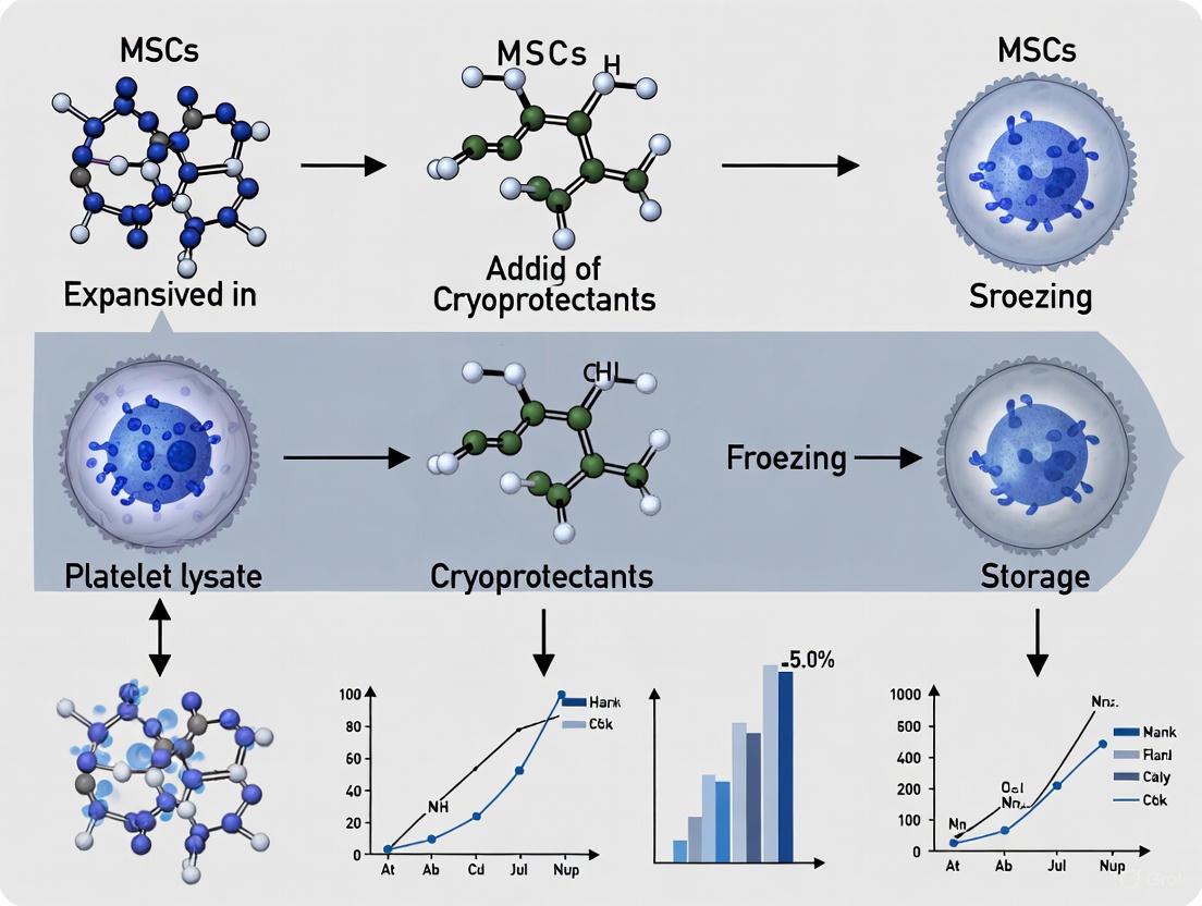
GMP-Compliant Protocols for Cryopreserving PL-Expanded MSCs
The use of platelet lysate (PL) as a supplement for the ex vivo expansion of Mesenchymal Stem Cells (MSCs) represents a significant advancement in the development of clinically applicable advanced therapy medicinal products (ATMPs) [2]. PL, rich in a plethora of growth factors and cytokines, not only replaces fetal bovine serum (FBS) to mitigate xenogenic risks but also enhances MSC proliferation rates [2]. For these PL-expanded MSCs (PL-MSCs) to be utilized as reliable "off-the-shelf" therapeutics, a robust and standardized cryopreservation process is indispensable. This protocol details a comprehensive, step-by-step procedure for the cryopreservation of PL-MSCs, designed to ensure high post-thaw viability, recovery, and, crucially, the retention of their critical biological functions, including immunomodulatory potential and multi-lineage differentiation capacity [26] [24]. The methodology outlined herein is framed within a GMP-compliant framework, incorporating key quality control checkpoints to guarantee batch-to-batch consistency and product safety [27].
Materials and Reagent Solutions
Research Reagent Solutions
The selection of high-quality, defined reagents is critical for the successful cryopreservation of PL-MSCs. The table below lists the essential materials and their functions.
Table 1: Essential Reagents and Materials for PL-MSC Cryopreservation
| Item | Function/Description | Example/Note |
|---|---|---|
| Basal Medium | Serves as the base for cryopreservation solution. | DMEM or other MSC-approved basal medium. |
| Cryoprotectant | Penetrating agent to protect against intracellular ice formation. | DMSO (e.g., 5-10%) [24]. |
| Cryopreservation Additive | Non-penetrating agent for extracellular protection and osmotic balance. | Human Serum Albumin (HSA) or Sucrose (0.2 M) [28]. |
| Complete Freezing Medium | Ready-to-use, defined cryopreservation solution. | CryoStor CS10 [29] [27] or similar GMP-grade media. |
| Cell Dissociation Reagent | To harvest adherent PL-MSCs. | TrypLE Express or other animal-origin-free enzymes [27]. |
| Wash Buffer | To remove enzyme and resuspend cells pre-freezing. | Phosphate Buffered Saline (PBS), without Ca2+/Mg2+. |
| Cryogenic Vials | For storage of cell product. | Internally-threaded, sterile vials for safe liquid nitrogen storage [29]. |
| Controlled-Rate Freezer (CRF) | Ensures consistent, optimal cooling rate. | Default profile of -1°C/min is typically effective [30]. |
Pre-freeze Equipment and Quality Control
- Equipment: Biosafety cabinet, refrigerated centrifuge, automated cell counter or hemocytometer, controlled-rate freezing device or passive freezing container (e.g., CoolCell or Mr. Frosty) [29].
- Quality Control (Pre-freeze): Confirm that cells are in the logarithmic growth phase (≥80% confluency) and are free from microbial contamination (e.g., mycoplasma) [29]. Cell viability before freezing should be ≥90% [31].
Step-by-Step Cryopreservation Protocol
Protocol Workflow
The following diagram illustrates the complete cryopreservation workflow for PL-MSCs, from cell harvest to final storage.
Detailed Experimental Method
Step 1: Cell Harvesting
- Aspirate the PL-containing culture medium from the flask and wash the cell monolayer with pre-warmed PBS to remove residual serum components.
- Add a sufficient volume of a cell dissociation reagent (e.g., TrypLE Express) to cover the monolayer and incubate at 37°C until cells detach (typically 3-5 minutes) [27].
- Neutralize the dissociation reagent with a volume of wash buffer or PL-containing medium that is at least equal to the volume of reagent used.
Step 2: Cell Washing and Counting
- Transfer the cell suspension to a conical tube and centrifuge at 300-400 × g for 5 minutes at 4°C [29].
- Carefully aspirate the supernatant and gently resuspend the cell pellet in a suitable volume of cold wash buffer or a serum-free buffer.
- Perform a cell count and viability assessment using an automated cell counter or Trypan Blue exclusion on a hemocytometer. It is critical that the starting cell population has >90% viability [31].
Step 3: Preparation in Freezing Medium
- Centrifuge the cell suspension again as in Step 2 and thoroughly aspirate the supernatant.
- Resuspend the cell pellet in the chosen, ice-cold freezing medium to achieve a final concentration of 1-5 × 10^6 cells/mL [29] [26]. For a home-made formulation, this could be a mixture of a basal medium supplemented with 10% DMSO and 2% HSA, or 10% DMSO and 0.2M Sucrose [28]. GMP-compliant, ready-to-use media like CryoStor CS10 are highly recommended [27].
- Keep the cell suspension on ice at all times to minimize cryoprotectant toxicity.
Step 4: Aliquot and Begin Freezing
- Quickly aliquot the cell suspension into pre-chilled, labeled cryogenic vials (e.g., 1 mL/vial).
- Immediately transfer the vials to a controlled-rate freezer (CRF) programmed with a standard profile of -1°C/min [30]. If a CRF is unavailable, use a passive freezing container placed in a -80°C freezer, which provides an approximate cooling rate of -1°C/min [29].
- After the program completes, promptly transfer the vials to the long-term storage location.
Step 5: Long-Term Storage
- Store cryogenic vials in the vapor phase of liquid nitrogen (below -135°C) to prevent potential contamination and ensure long-term stability [29]. Avoid storage in mechanical -80°C freezers for periods exceeding one month.
Quality Assessment and Functional Validation
Post-Thaw Analysis Workflow
Upon retrieval, a representative vial must be thawed and analyzed to validate the success of the cryopreservation process. The key parameters to assess are outlined below.
Table 2: Key Quality Control Assays for Post-Thaw PL-MSCs
| Parameter | Assay/Method | Acceptance Criterion | Rationale |
|---|---|---|---|
| Viability | Flow cytometry (Annexin V/PI) or Live/Dead staining (Calcein AM/EthD-1) [26]. | >70-80% viable cells [24]. | Measures direct survival from cryoinjury. |
| Immunophenotype | Flow cytometry for CD73, CD90, CD105 (positive) and CD34, CD45, CD14 (negative) [26] [24]. | ≥95% expression of positive markers; ≤2% for negative markers [24]. | Confirms MSC identity and purity. |
| Clonogenic Potential | Colony-Forming Unit Fibroblast (CFU-F) assay [28]. | Varies by cell source; stable colony number post-thaw. | Assesses self-renewal capacity. |
| Differentiation Potential | Trilineage Induction:- Osteogenic: Alizarin Red S- Adipogenic: Oil Red O- Chondrogenic: Alcian Blue [26] [28]. | Positive staining for lineage-specific markers. | Validates retention of multi-potency. |
| Immunomodulatory Ability | In vitro suppression of T-cell or PBMC proliferation [13] [26]. | Significant suppression of immune cell proliferation. | Critical for therapeutic efficacy. |
Troubleshooting Common Issues
- Low Post-Thaw Viability: Optimize the DMSO concentration and ensure a consistent, controlled cooling rate of -1°C/min [24]. Verify that cells were in the log phase of growth at harvest.
- Reduced Immunosuppressive Function: Note that some studies report a transient, ~50% reduction in immunosuppressive capacity post-thaw in specific in vitro assays; this should be considered during potency assay design and timing [13].
- Poor Recovery/Function after Extended Storage: Ensure stable storage in liquid nitrogen vapor phase without temperature fluctuations and use of a GMP-grade, defined freezing medium to enhance long-term stability [29].
This application note provides a standardized, detailed protocol for the cryopreservation of PL-expanded MSCs. Adherence to this protocol, with emphasis on pre-freeze quality control, a controlled freezing rate, and comprehensive post-thaw functional validation, ensures the production of a high-quality cellular product. The implementation of such a robust and reliable cryopreservation process is a cornerstone for the translational success of PL-MSCs in regenerative medicine and cell-based therapies.
The cryopreservation of mesenchymal stromal cells (MSCs) is a critical step in ensuring their off-the-shelf availability for clinical and research applications in regenerative medicine. For MSCs expanded in human platelet lysate (HPL)—a preferred, xeno-free culture supplement—selecting an appropriate cryoprotectant is paramount to maintaining post-thaw viability, functionality, and genetic stability [6] [32]. Dimethyl sulfoxide (DMSO) has long been the standard cryoprotective agent (CPA), but concerns regarding its toxicity to both cells and patients have spurred the development of alternative formulations and mitigation strategies [33] [34]. This application note provides a structured evaluation of DMSO-based and DMSO-free cryoprotectants, summarizing quantitative performance data and detailing standardized protocols for their assessment, specifically within the context of HPL-expanded MSC cultures.
Quantitative Comparison of Cryoprotectant Performance
The following tables consolidate key quantitative findings from recent, multi-center studies to facilitate direct comparison of cryoprotectant options.
Table 1: Post-Thaw Cell Recovery and Viability of MSCs Cryopreserved with Different Formulations
| Cryoprotectant Formulation | Average Post-Thaw Viability (%) | Average Recovery of Viable MSCs (%) | Key Findings |
|---|---|---|---|
| 5-10% DMSO (In-house solutions) | 89.8 [34] | 87.3 [34] | Considered the conventional standard, but carries potential toxicity concerns [33]. |
| SGI Solution (DMSO-free) | 82.9 [34] [35] | 92.9 [34] [35] | Slightly lower viability but superior cell recovery; immunophenotype and gene expression comparable to DMSO [34]. |
| 2.5% DMSO + Hydrogel Microcapsule | >70 [36] | Information Not Specified | Meets the minimum clinical threshold for viability while significantly reducing DMSO exposure [36]. |
| Trehalose via Ultrasonication | Comparable to DMSO controls [37] | Information Not Specified | Preserves multipotency; a biocompatible, non-toxic alternative requiring advanced delivery [37]. |
Table 2: Safety and Functional Profile of DMSO in MSC Therapies
| Evaluation Parameter | Finding | Clinical Context |
|---|---|---|
| Typical DMSO Dose in MSC Products | 2.5 to 30 times lower than the 1 g/kg accepted for HSC transplantation [33] [38]. | Lower systemic exposure per infusion [33] [38]. |
| Reported Infusion-Related Reactions | Isolated incidents, generally with adequate premedication [33] [38]. | Safety profile appears favorable when DMSO dose and concentration are controlled [33]. |
| Post-Thaw Immunosuppression Function | MSCs cryopreserved in FBS-supplemented media showed greater T-cell inhibition than those in HPL [6]. | Media supplement can influence cell function independently of the cryopreservation method [6]. |
Detailed Experimental Protocols
Protocol 1: Multicenter Evaluation of a Novel DMSO-Free Cryoprotectant
This protocol is adapted from an international PACT/BEST collaborative study [34] [35].
- Objective: To compare the efficacy of a novel DMSO-free solution (SGI) against standard DMSO-containing solutions for cryopreserving MSCs.
- Materials:
- SGI Solution: Sucrose, glycerol, and isoleucine in a Plasmalyte A base.
- In-house DMSO Solutions: 5-10% DMSO prepared locally at participating centers.
- MSCs: Expanded from bone marrow or adipose tissue per local protocols, preferably in HPL-supplemented media.
- Equipment: Controlled-rate freezer, liquid nitrogen storage, cell counter/analyzer, flow cytometer.
- Methodology:
- Cell Preparation: Harvest MSCs at the desired passage and create a single-cell suspension.
- Aliquoting and Cryopreservation: Aliquot the MSC suspension into cryovials or bags. For each solution (SGI and in-house DMSO), cryopreserve at least three replicate vials.
- Freezing Process: Place vials in a controlled-rate freezer, cooling at a standardized rate (e.g., -1°C/min) before transfer to liquid nitrogen for at least one week.
- Thawing and Assessment: Rapidly thaw cells in a 37°C water bath.
- Post-Thaw Analysis:
- Viability & Recovery: Assess using trypan blue exclusion or automated cell counting.
- Immunophenotype: Confirm MSC identity via flow cytometry for CD73, CD90, CD105 (positive) and CD45, CD34, CD14, HLA-DR (negative).
- Gene Expression: Perform global transcriptome analysis (e.g., RNA-Seq) to compare profiles.
Protocol 2: Hydrogel Microencapsulation to Enable Low-DMSO Cryopreservation
This protocol is based on research using hydrogel microcapsules to reduce cryoinjury [36].
- Objective: To cryopreserve MSCs using a low concentration (2.5%) of DMSO by employing alginate hydrogel microencapsulation.
- Materials:
- Sodium Alginate Solution: Sterile, low-viscosity alginate in culture-grade water.
- Calcium Chloride Solution: 100mM CaCl₂ for cross-linking.
- Core Solution: Contains mannitol and hydroxypropyl methylcellulose.
- Equipment: High-voltage electrostatic coaxial spraying device, infusion pumps.
- Methodology:
- Cell Encapsulation:
- Resuspend the MSC pellet in the core solution mixed with type I collagen.
- Use the coaxial spraying device with the core solution (inner flow) and sodium alginate (outer flow) to generate droplets that fall into the CaCl₂ solution, forming gelled microcapsules.
- Pre-culture: Culture the microcapsules in complete medium for 24-48 hours.
- Cryopreservation: Suspend the microcapsules in culture medium supplemented with 2.5% (v/v) DMSO. Transfer to cryovials and freeze using a standard controlled-rate protocol.
- Thawing and Release: Rapidly thaw microcapsules. Dissolve the alginate shell using a chelating agent like sodium citrate to release MSCs for analysis.
- Post-Thaw Analysis: Assess viability, differentiation potential (osteogenic, adipogenic, chondrogenic), and expression of stemness-related genes.
- Cell Encapsulation:
Visualizing Cryoprotectant Evaluation and Application Strategies
The following diagrams outline the core strategies for evaluating cryoprotectants and applying advanced low-DMSO techniques.
Comprehensive Cryoprotectant Evaluation Workflow
Hydrogel Microencapsulation for Low-DMSO Cryopreservation
The Scientist's Toolkit: Essential Reagents and Materials
Table 3: Key Research Reagent Solutions for MSC Cryopreservation Studies
| Reagent / Material | Function / Application | Examples / Notes |
|---|---|---|
| Human Platelet Lysate (HPL) | Xeno-free supplement for MSC expansion media; reduces immunogenic risk vs. FBS [6] [32]. | Multiple commercial sources; variability between lots should be monitored [6]. |
| DMSO (Clinical Grade) | Penetrating cryoprotectant; standard of care but requires toxicity management [33] [34]. | Use at lowest effective concentration (e.g., 5-10%); associated with patient side effects at high doses [33] [36]. |
| SGI Solution | DMSO-free cryoprotectant containing Sucrose, Glycerol, Isoleucine in Plasmalyte A [34] [35]. | Shows comparable recovery and phenotype to DMSO; a promising non-toxic alternative [34]. |
| Sodium Alginate | Natural biomaterial for forming hydrogel microcapsules that protect cells during freezing [36]. | Used in microencapsulation strategies to enable drastic DMSO reduction to 2.5% [36]. |
| Trehalose | Non-penetrating, biocompatible disaccharide cryoprotectant [37]. | Requires ultrasonication with microbubbles for intracellular delivery; avoids chemical toxicity [37]. |
The move towards xeno-free cell manufacturing, using HPL for expansion, must be matched by advances in cryopreservation to ensure final product safety and efficacy. While DMSO remains a functionally effective cryoprotectant, evidence indicates that DMSO-free solutions like SGI and technologies such as hydrogel microencapsulation are viable and often superior from a safety perspective. Future work should focus on standardizing these protocols across manufacturing centers, as local processes significantly impact outcomes [6], and on validating the long-term functional potency of MSCs preserved with these advanced methods in preclinical models.
In the field of advanced therapy medicinal products (ATMPs), Mesenchymal Stromal Cells (MSCs) have emerged as a cornerstone for cell-based therapies, demonstrating significant potential in treating a wide range of diseases, from graft-versus-host disease to intestinal inflammation [39]. The transition of MSC therapies from research to clinical application necessitates strict adherence to Good Manufacturing Practice (GMP) standards, requiring standardized protocols that ensure consistent product quality, safety, and efficacy [40] [41]. A critical determinant of MSC quality throughout the manufacturing process is the precise control of culture parameters, particularly the optimization of harvesting at specific confluence states and passage numbers.
The practice of harvesting MSCs at the correct confluence and passage is not merely a procedural step but a fundamental aspect of quality control that directly impacts critical quality attributes (CQAs) of the final cellular product. These attributes include cell viability, proliferative capacity, immunophenotype stability, differentiation potential, and therapeutic potency [42] [40]. Operating within the specific context of platelet lysate-expanded cultures—which are increasingly replacing fetal bovine serum (FBS) to meet regulatory and safety concerns—this protocol details the evidence-based procedures for determining and executing the optimal harvest point in MSC manufacturing processes.
Quantitative Benchmarks for Harvest Decisions
Extensive research has established correlations between culture confluence, passage number, and key MSC characteristics. The tables below summarize critical quantitative benchmarks to guide harvest decisions.
Table 1: Impact of Passage Number on MSC Culture Characteristics
| Passage Range | Proliferation Kinetics | Characteristic Stability | Recommended Use |
|---|---|---|---|
| Early (P1-P3) | Highest proliferation rate; shortest population doubling time [41] | Stable immunophenotype and differentiation potential [2] | Master Cell Bank creation; Clinical-scale expansion |
| Mid (P4-P6) | Consistent proliferation in optimized media (e.g., MSC-Brew) [41] | Maintains critical functions; genomic stability should be monitored [40] | Large-scale production for clinical trials |
| Late (P7+) | Significantly decreased proliferation capacity; elongated doubling time [40] | Increased risk of senescence; potential loss of function [40] | Not recommended for clinical applications |
Table 2: Confluence Guidelines for MSC Harvesting
| Confluence Stage | Morphological Cues | Performance Outcomes | Harvest Recommendation |
|---|---|---|---|
| Sub-confluent (70-80%) | Cells are spindle-shaped, evenly distributed, with minimal contact inhibition [42] | Optimal yield of viable, proliferative cells for subsequent passages [42] | Ideal for routine passaging and continued expansion |
| Fully Confluent (90-100%) | Dense, monolayer formation; some flattening may occur [42] | Potential onset of contact inhibition and spontaneous differentiation [40] | Harvest promptly; avoid prolonged maintenance |
| Over-confluent (>100%) | Pronounced cell flattening, granular appearance, potential vacuolization [42] | Reduced recovery post-thaw, decreased proliferative capacity, senescence [40] | Avoid for clinical product collection |
Detailed Experimental Protocols
Protocol 1: Routine Monitoring and Determination of Harvest Point
This protocol ensures consistent monitoring and objective assessment of culture confluence and cellular morphology to identify the optimal harvest window.
Materials:
- Phase-contrast microscope
- Hemocytometer or automated cell counter
- Culture vessels (flasks, cell stacks, or bioreactors)
- Phosphate-Buffered Saline (PBS)
- Trypsin/EDTA or other GMP-compliant dissociation reagent
- Platelet lysate (PL)-supplemented medium [2]
Procedure:
- Daily Microscopic Examination:
- Using a phase-contrast microscope, visually assess cultures daily once they exceed 50% confluence.
- Document cell morphology, noting a healthy, spindle-shaped, fibroblast-like appearance. Be alert for increased flatness, granularity, or irregular shapes, which may indicate less optimal states [42].
- Estimate the percentage of the culture surface covered by cells (confluence). For greater objectivity, use image analysis software if available.
- Harvest Trigger:
- Initiate the harvest procedure when cultures reach 70-90% confluence [42]. This range typically provides the best balance between high cell yield and the preservation of progenitor characteristics and viability.
- Do not allow cultures to remain at >100% confluence for extended periods, as this accelerates senescence and functional decline [40].
Protocol 2: Harvesting and Post-Harvest Quality Assessment
This protocol covers the harvesting process and the essential quality control checks to validate the success of the harvest timing.
Materials:
- Trypsin/EDTA or a GMP-compliant recombinant enzyme (e.g., TrypLE)
- Centrifuge
- PL-supplemented medium or a clinically compatible cryopreservation medium
- Flow cytometer with antibodies for CD73, CD90, CD105, and hematopoietic negativity panel (CD45, CD34, etc.) [40]
Harvesting Procedure:
- Aspirate the culture medium from the vessel and wash the cell layer gently with PBS to remove residual serum/proteins.
- Add a pre-warmed, GMP-compliant dissociation reagent (e.g., TrypLE) sufficient to cover the cell layer.
- Incubate the vessel at 37°C for the time recommended by the reagent manufacturer (typically 3-5 minutes). Monitor detachment visually under a microscope.
- Neutralize the dissociation reagent by adding a volume of PL-supplemented medium that is at least equal to the volume of the reagent used.
- Collect the cell suspension and centrifuge (e.g., 300-400 x g for 5-10 minutes). Resuspend the cell pellet in the appropriate medium for counting, passaging, or cryopreservation.
Post-Harvest Quality Control:
- Viability and Yield: Determine cell count and viability using Trypan Blue exclusion or an automated cell counter. Expected viability should be >95% for high-quality cultures [41].
- Immunophenotype Verification: Perform flow cytometry on a sample of the harvested cells to confirm expression of typical MSC markers (CD105, CD73, CD90 > 95%) and lack of hematopoietic markers (CD45, CD34, CD11b, CD19, HLA-DR < 2%) [40].
- Functional Potency (Lot Release): Perform a colony-forming unit fibroblast (CFU-F) assay to confirm clonogenic capacity. Cultures harvested at the optimal state should show robust colony formation [41].
Workflow Integration and Decision Pathways
The following diagram illustrates the integrated workflow for culture maintenance, harvest decision-making, and subsequent processing within a GMP-compliant framework for platelet lysate-expanded MSCs.
The Scientist's Toolkit: Essential Reagents and Platforms
The successful implementation of these harvesting protocols relies on the use of specific, quality-assured materials. The following table details key research reagent solutions for GMP-compliant MSC manufacturing.
Table 3: Essential Reagents and Platforms for MSC Culture and Harvest Optimization
| Reagent/Solution | Function | GMP-Compliant Examples & Notes |
|---|---|---|
| Human Platelet Lysate (hPL) | Xeno-free culture supplement; provides growth factors and adhesion proteins [2]. | Pooled, pathogen-inactivated batches from certified blood banks. Must be tested for endotoxin and growth factor levels [2] [3]. |
| Serum-Free/Xeno-Free Media | Chemically defined media eliminating lot-to-lot variability and safety risks of animal sera [42]. | MSC-Brew GMP Medium (Miltenyi), StemMACS MSC XF (Miltenyi), NutriStem XF (Biological Industries) [42] [41]. |
| GMP-Compliant Dissociation Agents | Enzymatic detachment of adherent MSCs for passaging and harvesting. | Recombinant trypsin substitutes (e.g., TrypLE), which are animal-origin-free and gentle on cell surface proteins [41]. |
| Cryopreservation Solutions | Maintain cell viability and functionality during freeze-thaw cycles. | Should contain protein (e.g., 2% Human Serum Albumin) to prevent thawing-induced cell loss [19]. DMSO concentration should be controlled and minimized [43]. |
| Automated Bioreactor Systems | Scalable, closed-system platforms for consistent large-scale MSC expansion. | Quantum Cell Expansion System (Terumo BCT), CliniMACS Prodigy (Miltenyi Biotec). Ensure homogeneous culture conditions and reduce manual handling [40]. |
Harvesting MSCs at the correct confluence and passage is a critical process parameter that directly determines the quality, functionality, and therapeutic consistency of the final cell product. The protocols and benchmarks outlined here, specifically tailored for platelet lysate-expanded cultures, provide a actionable framework for researchers and manufacturers. By integrating daily morphological assessments with stringent post-harvest quality controls and leveraging GMP-compliant reagents and automated systems, it is possible to standardize MSC production. This standardization is paramount for achieving the reproducibility required to advance promising MSC therapies from the research bench into reliable clinical applications, ultimately fulfilling their potential in regenerative medicine.
Within research on platelet lysate (PL)-expanded Mesenchymal Stromal Cells (MSCs), cryopreservation is not merely a storage technique but a critical determinant of cell quality and therapeutic efficacy. The choice between home-made and commercial Good Manufacturing Practice (GMP)-grade freezing media carries significant implications for experimental reproducibility, regulatory compliance, and clinical translation. This application note provides a detailed comparison of these two approaches, supported by quantitative data and standardized protocols, to guide researchers and drug development professionals in optimizing their cryopreservation strategies for PL-expanded MSCs.
The transition from research to clinical application necessitates stringent quality controls. Regulatory authorities increasingly discourage the use of fetal bovine serum (FBS) in freezing media due to concerns about undefined components, lot-to-lot variability, and the risk of transmitting zoonotic infections [2]. Human platelet lysate has emerged as a superior alternative for MSC expansion, offering a well-characterized, xeno-free supplement that supports high cell proliferation without compromising immunophenotype or differentiation potential [2]. This same principle of defined composition and safety extends to the critical choice of cryopreservation media.
Comparative Analysis: Home-Made vs. GMP-Grade Media
The selection of cryopreservation media fundamentally influences post-thaw cell viability, recovery, and functional characteristics. The table below summarizes the core differences between home-made and commercial GMP-grade formulations.
Table 1: Key Characteristics of Home-Made vs. GMP-Grade Freezing Media
| Characteristic | Home-Made Freezing Media | Commercial GMP-Grade Media |
|---|---|---|
| Typical Composition | Culture medium (e.g., DMEM) with 10% FBS and 10% DMSO [44] | Pre-formulated, often serum- and protein-free; defined DMSO concentrations (e.g., 2%, 5%, 10%) [45] |
| Regulatory Status | Poorly defined; not intended for clinical use | Manufactured under cGMP principles; suitable for clinical applications [45] [46] |
| Batch-to-Batch Consistency | High variability due to serum components [45] [2] | High consistency; rigorous quality control and multi-year stability profiles [45] [44] |
| Mitigation of Cell Stress | Limited and undefined | Engineered to mitigate temperature-induced molecular stress and reduce cryopreservation-induced delayed-onset cell death [45] |
| Primary Application | Basic research with robust cell lines | Sensitive cells (e.g., MSCs, iPSCs), clinical trials, and commercial cell-based therapies [45] [44] |
Commercial GMP-grade media, such as CryoStor and Bambanker, are specifically designed to address the molecular stress responses that occur during freezing and thawing [45] [47]. Their defined, serum-free formulations not only enhance post-thaw viability but also simplify regulatory approval by ensuring consistent production and control according to established quality standards [45] [46].
Empirical data from published studies provides compelling evidence for the functional impact of different freezing media and protocols. The following table consolidates key quantitative findings relevant to PL-expanded MSCs.
Table 2: Summary of Quantitative Findings on Cryopreservation from Scientific Literature
| Study Model | Freezing Protocol | Key Quantitative Outcome | Reference |
|---|---|---|---|
| Human B Cells (from donors) | CryoStor CS10 | Post-thaw viability: 94.3 - 97.9% (PI staining) | [45] |
| Bone Marrow Aspirate Concentrate (BMAC) | 10% DMSO in autologous plasma, passive freezing (-1°C/min) | MSC proliferation and multilineage differentiation preserved after 4 weeks at -80°C; cartilage repair in OA rat model equivalent to fresh BMAC. | [48] |
| Adipose-Derived MSCs | DMSO-based cryoprotectant | Up to 50% cell loss when thawed in protein-free solutions; >90% viability with no cell loss for 4h when reconstituted in isotonic saline. | [19] |
| Clinical-Grade BM-MSCs | Proprietary GMP protocol (PL-based expansion) | Superior viability and recovery post-thaw; phenotype and differentiation unaltered; ~50% reduced performance in in vitro IDO-pathway immunosuppression assay. | [21] |
A critical finding from recent research is that the thawing and reconstitution process is as vital as the freezing itself. One study demonstrated that reconstituting MSCs at concentrations below 100,000 cells/mL in protein-free vehicles resulted in instant cell loss exceeding 40% [19]. The addition of clinical-grade Human Serum Albumin (HSA) was shown to prevent this thawing- and dilution-induced cell loss, highlighting the need for standardized post-thaw handling [19].
Detailed Experimental Protocols
Protocol 1: Cryopreservation of PL-Expanded MSCs Using Commercial GMP-Grade Media
This protocol utilizes CryoStor CS10, designed for sensitive cell types like MSCs, to ensure high viability and functionality post-thaw [45].
Materials:
- Cells: PL-expanded MSCs at desired passage (e.g., P2-P4) [21].
- Freezing Media: Pre-chilled CryoStor CS10 [45].
- Other Reagents: DPBS without Ca2+/Mg2+.
- Equipment: Controlled-rate freezer (e.g., CoolCell or equivalent), cryogenic vials, centrifuge.
Procedure:
- Cell Harvest: Detach MSCs using a clinical-grade enzyme (e.g., TrypLE Select). Neutralize the enzyme with a PL-containing culture medium.
- Cell Washing: Centrifuge the cell suspension and carefully aspirate the supernatant. Resuspend the cell pellet in DPBS to remove residual enzymes and PL.
- Cell Counting: Perform a viable cell count using an automated cell counter (e.g., NucleoCounter NC-100) or hemocytometer [21].
- Formulating Final Suspension: Centrifuge the cells again and aspirate the DPBS. Resuspend the cell pellet in pre-chilled CryoStor CS10 to a final concentration of 1-5 x 10^6 cells/mL.
- Aliquoting: Dispense 1 mL of the cell suspension into each cryogenic vial.
- Controlled-Rate Freezing: Place the vials in a controlled-rate freezer and initiate a freeze program with a cooling rate of -1°C/min until reaching at least -80°C [48] [21].
- Transfer to Long-Term Storage: After freezing, immediately transfer the vials to a -150°C mechanical freezer or liquid nitrogen vapor phase for long-term storage.
Protocol 2: Assessing Post-Thaw MSC Potency and Recovery
This protocol outlines a colony-forming unit (CFU) assay to assess the clonogenic capacity and functional recovery of MSCs post-thaw, a key quality attribute.
Materials:
- Thawed MSCs: From Protocol 1.
- Culture Medium: Alpha-MEM supplemented with 20% FBS, 1% Penicillin/Streptomycin, and 10 ng/mL FGF-2 [48].
- Staining Reagents: 4% Paraformaldehyde (PFA), 1% Crystal Violet solution.
Procedure:
- Thaw and Plate: Rapidly thaw the MSC vial in a 37°C water bath. Immediately dilute the cell suspension in pre-warmed culture medium. Centrifuge to remove DMSO and resuspend in fresh culture medium.
- Cell Seeding: Seed 300,000 mononuclear cells per well of a 6-well plate. Use at least three technical replicates per condition [48].
- Culture: Incubate the plates for 14 days at 37°C and 5% CO2, replacing the culture medium twice weekly.
- Fix and Stain: On day 14, aspirate the medium. Fix the cells with 4% PFA for 15 minutes, then stain with 1% Crystal Violet solution for 30 minutes.
- Count Colonies: Gently rinse the plates with water and air-dry. Manually count colonies defined as aggregates of over 100 cells with a distinct clonal center [48].
- Analysis: Calculate the number of CFU-f per 300,000 seeded cells. Compare results between fresh and frozen MSCs to determine the recovery of clonogenic progenitors.
Workflow and Pathway Diagrams
MSC Cryopreservation and Post-Thaw Analysis Workflow
The diagram below illustrates the critical steps in the cryopreservation, thawing, and quality assessment of platelet lysate-expanded MSCs.
Impact of Cryopreservation on MSC Immunomodulatory Pathways
Cryopreservation can selectively impact specific immunomodulatory mechanisms of MSCs. This diagram outlines the potential effects on the IDO-mediated pathway, which is often more susceptible to freezing stress.
The Scientist's Toolkit: Essential Reagents and Materials
Successful cryopreservation of PL-expanded MSCs requires a suite of standardized, quality-assured materials. The following table details key reagents and their functions in the workflow.
Table 3: Essential Research Reagent Solutions for MSC Cryopreservation
| Reagent/Material | Function & Importance | Example Products / Formulations |
|---|---|---|
| GMP-Grade Freezing Media | Pre-formulated, serum-free solutions designed to mitigate freezing-induced cell stress and ensure batch-to-batch consistency for regulatory compliance. | CryoStor [45], Bambanker [47], BloodStor [45] |
| Human Platelet Lysate (PL) | Xeno-free, clinically compliant supplement for MSC expansion, providing a plethora of growth factors that support proliferation without altering MSC characteristics. | Pooled, pathogen-inactivated PL from qualified donors [2] [21] |
| Protein-Containing Reconstitution Solution | Essential for preventing massive cell loss during thawing and dilution; provides osmotic protection and stabilizes cell membranes. | Isotonic Saline with 2% Human Serum Albumin (HSA) [19] |
| Controlled-Rate Freezer (CRF) | Provides precise control over cooling rate (-1°C/min), a critical process parameter to minimize intracellular ice formation and osmotic shock. | Various GMP-compliant vendors [30] |
| Automated Cell Thawing System | Ensures consistent, GMP-compliant thawing, eliminating contamination risks from water baths and standardizing the warming rate. | ThawSTAR CFT2 [45] |
The choice between home-made and GMP-grade freezing media is a strategic decision that extends beyond simple cell preservation. For preclinical research with PL-expanded MSCs, commercial GMP-grade media offer defined composition, superior cytoprotection, and enhanced reproducibility. When transitioning to clinical trials or cell banking, the regulatory compliance, documented quality control, and optimized performance of GMP-grade media become indispensable.
Researchers must be aware that cryopreservation is a system, not just a reagent. The freezing protocol, thawing method, and post-thaw handling are integral to maintaining MSC quality and critical functions, such as immunomodulation. Adopting the standardized protocols and quality-focused materials outlined in this application note will provide a robust foundation for reliable and translatable results in MSC-based research and therapy development.
The cryopreservation of Mesenchymal Stromal Cells (MSCs) expanded in human platelet lysate (hPL) is a critical process in regenerative medicine and advanced therapy medicinal product (ATMP) development. As regulatory authorities increasingly discourage the use of fetal bovine serum (FBS) due to concerns about xenogenic antigens and zoonotic infections, hPL has emerged as an effective, clinically compatible alternative for MSC expansion [49]. Within this context, selecting an appropriate cryopreservation strategy becomes paramount for maintaining cell functionality, viability, and therapeutic potential. The two principal techniques—slow freezing and vitrification—operate on fundamentally different principles of rate control and cryoprotectant utilization, each with distinct implications for MSC-based therapies. This application note provides a detailed comparative analysis of these techniques, supported by quantitative data and standardized protocols, to guide researchers in selecting and optimizing cryopreservation methods for hPL-expanded MSCs.
Technical Comparison: Fundamental Principles and MSC Outcomes
Mechanism of Action
Slow Freezing relies on controlled, gradual cooling typically at -1°C to -3°C per minute, facilitating cellular dehydration and minimizing intracellular ice crystal formation [50] [29]. This technique uses relatively low concentrations of cryoprotectants (usually 5-10% DMSO) and requires specialized equipment like programmable freezers or passive cooling devices such as Mr. Frosty or CoolCell [29].
Vitrification employs high cooling rates and high concentrations of cryoprotectants (typically 6-8M) to achieve an amorphous, glass-like state without ice crystal formation [51] [50]. This method is characterized by ultrarapid cooling, often directly immersing samples in liquid nitrogen, and can be completed more quickly than slow freezing [52].
Comparative Performance Metrics for MSCs
Table 1: Comparative Analysis of Slow Freezing vs. Vitrification for MSCs
| Parameter | Slow Freezing | Vitrification | References |
|---|---|---|---|
| Typical Cell Viability | 70-80% post-thaw survival | 96% demonstrated in optimized 3D systems | [50] [53] |
| Cooling Rate | -1°C/min to -3°C/min | Ultra-rapid (> -100°C/min) | [50] [29] |
| CPA Concentration | Low (e.g., 10% DMSO) | High (e.g., 38% EG, 20% DMSO) | [51] [54] |
| Equipment Needs | Programmable freezer or passive cooling device | Liquid nitrogen, specialized carriers | [29] [52] |
| Process Duration | Several hours | 30 minutes to 1 hour | [53] [52] |
| Structural Impact | Potential for extracellular ice damage | Avoids ice crystal formation | [50] [52] |
| Genetic Stability | Generally maintained | Transcriptomic changes reported | [51] |
| Clinical Suitability | Well-established, current gold standard | Emerging protocols, requires optimization | [21] [52] |
Research indicates that vitrification can achieve excellent post-thaw viability, with one study reporting 96% viability for 3D-human MSCs encapsulated in GelMA hydrogel [53]. However, this often requires optimized conditions, as vitrification poses challenges including potential cytotoxicity from high cryoprotectant agent (CPA) concentrations and the need for precise protocol standardization [51] [50].
For slow-frozen MSCs, studies report generally maintained immunophenotype (expression of CD105, CD73, CD90) and differentiation potential post-thaw, though some report a reduced in vitro immunosuppressive capacity specifically related to the IDO pathway [21]. Vitrification has also been shown to preserve MSC surface markers and differentiation potential, with one study suggesting ethylene glycol (EG) may offer better protection of cell viability and proliferation compared to DMSO [51].
Detailed Experimental Protocols
Protocol for Slow Freezing of hPL-Expanded MSCs
Principle: Controlled-rate freezing minimizes intracellular ice formation by allowing sufficient cellular dehydration [50] [29].
Materials:
- MSC suspension (≥80% confluent, during log phase)
- Pre-cooled freezing medium: D-MEM low glucose + 40 IU/ml heparin + 10% hPL + 10% DMSO
- Cryogenic vials (e.g., Corning)
- Passive cooling container (e.g., Nalgene Mr. Frosty) or controlled-rate freezer
- -80°C freezer
- Liquid nitrogen storage tank
Procedure:
- Harvesting: Detach MSCs using TrypLE Select and determine viability/count (e.g., via NucleoCounter) [21].
- Centrifugation: Centrifuge cell suspension and carefully remove supernatant [29].
- Freezing Medium Preparation: Resuspend cell pellet in pre-cooled freezing medium to a final concentration of 1×10^6 cells/mL [29]. Critical: Work quickly to minimize DMSO exposure at room temperature.
- Aliquoting: Dispense 1 mL of cell suspension into each cryogenic vial [29].
- Freezing:
- Long-Term Storage: Transfer vials to liquid nitrogen tank (-135°C to -196°C) for long-term storage [29].
Protocol for Vitrification of hPL-Expanded MSCs
Principle: Ultrafast cooling combined with high CPA concentration induces a glassy state without ice crystal formation [51] [53].
Materials:
- MSC suspension
- Equilibration Solution: 3.8% ethylene glycol (EG) + 0.5M sucrose + 6% SSS in MEM-Glumax
- Vitrification Solution: 38% EG + 0.5M sucrose + 6% SSS in MEM-Glumax
- Warming Solutions: Sucrose gradients (0.5M, 0.25M, 0.125M, 0M) + 6% SSS in basic medium
- Liquid nitrogen
- Vitrification device (e.g., Cryotop, metallic grid)
- 37°C water bath
Procedure:
- Equilibration: Transfer ovarian tissues to equilibration solution for 3 minutes at room temperature [54].
- Vitrification Solution Exposure: Transfer to vitrification solution for 11 minutes at room temperature [54].
- Cooling: Place tissue on metallic grid and plunge directly into liquid nitrogen [54].
- Storage: Transfer to cryotubes for long-term storage in liquid nitrogen [54].
- Warming: Rapidly warm by immersing in 37°C water bath for 2 minutes [51].
- CPA Removal: Incubate in sucrose gradient solutions (0.5M, 0.25M, 0.125M, 0M) for 5 minutes each at room temperature to remove cryoprotectants [54].
Post-Thaw Handling and MSC Stability
Proper post-thaw handling is crucial for maintaining MSC quality and functionality. Research indicates that thawing cryopreserved MSCs in protein-free solutions can result in up to 50% cell loss [19]. The addition of human serum albumin (HSA) to thawing solutions has been shown to prevent this cell loss [19].
Table 2: Optimized Post-Thaw Handling for Cryopreserved MSCs
| Parameter | Recommendation | Impact on MSC Quality |
|---|---|---|
| Thawing Method | Rapid thawing in 37°C water bath | Minimizes ice recrystallization damage |
| Reconstitution Solution | Isotonic saline with 2% HSA | Prevents up to 50% cell loss |
| Post-Thaw Cell Concentration | ≥5×10^6 cells/mL | Prevents dilution-induced cell death |
| Post-Thaw Storage Duration | ≤4 hours at room temperature | Maintains >90% viability |
| Viability Assessment | Flow cytometry with 7-AAD | Accurate quantification of live/dead cells |
For clinical applications, it is recommended to reconstitute MSCs in simple isotonic saline with 2% HSA, which ensures >90% viability with no significant cell loss for at least 4 hours at room temperature [19]. Diluting MSCs to concentrations below 10^5/mL in protein-free vehicles should be avoided as it results in instant cell loss (>40%) and reduced viability [19].
The Scientist's Toolkit: Essential Research Reagents
Table 3: Essential Reagents for MSC Cryopreservation in hPL Systems
| Reagent/Category | Specific Examples | Function & Application Note |
|---|---|---|
| Basal Media | D-MEM low glucose, MEM-Glumax | Form the base for both culture and cryopreservation media |
| Cryoprotectants (CPAs) | DMSO, Ethylene Glycol (EG) | Penetrating agents that reduce ice crystal formation |
| Non-Penetrating CPAs | Sucrose, Trehalose | Osmotically active agents that support dehydration |
| Protein Supplement | Human Platelet Lysate (hPL) | Defined, clinical-grade alternative to FBS for expansion |
| Stabilizing Additive | Human Serum Albumin (HSA) | Prevents cell loss during thawing and reconstitution |
| Freezing Media | CryoStor CS10, MesenCult-ACF | Pre-formulated, GMP-compliant options |
| Vitrification Carriers | Cryotop, Metallic grids | Enable ultra-rapid cooling for vitrification |
Both slow freezing and vitrification present viable pathways for cryopreserving MSCs expanded in platelet lysate, yet with distinct advantages and limitations. Slow freezing remains the established method for clinical applications, offering robust results and standardized protocols. Vitrification shows promising potential with reportedly less DNA fragmentation and improved stromal preservation, though protocol standardization requires further development. The selection between these techniques should be guided by specific research or clinical requirements, considering factors such as viability needs, equipment availability, and regulatory compliance. As MSC therapies continue to advance, optimized cryopreservation protocols incorporating defined components like hPL and HSA will be essential for ensuring consistent, high-quality cell products for regenerative medicine applications.
Within the expanding field of regenerative medicine, cryopreserved mesenchymal stromal cells (MSCs) expanded in human platelet lysate (hPL) have emerged as a cornerstone for off-the-shelf cellular therapies. The transition of these living products from liquid nitrogen storage to a ready-to-infuse suspension represents a critical juncture, where suboptimal handling can significantly compromise cell viability, yield, and therapeutic potency. Variations in post-thaw reconstitution protocols present a major hurdle for the clinical standardization of MSC therapies [56]. This Application Note synthesizes current research to provide detailed, evidence-based protocols for the thawing and recovery of MSCs, specifically within the context of hPL-expanded cultures, to ensure maximum post-thaw viability and function for research and drug development.
Critical Parameters for Post-Thaw Recovery
The recovery of MSCs after thawing is not a single event but a process influenced by several interdependent parameters. Understanding these variables is essential for developing a robust standard operating procedure.
The Necessity of Protein in Thawing Solutions
A primary finding from recent investigations is that the presence of protein in the thawing solution is not optional but essential for preventing massive cell loss. A 2023 study demonstrated that thawing cryopreserved MSCs in protein-free solutions can lead to the immediate loss of up to 50% of the cell population [56] [19]. This cell loss is attributed to acute osmotic and mechanical stress during the rapid transition from the frozen state.
The addition of clinical-grade Human Serum Albumin (HSA) at a concentration of 2% to the thawing solution has been shown to effectively prevent this cell loss [56] [19]. HSA likely acts by providing oncotic pressure and coating surfaces, thereby protecting the fragile, newly thawed cell membranes.
Optimal Reconstitution and Storage Solutions
Following thawing and the removal of cryoprotectant, cells are reconstituted in a final vehicle for administration or short-term storage. The choice of this solution profoundly impacts MSC stability.
Table 1: Impact of Different Reconstitution Solutions on MSC Stability at Room Temperature [56] [19]
| Reconstitution Solution | Cell Viability after 1 Hour | Cell Loss after 1 Hour | Suitability for Post-Thaw Storage |
|---|---|---|---|
| Culture Medium | < 80% | > 40% | Poor |
| Phosphate Buffered Saline (PBS) | < 80% | > 40% | Poor |
| Isotonic Saline (with or without 2% HSA) | > 90% | Minimal | Good |
| Ringer's Acetate (with or without 2% HSA) | > 90% | Minimal | Good |
Contrary to what might be assumed, commonly used solutions like culture medium and PBS are poor choices for holding thawed MSCs, leading to significant viability drop and cell loss within an hour [56]. Simple isotonic saline emerged as a superior alternative, maintaining >90% viability with no observable cell loss for at least 4 hours at room temperature [56] [19]. The addition of 2% HSA to saline further enhances its protective capacity.
Critical Minimum Cell Concentration
Reconstituting the final MSC product to an appropriate concentration is critical. Diluting MSCs to too low a density, specifically below 100,000 cells/mL (10^5/mL), in protein-free vehicles results in instant cell loss exceeding 40% and reduced viability below 80% [56] [19]. This suggests that a certain cell density is required for survival, potentially through autocrine and paracrine signaling. Therefore, maintaining a concentration at or above 5 x 10^6 cells/mL during reconstitution is recommended for optimal stability [56].
Detailed Experimental Protocols
Protocol: Thawing and Reconstitution of hPL-Expanded MSCs
This protocol is optimized for clinical compatibility, ensuring high MSC yield, viability, and short-term stability [56] [57] [19].
Materials Required:
- Cryopreserved vial of MSCs (expanded in hPL, cryopreserved in DMSO-based cryoprotectant like CryoStor CS10)
- Water bath or dry bead bath (37°C)
- Pre-warmed thawing solution: Plasmalyte-A or Saline supplemented with 2% clinical-grade Human Serum Albumin (HSA)
- Reconstitution/Storage solution: Isotonic Saline (with or without 2% HSA)
- Centrifuge
- Pipettes and sterile tubes
Workflow:
- Preparation: Warm the thawing solution to room temperature or 37°C. Label appropriate sterile tubes.
- Rapid Thaw: Remove the MSC cryovial from liquid nitrogen storage and immediately place it in a 37°C water bath or dry bath. Gently agitate until only a small ice crystal remains (approximately 2 minutes). Critical Note: Thawing quickly is essential to minimize the toxic effects of DMSO and ice crystal recrystallization [24].
- Transfer and Dilute: Aseptically transfer the thawed cell suspension from the cryovial into a sterile tube containing a pre-determined volume of warm thawing solution (e.g., dilute 1:10 to rapidly reduce DMSO concentration).
- Gentle Centrifugation: Centrifuge the cell suspension at approximately 300-400 x g for 5 minutes to pellet the cells and remove the DMSO-containing supernatant [24] [48].
- Reconstitution: Carefully decant the supernatant. Resuspend the cell pellet in the desired volume of isotonic saline to achieve the final administration concentration. Critical Note: Maintain a minimum concentration of 5 x 10^6 cells/mL to prevent dilution-induced cell death [56] [19].
- Post-Thaw Storage: The reconstituted cell product can be stored in isotonic saline at room temperature for up to 4 hours without significant loss of viability or cell number [56] [19].
Diagram 1: Optimized workflow for MSC thawing and recovery, highlighting critical parameters.
Protocol: Post-Thaw Viability and Recovery Assessment
Accurate assessment of post-thaw cell number and viability is crucial for dosing and quality control.
Materials Required:
- Reconstituted MSC product
- Trypan Blue solution (0.4%) or Automated Cell Counter
- Hemocytometer
- Flow Cytometer
- Annexin V / Propidium Iodide (PI) or 7-AAD staining kit
Method:
- Cell Counting and Viability (Trypan Blue Exclusion):
- Mix a small aliquot of the reconstituted cell suspension with an equal volume of 0.4% Trypan Blue.
- Load onto a hemocytometer and count the cells under a microscope.
- Viable cells will exclude the dye and appear bright, while non-viable cells will take up the dye and appear blue.
- Calculate total cell count and percentage viability [57].
Apoptosis and Necrosis Assessment (Flow Cytometry):
- Use Annexin V / PI staining for a more precise quantification of viable (Annexin V-/PI-), early apoptotic (Annexin V+/PI-), late apoptotic (Annexin V+/PI+), and necrotic (Annexin V-/PI+) cell populations [57].
- Alternatively, 7-Aminoactinomycin D (7-AAD) can be used with flow cytometry to determine viability, as described in studies using 7-AAD to identify non-viable cells [56].
Recovery Calculation:
- Calculate the post-thaw recovery percentage by dividing the total number of viable cells recovered after thaw and reconstitution by the total number of cells that were originally cryopreserved in the vial.
The Scientist's Toolkit: Essential Research Reagents
Table 2: Key Reagents for MSC Thawing and Recovery Protocols
| Reagent / Material | Function / Purpose | Examples / Notes |
|---|---|---|
| Human Serum Albumin (HSA) | Protein source for thawing solutions; prevents cell loss by mitigating osmotic shock and surface adhesion. | Use clinical-grade, 2% concentration in isotonic solutions [56] [19]. |
| Isotonic Saline | Reconstitution and post-thaw storage vehicle; provides superior stability compared to PBS or culture medium. | 0.9% Sodium Chloride. Simple and clinically compatible [56] [19]. |
| Plasmalyte-A | Buffer solution; can be used as a base for thawing solutions or for diluting DMSO post-thaw. | Used in PHD10 cryopreservation formulation (PLA/5%HA/10%DMSO) [57]. |
| DMSO-Reduction Diluent | Solution used to dilute high cell concentration products post-thaw to reduce final DMSO concentration for infusion. | e.g., Plasmalyte-A with 5% HSA [57]. |
| Cryopreservation Solutions | Protects cells during freezing and storage. DMSO is the most common permeating cryoprotectant. | CryoStor CS10, CryoStor CS5, or in-house formulations (e.g., with 10% DMSO in autologous plasma) [48] [57] [25]. |
The journey of a cryopreserved MSC from a frozen vial to a functional therapeutic is precarious. This Application Note establishes that a standardized, clinically compatible protocol for thawing and recovery is fundamental to success. The key pillars of this protocol are: 1) the mandatory inclusion of protein (e.g., 2% HSA) in the thawing solution, 2) the use of simple isotonic saline for reconstitution and short-term storage, and 3) maintaining a sufficiently high cell concentration (>5x10^6/mL) to prevent dilution-induced stress. By adhering to these evidence-based practices, researchers and developers can ensure the reliable delivery of viable, potent MSCs, thereby enhancing the reproducibility and efficacy of cellular therapies in regenerative medicine.
Solving Common Challenges in PL-MSC Cryopreservation
Troubleshooting Low Post-Thaw Viability and Cell Recovery
Achieving high viability and cell recovery after thawing is a critical bottleneck in the clinical application of mesenchymal stromal cells (MSCs). For MSCs expanded in human platelet lysate (hPL)—a common clinical-grade culture supplement—optimizing post-thaw outcomes is essential for ensuring product quality and therapeutic efficacy. This Application Note synthesizes recent research findings to identify the primary causes of low post-thaw viability and recovery and provides evidence-based, clinically compatible protocols to overcome these challenges. The recommendations are framed within the broader research context of developing robust, standardized cryopreservation processes for advanced therapy medicinal products (ATMPs).
Key Challenges and Quantitative Assessment
Research indicates that the immediate post-thaw period is when MSCs are most vulnerable. The freezing and thawing process can induce acute cellular stress, leading to several measurable deficits.
Table 1: Key Post-Thaw Deficits in Cryopreserved MSCs
| Cell Attribute | Impact of Cryopreservation (Immediately Post-Thaw) | Quantitative Data | Recovery Timeline | Primary Citation |
|---|---|---|---|---|
| Viability | Reduced due to immediate apoptosis and necrosis. | Significantly increased apoptosis (Annexin V+ cells). | Recovers within 24 hours. | [22] [58] |
| Metabolic Activity | Acutely impaired. | Significantly increased metabolic activity in freshly thawed (FT) cells, indicating stress. | May remain lower than fresh cells even at 24 hours. | [22] [58] |
| Adhesion Potential | Significantly impaired. | Reduced adhesion potential in the first 4-24 hours post-thaw. | Requires >24 hours for full recovery. | [22] |
| Immunophenotype | Alteration in specific surface markers. | Decreased expression of CD44 and CD105 in FT cells. | Recovers after 24-hour acclimation. | [58] |
| Immunosuppressive Function | Potentially reduced, depending on the mechanism. | ~50% reduced performance in in vitro T-cell suppression assays (IDO-pathway specific). | Recovers after 24-hour acclimation. | [21] [58] |
| Cell Yield/Loss | Significant loss during thawing and reconstitution if protocol is suboptimal. | Up to 50% cell loss when thawed in protein-free solutions. | N/A – Preventable with correct protocol. | [56] [19] |
A quantitative study demonstrated that cryopreservation reduces cell viability and increases apoptosis immediately after thawing, with metabolic activity and adhesion potential remaining impaired for at least 24 hours [22]. Furthermore, a 24-hour acclimation period post-thaw was shown to be critical for MSCs to "reactivate" and regain functional potency, including upregulated expression of angiogenic and anti-inflammatory genes and enhanced immunomodulatory capacity [58].
Optimized Experimental Protocols
Clinically Compatible Thawing and Reconstitution Protocol
The following protocol, optimized for hPL-expanded MSCs, addresses the major pitfalls of thawing and reconstitution [56] [19].
Workflow Overview:
Detailed Methodology:
Preparation of Thawing Solution:
Thawing and Dilution:
- Remove the cryovial from liquid nitrogen storage.
- Thaw the vial rapidly by gentle agitation in a 37°C water bath for approximately 1 minute, until only a small ice crystal remains.
- Transfer the contents of the vial to a sterile tube.
- Slowly dilute the cell suspension 1:10 by dropwise addition of the pre-warmed thawing solution while gently swirling the tube. This step dilutes the cytotoxic cryoprotectant (DMSO).
Centrifugation and Reconstitution:
- Centrifuge the cell suspension at 400–500 × g for 5 minutes at room temperature.
- Carefully aspirate the supernatant without disturbing the cell pellet.
- Resuspend the cell pellet in the preferred administration or storage solution (see Section 3.2).
- Critical Step: Resuspend cells to a final concentration not less than 5 × 10^5 cells/mL. Diluting below 1 × 10^5 cells/mL in protein-free vehicles causes instant and significant cell loss (>40%) [19].
Post-Thaw Storage and Acclimation
The choice of solution for post-thaw storage is a key determinant of cell stability.
Table 2: Impact of Storage Solution on MSC Stability at Room Temperature [56] [19]
| Storage Solution | Cell Viability after 1h | Cell Loss after 1h | Recommendation |
|---|---|---|---|
| Culture Medium | <80% | >40% | Not recommended for short-term storage. |
| Phosphate Buffered Saline (PBS) | <80% | >40% | Poor choice; leads to rapid decline. |
| Isotonic Saline (0.9% NaCl) | >90% | No observed cell loss for up to 4h | Recommended for clinical compatibility and stability. |
| Ringer's Acetate | >90% (with HSA) | Minimal (with HSA) | Good alternative, especially with HSA. |
- For Immediate Use (≤4 hours): Reconstitute in simple isotonic saline. This ensures >90% viability with no significant cell loss for at least 4 hours at room temperature [19].
- For a 24-hour Acclimation Period to Regain Function:
- After thawing and centrifugation as in Section 3.1, resuspend the cell pellet in standard culture medium (e.g., α-MEM or DMEM supplemented with hPL).
- Seed the cells at a density of 1,000–5,000 cells/cm² in a tissue culture flask.
- Incubate at 37°C and 5% CO₂ for 24 hours.
- After this period, harvest the cells as usual. These "acclimated" MSCs demonstrate recovered immunophenotype, clonogenic capacity, metabolic activity, and enhanced immunomodulatory function compared to freshly thawed cells [58].
The Scientist's Toolkit: Essential Research Reagents
Table 3: Key Reagents for Optimizing MSC Post-Thaw Recovery
| Reagent / Material | Function / Rationale | Clinical-Grade Considerations |
|---|---|---|
| Human Platelet Lysate (hPL) | Xeno-free supplement for MSC expansion; rich in growth factors (PDGF, TGF-β, IGF) promoting proliferation. Preferable to FBS for clinical applications [2] [59]. | Use pooled, pathogen-inactivated (e.g., with Intercept technology) PL from certified blood establishments to ensure safety and standardization [60]. |
| Dimethyl Sulfoxide (DMSO) | Penetrating cryoprotectant; standard for MSC cryopreservation. Protects against intracellular ice crystal formation [25] [61]. | Clinical-grade (GMP) versions are available. Cytotoxic upon thawing; must be diluted or washed away. |
| Human Serum Albumin (HSA) | Exocellular cryoprotectant and stabilizer. Prevents cell loss during thawing and dilution by providing a protective protein environment [56] [19]. | Use clinical-grade HSA. Essential component of thawing and reconstitution solutions. |
| Isotonic Saline (0.9% NaCl) | Reconstitution and storage solution. Provides an isotonic, clinically compatible environment that maintains MSC viability and stability post-thaw [19]. | Readily available as a GMP-grade solution. Superior to PBS for short-term storage. |
| Programmable Freezer or Freezing Container | Controls cooling rate. A consistent, slow cooling rate of -1°C/min is widely used and critical for high recovery [22] [61]. | Validated controlled-rate freezers are ideal for GMP. "Mr. Frosty"-type containers filled with isopropanol can be used in research settings. |
Low post-thaw viability and recovery of MSCs are not inevitable and can be significantly mitigated through standardized, evidence-based protocols. The key strategies include:
- Thawing in a protein-containing solution (e.g., Saline + 2% HSA) to prevent massive cell loss.
- Reconstituting at an adequate cell concentration (>5x10^5 cells/mL) to avoid dilution-induced death.
- Using simple isotonic saline for short-term post-thaw storage to maintain stability.
- Implementing a 24-hour post-thaw acclimation period when the experimental or clinical scenario allows, to ensure full recovery of MSC functional potency.
Implementing these optimized protocols will enhance the reliability and standardization of MSC-based therapies, facilitating their transition from research tools to robust clinical products.
The therapeutic efficacy of Mesenchymal Stromal Cells (MSCs) hinges upon two cornerstone biological properties: their capacity to modulate immune responses and their potential to differentiate into multiple cell lineages. Within the context of platelet lysate (PL)-expanded cultures—increasingly adopted as a xeno-free alternative to fetal bovine serum (FBS)—the impact of cryopreservation on these critical functions presents a significant consideration for clinical translation [2]. While cryopreservation enables the creation of "off-the-shelf" MSC products essential for acute care scenarios, the freezing and thawing processes can induce cellular stress, alter phenotype, and transiently impair function [62] [22]. This application note synthesizes current research to provide detailed methodologies for quantifying immunomodulatory potency and differentiation capacity in cryopreserved, PL-expanded MSCs, supporting the development of robust and reproducible cell therapies.
Quantitative Impact of Cryopreservation on MSC Functional Properties
A quantitative assessment of post-thaw MSC function is vital for evaluating the success of a cryopreservation protocol. The data below summarize key functional metrics reported in recent studies, comparing freshly cultured MSCs to their cryopreserved counterparts, with a specific focus on immunomodulation and differentiation.
Table 1: Impact of Cryopreservation on Immunomodulatory Potency
| Functional Assay | Fresh MSCs | Freshly Thawed MSCs | Thawed & Acclimated MSCs (24h) | Reference |
|---|---|---|---|---|
| T-cell Proliferation Inhibition | 13-38% inhibition | Comparable to fresh (no significant difference) | Significantly more potent than freshly thawed | [62] [63] |
| In Vitro Phagocytosis Recovery | Restores to 71% (Donor 1) | Comparable to donor-matched fresh cells | Not Reported | [63] |
| Apoptosis/Necrosis Rate | Baseline | Significantly increased immediately post-thaw | Significantly reduced vs. freshly thawed | [62] |
| Metabolic Activity | Baseline | Significantly decreased | Partial recovery, remains lower than fresh | [22] |
Table 2: Impact of Cryopreservation on Differentiation Capacity and Other Attributes
| Attribute | Fresh MSCs | Cryopreserved MSCs | Notes | Reference |
|---|---|---|---|---|
| Osteogenic Potential | Maintained | Maintained (Variable effects reported between cell lines) | Assessed by Alizarin Red staining | [62] [22] |
| Chondrogenic Potential | Maintained | Maintained | Assessed by Alcian Blue staining | [62] |
| Adipogenic Potential | Maintained | Variably affected (Donor-dependent) | Not consistently altered | [22] |
| Surface Marker Expression | Typical MSC phenotype | Decrease in CD44, CD105 post-thaw; recovers after acclimation | Generally unaltered when measured after recovery | [62] [64] [63] |
| Cell Viability | >90% | >90% post-thaw (can drop after 4-6 hours without acclimation) | Viability recovers by 24h; apoptosis peaks 2-4h post-thaw | [22] [63] |
Experimental Protocols for Functional Potency Assessment
Protocol 1: Lymphocyte Proliferation Inhibition Assay
This assay quantifies the ability of MSCs to suppress the proliferation of activated immune cells, a key immunomodulatory mechanism [65] [62] [63].
Workflow Overview:
Detailed Methodology:
- PBMC Preparation: Isulate Peripheral Blood Mononuclear Cells (PBMCs) from healthy donor buffy coats using density gradient centrifugation (e.g., LymphoPrep) [65].
- CFSE Labeling: Resuspend PBMCs in PBS with 2.5% FBS and label with 5 µM Carboxyfluorescein succinimidyl ester (CFSE) for 10 minutes at 37°C. Quench the reaction with complete medium and wash cells twice [65].
- Mitogen Activation: Seed CFSE-labeled PBMCs (1×10⁵ cells/well in a 96-well round-bottom plate) and activate them with a mitogen.
- Recommended Mitogens: Phytohemagglutinin (PHA, 5 µg/ml) or antibody-based CD3/CD28 activation (e.g., TransAct, 1:100 dilution) provide robust and reproducible proliferation [65].
- Co-culture Setup: Add the test MSCs (fresh, freshly thawed, or thawed and acclimated) to the activated PBMCs at various MSC:PBMC ratios (e.g., 1:5, 1:10). Include controls for non-activated PBMCs and activated PBMCs without MSCs.
- Culture and Analysis: Co-culture for 5 days. Harvest cells and analyze CFSE dilution using flow cytometry. The percentage of proliferated cells is determined by the reduction in CFSE fluorescence.
- Calculation: Calculate the percentage inhibition of proliferation using the formula:
% Inhibition = (1 - (% Proliferation with MSCs / % Proliferation without MSCs)) × 100[63].
Protocol 2: Trilineage Differentiation Capacity Assessment
This protocol confirms the retention of stemness and multipotency following cryopreservation, as per International Society for Cellular Therapy (ISCT) guidelines [62] [2].
Workflow Overview:
Detailed Methodology:
Osteogenic Differentiation:
- Culture: Seed MSCs at high density (1.6×10⁴ cells/cm²) and culture in commercial osteogenic induction media (e.g., StemPro Osteogenesis Differentiation Kit) for 21 days, changing media twice weekly.
- Analysis: Fix cells with 4% paraformaldehyde and stain with 2% Alizarin Red S (pH 4.2) for 20-30 minutes to detect calcium deposits [62].
Chondrogenic Differentiation:
- Culture: Create a micromass by centrifuging 2×10⁵ MSCs. Culture the pellet in commercial chondrogenic induction media (e.g., StemPro Chondrogenesis Differentiation Kit) for 21 days, changing media every 2-3 days.
- Analysis: Fix the pellet with 4% paraformaldehyde, embed in paraffin, section, and stain with Alcian Blue to detect sulfated proteoglycans in the extracellular matrix [62].
Adipogenic Differentiation:
- Culture: Seed MSCs at high density (2×10⁴ cells/cm²). Upon confluence, switch to commercial adipogenic induction media (e.g., StemPro Adipogenesis Differentiation Kit) for 14-21 days, changing media twice weekly.
- Analysis: Fix cells with 4% paraformaldehyde and stain with Oil Red O in isopropanol for 30-60 minutes to visualize lipid vacuoles [22].
The Scientist's Toolkit: Essential Reagents and Materials
Successful assessment of MSC potency requires carefully selected reagents. The following table outlines key solutions for the protocols described herein.
Table 3: Research Reagent Solutions for MSC Potency Assays
| Reagent/Material | Function/Application | Examples & Specifications |
|---|---|---|
| Platelet Lysate (PL) | Xeno-free culture supplement for MSC expansion; provides growth factors and adhesion proteins [2]. | Pooled, pathogen-inactivated human PL. Typically used at 5-10% v/v in basal medium. Must pass sterility and endotoxin testing. |
| DMSO & Cryoprotectants | Penetrating cryoprotective agent (CPA) for slow-freezing protocols; prevents intracellular ice crystal formation [24]. | Pharmaceutical grade DMSO. Used at 5-10% in a carrier solution (e.g., PL, HSA, or FBS). CryoStor CS10 is a GMP-compliant, ready-to-use alternative [65] [64]. |
| Basal Media | Foundation for cell culture and differentiation media. | DMEM low glucose, α-MEM, or RPMI-1640, supplemented with antibiotics/antimycotics as required. |
| Trilineage Differentiation Kits | Standardized, quality-controlled media for inducing osteogenic, chondrogenic, and adipogenic lineages. | StemPro Differentiation Kits (Thermo Fisher Scientific) or equivalent GMP-grade formulations. |
| Mitogens & Activation Reagents | Stimulate lymphocyte proliferation in immunomodulatory potency assays. | PHA-L (5 µg/ml), anti-CD3/anti-CD28 antibodies (e.g., TransAct), or CD3/CD28 Dynabeads [65]. |
| Flow Cytometry Reagents | Cell labeling and analysis for phenotype and functional assays. | CFSE cell division tracker, antibody panels for MSC surface markers (CD73, CD90, CD105, CD45, etc.) and lymphocyte markers (CD3, CD4, CD8). |
Critical Considerations for Protocol Implementation
- Post-Thaw Acclimation: A 24-hour acclimation period post-thaw is strongly recommended. Data indicate that while viability may appear high immediately after thaw, metabolic activity, adhesion potential, and immunomodulatory potency are often suboptimal and recover significantly after a short culture period [62] [22].
- Donor Variability: Intrinsic biological differences between MSC donors can lead to variable responses to cryopreservation and differentiation induction [22]. Testing multiple donors or cell lines is crucial for robust conclusions.
- Assay Validation: The choice of mitogen in lymphocyte assays can influence the outcome and interpretation of immunomodulatory potency. PHA and CD3/CD28 activation provide strong, reproducible stimuli ideal for assay standardization [65].
- Xeno-Free Cryopreservation: For clinical applications, transitioning from FBS-based cryomedia to xeno-free alternatives like human serum albumin (HSA) or proprietary GMP-grade cryomedia (e.g., CryoStor) is essential for product quality and regulatory compliance [65] [64].
Managing Batch-to-Batch Variability in PL Sourcing and Production
The transition from fetal bovine serum (FBS) to human platelet lysate (hPL) as a growth supplement for mesenchymal stromal cell (MSC) expansion represents a significant advancement in cell therapy, aligning with clinical safety requirements by eliminating xenogeneic components [1]. However, this shift introduces a critical challenge: managing the inherent batch-to-batch variability in hPL production. This variability stems from differences in donor populations, platelet concentrate preparation, and lysate manufacturing protocols [66]. For researchers focusing on the cryopreservation of MSCs expanded in hPL, this inconsistency poses a substantial risk to experimental reproducibility and cell product quality. This application note outlines standardized protocols and quality control measures to minimize variability, ensuring reliable and consistent outcomes in MSC-based research and therapy development.
The composition and performance of hPL can be influenced by multiple factors throughout the production chain. A recent multinational study highlighted that variations in the primary platelet concentrate—affected by local blood collection practices and regulations—contribute significantly more to variability than differences in the subsequent hPL manufacturing process itself [66]. Key sources of variability include:
- Donor-Related Factors: The health, age, and genetics of platelet donors can influence growth factor profiles [66].
- Platelet Concentrate Preparation: The number of donors per pool, the use of pathogen inactivation methods (e.g., amotosalen/UV light), and the type of platelet additive solution (PAS) used vary between blood establishments [66].
- Lysate Manufacturing Method: The technique used for platelet lysis (e.g., freeze-thaw cycles, chemical activation, or physical disruption) and subsequent processing steps (e.g., centrifugation and filtration) can affect the final product's composition [1] [67].
The following diagram illustrates the major sources of variability and the recommended control points in the hPL production workflow.
Figure 1: Workflow diagram highlighting key sources of variability in hPL production and corresponding control points for standardization.
Quantitative Analysis of hPL Variability
A standardized approach to hPL manufacturing begins with understanding the typical range of critical biochemical and functional properties. The following table summarizes key quality attributes and their typical values from published studies on pooled hPL batches.
Table 1: Key Quality Attributes of Human Platelet Lysate
| Parameter | Typical Range or Value | Measurement Technique | Significance for MSC Culture |
|---|---|---|---|
| Total Protein | Consistent across batches [66] | Spectrophotometry (e.g., NanoDrop) | Indicates general supplement richness. |
| pH | Consistent across batches [66] | pH electrode | Critical for maintaining physiological cell culture conditions. |
| Growth Factors (Pooled hPL) | Luminex MAGPIX multiplex assay [66] | Directly influences MSC proliferation and potency. | |
- PDGF-AB/BB |
~100-200 ng/mL [66] | Potent mitogen for MSCs. | |
- TGF-β1 |
~100-150 ng/mL [66] | Supports immunomodulation and matrix production. | |
- VEGF |
~1-5 ng/mL [66] | Promotes angiogenic paracrine activity. | |
- FGF2 |
~1-3 ng/mL [66] | Enhances proliferation and stemness. | |
| Fibrinogen | largely removed [66] | Immunoassay | Prevents gelation of media; crucial for handling. |
| Immunomodulatory Potency | MSC immunosuppression maintained or improved vs. FBS [68] [67] | Mixed Lymphocyte Reaction (MLR) | Ensures functional quality for therapeutic applications. |
Standardized Protocol for hPL Production
This protocol is adapted from multinational standardization efforts and is designed to minimize batch-to-batch variability, making it suitable for the production of research-grade hPL for MSC cryopreservation studies [66].
Reagents and Equipment
- Starting Material: Pooled human platelet concentrates (PCs). Using PCs derived from 5-8 buffy coats each is recommended to establish a robust donor pool [66].
- Pathogen Inactivation: Amotosalen/UV light treatment system (e.g., INTERCEPT Blood System) or similar, if available [66].
- Platelet Additive Solution: T-PAS+ or SSP+ [66].
- Production Set: Single-use, sterile bag system capable of clot formation and contraction [66].
- Glass Beads: 3 mm diameter, sterile [66].
- Calcium Chloride: 100 mM sterile solution.
- Filtration Unit: 0.65 µm pore size [69].
- Storage Bags: Sterile, low-adsorption bags.
Step-by-Step Procedure
- Pooling and Preparation: Obtain expired human platelet concentrates from a licensed blood establishment. Pool a minimum of 30-50 individual donor units to balance donor-specific variations [66] [69]. Record the total platelet count per mL for the pool.
- Clot Formation and Growth Factor Release:
- Aseptically transfer the pooled PC to a single-use production set containing sterile glass beads.
- Add sterile CaCl₂ to a final concentration of 10-20 mM to initiate the clotting cascade.
- Incubate the sealed bag for 3 hours at room temperature (22°C ± 2°C) with continuous agitation to allow for complete clot contraction and growth factor release from platelets [66].
- Clarification and Filtration:
- Following incubation, freeze the entire bag at -80°C for a minimum of 48 hours.
- Thaw the bag overnight at room temperature or in a 4°C refrigerator.
- Aseptically express the clarified, growth factor-rich supernatant away from the fibrin clot.
- Pass the supernatant through a 0.65 µm filter to remove remaining cellular debris and platelet fragments [69].
- Aliquoting and Storage:
- Dispense the final lysate into single-use aliquots in sterile containers.
- Store the hPL at -80°C until use. Avoid repeated freeze-thaw cycles.
Critical Control Points and Validation
- Sterility Testing: Perform bacterial and fungal culture tests on a representative sample from each production batch.
- Growth Factor Profiling: Use a multiplex immunoassay (e.g., Luminex) to quantify key growth factors (PDGF, TGF-β, VEGF, FGF) in a sample from each batch to ensure consistency with predefined specifications [66].
- Performance Testing: Validate each new batch by testing its ability to support the isolation and expansion of MSCs, comparing proliferation rates (e.g., Population Doubling Time) and immunophenotype to a reference batch [68] [67].
The Scientist's Toolkit: Essential Reagents for hPL-MSC Research
Table 2: Key Research Reagent Solutions for hPL-Based MSC Culture
| Reagent / Material | Function / Application | Example Products / Notes |
|---|---|---|
| Pooled Platelet Concentrates | Raw material for producing consistent hPL. | Sourced from licensed blood banks; aim for large, diverse donor pools [66]. |
| Luminex MAGPIX Multiplex Analyzer | Quantification of multiple growth factors (PDGF, TGF-β, VEGF) in hPL for quality control. | MILLIPLEX Human Cytokine/Chemokine/Growth Factor Panels [66]. |
| Standardized hPL Production Set | Closed-system bag for sterile clot formation and lysate collection. | Single-use sets with integrated glass beads for clot contraction [66]. |
| Mesenchymal Stem Cell Basal Medium | Serum-free basal medium for MSC expansion when supplemented with hPL. | Alpha-MEM, DMEM; must be supplemented with heparin to prevent gelation [1]. |
| Heparin | Anticoagulant; prevents gelation of hPL-supplemented media by binding fibrinogen. | Required at a concentration of 2-4 IU/mL of complete culture medium [1]. |
| Automated Cell Counter | Determines cell viability and concentration during MSC expansion. | Used for calculating Population Doubling Time to assess hPL performance [68]. |
| Flow Cytometry Antibodies | Confirms MSC immunophenotype (CD73+, CD90+, CD105+, CD45-, CD34-). | Essential for quality control of MSCs expanded in any new hPL batch [67] [69]. |
The successful standardization of hPL is paramount for the reliability of research on cryopreserved, hPL-expanded MSCs. By implementing controlled sourcing, adhering to a detailed production SOP, and employing rigorous quality control, researchers can significantly reduce batch-to-batch variability.
- Prioritize Large Pools: The most effective strategy is to use hPL manufactured from large, pooled donor cohorts. International pooling efforts have demonstrated lower variability compared to smaller, nationally sourced batches [66].
- Functional Potency is Key: Beyond biochemical profiling, the definitive test for any hPL batch is its performance in supporting MSC expansion, maintaining phenotype, and preserving immunomodulatory function post-cryopreservation [68] [67].
- Embrace Automation: For large-scale clinical production, automated bioreactor systems (e.g., Quantum Cell Expansion System) have been shown to effectively expand MSCs using hPL, reducing process-related variability and improving reproducibility [40].
Adoption of these standardized protocols and quality frameworks will enhance the consistency of hPL, thereby strengthening the validity and translational potential of MSC-based research and therapies.
Optimizing Cryoprotectant Concentration and Removal to Minimize Toxicity
The cryopreservation of mesenchymal stromal cells (MSCs) expanded in platelet lysate represents a critical process in advanced therapy medicinal product (ATMP) manufacturing, enabling off-the-shelf availability for clinical applications in regenerative medicine and immunomodulation [39] [70]. Achieving optimal post-thaw viability and functionality requires careful balancing of cryoprotective agent (CPA) concentration and efficient removal strategies to mitigate CPA-associated toxicity. Current challenges in the field include the narrow therapeutic window of CPAs, where concentrations sufficient for ice formation prevention often coincide with toxic thresholds that compromise cell integrity and function [71] [72]. This application note provides detailed protocols and optimization strategies for CPA management in platelet lysate-expanded MSC cultures, focusing on quantitative approaches to maximize cell recovery while maintaining immunosuppressive potency and differentiation capacity.
Cryoprotectant Toxicity and Permeability Profiling
Fundamental Principles of Cryoprotectant Action
CPAs protect cells during freezing by suppressing intracellular ice formation through several mechanisms: creating strong bonds with water molecules, decreasing salt concentrations to protect protein structures, and interacting with membrane components to prevent ice crystal damage [25]. These compounds are broadly classified into two categories based on their membrane interaction properties:
- Penetrating (Endocellular) CPAs: Low molecular weight compounds (e.g., DMSO, glycerol, ethylene glycol) that cross cell membranes and provide intracellular protection by forming hydrogen bonds with water molecules. These agents are generally more effective but often exhibit higher toxicity due to their penetration capacity [25].
- Non-Penetrating (Exocellular) CPAs: Macromolecular compounds (e.g., sucrose, trehalose, hydroxyethyl starch) that remain extracellular and protect cells through osmotic regulation, inhibition of extracellular ice crystal growth, and physical separation of cells from ice crystals [25].
High-Throughput Screening of CPA Toxicity and Permeability
Recent advances in automated screening technologies enable rapid assessment of CPA properties essential for optimization. A high-throughput method using intracellular calcein as a volume-dependent fluorescence marker allows simultaneous measurement of membrane permeability and toxicity in 96-well plates [72]. This approach facilitates screening of multiple CPA candidates and mixtures under identical conditions, generating comparative data for informed selection.
Table 1: Membrane Permeability Parameters of Common Cryoprotectants in Endothelial Cells
| Cryoprotectant | Permeability at 4°C (μm/s) | Permeability at 25°C (μm/s) | Activation Energy (kJ/mol) |
|---|---|---|---|
| DMSO | 0.47 ± 0.05 | 1.82 ± 0.19 | 45.2 |
| Ethylene Glycol | 0.89 ± 0.11 | 2.95 ± 0.32 | 38.7 |
| Glycerol | 0.08 ± 0.01 | 0.35 ± 0.04 | 49.1 |
| Propylene Glycol | 0.62 ± 0.07 | 2.14 ± 0.23 | 41.3 |
| Formamide | 0.21 ± 0.03 | 0.79 ± 0.09 | 43.6 |
Table 2: Toxicity Profiles of Single CPA Solutions (20-minute exposure at 25°C)
| Cryoprotectant | Concentration (mol/kg) | Cell Viability (%) | Toxicity Classification |
|---|---|---|---|
| DMSO | 2.0 | 92.5 ± 3.2 | Low |
| DMSO | 4.0 | 78.3 ± 4.1 | Moderate |
| DMSO | 6.0 | 45.7 ± 5.6 | High |
| Ethylene Glycol | 2.0 | 95.2 ± 2.8 | Low |
| Ethylene Glycol | 4.0 | 88.7 ± 3.5 | Low |
| Ethylene Glycol | 6.0 | 72.4 ± 4.3 | Moderate |
| Glycerol | 2.0 | 89.6 ± 3.7 | Low |
| Glycerol | 4.0 | 68.9 ± 4.9 | Moderate |
| Glycerol | 6.0 | 32.1 ± 6.2 | High |
CPA Mixture Optimization Strategy
Binary and ternary CPA mixtures can significantly reduce overall toxicity through two primary mechanisms: mutual dilution (each CPA lowers the concentration of others) and toxicity neutralization (one CPA counteracts the toxic effects of another) [71]. Research has identified several binary combinations that exhibit reduced toxicity compared to single-CPA solutions at equivalent concentrations, including formamide/glycerol and DMSO/1,3-propanediol mixtures [71].
Diagram 1: CPA Formulation Optimization Workflow
Experimental Protocols
Protocol 1: High-Throughput CPA Toxicity and Permeability Screening
Purpose: To simultaneously assess membrane permeability and toxicity of candidate CPAs using an automated fluorescence-based system.
Materials:
- Bovine Pulmonary Artery Endothelial Cells (BPAECs) or human MSCs
- 96-well cell culture plates
- Automated plate reader with temperature control
- Hamilton Microlab STARlet liquid handling system or equivalent
- Candidate CPAs (molecular weight 60-200 g/mol, logP < 0)
- Calcein-AM fluorescence dye
- HEPES-buffered saline (HBS)
- PrestoBlue cell viability reagent
Procedure:
- Cell Preparation: Seed BPAECs or MSCs at 20,000 cells/well in 96-well plates and culture until 80-90% confluent.
- Calcein Loading: Incubate cells with 2 μM Calcein-AM in HBS for 30 minutes at 37°C.
- Baseline Measurement: Record baseline fluorescence (excitation: 494 nm, emission: 517 nm) in HBS.
- CPA Exposure: Using automated liquid handling, rapidly replace medium with CPA solutions at target concentrations (1-6 mol/kg) in HBS.
- Fluorescence Monitoring: Continuously monitor fluorescence for 20 minutes at 15-second intervals.
- Permeability Calculation: Fit fluorescence data to a mass transport model to determine permeability coefficients.
- Viability Assessment: Remove CPA solutions and measure fluorescence retention and PrestoBlue conversion.
Data Analysis:
- Calculate permeability parameters using the following equation derived from the Kedem-Katchalsky model:
( \frac{dV}{dt} = L_p A RT [\Phi \Delta \pi - \sigma \Delta \Pi] )
where (L_p) is hydraulic permeability, (A) is membrane area, (R) is gas constant, (T) is temperature, (\Phi) is reflection coefficient, and (\pi) is osmotic pressure [72].
- Normalize viability to isotonic controls and classify compounds with >80% viability as low toxicity.
Protocol 2: Optimized Cryopreservation of Platelet Lysate-Expanded MSCs
Purpose: To preserve MSC viability, immunomodulatory potential, and differentiation capacity using toxicity-optimized CPA formulations.
Materials:
- Passage 3-5 MSCs expanded in human platelet lysate-supplemented media
- Optimized CPA formulation (e.g., 5% DMSO + 3% ethylene glycol + 5% hydroxyethyl starch)
- Controlled-rate freezer
- Water bath (37°C)
- Centrifuge with swinging bucket rotor
- Cryostorage containers or bags
Procedure:
- Pre-freeze Assessment: Determine cell count, viability, and immunophenotype (CD73+, CD90+, CD105+, CD34-, CD45-) prior to harvesting.
- CPA Addition: Gently mix concentrated MSC suspension with pre-cooled (4°C) CPA formulation to achieve final concentration of 10-20 × 10^6 cells/mL.
- Controlled-Rate Freezing:
- Start at 4°C
- Cool at -1°C/min to -4°C
- Induce nucleation by manual seeding
- Cool at -5°C/min to -40°C
- Cool at -10°C/min to -90°C
- Transfer to liquid nitrogen vapor phase
- Thawing and CPA Removal:
- Rapidly thaw in 37°C water bath with gentle agitation (approximately 2 minutes)
- Immediately transfer to pre-warmed platelet lysate medium
- Centrifuge at 300 × g for 7 minutes
- Carefully remove supernatant containing CPAs
- Resuspend in fresh platelet lysate medium
Quality Control:
- Post-thaw viability should exceed 85% by trypan blue exclusion
- Immunosuppressive function assessed by ≥40% inhibition of PHA-stimulated lymphocyte proliferation [70]
- Recovery rate should be ≥80% compared to pre-freeze counts
Protocol 3: Dilution-Filtration System for CPA Removal
Purpose: To efficiently remove CPAs from thawed MSC products while minimizing osmotic damage using an optimized dilution-filtration approach.
Materials:
- Dilution-filtration system with hemofilter (Plasmflo AP-05H/L or equivalent)
- Peristaltic pumps with flow control
- Isotonic saline solution (290 mOsm)
- Blood collection bags or appropriate MSC-compatible containers
- Conductivity meter for osmolality monitoring
Procedure:
- System Priming: Prime the dilution-filtration circuit with isotonic saline, ensuring removal of air bubbles.
- Sample Loading: Introduce thawed MSC suspension into the system at a controlled flow rate (Q_b = 10-20 mL/min).
- Automated Dilution: Implement optimized diluent flow rate (Q_d) using discrete mass transfer principles:
( \frac{dVc}{dt} = L{p,c} Ac RT[(m{n,1} - m{n,2}) + (m{s,1} - m{s,2})] - \overline{Vs} P{s,c} Ac (m{s,1} - m{s,2}) )
where (Vc) is cell volume, (L{p,c}) is hydraulic permeability, (Ac) is membrane area, (m{n,1}) and (m{n,2}) are intracellular and extracellular NaCl concentrations, (m{s,1}) and (m{s,2}) are intracellular and extracellular CPA concentrations, and (P{s,c}) is solute permeability [73].
- Continuous Filtration: Remove CPA-containing filtrate while retaining MSCs in the circulation loop.
- Concentration Monitoring: Monitor cell volume changes indirectly through conductivity measurements.
- Product Collection: When CPA concentration falls below 0.1% (w/v), collect the washed MSC product.
Optimization Parameters:
- Initial CPA concentration: 6500 mOsm (for glycerol-based systems)
- Hematocrit: 30% (cell density adjusted for MSCs)
- Optimal diluent flow rate reduces processing time by >50% compared to fixed flow rates [73]
- Upper volume tolerance limit: 1.3× isotonic volume to prevent swelling-induced damage
Diagram 2: CPA Removal via Dilution-Filtration
The Scientist's Toolkit: Research Reagent Solutions
Table 3: Essential Materials for Cryoprotectant Optimization Studies
| Reagent/Equipment | Function | Specific Recommendations |
|---|---|---|
| Platelet Lysate | Xeno-free culture supplement for MSC expansion | Human platelet lysate, 5-10% concentration, pathogen-inactivated |
| Permeating CPAs | Intracellular ice suppression | DMSO (cell culture grade), ethylene glycol, propylene glycol |
| Non-Permeating CPAs | Extracellular protection | Sucrose, trehalose, hydroxyethyl starch (HES) |
| Viability Assays | Assessment of cell integrity | Calcein-AM, PrestoBlue, trypan blue exclusion |
| Automated Liquid Handler | High-throughput screening | Hamilton Microlab STARlet or equivalent |
| Controlled-Rate Freezer | Reproducible freezing protocols | Planar freezing or programmable freezer |
| Dilution-Filtration System | CPA removal with minimal osmotic stress | Hemofilter system with adjustable flow rates |
| Osmolality Meter | Solution concentration verification | Freezing point depression osmometer |
Analytical Methods for Functional Assessment
Immunomodulatory Potency Testing
Post-thaw MSC functionality must be verified through immunomodulatory potency assays. The standard approach involves co-culturing MSCs with peripheral blood mononuclear cells (PBMCs) at varying ratios (1:10 to 1:100) in the presence of mitogens such as phytohemagglutinin (PHA). Immunosuppressive capacity is quantified by comparing lymphocyte proliferation in test versus control cultures using 3H-thymidine incorporation or CFSE dilution assays [70]. Platelet lysate-expanded MSCs should demonstrate ≥40% suppression of lymphocyte proliferation at optimal cell ratios.
Osmotic Damage Quantification
Cell volume changes during CPA addition and removal can be modeled using the Kedem-Katchalsky equations for non-ideal osmotic behavior:
( \frac{dV}{dt} = Lp A RT \left[ \ln \left( \frac{pi}{pe} \right) - \sum \sigmak \left( \frac{C{k,i} - C{k,e}}{C_{k,ref}} \right) \right] )
where (pi) and (pe) are internal and external osmotic pressures, (\sigmak) is reflection coefficient for solute k, and (C{k,i}), (C{k,e}), and (C{k,ref}) are intracellular, extracellular, and reference concentrations respectively [73].
Optimizing cryoprotectant concentration and removal represents a critical step in manufacturing MSC-based advanced therapies. The integration of high-throughput screening technologies with physiologically-based transport modeling enables rational design of CPA formulations that balance ice prevention efficacy with minimal cellular toxicity. For platelet lysate-expanded MSCs, combination approaches utilizing permeating and non-permeating CPAs followed by optimized dilution-filtration removal can achieve post-thaw viabilities exceeding 85% while maintaining immunomodulatory potency. These protocols provide a foundation for standardized manufacturing processes essential for clinical translation of MSC therapies, particularly in the context of allogeneic banking and off-the-shelf availability for acute applications.
Within the development of advanced therapy medicinal products (ATMPs), cryopreservation is an indispensable step that enables the storage and off-the-shelf availability of mesenchymal stromal cells (MSCs). For MSCs expanded in human platelet lysate (HPL)—a clinically preferred alternative to fetal bovine serum—understanding the impact of freezing and thawing on cell phenotype is paramount. This application note synthesizes recent research to detail the specific alterations in MSC immunophenotype induced by cryopreservation and provides standardized protocols for its reliable assessment. The data confirms that while a cryopreservation step is feasible for clinical manufacturing, it introduces significant and quantifiable changes in surface marker expression and immunomodulatory function that must be accounted for in product characterization and potency testing.
Key Findings on Immunophenotypic Alterations
Cryopreservation imposes a cold stress on MSCs that can alter the expression of critical surface markers, which are essential for cell identity, homing, and immunomodulatory function. The table below summarizes the primary immunophenotypic changes documented in the literature.
Table 1: Key Immunophenotypic Alterations in MSCs Post-Cryopreservation
| Surface Marker | Reported Change Post-Thaw | Functional Implication | Citation |
|---|---|---|---|
| CD105 | Decreased expression | Potential impact on TGF-β signaling and multipotency | [62] |
| CD44 | Decreased expression | Impaired hyaluronan binding and cell migration | [62] |
| CD49d (VLA-4) | Altered expression (HPL cultures) | Potential disruption of VCAM-1-mediated adhesion | [74] |
| αvβ3 Integrin | Altered expression (HPL cultures) | Potential impact on cell migration and survival | [74] |
| PVR / Nectin-2 | Altered expression (HPL cultures) | Modulation of interactions with NK and T cells | [74] |
| HCELL | Requires optimization to maintain | Critical for E-selectin mediated homing to inflammation sites | [75] |
The evidence indicates that the expression of key markers defining MSC identity (CD73, CD90, CD105) can be perturbed, with CD105 and CD44 showing particular vulnerability [62]. Furthermore, culture conditions pre-freezing are critical; MSCs expanded in HPL already display a different surface marker profile compared to those in FBS, which is then further modified by the cryopreservation process [74]. For instance, HPL-cultured MSCs show altered expression of adhesion molecules like CD49d and DNAM-1 ligands, which can affect their subsequent interactions with immune cells [74].
Impact on Immunomodulatory Function
The phenotypic shifts induced by cryopreservation have direct functional consequences, particularly on the hallmark immunomodulatory capacity of MSCs.
Table 2: Functional Consequences of Cryopreservation on MSCs
| Functional Attribute | Impact of Cryopreservation | Notes on Recovery | Citation |
|---|---|---|---|
| Viability | Significantly reduced immediately post-thaw | Recovers to near-baseline levels after 24h acclimation | [62] [22] |
| Metabolic Activity | Significantly reduced | May remain lower than fresh cells even at 24h post-thaw | [22] |
| T-cell Suppression | Impaired inhibitory capacity | Reduced, but not abolished; recovers after 24h acclimation | [74] [62] [21] |
| NK-cell Suppression | Impaired inhibitory capacity & cytotoxicity | Notably decreased in HPL-expanded MSCs | [74] |
| Secretion of PGE2 | Decreased constitutive production | PGE2 is a key mediator of immunomodulation | [74] |
| Adhesion Potential | Impaired | Remains lower than in fresh cells at 24h post-thaw | [22] |
A critical finding is the impaired inhibitory capacity on T-cell and NK-cell proliferation and function [74] [21]. This has been linked to a decrease in the constitutive production of key soluble mediators like PGE2, while the secretion of other factors like IL-6, IL-8, and RANTES may be upregulated [74]. It is important to note that a 24-hour post-thaw acclimation period in standard culture conditions allows MSCs to recover much of their diminished immunomodulatory function and reduce apoptosis [62].
Detailed Experimental Protocols
Protocol 1: Assessing Post-Thaw Immunophenotype by Flow Cytometry
This protocol is designed for the quantitative assessment of MSC surface markers before and after cryopreservation.
Materials & Reagents:
- Flow Cytometry Staining Buffer: PBS supplemented with 1% Bovine Serum Albumin (BSA) or Fetal Bovine Serum (FBS).
- Viability Stain: 7-Aminoactinomycin D (7-AAD) or propidium iodide.
- Antibody Panel: Fluorochrome-conjugated antibodies against CD73, CD90, CD105, CD44, CD34, CD45, HLA-DR. Include isotype controls.
- Facility Equipment: Flow cytometer, refrigerated centrifuge.
Procedure:
- Cell Preparation: Thaw cryopreserved MSCs rapidly in a 37°C water bath. Dilute the cell suspension in pre-warmed culture medium supplemented with a protein source (e.g., 2% HSA) to mitigate osmotic shock and improve cell yield [19]. Centrifuge to remove cryoprotectant.
- Cell Counting and Viability Assessment: Count cells using an automated cell counter or hemocytometer. Viability should be determined using a trypan blue exclusion test.
- Staining: Aliquot 1 x 10^5 - 5 x 10^5 cells per test tube. Resuspend cells in staining buffer containing an Fc receptor blocker to reduce non-specific binding. Incubate for 10 minutes at 4°C.
- Antibody Incubation: Add the pre-titrated antibody cocktail. Vortex gently and incubate for 20-30 minutes in the dark at 4°C.
- Washing: Add 2 mL of staining buffer to each tube, centrifuge at 300-400 x g for 5 minutes. Decant the supernatant completely.
- Viability Staining: Resuspend the cell pellet in staining buffer containing the viability dye (e.g., 7-AAD) and incubate for 5-10 minutes in the dark on ice. Do not wash after this step.
- Acquisition: Resuspend cells in an appropriate volume of staining buffer and acquire data on a flow cytometer within 1-2 hours. Analyze a minimum of 10,000 events per sample.
Protocol 2: In Vitro Immunosuppression Assay
This co-culture assay evaluates the functional capacity of thawed MSCs to suppress the proliferation of activated immune cells.
Materials & Reagents:
- Responder Cells: Peripheral Blood Mononuclear Cells (PBMCs) isolated from healthy donors.
- T-cell Activator: Mitogens such as Phytohemagglutinin (PHA) or Concanavalin A (ConA), or anti-CD3/CD28 activation beads.
- Proliferation Dye: Carboxyfluorescein succinimidyl ester (CFSE) or similar cell proliferation dyes.
- Culture Medium: RPMI-1640 supplemented with 10% FBS or appropriate serum-free supplements.
Procedure:
- PBMC Preparation: Isolate PBMCs from buffy coats using density gradient centrifugation (e.g., Ficoll-Paque). Label PBMCs with CFSE according to the manufacturer's protocol.
- Co-culture Setup: Seed irradiated (or mitomycin C-treated) MSCs (fresh or post-thaw) in a 96-well plate. Allow them to adhere for 4-6 hours. Add CFSE-labeled PBMCs at a predetermined ratio (e.g., 1:10 MSC:PBMC). Include controls for PBMC proliferation alone (without MSCs) and background (unstimulated PBMCs).
- Activation: Activate the PBMCs in the co-culture system by adding PHA (1-5 µg/mL) or anti-CD3/CD28 beads.
- Incubation: Incubate the co-cultures for 3-5 days at 37°C and 5% CO2.
- Analysis: Harvest the cells and analyze the CFSE dilution profile of the lymphocyte population (e.g., CD3+ T cells) using flow cytometry. The immunosuppressive capacity is calculated as the percentage reduction in the proliferating T-cell fraction compared to the PBMC-alone control.
Diagram 1: Post-Thaw MSC Characterization Workflow
The Scientist's Toolkit: Essential Research Reagents
Successful characterization of cryopreserved MSCs relies on a standardized set of high-quality reagents.
Table 3: Essential Reagents for Post-Thaw MSC Characterization
| Reagent / Material | Function / Application | Key Considerations |
|---|---|---|
| Human Serum Albumin (HSA) | Critical component of thawing and reconstitution solutions to prevent massive cell loss. | Use clinical-grade (e.g., Albutein) at 2% concentration for optimal cell yield and viability [19]. |
| Defined Cryoprotectant | Protects cells from freezing damage. | DMSO is standard (typically 10%). Cytotoxicity is a concern; consider lower concentrations or DMSO-free alternatives [24]. |
| Viability Stains (7-AAD, PI) | Distinguish live/dead cells in flow cytometry. | Essential for accurate immunophenotyping; gates should be set on viable cells only. |
| Validated Antibody Panels | Detection of MSC positive/negative markers and functional molecules. | Must include ISCT minimal panel (CD73+, CD90+, CD105+, CD34-, CD45-, HLA-DR-) plus investigational markers (e.g., CD44, CD106). |
| Platelet Lysate (PL) | Serum substitute for clinical-grade MSC expansion. | Mitogenic but can alter baseline immunophenotype (e.g., adhesion molecules) pre-freezing [74] [2]. |
| T-cell Activation Reagents | For functional immunosuppression assays (PHA, anti-CD3/CD28). | Required to stimulate responder PBMCs in co-culture systems. |
Cryopreservation is a critical unit operation in the manufacturing of MSCs that directly impacts the product's immunophenotype and functional potency. The data unequivocally shows that a post-thaw acclimation period of approximately 24 hours is crucial for the recovery of MSC immunomodulatory function [62]. Furthermore, the choice of pre-freezing culture supplement (HPL vs. FBS) and the composition of the thawing solution are key determinants of the final product profile [74] [19]. A comprehensive quality control strategy for HPL-expanded, cryopreserved MSCs must therefore integrate detailed immunophenotyping with robust functional potency assays, using standardized protocols as outlined in this application note, to ensure product consistency, safety, and efficacy for clinical applications.
Preventing Contamination and Ensuring Aseptic Handling Throughout the Process
Within the rapidly advancing field of mesenchymal stromal cell (MSC)-based advanced therapies, the combination of platelet lysate-expanded cultures and subsequent cryopreservation represents a critical manufacturing axis. This process demands an uncompromising approach to aseptic handling to ensure both product safety and functional efficacy. Contamination events, whether microbial or cross-cellular, can compromise entire cell batches, leading to significant therapeutic and financial losses. This application note provides detailed protocols and evidence-based strategies for maintaining sterility from the initial donor material acquisition through to the final cryopreserved product, directly supporting the rigorous requirements of preclinical and clinical MSC production.
Aseptic Processing and Environmental Control
Closed-System Processing
The foundation of aseptic processing for human platelet lysate (hPL) production and MSC expansion is the implementation of closed-system technologies. Utilizing sterile, closed-loop bag systems and sterile tube welding for all fluid transfers effectively minimizes the risk of microbial ingress from the environment [76] [66]. One multicenter study highlighted that standardizing this aspect of production was key to reducing batch-to-batch variability and preventing contamination during hPL manufacture [66].
Aseptic Workstation and Personnel Practice
All open or semi-open manipulations, such as the addition of supplements to culture media or the transfer of cells into cryovials, must be performed within a Class II biological safety cabinet (BSC). The BSC should be certified regularly, and surfaces should be decontaminated before and after use with appropriate sporicidal agents. Personnel must be trained in rigorous aseptic techniques, including proper gowning, gloving, and the use of sterile single-use materials to prevent adventitious agent introduction [77].
Table: Key Environmental Monitoring Parameters
| Parameter | Target Specification | Monitoring Frequency |
|---|---|---|
| Viable Airborne Particulates | < 1 CFU/m³ | Per session |
| Surface Viable Count (Contact Plates) | < 1 CFU/25 cm² | Weekly |
| BSC Airflow Velocity | 0.45 ± 0.05 m/s | Pre-use, annually |
| Personnel Glove Imprint | No growth | Per session |
Sourcing and Testing of Starting Materials
Donor Selection and Pathogen Testing
The safety profile of the final MSC product is fundamentally linked to the quality of the starting materials. For hPL production, blood donors must be carefully selected according to blood bank standards. This includes using non-transfused male donors (aged 18-60) to mitigate the risk of HLA/HNA antibody transmission [76]. All platelet concentrate units must test negative for relevant communicable diseases via serology (HIV 1/2, HBV, HCV, Treponema pallidum) and Nucleic Acid Testing (NAT) for HIV, HBV, and HCV [76] [16]. Implementing pathogen inactivation technologies, such as the amotosalen and ultraviolet light method, provides an additional layer of safety [66].
Sterility Assurance
The sterility of the final hPL batch and cell cultures must be confirmed through rigorous testing. This includes tests for fungi, aerobic, and anaerobic bacteria [76]. The use of BacT/ALERT or similar systems for bacterial culture of platelet concentrates before their use in hPL production is a recommended best practice [66].
Protocols for Aseptic hPL Production and MSC Handling
Standardized Aseptic Production of Human Platelet Lysate
The following protocol, adapted from multicenter standardization studies, ensures a consistent and sterile hPL product [76] [66].
Materials:
- Pathogen-tested platelet concentrates (PCs)
- Closed-system hPL production set (including bags and connectors)
- Sterile TSCD-II connectors
- -80°C freezer and +37°C water bath
- Centrifuge
- 0.2 µm vacuum or syringe filters
Procedure:
- Pooling: Aseptically transfer ABO-matched PCs into a single pooling bag using sterile welding.
- Lysis: Subject the platelet pool to multiple freeze-thaw cycles (e.g., three to four cycles between -80°C and +37°C) to ensure complete platelet lysis and growth factor release [76].
- Clarification: Centrifuge the lysate at 5,000 × g for 25 minutes at 22°C to remove platelet debris.
- Filtration: Sterile-filter the supernatant through a 0.2 µm filter into a final sterile container bag.
- Aliquoting and Storage: Aseptically aliquot the filtered hPL into single-use sterile containers. Store at -80°C until use.
Aseptic MSC Expansion and Cryopreservation
This protocol details the critical steps for maintaining sterility during the expansion and cryopreservation of MSCs using hPL-supplemented media [77] [39].
Materials:
- Log-phase MSCs
- Complete growth medium (e.g., MEM-α) supplemented with 5-10% hPL and 2-4 IU/mL heparin
- Sterile DPBS (without calcium or magnesium)
- Dissociation reagent (e.g., TrypLE Express or trypsin)
- Cryoprotective Agent (e.g., DMSO)
- Sterile cryogenic vials
- Controlled-rate freezer or isopropanol freezing chamber
Procedure:
- Feeding and Passaging:
- Perform all medium changes and cell passaging within a BSC.
- Pre-warm all media and reagents to 37°C to minimize cell stress.
- For passaging, rinse the cell layer with sterile DPBS, then add a minimal volume of dissociation reagent. Neutralize the reagent with a complete growth medium containing hPL immediately once cells detach.
- Pre-Freezing Preparation:
- Harvest MSCs in their log phase of growth at a high viability (>90%) [77].
- Centrifuge the cell suspension and resuspend the pellet in a pre-chilled cryopreservation medium. A typical formulation is fresh culture medium supplemented with 10% DMSO, or a commercially available, sterile, defined cryopreservation medium [77].
- Aseptic Filling:
- Aseptically dispense the cell suspension into pre-labeled sterile cryovials inside the BSC. Mix the cell suspension gently but frequently during aliquoting to ensure a homogeneous cell count per vial.
- Controlled-Rate Freezing:
- Use a controlled-rate freezing apparatus to cool the vials at approximately -1°C per minute. Alternatively, place vials in an isopropanol chamber and store at -80°C for 24 hours [77].
- Liquid Nitrogen Storage:
- Transfer frozen vials to the vapor phase of a liquid nitrogen tank (below -135°C) for long-term storage. Storing in the vapor phase is critical to mitigate the explosion risk associated with liquid-phase storage [77].
Diagram: Integrated Aseptic Workflow for MSC Cryopreservation. This workflow outlines the critical control points from donor material to final cryopreserved product, highlighting steps where closed systems and aseptic technique are paramount.
Quality Control and Validation of Aseptic Processes
A robust Quality Control (QC) system is essential for validating the success of aseptic practices. Key QC tests for final cell products include sterility testing according to pharmacopoeial standards (e.g., USP <71>), mycoplasma testing, and endotoxin testing via LAL assay [39]. Furthermore, the identity and purity of MSCs should be confirmed via flow cytometry for standard positive (CD73, CD90, CD105) and negative (CD34, CD45, HLA-DR) markers, ensuring no cross-contamination with other cell types has occurred [6].
Table: Research Reagent Solutions for Aseptic MSC Culture & Cryopreservation
| Reagent / Material | Function / Application | Key Considerations for Aseptic Handling |
|---|---|---|
| Human Platelet Lysate (hPL) | Xeno-free supplement for MSC expansion medium | Source from standardized, pathogen-tested batches; pre-filter sterilized; use closed systems for pooling [76] [66]. |
| Cryopreservation Medium (e.g., with DMSO) | Protects cells from freezing damage | Use sterile, ready-to-use formulations or prepare aseptically; keep chilled; minimize exposure to air [77]. |
| Serum-Free Dissociation Reagent (e.g., TrypLE) | Detaches adherent MSCs non-enzymatically | Pre-aliquot into single-use volumes to prevent repeated freeze-thaw cycles and contamination [77]. |
| Sterile Cryogenic Vials | Long-term storage of cell stocks | Ensure seals are intact; use in a BSC; do not submerge in liquid nitrogen to prevent contamination [77]. |
| Closed-System Transfer Sets | For sterile fluid transfers between bags | Use sterile tube welders or needleless connectors to maintain a closed pathway [76] [66]. |
Diagram: Quality Control Framework for Aseptic Assurance. This diagram shows the three pillars of quality control: in-process controls, final product testing, and overarching process validation, which together ensure the sterility and safety of the final cell product.
Preventing contamination in the cryopreservation of platelet lysate-expanded MSCs is not a single step but a comprehensive quality system integrated into every facet of production. By adhering to strict donor screening, implementing closed-system processing where possible, rigorously applying aseptic techniques in open steps, and validating all processes through a robust QC program, researchers and manufacturers can significantly mitigate risks. This multi-layered approach is fundamental to ensuring the consistent production of safe, potent, and reliable MSC-based therapeutics for clinical applications.
Benchmarking Success: Validating and Comparing Cryopreserved PL-MSCs
Within advanced therapeutic medicinal product (ATMP) development, the cryopreservation of mesenchymal stromal cells (MSCs) is a critical unit operation that ensures the availability of cell stocks for clinical applications. The choice of culture supplement—xenogenic fetal bovine serum (FBS) or xenogen-free human platelet lysate (PL)—profoundly influences MSC characteristics and post-thaw recovery. This application note provides a detailed, head-to-head comparison of functional assays for evaluating PL- versus FBS-expanded MSCs post-thaw, delivering essential protocols and datasets to support robust cryopreservation workflows in cell therapy development.
Comparative Performance of PL vs. FBS in MSC Culture and Cryopreservation
Table 1: Summary of Key Comparative Studies
| Study Focus | Cell Types | Major Findings (PL vs. FBS) | Reference |
|---|---|---|---|
| Culture & Cryopreservation | Human dermal fibroblasts, WJ-MSC, AdMSC | - Superior cell proliferation in PL/PLS.- Higher growth factors/cytokines in PL/PLS.- Better cryopreservation results with PLS and FBS. | [78] |
| Industrial-Scale Expansion | Bone Marrow MSCs | - All PL lots as good as or better than FBS in expanding MSCs.- PL stored at -80°C remained stable for over two years. | [79] |
| Surface Phenotype | Bone Marrow MSCs (MSC-PLT vs. MSC-FCS) | - MSC-PLT showed differential surface protein expression.- 13 surface proteins significantly enriched on MSC-PLT.- MSC-PLT exhibited higher proliferative capacity. | [80] |
| Alternative Source Efficacy | Bone Marrow MSCs | - MSCs cultured with filter-derived hPL (f-hPL) showed proliferation rates 300% higher than FBS. | [16] |
The body of evidence consistently demonstrates that PL is not merely an equivalent substitute for FBS but often a superior one, particularly in enhancing MSC proliferation while maintaining critical cellular functions. Furthermore, the use of PL mitigates risks associated with FBS, such as xenogenic immune responses and transmission of animal-derived pathogens [1].
Post-Thaw Functional Assay Protocols
A panel of functional assays is crucial for comprehensively evaluating the quality of cryopreserved MSCs. The following protocols are optimized for head-to-head comparison of PL- and FBS-cultured MSCs.
Post-Thaw Viability and Proliferation Assay
Objective: To quantify the recovery, viability, and proliferative capacity of MSCs after thawing.
Materials:
- Test Articles: Cryopreserved MSCs (P3-P5) expanded in 5% PL/PLS and 10% FBS.
- Culture Medium: α-MEM supplemented with the respective original supplement (5% PL or 10% FBS).
- Reagents: Trypan blue, Trypsin/EDTA, MTT reagent (3-(4,5-dimethylthiazol-2-yl)-2,5-diphenyltetrazolium bromide) or PrestoBlue/CCK-8.
Protocol:
- Thawing: Rapidly thaw cryovials in a 37°C water bath. Immediately transfer cell suspension to a pre-warmed culture medium.
- Centrifugation: Centrifuge at 300-400 x g for 5 minutes. Discard supernatant and resuspend in fresh complete medium.
- Seeding for Viability: Seed cells at a density of 5,000 - 10,000 cells/cm² in 6-well plates for manual counting (in triplicate).
- Seeding for Proliferation: In parallel, seed cells in 96-well plates at 2,000 - 5,000 cells/well for MTT/colorimetric assays (in quintuplicate).
- Viability Assessment (24 hours post-thaw): Harvest cells from 6-well plates and perform a cell count using an automated cell counter or hemocytometer with trypan blue exclusion.
- Proliferation Assessment (72 hours post-thaw):
- For MTT assay, add MTT reagent to each well and incubate for 4 hours at 37°C. Solubilize formed formazan crystals with SDS-HCl and measure absorbance at 570 nm.
- For PrestoBlue/CCK-8 assay, add reagent directly to the medium, incubate for 1-4 hours, and measure fluorescence/absorbance.
Data Analysis:
- Calculate post-thaw viability: (Viable cell count / Total cell count) x 100.
- Calculate population doubling time or growth ratios (PL/FBS) based on cell counts or absorbance/fluorescence values.
Immunophenotype Analysis by Flow Cytometry
Objective: To confirm that cryopreservation and the culture supplement do not alter the standard MSC immunophenotype.
Materials:
- Antibodies: Fluorochrome-conjugated monoclonal antibodies against CD73, CD90, CD105, CD45, CD34, CD14 or CD11b, CD19 or CD79α, and HLA-DR.
- Staining Buffer: PBS containing 1-2% FBS or BSA.
- Equipment: Flow cytometer.
Protocol:
- Cell Harvesting: Harvest MSCs (~1x10^5 cells per test) 48-72 hours post-thaw.
- Staining: Incubate cells with antibody cocktails for 20-30 minutes in the dark at 4°C.
- Washing: Wash cells twice with staining buffer to remove unbound antibody.
- Fixation: Fix cells in 1-2% paraformaldehyde (optional) and resuspend in staining buffer for analysis.
- Acquisition: Acquire data on a flow cytometer, collecting a minimum of 10,000 events per sample.
- Analysis: Use fluorescence-minus-one (FMO) or isotype controls for gating. The population must be ≥95% positive for CD73, CD90, CD105, and ≤2% positive for hematopoietic markers.
Trilineage Differentiation Potential Assay
Objective: To verify the retained multipotency of MSCs post-thaw, a defining feature per ISCT criteria.
Materials:
- Induction Media: Commercial or in-house prepared osteogenic, adipogenic, and chondrogenic differentiation media.
Protocol:
- Seeding: Seed MSCs at standard densities (e.g., 2.1x10^4 cells/cm² for adipogenesis, 3.1x10^3 cells/cm² for osteogenesis) in 24-well plates.
- Induction: Once cells reach ~80-100% confluence, replace growth medium with specific differentiation induction media. Maintain cultures for 2-4 weeks, changing media every 3-4 days. Include control wells with standard growth medium.
- Staining and Analysis:
- Adipogenesis: Fix cells with 4% PFA and stain with Oil Red O to visualize lipid droplets.
- Osteogenesis: Fix cells and stain with Alizarin Red S to detect calcium deposits or Von Kossa for mineralization.
- Chondrogenesis: Pellet culture is preferred. Fix micromasses and stain with Alcian Blue to visualize sulfated glycosaminoglycans in the extracellular matrix.
The Scientist's Toolkit: Essential Research Reagents
Table 2: Key Reagent Solutions for Post-Thaw MSC Functional Analysis
| Reagent / Material | Function in Assay | Key Considerations |
|---|---|---|
| Human Platelet Lysate (PL) | Xenogen-free culture supplement for MSC expansion. | Check for heparin addition to prevent gelation; assess batch-to-batch variability [79] [1]. |
| Fetal Bovine Serum (FBS) | Traditional xenogenic culture supplement. | Source from BSE-free countries; pre-test batches for MSC growth; document risks for clinical applications [1]. |
| Cryopreservation Medium | Protects cells during freeze-thaw cycle (e.g., DMSO-based). | Standardize DMSO concentration (typically 10%); use controlled-rate freezer for reproducibility. |
| Defined MSC Basal Medium (e.g., α-MEM) | Base nutrient solution for culture and assay setup. | Ensure compatibility with both PL and FBS supplements. |
| Viability Stain (Trypan Blue) | Distinguishes live from dead cells for post-thaw counts. | Use rapidly after mixing with cells; manual counting is subjective; consider automated counters. |
| MTT / PrestoBlue / CCK-8 | Colorimetric/Fluorometric metrics for cell viability/proliferation. | MTT requires solubilization; newer assays are more sensitive and homogenous. |
| Flow Cytometry Antibody Panel | Validates MSC immunophenotype per ISCT criteria. | Panel must include positive (CD73, CD90, CD105) and negative (CD45, CD34, HLA-DR) markers. |
| Trilineage Differentiation Media | Induces differentiation to confirm multipotency post-thaw. | Use validated, high-quality media kits for consistent results across labs. |
Workflow and Signaling Pathways
Experimental Workflow for Post-Thaw MSC Analysis
The following diagram illustrates the sequential workflow for the comprehensive functional characterization of MSCs post-thaw, from cell expansion to final data analysis.
Signaling Pathways in PL-Mediated Enhanced Recovery
The superior performance of PL is largely attributed to its rich and diverse cargo of growth factors that activate key signaling pathways promoting survival, proliferation, and migration post-thaw. The following diagram summarizes these interactions.
The transition from FBS to PL for MSC expansion and cryopreservation is strongly supported by robust functional data. PL consistently promotes superior cell proliferation and can enhance post-thaw recovery and function. The comprehensive panel of assays detailed herein—assessing viability, immunophenotype, and functional potency—provides a critical framework for developers to validate their MSC products, ensuring they meet the stringent requirements for safety, potency, and efficacy in clinical applications. This head-to-head comparison underscores PL's viability as a superior, clinically relevant supplement for the manufacturing of MSC-based ATMPs.
Within advanced therapy medicinal product (ATMP) development, the cryopreservation of mesenchymal stromal cells (MSCs) is a critical unit operation that enables off-the-shelf availability and completion of quality control testing before patient administration [13]. For MSCs expanded in human platelet lysate (PLT)—a superior, clinically relevant alternative to fetal calf serum (FCS)—ensuring post-thaw phenotypic stability is paramount for product potency and regulatory compliance [81] [2]. This application note details integrated protocols for validating the phenotypic stability of PLT-expanded MSCs (MSC-PLTs) post-cryopreservation using high-throughput and multiparameter flow cytometry, providing a critical framework for robust manufacturing and release in clinical-scale production.
The Impact of Culture Supplement on MSC Phenotype
The choice of culture supplement fundamentally influences the MSC surface proteome. High-throughput flow cytometry analysis of 356 surface markers reveals that MSC-PLTs exhibit a distinct phenotypic profile compared to their FCS-expanded (MSC-FCS) counterparts [81] [82].
Table 1: Significantly Enriched Surface Markers on MSC-PLTs vs. MSC-FCS
| Surface Marker | Alternative Name | Fold Increase (Positive Cells) | Proposed Functional Relevance |
|---|---|---|---|
| CD318 [81] | CUB domain-containing protein 1 (CDCP1) | ≥1.5 | Increased proliferation and migration |
| MSCA-1 [81] | Tissue non-specific alkaline phosphatase (TNAP/ALPL) | ≥1.5 | Enhanced chondrogenic and osteogenic potential |
| CD26 [81] | Dipeptidyl peptidase-4 (DPP4) | ≥1.5 | Cellular motility, inflammatory response |
| CD54 [81] | Intracellular Adhesion Molecule-1 (ICAM-1) | ≥1.5 | Immunomodulation, cell adhesion |
| CD106 [81] | Vascular Cell Adhesion Molecule-1 (VCAM-1) | ≥1.5 | Immunomodulation, hematopoietic stem cell support |
| CD312 [81] | EMR2 | ≥1.5 | Cellular motility |
| CD49d [81] | Integrin alpha-4 | ≥1.5 | Cell migration and homing |
This enriched phenotype is associated with enhanced biological functions, including increased proliferative and migratory capacity, as well as improved chondrogenic and osteogenic differentiation potential [81]. Network analysis indicates these proteins participate in networks governing inflammatory responses, carbohydrate metabolism, and cellular motility [81]. This baseline phenotypic characterization is essential for designing stability-indicating assays for cryopreserved MSC-PLTs.
Experimental Protocols
High-Throughput Phenotypic Screening of MSC-PLTs
This protocol uses a barcoding strategy for parallel analysis of MSC-PLTs and control populations [81].
Workflow:
Materials:
- Cells: Paired MSC-PLTs and control cells (e.g., MSC-FCS) at passage 3 [81].
- Dye: CellTrace Violet Cell Proliferation Kit (e.g., Thermo Fisher Scientific) [81].
- Screening Plate: Lyoplate screening panel (e.g., BD Biosciences) containing 356 APC-conjugated monoclonal antibodies and isotype controls [81].
- Buffer: Flow cytometry staining buffer (PBS containing 1-2% FCS or human serum albumin).
Procedure:
- Cell Barcoding: Harvest and wash MSC-PLTs. Resuspend the cell pellet at 1-5 million cells/mL in pre-warmed PBS containing 1-5 µM CellTrace Violet. Incubate for 20 minutes at 37°C protected from light. Quench the reaction with 5 volumes of complete culture medium for 5 minutes. Pellet cells and resuspend in flow cytometry buffer [81].
- Cell Pooling: Mix the labeled MSC-PLTs with an equal number of unlabeled control cells (e.g., MSC-FCS) in a single tube [81].
- Antibody Staining: Dispense the pooled cell suspension into the 96-well Lyoplate. Centrifuge the plate and incubate with APC-conjugated antibodies per manufacturer's instructions. Wash cells twice with flow cytometry buffer [81].
- Data Acquisition: Resuspend cells in buffer and acquire data on a high-throughput flow cytometer (e.g., BD FACSVerse or similar) capable of automated plate reading [81].
- Data Analysis: Gate on the CellTrace Violet-positive (MSC-PLT) and negative (control) populations. Determine the percentage of APC-positive cells for each marker in each population. A positivity threshold of ≥5.5% is commonly used [81].
Validation of Phenotypic Stability Post-Cryopreservation
This protocol uses multiparameter flow cytometry to track specific stability-indicating markers before and after cryopreservation.
Workflow:
Materials:
- Antibody Panel: Fluorochrome-conjugated antibodies against classical (CD73, CD90, CD105) and enriched (CD318, CD26, CD106, CD49d) markers, plus viability dye (e.g., 7-AAD or Propidium Iodide) [81] [83].
- Cryopreservation Medium: Culture medium (e.g., MEM-α) supplemented with 10% DMSO [84]. Clinical-grade protocols often use 90% medium + 10% DMSO [13] [84].
Procedure:
- Baseline Analysis: Harvest a sample of freshly expanded MSC-PLTs. Stain with the validated antibody panel and acquire data to establish the pre-cryopreservation phenotype.
- Cryopreservation: Harvest and resuspend MSC-PLTs in cryopreservation medium at 1-5 x 10^6 cells/mL [84]. Aliquot into cryovials. Use a controlled-rate freezer or a passive freezing container (e.g., "Mr. Frosty") at -80°C before transferring to liquid nitrogen for long-term storage [84] [85].
- Post-Thaw Analysis: Rapidly thaw a cryovial in a 37°C water bath [13] [84]. Transfer cells to pre-warmed culture medium, centrifuge to remove DMSO, and resuspend in fresh PLT-containing medium. Allow cells to recover in culture for 24-48 hours [13].
- Staining and Acquisition: Harvest, count, and assess viability of post-thaw cells. Stain with the same antibody panel used for baseline analysis, including a viability marker. Acquire data on a flow cytometer using standardized instrument settings (PMT voltages, compensation) to ensure longitudinal comparability [86].
- Validation: Compare the post-thaw Median Fluorescence Intensity (MFI) and percentage of positive cells for each marker to the pre-cryopreservation baseline. A significant change (e.g., >20% reduction) in key markers may indicate loss of phenotypic stability.
The Scientist's Toolkit: Essential Research Reagents
Table 2: Key Reagent Solutions for Phenotypic Validation
| Reagent / Material | Function / Application | Example & Notes |
|---|---|---|
| Human Platelet Lysate (PLT) | Clinical-grade xeno-free culture supplement for MSC expansion. Promotes proliferation and influences surface marker expression [81] [2]. | Pooled, pathogen-inactivated PLT from accredited blood banks. Must pass sterility and endotoxin testing [2]. |
| High-Throughput Screening Panel | Simultaneous screening of hundreds of surface proteins to define a global phenotypic "fingerprint" [81]. | BD Lyoplate (356 markers). Enables discovery of novel, functionally relevant markers [81]. |
| Validated Antibody Panels | Multiparameter validation of stability-indicating markers for routine quality control [81] [83]. | Custom panels including CD73, CD90, CD105 and PLT-enriched markers (CD318, CD26). Requires titration and compensation controls [81]. |
| Cryopreservation Medium | Protects cell viability and functionality during freeze-thaw cycles [13] [84]. | 90% Basal Medium (e.g., MEM-α) + 10% DMSO. Clinical-grade DMSO is recommended [84]. |
| Controlled-Rate Freezer | Ensures reproducible, optimal freezing kinetics to minimize cryo-injury [13]. | Critical for GMP compliance. Passive freezing containers (e.g., Mr. Frosty) are an alternative for research [84]. |
Rigorous validation of phenotypic stability is a cornerstone of manufacturing robust MSC-PLT products. The protocols outlined herein, combining high-throughput discovery with targeted, multiparameter validation, provide a comprehensive framework for ensuring that the critical quality attributes of these advanced therapies are maintained throughout the cryopreservation process. This approach is essential for correlating phenotype with product potency and meeting the stringent requirements of regulatory authorities for clinical application.
Mesenchymal stem cells (MSCs) have emerged as a highly promising therapeutic strategy in regenerative medicine due to their unique immunomodulatory properties and paracrine activity [87]. The therapeutic potential of MSCs from different tissues has been widely explored in preclinical models and clinical trials for various human diseases, ranging from autoimmune diseases and inflammatory disorders to neurodegenerative diseases and orthopedic injuries [87]. Unlike traditional cell therapies that rely on engraftment and differentiation, MSCs primarily function through paracrine signaling—secreting bioactive molecules like vascular endothelial growth factor (VEGF), transforming growth factor-beta (TGF-β), and exosomes [88]. These factors contribute to tissue repair, promote angiogenesis, and modulate immune responses in damaged or inflamed tissues [88].
Recent studies have identified mitochondrial transfer as a novel therapeutic mechanism, where MSCs donate mitochondria to injured cells, restoring their bioenergetic function [88]. This has expanded the therapeutic potential of MSCs to include conditions such as acute respiratory distress syndrome (ARDS) and myocardial ischemia [88]. The potency of MSC-based therapies—defined as their specific biological activity—must be rigorously assessed through standardized assays that measure both immunosuppressive capacity and paracrine activity. This is particularly crucial when MSCs are expanded in human platelet lysate (hPL) cultures, as the composition of the expansion medium can significantly influence MSC functionality and therapeutic potential [15] [16].
Quantifying Immunosuppressive Capacity
Mechanism of Immunomodulation
MSCs can interact with various immune cells, such as T cells, B cells, dendritic cells (DCs), and macrophages, modulating the immune response through both direct cell–cell interactions and the release of immunoregulatory molecules [87]. In terms of immunomodulation, MSCs interact with both innate and adaptive immune systems to help restore immune balance [88]. They inhibit T-cell proliferation through the secretion of immunosuppressive agents such as prostaglandin E2 (PGE2), indoleamine 2,3-dioxygenase (IDO), and programmed death-ligand 1 (PD-L1), thereby tempering overactive immune responses [88]. Moreover, MSCs guide macrophage polarization by converting pro-inflammatory M1 macrophages into anti-inflammatory M2 phenotypes through signaling molecules like interleukin-10 (IL-10) and transforming growth factor-beta (TGF-β) [88].
Table 1: Key Immunomodulatory Molecules Secreted by MSCs
| Molecule | Type | Primary Function | Target Immune Cells |
|---|---|---|---|
| PGE2 | Lipid mediator | Inhibits T-cell proliferation, promotes M2 macrophage polarization | T cells, macrophages |
| IDO | Enzyme | Depletes tryptophan, suppresses T-cell activation | T cells |
| PD-L1 | Surface protein | Binds PD-1 on T cells, inhibits T-cell function | T cells |
| IL-10 | Cytokine | Anti-inflammatory, promotes regulatory T cells | Macrophages, T cells |
| TGF-β | Growth factor | Suppresses T-cell responses, promotes Treg differentiation | T cells, macrophages |
| HLA-G | Non-classical MHC | Immunosuppressive, induces regulatory T cells | T cells, NK cells |
T-cell Suppression Assay
Protocol Principle: This assay measures the ability of MSCs to suppress the proliferation of activated T cells, representing a fundamental aspect of MSC immunomodulatory potency.
Materials:
- MSCs expanded in hPL (test article)
- Peripheral blood mononuclear cells (PBMCs) from healthy donors
- Anti-CD3/CD28 activation beads or mitogens (PHA, ConA)
- Cell culture medium (RPMI-1640 with 10% FBS or appropriate supplement)
- CFSE dye or similar proliferation tracking dye
- Flow cytometer with appropriate antibodies (CD3, CD4, CD8)
- 96-well U-bottom plates
Procedure:
- Prepare MSCs: Seed MSCs at varying densities (e.g., 1:10, 1:50, 1:100 MSC:PBMC ratios) in 96-well U-bottom plates and allow to adhere overnight.
- Isolate and Label PBMCs: Isolate PBMCs from healthy donor blood using density gradient centrifugation. Label PBMCs with CFSE according to manufacturer's protocol.
- Activate T-cells: Stimulate CFSE-labeled PBMCs with anti-CD3/CD28 beads (1 bead per cell) or PHA (5 μg/mL).
- Coculture: Add activated PBMCs to MSC-containing wells. Include controls (PBMCs alone without activation, PBMCs with activation but without MSCs).
- Incubate: Culture for 3-5 days at 37°C, 5% CO₂.
- Analyze: Harvest cells and analyze T-cell proliferation by flow cytometry using CFSE dilution in CD3+ populations.
- Calculate Suppression: Determine percentage suppression using the formula:
% Suppression = [1 - (Proliferation with MSCs / Proliferation without MSCs)] × 100
Quality Controls:
- Include reference MSCs with known immunosuppressive capacity
- Use PBMCs from at least 3 different donors to account for donor variability
- Ensure MSC viability >90% before assay initiation
- Monitor cytokine secretion (IFN-γ, TNF-α, IL-10) in supernatant for additional validation
IDO Activity Assay
Protocol Principle: Indoleamine 2,3-dioxygenase (IDO) is a key immunomodulatory enzyme expressed by MSCs in response to inflammatory signals, particularly IFN-γ. This assay quantifies IDO activity through measurement of tryptophan and kynurenine levels.
Materials:
- MSCs expanded in hPL
- Culture medium with and without IFN-γ (50 ng/mL)
- Tryptophan standard solution
- Kynurenine standard solution
- Trichloroacetic acid
- Ehrlich's reagent (p-dimethylaminobenzaldehyde)
- Spectrophotometer or HPLC system
- 96-well flat-bottom plates
Procedure:
- Stimulate MSCs: Seed MSCs at 5×10⁴ cells/well in 24-well plates. After adherence, stimulate with IFN-γ (50 ng/mL) for 24-48 hours.
- Collect Supernatant: Harvest culture supernatant and centrifuge to remove debris.
- Deproteinize: Mix supernatant with equal volume of 30% trichloroacetic acid, vortex, and centrifuge at 10,000×g for 5 minutes.
- Kynurenine Detection: Transfer supernatant to a 96-well plate, add equal volume of Ehrlich's reagent (2% p-dimethylaminobenzaldehyde in glacial acetic acid).
- Measure Absorbance: Read absorbance at 490 nm after 10-minute incubation.
- Quantification: Calculate kynurenine concentration using a standard curve (0-100 μM).
- Tryptophan Measurement: For complete IDO activity assessment, measure tryptophan depletion in the same samples using HPLC with fluorescence detection.
Data Interpretation:
- High IDO activity correlates with strong immunosuppressive capacity
- Results should be normalized to cell number or total protein content
- Compare IFN-γ stimulated vs. unstimulated conditions to determine inducibility
Table 2: Expected IDO Activity Ranges in MSCs
| MSC Source | Basal Kynurenine (μM) | IFN-γ Stimulated Kynurenine (μM) | Typical Fold Induction |
|---|---|---|---|
| Bone Marrow | 2-5 | 25-60 | 10-15 |
| Adipose Tissue | 1-4 | 20-50 | 8-12 |
| Umbilical Cord | 3-6 | 30-70 | 12-18 |
| hPL-Expanded | 4-8 | 40-80 | 15-20 |
Analyzing Paracrine Activity
Mechanisms of Paracrine Signaling
The therapeutic effects of MSCs can be mediated through the release of bioactive molecules, including growth factors, cytokines, and extracellular vesicles, which play crucial roles in modulating the local cellular environment, promoting tissue repair, angiogenesis, and cell survival, and exerting anti-inflammatory effects [87]. Although early research focused heavily on their ability to differentiate, more recent findings emphasize that the predominant therapeutic impact of MSCs arises from their paracrine activity [88]. This includes the release of extracellular vesicles (EVs), cytokines, and growth factors that influence surrounding cells and tissues [88].
In addition to their immunomodulatory effects, MSCs secrete a wide array of trophic factors that support tissue repair. Their secretome contains growth factors, chemokines, and EVs that collectively foster regeneration [88]. For example, vascular endothelial growth factor (VEGF) and basic fibroblast growth factor (bFGF) promote new blood vessel formation, improving perfusion to injured areas [88]. Hepatocyte growth factor (HGF) contributes to antifibrotic effects by limiting collagen accumulation in organs like the liver and lungs [88]. Meanwhile, insulin-like growth factor 1 (IGF-1) and stromal-derived factor-1 (SDF-1) play protective roles by inhibiting cell death and preserving tissue structure [88].
Secretome Analysis by Multiplex Immunoassay
Protocol Principle: This protocol describes the comprehensive analysis of MSC secretome using multiplex immunoassay technology to simultaneously quantify multiple growth factors and cytokines in conditioned media.
Materials:
- MSCs expanded in hPL (P3-P5 recommended)
- Serum-free basal medium (to avoid serum protein interference)
- Multiplex immunoassay kits (e.g., Luminex, MSD, or similar platforms)
- Conditioned media collection tubes
- Centrifuge and microplate shaker
- Plate reader compatible with multiplex assays
- Analysis software for standard curve fitting and concentration calculation
Procedure:
- Prepare Conditioned Media:
- Culture MSCs to 80% confluence in complete growth medium
- Wash cells 3× with PBS to remove residual serum proteins
- Add serum-free basal medium and culture for 24-48 hours
- Collect conditioned media and centrifuge at 2,000×g for 10 minutes to remove cells and debris
- Aliquot and store at -80°C until analysis
Perform Multiplex Immunoassay:
- Thaw conditioned media on ice and keep cold
- Prepare standards, controls, and samples according to kit instructions
- Add samples to pre-coated plates and incubate with shaking
- Wash plates thoroughly between incubations
- Add detection antibodies and streptavidin-conjugated reporter
- Add substrate solution and measure signal intensity
Data Analysis:
- Generate standard curves for each analyte
- Calculate concentrations in conditioned media
- Normalize to cell number, total protein, or time of collection
- Compare with negative control (basal medium without cells)
Critical Factors:
- Maintain consistent cell density across experiments
- Use the same collection time points for all comparisons
- Include quality control samples with known concentrations
- Account for potential matrix effects in conditioned media
Table 3: Key Paracrine Factors in MSC Conditioned Media
| Analyte | Function | Expected Concentration Range | Significance in Potency |
|---|---|---|---|
| VEGF | Angiogenesis | 500-2000 pg/10⁶ cells/24h | Tissue repair, vascularization |
| HGF | Anti-fibrotic, mitogenic | 300-1500 pg/10⁶ cells/24h | Reduction of scar tissue |
| TGF-β1 | Immunomodulation, fibrosis | 200-1000 pg/10⁶ cells/24h | Mixed effects (context-dependent) |
| IGF-1 | Cell survival, proliferation | 400-1200 pg/10⁶ cells/24h | Tissue protection |
| FGF-2 | Angiogenesis, proliferation | 100-600 pg/10⁶ cells/24h | Wound healing |
| IL-6 | Pleiotropic inflammation | 50-400 pg/10⁶ cells/24h | Context-dependent pro/anti-inflammatory |
| MCP-1 | Leukocyte recruitment | 200-800 pg/10⁶ cells/24h | Immune cell trafficking |
| PGE2 | Immunosuppression | 1-10 ng/10⁶ cells/24h | T-cell suppression |
Extracellular Vesicle Characterization
Protocol Principle: Extracellular vesicles (EVs), particularly exosomes, are critical mediators of MSC paracrine effects. This protocol describes the isolation and characterization of EVs from MSC conditioned media.
Materials:
- Ultracentrifuge with fixed-angle and swinging-bucket rotors
- Polycarbonate ultracentrifuge bottles/tubes
- PBS, filtered (0.1 μm)
- Transmission electron microscope
- Nanoparticle tracking analyzer (e.g., Malvern Nanosight)
- BCA protein assay kit
- Western blot equipment and antibodies (CD63, CD81, TSG101, Alix)
Procedure:
- Conditioned Media Collection:
- Culture MSCs in serum-free medium for 48 hours
- Collect conditioned media and perform sequential centrifugation:
- 300×g for 10 minutes to remove cells
- 2,000×g for 20 minutes to remove dead cells
- 10,000×g for 30 minutes to remove cell debris
EV Isolation by Ultracentrifugation:
- Transfer supernatant to ultracentrifuge tubes
- Centrifuge at 100,000×g for 70 minutes at 4°C
- Discard supernatant, resuspend pellet in large volume of PBS
- Repeat ultracentrifugation step for washing
- Resuspend final EV pellet in 50-100 μL PBS
EV Characterization:
- Nanoparticle Tracking: Dilute EVs 1:1000 in PBS, measure size distribution and concentration
- TEM: Negative stain with uranyl acetate, image vesicle morphology
- Protein Quantification: Determine total protein content using BCA assay
- Western Blot: Confirm presence of EV markers (CD63, CD81, TSG101) and absence of contaminants (calnexin)
Functional Assays:
- Treat target cells with EVs and assess functional responses
- Measure angiogenesis using tube formation assay
- Assess immunomodulation using T-cell suppression assay
- Evaluate regenerative potential in scratch wound assay
The Scientist's Toolkit: Essential Research Reagents
Table 4: Key Research Reagent Solutions for MSC Potency Assays
| Reagent/Category | Specific Examples | Function in Potency Assessment |
|---|---|---|
| Cell Culture Supplements | Human Platelet Lysate (hPL) | Xeno-free expansion medium that maintains MSC functionality and potency [15] [16] |
| Immunoassay Kits | Luminex multiplex panels, ELISA kits | Quantification of secreted factors (VEGF, HGF, TGF-β, PGE2) in conditioned media |
| Flow Cytometry Reagents | CFSE, anti-CD3/CD28/CD4/CD8 antibodies | T-cell suppression assays and immune cell profiling |
| EV Isolation Tools | Ultracentrifuge, size exclusion columns, precipitation kits | Isolation of extracellular vesicles for paracrine activity evaluation |
| Molecular Biology Kits | RNA extraction kits, qPCR reagents, Western blot materials | Analysis of gene expression (IDO, COX-2, TSG-6) and protein characterization |
| Functional Assay Materials | Matrigel, transwell plates, scratch assay tools | Assessment of angiogenic, migratory, and wound healing capabilities |
| Reference Materials | International reference MSCs, standardized hPL batches | Assay standardization and inter-laboratory comparison |
Experimental Workflow Integration
Comprehensive potency assays measuring both immunosuppressive capacity and paracrine activity are essential for characterizing MSC-based therapeutics, particularly when expanded in hPL cultures. The protocols described herein provide a framework for assessing critical quality attributes that predict therapeutic efficacy. As the field advances toward greater standardization, these assays will play an increasingly important role in ensuring the consistent manufacturing of MSC products with predictable clinical performance. The integration of these potency measures with other critical quality attributes, including cell surface marker expression, differentiation potential, and genetic stability, provides a holistic approach to MSC quality control that supports their successful translation from bench to bedside [87] [88].
Genomic and Epigenetic Stability After Freeze-Thaw Cycles
Within the advancing field of mesenchymal stem cell (MSC) therapeutics, the transition from research to clinical application hinges on robust and reliable cryopreservation protocols. For MSCs expanded in xeno-free human platelet lysate (HPL), which is increasingly adopted to meet clinical safety standards, understanding the impact of freeze-thaw cycles on genomic and epigenetic stability is paramount [89]. While cryopreservation enables the creation of "off-the-shelf" cell therapies, the process subjects cells to substantial stress, potentially compromising their genetic integrity and functionality [24] [90]. This application note synthesizes current research to outline the specific risks to genomic and epigenetic stability post-thaw and provides detailed protocols for their assessment in HPL-expanded MSC cultures, offering a critical toolkit for researchers and drug development professionals aiming to ensure the production of safe and potent cell-based products.
The Impact of Cryopreservation on Genetic Integrity
Cryopreservation can induce multiple forms of cellular stress that threaten genomic stability. A fundamental mechanism of cryoinjury identified in MSCs is the occurrence of DNA double-stranded breaks (DSBs), which are particularly pronounced in cells undergoing DNA replication (S phase) during freezing [91]. This finding indicates that the cryopreservation process itself can directly damage the genome.
The table below summarizes the key types of genomic and epigenetic alterations that can occur in MSCs following cryopreservation, along with their potential consequences.
Table 1: Potential Genomic and Epigenetic Alterations in Cryopreserved MSCs
| Type of Alteration | Specific Change | Potential Consequence on MSCs |
|---|---|---|
| Genomic Instability | DNA Double-Stranded Breaks [91] | Compromised genomic integrity, delayed apoptosis, reduced immunomodulatory function. |
| Alterations in Telomere Length [24] | Impacts on cellular senescence and long-term proliferative capacity. | |
| Random Loss of Genomic Regions [24] | Potential loss of tumor suppressor genes or other critical genetic elements. | |
| Epigenetic Modifications | Changes in DNA Methylation Patterns [92] | Altered gene expression, potentially affecting stemness, differentiation potential, and therapeutic function. |
| Disruption of Histone Modifications [92] | Changes in chromatin structure and gene regulation. |
The current body of evidence presents a complex picture. A systematic review on bone marrow-derived MSCs concluded that the effects of cryopreservation on genomic stability remain undefined, largely due to significant variabilities in cryopreservation protocols and a lack of standardized assays across studies [93]. This highlights the critical need for consistent and rigorous stability testing within individual manufacturing workflows.
Experimental Protocols for Stability Assessment
To ensure the genomic and epigenetic stability of HPL-expanded MSCs, the following detailed protocols are recommended for evaluation pre- and post-cryopreservation.
Protocol for Assessing Genomic Stability
Objective: To detect DNA double-stranded breaks and evaluate chromosomal integrity after freeze-thaw cycles.
Materials:
- Research Reagent Solutions: Primary antibody: Anti-γH2AX (phosphorylated histone H2AX); Secondary antibody: Fluorescently-labeled IgG; DAPI (4',6-diamidino-2-phenylindole) staining solution; Phosphate Buffered Saline (PBS); Cell culture medium; Methanol or paraformaldehyde (fixation agents).
Methodology:
- Cell Culture and Freezing: Culture HPL-expanded MSCs to ~70% confluency. Synchronize the cell cycle by serum starvation (e.g., growth factor deprivation for 48-72 hours) to arrest cells in G0/G1 phase, which has been shown to reduce post-thaw DNA damage [91].
- Cryopreservation: Harvest and cryopreserve cells using a standard slow-freezing protocol (e.g., at a cooling rate of -1°C/min in a cryoprotectant solution containing 10% DMSO) before storing in liquid nitrogen [24] [93].
- Post-Thaw Analysis: Thaw cells rapidly in a 37°C water bath and seed onto chamber slides. After 24 hours of recovery, proceed with analysis.
- Immunofluorescence for γH2AX Foci:
- Fix cells with 4% paraformaldehyde for 15 minutes.
- Permeabilize with 0.1% Triton X-100 for 10 minutes.
- Block with 1% BSA for 1 hour.
- Incubate with anti-γH2AX primary antibody overnight at 4°C.
- Incubate with fluorescent secondary antibody for 1 hour at room temperature.
- Counterstain nuclei with DAPI.
- Visualize and count γH2AX foci (indicative of DSBs) using a fluorescence microscope. Compare the number of foci per cell between fresh and cryopreserved samples.
- Karyotype Analysis (G-banding):
- Culture thawed MSCs for 24-48 hours.
- Add a mitotic inhibitor (e.g., colcemid) to the culture to arrest cells in metaphase.
- Harvest cells, treat with a hypotonic solution, and fix with Carnoy's fixative.
- Spread chromosomes on slides, stain with Giemsa, and analyze at least 20 metaphase spreads for numerical and structural chromosomal abnormalities under a microscope [93] [89].
- Immunofluorescence for γH2AX Foci:
Protocol for Assessing Phenotypic and Functional Stability
Objective: To confirm that cryopreservation does not alter the critical defining characteristics of HPL-expanded MSCs.
Materials:
- Research Reagent Solutions: Flow cytometry antibodies (CD105, CD73, CD90, CD45, CD34, CD14, CD11b, HLA-DR); Tri-lineage differentiation induction media (osteogenic, adipogenic, chondrogenic); Oil Red O, Alizarin Red S, Alcian Blue staining solutions; Colony Forming Unit (CFU) assay reagents [92] [94].
Methodology:
- Immunophenotype by Flow Cytometry:
- Harvest fresh and post-thaw HPL-expanded MSCs.
- Incubate cells with fluorochrome-conjugated antibodies against positive (CD105, CD73, CD90) and negative (CD45, CD34, CD14/CD11b, HLA-DR) markers.
- Analyze using a flow cytometer. Post-thaw MSCs should maintain >95% expression for positive markers and <2% for negative markers, consistent with International Society for Cellular Therapy standards [24] [94].
- Trilineage Differentiation Potential:
- Adipogenic Differentiation: Culture cells in adipogenic induction medium for 14-21 days. Fix and stain with Oil Red O to visualize lipid droplets.
- Osteogenic Differentiation: Culture cells in osteogenic induction medium for 21-28 days. Fix and stain with Alizarin Red S to detect calcium deposits.
- Chondrogenic Differentiation: Pellet cells and culture in chondrogenic induction medium for 21-28 days. Embed in paraffin, section, and stain with Alcian Blue to detect glycosaminoglycans [92] [94].
- Colony-Forming Unit (CFU) Assay:
Table 2: Key Research Reagent Solutions for Stability Assessment
| Reagent / Assay | Function in Stability Assessment |
|---|---|
| Anti-γH2AX Antibody | Critical biomarker for detecting DNA double-stranded breaks, a key indicator of genomic instability [91]. |
| Karyotyping (G-banding) | Gold-standard method for visualizing gross chromosomal abnormalities and numerical changes [93] [89]. |
| Flow Cytometry Antibody Panel | Validates immunophenotypic stability post-thaw, ensuring cells retain their identity (CD73+, CD90+, CD105+) and lack hematopoietic contamination [24] [94]. |
| Trilineage Differentiation Kits | Functional assays to confirm retention of multi-lineage potential, a core property of MSCs, after cryopreservation [92] [94]. |
| Colony-Forming Unit (CFU) Assay | Assesses clonogenic capacity and stemness, which can be sensitive to cryopreservation-induced stress [89]. |
Mitigation Strategies and Best Practices
Several strategies can be employed to minimize cryopreservation-induced damage and enhance the genomic stability of thawed MSCs.
Cell Cycle Synchronization: As previously mentioned, arresting HPL-expanded MSCs in the G0/G1 phase via growth factor deprivation (serum starvation) prior to freezing has been shown to significantly reduce post-thaw apoptosis and dysfunction by protecting replicating DNA from cryopreservation-induced DSBs [91]. This is a highly effective, pharmacology-free strategy.
Optimized Cryopreservation Formulations: While DMSO is the most common cryoprotectant, its concentration and combination with other agents can be optimized. Consider using lower concentrations of DMSO (e.g., 5-7.5%) supplemented with non-penetrating cryoprotectants like sucrose or trehalose. These sugars help stabilize cell membranes and reduce osmotic stress, potentially lessening overall cell damage [95] [90]. The development of fully defined, xeno-free cryomedias is also a key area of innovation for clinical applications [92].
Controlled Freezing and Rapid Thawing: The use of a controlled-rate freezer, which maintains a consistent, slow cooling rate (typically -1°C/min), is recommended to minimize intracellular ice crystal formation [92] [93]. Conversely, thawing should be rapid to reduce the time cells are exposed to damaging solute concentrations and ice recrystallization [24].
The following workflow diagram summarizes the key steps from cell culture to post-thaw analysis, integrating the primary mitigation strategy.
Maintaining the genomic and epigenetic stability of HPL-expanded MSCs through freeze-thaw cycles is a non-negotiable requirement for clinical-grade cell manufacturing. Evidence indicates that while cryopreservation can induce specific injuries like DNA double-stranded breaks, these risks can be effectively managed through strategic interventions such as cell cycle synchronization and protocol optimization. The experimental protocols detailed herein for assessing genetic integrity, immunophenotype, and functionality provide a foundational framework for quality control. By rigorously implementing these application notes, researchers can significantly advance the safety, efficacy, and reliability of MSC-based therapies, ensuring that cryopreserved products delivered to patients are both potent and genomically stable.
Within the context of advancing the cryopreservation of Mesenchymal Stem/Stromal Cells (MSCs), a critical preliminary step is the selection and expansion of an optimal cell source. The move towards xeno-free culture systems, particularly those using human platelet lysate (hPL), is essential for manufacturing clinical-grade cells [96] [40]. This application note provides a detailed comparative analysis of MSCs derived from bone marrow (BM-MSCs), adipose tissue (AT-MSCs), and Wharton's jelly (WJ-MSCs) following expansion in hPL. We present standardized protocols and quantitative data to guide researchers in selecting the most appropriate MSC source for their specific therapeutic applications, with a focus on generating high-quality cells for subsequent cryopreservation studies.
The functional properties of MSCs vary significantly depending on their tissue of origin, even when cultured under standardized conditions in PL-supplemented media. The table below summarizes key comparative characteristics.
Table 1: Comparative Analysis of MSC Sources Expanded in PL-Supplemented Media
| Parameter | Bone Marrow (BM-MSCs) | Adipose Tissue (AT-MSCs) | Wharton's Jelly (WJ-MSCs) |
|---|---|---|---|
| Growth Kinetics | Slowest proliferation rate [97] | Moderate proliferation rate [97] | Highest proliferation rate; shortest population doubling time [97] |
| Population Doubling Time | ~99 hours [97] | ~40 hours [97] | ~21 hours [97] |
| Time to Clinical-Scale Expansion | Longest (~2x longer than AT-/WJ-MSCs) [97] | Moderate [97] | Shortest [97] |
| Immunophenotype (Variations) | High expression of MSCA-1 and SSEA-4 [97] | Variable CD34+ expression; low SSEA-4 [97] | CD146+ expression; no MSCA-1; high SSEA-4 [97] |
| Immunomodulatory Potency | Highest suppression of PBMC proliferation [97] | Moderate suppression, contact-dependent [97] | Moderate suppression, contact-dependent [97] |
| Secretome Profile | Lower secretion of neurotrophic factors [97] | Robust secretome; high neurotrophic potential [97] | Robust secretome; high neurotrophic potential [97] |
| Therapeutic Strengths | Superior immunomodulation [97] | Accessible source; good secretory profile [97] | Rapid expansion; potent paracrine activity [97] |
Detailed Experimental Protocols
Protocol: Isolation and Expansion of MSCs in hPL
This standardized protocol ensures clinical-grade, xeno-free expansion of MSCs from different sources for downstream cryopreservation research [96] [97].
3.1.1 Materials and Reagents
- Source Tissue: Bone marrow aspirate, lipoaspirate (adipose tissue), or umbilical cord Wharton's Jelly.
- Basal Medium: Alpha Minimum Essential Medium (α-MEM) [96] [97].
- Growth Supplement: 5-10% human platelet lysate (hPL) [98] [97].
- Supplements: 1-2 mM L-glutamine, 1% antibiotic-antimycotic solution [99].
- Culture Vessels: T-flasks, cell factories, or automated bioreactor systems (e.g., Quantum or CliniMACS Prodigy) [40].
3.1.2 Step-by-Step Procedure
- Cell Isolation:
- BM-MSCs: Isolate mononuclear cells (MNCs) from bone marrow aspirate via density gradient centrifugation or gravity sedimentation [100] [98].
- AT-MSCs: Mince lipoaspirate tissue and digest with collagenase. Recover the stromal vascular fraction (SVF) by centrifugation [97].
- WJ-MSCs: Mechanically mince Wharton's jelly and explant tissue pieces for outgrowth or enzymatically digest to isolate cells [97].
- Primary Culture: Seed the isolated cells in a culture vessel with complete culture medium (α-MEM supplemented with 5-10% hPL, L-glutamine, and antibiotics). Maintain cultures at 37°C in a humidified 5% CO₂ incubator [97].
- Cell Expansion: Refresh the medium twice weekly. Once cells reach 70-80% confluence, passage them using trypsin/EDTA or a recombinant enzyme solution.
- Cell Harvesting: For cryopreservation, harvest cells at the desired passage, wash, and resuspend in a suitable cryopreservation medium, such as a GMP-compliant solution like CS-SC-D1 [101].
Diagram 1: MSC expansion and analysis workflow
Protocol: Functional Characterization of MSCs
Rigorous characterization is vital to confirm MSC identity and therapeutic potential post-expansion [97].
3.2.1 Immunophenotyping by Flow Cytometry
- Procedure: Detach and wash ~1x10⁵ cells. Incubate with fluorochrome-conjugated antibodies against MSC markers (CD73, CD90, CD105) and hematopoietic exclusion markers (CD34, CD45, HLA-DR) for 30 minutes at 4°C. Analyze on a flow cytometer. Cells must be >95% positive for positive markers and <5% positive for negative markers [102] [97].
3.2.2 Trilineage Differentiation Assay
- Adipogenic Differentiation: Culture MSCs in adipogenic induction medium (e.g., containing dexamethasone, insulin, and indomethacin) for 14-21 days. Confirm differentiation by staining lipid vacuoles with Oil Red O [96] [97].
- Osteogenic Differentiation: Culture MSCs in osteogenic induction medium (e.g., containing dexamethasone, ascorbate-2-phosphate, and β-glycerophosphate) for 21-28 days. Confirm differentiation by staining calcium deposits with Alizarin Red S [99] [97].
- Chondrogenic Differentiation: Pellet MSCs and culture in chondrogenic induction medium (e.g., containing TGF-β3) for 21-28 days. Confirm differentiation by staining sulfated proteoglycans with Alcian Blue [97].
3.2.3 Immunomodulatory Assay
- Procedure: Co-culture irradiated MSCs with peripheral blood mononuclear cells (PBMCs) stimulated with phytohaemagglutinin (PHA). Use ratios from 1:1 to 40:1 (PBMC:MSC). After 3-5 days, measure PBMC proliferation using a BrdU or ³H-thymidine incorporation assay. BM-MSCs typically show superior suppression, especially at higher ratios [97].
The Scientist's Toolkit: Essential Research Reagents
Table 2: Key Reagents for MSC Expansion in PL
| Reagent / Solution | Function / Purpose | Example & Notes |
|---|---|---|
| Human Platelet Lysate (hPL) | Xeno-free supplement providing growth factors, cytokines, and adhesion proteins for robust MSC expansion. Replaces fetal bovine serum (FBS). | Must be screened for pathogens. Batch-to-batch variability should be controlled [96] [99] [98]. |
| Alpha-MEM (α-MEM) | Basal culture medium formulation. Optimized for supporting high growth rates and maintaining MSC properties in hPL-supplemented cultures. | Superior performance compared to DMEM for MSC proliferation in hPL [96] [97]. |
| Clinical-Grade Cryopreservation Medium | Protects cell viability and functionality during freeze-thaw. DMSO-free options enhance product safety and post-thaw recovery. | e.g., CellStore's CS-SC-D1 (NMPA-approved). Maintains >90% post-thaw viability [101]. |
| GMP-Grade Enzymes | For cell detachment during passaging. Recombinant, animal-origin-free enzymes ensure process consistency and reduce contamination risk. | e.g., TrypLE or similar recombinant trypsin replacements. |
| Automated Bioreactor Systems | Enable closed, scalable, and GMP-compliant expansion of MSCs, minimizing manual handling and contamination risk. | e.g., Quantum (hollow fiber bioreactor) or CliniMACS Prodigy (integrated system) [40]. |
The choice of MSC source, expanded under standardized xeno-free conditions, directly influences critical quality attributes and informs downstream cryopreservation strategy development. BM-MSCs are the preferred choice for applications demanding potent immunomodulation, whereas WJ-MSCs and AT-MSCs offer advantages in rapid expansion and rich secretome production for regenerative applications [97]. Future research should focus on integrating these optimized expansion protocols with advanced cryopreservation methodologies to ensure that the specific biological strengths of each MSC source are effectively preserved, ultimately enhancing the efficacy and reliability of MSC-based therapies.
Application Note
Mesenchymal stem/stromal cells (MSCs) hold immense potential in regenerative medicine and cell therapy due to their remarkable regenerative, immunomodulatory, and multi-lineage differentiation properties [103]. Classified as Advanced Therapy Medicinal Products (ATMPs), their clinical application necessitates manufacturing under stringent Good Manufacturing Practice (GMP) guidelines to ensure product quality, safety, and efficacy [103] [104]. A primary challenge in MSC-based therapies is the generation of clinically relevant cell numbers, which often requires extensive ex vivo expansion from a low initial frequency in native tissues [103]. This expansion process must be meticulously controlled to prevent contamination and maintain critical quality attributes.
This application note details a standardized workflow for the GMP-compliant production and cryopreservation of human bone marrow-derived MSCs (BM-MSCs) expanded in human platelet lysate (hPL), a xeno-free culture supplement. Replacing fetal bovine serum (FBS) with hPL mitigates risks of zoonotic infections and xenogeneic immune reactions, aligning with regulatory expectations for clinical-grade manufacturing [49] [105]. Furthermore, we outline a optimized cryopreservation protocol utilizing a novel, DMSO-free solution, enhancing product safety for patient administration [34].
Experimental Design & Workflow
The entire process, from cell isolation to cryopreserved product, was designed with GMP principles in mind, emphasizing aseptic processing, closed systems where possible, and in-process controls. The workflow integrates automated bioreactor expansion and a safety-focused cryopreservation strategy. Key components of the experimental design include:
- Cell Source: Bone marrow aspirates from healthy, consented donors.
- Culture Supplement: Pooled, pathogen-reduced human platelet lysate, compliant with GMP standards [106] [105].
- Expansion Platform: Use of automated, closed-system bioreactors to ensure scalability and reproducibility.
- Cryopreservation Strategy: A comparative analysis of a traditional DMSO-containing cryoprotectant versus a novel DMSO-free solution (SGI) [34].
The overarching experimental workflow is summarized in the diagram below.
Key Outcomes & Significance
The implementation of this protocol resulted in the successful production of clinical-grade MSCs. Expansion in hPL significantly enhanced cell proliferation compared to traditional FBS-supplemented media, while maintaining the essential MSC immunophenotype and functionality as defined by the International Society for Cellular Therapy (ISCT) [49] [81]. A critical finding was that cryopreservation in the novel DMSO-free solution (SGI) yielded post-thaw cell viability above the clinically acceptable threshold of 80%, with viable cell recovery comparable to, and in some cases superior to, standard DMSO-containing formulations [34]. This demonstrates a viable path forward for producing safer, ready-to-use MSC therapies with reduced patient risk from cryoprotectant toxicity.
Protocols
Protocol 1: GMP-Compliant MSC Expansion in Human Platelet Lysate
Objective: To isolate and expand BM-MSCs under GMP-compliant conditions using human platelet lysate as a culture supplement, ensuring a robust and consistent cell product for clinical use.
Materials:
- Bone Marrow Aspirate: Obtain under informed consent and ethical approval.
- Culture Medium: Dulbecco's Modified Eagle Medium (DMEM) low glucose, supplemented with 5-10% v/v GMP-compliant human platelet lysate and 2 mM L-glutamine [49] [105]. No heparin addition is required if using a fibrinogen-depleted hPL [106] [105].
- Bioreactor System: Quantum Cell Expansion System or equivalent automated, closed-system bioreactor [103].
- QC Reagents: Flow cytometry antibodies for CD73, CD90, CD105, CD45, CD34, HLA-DR; sterility test kits (e.g., BactALERT); endotoxin testing kit (LAL).
Methodology:
- Cell Isolation: Process the bone marrow aspirate using density gradient centrifugation (e.g., Ficoll-Paque) within a GMP-grade biological safety cabinet to isolate mononuclear cells [103].
- Seeding: Seed the isolated cells into a multilayer flask (e.g., CellSTACK) or directly into the bioreactor system. For the Quantum system, coat the hollow fibers with a GMP-grade substrate like fibronectin prior to seeding [103]. Initial seeding density should be optimized (e.g., 20 × 10^6 cells for the Quantum system [103]).
- Expansion Culture:
- Maintain cultures at 37°C, 5% CO2 in a GMP-grade incubator.
- For flask-based systems, perform medium changes every 2-3 days.
- In automated bioreactors like the Quantum or CliniMACS Prodigy, the system automatically manages continuous medium perfusion and gas exchange, significantly reducing open manipulations [103].
- Culture until 70-80% confluence is reached.
- Cell Harvesting: For manual systems, wash cells with PBS and detach using a GMP-grade detachment enzyme (e.g., trypsin/EDTA). In automated systems, the harvest function is integrated into the platform [103].
- Quality Control (In-process and Lot-Release):
- Viability: Assess via Trypan Blue exclusion.
- Immunophenotype: Confirm expression of CD73, CD90, CD105 (>95%) and lack of expression of CD45, CD34, HLA-DR (<5%) using flow cytometry [103] [81].
- Sterility: Perform tests for bacterial and fungal contamination.
- Endotoxin: Test using the LAL method to ensure levels are within acceptable limits.
- Potency: Perform in vitro trilineage differentiation assays (osteogenic, adipogenic, chondrogenic) [103] [105].
Table 1: Quantitative Outcomes of MSC Expansion in hPL vs. FBS
| Expansion Parameter | hPL-Supplemented Media | FBS-Supplemented Media | Citation |
|---|---|---|---|
| Proliferation (Cumulative Population Doublings) | Significantly Higher | Baseline | [81] |
| Typical Yield (from 20x10^6 seeded cells) | 100–276 × 10^6 cells | Not Reported | [103] |
| Expression of HLA-DR | 1.30% (with HPL-S method) | 14.10% (with HPL-E method) | [105] |
| Post-Expansion Viability | >90% | Comparable | [49] |
Protocol 2: DMSO-Free Cryopreservation of MSCs
Objective: To cryopreserve expanded MSCs using a DMSO-free cryoprotectant solution, thereby minimizing potential toxicity for both the cells and the patient, while maintaining high post-thaw viability and functionality.
Materials:
- DMSO-Free Cryoprotectant (SGI Solution): Comprising Sucrose, Glycerol, and Isoleucine in a Plasmalyte A base [34].
- Standard DMSO Cryoprotectant (for comparison): 5-10% DMSO in culture medium.
- Controlled-Rate Freezer: e.g., CryoMed or similar.
- Cryogenic Storage Vessels: Cryobags or cryovials.
Methodology:
- Cell Preparation: After harvesting and QC, resuspend the MSC pellet in the pre-chilled (4°C) cryoprotectant solution at a final concentration of 5-20 × 10^6 cells/mL.
- Aliquoting: Aseptically aliquot the cell suspension into cryogenic vials or bags.
- Slow Freezing:
- Place the aliquots in a controlled-rate freezer.
- Apply the following standard freezing curve:
- Cool from 4°C to -4°C at -3°C/min.
- Hold at -4°C for 10 minutes (seeding can be performed here).
- Cool from -4°C to -40°C at -3°C/min.
- Cool from -40°C to -100°C at -10°C/min.
- Finally, transfer the vials directly to the vapor or liquid phase of liquid nitrogen for long-term storage [24].
- Thawing and CPA Removal:
- Rapidly thaw the cryovial in a 37°C water bath (using an overwrap to prevent contamination) or a dry-thawing device until only a small ice crystal remains [24].
- Gently transfer the cell suspension to a tube containing pre-warmed, complete culture medium (with hPL) in a drop-wise manner to gradually dilute the cryoprotectant.
- Centrifuge the cell suspension to pellet the cells and remove the supernatant containing the cryoprotectant.
- Resuspend the cell pellet in fresh culture medium for subsequent QC or administration.
Table 2: Post-Thaw Analysis of MSCs Cryopreserved with DMSO vs. DMSO-Free (SGI) Solution
| Cryopreservation Parameter | DMSO-Free (SGI) Solution | DMSO-Containing Solution | Citation |
|---|---|---|---|
| Average Post-Thaw Viability | >80% (Clinically acceptable) | ~89.8% (4.5% decrease from fresh) | [34] |
| Viable Cell Recovery | 92.9% | 87.3% (5.6% lower than SGI) | [34] |
| Immunophenotype | Comparable to pre-freeze profile | Comparable to pre-freeze profile | [34] |
| Global Gene Expression | No significant differences | No significant differences | [34] |
The cryopreservation and thawing workflow, highlighting the parallel paths for the two cryoprotectant solutions, is illustrated below.
The Scientist's Toolkit
Table 3: Essential Research Reagent Solutions for GMP-Grade MSC Manufacturing
| Reagent / Material | Function & Role in GMP Context | Key Characteristics & Notes |
|---|---|---|
| Human Platelet Lysate (hPL) | Xeno-free supplement providing growth factors and nutrients for cell expansion. Replaces FBS. | GMP-compliant, pathogen-reduced; pooled from >100 donors to minimize lot-to-lot variability; may be fibrinogen-depleted to eliminate heparin requirement [106] [105]. |
| DMSO-Free Cryoprotectant (SGI) | Protects cells from cryo-injury during freezing without DMSO-related toxicity. | Contains Sucrose, Glycerol, Isoleucine in Plasmalyte A; ensures high post-thaw viability and recovery [34]. |
| Automated Bioreactor (e.g., Quantum) | Scalable, closed-system platform for cell expansion. | Minimizes open manipulations (reduces steps from ~54,400 to 133 vs. flasks); provides controlled, reproducible environment; equivalent to 120 T-175 flasks [103]. |
| GMP-Grade Culture Medium | Base medium for cell growth. | Formulated for clinical use, often serum-free and xeno-free, to be supplemented with hPL. |
| Characterization Antibody Panels | Quality control to verify MSC identity (ISCT criteria). | Antibodies against CD73, CD90, CD105 (positive) and CD45, CD34, HLA-DR (negative) for flow cytometry [103] [81]. |
Conclusion
The cryopreservation of MSCs expanded in human platelet lysate represents a robust and clinically superior pathway for producing advanced therapy medicinal products. By integrating a defined, xeno-free culture system with optimized cryopreservation protocols, researchers can ensure consistent cell product quality, enhanced post-thaw functionality, and improved safety profiles. Future directions should focus on the standardization of PL production, the development of next-generation, DMSO-free cryoprotectant solutions, and the execution of large-scale clinical trials that directly compare the therapeutic efficacy of cryopreserved PL-MSCs with their freshly cultured counterparts. Mastering this process is paramount for realizing the full potential of off-the-shelf MSC therapies in regenerative medicine and immunology.
References
- 1. Evaluation of human platelet lysate versus fetal bovine ... [https://www.sciencedirect.com/science/article/abs/pii/S1465324913007779]
- 2. Human platelet lysate in mesenchymal stromal cell expansion ... [https://stemcellres.biomedcentral.com/articles/10.1186/s13287-018-0863-8]
- 3. Comparison between Platelet Lysate ... [https://www.mdpi.com/2227-9059/12/1/140]
- 4. Human platelet lysate produced from leukoreduction filter ... [https://pmc.ncbi.nlm.nih.gov/articles/PMC12020118/]
- 5. Mesenchymal stem cells: opening a new chapter in the ... [https://pmc.ncbi.nlm.nih.gov/articles/PMC12465317/]
- 6. Local manufacturing processes contribute to variability in ... [https://www.sciencedirect.com/science/article/abs/pii/S1465324923011015]
- 7. Expression of HLA-DR by mesenchymal stromal cells in ... [https://pmc.ncbi.nlm.nih.gov/articles/PMC10775607/]
- 8. Cryopreserved Mesenchymal Stromal Cells Maintain Potency in a Retinal Ischemia/Reperfusion Injury Model: Toward an off-the-shelf Therapy | Scientific Reports [https://www.nature.com/articles/srep26463]
- 9. Cryostorage of Mesenchymal Stem Cells and Biomedical Cell-Based Products - PMC [https://www.ncbi.nlm.nih.gov/pmc/articles/PMC9454587/]
- 10. Evaluation of Cytokines after Platelet-Lysate-Expanded ... [https://www.sciencedirect.com/science/article/pii/S0006497118488714]
- 11. MSCs' conditioned media cytokine and growth factor profiles ... [https://stemcellres.biomedcentral.com/articles/10.1186/s13287-023-03381-w]
- 12. Human platelet lysate enhances small lipid droplet ... [https://stemcellres.biomedcentral.com/articles/10.1186/s13287-024-04085-5]
- 13. The Utilization of Freezing Steps in Mesenchymal Stromal ... [https://pubmed.ncbi.nlm.nih.gov/31379832/]
- 14. Mechanisms underlying the protective effects of ... [https://pmc.ncbi.nlm.nih.gov/articles/PMC7223321/]
- 15. Towards the standardization of human platelet lysate ... [https://pmc.ncbi.nlm.nih.gov/articles/PMC12245862/]
- 16. Human platelet lysate produced from leukoreduction filter ... [https://stemcellres.biomedcentral.com/articles/10.1186/s13287-025-04329-y]
- 17. Chemically defined and xeno-free media enables the ... [https://pmc.ncbi.nlm.nih.gov/articles/PMC12123846/]
- 18. Guidance on the clinical application of extracellular vesicles [https://www.sciencedirect.com/science/article/pii/S2352320425000343]
- 19. Reconstitution and post-thaw storage of cryopreserved ... [https://www.sciencedirect.com/science/article/pii/S2352320423000275]
- 20. Human Platelet Lysate Supports Efficient Expansion and ... [https://pmc.ncbi.nlm.nih.gov/articles/PMC7503902/]
- 21. The Utilization of Freezing Steps in Mesenchymal Stromal ... [https://www.frontiersin.org/journals/immunology/articles/10.3389/fimmu.2019.01627/full]
- 22. Quantitative assessment of the impact of cryopreservation ... [https://pmc.ncbi.nlm.nih.gov/articles/PMC7734731/]
- 23. Platelet lysate for expansion or osteogenic differentiation of ... [https://www.sciencedirect.com/science/article/pii/S2352320423000755]
- 24. Effects, methods and limits of the cryopreservation on ... [https://stemcellres.biomedcentral.com/articles/10.1186/s13287-024-03954-3]
- 25. Cryostorage of Mesenchymal Stem Cells and Biomedical Cell ... [https://pmc.ncbi.nlm.nih.gov/articles/PMC9454587/]
- 26. Dealing with Cryopreservation Challenges in ... [https://www.atlantisbioscience.com/blog/dealing-with-cryopreservation-challenges-in-mesenchymal-stem-cell-culture/?srsltid=AfmBOoql5X-2Mq4Cz2ECzSIfaZPc2XNgjcSZofbEtNJ7i65u4UkvNpx2]
- 27. Streamlined Methods for Processing and Cryopreservation ... [https://en.bio-protocol.org/en/bpdetail?id=4900&type=0]
- 28. Cryopreservation of Human Mesenchymal Stem Cells in an ... [https://www.nature.com/articles/s41598-017-16134-6]
- 29. Protocols and Best Practices for Freezing Cells [https://www.stemcell.com/cryopreservation-basics-protocols-and-best-practices-for-freezing-cells]
- 30. Cryopreservation - Key Survey Findings from the ISCT ... [https://www.isctglobal.org/telegrafthub/blogs/ken-ip1/2025/05/28/cryopreservation-key-survey-findings-from-the-isct]
- 31. Cryopreservation and Recovery of Primary Cells [https://www.intechopen.com/online-first/1231876]
- 32. Fetal bovine serum: how to leave it behind in the pursuit of ... [https://www.frontiersin.org/journals/toxicology/articles/10.3389/ftox.2025.1612903/full]
- 33. Dimethyl sulfoxide in cryopreserved mesenchymal stromal cell ... [https://translational-medicine.biomedcentral.com/articles/10.1186/s12967-025-06807-6]
- 34. Cryopreservation of mesenchymal stem/stromal cells using ... [https://www.sciencedirect.com/science/article/abs/pii/S1465324924007734]
- 35. Cryopreservation of mesenchymal stem/stromal cells using a ... [https://pubmed.ncbi.nlm.nih.gov/39066775/]
- 36. Hydrogel microcapsulation technology reduces the DMSO ... [https://pmc.ncbi.nlm.nih.gov/articles/PMC12639263/]
- 37. Mesenchymal stem cell cryopreservation with cavitation- ... [https://www.nature.com/articles/s44172-024-00265-6]
- 38. Dimethyl sulfoxide in cryopreserved mesenchymal stromal ... [https://pubmed.ncbi.nlm.nih.gov/40826088/]
- 39. Optimization of Mesenchymal Stromal Cell (MSC ... [https://pmc.ncbi.nlm.nih.gov/articles/PMC9261977/]
- 40. Automated Manufacturing Processes and Platforms for ... [https://link.springer.com/article/10.1007/s12015-024-10812-5]
- 41. Optimizing mesenchymal stem cell therapy: from isolation ... [https://pmc.ncbi.nlm.nih.gov/articles/PMC12054297/]
- 42. Expansion and characterization of bone marrow derived ... [https://www.nature.com/articles/s41598-021-83088-1]
- 43. Rapid and Continuous Cryopreservation of Stem Cells with ... [https://www.mdpi.com/2072-666X/13/9/1516]
- 44. How to Choose the Right Freeze Media [https://www.biolifesolutions.com/resource-center/insights-blog/how-to-choose-the-right-freeze-media/]
- 45. Cell Freezing and Preservation Media | Cell Storage [https://www.stemcell.com/cell-storage-media-overview.html]
- 46. Understanding GMP Grade Cell Freezing Media Trends ... [https://www.datainsightsmarket.com/reports/gmp-grade-cell-freezing-media-1182918]
- 47. Bambanker - Cell Freezing Medium [https://www.nippongenetics.eu/en/products/cell-biology/freezing-media/for-all-cell-lines/bambanker/]
- 48. Short-Term Cryopreservation Preserved the Function of ... [https://www.mdpi.com/2073-4409/14/19/1569]
- 49. Human platelet lysate in mesenchymal stromal cell ... [https://pmc.ncbi.nlm.nih.gov/articles/PMC5930506/]
- 50. Effects, methods and limits of the cryopreservation on ... [https://pmc.ncbi.nlm.nih.gov/articles/PMC11441116/]
- 51. Vitrification of Rhesus Macaque Mesenchymal Stem Cells and ... [https://pmc.ncbi.nlm.nih.gov/articles/PMC5674518/]
- 52. Vitrification versus slow freezing for human ovarian tissue ... [https://www.nature.com/articles/s41598-017-09005-7]
- 53. Vitrification of 3D-MSCs encapsulated in GelMA hydrogel [https://www.sciencedirect.com/science/article/pii/S014181302500265X]
- 54. Comparative effectiveness of vitrification and slow freezing ... [https://bmcwomenshealth.biomedcentral.com/articles/10.1186/s12905-024-03505-1]
- 55. Comparison between slow freezing and vitrification of ... [https://pmc.ncbi.nlm.nih.gov/articles/PMC8956766/]
- 56. Reconstitution and post-thaw storage of cryopreserved human ... [https://pmc.ncbi.nlm.nih.gov/articles/PMC10154666/]
- 57. Key quality parameter comparison of mesenchymal stem ... [https://www.frontiersin.org/journals/bioengineering-and-biotechnology/articles/10.3389/fbioe.2024.1412811/full]
- 58. Cryopreserved mesenchymal stem cells regain functional ... [https://translational-medicine.biomedcentral.com/articles/10.1186/s12967-019-2038-5]
- 59. Human Platelet Lysate (hPL) Improves Expansion Of MSCs [https://www.cellandgene.com/doc/human-platelet-lysate-hpl-improves-expansion-of-mscs-0001]
- 60. Pathogen-free, plasma-poor platelet lysate and expansion of ... [https://translational-medicine.biomedcentral.com/articles/10.1186/1479-5876-12-28]
- 61. Mesenchymal stromal cells derived from various tissues [https://www.sciencedirect.com/science/article/pii/S0011224015002047]
- 62. Cryopreserved mesenchymal stem cells regain functional ... [https://pmc.ncbi.nlm.nih.gov/articles/PMC6716839/]
- 63. Thawed Mesenchymal Stem Cell Product Shows ... [https://www.nature.com/articles/s41598-019-54462-x]
- 64. The Utilization of Freezing Steps in Mesenchymal Stromal ... [https://pmc.ncbi.nlm.nih.gov/articles/PMC6646664/]
- 65. Optimizing an immunomodulatory potency assay for ... [https://pmc.ncbi.nlm.nih.gov/articles/PMC9791065/]
- 66. Human platelet lysate standardization across three ... [https://stemcellres.biomedcentral.com/articles/10.1186/s13287-025-04445-9]
- 67. Evaluation of platelet lysate as a substitute for FBS in ... [https://www.nature.com/articles/s41598-018-30772-4]
- 68. Potency testing of mesenchymal stromal cell growth ... [https://pmc.ncbi.nlm.nih.gov/articles/PMC4997686/]
- 69. Evaluation of Peripheral Blood and Cord Blood Platelet ... [https://www.mdpi.com/2306-5354/5/1/19]
- 70. Optimizing in vitro conditions for immunomodulation and ... [https://www.sciencedirect.com/science/article/abs/pii/S1465324909702579]
- 71. High-throughput evaluation of cryoprotective agents for ... [https://www.sciencedirect.com/science/article/pii/S0011224025001221]
- 72. High throughput method for simultaneous screening of ... [https://www.nature.com/articles/s41598-025-85509-x]
- 73. Theoretical optimization of the removal of cryoprotective ... [https://biomedical-engineering-online.biomedcentral.com/articles/10.1186/1475-925X-13-120]
- 74. Mesenchymal stem cells expanded in human platelet ... [https://pubmed.ncbi.nlm.nih.gov/21874650/]
- 75. Optimizing cryopreservation conditions for use of ... [https://www.frontiersin.org/journals/immunology/articles/10.3389/fimmu.2024.1385691/full]
- 76. A Blood Bank Standardized Production of Human Platelet ... [https://pmc.ncbi.nlm.nih.gov/articles/PMC8160451/]
- 77. Cell Freezing Protocols | Thermo Fisher Scientific - US [https://www.thermofisher.com/us/en/home/references/gibco-cell-culture-basics/cell-culture-protocols/freezing-cells.html]
- 78. Comparison between Platelet Lysate ... [https://pmc.ncbi.nlm.nih.gov/articles/PMC10813006/]
- 79. Comparative Analyses of Industrial Scale Human Platelet ... [https://pmc.ncbi.nlm.nih.gov/articles/PMC5725250/]
- 80. Global phenotypic characterisation of human platelet ... lysate expanded [https://www.nature.com/articles/s41598-018-22326-5?error=cookies_not_supported&code=17a1eefd-c21a-47a8-936d-1e966a2d671d]
- 81. Global phenotypic characterisation of human platelet ... [https://www.nature.com/articles/s41598-018-22326-5]
- 82. Global phenotypic characterisation of human platelet lysate ... [https://pubmed.ncbi.nlm.nih.gov/29500387/]
- 83. Identification and validation of multiple cell surface markers ... [https://pmc.ncbi.nlm.nih.gov/articles/PMC4982273/]
- 84. Cryopreservation and validation of differentiated PDGFRα ... [https://bmcresnotes.biomedcentral.com/articles/10.1186/s13104-023-06549-y]
- 85. A practical cryopreservation and staining protocol for ... [https://pmc.ncbi.nlm.nih.gov/articles/PMC6060642/]
- 86. A cross‐standardized flow cytometry platform to assess ... [https://pmc.ncbi.nlm.nih.gov/articles/PMC9292200/]
- 87. Mesenchymal stem cells in treating human diseases [https://www.nature.com/articles/s41392-025-02313-9]
- 88. From bench to bedside: translating mesenchymal stem cell ... [https://pmc.ncbi.nlm.nih.gov/articles/PMC12344367/]
- 89. Systematic review and meta-analysis on the use of human ... [https://stemcellres.biomedcentral.com/articles/10.1186/s13287-022-02815-1]
- 90. Cryostorage of Mesenchymal Stem Cells and Biomedical ... [https://www.mdpi.com/2073-4409/11/17/2691]
- 91. Identification of a fundamental cryoinjury mechanism in ... [https://www.sciencedirect.com/science/article/pii/S0011224023000913]
- 92. Dealing with Cryopreservation Challenges in ... [https://www.atlantisbioscience.com/blog/dealing-with-cryopreservation-challenges-in-mesenchymal-stem-cell-culture/?srsltid=AfmBOor3zZQJ9uXrS8CPyg6jYHvK0rTzdgUx-TeMvo-Jknr3p5maeAty]
- 93. The impact of cryopreservation on bone marrow-derived ... [https://translational-medicine.biomedcentral.com/articles/10.1186/s12967-019-02136-7]
- 94. Comparative analysis of cryopreserved adipose stem cells ... [https://www.nature.com/articles/s41598-024-83255-0]
- 95. Dimethyl sulfoxide in cryopreserved mesenchymal stromal cell ... [https://pmc.ncbi.nlm.nih.gov/articles/PMC12362920/]
- 96. Comparison of production methods for mesenchymal stem ... [https://www.nature.com/articles/s41598-025-21218-9]
- 97. A Comparative Analysis of Multipotent Mesenchymal ... [https://www.nature.com/articles/s41598-020-61167-z]
- 98. Optimization of mesenchymal stem cell expansion ... [https://www.sciencedirect.com/science/article/pii/S0301472X08001264]
- 99. hPL-conditioned MSC-derived extracellular vesicles exhibit ... [https://pmc.ncbi.nlm.nih.gov/articles/PMC12452434/]
- 100. Long-Term Effectiveness of Intradiscal Culture-Expanded ... [https://pmc.ncbi.nlm.nih.gov/articles/PMC12561664/]
- 101. Powering China's Stem Cell Therapy Revolution in 2025 [http://www.cellstore-global.com/news/cellstore-stem-cell-cryopreservation-medium-powering-chinas-stem-cell-therapy-revolution-in-2025.html]
- 102. Mesenchymal Stem/Stromal Cells (MSCs) from Mouse ... [https://www.mdpi.com/2073-4409/14/4/274]
- 103. Automated Manufacturing Processes and Platforms for ... [https://pmc.ncbi.nlm.nih.gov/articles/PMC11872983/]
- 104. Facts About the Current Good Manufacturing Practice ... [https://www.fda.gov/drugs/pharmaceutical-quality-resources/facts-about-current-good-manufacturing-practice-cgmp]
- 105. A New Human Platelet Lysate for Mesenchymal Stem Cell ... [https://www.mdpi.com/1422-0067/23/6/3234]
- 106. Human Platelet Lysate, Pathogen-Reduced, GMP-Compliant [https://www.stemcell.com/products/human-platelet-lysate-pathogen-reduced-gmp-compliant.html]