Optimizing Needle Gauge and Lumen Diameter for Cell Viability: A Strategic Guide for Researchers and Clinicians
This article provides a comprehensive guide for researchers, scientists, and drug development professionals on the critical impact of needle selection on cell viability in therapeutic and research applications.
Optimizing Needle Gauge and Lumen Diameter for Cell Viability: A Strategic Guide for Researchers and Clinicians
Abstract
This article provides a comprehensive guide for researchers, scientists, and drug development professionals on the critical impact of needle selection on cell viability in therapeutic and research applications. It synthesizes foundational principles, methodological applications, and advanced optimization strategies, covering the effects of shear stress, needle geometry, and flow rate on various cell types, including mesenchymal stromal cells. The content also addresses validation techniques through cell viability assays and offers comparative analyses of different delivery systems to ensure high cell survival and therapeutic efficacy from benchtop to bedside.
The Critical Link Between Needle Gauge, Shear Stress, and Cell Survival
Frequently Asked Questions
How does needle gauge selection impact cell viability? Using a needle with an incorrect diameter can subject cells to damaging shear stress. Smaller lumen diameters (higher gauge numbers) increase fluid velocity and shear forces, which can compromise cell membrane integrity, reduce viability, and alter cell phenotype [1] [2]. Selecting the optimal gauge is a balance between minimizing shear stress and achieving the required flow for the procedure.
What is the optimal needle gauge for injecting sensitive cells like dendritic cells? A study on tolerogenic dendritic cell (tolDC) vaccines found that needles as small as 30G did not significantly impact cell viability or phenotype when ejected at a constant flow rate of 13.5 µL/s [2]. This indicates that a 30G needle is suitable for intradermal injection of these cells, also improving patient comfort compared to larger needles like 26G.
Besides gauge, what other factors affect shear stress during liquid handling? The pipetting technique is critical. To reduce shear forces, you should always pipette slowly and carefully, especially during aspiration. Using electronic liquid handlers with smooth, controlled piston movements is preferable to manual pipetting for better reproducibility [1]. Furthermore, the design of the tip orifice plays a role, with smaller diameters generating higher shear forces.
How does needle design affect fluid flow in confined spaces? In applications like root canal irrigation, computational fluid dynamics (CFD) models show that needle design significantly influences flow patterns. 30-gauge, side-vented needles create a side-impinging jet with high velocity near the outlet, which can be directed by rotating the needle's orientation [3]. Open-ended needles can achieve deeper irrigant penetration but carry a higher risk of extrusion compared to side-vented designs [4].
The Scientist's Toolkit: Research Reagent Solutions
| Item | Function/Benefit |
|---|---|
| Density Gradient Media (e.g., Ficoll, Histopaque) | Separates PBMCs from other blood components (like red blood cells and granulocytes) based on cell density during centrifugation [5]. |
| Cryoprotectant (e.g., DMSO) | Protects cells from intracellular ice formation and osmotic stress during the cryopreservation process. Must be used at low concentrations (<10%) and with minimal exposure time to avoid toxicity [5]. |
| CD15/CD16 MicroBeads | Magnetic beads used for the specific depletion of contaminating granulocytes from a PBMC fraction, helping to increase purity for downstream applications [5]. |
| Mr. Frosty Freezing Container | A device filled with isopropanol that, when placed in a -80°C freezer, provides a controlled freezing rate of approximately -1°C/minute, which is critical for preserving cell viability during cryopreservation [5]. |
| Anticoagulant Tubes/Bags | Prevents blood from clotting after collection, which is the first essential step in ensuring high PBMC recovery and viability [5]. |
| Syringe Pump | Provides a constant, controlled flow rate during injection or ejection procedures, which is essential for standardized testing of shear stress effects on cells [2]. |
Experimental Data and Protocols
Table 1: Needle Gauge and Tolerogenic Dendritic Cell Viability Data from a shear stress test determining the optimal needle diameter for injection [2].
| Needle Gauge | Cell Viability Post-Ejection | Phenotype Consistency |
|---|---|---|
| 23G | No significant difference from control | No significant difference from control |
| 26G | No significant difference from control | No significant difference from control |
| 27G | No significant difference from control | No significant difference from control |
| 30G | No significant difference from control | No significant difference from control |
Table 2: Irrigant Penetration Depth by Needle Type Data from an in-vitro assessment of needle and irrigant penetration in root canals [4].
| Needle Type | Gauge | Mean Needle Penetration | Mean Irrigant Penetration |
|---|---|---|---|
| Multi-vented Polymer | 30G | 99% | 98% |
| Open-ended Metal | 30G | Information Missing | 99% |
| Side-vented Polymer | 30G | Information Missing | Information Missing |
| Notched Metal (Control) | 27G | Information Missing | Information Missing |
Detailed Experimental Protocol: Shear Stress Test for Needle Selection
This protocol is adapted from a study investigating the optimal needle diameter for injecting dendritic cells [2].
Objective: To assess the impact of needle gauge-induced shear stress on the viability and phenotype of sensitive cell populations.
Materials:
- Cell suspension (e.g., tolerogenic dendritic cells)
- Syringe pump
- Test needles (e.g., 23G, 26G, 27G, 30G)
- Syringes compatible with the needles
- Flow cytometer with viability and phenotypic markers (e.g., propidium iodide, CD86, CD80, HLA-DR, CD40)
Method:
- Preparation: Prepare a homogeneous cell suspension at the required concentration.
- Control Sample: Without ejecting through a needle, set aside a 100 µL aliquot of the cell suspension as a control.
- Test Setup: For each needle gauge being tested, load 300 µL of the cell suspension into a syringe and attach the needle.
- Ejection: Mount the syringe on a syringe pump. Eject the 300 µL of cell suspension through the needle in three separate 100 µL aliquots, maintaining a constant flow rate of 13.5 µL/s.
- Analysis: Collect the ejected test samples and the control sample. Analyze all samples using a flow cytometer to assess cell viability (e.g., using propidium iodide) and phenotype (using relevant antibodies).
- Comparison: Statistically compare the viability and phenotype markers of the test samples against the control sample using an appropriate test, such as the Wilcoxon test for non-parametric comparisons.
Troubleshooting Guide
| Problem | Potential Cause | Solution |
|---|---|---|
| Low cell viability after injection | Excessive shear stress from too small a needle gauge (high gauge number) or overly rapid aspiration/flow rate. | Use a larger diameter needle (lower gauge). For pipetting, aspirate the cell suspension slowly and use electronic pipettes for controlled movement [1]. Validate the flow rate for your specific cell type. |
| Low post-thaw cell recovery | Intracellular ice crystal formation during cryopreservation. | Ensure the use of an appropriate cryoprotectant like DMSO and a controlled-rate freezing device (e.g., Mr. Frosty) to achieve a cooling rate of -1°C/min [5]. |
| Poor separation of PBMCs using density gradient | Reagents or blood used while cold. | Allow blood and all buffers to equilibrate to room temperature (15-25°C) before starting the separation. At room temperature, red blood cells aggregate properly, leading to a cleaner PBMC layer [5]. |
| Hemolysis during blood draw | Use of an improperly sized needle. | Use a standard 21- or 22-gauge needle for routine blood collection. A needle that is too small can cause excess vacuum force, while one that is too large can cause shear stress on the cell walls, both leading to hemolysis [5]. |
Principles and Relationships
Shear stress is a mechanical force that induces deformation by applying a tangential force to a surface. In biological systems, cells are highly sensitive to shear stress, which can influence their mechanical state, transcriptional activity, and overall viability at magnitudes of just a few pascals [6]. This technical guide explores the mechanisms through which shear stress compromises cell membrane integrity, with a specific focus on the context of cell injection through hypodermic needles—a critical procedure in regenerative medicine and cell-based therapies. Understanding these mechanisms is fundamental to optimizing laboratory and clinical protocols to maintain maximum cell viability and therapeutic efficacy.
FAQ: Shear Stress and Cell Damage
1. What is shear stress in the context of cell biology? Shear stress is a type of mechanical stress that acts coplanar with a surface, causing deformation. In cell biology, it commonly arises from fluid flow or friction, such as when a cell suspension is passed through a narrow-gauge needle. Cells are remarkably sensitive to these forces, which can trigger rapid protein modifications or long-term transcriptional changes, ultimately affecting cell behavior and fate [6].
2. How does needle gauge affect cell viability? Needle gauge directly influences the shear stress experienced by cells during injection. Smaller gauge needles have a smaller internal diameter, which dramatically increases the shear stress on cells passing through them. This can lead to immediate cell death or the initiation of apoptosis.
Table: Effect of Needle Gauge on Equine Mesenchymal Stromal Cell Viability
| Needle Gauge | Internal Diameter (Approx.) | Relative Cell Viability | Key Observations |
|---|---|---|---|
| 18-20 Ga | Larger | Higher | Recommended for aspiration and injection to minimize damage [7] [8]. |
| 23-25 Ga | Medium | Intermediate | Viability decreases compared to larger needles; increase in apoptotic cells noted [7]. |
| 27-30 Ga | Smaller (e.g., 160 µm for 30Ga) | Lower | Significant decrease in viability and increase in cellular debris [7] [8]. |
3. What are the primary mechanisms of shear-induced cell damage? The main mechanism is the compromise of plasma membrane integrity. Excessive shear stress can cause immediate physical rupture of the membrane or the formation of transient pores, leading to a loss of osmotic balance, influx of ions, and leakage of essential intracellular components. This ultimately results in cell death [9].
4. Does the process of aspirating cells differ from injecting them in terms of damage? Yes. Studies on equine mesenchymal stromal cells (MSCs) have shown that the aspiration process—drawing cells into a syringe through a needle—significantly decreases immediate cell viability, especially with smaller gauge needles (20 Ga and smaller). In contrast, manual injection through the same range of needle sizes (18-30 Ga) did not significantly affect immediate viability [8]. This suggests that the forces during aspiration are particularly damaging, possibly due to the flow dynamics as cells enter the needle constriction.
Troubleshooting Guide: Mitigating Shear Stress in Experiments
Problem: Low cell viability following injection or aspiration.
| Potential Cause | Recommended Solution | Rationale |
|---|---|---|
| Needle gauge too small | Use the largest gauge needle practicable for your application. For aspiration, 18 Ga or larger is recommended [8]. | Larger needle diameter reduces shear stress experienced by cells [9]. |
| High injection/flow rate | Reduce the rate of injection or aspiration. Use a syringe pump for consistent, controlled flow rates. | Lower flow rates decrease shear stress, as shear stress is directly proportional to flow rate [9]. |
| High cell concentration | Optimize cell concentration to balance delivery requirements with viability. Avoid highly viscous suspensions. | High-density suspensions can expose cells to increased shear forces and risk needle clogging [9]. |
| Suboptimal suspension vehicle | Use a balanced, nutrient-rich solution designed for cell suspension rather than basic buffers like PBS for extended periods. | The suspension vehicle affects pre-delivery viability; some solutions better maintain membrane integrity [9]. |
Problem: Inconsistent viability results between experiments.
- Action: Standardize the protocol. Ensure all users follow the same procedures for aspiration, injection speed, and needle handling. The use of automated systems like syringe pumps can minimize user-dependent variability.
- Action: Account for cell source and size. Different cell types (e.g., bone marrow-derived vs. cord blood-derived MSCs) can have varying sizes and resilience. Measure the diameter of your specific cells in suspension to better inform needle gauge selection [8].
Key Experiments and Data
Quantitative Impact of Needle Gauge
A study on equine bone marrow-derived MSCs directly quantified the impact of needle diameter. When cell suspensions were passed through a 20-gauge needle, viability was significantly higher, and the percentage of intact cells was greater compared to a 25-gauge needle. Conversely, the percentage of cellular debris increased as the needle diameter decreased [7].
Table: Experimental Findings on Needle Gauge and Cell Damage
| Study Focus | Cell Type | Key Experimental Parameters | Principal Finding |
|---|---|---|---|
| Needle Gauge Effect [7] | Equine Bone Marrow MSCs | Needles: 20, 22, 23, 25-ga; 3 aspiration/injection cycles to simulate clinical prep. | Cell damage is more likely when MSCs are passed through 25-ga rather than 20-ga needles. |
| Aspiration vs. Injection [8] | Equine Cord Blood & Bone Marrow MSCs | Separate tests for aspiration and injection through 18-30 Ga needles. | Aspiration through 20 Ga and smaller needles decreased immediate viability. Injection did not affect viability. |
| Flow Dynamics [9] | General Cell Therapies | Analysis of shear stress (τ) using Poiseuille’s equation: ( \tau = \frac{4Q\eta}{\pi R^3} ) | Shear stress is inversely proportional to the cube of the needle radius, highlighting the critical impact of small diameter changes. |
Experimental Protocol: Assessing Cell Viability Post-Injection
This protocol is adapted from methodologies used to evaluate shear stress damage in mesenchymal stromal cells [7] [8].
Objective: To determine the immediate impact of needle gauge and flow on cell membrane integrity and viability.
Materials:
- Cell Suspension: Mesenchymal stromal cells (MSCs) or other relevant cell type, suspended at a standard concentration (e.g., 5-10 million cells/mL) in an appropriate buffer or medium.
- Equipment: Syringes (e.g., 3 mL Luer-lock), hypodermic needles of various gauges (e.g., 18, 20, 22, 25, 27 Ga).
- Viability Stains:
- Propidium Iodide (PI): A membrane-impermeant dye that enters dead cells, intercalates into DNA, and fluoresces red. It is used for flow cytometry [10].
- Fluorescein Diacetate (FDA): A cell-permeant dye converted to green-fluorescent fluorescein by live-cell esterases.
- Analysis Tool: Flow cytometer or automated fluorescence-based cell counter.
Procedure:
- Preparation: Gently resuspend the cell pellet to ensure a homogeneous single-cell suspension.
- Treatment:
- For each needle gauge being tested, aspirate the cell suspension into the syringe and then expel it back into the vial. Repeat this for a set number of cycles (e.g., 3 times) to simulate clinical preparation.
- Include a control group manipulated with a wide-bore pipette tip instead of a needle.
- Viability Staining:
- Following treatment, aliquot cells and stain with PI (e.g., 5-10 µL of a 10 µg/mL solution per 100 µL of cells) immediately before analysis [10].
- Alternatively, use a dual-stain kit like LIVE/DEAD which may contain calcein-AM (for live cells) and ethidium homodimer-1 (for dead cells), a principle similar to PI staining [11].
- Analysis:
- Analyze samples using flow cytometry. PI fluorescence is typically measured in the FL-2 or FL-3 channel.
- The intact plasma membrane of viable cells will exclude PI, resulting in a low fluorescence signal. Cells with compromised membranes will show high PI fluorescence and are classified as dead.
- Count a sufficient number of events (e.g., >1,000 cells) to ensure statistical significance [12].
Workflow for viability assessment after shear stress.
The Scientist's Toolkit: Essential Reagents & Materials
Table: Key Research Reagents for Shear Stress and Viability Studies
| Item | Function/Description | Example Use |
|---|---|---|
| Propidium Iodide (PI) | Membrane-impermeant nucleic acid stain. Labels cells with compromised membranes. | Quantitative dead cell discrimination in flow cytometry [10]. |
| Fluorescein Diacetate (FDA) | Cell-permeant substrate converted to fluorescent fluorescein by intracellular esterases in live cells. | Used in combination with PI for dual-fluorescence viability counting under microscopy [7]. |
| Fixable Viability Dyes | Amine-reactive dyes that covalently bind to proteins in dead cells; compatible with cell fixation. | Allows for intracellular staining workflows while preserving viability information [11]. |
| L-Glutamine/GlutaMAX | Essential cell culture supplement providing an energy source for rapidly dividing cells. | Maintains cell health in culture pre- and post-experiment [13]. |
| Recombinant Albumin | Non-animal origin protein supplement; used as a stabilizer in suspension vehicles. | Reduces variability and risk of contamination in cell suspension preparations [14]. |
| Hypodermic Needles (Various Gauges) | Tools for applying controlled shear stress. Larger gauges (e.g., 18-20G) minimize damage. | Comparing the effect of shear stress on viability across different lumen diameters [7] [8]. |
Shear Stress Signaling and Damage Pathway
The cellular response to shear stress involves rapid sensing and complex signaling. Mechanosensors on the cell membrane, such as integrins and ion channels, detect the force. This triggers intracellular signaling cascades that can lead to adaptive changes (like cytoskeletal reorganization and gene expression) or, if the stress is excessive, destructive pathways. Extreme shear forces cause direct physical damage to the plasma membrane, leading to a loss of integrity, influx of calcium, and ultimately, cell death.
Cellular response pathways to shear stress.
FAQs on Needle Gauge and Cell Viability
Q1: How does needle choice directly impact cell viability during injection? The mechanical forces experienced by cells as they pass through a narrow-gauge needle are a major cause of acute cell death. Studies have shown that injection processes alone can result in viabilities as low as 1–32% post-transplantation. The primary damaging forces include extensional flow (stretching forces at the entrance of the needle) and shear stress (frictional forces from the fluid flow against the needle wall) [9] [15]. Using inappropriately small needle gauges exacerbates these forces, leading to significant membrane disruption and cell lysis.
Q2: What are the critical post-thaw handling factors for cryopreserved cells prior to injection? Post-thaw handling is a critical determinant of final cell yield. Key factors include:
- Reconstitution Solution: Using a protein-free solution like pure PBS or saline can cause instant cell loss of over 40%. The addition of a protein like 2% Human Serum Albumin (HSA) is proven to be essential to prevent this loss and maintain viability above 90% [16].
- Post-Thaw Concentration: Diluting cryopreserved cells to too low a concentration (e.g., < 10⁵ cells/mL) in a protein-free vehicle leads to significant instant cell death [16].
- Storage Time: While reconstitution in simple isotonic saline with HSA can ensure high viability for at least 4 hours at room temperature, prolonged storage in suboptimal solutions leads to rapid viability decline [16].
Q3: Besides needle gauge, what other injection parameters should be optimized? A holistic approach to injection protocol design is necessary. Other key parameters include [9]:
- Injection Flow Rate: Higher flow rates increase shear and extensional stresses.
- Cell Suspension Density: Highly concentrated cell suspensions can increase viscosity and lead to needle clogging, but may reduce the percentage of cells lost to the delivery device.
- Suspension Vehicle: The choice of buffer or hydrogel carrier can profoundly protect cells from mechanical damage.
Troubleshooting Guide: Low Post-Injection Viability
| Symptom | Potential Cause | Solution |
|---|---|---|
| Low cell viability immediately after injection. | Excess mechanical damage from extensional and shear flow in small-bore needles [15]. | Increase needle gauge diameter (use a smaller gauge number). Utilize a protective hydrogel carrier with optimized viscoelastic properties (e.g., alginate, G' ~30 Pa) [15]. |
| Clogging during injection. | Needle diameter is too small for the cell type or concentration. Cell aggregates are present [9]. | Use a larger needle gauge (smaller number). Filter cells through a mesh to remove aggregates prior to loading. Reduce cell concentration if viability permits [9]. |
| Low viability after injecting cryopreserved cells. | Improper post-thaw handling, including reconstitution in a protein-free solution or excessive dilution [16]. | Reconstitute thawed cells in saline or buffer containing 2% HSA. Ensure post-thaw cell concentration is maintained above 1 x 10⁵ cells/mL [16]. |
| Inconsistent cell delivery and viability between users. | Lack of a standardized protocol for injection flow rate, needle type, and suspension vehicle [9]. | Establish and adhere to a Standard Operating Procedure (SOP) that specifies needle gauge, syringe type, flow rate, and suspension vehicle. |
Experimental Protocols for Optimization
Protocol 1: Quantifying the Impact of Needle Gauge and Flow Rate
This protocol helps researchers empirically determine the optimal injection parameters for their specific cell type.
Key Materials:
- Cell suspension (e.g., MSCs, neuronal cells)
- Syringes (e.g., 1 mL)
- Needles of various gauges (e.g., 25G, 27G, 30G)
- Syringe pump
- Cell viability stain (e.g., Trypan Blue) and analyzer or flow cytometer
Methodology:
- Prepare Cells: Harvest and suspend cells at the desired concentration in a standard buffer or a protective hydrogel like alginate [15].
- Set Up Syringe Pump: Load the cell suspension into a syringe and attach the needle. Mount the syringe on a pump.
- Inject and Collect: Eject the cell suspension through the needle into a collection tube at a defined, constant flow rate (e.g., 100-1000 µL/min). Repeat for each needle gauge and flow rate combination.
- Analyze Viability: Measure the viability of the collected cells using a viability stain and cell counter or flow cytometer. Compare to the viability of the pre-injection suspension [15].
- Calculate Pressure: Use a force sensor to measure the ejection force, which can be converted to pressure drop. Higher pressures indicate greater fluid dynamic stress [15].
Protocol 2: Testing Hydrogel Protectants During Injection
This protocol evaluates the effectiveness of viscoelastic materials in shielding cells from mechanical damage.
Key Materials:
- Crosslinkable alginate (e.g., 1% wt/vol, G' ~30 Pa) [15]
- Control solutions (culture media, PBS)
- 28-gauge syringe needles
Methodology:
- Encapsulate Cells: Mix the cell suspension with the alginate solution and crosslink according to manufacturer instructions to form a hydrogel.
- Inject Cells: Load the cell-laden hydrogel into a syringe and eject through a 28-gauge needle at a clinically relevant flow rate (e.g., 1000 µL/min).
- Assess Acute Viability: Immediately after injection, assess cell viability and compare it to cells injected in a standard Newtonian fluid like PBS. A successful protective hydrogel will show significantly higher post-injection viability [15].
Experimental Workflow for Injection Parameter Optimization
Quantitative Data for Experimental Design
Table 1: Needle Gauge Specifications and Associated Shear Stress
| Needle Gauge | Inner Diameter (mm) | Approx. Max Shear Stress at 1000 µL/min (dyn/cm²) | Relative Cell Viability in Buffer* |
|---|---|---|---|
| 25G | 0.260 | ~2,900 [15] | High |
| 27G | 0.210 | ~4,500 [15] | Medium |
| 28G | 0.184 | ~6,800 [15] | Low |
| 30G | 0.159 | ~12,000 (Est.) | Very Low |
*Relative viability is cell-type dependent. Values are illustrative based on model systems [15].
Table 2: Post-Thaw Reconstitution Conditions for MSCs
| Reconstitution Vehicle | Post-Thaw Cell Loss (%) | Viability after 1h (%) | Clinical Compatibility |
|---|---|---|---|
| Protein-Free PBS/Saline | ~40-50% | <80% | Low |
| Culture Medium | ~40% | <80% | Medium |
| Isotonic Saline + 2% HSA | <5% | >90% | High [16] |
| Ringer's Acetate + 2% HSA | <5% | >90% | High [16] |
Mechanisms of Injection-Induced Cell Damage and Protection Strategies
The Scientist's Toolkit: Essential Reagents & Materials
| Item | Function | Example & Notes |
|---|---|---|
| Defined Cryopreservation Media | Protects cells from ice crystal damage during freezing and thawing. Prevents lot-to-lot variability. | CryoStor CS10 [17] or similar GMP-manufactured, serum-free media. |
| Human Serum Albumin (HSA) | Prevents cell loss during post-thaw reconstitution and dilution. Critical for maintaining yield [16]. | Use clinical-grade 2% HSA in isotonic saline [16]. |
| Protective Hydrogels | Shields cells from mechanical shear and extensional forces during syringe needle flow. | Crosslinked alginate with a plateau storage modulus (G') of ~30 Pa [15]. |
| Controlled-Rate Freezer | Ensures a consistent, optimal cooling rate (typically -1°C/min) for high post-thaw viability [17]. | Corning CoolCell or programmable freezing units [17] [18]. |
| Internal-Thread Cryovials | For secure, sterile long-term storage in liquid nitrogen. Reduces risk of contamination [17] [18]. | Compatible with automated handling systems. |
Troubleshooting Guides
FAQ: How does needle gauge selection impact MSC viability during injection?
Problem: Clinicians prefer using the smallest possible bore size needles (e.g., 26-30G) for patient comfort and to minimize bleeding during MSC injections, particularly for intravascular or intradermal applications. However, there is a significant concern that the resulting shear forces during expulsion could damage cells, reduce viability, and impair therapeutic function.
Explanation: The bore size of the needle directly influences the shear stress experienced by cell suspensions. Excessive shear stress can compromise cell membrane integrity, leading to reduced viability and potentially altering the cell's phenotype and secretory profile, which are crucial for therapeutic efficacy [19].
Solution: A systematic laboratory investigation determined that a 26-gauge (26G) needle is the smallest bore size that can be used without adversely affecting MSC viability and function [19].
Step-by-Step Verification Protocol:
- Prepare Cells: Harvest and resuspend a batch of MSCs at the required clinical concentration in the final injection vehicle.
- Set Up Experiment: Aliquot the cell suspension into 1 ml syringes. Fit syringes with needles of different bore sizes (e.g., 24G, 25G, 26G). Include a control group where cells are expelled through a syringe without a needle.
- Perform Injection: Expel the cell suspension through the needles at a standardized flow rate (e.g., 2000 µL/min) into a sterile collection tube [19].
- Assess Viability and Function: Culture the ejected cells and compare them to the control group using the following assays:
- Viability: Use 7-AAD staining and flow cytometry to quantify the percentage of live/dead cells [19].
- Phenotype: Confirm the retention of standard MSC surface markers (CD73, CD90, CD105) via flow cytometry [19].
- Functionality: Perform in vitro differentiation assays (osteogenic, adipogenic, chondrogenic) to confirm multipotency is retained [19].
- Validate for Repeated Use: If multiple injections are planned, repeat the ejection process (e.g., 10 times) through the 26G needle and repeat the assessments to confirm no cumulative damage occurs [19].
Summary of Key Findings from Needle Gauge Study [19]:
| Needle Gauge | Viability Post-Ejection | Phenotype Retention | Differentiation Potential | Key Finding |
|---|---|---|---|---|
| Control (No Needle) | Maintained | Maintained | Maintained | Baseline control |
| 24G | Maintained | Maintained | Maintained | Safe for use |
| 25G | Maintained | Maintained | Maintained | Safe for use |
| 26G | Maintained | Maintained | Maintained | Smallest safe bore size |
FAQ: How do shear forces in bioreactors and perfusion systems affect MSC viability and attachment?
Problem: During scaled-up production of MSCs in stirred-tank bioreactors, cells are subjected to fluid shear stress. Furthermore, when using perfusion systems with cell retention devices to automate medium exchange, MSCs circulating through the system can experience additional shear, leading to detachment from microcarriers or reduced viability [20].
Explanation: The hydrodynamic environment in bioreactors is a critical process parameter. Excessive agitation or shear from pumps and retention devices can physically strip adherent MSCs from their growth surface (e.g., microcarriers) or damage cells in suspension, directly impacting yield and process consistency [20].
Solution: Optimize bioreactor operation parameters and select appropriate cell retention technologies to minimize detrimental shear forces.
Step-by-Step Investigation Protocol:
- System Setup: Conduct MSC expansions on microcarriers in stirred-tank bioreactors. Compare different operation modes: repeated-batch (as a control) versus perfusion mode using different cell retention devices [20].
- Parameter Monitoring: Track key performance indicators over the cultivation period, including:
- Compare Technologies: Evaluate different perfusion devices. For example:
- Alternating Tangential Flow (ATF) Filtration: Found to constrain microcarrier aggregate size, indicating controlled shear, and enable high cell densities (~2.9 × 10^6 cells mL⁻¹) without significant viability loss [20].
- Tangential Flow Depth Filtration (TFDF): Higher shear forces in its recirculation loop can strip cells from microcarriers, leading to the formation of proliferating spheroids but at a decreased rate [20].
- Leverage for Harvesting: Utilize the ATF system not only for perfusion but also for gentle medium removal and washing steps prior to cell detachment, reducing manual handling and contamination risk [20].
Summary of Bioreactor Shear Impact Findings [20]:
| Bioreactor Operation Mode | Cell Retention Device | Impact on Cells & Microcarriers | Viable Cell Concentration (cells mL⁻¹) |
|---|---|---|---|
| Repeated-Batch (Control) | N/A | Large MC aggregates (median 470 µm) | ≈ 2.9 · 10⁶ |
| Perfusion | ATF | Constrained MC aggregates (median 250 µm) | ≈ 2.9 · 10⁶ |
| Perfusion | TFDF | Cells stripped from MCs; spheroid formation | Decreased proliferation rate |
FAQ: How can I optimize my cell viability assays for reliable results with MSCs?
Problem: Inconsistent or unreliable data from cell viability assays (e.g., MTT) when testing the impact of solvents, drugs, or culture conditions on MSCs.
Explanation: The accuracy of colorimetric viability assays like MTT is highly dependent on cell seeding density and the careful management of solvent concentrations. An incorrect cell density can lead to signal saturation or weak signals, while solvents like DMSO, commonly used for compound dissolution, have intrinsic cytotoxic properties that can confound results if not properly controlled [21].
Solution: Systematically optimize the seeding density for your specific MSC type and passage number, and establish safe, non-cytotoxic thresholds for all solvents used in your assays.
Step-by-Step Optimization Protocol [21]:
- Cell Density Optimization:
- Harvest MSCs during exponential growth and prepare a series of cell suspensions at different densities (e.g., from 1,000 to 8,000 cells per well for a 96-well plate).
- Seed cells in triplicate for each density and allow them to adhere.
- At the desired time points (e.g., 24, 48, 72 h), perform the MTT assay. Add MTT reagent and incubate for 4 hours at 37°C to allow formazan crystal formation. Dissolve crystals and measure absorbance at 570 nm.
- Generate a standard curve of absorbance versus cell number. Select the density that falls within the linear range of the curve for future experiments. A density of 2000 cells/well has been shown to be a good starting point for several mammalian cell lines [21].
- Solvent Cytotoxicity Testing:
- Prepare a dilution series of the solvent (e.g., DMSO, ethanol) in culture medium. A typical range is 5% to 0.3125% (v/v).
- Seed MSCs at the pre-optimized density. After 24 hours, replace the medium with medium containing the solvent dilutions.
- Incubate for 24, 48, and 72 hours, then perform the MTT assay.
- Calculate cell viability relative to the untreated control. A reduction in viability of more than 30% is considered cytotoxic according to ISO 10993-5:2009 [21]. For DMSO, concentrations ≤0.3125% are generally well-tolerated by many cell lines, but this must be verified for your specific MSCs [21].
Experimental Workflow for Assay Optimization:
The Scientist's Toolkit: Essential Reagents and Materials
This table lists key materials used in the critical studies cited in this guide.
| Item | Function / Application | Key Consideration |
|---|---|---|
| 26G Needle [19] | Safe delivery of MSC suspensions for clinical/therapeutic injections. | Smallest bore size verified to not damage MSCs or alter their function. |
| ATF (Alternating Tangential Flow) System [20] | Cell retention in perfusion bioreactors; gentle medium exchange and washing. | Minimizes shear stress compared to other filtration methods, protecting MC-based cultures. |
| DMSO (Dimethyl Sulfoxide) [21] | Common solvent for dissolving water-insoluble compounds in viability assays. | Intrinsically cytotoxic; concentrations ≤0.3125% are typically safe, but cell-specific validation is required. |
| 7-AAD Stain [19] | Flow cytometry-based viability assay. | Distinguishes between live and dead cells by staining DNA in cells with compromised membranes. |
| MTT Assay Kit [21] | Colorimetric measurement of cell metabolic activity as a proxy for viability. | Sensitivity is highly dependent on optimal cell seeding density. |
| Polydopamine (PDA) Coating [22] [23] | Enhances adhesion of MSCs to artificial surfaces (e.g., vascular grafts). | Improves cell seeding efficiency, a critical step in tissue engineering applications. |
| xCELLigence RTCA System [24] | Label-free, real-time monitoring of cell proliferation, viability, and barrier integrity. | Provides continuous data on cell kinetics, overcoming the single-time-point limitation of endpoint assays. |
Visualizing the Experimental Workflow for Needle Gauge Validation
The following diagram summarizes the key steps for validating the impact of needle gauge on MSC quality, as derived from the foundational study [19].
Fundamental Definitions
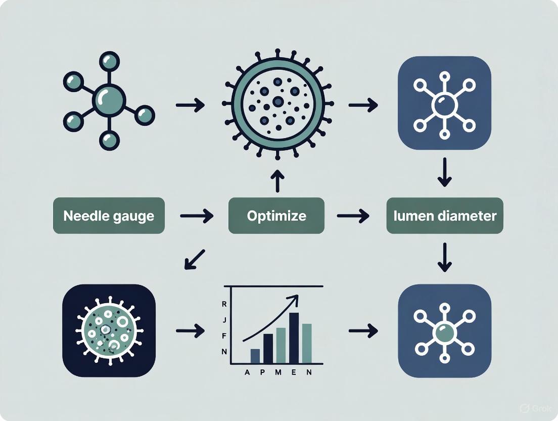
What is needle gauge? The needle gauge (G) is a standardized number that indicates the size of a needle. It is based on the Birmingham Wire Gauge (BWG) system, where the gauge number has an inverse relationship with the needle's outer diameter: a higher gauge number means a thinner needle. This system originated from 19th-century wire manufacturing. [25]
What is inner diameter? The inner diameter is the measurement across the open space inside the needle through which fluid flows. It is the actual physical measurement of the lumen opening, typically reported in millimeters (mm). The inner diameter determines the flow rate and shear stress of the fluid passing through the needle. [26]
What is a lumen? In the context of needles, the lumen is the hollow internal channel of the needle. In cell biology, the same term refers to the fluid-filled cavity inside cellular structures like cysts and organoids. In both cases, it is the central space through which substances pass or are contained. [26] [27]
The Critical Relationship: Gauge, Inner Diameter, and Experimental Outcomes
How are gauge and inner diameter related? While gauge determines the needle's outer size, the inner diameter is the critical parameter for experimental design. The relationship between gauge and inner diameter is not linear and can vary by manufacturer, so always verify the inner diameter for precise calculations. [26] [25]
The table below shows how inner diameter changes with gauge for standard needles.
Table 1: Common Needle Gauge Sizes and Inner Diameters
| Gauge Size | Typical Inner Diameter (mm) | Common Color Code (ISO 6009) |
|---|---|---|
| 18G | ~0.864 mm (metal, tapered) [26] | Pink [25] |
| 21G | Information missing from search results | Deep Green [25] |
| 22G | ~0.413 mm (plastic, straight) [26] | Black [25] |
| 23G | ~0.330 mm (plastic, straight) [26] | Orange [25] |
| 25G | ~0.250 mm (plastic, straight) [26] | Red [25] |
| 26G | ~0.240 mm (plastic, straight) [26] | Peach [26] |
| 27G | ~0.200 mm (plastic, straight) [26] | Clear [26] |
| 30G | ~0.152 mm (plastic, straight) [26] | Lavender [26] |
Why does this relationship matter for cell viability? The inner diameter directly influences two key, competing factors in cell-based experiments:
- Shear Stress: A smaller inner diameter increases the pressure required to extrude material. This exerts higher shear stress on cells, which can damage cell membranes and decrease cell viability. [26] [9]
- Resolution vs. Viability: While a higher gauge (smaller inner diameter) can offer higher printing resolution in bioprinting, it comes with the trade-off of increased cell damage. Selecting a needle is often a balance between the desired resolution and maintaining cell health. [26]
FAQs and Troubleshooting Guide
FAQ 1: How do I choose the correct needle gauge for my cell type? Selection depends on cell size, sensitivity, and viscosity of the carrier solution.
- For large or sensitive cells (e.g., neurons, pancreatic islets): Use higher gauges (e.g., 25G-30G) to minimize shear stress and membrane damage. [9]
- For viscous solutions or high cell-density suspensions: Use lower gauges (e.g., 18G-22G) to reduce required pressure and prevent clogging. Be aware that this may increase shear forces. [25] [9]
- Standard guideline: A 21G or 22G needle is often suitable for routine blood collection and can serve as a starting point for many cell suspensions to minimize hemolysis or damage. [5]
FAQ 2: My cell viability is low after injection. Could the needle be the cause? Yes. Low post-injection viability is a common issue directly linked to needle choice and injection parameters.
- Primary Cause: High shear stress from forcing cells through a narrow lumen (high gauge) or at high flow rates. [26] [9]
- Troubleshooting Steps:
- Increase the inner diameter: Switch to a lower gauge needle (e.g., from 27G to 22G).
- Reduce injection speed: Use a slower, steady flow rate to decrease shear stress.
- Verify cell concentration: Overly concentrated suspensions increase viscosity and damage. [9]
FAQ 3: The flow rate of my cell suspension is inconsistent. What should I check? Inconsistent flow often indicates a physical obstruction or inappropriate needle selection.
- Check for clogging: Cell aggregates or debris can block the lumen. Use a filter or ensure a homogeneous single-cell suspension.
- Verify needle gauge for viscosity: The suspension may be too viscous for a high-gauge needle. Switch to a needle with a larger inner diameter. [25]
- Inspect for bubbles: Air bubbles can disrupt flow. Ensure the syringe and needle are properly primed with the cell suspension.
FAQ 4: Why is the term "lumen" used in both needles and biology, and are the concepts related? The concepts are analogous. In both contexts, a lumen is a defined, contained space.
- Needle Lumen: The physical channel that confines and directs fluid flow. [26]
- Biological Lumen: The fluid-filled cavity inside structures like MDCK cysts or organoids, which is enclosed and pressurized by cells. [27] [28] The key connection for your research is that the physical forces within a needle lumen (e.g., pressure, shear stress) directly affect the health of cells that themselves may form biological lumens.
Essential Research Reagent Solutions
Table 2: Key Reagents for Cell Viability and Cytotoxicity Assays
| Reagent / Assay | Function | Key Feature |
|---|---|---|
| CellTiter-Glo Luminescent Assay [29] | Measures ATP levels as a marker of metabolically active cells. | Highly sensitive, provides a bright, stable luminescent signal. |
| RealTime-Glo MT Cell Viability Assay [29] | Measures cell viability in real-time using a luciferase-based method. | Allows for kinetic monitoring without lysing cells. |
| MTT Tetrazolium Assay [30] [29] | Measures metabolic activity via conversion of MTT to purple formazan. | Requires a solubilization step; classic endpoint assay. |
| CellTiter-Blue Cell Viability Assay (Resazurin) [29] | Measures the reduction of resazurin to fluorescent resorufin. | Highly sensitive, more so than tetrazolium assays. |
| CytoTox-Glo Cytotoxicity Assay [29] | Measures dead-cell protease activity released upon loss of membrane integrity. | Specifically detects dead cells; can be multiplexed with viability assays. |
| Dimethyl Sulfoxide (DMSO) [5] | A cryoprotectant used to preserve cells during freezing. | Prevents intracellular ice crystal formation; toxic if left on cells too long. |
| Ficoll / Histopaque [5] | Density gradient solutions for isolating PBMCs from whole blood. | Separates cells based on density; critical for purifying specific cell types. |
Experimental Protocol: Correlating Needle Gauge with Post-Injection Cell Viability
Aim: To quantitatively assess the impact of needle gauge (inner diameter) on the viability and functionality of a specific cell line post-injection.
Materials:
- Cell culture of interest (e.g., mesenchymal stem cells, primary neurons).
- Syringes and needles of various gauges (e.g., 18G, 22G, 27G).
- Appropriate cell suspension vehicle (e.g., PBS with glucose, saline). [9]
- Cell viability assay kit (e.g., CellTiter-Glo for ATP measurement). [29]
- Laminar flow hood, cell culture incubator, microplate reader.
Methodology:
- Cell Preparation: Harvest and concentrate cells to a standard density (e.g., 50,000 cells/µL). Keep the suspension homogeneous and on ice until injection to minimize metabolic changes. [9]
- Experimental Setup: Load the cell suspension into separate syringes, each fitted with a different gauge needle. Record the inner diameter for each needle from manufacturer specifications. [26]
- Injection Simulation: Expel the cell suspension through each needle into a microcentrifuge tube using a syringe pump to maintain a constant, physiologically relevant flow rate (e.g., 1-5 µL/min). [9]
- Control: Do not pass a sample of the initial cell suspension through any needle.
- Viability Assessment: a. Collect the injected samples and the control. b. Use the CellTiter-Glo Assay according to the manufacturer's protocol: mix equal volumes of sample and reagent, incubate for 10 minutes to stabilize the signal, and record luminescence. [29] c. Calculate the percentage viability relative to the control sample.
- Data Analysis: Plot percentage viability against needle inner diameter. Expect to see a positive correlation, where larger inner diameters (lower gauges) result in higher post-injection viability.
Practical Protocols: Selecting the Right Needle for Cell Injection and Bioprinting
Core Concepts in Needle Selection
Selecting the appropriate needle is a critical step in injectable cell therapy that directly impacts cell viability and therapeutic outcomes. The core parameters—gauge, length, and application-specific considerations—form a interdependent system. The mechanical forces cells experience during injection, particularly shear stress, are a primary cause of post-transplantation cell death, with some studies showing fewer than 5% of injected cells persisting at the injection site within days [9]. The following table summarizes the key parameters and their competing considerations.
| Parameter | Definition & Measurement | Impact on Cell Viability & Delivery | Conflicting Research Findings |
|---|---|---|---|
| Needle Gauge (Inner Diameter) | The internal diameter of the needle lumen; a lower gauge number indicates a larger diameter [31]. | Smaller diameters (higher gauge) increase shear stress (τ), calculated by (\tau = \frac{{4Q\eta }}{{\pi {R^3}}}), where (R) is needle radius, (Q) is flow rate, and (\eta) is viscosity [9]. This can reduce viability [32]. | Effect is cell-type dependent. One study on Muscle-Derived Cells found viability was not significantly impacted by 23G vs. 27G needles [31], whereas other studies on fibroblasts show a negative impact [32]. |
| Needle Length | The distance a cell suspension must travel before ejection. | Longer needles increase exposure to shear forces and the risk of cell sedimentation and needle clogging, especially in high-density suspensions [9] [31]. | Studies on muscle-derived cells showed no significant impact of needle length (1.5 in to 9.5 in) on immediate cell viability [31]. |
| Ejection Flow Rate (Q) | The speed at which the cell suspension is expelled, often controlled by a syringe pump (e.g., µL/min) [31] [32]. | Higher flow rates (Q) exponentially increase shear stress (τ) [9]. However, one study found ejecting fibroblasts at 150 µL/min yielded the highest viable cell dose compared to slower rates, which increased apoptosis [32]. | Requires balancing mechanical stress against other factors. An optimal rate exists that minimizes total damage. |
| Cell Suspension Vehicle | The solution or biomaterial in which cells are suspended (e.g., PBS, alginate hydrogels, collagen) [31] [32]. | Viscous vehicles like alginate or collagen can have a protective effect, improving viability by reducing mechanical shock [32]. The dynamic viscosity of the vehicle (η) is a key variable in the shear stress equation [9] [31]. | Highly viscous vehicles may be difficult to inject through small-bore needles and can increase injection pressure. |
Experimental Protocols for Assessing Cell Viability Post-Injection
Protocol 1: Multiparametric Analysis of Cell Health After Ejection
This protocol provides a comprehensive methodology for evaluating the impact of the injection process on NIH 3T3 fibroblasts, adaptable to other cell types [32].
1. Cell Preparation and Injection
- Culture & Harvest: Culture Swiss mouse embryonic fibroblast cell lines (NIH 3T3) in Dulbecco's Modified Eagle's Medium (DMEM) supplemented with 10% fetal calf serum. Detach cells using a standard trypsinization protocol or Accutase for apoptosis assays [32].
- Reconstitution: Centrifuge cells at 180 × g for 5 minutes and reconstitute to a density of 5 × 10^5 cells/mL in phosphate-buffered saline (PBS) or the test vehicle [32].
- Loading and Ejection: Load 100 µL aliquots of cell suspension into Hamilton Gastight syringes fitted with the test needles. Use a programmable syringe pump (e.g., Harvard Infuse/Withdraw syringe pump) to eject the suspension at a controlled rate into 1 mL of complete media. Directly pipette a sample to serve as a non-injected control [32].
2. Viability and Functionality Assays
- Immediate Viability (Trypan Blue): Immediately after ejection, mix 10 µL of cell suspension with 10 µL trypan blue. Gently mix and count viable (unstained) and non-viable (blue) cells using an improved Neubauer haemocytometer [32].
- Metabolic Activity (PrestoBlue): Plate ejected cells and incubate. At 6-hour and 24-hour time points, add PrestoBlue reagent (1:9 in culture medium) and incubate at 37°C for 45 minutes in the dark. Measure fluorescence (Exc/Em 560/590 nm) with a microplate reader to assess metabolic activity and proliferation [32].
- Membrane Integrity (Live/Dead): At designated time points, stain cells with a solution containing Calcein AM (labels live cells green) and ethidium homodimer-1 (labels dead cells red). Visualize using fluorescence microscopy [32].
- Apoptosis and Necrosis (Flow Cytometry): Analyze cell suspensions using an Annexin V/PI apoptosis kit. Use a flow cytometer (e.g., Beckman Coulter Cytomics FC500) with a 488 nm laser to distinguish between live (Annexin V-/PI-), early apoptotic (Annexin V+/PI-), late apoptotic (Annexin V+/PI+), and necrotic (Annexin V-/PI+) cell populations [32].
Protocol 2: Testing Muscle-Derived Cells with Different Delivery Vehicles
This protocol is tailored for investigating Autologous Muscle-Derived Cells (AMDCs) and Motor Endplate-Expressing Cells (MEEs) [31].
1. Cell Culture and Differentiation
- AMDC Isolation: Isolate AMDCs from skeletal muscle tissue and culture for 2-3 passages in DMEM with 20% fetal bovine serum [31].
- MEE Differentiation: Allow AMDCs to reach confluence. Culture in differentiation media (DMEM with 2% horse serum) for 5 days. Then, add induction media containing agrin (10 nM), neuregulin (2 nM), and acetylcholine (10 nM) for 5 days to induce motor endplate formation. Confirm differentiation via immunostaining with Alexa Fluor 594 conjugated bungarotoxin [31].
2. Injection and Viability Testing
- Suspension Preparation: Reconstitute AMDCs or MEEs to 1 × 10^7 cells/mL in either PBS or polymerizable type I oligomeric collagen [31].
- Ejection: Load 1 mL of cell suspension into a syringe attached to the test needle. Use a syringe pump (e.g., NE-500) to eject at a constant flow rate of 2 mL/min. For a 27G orotracheal needle that doesn't fit the pump, perform manual injection at an equivalent rate [31].
- Viability Assessment: For PBS-suspended cells, measure viability immediately after ejection and after 24/48 hours of incubation in serum-deprived media. For collagen-suspended cells, eject the suspension, allow it to polymerize, and then incubate with serum-deprived media for 24/48 hours before live/dead staining and imaging with a confocal microscope [31].
Troubleshooting Common Injection Problems
FAQ 1: I observe high rates of cell death immediately after injection. What are the primary causes and solutions?
- Cause: Excessive shear stress during ejection is a likely culprit. As defined by Poiseuille’s equation, shear stress (τ) is inversely proportional to the cube of the needle radius (τ ∝ 1/R³). A small reduction in needle diameter dramatically increases shear forces [9].
- Solution:
- Increase Needle Diameter: Use the largest practicable needle gauge (smallest gauge number) for your application. Switching from a 27G to a 22G needle can reduce shear stress by nearly 90% due to the larger radius [9] [31].
- Optimize Ejection Rate: Reduce the flow rate (Q), as it is a linear factor in the shear stress equation. Systematically test rates (e.g., 50-300 µL/min) to find the optimum for your cell type [32].
- Use a Protective Vehicle: Suspend cells in a viscous, protective biomaterial like alginate hydrogel or type I oligomeric collagen, which can shield cells from mechanical forces [31] [32].
FAQ 2: My needle frequently clogs during the injection procedure. How can I prevent this?
- Cause: Clogging is often due to high cell density or sedimentation within the syringe and needle. Cell suspensions over 100,000 cells/µL are considered highly concentrated and can be viscous, leading to clogging and uneven flow [9].
- Solution:
- Optimize Cell Density: Reduce the cell concentration in the suspension vehicle. Express the cellular component as a volume fraction to standardize across cell types of different sizes [9].
- Minimize Sedimentation: Avoid long pauses after loading the syringe. Use the suspension within a short, defined timeframe or gently agitate the syringe to maintain a homogeneous mixture.
- Consider Multiple Injections: For large therapeutic doses, consider making multiple, lower-volume injections rather than a single, high-volume injection with a highly concentrated solution [9].
FAQ 3: How can I accurately standardize and report my viability findings for comparison with other studies?
- Cause: Inconsistent methodology and reporting make cross-study comparisons difficult. Viability can be measured using different assays (e.g., Trypan Blue, flow cytometry, metabolic activity) at different time points (immediately post-ejection vs. 24 hours later) [32].
- Solution:
- Use Multiplex Assays: Employ a combination of assays to get a complete picture of cell health. For example, use Trypan Blue for immediate viability, a metabolic assay for 24-hour function, and Annexin V/PI staining for apoptosis [32].
- Report Detailed Parameters: Always document the complete injection setup: needle gauge and length, inner diameter, ejection flow rate, cell type, cell density, suspension vehicle, and the specific viability assay(s) used. This allows for true experimental replication [31] [32].
- Include Internal Controls: Always compare ejected samples to a non-ejected control that has been pipetted directly from the source suspension [31].
Experimental Workflow and Parameter Relationships
Parameter Optimization Workflow
Parameter Impact on Viability
Research Reagent Solutions
The following table lists key materials and reagents essential for conducting rigorous needle selection and cell viability experiments.
| Reagent / Material | Function / Application | Example Specifications & Notes |
|---|---|---|
| Programmable Syringe Pump | Provides precise, automated control over ejection flow rate (Q), a critical variable in shear stress calculation [31] [32]. | e.g., NE-500 (New Era Syringe Pump Inc.) or Harvard PHD 2000. Ensures reproducibility between experiments and operators. |
| Hamilton Gastight Syringes | High-precision syringes designed to eliminate dead volume and provide smooth, consistent plunger movement, reducing variability in cell delivery [32]. | Model 1710RN with removable needles (RN). Essential for high-accuracy cell therapy applications. |
| Annexin V/Dead Cell Apoptosis Kit | Multiplex flow cytometry assay to distinguish between live, early apoptotic, late apoptotic, and necrotic cell populations post-ejection [32]. | e.g., Alexa Fluor 488 Annexin V/Dead Cell Kit (Molecular Probes). More informative than simple viability stains. |
| PrestoBlue Cell Viability Reagent | A resazurin-based solution used to measure metabolic activity and proliferation of cells at 6-hour and 24-hour time points after injection [32]. | Added directly to culture wells. Fluorescence (Exc/Em 560/590 nm) is proportional to metabolic activity. |
| Type I Oligomeric Collagen | A polymerizable, viscous delivery vehicle. Can shield cells from biomechanical stress during injection, improving post-injection survival [31]. | e.g., GeniPhys OM10027. Dynamic viscosity ~49.7 × 10⁻³ kg/(m·s). Allows formation of a 3D scaffold upon ejection. |
| AlignFlow Flow Cytometry Alignment Beads | Fluorescent beads used to calibrate and ensure the optimal performance of the flow cytometer before analyzing cell samples for viability and apoptosis [33]. | Laser-specific versions available (UV, Blue, Red). Critical for generating reproducible and reliable flow cytometry data. |
FAQs and Troubleshooting Guides
FAQ: Why does needle size affect MSC viability? Mesenchymal Stem Cells (MSCs) are relatively large cells (approximately 12–19 μm in diameter) [34]. When forced through a narrow-bore needle, they are subjected to significant shear stress [9]. This mechanical force can damage the cell membrane, leading to immediate cell death or the initiation of apoptosis (programmed cell death) that manifests hours later [34]. Using a smaller needle gauge (larger diameter) reduces this shear stress, preserving cell viability and function [7].
Troubleshooting: My post-injection cell viability is low. What should I check?
- Verify Needle Gauge: Immediately confirm that you are using a needle no smaller than the recommended minimum size (e.g., 20-gauge or 22-gauge for initial resuspension) [7].
- Inspect Injection Rate: A high flow rate can increase shear stress. If you must use a smaller needle, consider reducing the ejection rate to mitigate cell damage [35].
- Check Cell Preparation: Ensure cells are not forming large clumps that could increase pressure and shear stress during injection. Use a homogeneous single-cell suspension.
FAQ: What is the minimum recommended needle size for MSC delivery? For equine MSCs, one study strongly recommends using needles larger than 25-gauge to maintain viability [7]. For human MSCs, research indicates that while viability may not drop significantly immediately after injection through a 25-gauge needle, a delayed decrease in viability can be observed at 24 hours [34]. Therefore, a 20-gauge or 22-gauge needle is a safer choice for critical injections to ensure high medium-to-long-term survival.
Troubleshooting: The injection flow is inconsistent or the needle keeps clogging. This is often a sign that the needle gauge is too small for the cell density or that cells have settled in the syringe.
- Prevention: Use a larger gauge needle (e.g., 20-gauge) for the initial aspiration and resuspension of the cell pellet before switching to a finer needle for the actual injection, if necessary [7].
- Action: Gently mix the cell suspension immediately before loading it into the syringe to ensure a homogeneous mixture and prevent sedimentation.
The following tables summarize key quantitative findings from scientific literature on how needle gauge and flow rate impact MSC viability and function.
Table 1: Effect of Needle Gauge on Equine MSC Viability [7]
| Needle Gauge | Internal Diameter (mm) | Relative Cell Viability | Key Findings |
|---|---|---|---|
| 20-Ga | ~0.91 | Highest | Higher viability and a larger percentage of intact cells compared to 25-Ga. |
| 22-Ga | ~0.71 | Intermediate | Viability lower than 20-Ga but higher than smaller gauges. |
| 23-Ga | ~0.64 | Intermediate | Viability lower than 20-Ga but higher than smaller gauges. |
| 25-Ga | ~0.51 | Lowest | Significantly lower viability; highest percentage of cell debris. |
Table 2: Effect of Needle Gauge and Flow Rate on Human MSC (hMSC) Health [35] [34]
| Parameter | Conditions Tested | Impact on hMSCs |
|---|---|---|
| Needle Gauge | 30-Ga, 34-Ga [35] | Slower ejection rates (10 µl/min) resulted in higher apoptosis and significant downregulation of CD105 expression. |
| Flow Rate | 20-Ga, 25-Ga, 30-Ga [34] | No immediate clinically significant effect on viability post-injection. A delayed decrease in viability was observed at 24 hours. |
| Cell Function | Various needle gauges [34] | No significant changes in surface markers or capacity for multilineage differentiation were observed post-injection. |
Detailed Experimental Protocols
Protocol 1: Direct Assessment of Needle Gauge on MSC Viability This protocol is adapted from a study investigating the effect of needle diameter on equine bone marrow-derived MSCs [7].
- Objective: To evaluate the effect of different needle diameters on the viability of MSCs when handled under simulated clinical shipping conditions.
- Materials:
- Phosphate Buffered Saline (PBS)
- 3 ml syringes
- Hypodermic needles (20-, 22-, 23-, and 25-gauge)
- Fluorescein diacetate and propidium iodide (or other viability stain)
- Hemocytometer or flow cytometer
- Methodology:
- Cell Preparation: Suspend MSCs in PBS at a concentration of 1 × 10⁷ cells/mL. Hold the samples at room temperature for 7 hours to mimic shipping conditions.
- Needle Simulation: For each needle size, gently resuspend the cells and aspirate the suspension into a 3 mL syringe fitted with the test needle. Reinject the cells back into the holding vial. Repeat this aspiration/injection cycle 3 times to simulate clinical resuspension.
- Viability Assessment: Stain the cells with fluorescein diacetate (labels live cells) and propidium iodide (labels dead cells). Calculate the percentage of viable cells using a hemocytometer for manual counting or flow cytometry for automated, high-throughput analysis.
- Flow Cytometry for Debris: Use flow cytometry to measure forward scatter (FSC), which helps distinguish intact cells from smaller cell debris.
Protocol 2: Evaluating Combined Effects of Needle Gauge and Ejection Rate This protocol is based on studies using human MSCs (hMSCs) to assess cellular health after ejection [35].
- Objective: To determine the impact of clinically relevant needle gauges and ejection rates on hMSC viability, apoptosis, and differentiation capacity.
- Materials:
- 100-µl Hamilton GASTIGHT syringes
- Customized removable needles (e.g., 30-gauge and 34-gauge)
- Harvard Infuse/Withdraw syringe pump (or equivalent)
- Apoptosis detection kit (e.g., Annexin V)
- Differentiation media (adipogenic, osteogenic, chondrogenic)
- Methodology:
- Cell Loading: Reconstitute trypsinized hMSCs in PBS at a density of 7 × 10⁵ cells/mL. Draw the cell suspension into a 100-µl syringe with the test needle attached.
- Controlled Ejection: Use a syringe pump to eject the cell suspension at defined rates (e.g., ranging from 10 µl/minute to 300 µl/minute) into a collection tube.
- Post-Ejection Analysis:
- Viability & Apoptosis: Measure immediate viability using a stain like thiazole orange and propidium iodide. Plate a subset of cells and measure apoptosis (e.g., using Annexin V stain) after 24 hours of incubation.
- Phenotype & Function: Culture the ejected cells and perform flow cytometric immunophenotyping to check for surface markers (e.g., CD105). Induce multilineage differentiation to assess if ejection parameters affect the cells' functional capacity.
The Scientist's Toolkit
Table 3: Essential Materials for MSC Delivery Experiments
| Item | Function/Application |
|---|---|
| Hamilton GASTIGHT Syringes | Precision syringes designed to minimize dead volume, providing high accuracy in dispensing small volumes of cell suspensions [35]. |
| Programmable Syringe Pump | Allows for highly controlled and reproducible ejection rates during injection experiments, eliminating user-induced variability [35]. |
| Annexin V Apoptosis Kit | A fluorescence-based assay to detect early-stage apoptosis in cells, which is crucial for identifying delayed cell death post-injection [34]. |
| Flow Cytometer | An essential instrument for quantitative analysis of cell viability, apoptosis, immunophenotyping (surface marker expression), and detection of cell debris [7] [34]. |
| Differentiation Media Kits | Pre-formulated media supplements used to induce and assess the adipogenic, osteogenic, and chondrogenic potential of MSCs after experimental manipulation [34]. |
Experimental Workflow and Decision Pathway
MSC Injection Experimental Workflow
Impact of Needle Gauge on MSC Therapy
In extrusion-based bioprinting, achieving high cell viability and high structural fidelity requires navigating inherent trade-offs between needle gauge, extrusion pressure, and printing resolution. The mechanical stress experienced by cells during the bioprinting process is a direct function of these parameters. Fundamentally, smaller needle diameters (higher gauge numbers) enable finer printing resolution but increase shear stress on cells, potentially compromising viability. Conversely, larger needles reduce shear stress but limit the ability to print intricate structures. This guide details a systematic approach to optimizing these parameters for robust and reproducible research outcomes.
Troubleshooting Guides
FAQ: Addressing Common Experimental Challenges
Q1: My needle tip keeps colliding with the print bed during movement. How can I fix this?
- Cause: Incorrectly set Z-axis home position or G-code coordinates.
- Solution: Accurately locate and set the center point coordinates of your print area in the G-code. Use commands like
G1 Z5 F200to adjust the print bed or print head away from the needle tip before the extruder head moves. Always ensure the XYZ coordinates are properly calibrated [36].
Q2: I am observing air bubbles in my bioink when loading the syringe. What should I do?
- Cause: Air entrapment during bioink mixing or loading.
- Solution: To eliminate air bubbles, centrifuge the bioink at a low RPM for about 30 seconds. Avoid high RPMs to prevent cell clustering. Alternatively, triturate the bioink slowly by gently dispensing it along the walls of the falcon tube during mixing, which minimizes bubble formation [36].
Q3: I am experiencing frequent needle clogging during bioprinting. How can I resolve this?
- Cause: Bioink inhomogeneity, particle agglomeration, or using a needle gauge that is too small for the cell aggregates or particles in the bioink.
- Solution:
- Ensure bioink homogeneity. If it appears uniform, briefly increase the pressure to clear the clog, but do not exceed 2 bar when working with cells to avoid viability loss.
- If clogging persists, change to a larger needle gauge.
- When using nanoparticles, confirm the particle size is smaller than the needle's internal diameter. Pre-characterize particle size and ensure they are well-dispersed to prevent agglomeration [36].
Q4: The layers of my multi-layer construct are merging or collapsing, resulting in a 2D-like structure. What is wrong?
- Cause: Insufficient bioink viscosity or overly rapid crosslinking time, preventing the bottom layers from supporting the weight of subsequent layers.
- Solution: Perform rheological tests to understand the thixotropic (shear-thinning and recovery) nature of your bioink. Optimize the crosslinking time to ensure that each layer gains sufficient structural integrity before the next layer is deposited [36].
Q5: After printing, my scaffolds lack structural integrity and collapse. How can I improve this?
- Cause: Inadequate crosslinking of the bioink post-printing.
- Solution: Crosslinking is critical for mechanical and physiochemical properties. Choose and optimize the correct crosslinking method for your bioink:
- Photocrosslinking: Determine the appropriate wavelength and exposure time.
- Ionic Crosslinking: Characterize the optimal crosslinker (e.g., CaCl₂ for alginate) concentration.
- Thermal Crosslinking: Optimize the build plate temperature.
- Self-crosslinking polymers: Print at very low speeds to allow sufficient time for automatic crosslinking [36].
Parameter Optimization and Cell Viability
Q6: How do needle gauge and extrusion pressure directly impact cell viability? The selection of needle gauge and pressure is a critical balance. While one study on non-bioprinting cell injection found that needle diameters as small as 30-gauge (160 µm internal diameter) did not significantly affect the viability of injected equine mesenchymal stromal cells (which have a diameter of ~20 µm) [8], the high shear rates of bioprinting change this dynamic.
- Pressure and Viability: Higher extrusion pressures lead to greater cell death. A study on 3D-bioprinted lung cells confirmed that increased extrusion pressure directly reduces cell viability [37].
- Shear Stress: The combination of high pressure and small needle diameter dramatically increases shear stress, which is a primary cause of cell membrane damage and death during extrusion [38]. Using tapered needle tips can decrease the pressure required for printing, thereby reducing shear stress on cells [39].
Q7: What is a systematic method to find the optimal balance between pressure and print speed? A structured, data-driven approach is recommended over trial-and-error. One effective protocol involves three sequential tests [40]:
- Extrudability Test: Quantify the mass deposition rate under varying pressures to identify the pressure range that ensures a consistent, uninterrupted flow.
- Filament Deposition Test: Print straight lines at various combinations of pressure and speed. Measure the resulting filament diameters; the optimal parameters are those that produce a filament diameter closest to the nozzle's internal diameter.
- Printability Test: Print multi-layered grid structures (e.g., 10 mm x 10 mm, 2 layers) to assess structural fidelity. Parameters that successfully create grids with well-defined pores and no layer sagging or merging are considered optimal. Using this workflow, researchers identified 75 kPa pressure and 600 mm/min print speed as optimal for a GelMA-based ink [40].
Experimental Protocols
Protocol 1: Systematic Parameter Optimization Using Design of Experiments (DoE)
This protocol uses a factorial DoE to efficiently establish a relationship between process parameters and filament quality [41].
1. Objective: Develop an analytical model to predict Filament Width (FW) based on key bioprinting parameters. 2. Key Parameters and Levels:
- Nozzle Diameter (ND): 210 µm (-1) and 410 µm (+1)
- Extrusion Pressure (EP): 110 kPa (-1) and 170 kPa (+1)
- Print Speed (PS): 5 mm/s (-1) and 15 mm/s (+1)
- Print Distance (PD): Held constant (e.g., 0.30 mm) 3. Methodology: a. Design: Set up a full factorial experiment using the parameter levels above. b. Printing: Fabricate filaments for each parameter combination using your bioink. c. Image Acquisition: Capture images of the printed filaments within 1-2 minutes of printing using a bright-field microscope. d. Image Analysis: Process images in MATLAB or similar software. Binarize images and measure the FW by calculating the distance between the first and last pixels across the filament width. e. Modeling: Use statistical analysis software to fit the experimental data to a model that predicts FW based on ND, EP, and PS. 4. Outcome: A predictive model that allows researchers to select parameters to achieve a target filament width, thereby controlling scaffold porosity and architecture [41].
Protocol 2: Assessing the Impact of Aspiration and Injection on Cell Viability
This protocol is designed to specifically evaluate how the processes of loading (aspiration) and dispensing (injection) a cell suspension affect immediate and long-term cell health [8].
1. Cell Preparation: * Prepare a cell suspension at a clinically relevant concentration (e.g., 5x10⁶ cells/mL). 2. Aspiration Study: * Slowly aspirate 0.5 mL of cell suspension through different needle gauges (e.g., no needle, 20G, 25G, 30G) at a controlled rate (e.g., 0.25 mL/s). * Remove the needle before gently ejecting the suspension for analysis. * Assess immediate viability using an automated fluorescence-based cell counter or trypan blue exclusion. 3. Injection Study: * Attach various needles (e.g., 18G to 30G) to a syringe. * Inject 0.5 mL of cell suspension over 2 seconds into a vial. * Assess immediate viability as above. 4. Delayed Viability Assessment: * Seed the aspirated or injected cells into a culture plate. * Culture for 24 hours and assess metabolic activity using a resazurin-based assay or similar. 5. Key Findings to Inform Your Research: * Injection: Needle diameter (18G-30G) may not significantly affect immediate or delayed viability [8]. * Aspiration: Aspiration through smaller needles (20G and smaller) can significantly decrease immediate cell viability [8]. Therefore, use an 18G or larger needle for aspiration to minimize cell damage.
Data Presentation
Quantitative Data Tables
Table 1: Experimentally Determined Optimal Parameters for Different Bioinks
| Bioink Formulation | Optimal Pressure (kPa) | Optimal Speed (mm/min) | Nozzle Diameter (µm) | Key Outcome | Source |
|---|---|---|---|---|---|
| GelMA + Egg White Protein | 70 - 80 (75 optimal) | 300 - 900 (600 optimal) | Not Specified | Reliable extrusion flow & high structural fidelity | [40] |
| ALGEC (Alginate-Gelatin-TO-NFC) | Model-Dependent | Model-Dependent | Not Specified | Data-driven optimization of viscosity and printability | [42] |
| Alginate-CMC-Gelatin (FRESH) | Optimized via DoE | Optimized via DoE | ~250 (achieved resolution) | High shape fidelity & ~100% metabolic activity vs. control at day 7 | [43] |
Table 2: The Impact of Needle Gauge and Aspiration/Injection on Cell Viability
| Process | Needle Gauge | Internal Diameter (µm) | Effect on Immediate Viability | Recommendation | Source |
|---|---|---|---|---|---|
| Injection | 18G - 30G | ~838 - 160 | No significant effect | Needle selection can be based on clinical/experimental preference | [8] |
| Aspiration | 20G and smaller (e.g., 25G, 30G) | ~603 and smaller | Significant decrease | Use 18G or larger for aspiration | [8] |
The Scientist's Toolkit: Research Reagent Solutions
Table 3: Essential Materials and Their Functions in Bioink Formulation
| Material | Function in Bioink | Key Characteristics |
|---|---|---|
| Sodium Alginate | Provides scaffold framework and excellent printability | Biocompatible; forms stable gels via ionic crosslinking (e.g., with CaCl₂) [43] [42]. |
| Gelatin | Promotes cell adhesion and viability | Denatured collagen; often modified (e.g., GelMA) for photo-crosslinking; provides a cell-friendly environment [43] [42]. |
| Carboxymethyl Cellulose (CMC) | Enhances printability and viscosity | Provides a microfibrillar structure resembling the ECM; remains un-crosslinked [43]. |
| TEMPO-Oxidized NFC (TO-NFC) | Improves structural fidelity and homogeneity | Nanofibrillated cellulose; modifies rheology for better shape retention without compromising biocompatibility [42]. |
| Collagen | Mimics the native extracellular matrix | High biocompatibility; often mixed with other materials like alginate to support cell function and viability [37]. |
Workflow Visualization
Bioprinting Parameter Optimization Logic
Diagram 1: A logic flow for systematically troubleshooting and optimizing bioprinting parameters.
Key Parameter Interrelationships
Diagram 2: Interplay of bioprinting parameters shows how changes to one parameter often involve a trade-off between cell viability and structural fidelity.
Troubleshooting Guide: Frequently Asked Questions
FAQ: How does needle gauge affect the viability of cells during injection?
Research demonstrates a clear correlation between smaller needle diameters (higher gauges) and decreased cell viability due to increased shear stress. For mesenchymal stem cells (MSCs), viability was significantly higher when passed through a 20-gauge needle compared to a 25-gauge needle. Suspensions passed through the 20-gauge needle also contained a larger percentage of intact cells and less debris [7]. While one study on fetal brain cells found that fully dispersed cells had less viability than cell clumps, it noted a tendency for narrower needles to adversely affect both types [44].
- Recommendation: For injecting sensitive cells like MSCs, use a needle larger than 25-gauge. A 20-gauge or 22-gauge needle is generally recommended to maintain high viability [7].
FAQ: What is the optimal cell concentration for cryopreservation?
The optimal concentration for freezing cells varies by cell type. A very low concentration can lead to low post-thaw viability, while a very high concentration can cause undesirable cell clumping. A general range is 1x10^3 to 1x10^6 cells/mL [17].
- Recommendation: For best results, you should test freezing your specific cell type at multiple concentrations to determine which gives the desired viability, recovery, and functionality upon thawing [17].
FAQ: Why is a controlled freezing rate critical for cryopreservation?
The rate at which cells are frozen significantly impacts survival. Slow freezing at approximately -1°C per minute helps maximize cell viability and integrity by reducing the formation of damaging intracellular ice crystals. This can be achieved using a controlled-rate freezer or an isopropanol freezing container placed in a -80°C freezer [17] [45].
FAQ: Are standard 2D cell viability assays accurate for 3D hydrogel cultures?
No, using standard 2D viability assays on 3D hydrogel constructs can lead to inaccurate cellular health readings. Assays like CellTiter-Glo, MTS, and PrestoBlue showed variable and unreliable results across different hydrogel formulations (e.g., collagen, HA-based, synthetic) [46].
- Recommendation: For accurate viability assessment in 3D cultures, these indirect assays should be used in combination with direct microscopic imaging for validation [46].
Needle Gauge and Cell Viability: A Quantitative Guide
The following table consolidates data on how needle gauge impacts cell viability and provides standard dimensional data to inform your experimental setup.
Table 1: Impact of Needle Gauge on Cell Viability and Technical Specifications
| Gauge (G) | Nominal Inner Diameter (mm) | Recommended for Cell Types | Effect on Viability and Key Findings |
|---|---|---|---|
| 20G | 0.603 [47] | Equine MSCs [7] | Higher viability and a larger percentage of intact cells compared to smaller gauges [7]. |
| 22G | 0.413 [47] | General cell injection | A common size that offers a balance between flow and minimal shear. |
| 23G | 0.337 [47] | General cell injection | -- |
| 25G | 0.260 [47] | -- | Significantly reduced MSC viability and increased cellular debris compared to 20G needles [7]. |
Detailed Experimental Protocols
Protocol 1: Cryopreservation of Cells Using a Freezing Container
This protocol outlines the steps for freezing adherent and suspension cells for long-term storage [17] [45].
- Key Materials: Log-phase cells (>80% confluency, >90% viability), complete growth medium, cryoprotectant (e.g., DMSO), freezing medium (e.g., CryoStor CS10 or lab-made formulation), sterile cryogenic vials, isopropanol freezing container (e.g., "Mr. Frosty" or CoolCell), -80°C freezer, liquid nitrogen storage tank [17] [45].
- Harvest: Gently detach adherent cells using a dissociation reagent like trypsin. Resuspend the cells in complete growth medium [45].
- Count & Centrifuge: Determine viable cell count and concentration. Centrifuge the cell suspension (100–400 × g for 5–10 min) and carefully aspirate the supernatant [45].
- Resuspend in Freezing Medium: Resuspend the cell pellet in cold freezing medium at the recommended density (e.g., 1x10^3–1x10^6 cells/mL). Gently mix to ensure a homogeneous suspension [17] [45].
- Aliquot: Dispense the cell suspension into sterile cryogenic vials [45].
- Freeze: Place the vials in an isopropanol freezing container and transfer it immediately to a -80°C freezer for overnight (approx. -1°C/minute cooling rate) [17] [45].
- Long-term Storage: The next day, transfer the cryovials to a liquid nitrogen tank for storage at below -135°C [17].
Protocol 2: Assessing Cell Viability in 3D Hydrogel Constructs
This protocol summarizes the methodology for evaluating cell health within hydrogels, highlighting key considerations for assay selection [46].
- Key Materials: Hydrogel components (e.g., Collagen, HyStem, HA:Col1 hybrid), cells (e.g., HCT-116), cell culture medium, viability assay reagents (e.g., CellTiter-Glo 3D, PrestoBlue), multiwell plates.
- Prepare Hydrogel Constructs:
- Trypsinize and count cells from 2D culture.
- Mix the cell suspension with the desired hydrogel precursor solution on ice to ensure even distribution.
- Dispense the cell-hydrogel mix into wells of a PDMS-coated plate to prevent adhesion. Polymerize or cross-link the hydrogel according to the manufacturer's instructions (e.g., incubate at 37°C for collagen, UV light for some synthetic gels) [46] [48].
- Culture: Add cell culture medium to cover the hydrogel constructs and incubate at 37°C [48].
- Select and Perform Viability Assay:
- Note that standard 2D assays may provide inaccurate readings. The CellTiter-Glo 3D assay is specifically designed for better lytic capability in 3D matrices [46].
- Follow the manufacturer's protocol for the chosen assay, ensuring reagents equilibrate to room temperature.
- Validate with Imaging: For accurate results, combine indirect assay readings with direct microscopic imaging to visually confirm cell viability and morphology within the hydrogel [46].
The Scientist's Toolkit: Essential Research Reagents & Materials
Table 2: Key Reagents and Materials for Cell Handling and 3D Culture
| Item | Function | Example & Notes |
|---|---|---|
| Cryopreservation Medium | Protects cells from ice crystal damage during freeze-thaw cycles. | CryoStor CS10: A ready-to-use, serum-free option [17]. Synth-a-Freeze: A chemically-defined, protein-free medium [45]. |
| Controlled-Rate Freezer | Ensures a consistent, slow cooling rate (~1°C/min). | Mr. Frosty (isopropanol container): A low-cost alternative to programmable freezers [17] [45]. |
| Hydrogel Kit | Provides a 3D scaffold that mimics the native extracellular matrix (ECM). | TrueGel3D: A chemically-defined, slow-gelling hydrogel kit for cell encapsulation [48]. Collagen I: A natural, organic hydrogel material [46]. |
| 3D Viability Assay | Accurately measures metabolic activity or ATP levels in 3D constructs. | CellTiter-Glo 3D: A luminescent assay optimized for lysis in 3D models [46]. |
| Hypodermic Needles | For aspirating and injecting cell suspensions with minimal shear stress. | Use 20G-22G needles for MSCs to maximize viability [7]. Refer to gauge charts for precise inner diameters [47] [49]. |
This case study investigates the critical impact of needle gauge selection on the viability and quality of Mesenchymal Stem Cell (MSC) suspensions during routine laboratory resuspension and injection procedures. MSCs are a cornerstone of regenerative medicine research and clinical applications, and their therapeutic potential is highly dependent on the delivery of a high proportion of viable, functional cells to the target site. The physical stresses imposed during cell handling—specifically, the aspiration of cell suspensions through hypodermic needles—can significantly compromise cell integrity. This report provides a detailed comparative analysis, using published experimental data, of using 20-gauge (G) versus 25-G needles. The findings are synthesized into evidence-based troubleshooting guides and FAQs to support researchers in optimizing their injection protocols, thereby enhancing the reliability and reproducibility of their experimental and therapeutic outcomes.
Core Experimental Data and Comparison
The following tables summarize key quantitative findings from relevant studies on needle gauge effects on MSC viability.
Table 1: Comparative Viability of Equine Bone Marrow-Derived MSCs Post-Needle Passage
| Needle Gauge | Approximate Internal Diameter | Reported Cell Viability | Key Observations | Source |
|---|---|---|---|---|
| 20-G | ~0.584 mm | Higher viability compared to 25-G | Larger percentage of intact cells; recommended for clinical use in horses. | [7] |
| 25-G | ~0.240 mm | Lower viability compared to 20-G | Increased percentage of cellular debris in suspension; more likely to cause cell damage. | [7] |
| Control (No Needle) | N/A | Baseline viability | Used for comparison to establish baseline cell health. | [7] |
Table 2: Differential Effects of Aspiration vs. Injection on Equine MSC Viability
| Procedure | Needle Gauge | Impact on Immediate Viability | Impact on Delayed Viability (24h) | Source |
|---|---|---|---|---|
| Injection | 18-G to 30-G | No significant effect observed. | No significant effect observed. | [8] |
| Aspiration | 20-G, 25-G, 30-G | Significant decrease in viability. | No significant change compared to non-aspirated controls. | [8] |
Experimental Protocols from Key Studies
Protocol: Effect of Needle Diameter on Equine MSC Viability
This protocol is adapted from the study that provides the core comparative data for 20-G vs. 25-G needles [7].
1. Cell Preparation:
- Source equine bone marrow-derived MSCs and culture using standard methods.
- Wash cells and resuspend in phosphate-buffered saline (PBS) at a density of 1 x 10^7 cells/mL.
- Hold cell suspensions in cryovials at room temperature for approximately 7 hours to simulate shipping conditions for clinical use. This allows cells to settle into a soft pellet.
2. Needle Aspiration & Injection Simulation:
- Assign sample vials to control or experimental groups (20-G, 22-G, 23-G, 25-G needles).
- Gently invert the vial to resuspend the cell pellet.
- Using a 3 mL syringe, aspirate the cell suspension through the designated needle and then re-inject it back into the original vial.
- Repeat this aspiration/injection cycle 3 times to mimic the rigorous resuspension performed prior to clinical injection.
3. Viability Assessment:
- Stain cells with fluorescent dyes: Fluorescein Diacetate (FDA) for live cells (green) and Propidium Iodide (PI) for dead cells (red).
- Count viable and non-viable cells using fluorescent microscopy.
- Perform flow cytometry to analyze the population for the percentage of intact cells versus cellular debris based on forward scatter (FSC).
Protocol: Separate Analysis of Aspiration and Injection
This protocol highlights the critical distinction between the two procedures [8].
1. Cell Preparation:
- Use equine cord blood or bone marrow-derived MSCs.
- Detach and suspend cells in culture media at a high density (e.g., 5 x 10^6 cells/mL).
2A. Injection-Only Experiment:
- Attach various needles (18-G to 30-G) to a 3 mL syringe.
- Manually inject 0.5 mL of cell suspension over 2 seconds into a collection vial.
- Assess immediate viability using an automated fluorescence-based cell counter.
2B. Aspiration-Only Experiment:
- Slowly aspirate 0.5 mL of cell suspension through a needle (20-G, 25-G, 30-G) at a set rate (e.g., 0.25 mL/s) using a 3 mL syringe.
- Remove the needle before ejecting the suspension from the syringe for testing.
- Assess immediate and delayed viability (the latter via a resazurin-based metabolic assay after 24 hours in culture).
Troubleshooting Guides & FAQs
Frequently Asked Questions
Q1: I need to inject MSCs into a mouse joint, which requires a very small needle like a 30-G. Will this kill my cells? A: The injection process itself through a 30-G needle may not catastrophically reduce viability [8]. The greater risk comes from the initial aspiration step. To maximize viability, aspirate your cell suspension using a larger-bore needle (e.g., 18-G or 20-G), then switch to the 30-G needle for the actual injection.
Q2: My protocol requires me to repeatedly pass cells through a needle to ensure a single-cell suspension. Is this safe? A: Exercise caution. Studies that showed significant viability loss often involved multiple (e.g., 3) aspiration/injection cycles to simulate resuspension [7]. You should empirically determine the minimum number of passages needed and use the largest feasible needle for this step. Consider alternative, gentler resuspension methods if possible.
Q3: Why does aspiration damage cells more than injection? A: During aspiration, the negative pressure (suction) created by pulling the syringe plunger can subject cells to rapid pressure changes and shear stresses as they accelerate into the narrow needle lumen. During injection, the positive pressure, while still generating shear, may subject cells to a different, and potentially less damaging, flow profile [8].
Q4: Beyond viability, can needle gauge affect other critical cell functions? A: Yes. Some studies report an increase in apoptotic cells and cell debris when using smaller needles [7]. While other research indicates no change in surface markers or differentiation potential immediately after injection [34], one study found a delayed decrease in human MSC viability 24 hours post-injection through smaller catheters, suggesting the initiation of apoptosis [34]. The mechanical stress can also alter the cell's mechanical phenotype (deformability), which is linked to homing efficiency [50].
Troubleshooting Guide
| Problem | Potential Cause | Solution | Preventive Tip |
|---|---|---|---|
| Low immediate cell viability after preparation. | Cell damage during aspiration through a small-bore needle. | Use a larger needle (18-G or 20-G) for the initial aspiration from the storage vial. | Implement a standard operating procedure (SOP) that mandates needle gauge for aspiration vs. injection. |
| High levels of cellular debris in flow cytometry. | Excessive shear stress from small needles or multiple passages. | Increase needle gauge; reduce the number of aspiration/injection cycles; avoid creating bubbles. | Use a Luer-lock syringe to prevent accidental needle detachment and fluid leakage, which can cause shear. |
| Poor cell survival 24 hours after injection. | Delayed onset of apoptosis or loss of function due to injection stress. | Use the largest needle gauge clinically or experimentally permissible. Confirm viability with a 24-hour metabolic assay, not just immediate dye exclusion. | Optimize cell concentration and carrier solution (e.g., reducing DMSO concentration to <0.5% can improve viability [51]). |
Essential Signaling Pathways and Workflows
The following diagram illustrates the key experimental workflow for evaluating needle gauge effects on MSCs, as described in the protocols.
Diagram 1: Needle Gauge MSC Viability Workflow
The decision tree below provides a clear, actionable pathway for researchers based on the findings of this case study.
Diagram 2: MSC Injection Needle Selection Guide
The Scientist's Toolkit: Research Reagent Solutions
Table 3: Essential Materials and Reagents for MSC Injection Studies
| Item | Function / Purpose | Example from Research |
|---|---|---|
| Hypodermic Needles (18-G, 20-G, 25-G, etc.) | To aspirate and inject cell suspensions; the independent variable in gauge optimization studies. | 20-G, 22-G, 23-G, 25-G needles were compared in the core case study [7]. |
| Luer-Lock Syringes | To securely attach needles and prevent accidental leakage or separation, which can introduce shear. | 3 mL syringes were used in multiple referenced studies [7] [8]. |
| Viability Stains (FDA & PI) | Fluorescent dyes for simultaneous visualization of live (FDA, green) and dead (PI, red) cells. | Used for viability counts via fluorescent microscopy [7]. |
| Propidium Iodide (PI) & 7-AAD | Membrane-impermeant DNA dyes used in flow cytometry to identify dead cells. | PI used with thiazole orange for flow cytometry [34]; 7-AAD used for flow-based viability [19]. |
| Flow Cytometer | To quantitatively analyze cell populations for viability, apoptosis, and the percentage of cellular debris. | Used to compare events with forward scatter consistent with debris vs. intact cells [7]. |
| Phosphate Buffered Saline (PBS) | A common isotonic buffer for washing and resuspending cells during experimental procedures. | Used as the suspension medium for equine MSCs to mimic clinical shipping [7]. |
| Resazurin-Based Assay | A metabolic assay used to assess delayed viability and proliferation 24 hours after treatment. | Used to evaluate delayed viability post-injection/aspiration [8]. |
Advanced Strategies for Maximizing Viability: From Needle Choice to Flow Rate
Troubleshooting Guides and FAQs
How does needle gauge affect flow rate during cell aspiration?
The needle gauge directly determines the inner diameter of the lumen, which significantly impacts flow rate according to fluid dynamics principles. Higher gauge numbers indicate smaller diameters, which substantially reduce flow rates and require greater extrusion force, particularly important for viscous biological fluids [52] [53] [54].
Troubleshooting Tips:
- For high cell density suspensions, use lower gauge needles (16G-18G) to prevent clogging and maintain efficient flow rates
- For standard cell cultures, medium gauge needles (19G-21G) typically provide the optimal balance between flow rate and cell viability
- If experiencing slow aspiration, consider switching to a thin-walled needle design, which provides a larger inner diameter at the same gauge for improved flow [54]
Does follicular flushing with double-lumen needles improve oocyte yield?
Recent evidence suggests limited benefits. Multiple randomized controlled trials demonstrate that while double-lumen needles (DLNs) allow for follicular flushing, this does not significantly increase the number of oocytes retrieved, mature (MII) oocytes, or clinical pregnancy rates compared to single-lumen needles (SLNs). However, DLN procedures are consistently longer in duration [55].
Troubleshooting Tips:
- Reserve DLNs for specific cases where initial aspiration yields are unexpectedly low
- For standard procedures, SLNs provide equivalent efficacy with significantly shorter procedure times
- Monitor procedure duration closely when using DLNs, as increased operation time may impact workflow efficiency
What role does cell density play in assessing cellular state?
Cell density serves as a sensitive biomarker of cellular state and function. Research reveals that density changes reflect critical biological processes including:
- Cell activation: T cells show density decreases from approximately 1.08 g/mL to 1.06 g/mL upon activation, indicating water and molecular content changes [56]
- Drug response: Density measurements can predict tumor cell susceptibility to chemotherapeutic agents [56]
- Metabolic state: Density variations track with proliferation, differentiation, and apoptosis [57]
Troubleshooting Tips:
- Implement regular density monitoring when assessing immune cell activation states
- Consider density measurements as early predictors of drug efficacy in screening assays
- Recognize that small density changes (0.001 g/mL) can be biologically significant, requiring precise measurement techniques
How can I accurately measure cell density in my experiments?
Traditional density gradient centrifugation provides population averages but lacks single-cell resolution. Emerging technologies offer superior precision:
Advanced Measurement Options:
- Suspended Microchannel Resonator (SMR) with Fluorescence: Combines mass measurement with fluorescent volume detection to calculate density at ~30,000 cells/hour throughput [56]
- Densimeter-on-Chip (DoC): Microfluidic approach using hydrodynamic sedimentation for single-cell density measurement with 0.001 sensitivity [57]
Troubleshooting Tips:
- For population-level studies, density gradient centrifugation remains adequate
- For heterogeneous samples requiring single-cell resolution, invest in SMR or microfluidic approaches
- Validate any new method with control cells of known density characteristics
Experimental Protocols
Protocol 1: Comparative Analysis of Single vs. Double-Lumen Needles
Methodology from Recent Clinical Studies [55]:
- Needle Specifications: Use 17G SLN and 17G DLN with standardized length (300-350 mm)
- Aspiration Parameters: Maintain consistent vacuum pressure (typically 100-120 mmHg) across both needle types
- Flushing Protocol (DLN only): Flush with 2mL flushing media 1-3 times based on established clinical protocols
- Outcome Measures: Record oocyte yield, metaphase II oocyte count, procedure duration, and fertilization rates
- Statistical Analysis: Employ appropriate sample sizes (n=100-150 per group) with randomized allocation
Protocol 2: Measuring Cell Density via Integrated Mass and Volume Analysis
Adapted from MIT SMR Technology [56]:
- Sample Preparation: Suspend cells in fluorescent dye that cannot penetrate cell membranes
- Volume Measurement: Flow cells past fluorescence microscope; measure signal dip to calculate cell volume
- Mass Measurement: Direct cells through microchannel resonator to measure buoyant mass
- Density Calculation: Compute density using mass and volume data: ρ = M/V
- Quality Control: Include standard particles of known density for calibration; maintain throughput of ~30,000 cells/hour
Data Tables
| Gauge | Outer Diameter (mm) | Inner Diameter - Regular Wall (mm) | Inner Diameter - Thin Wall (mm) | Relative Flow Rate* | Typical Applications |
|---|---|---|---|---|---|
| 16G | 1.60-1.69 | 1.10 | 1.28 | High | Bioreactor harvesting, viscous fluids |
| 17G | 1.40-1.51 | 0.95 | 1.16 | High-medium | Oocyte retrieval, tissue aspiration |
| 18G | 1.20-1.30 | 0.79 | 0.91 | Medium | Standard cell culture applications |
| 19G | 1.03-1.10 | 0.65 | 0.75 | Medium-low | General laboratory use |
| 20G | 0.86-0.92 | 0.56 | 0.64 | Low | Precision sampling |
| 21G | 0.80-0.83 | 0.49 | 0.55 | Very Low | Sensitive cell types |
*Relative flow rate based on Poiseuille's Law relationship to inner diameter
| Parameter | Single-Lumen Needle (SLN) | Double-Lumen Needle (DLN) | Statistical Significance |
|---|---|---|---|
| Oocyte Yield | Comparable | Comparable | NSD |
| MII Oocytes | Comparable | Comparable | NSD |
| Fertilization Rate | 75-80% | 75-80% | NSD |
| Procedure Duration | 15-20 minutes | 30-40 minutes | p < 0.05 |
| Clinical Pregnancy Rate | 45-50% | 45-50% | NSD |
| Live Birth Rate | 40-45% | 40-45% | NSD |
| Follicle-to-Oocyte Index | 70-98% | 60-93% | NSD |
NSD = No Significant Difference
| Cell Type | Density Range (kg/m³) | Density (g/mL) | Physiological Significance |
|---|---|---|---|
| Red Blood Cells | 1159 ± 29.5 | 1.159 ± 0.030 | Oxygen transport efficiency |
| Lymphocytes | 1073 ± 49 | 1.073 ± 0.049 | Immune activation status |
| Neutrophils | 1093 ± 27 | 1.093 ± 0.027 | Inflammatory response |
| Activated T-cells | ~1060 | ~1.060 | Transition to proliferative state |
| Quiescent T-cells | ~1080 | ~1.080 | Resting metabolic state |
Visualization Diagrams
The Scientist's Toolkit: Research Reagent Solutions
Table 4: Essential Materials for Needle Optimization and Cell Density Studies
| Item | Function | Application Notes |
|---|---|---|
| Single-Lumen Aspiration Needles (17G-19G) | Standard follicular aspiration | Provides efficient flow with minimal procedure time [55] |
| Double-Lumen Aspiration Needles (17G) | Follicular flushing capability | Reserved for specific cases where initial aspiration yield is low [55] |
| Fluorescent Cell Tracking Dyes | Volume measurement in SMR systems | Must not penetrate cell membranes for accurate volumetry [56] |
| Microfluidic DoC Chips | Single-cell density measurement | Enables high-precision sedimentation-based analysis [57] |
| Density Gradient Media | Population-level density separation | Traditional method for average density determination [57] |
| Vibrating Disc Bioreactors | Low-shear cell culture | Maintains cell viability during large-scale culture [58] |
Troubleshooting Guide: Key Questions and Answers
Q1: How do needle gauge and flow rate directly impact cell viability during injection?
The diameter of your needle (gauge) and the speed at which you eject the cell suspension (flow rate) are primary factors generating shear stress. Smaller diameter needles (higher gauge) and higher flow rates create greater shear forces, which can damage cell membranes and lead to reduced viability [59] [32]. One study found that for NIH 3T3 fibroblasts, an ejection rate of 150 µL/min resulted in the highest delivery of viable cells compared to other rates, and slower ejection rates were linked to higher proportions of apoptotic cells 48 hours post-ejection [32]. The internal diameter of the needle is directly related to shear stress; for example, a 27-gauge needle has an internal diameter of 0.21 mm, while a 22-gauge needle is 0.41 mm [31].
Q2: Does needle length affect cell survival?
Surprisingly, needle length may have a less significant impact than other factors. A study on autologous muscle-derived cells (AMDCs) showed that viability post-injection was not significantly impacted by needle length, but was greatly affected by the delivery vehicle used [31]. However, longer needles can contribute to increased fluid resistance, which may require higher pressure to maintain flow, indirectly influencing the stress on cells.
Q3: What role does the delivery vehicle or bioink play in protecting cells?
The solution carrying your cells, known as the delivery vehicle or bioink, is critical for shielding them from mechanical stress. Hydrogels with higher viscosity can act as a protective barrier. Research has demonstrated that co-delivery with alginate hydrogels has a protective action on cell payloads [32]. Another study confirmed that using a polymerizable type I oligomeric collagen as a delivery vehicle maintained significantly higher viability for muscle-derived cells compared to phosphate-buffered saline (PBS) [31]. The dynamic viscosity of the vehicle influences the force needed for the suspension to flow; for instance, collagen solution has a much higher dynamic viscosity (~49.7 x 10⁻³ kg/(m·s)) than PBS (~0.92 x 10⁻³ kg/(m·s)), which helps cushion cells [31].
Q4: What are the best practices for maintaining viability in 3D bioprinting processes?
For 3D bioprinting, the principles are similar but are integrated into the printing parameters. Key variables to optimize include [39]:
- Needle Type: Use tapered needle tips and larger diameters where possible. Smaller needle diameters increase shear stress.
- Print Pressure: Increased print pressure directly increases shear stress. Test a range of pressures to find the minimum required for consistent printing.
- Print Time: The total duration of the printing process can affect viability. Prolonged exposure to environmental stresses and the printing process itself should be minimized.
Q5: How can I systematically test and identify the source of viability loss in my experiments?
Establishing proper controls is essential for troubleshooting. It is recommended to use the following controls to pinpoint issues [39]:
- 2D Control: A standard cell culture to ensure your initial cells are healthy.
- 3D Pipette Control: A non-printed, pipetted sample of your cell-laden bioink to identify problems with the bioink material or crosslinking process.
- 3D Print Control: A simple printed structure (like a thin film) using your chosen parameters to isolate the effects of the printing process itself.
Experimental Protocols for Assessing Shear Stress Damage
Protocol 1: Evaluating Needle Gauge and Flow Rate Parameters
This protocol helps determine the optimal needle and flow rate combination for your specific cell type.
- Cell Preparation: Culture and harvest your cells (e.g., NIH 3T3 fibroblasts or your primary cells of interest) using a standard method like trypsinization [32].
- Suspension Preparation: Centrifuge cells and reconstitute them in your chosen delivery vehicle (e.g., PBS or a protective hydrogel like alginate or collagen) at a standard concentration (e.g., 5 x 10⁵ cells/mL or 1 x 10⁷ cells/mL) [32] [31].
- Parameter Setup: Load the cell suspension into a syringe pump. Prepare syringes fitted with needles of varying gauges (e.g., 22G, 23G, 27G) and lengths [31].
- Ejection: Eject the cell suspension through each needle at different, controlled flow rates (e.g., 150 µL/min, 300 µL/min, 2 mL/min) into a collection tube containing complete culture media [32] [31].
- Viability Assessment: Immediately after ejection, measure cell viability using a method like the trypan blue exclusion assay or a Live/Dead viability/cytotoxicity assay [32] [31]. For longer-term effects, measure viability and apoptosis again after 24 and 48 hours [32].
Protocol 2: Testing the Protective Effect of a Delivery Vehicle
This protocol assesses whether a viscous biomaterial can improve cell survival.
- Vehicle Preparation: Prepare two versions of your cell suspension: one in a standard buffer like PBS and one in the test hydrogel (e.g., type I oligomeric collagen) [31].
- Challenge Setup: Eject both suspensions through the same, challenging needle (e.g., a high-gauge 27G needle) at a constant, predefined flow rate using a syringe pump [31].
- Control and Test Groups: Include a pipetted control for each suspension that is not ejected through a needle [31].
- Analysis: Compare the viability of the cells ejected in PBS versus those ejected in the hydrogel. A significant increase in viability for the hydrogel group indicates a protective effect. Furthermore, you can incubate the ejected hydrogel-cell constructs for 24-48 hours and then perform Live/Dead staining and imaging to assess viability and cell distribution in 3D [31].
Table 1: Impact of Ejection Parameters on Cell Viability
| Parameter | Experimental Finding | Cell Type | Source |
|---|---|---|---|
| Ejection Rate | 150 µL/min yielded the highest percentage of viable delivered cells. Slower rates increased apoptosis at 48h. | NIH 3T3 fibroblasts | [32] |
| Needle Gauge | Viability was not significantly impacted by gauge (22G vs 27G) or length when using a protective vehicle. | Porcine AMDCs/MEEs | [31] |
| Delivery Vehicle | Collagen vehicle maintained significantly higher cell viability compared to PBS post-injection. | Porcine AMDCs/MEEs | [31] |
| Bioink Viscosity | Higher viscosity bioinks (e.g., collagen) can protect cells from biomechanical stress during delivery. | Muscle-derived cells | [31] |
Table 2: Dynamic Viscosity of Common Delivery Vehicles
| Delivery Vehicle | Dynamic Viscosity (kg/(m·s)) | Notes |
|---|---|---|
| Phosphate-Buffered Saline (PBS) | ~0.92 x 10⁻³ | Standard low-viscosity buffer [31] |
| Polymerizable Collagen | ~49.7 x 10⁻³ | High viscosity provides protective cushioning [31] |
Research Reagent Solutions
Table 3: Essential Materials for Shear Stress Mitigation Experiments
| Item | Function/Description | Example |
|---|---|---|
| Syringe Pump | Provides precise, controlled ejection rates for standardized and reproducible experiments. | Harvard Infuse/Withdraw syringe pump [32] |
| Gastight Syringes | Prevents air bubbles and ensures accurate volume delivery, avoiding pressure fluctuations. | Hamilton Gastight syringes [32] |
| Removable Needles (varying gauges) | Allows for systematic testing of different internal diameters on cell viability. | Standard and customised removable stainless steel needles [32] |
| Protective Hydrogels | Viscous delivery vehicles that cushion cells against shear forces. | Alginate hydrogels [32], Type I oligomeric collagen [31] |
| Viability/Cytotoxicity Assay | Quantifies the percentage of live and dead cells immediately after the ejection process. | Live/Dead assay (Calcein AM/EthD-1) [32] [31] |
| Apoptosis Detection Kit | Assesses delayed cell death (apoptosis) triggered by shear stress, often visible 24-48 hours post-ejection. | Alexa Fluor 488 Annexin V/Dead Cell Apoptosis Kit [32] |
Process Diagrams
Troubleshooting Viability Loss
Shear Stress Cause and Effect
This guide addresses frequent challenges in cell culture, providing targeted solutions to enhance experimental reproducibility and cell health, with a specific focus on implications for injection-based delivery systems.
Frequently Asked Questions
What are the primary causes of cell clumping? Cell clumping occurs due to several factors:
- Presence of Free DNA and Cell Debris: Following cell lysis, released DNA is sticky and causes cells and debris to aggregate into large clumps [60].
- Over-digestion: Excessive treatment with proteolytic enzymes like trypsin during cell detachment can damage cells and promote clumping [60] [61].
- Environmental Stress: Mechanical force from rough handling or excessive pipetting, as well as repeated freeze-thaw cycles, can accelerate cell death and increase cell-cell adhesion [60] [61].
- High Cell Density & Overgrowth: When cultures reach over-confluence, excessive buildup of debris and free DNA from cell lysis occurs, creating a sticky matrix that promotes clumping [60] [61].
- Inherent Cell Properties: Cells like Mesenchymal Stem Cells (MSCs) naturally express adhesion molecules (e.g., cadherin, integrin) and produce extracellular matrix (ECM) that acts as a scaffold for aggregation [61].
How can I effectively remove dead cells and cellular debris from my culture? Several techniques are available, each with advantages and limitations:
- Density-Gradient Centrifugation: This method separates particles based on density using high-speed rotation. Dead cells and debris are less dense and will group separately from viable cells. Reagents like Ficoll or Percoll can enhance purification [62].
- Fluorescence-Activated Cell Sorting (FACS): A flow cytometer equipped with sorting capabilities can identify and separate dead cells using dyes that penetrate compromised membranes. However, the process can cause cell shearing and requires specialized equipment [62].
- Magnetic-Activated Cell Sorting (MACS): Magnetic beads conjugated with antibodies (e.g., against exposed phosphatidylserine on dead cells) bind to targets, allowing their removal with a magnetic field. Throughput can be a limitation, and the magnetic field may damage fragile cells [62].
- Buoyancy-Activated Cell Sorting (BACS): This gentle method uses Annexin V-conjugated microbubbles to bind dead and dying cells, floating them to the surface for easy removal. It requires no additional equipment and minimizes shear stress [62].
What is the relationship between delayed neutrophil apoptosis and inflammation in conditions like ARDS? In Acute Respiratory Distress Syndrome (ARDS), neutrophil apoptosis is delayed, leading to extended cell lifespan. These longer-lived neutrophils exhibit enhanced formation of Neutrophil Extracellular Traps (NETs). While NETs have bactericidal functions, their excessive production in ARDS drives inflammation and tissue damage. Promoting apoptosis with agents like the CDK inhibitor AT7519 can reduce NET formation, thereby inhibiting inflammation and alleviating ARDS [63].
How do injection parameters affect cell viability and apoptosis? The process of injecting cells through narrow-bore needles can significantly impact cell health.
- Shear and Extensional Forces: As cells pass from a wider syringe into a narrower needle, they experience extensional and shear forces, which can rupture membranes and induce damage [32].
- Ejection Rate: One study on NIH 3T3 fibroblasts found that an ejection rate of 150 µl/min resulted in the highest delivery of viable cells. Slower rates were associated with higher proportions of apoptotic cells 48 hours post-ejection [32].
- Protective Formulations: Co-delivery of cells with protective agents like alginate hydrogels can shield the cell payload from mechanical damage during injection [32].
Troubleshooting Guides
Cell Clumping and Debris
Prevention and Mitigation Strategies
- Optimize Cell Seeding Density: Avoid over-confluence. Subculture adherent cells at approximately 80% confluence using low seeding densities (e.g., 5,000-6,000 cells/cm² for MSCs) to minimize cell-to-cell contact [61].
- Gentle and Optimized Dissociation:
- Avoid over-trypsinization; use gentler alternatives like TrypLE for sensitive cells [61].
- Incorporate chelating agents like EDTA to block surface adhesion molecules [61].
- Use wide-bore pipette tips and minimize vigorous pipetting or vortexing to prevent mechanical stress [61].
- Centrifuge cells gently (e.g., 3-5 min at 200-300g) to pellet cells without promoting clumping [61].
- Modify Culture Conditions: Coating culture surfaces with ECM proteins (e.g., fibronectin, collagen) encourages cell adhesion to the substrate rather than to each other [61]. For large-scale cultures, bioreactors can maintain homogeneous suspension and nutrient distribution [61].
Experimental Protocol: Debris Removal via Density-Gradient Centrifugation
- Prepare Gradient: Carefully layer a cell separation reagent (e.g., Ficoll-Paque) under your cell suspension in a centrifuge tube.
- Centrifuge: Spin the tube at a specified speed and time (e.g., 400-800g for 15-30 minutes at room temperature) without the brake.
- Collect Cells: After centrifugation, dead cells, debris, and platelets will be in the upper plasma layer, while the dense separation medium forms a barrier. The viable mononuclear cells form a distinct band at the medium-plasma interface. Carefully collect this band with a pipette.
- Wash Cells: Transfer the collected cells to a new tube, add buffer, and centrifuge to wash away residual separation medium [62].
Delayed Apoptosis
Investigation and Intervention
Experimental Protocol: Assessing Apoptosis via Flow Cytometry
- Cell Preparation: Harvest and wash cells. Resuspend the cell pellet in a binding buffer.
- Staining: Add Annexin V-FITC and Propidium Iodide (PI) to the cell suspension. Incubate for 15 minutes in the dark at room temperature.
- Analysis: Analyze by flow cytometry within 1 hour.
- Annexin V-FITC+/PI-: Early apoptotic cells.
- Annexin V-FITC+/PI+: Late apoptotic or necrotic cells.
- Annexin V-FITC-/PI-: Viable, non-apoptotic cells [63].
Key Research Reagent Solutions
| Reagent | Function/Application |
|---|---|
| Annexin V | Binds to phosphatidylserine (PS) exposed on the outer leaflet of the cell membrane during early apoptosis. Used for detection via flow cytometry or removal kits [62] [63]. |
| TrypLE | A gentler, recombinant enzyme alternative to trypsin for dissocating adherent cells, helping to preserve cell surface proteins and reduce clumping [61]. |
| CDK Inhibitors (e.g., AT7519) | Promotes apoptosis in neutrophils by reducing levels of the anti-apoptotic protein Mcl-1, shown to reduce NET formation and inflammation in ARDS models [63]. |
| Alginate Hydrogels | Used as a protective additive in cell suspensions during injection to shield cells from shear and extensional forces, improving post-ejection viability [32]. |
| Ficoll/Percoll | Polysaccharide-based solutions used in density-gradient centrifugation to separate viable cells from dead cells and debris based on density differences [62]. |
Data and Experimental Summaries
Quantitative Analysis of Injection Parameters on Cell Viability
The following table summarizes key findings from research on injecting NIH 3T3 fibroblasts [32].
| Parameter | Tested Conditions | Key Finding on Viability/Apoptosis |
|---|---|---|
| Ejection Rate | 5 µl/min - 1000 µl/min | Highest % of viable dose delivered at 150 µl/min; slower rates showed higher apoptosis at 48h. |
| Needle Gauge | Various narrow-bore needles | Smaller gauge (higher diameter) needles generate more shear/extensional force, reducing viability. |
| Protective Excipient | Alginate Hydrogels | Demonstrated a protective action on the cell payload during the injection process. |
Experimental Conditions for pH and Angiogenesis
The following table summarizes the design of a study investigating how pH affects capillary lumen diameter in tissue engineering [64].
| Experimental Aspect | Conditions / Methodologies |
|---|---|
| pH Conditions Tested | Culture media adjusted to pH 6.4, 6.6, 6.8, 7.0, 7.2, 7.4, 7.6, and 7.8. |
| Cytosolic pH Measurement | Flow cytometry using the pH-sensitive fluorescent probe BCECF-AM. |
| Functional Assays | Bromodeoxyuridine (BrdU) staining for proliferation; wound-healing assay for migration; tube formation assay on Matrigel. |
| Key Finding on Lumen Diameter | Optimal lumen diameter for capillary formation was observed when the medium pH was between 7.4 and 7.6. |
Signaling Pathways and Experimental Workflows
Signaling in Delayed Apoptosis and NET Formation
Cell Debris Removal Workflow
The Scientist's Toolkit
Essential Materials for Featured Experiments
- Annexin V-FITC/PI Apoptosis Detection Kit: Essential for quantifying apoptotic cells by flow cytometry. The FITC conjugate binds to exposed PS, while PI stains cells with compromised membrane integrity [63].
- EasySep Direct Human Neutrophil Isolation Kit: A specialized immunomagnetic cell separation kit for directly isolating neutrophils from whole blood, used in the cited ARDS research [63].
- SYTOX Green Nucleic Acid Stain: An impermeant dye that stains the DNA of dead cells and is used to visualize and quantify NET formation in immunofluorescence assays [63].
- Hamilton Gastight Syringes: Used with removable needles for high-accuracy injection studies, allowing precise control over ejection parameters [32].
- Matrigel: A reconstituted basement membrane matrix extracted from Engelbreth-Holm-Swarm mouse sarcoma cells. Used for in vitro tube formation assays to study angiogenesis and the effect of pH on capillary lumen diameter [64].
Frequently Asked Questions
How does needle profile affect cell viability during procedures like oocyte retrieval? Using a needle with a tapered profile (e.g., a reduced needle with a thin tip and thicker shaft) has been shown to significantly reduce procedural pain and genital bleeding while maintaining oocyte retrieval rates, compared to straight-profile needles. This design minimizes tissue damage at the puncture site. For optimal outcomes with such tapered needles, the aspiration pressure should be calibrated for the thicker shaft diameter rather than the thin tip to prevent a decrease in fertilization rates [65].
What are the key material properties to consider for a cell viability research needle? The key properties are biocompatibility, corrosion resistance, and mechanical strength. Surgical-grade stainless steel, particularly grade 316L, is often the gold standard as it is highly biocompatible, resists corrosion from bodily fluids, and maintains structural integrity during penetration, thereby minimizing the risk of tissue irritation and needle failure [66].
Does a sharper needle tip always mean less cell damage? Not necessarily. While extremely sharp tips can reduce penetration force, research on nanoneedles has shown that tip geometry and surface smoothness are also critical. Probes with sharper tips and smoother surfaces can significantly reduce cell membrane deformation and friction during penetration, leading to less cell disturbance. However, an ultra-fine tip may be more prone to bending or breakage [67].
Can the material of the needle impact my experimental results? Yes. The material can affect everything from cell viability to sample purity. For instance, stainless steel provides rigidity for precise penetration, while certain coatings can reduce friction. Silver needles, though less common, are sometimes chosen for their believed antimicrobial properties. Choosing a material that is inert and non-toxic to your specific cell type is crucial [66] [68].
We are developing a new intracellular delivery protocol. Should we use a straight or long-taper needle? For microinjection into delicate cells (e.g., oocytes, neurons, early-stage embryos), a long-taper needle is strongly recommended. Its geometry provides superior precision for targeting specific cellular compartments, minimizes mechanical stress on the cell membrane, and ensures consistent delivery volumes, all of which are vital for cell survival and experimental reproducibility [69].
What is the relationship between aspiration pressure and needle diameter? The relationship is defined by the Hagen-Poiseuille law, where flow rate is proportional to the fourth power of the needle's radius. Thinner needles generally require higher aspiration pressures to achieve adequate flow. However, for tapered ("reduced") needles, the pressure should be optimized for the entire system's flow resistance, not just the narrowest point [65].
Troubleshooting Guides
Problem: Low Cell Viability Post-Procedure
| Potential Cause | Diagnostic Steps | Recommended Solution |
|---|---|---|
| Excessive Mechanical Stress | Review force/indentation data from penetration; check for membrane lacerations under microscope. | Switch to a finer gauge needle with a long-taper profile to reduce membrane damage and cytoplasmic leakage [69]. |
| Suboptimal Penetration Speed | Systematically test speeds between 3–10 μm/s while measuring indentation length and force. | For nanoneedles, an approaching speed of 10 μm/s may be optimal to balance efficiency and minimizing disturbance [67]. |
| Inappropriate Needle Material | Verify material biocompatibility certificates (e.g., ISO 13485, ROHS). Check for signs of corrosion. | Use medical-grade stainless steel 316L for enhanced corrosion resistance and biocompatibility, especially with aggressive biological fluids [66]. |
Problem: Clogging or Inconsistent Flow Rates
| Potential Cause | Diagnostic Steps | Recommended Solution |
|---|---|---|
| Improper Aspiration Pressure | Compare actual flow rate against theoretical rate calculated using the Hagen-Poiseuille law. | For a tapered needle (e.g., 20/17-gauge), set pressure for the shaft diameter (e.g., 160 mmHg) rather than the tip diameter to improve flow and outcomes [65]. |
| Needle Geometry | Inspect tip for damage or irregularities; test with a different needle lot. | Use needles manufactured with precision tubing and CNC machining for a consistent, burr-free inner diameter and smooth flow [66]. |
Problem: High Variability in Delivery Volumes
| Potential Cause | Diagnostic Steps | Recommended Solution |
|---|---|---|
| Inconsistent Tip Geometry | Measure inner diameter (ID) of multiple tips under a microscope. | Use long-taper glass pipettes, where the ID can be delicately controlled during pulling, ensuring reliable and repeatable sample delivery [69]. |
| Turbulent Flow in Tip | Check for air bubbles or pulsatile flow in the delivery line. | The gradual taper of a long-tip pipette creates a smoother flow profile, decreasing turbulence and improving injection consistency [69]. |
| Grade | Corrosion Resistance | Mechanical Strength | Biocompatibility | Ideal Application |
|---|---|---|---|---|
| 304 | Good | Moderate | High | General-purpose, disposable needles. |
| 316L | Excellent (best) | High | Very High | Biopsy/puncture needles exposed to aggressive body fluids. |
| 321 | Good (high-temp) | Very High | High | Reusable, high-fatigue needles (e.g., spinal). |
| Parameter | 20-gauge Straight Needle | 20/17-gauge Tapered (Reduced) Needle | Significance |
|---|---|---|---|
| Pain and Bleeding | Higher | Reduced | Improved patient tolerance. |
| Procedure Time | Longer | Shorter | Maintains efficiency. |
| Oocyte Retrieval Rate | Equivalent | Equivalent | No compromise on yield. |
| Fertilization Rate | Can be lower if pressure is too high | Higher (with optimal pressure: 160 mmHg) | Critical for success. |
| Probe Characteristic | Penetration Force | Membrane Deformation (Indentation) | Cell Disturbance |
|---|---|---|---|
| Smaller Diameter | Decreases | Decreases | Minimized |
| Sharper Tip Apex | Decreases | Decreases | Minimized |
| Smoother Surface | Decreases (friction) | - | Minimized |
The Scientist's Toolkit: Essential Research Reagents & Materials
| Item | Function | Application Note |
|---|---|---|
| Stainless Steel Needles (316L) | Provides strength and corrosion resistance for penetrating tissue and fluid environments. | The gold standard for most clinical and research needles; ideal for biopsies and punctures [66]. |
| Long-Taper Glass Pipettes | Enables precise microinjection into specific cellular compartments with minimal cell damage. | Critical for transfection of delicate cells (oocytes, neurons) [69]. |
| EBD Carbon Nanoprobes | Allows for intracellular probing and imaging with minimal membrane deformation. | Used in advanced AFM for studying cellular mechanics in living cells [67]. |
| Leibovitz L-15 Buffer | Maintains cell condition during live-cell measurements without the need for CO₂ equilibration. | Used for AFM nanoneedle experiments in living HeLa cells [67]. |
| Serum-Free Cell Culture Media | Provides a defined environment for cell culture, free of unknown variables in serum. | Optimized media can significantly increase cell concentration; machine learning is used for reformulation [70]. |
Experimental Workflow & Pathway Diagrams
Needle Selection Workflow for Cell Viability
Aspiration Pressure Optimization
Validation and Benchmarking: Ensuring Efficacy Through Viability Assays and Comparative Data
Troubleshooting Guides and FAQs
Troubleshooting Common Viability Assay Problems
Problem: Low Cell Count or Recovery After Isolation
- Potential Cause: Cell clumping due to micro-clots from suboptimal blood collection or handling.
- Solution: Ensure proper mixing of blood with anticoagulant immediately upon collection. Avoid continuous rocking of blood samples during storage, as this can promote micro-clotting. For isolated cells, use DNase to degrade extracellular DNA from dead cells that causes clumping [5] [71].
- Thesis Context: The needle gauge used for blood collection is critical. A standard 21- or 22-gauge needle is recommended. A needle that is too small can cause excess vacuum force, while one that is too large can cause shear stress on cells, both leading to hemolysis and cell loss [5].
Problem: High Background Noise in Fluorescence Microscopy
- Potential Cause: Autofluorescence from the biomaterial itself (e.g., certain polymers or glasses) or from the sample handling process.
- Solution: If possible, select biomaterials with low autofluorescence. For fluorescence microscopy (FM), ensure thorough washing to remove unbound dye. For flow cytometry (FCM), use a viability dye that is compatible with fixation and include an unstained control to set baselines [72] [73].
Problem: Poor Viability Staining in Flow Cytometry
- Potential Cause (A): Using a membrane-impermeant dye like Propidium Iodide (PI) in an intracellular staining protocol.
- Solution A: Use a fixable viability dye (FVD) that covalently binds to cellular amines and remains stable after permeabilization [74].
- Potential Cause (B): Trypsin or other detachment methods have damaged the cell membrane.
- Solution B: After detaching adherent cells, allow a recovery period of 30-45 minutes in culture medium before staining to allow the membrane to repair [73].
- Potential Cause (C): The staining buffer contains proteins or azides that interfere with the FVD.
- Solution C: For the most robust FVD staining, use an azide-free and protein-free PBS buffer [74].
Problem: Low Event Rate in Flow Cytometry
- Potential Causes: The system may be clogged, the sample may be too dilute, or the threshold may be set too high.
- Solution: Check for clogs, ensure the sample is adequately mixed and concentrated (e.g., 1-10 million cells/mL), and adjust the threshold setting on the instrument [73].
Problem: Inconsistent Viability Results Between Techniques
- Potential Cause: Different techniques have different sensitivities and sampling biases. Flow cytometry analyzes tens of thousands of cells in suspension, providing high statistical power, while fluorescence microscopy may only analyze a few fields of view and can be influenced by material autofluorescence [72].
- Solution: Understand the limitations of each method. FCM is generally more precise and quantitative, especially under high cytotoxic stress. Correlate results from both methods to validate findings [72].
Detailed Experimental Protocols
Protocol A: Staining Dead Cells with Fixable Viability Dyes (FVD) for Flow Cytometry
This protocol is essential for experiments requiring subsequent fixation or permeabilization, as it preserves the viability stain [74].
Materials:
- Fixable Viability Dye (e.g., eFluor 780)
- Phosphate-buffered saline (PBS), azide- and protein-free
- Flow Cytometry Staining Buffer
- 12 x 75 mm round-bottom tubes
Procedure:
- Harvest and wash cells twice in azide-free, protein-free PBS.
- Resuspend cell pellet at a concentration of 1–10 x 10⁶ cells/mL in the same PBS. A minimum volume of 0.5 mL is recommended.
- Add 1 µL of FVD stock solution per 1 mL of cell suspension. Vortex immediately to ensure even distribution.
- Incubate for 30 minutes at 2–8°C. Protect the tube from light.
- Wash cells 1-2 times with an excess of Flow Cytometry Staining Buffer to remove unbound dye.
- The cells are now ready for subsequent surface or intracellular staining steps.
Protocol B: Simultaneous Staining of Live and Dead Cells for Fluorescence Microscopy
This protocol uses FDA and PI to provide a direct visual assessment of cell viability [72].
Materials:
- Fluorescein Diacetate (FDA)
- Propidium Iodide (PI)
- Appropriate culture medium or buffer
- Fluorescence microscope with FITC and TRITC/Rhodamine filter sets
Procedure:
- Prepare working solutions of FDA and PI according to manufacturer recommendations.
- Aspirate the culture medium from the cells (e.g., in a culture dish or plate).
- Add the FDA/PI staining solution to cover the cells.
- Incubate for a predetermined time (typically 5-20 minutes) at 37°C, protected from light.
- Gently rinse with fresh buffer to reduce background fluorescence.
- Observe immediately under the fluorescence microscope.
- Viable cells: Green fluorescence (FDA metabolized to fluorescein).
- Non-viable cells: Red fluorescence (PI bound to nuclear DNA).
Research Reagent Solutions
The following table details key reagents used in cell viability assays.
- Key Research Reagents for Cell Viability Assays
| Reagent | Function | Key Considerations |
|---|---|---|
| Propidium Iodide (PI) | Membrane-impermeant DNA dye stains nuclei of dead cells [72] [74]. | Not compatible with intracellular staining; must be present in buffer during acquisition [74]. |
| Fixable Viability Dyes (FVD) | Amine-reactive dyes that covalently label dead cells; compatible with fixation/permeabilization [74]. | Must be used in protein/azide-free PBS for brightest staining; allow dye to equilibrate to room temp before use [74]. |
| Calcein AM | Cell-permeant dye converted to green-fluorescent calcein in live cells by esterases [74]. | Used to stain live cells; apoptotic/dead cells do not retain the dye [74]. |
| Annexin V | Binds to phosphatidylserine (PS) exposed on the outer leaflet of the cell membrane during early apoptosis [72]. | Often used in combination with PI to distinguish early apoptotic (Annexin V+/PI-) from late apoptotic/necrotic (Annexin V+/PI+) cells [72]. |
| Fc Block | Monoclonal antibody that blocks Fc receptors on immune cells to prevent non-specific antibody binding [71]. | Critical for reducing background when staining immune cells like monocytes, B cells, and NK cells [71]. |
| Staining Buffer | Preserves cell viability, minimizes non-specific binding, and prevents cell clumping [71]. | Typically consists of PBS with 0.5-1% BSA or 5-10% FBS, EDTA, and optional sodium azide [71]. |
Data Presentation: Comparative Viability Analysis
A study directly comparing Fluorescence Microscopy (FM) and Flow Cytometry (FCM) for assessing the cytotoxicity of Bioglass 45S5 particles on SAOS-2 cells revealed a strong correlation but also key differences in sensitivity and resolution [72].
- Quantitative Comparison of FM and FCM Viability Assessment
| Condition (Particle Size & Concentration) | Fluorescence Microscopy (FDA/PI) Viability | Flow Cytometry (Multiparametric Stain) Viability |
|---|---|---|
| Control (Untreated) | > 97% [72] | > 97% [72] |
| < 38 µm, 100 mg/mL (3 h) | 9% [72] | 0.2% [72] |
| < 38 µm, 100 mg/mL (72 h) | 10% [72] | 0.7% [72] |
| Correlation between FM and FCM | \( r = 0.94, R^2 = 0.8879, p < 0.0001 \) [72] |
This data demonstrates that while both techniques confirm the same trends (smaller particles and higher concentrations cause greater cytotoxicity), FCM provided a more sensitive and precise measurement, especially under conditions of extreme cytotoxic stress where viability was very low [72]. Furthermore, FCM's multiparametric staining allowed for the distinction of viable, early apoptotic, late apoptotic, and necrotic cell populations, offering a deeper biological insight beyond simple live/dead classification [72].
Experimental Workflow and Decision Pathways
The following diagrams outline the logical workflow for selecting a viability assay and the key steps in a flow cytometry staining protocol.
- Viability Assay Selection Workflow
- *Flow Cytometry Viability Staining Steps
Key FAQ: Understanding Viability Timepoints
What is the difference between immediate and 24-hour post-injection cell viability?
Immediate viability refers to the percentage of living cells measured directly after the injection process. It primarily detects acute physical damage, such as cell membrane rupture from shear forces during ejection [32] [8].
24-hour post-injection viability assesses the percentage of cells that remain viable and metabolically active after a day in culture. This measurement captures delayed cell death, including apoptosis (programmed cell death) initiated by the injection stress, and indicates the population's ability to recover and proliferate [32] [75].
A significant drop in viability at 24 hours compared to immediate measurement suggests that the injection process, while not immediately lethal, has triggered secondary, delayed cell death pathways that compromise the long-term therapeutic potential of the cell product [75].
Troubleshooting Guide: Discrepancies Between Immediate and Delayed Viability
| Observed Problem | Potential Causes | Recommended Solutions |
|---|---|---|
| High immediate viability but low 24-hour viability | - Significant apoptosis induction [32]- Transient metabolic shock [75]- Activation of senescence pathways [32] | - Use larger needle bore (e.g., 19G vs. 23G) [75]- Optimize ejection rate [32]- Use protective delivery vehicles (e.g., collagen, alginate hydrogels) [32] [31] |
| Low viability at both timepoints | - Extreme shear stress causing acute lysis [32]- Overly high cell suspension density [32]- Needle gauge too narrow for cell type [32] | - Increase needle internal diameter [8]- Reduce ejection pressure/flow rate [32]- Verify cell concentration is appropriate for injection [32] |
| Viable cell count decreases over 24 hours despite high viability percentages | - Loss of cell adhesion capacity due to membrane damage [75]- Release of cytotoxic agents from a small number of dying cells [32] | - Ensure delivery vehicle is compatible with cell type [31]- Allow cells a recovery period post-injection before assessment [75] |
Experimental Protocols for Assessing Viability
Protocol 1: Measuring Immediate Viability via Flow Cytometry
This protocol is ideal for obtaining a precise, quantitative count of viable cells immediately after ejection [32] [8].
- Cell Ejection: Eject the cell suspension through the test needle and syringe setup into a collection tube [32].
- Staining: Transfer the collected sample to a flow cytometry tube. Add a viability stain, such as a LIVE/DEAD stain kit, which typically uses calcein AM to stain live cells and ethidium homodimer-1 or propidium iodide to stain dead cells [32] [76].
- Analysis: Analyze the sample using a flow cytometer. A sorting parameter of 50,000 total events is often sufficient [32].
- Calculation: Viability is determined by dividing the number of events fluorescing for live cells by the total number of events counted [32].
Protocol 2: Measuring 24-Hour Post-Injection Viability via Resazurin (AlamarBlue) Assay
This metabolic assay assesses the health and proliferative capacity of cells after a recovery period [75] [8].
- Cell Seeding: After ejection, seed the cell suspension into a multi-well plate (e.g., 96-well plate) at a standardized density (e.g., 5,000 cells/cm²) [75] [8].
- Recovery Culture: Culture the cells for 24 hours in a complete growth medium at standard culture conditions (37°C, 5% CO₂) [75].
- Assay Incubation: After 24 hours, replace the culture media with a solution containing 10% resazurin dye in phosphate-buffered saline [8].
- Fluorescence Measurement: Incubate for approximately 4 hours, shielded from light. Then, measure the fluorescence using a microplate reader with excitation at 555 nm and emission at 585 nm [8]. The fluorescence signal is proportional to the number of metabolically active viable cells.
Experimental Workflow for Viability Assessment
Data Presentation: Key Findings from Literature
Table 1: Impact of Needle Gauge on Equine Mesenchymal Stromal Cell Viability
| Needle Gauge | Internal Diameter (mm) | Immediate Viability | 24-Hour Delayed Viability | Key Observations |
|---|---|---|---|---|
| 19 G [75] | ~1.06 | >89% [8] | No significant decrease [75] | Recommended for minimizing apoptosis [75]. |
| 21 G [75] | ~0.72 | >89% [8] | Significant increase in apoptotic cells [75] | Currently used in clinical practice but induces stress [75]. |
| 23 G [75] | ~0.33 | >89% [8] | Significant increase in apoptotic cells [75] | Smaller bores increase shear stress and apoptosis [75]. |
| 27 G [8] | ~0.21 | >89% | Not significantly different | Viability maintained despite small bore [8]. |
| 30 G [8] | ~0.16 | >89% | Not significantly different | Manual injection did not reduce viability [8]. |
Table 2: Effect of Ejection Rate and Delivery Vehicle on Cell Viability
| Parameter | Conditions Tested | Effect on Immediate Viability | Effect on 24-Hour Viability |
|---|---|---|---|
| Ejection Rate [32] | 150 µl/min | Highest % of viable cells delivered | Lower proportions of apoptotic cells at 48h |
| Slower rates (e.g., 5-50 µl/min) | - | Higher proportions of apoptotic cells | |
| Delivery Vehicle [31] | Phosphate-Buffered Saline (PBS) | Baseline viability | Decreased viability over time |
| Oligomeric Collagen | Maintained high viability | Significantly higher viability at 24h and 48h | |
| Alginate Hydrogels [32] | Protective action | Demonstrated protective effect on cell payload |
The Scientist's Toolkit: Essential Reagent Solutions
Table 3: Key Reagents for Cell Viability Assessment
| Reagent / Kit Name | Primary Function | Assay Principle | Key Consideration |
|---|---|---|---|
| LIVE/DEAD Viability/Cytotoxicity Kit [76] | Differentiates live/dead cells | Calcein AM (live, green) & EthD-1 (dead, red) fluorescence | Excellent for immediate viability via microscopy/flow cytometry [32] [76]. |
| Resazurin (AlamarBlue) [8] | Measures metabolic activity | Viable cells reduce blue resazurin to pink, fluorescent resorufin. | Ideal for 24-hour delayed viability and proliferation [75] [8]. |
| Annexin V/Propidium Iodide (PI) Kit [32] | Detects apoptosis & necrosis | Annexin V binds phosphatidylserine (early apoptosis), PI stains dead cells. | Crucial for identifying mechanism of delayed cell death [32] [75]. |
| PrestoBlue Assay [32] | Measures cell viability & proliferation | Resazurin-based; measures metabolic activity over time. | Can be used for 6h and 24h post-injection viability checks [32]. |
| Trypan Blue Exclusion [75] | Counts live/dead cells | Dead cells with compromised membranes uptake blue dye. | Common for immediate, simple viability checks via hemocytometer [75]. |
Cell Death Pathways Post-Injection
FAQs: Needle Gauge and Cell Viability
Q1: How does needle gauge impact the viability of mesenchymal stem cells (MSCs)?
A1: The impact of needle gauge on MSC viability is a subject of conflicting research findings, heavily influenced by cell type and specific procedures. Key factors include:
- Shear Stress: Passing cells through a narrow bore subjects them to greater shear forces, which can damage cell membranes [7].
- Cell Size: The size of the cells relative to the needle's inner diameter is critical. Equine bone marrow-derived MSCs (approximately 20 μm in diameter) showed reduced viability when passed through smaller 25-gauge (0.260 mm I.D.) needles compared to 20-gauge (0.603 mm I.D.) needles [7] [8].
- Procedure (Aspiration vs. Injection): One study found that aspiration of equine MSCs through 20, 25, and 30-gauge needles significantly decreased immediate cell viability, while injection through the same needles (18- to 30-gauge) did not affect viability [8] [77].
Q2: What is the smallest needle gauge recommended for injecting MSCs?
A2: Recommendations vary based on the specific cell type and study:
- For equine MSCs: One study recommends using needles larger than 25-gauge to maintain viability [7].
- For human bone marrow MSCs: Another study concluded that a 26-gauge needle can be used safely without affecting cell viability, phenotype, or function, even after multiple passages through the needle [19].
- General guidance: When high viability is critical, using a larger bore needle (e.g., 20- or 22-gauge) is a more conservative and safer approach. Smaller gauges (e.g., 27-30G) may be used with certain cell types, but viability should be verified experimentally [31].
Q3: Does the delivery vehicle (suspension solution) influence cell damage during injection?
A3: Yes, the delivery vehicle is a significant factor. Research on porcine muscle-derived cells showed that using a polymerizable type I oligomeric collagen as a delivery vehicle maintained higher cell viability post-injection compared to standard phosphate-buffered saline (PBS). The viscosity and composition of the vehicle can protect cells from biomechanical shear stress [31].
Q4: Besides viability, what other cell characteristics can be affected by needle gauge?
A4: Beyond immediate cell death, studies have investigated other functional impacts:
- Apoptosis: Injection through smaller-gauge needles (23- and 21-gauge) has been associated with an increase in apoptotic cells compared to larger needles (19-gauge) and non-injected controls [7].
- Phenotype and Differentiation: One study found that passing MSCs through 26-gauge needles did not adversely affect their phenotypic marker expression or their ability to differentiate into osteocytes, adipocytes, and chondrocytes [19].
Key Experimental Data and Comparisons
| Cell Type | Needle Gauge Tested | Key Viability Findings | Recommended Gauge | Citation |
|---|---|---|---|---|
| Equine Bone Marrow MSCs | 20, 22, 23, 25 Ga | Viability higher with 20Ga vs 25Ga; % debris increased with smaller diameter. | > 25 Ga | [7] |
| Equine Cord Blood & Bone Marrow MSCs | 18, 20, 22, 23, 25, 27, 30 Ga | Injection did not affect viability. Aspiration through ≤20Ga decreased immediate viability. | For injection: Based on clinical preference. For aspiration: ≥18 Ga | [8] [77] |
| Porcine Muscle-Derived Cells (AMDCs/MEEs) | 22, 23, 27 Ga | Needle gauge and length did not significantly impact viability; delivery vehicle was the critical factor. | Vehicle-dependent | [31] |
| Human Bone Marrow MSCs | 24, 25, 26 Ga | No negative impact on viability, phenotype, or differentiation after single or multiple injections. | 26 Ga is safe | [19] |
Table 2: Standard Hypodermic Needle Dimensions
| Gauge (G) | Nominal Inner Diameter (mm) | Nominal Inner Diameter (inches) |
|---|---|---|
| 18 Ga | 0.838 mm | 0.033 in |
| 20 Ga | 0.603 mm | 0.02375 in |
| 22 Ga | 0.413 mm | 0.01625 in |
| 23 Ga | 0.337 mm | 0.01325 in |
| 24 Ga | 0.311 mm | 0.01225 in |
| 25 Ga | 0.260 mm | 0.01025 in |
| 26 Ga | 0.260 mm | 0.01025 in |
| 27 Ga | 0.210 mm | 0.00825 in |
| 30 Ga | 0.159 mm | 0.00625 in |
Data compiled from needle gauge charts [47] [78]. Note: Slight variations may occur between manufacturers.
Detailed Experimental Protocols
Protocol 1: Assessing the Effect of Needle Gauge on Equine MSC Viability
This protocol is adapted from a study investigating equine bone marrow-derived mesenchymal stem cells [7].
1. Cell Preparation:
- Culture and expand equine bone marrow-derived MSCs to passage 2-4.
- Suspend MSCs in phosphate-buffered saline (PBS) at a concentration of 1 x 10^7 cells/mL.
- Hold the cell suspension at room temperature for 7 hours to mimic shipping conditions, allowing cells to settle into a soft pellet.
2. Needle Aspiration & Injection Simulation:
- Assign cell samples to control (micropipette) and test groups (20, 22, 23, and 25-gauge needles).
- Gently resuspend the cell pellet by inverting the vial.
- Using a 3 mL syringe, aspirate the cell suspension through the test needle and re-inject it back into the vial. Repeat this process 3 times to simulate clinical resuspension.
3. Viability Assessment:
- Stain the processed cells with fluorescent viability dyes (e.g., fluorescein diacetate for live cells and propidium iodide for dead cells).
- Analyze cell viability using fluorescent microscopy or flow cytometry.
- Use flow cytometry to measure forward scatter (FSC) and quantify the percentage of cellular debris versus intact cells.
Protocol 2: Separate Analysis of Aspiration vs. Injection on MSC Viability
This protocol is based on a study that treated aspiration and injection as independent variables [8].
1. Cell Preparation:
- Prepare cryopreserved equine MSCs (bone marrow or cord blood-derived), thaw, and culture.
- Detach MSCs using trypsin-EDTA and suspend in culture media at 5 x 10^6 cells/mL.
2A. Injection-Only Experiment:
- Attach a 3 mL Luer-lock syringe with various needles (18 to 30-gauge) or no needle (control).
- Manually inject 0.5 mL of cell suspension over 2 seconds into a collection vial.
- Assess immediate viability using an automated fluorescence-based cell counter.
- Assess delayed viability 24 hours post-injection using a resazurin-based metabolic proliferation assay.
2B. Aspiration-Only Experiment:
- Slowly aspirate 0.5 mL of cell suspension through a 3 mL syringe with test needles (20, 25, 30-gauge) at 0.25 mL/s.
- Remove the needle before ejecting the sample to isolate the effect of aspiration.
- Analyze immediate and delayed viability as described above.
The Scientist's Toolkit: Essential Research Reagents & Materials
Table 3: Key Reagents and Materials for Needle Gauge Viability Studies
| Item | Function/Application in Research |
|---|---|
| Mesenchymal Stem Cells (MSCs) | The primary model cell type used in viability studies, often derived from bone marrow or cord blood [7] [8] [19]. |
| Phosphate-Buffered Saline (PBS) | A common isotonic solution for suspending and shipping cells during experiments [7] [31]. |
| Fluorescein Diacetate (FDA) & Propidium Iodide (PI) | A pair of fluorescent vital dyes used for live/dead cell staining. FDA stains live cells green, while PI penetrates and stains dead cells red [7]. |
| Flow Cytometer | An essential instrument for quantitatively analyzing cell viability, apoptosis, and surface marker expression, and for distinguishing intact cells from debris [7] [19]. |
| Resazurin-Based Assay | A metabolic assay used to measure delayed viability and cell proliferation 24-48 hours after the injection procedure [8]. |
| Polymerizable Type I Collagen | A viscous biomaterial delivery vehicle that can protect cells from shear stress during injection, enhancing viability compared to PBS alone [31]. |
| Programmable Syringe Pump | Equipment used to standardize and control injection flow rates across different experimental groups, removing manual injection as a variable [31]. |
Experimental Workflow and Decision Pathway
The following diagram illustrates the key procedural steps and factors that influence cell viability outcomes in needle gauge studies, based on the cited research.
Frequently Asked Questions (FAQs)
Q1: What are the key FDA guidance documents for cell therapy clinical development?
The U.S. Food and Drug Administration (FDA) provides several guidance documents to assist sponsors in developing cell and gene therapy (CGT) products. Key recent documents include:
- Innovative Designs for Clinical Trials of Cellular and Gene Therapy Products in Small Populations (Sept 2025): Provides recommendations on clinical trial designs and endpoints to support product licensure for rare diseases [79] [80].
- Postapproval Methods to Capture Safety and Efficacy Data for Cell and Gene Therapy Products (Sept 2025): Discusses approaches for monitoring post-approval safety and efficacy data, including the use of real-world evidence (RWE) and decentralized data collection [81] [80].
- Expedited Programs for Regenerative Medicine Therapies for Serious Conditions (Sept 2025): Describes available FDA programs for regenerative medicine therapies, including RMAT designation [80].
These guidances represent the FDA's current thinking and are recommendations unless specific regulatory requirements are cited [79].
Q2: How does needle gauge selection impact cell viability during injection?
Research shows varying effects of needle gauge on viability, depending on cell type and delivery vehicle:
Table 1: Needle Gauge Impact on Cell Viability
| Cell Type | Needle Gauge | Viability Findings | Key Study Parameters |
|---|---|---|---|
| NIH 3T3 Fibroblasts [32] | Various narrow-bore | Ejection rate (150μL/min) showed highest viable cell delivery | Comprehensive viability, membrane integrity, apoptosis, and senescence assessment |
| Bone Marrow MSCs [51] | 25G vs. 27G | >82% viability maintained with both gauges | Cell density <2×10^7 cells/mL; DMSO concentration <0.5% |
| Muscle-Derived Cells [31] | 22G, 23G, 27G | No significant viability impact from gauge alone | Constant flow rate (2 mL/min); collagen delivery vehicle |
| Dispersed Foetal Brain Cells [44] | Various sizes | Narrower needles adversely affected viability | Vital staining method; clumped cells more resilient |
The relationship between needle gauge and viability is cell-type and context dependent. No single gauge is optimal for all applications, requiring empirical testing for each specific cell therapy.
Q3: What ejection rate parameters optimize cell viability?
Ejection rate significantly impacts cell viability and apoptosis:
Table 2: Ejection Rate Effects on Cell Viability
| Ejection Rate | Cell Viability Outcome | Apoptosis Observation | Study Details |
|---|---|---|---|
| 150 μL/min [32] | Highest percentage of viable cells delivered | - | NIH 3T3 fibroblasts; comprehensive viability assessment |
| Slower rates [32] | Reduced viable cell delivery | Higher apoptosis 48 hours post-ejection | Mechanical disruption during extended exposure |
| 2 mL/min [31] | Viability maintained with optimal delivery vehicle | - | Porcine muscle-derived cells; consistent flow rate |
Q4: How do delivery vehicles and excipients protect cells during injection?
Delivery vehicle composition significantly impacts cell survival through the injection process:
Hydrogel Protection: Alginate hydrogels demonstrate protective action on cell payloads during injection [32]. Hyaluronic acid (HA)-based hydrogels protect encapsulated cells from mechanical forces and provide a biocompatible environment for attachment, survival, and growth [51].
Viscosity Considerations: Polymerizable type I oligomeric collagen (49.7×10⁻³ kg/(m·s) dynamic viscosity) maintained higher viability for muscle-derived cells compared to PBS vehicle (0.92×10⁻³ kg/(m·s)) [31].
DMSO Concentration: For hydrogel-induced BM-MSC transplantation, maintain final DMSO concentration below 0.5% to preserve viability post-injection [51].
Troubleshooting Guides
Problem: Low cell viability post-injection
Potential Causes and Solutions:
Suboptimal needle gauge selection
- Solution: Test multiple needle gauges (22G-27G) with your specific cell type
- Validation: Use viability assays immediately post-ejection and at 24-48 hours [32]
Inappropriate ejection rate
Lack of protective formulation
Problem: High variability in viability results between operators
Standardization Protocol:
Implement standardized equipment
Establish viability assessment timeline
- Measure viability immediately post-ejection, at 24 hours, and 48 hours
- Include apoptosis assays as differences may manifest days post-injection [32]
Control cell preparation parameters
The Scientist's Toolkit: Research Reagent Solutions
Table 3: Essential Materials for Cell Injection Viability Research
| Reagent/Equipment | Function/Purpose | Example Specifications | Key Considerations |
|---|---|---|---|
| Hamilton Gastight Syringes [32] | Precision fluid delivery | Model 1710RN | Minimize dead volume; ensure compatibility with needle types |
| Programmable Syringe Pump [32] [31] | Controlled ejection rates | Harvard Apparatus PHD 2000; NE-500 | Enable consistent, reproducible flow rates across experiments |
| Hydrogel Delivery Systems [32] [51] | Cell protection during injection | Alginate, Hyaluronic acid-based (HyStem-C) | Provide mechanical protection; maintain biocompatibility |
| Viability Assay Kits [32] | Multiplex cell health assessment | Live/Dead, Annexin V/PI, PrestoBlue | Combine multiple assays for comprehensive viability profile |
| DMSO (Cryoprotectant) [51] | Cryopreservation agent | Pharmaceutical grade | Maintain concentration <0.5% in final formulation for injection |
Experimental Protocols
Comprehensive Viability Assessment Protocol
Based on: Effect of injection using narrow-bore needles on cell viability [32]
Methodology:
- Cell Preparation
- Culture NIH 3T3 fibroblasts (passages 29-41)
- Trypsinize, centrifuge at 180×g for 5 minutes
- Reconstitute to density of 5×10^5 cells/mL in PBS
Injection Setup
- Use Hamilton Gastight syringes (model 1710RN)
- Fit with removable stainless steel needles (various gauges)
- Mount on Harvard Apparatus syringe pump
- Draw cell suspension at constant rate (300μL/min)
Ejection Parameters
- Eject at varying rates into 1mL complete media
- Include pipette-only controls for baseline viability
Viability Assessment
- Immediate: Trypan blue exclusion, Live/Dead assay
- Short-term: PrestoBlue assay at 6h and 24h post-injection
- Apoptosis: Alexa Fluor 488 Annexin V/PI kit with flow cytometry
- Proliferation: Monitor over several days post-ejection
Hydrogel-Encapsulated Cell Viability Protocol
Based on: Cell density, DMSO concentration and needle gauge affect hydrogel-induced BM-MSC viability [51]
Methodology:
- Hydrogel Preparation
- Use HyStem-C hyaluronic acid-based hydrogel
- Mix 1mL 1.4% Glycosil with 75μL 1.0% Gelin-S
- Crosslink with 8.2% Extralink PEGDA
- Final concentration: 1.2% Glycosil, 0.06% Gelin-S, 0.8% PEGDA
Cell Encapsulation
- Use clinical-grade cGMP MSCs
- Control cell density (2×10^6 to 2×10^7 cells/mL)
- Maintain final DMSO concentration <0.5%
Injection and Assessment
- Pass through 25G or 27G needles
- Assess viability at 48 hours post-injection
- Compare to NIH 3T3 control cells
Compliance with Quality Standards
When developing cell therapy protocols, adhere to established quality frameworks:
- FACT Standards: The Foundation for the Accreditation of Cellular Therapy provides comprehensive standards for cellular therapy product collection, processing, and administration [82].
- GMP Compliance: Follow good manufacturing practices for cryopreservation, transportation, and large-scale cell supplementation [51].
- Comprehensive Reporting: Implement standardized reporting of viability parameters, including immediate post-ejection viability and longer-term functionality assessments [32] [83].
For specific regulatory guidance, FDA encourages early engagement with review staff to discuss innovative trial designs and development strategies [79] [80].
Troubleshooting Guides
Guide 1: Troubleshooting Automated Cell Counting and Viability Analysis
Automated cell counters and analysis systems are vital for generating consistent, high-quality viability data. The table below outlines common issues, their potential causes, and solutions.
| Problem | Possible Cause | Solution |
|---|---|---|
| Lower counts vs. manual hemocytometer | Cell clumping; manual user bias and calculation errors [84]. | Inspect image for clumps; increase instrument sensitivity to discriminate clumps; use filtered pipette tips to gently dissociate cells [84]. |
| Variable counts for the same sample | Changed instrument settings (size, circularity); poor sample mixing or pipetting [84]. | Verify and reset counting parameters (diameter, roundness); ensure sample is well-mixed with recently calibrated pipettors before loading [84]. |
| High % dead cells on viable cultures | Incorrect focus; prolonged trypan blue exposure; cells smaller than 7 µm [84]. | Re-focus so live cells have bright centers; prepare and count slides fresh; for cells <7 µm, use fluorescent viability stains instead of brightfield [84]. |
| Low Post-Thaw PBMC Viability | Slow or improper cryopreservation process; prolonged DMSO exposure [5]. | Use a controlled-rate freezer or isopropanol chamber (e.g., Mr. Frosty) at -1°C/min; limit DMSO exposure to less than 10% and work quickly [5]. |
| Poor PBMC Separation via Density Gradient | Use of cold reagents or old blood samples; slow blood draw causing microclots [5]. | Equilibrate all blood/buffers to room temperature before separation; use blood drawn <24 hours prior; ensure proper mixing with anticoagulant during draw [5]. |
Guide 2: Troubleshooting Automated Workflow Failures
Automating entire assay workflows, such as ELISA or high-throughput screening, introduces system-level challenges.
| Problem | Possible Cause | Solution |
|---|---|---|
| Inconsistent ELISA Results | Manual pipetting errors; uneven reagent mixing; inconsistent incubation timing [85]. | Implement automated liquid handling for precise volumes and mixing; use an automated system to control all incubation timings [85]. |
| Poor Data Reproducibility | Manual data transcription errors; contamination during human handling [85]. | Integrate a Lab Information Management System (LIMS) with automated machinery for real-time, error-free data transfer [85]. |
| System Failure or Malfunction | Damaged or misaligned equipment; incompatible legacy and new systems; power issues [86]. | Perform a complete diagnostic review; check activity logs and power connections; contact the automation vendor's service team for expert support [86]. |
| Granulocyte Contamination in PBMCs | Prolonged storage of whole blood at 2-8°C before processing [5]. | Process blood within 24 hours of draw; if prolonged storage is unavoidable, use CD15 or CD16 MicroBeads to deplete granulocytes from the PBMC fraction [5]. |
Frequently Asked Questions (FAQs)
Q1: How can I ensure my automated cell counter is accurately determining viability? For brightfield systems using trypan blue, precise focusing is critical. Ensure live cells show bright centers with defined edges, while dead cells are uniformly dark [84]. Let the cell suspension settle in the counting chamber for about 20 seconds to bring all cells into the same focal plane. For small cells (diameters approaching ~5 µm), the brightfield method is less accurate, and using a fluorescent viability stain (e.g., propidium iodide or SYTOX Green) on a capable instrument is recommended [84] [87].
Q2: My automated cell counter was working fine but now shows high variability. What should I check first? First, verify that the counting parameters (cell size, circularity, and brightness thresholds) have not been accidentally altered by another user [84]. Second, ensure your cell samples are well-mixed immediately before loading and that you are using properly calibrated pipettes for consistent sample transfer [84].
Q3: What are the key advantages of automated cell imaging over other viability methods like flow cytometry or MTT? Automated cell imaging directly counts every cell in a well using two fluorescent probes—one for all nuclei (e.g., Hoechst-33342) and one for dead cells (e.g., SYTOX Green) [87]. This method does not require detaching adherent cells, which can be a source of error in flow cytometry, and it directly assesses viability without relying on metabolic activity like the MTT assay, making it suitable for both adherent and suspension cell lines [87].
Q4: How does blood collection method impact downstream PBMC viability and recovery? The needle gauge used for venipuncture is critical. A needle that is too small can cause hemolysis due to excess vacuum force, while one that is too large can cause shear stress on cells. A standard 21- or 22-gauge needle is recommended for routine collection [5]. A slow blood draw can also lead to clotting if the blood does not mix immediately with the anticoagulant, resulting in poor PBMC recovery [5].
Q5: Can AI models be reliably used in biomedical research? Yes, AI and machine learning models are demonstrating high viability for specific, well-defined tasks. For example, one study developed a computer vision model that used 3-view X-rays to predict whether a patient was a candidate for knee arthroplasty with an accuracy of 87.8% and a high area under the curve (AUC) score [88]. This shows the potential for AI to become a valuable decision-support tool in structured analytical contexts.
Experimental Protocols & Workflows
Protocol 1: Automated Fluorescent Viability Assay Using Digital Microscopy
This protocol uses a two-dye fluorescence approach to accurately determine total and dead cell numbers in a high-throughput format [87].
- Cell Seeding: Seed cells into a 96-well flat-bottom plate. For adhesion cell lines, allow a 24-hour settling period. Suspension cells can be seeded and treated immediately, but must be allowed to settle before imaging [87].
- Staining: Add the fluorescent dyes to the culture medium. Hoechst-33342 (final concentration 500 µg/mL) stains the DNA of all cells. SYTOX Green (final concentration 1 µM) is a membrane-impermeant dye that only stains dead cells [87].
- Incubation: Incubate the plate for 1 hour at 37°C in the dark.
- Imaging and Analysis: Place the plate in an automated cell imaging system (e.g., ImageXpress PICO). Image the entire well using a 4x objective with DAPI and FITC channels. Use integrated software to analyze the images, counting Hoechst-positive objects as total cells and SYTOX Green-positive objects as dead cells [87].
Automated Fluorescent Viability Workflow
Protocol 2: Cryopreservation of PBMCs for Optimal Viability
Proper cryopreservation is essential for preserving PBMC viability for future assays [5].
- Isolation: Isolate PBMCs from whole blood or leukopak using a density gradient centrifugation method (e.g., Ficoll or Histopaque) with all reagents at room temperature [5].
- Cryoprotectant Preparation: Prepare a freezing medium containing 10% Dimethyl sulfoxide (DMSO) in fetal bovine serum. Keep this medium cold.
- Cell Resuspension: Gently resuspend the freshly isolated PBMC pellet in the cold freezing medium. Work quickly to minimize the time cells are exposed to the toxic effects of liquid DMSO at room temperature [5].
- Freezing: Aliquot the cell suspension into cryovials. Immediately place the vials in a controlled-rate freezing chamber (e.g., a Mr. Frosty container filled with isopropyl alcohol) and transfer it to a -80°C freezer. This setup achieves a cooling rate of approximately -1°C per minute, which is critical for preventing lethal intracellular ice crystal formation [5].
- Storage: After 24 hours, transfer the cryovials to the vapor phase of liquid nitrogen for long-term storage.
The Scientist's Toolkit: Key Research Reagent Solutions
| Item | Function |
|---|---|
| SYTOX Green / Propidium Iodide | Membrane-impermeant fluorescent nucleic acid stains. Used to identify and quantify dead cells with compromised membranes in automated imaging or flow cytometry [87]. |
| Hoechst-33342 | Cell-permeant fluorescent DNA stain. Labels the nuclei of all cells in a population, allowing for total cell counting in automated systems [87]. |
| Density Gradient Media (Ficoll) | A polysaccharide solution used to isolate PBMCs from whole blood via centrifugation based on cell density [5]. |
| Dimethyl Sulfoxide (DMSO) | A cryoprotectant agent. When added at concentrations <10%, it prevents intracellular ice crystal formation during the freezing process, thereby preserving cell viability [5]. |
| Trypan Blue | A vital dye used in brightfield cell counting. It is excluded by live cells with intact membranes but stains dead cells blue [84]. |
| CD15/CD16 MicroBeads | Magnetic beads conjugated to antibodies for granulocyte markers. Used to deplete contaminating granulocytes from a PBMC sample, improving purity [5]. |
Data Presentation: Comparison of Viability Assay Methods
The table below summarizes the performance characteristics of different viability assays, highlighting the strengths of automated methods.
| Assay Method | Principle | Best For | Key Limitations |
|---|---|---|---|
| Automated Digital Microscopy [87] | Fluorescent staining of all cells (Hoechst) and dead cells (SYTOX Green) with direct imaging. | Both adherent and suspension cells; high-throughput kinetic studies. | Requires expensive imaging equipment. |
| Flow Cytometry [87] | Propidium iodide (PI) staining of dead cells analyzed in a flow stream. | Suspension cells; multiparameter analysis. | Requires cell detachment for adherent lines; cells must remain intact. |
| MTT Assay [87] | Colorimetric measure of metabolic activity via mitochondrial reduction of MTT to formazan. | Measuring metabolic activity in adherent cells. | Does not directly measure cell number; can be inaccurate for suspension cells. |
| SRB Assay [87] | Colorimetric measure of cellular protein content after fixation. | Adherent cell lines; endpoint assays. | Not suitable for suspension cells due to loss during wash steps. |
| Trypan Blue (Automated) [84] [89] | Brightfield detection of dead cells stained by the dye, using size/roundness thresholds. | Quick, routine viability checks for common cell lines. | Inaccurate for cells <7 µm; prone to error from dye precipitate [84]. |
Conclusion
Selecting the appropriate needle gauge and lumen diameter is not a mere technical detail but a critical determinant of success in cell-based therapies and research. The synthesis of evidence confirms that larger diameter needles (e.g., 20-ga) generally preserve higher cell viability for delicate cells like MSCs, though a careful balance must be struck with clinical or bioprinting requirements for resolution and tissue access. Future directions must focus on standardizing delivery protocols, developing next-generation low-shear delivery systems, and further integrating real-time viability assessment technologies. By systematically applying these optimization strategies, researchers and clinicians can significantly enhance the translational potential and efficacy of regenerative medicine and advanced drug development.
References
- 1. FAQ: How to avoid Cell in Culture ... - Eppendorf US shear stress cell [https://www.eppendorf.com/us-en/lab-academy/life-science/cell-biology/how-to-avoid-shear-stress-in-cell-culture/]
- 2. Test for the Determination of the Optimal Shear ... Stress Needle [https://www.slideshare.net/slideshow/shear-stress-test-for-the-determination-of-the-optimal-needle-diameter-for-injection-of-dendritic-cells/111120961]
- 3. Evaluation of the Effect of Needle Position on Irrigant Flow ... [https://www.sciencedirect.com/science/article/abs/pii/S0099239915001089]
- 4. In vitro assessment of needle and irrigant penetration when ... [https://pmc.ncbi.nlm.nih.gov/articles/PMC11926552/]
- 5. Optimizing PBMC Viability and Recovery: Updated Best ... [https://cgt.global/pbmc-viability-and-recovery/]
- 6. Response of cells and tissues to shear stress - PubMed Central [https://pmc.ncbi.nlm.nih.gov/articles/PMC10560560/]
- 7. Effects of Needle diameter on the viability of equine bone ... [https://pmc.ncbi.nlm.nih.gov/articles/PMC5493497/]
- 8. Aspiration, but not injection, decreases cultured equine ... [https://bmcvetres.biomedcentral.com/articles/10.1186/s12917-016-0671-2]
- 9. Translational considerations in injectable cell-based ... [https://www.nature.com/articles/s41536-017-0028-x]
- 10. Propidium Iodide Cell Viability Flow Cytometry Protocol [https://www.rndsystems.com/resources/protocols/flow-cytometry-protocol-analysis-cell-viability-using-propidium-iodide]
- 11. Cell Viability Assays for Flow Cytometry [https://www.thermofisher.com/us/en/home/life-science/cell-analysis/flow-cytometry/flow-cytometry-assays-reagents/cell-viability-assays-flow-cytometry.html]
- 12. Quantitative assessment of cell viability based on flow ... [https://pubmed.ncbi.nlm.nih.gov/23081720/]
- 13. Cell Culture Media Supplements [https://www.thermofisher.com/us/en/home/life-science/cell-culture/mammalian-cell-culture/media-supplements.html]
- 14. Bioprocessing Recombinant Supplements [https://www.sigmaaldrich.com/US/en/products/pharma-and-biopharma-manufacturing/bioprocessing-cell-culture/recombinant-supplements?srsltid=AfmBOopAIAtjyQTGDfBI_1_mi9isi4rlpJ-9n4s05KGscS6cZc_t_X49]
- 15. Improving Viability of Stem Cells During Syringe Needle Flow ... [https://pmc.ncbi.nlm.nih.gov/articles/PMC3313609/]
- 16. Reconstitution and post-thaw storage of cryopreserved ... [https://www.sciencedirect.com/science/article/pii/S2352320423000275]
- 17. Protocols and Best Practices for Freezing Cells [https://www.stemcell.com/cryopreservation-basics-protocols-and-best-practices-for-freezing-cells]
- 18. Cryopreservation of Cells: Dos and Don'ts [https://www.corning.com/worldwide/en/products/life-sciences/resources/stories/at-the-bench/the-dos-and-donts-of-cryopreservation.html]
- 19. Impact of passing mesenchymal stem cells through smaller ... [https://translational-medicine.biomedcentral.com/articles/10.1186/1479-5876-10-229]
- 20. Cell retention in scalable, perfusion-based mesenchymal ... [https://pmc.ncbi.nlm.nih.gov/articles/PMC12271218/]
- 21. Optimizing Cell Density and Unveiling Cytotoxic Profiles of ... [https://pmc.ncbi.nlm.nih.gov/articles/PMC12388702/]
- 22. Gremlin1-MSCs seeded on small-diameter artificial blood ... [https://www.sciencedirect.com/science/article/abs/pii/S0142961225007689]
- 23. Gremlin1-MSCs seeded on small-diameter artificial blood ... [https://pubmed.ncbi.nlm.nih.gov/41260110/]
- 24. Optimization of Impedance-Based Real-Time Assay in ... [https://www.mdpi.com/2076-3417/15/15/8298]
- 25. Needle Gauge Meaning Decoded: Sizes, Uses, and ... [https://maggieframestore.com/fr/blogs/maggieframe-news/needle-gauge-meaning-decoded-sizes-uses-and-selection-guide]
- 26. Gauge Size Guide - Allevi [https://www.allevi3d.com/gauge-size-guide/]
- 27. Generic comparison of lumen nucleation and fusion in ... [https://www.nature.com/articles/s41467-025-60780-8]
- 28. Control of lumen morphology by lateral and basal cell ... [https://arxiv.org/html/2509.04316v1]
- 29. How to Measure Cell Viability [https://www.promega.com/resources/guides/cell-biology/cell-viability/]
- 30. Cell Viability Assays - Assay Guidance Manual - NCBI Bookshelf [https://www.ncbi.nlm.nih.gov/books/NBK144065/]
- 31. Impact of Needle Selection on Survival of Muscle-Derived ... [https://pmc.ncbi.nlm.nih.gov/articles/PMC10217785/]
- 32. The effect of injection using narrow‐bore needles on ... [https://pmc.ncbi.nlm.nih.gov/articles/PMC4964945/]
- 33. Flow Cytometer Calibration and Size Reference Beads [https://www.thermofisher.com/us/en/home/life-science/cell-analysis/flow-cytometry/flow-cytometry-calibration/flow-cytometer-alignment-and-linearity-beads.html]
- 34. Effect of Needle Diameter and Flow Rate on Rat and Human ... [https://pmc.ncbi.nlm.nih.gov/articles/PMC2943405/]
- 35. A Detailed Assessment of Varying Ejection Rate on ... [https://pmc.ncbi.nlm.nih.gov/articles/PMC4807669/]
- 36. Bioprinting 101: Troubleshooting common challenges and ... [https://nextbiginnovationlabs.com/bioprinting-101-troubleshooting-common-challenges-and-solutions/]
- 37. Building breath, layer by layer: 3D printing with living lung ... [https://stories.tamu.edu/news/2025/11/24/building-breath-layer-by-layer-3d-printing-with-living-lung-cells-in-extreme-environments/]
- 38. An Overview of 3D Bioprinting Impact on Cell Viability [https://www.mdpi.com/2079-4983/16/12/436]
- 39. Troubleshooting Guide: 3D Bioprinted Culture Viability - Allevi [https://www.allevi3d.com/troubleshooting-issues-with-3d-viability/]
- 40. An experimental workflow for bioprinting optimization [https://accscience.com/journal/IJB/articles/online_first/4815]
- 41. Factorial Design of Experiment Method to Characterize ... [https://pmc.ncbi.nlm.nih.gov/articles/PMC11683430/]
- 42. Data-Driven Optimization of Bioink Formulations for ... [https://pmc.ncbi.nlm.nih.gov/articles/PMC12533942/]
- 43. Fabrication of 3D soft polymeric constructs at high ... [https://www.sciencedirect.com/science/article/abs/pii/S2405886625000193]
- 44. Influence of needle lumen size on viability of injected ... [https://pubmed.ncbi.nlm.nih.gov/21551588/]
- 45. Cell Freezing Protocols | Thermo Fisher Scientific - US [https://www.thermofisher.com/us/en/home/references/gibco-cell-culture-basics/cell-culture-protocols/freezing-cells.html]
- 46. Cell Viability Assays in Three-Dimensional Hydrogels [https://pmc.ncbi.nlm.nih.gov/articles/PMC8309413/]
- 47. Syringe Needle Gauge Table [https://blog.darwin-microfluidics.com/syringe-needle-gauge-table/]
- 48. TrueGel3D™ Slow Gelling Hydrogels Preparation Protocol [https://www.sigmaaldrich.com/US/en/technical-documents/protocol/cell-culture-and-cell-culture-analysis/3d-cell-culture/truegel3d-slow-gelling?srsltid=AfmBOoqnStfYF4WsC2_cjohXPlX9QYgQJ4CvyMvGfADyexKARnT6Fgn2]
- 49. Needle Gauge Chart | Knowledge Center [https://www.hamiltoncompany.com/knowledge-base/article/needle-gauge-chart?srsltid=AfmBOortd4q7uDB0EQGYvrz7mHV5MH3-b1lsOim1f26ofIiamUHdLams]
- 50. The Therapeutic Significance of Mesenchymal Stem Cell ... [https://pmc.ncbi.nlm.nih.gov/articles/PMC12523856/]
- 51. Cell density, dimethylsulfoxide concentration and needle ... [https://www.sciencedirect.com/science/article/abs/pii/S1465324917306904]
- 52. Understanding the Basics of Needle Gauge [https://hmdhealthcare.com/blog/understanding-the-basics-of-needle-gauge/]
- 53. Needle Gauge Meaning Decoded: Sizes, Uses, and ... [https://maggieframestore.com/blogs/maggieframe-news/needle-gauge-meaning-decoded-sizes-uses-and-selection-guide]
- 54. Understanding Inner & Outer Needle Diameter [https://www.air-tite-shop.com/Articles/understanding-inner-and-outer-needle-diameter?srsltid=AfmBOopCvgj5os0VEe0zBto_Ez_nCggEi89Ku5ZgolOh8LAJiYg-hvAk]
- 55. Single or Double-Lumen Aspiration Needle? Revisiting ... [https://pmc.ncbi.nlm.nih.gov/articles/PMC12470649/]
- 56. Technique rapidly measures cells' density, reflecting health ... [https://news.mit.edu/2025/technique-rapidly-measures-cells-density-reflecting-health-developmental-state-0520]
- 57. measuring a single-cell mass density by sedimentation in ... [https://pubs.rsc.org/en/content/articlehtml/2025/lc/d4lc00916a]
- 58. Design, experimental optimization, and flow analysis of a ... [https://www.sciencedirect.com/science/article/abs/pii/S0263876224005938]
- 59. A review on cell damage, viability, and functionality during 3D ... [https://pmc.ncbi.nlm.nih.gov/articles/PMC9756521/]
- 60. Cell Clumping Troubleshooting [https://www.sigmaaldrich.com/US/en/technical-documents/technical-article/cell-culture-and-cell-culture-analysis/mammalian-cell-culture/cell-clumping-troubleshooting?srsltid=AfmBOopwe2LDKrP3oKK_dIuh5D4O15JATeDwPJlL_P5bITKt4KX94SGt]
- 61. Strategies to Mitigate Cell Clumping in Mesenchymal Stem ... [https://www.atlantisbioscience.com/blog/overcoming-a-sticky-situation-strategies-to-mitigate-cell-clumping-in-mesenchymal-stem-cell-cultures/?srsltid=AfmBOopwJkGCwR6-hj4XMadxgKPV7789fWJryikifXdcMctANooAtpms]
- 62. How to Separate Dead Cells and Cellular Debris [https://www.akadeum.com/types-of-cell-separation/dead-cell-removal/?srsltid=AfmBOopeSakiZ97WDDyYhQWBCRGtq1dnoYMIvA-5-MzXA_DcQuniBFwa]
- 63. neutrophil Delayed may enhance NET formation in ARDS... apoptosis [https://pmc.ncbi.nlm.nih.gov/articles/PMC9190136/]
- 64. pH regulates the lumen diameter of tissue-engineered ... [https://pmc.ncbi.nlm.nih.gov/articles/PMC8908470/]
- 65. Optimization of Aspiration Pressure in Oocyte Retrieval ... [https://pmc.ncbi.nlm.nih.gov/articles/PMC12515353/]
- 66. Why Stainless Steel Is the Gold Standard for Medical ... [https://eikomed.com/stainless-steel-medical-needle-guide/]
- 67. Significant reduction of cell invasiveness in nanoneedle ... [https://pubs.rsc.org/en/content/articlehtml/2025/nr/d4nr04497e]
- 68. Acupuncture Needle Market Report | Global Forecast From 2025 ... [https://dataintelo.com/report/acupuncture-needle-market]
- 69. The Importance of Long-Taper Pipettes for Cell Transfection [https://www.wpiinc.com/blog/post/The-Importance-of-Long-Taper-Pipettes-for-Cell-Transfection?srsltid=AfmBOop72r5l4p-voCXE9QksZjMm-FmMewlEfR0Lv3CI9maJ9qLZpW41]
- 70. Biology-aware machine learning for culture medium ... [https://www.sciencedirect.com/science/article/pii/S1871678425000731]
- 71. A Practical Guide to Flow Cytometry[v1] [https://www.preprints.org/manuscript/202508.1781/v1]
- 72. Evaluating cell viability assessment techniques [https://pmc.ncbi.nlm.nih.gov/articles/PMC12492793/]
- 73. Flow Cytometry Support—Troubleshooting [https://www.thermofisher.com/us/en/home/technical-resources/technical-reference-library/cell-analysis-support-center/flow-cytometry-support/flow-cytometry-support-troubleshooting.html]
- 74. BestProtocols: Viability Staining Protocol for Flow Cytometry [https://www.thermofisher.com/us/en/home/references/protocols/cell-and-tissue-analysis/protocols/viability-staining-flow-cytometry.html]
- 75. Viability of equine mesenchymal stem cells during transport ... [https://pmc.ncbi.nlm.nih.gov/articles/PMC4247703/]
- 76. Viability and Cytotoxicity Assay Reagents—Section 15.2 [https://www.thermofisher.com/us/en/home/references/molecular-probes-the-handbook/assays-for-cell-viability-proliferation-and-function/viability-and-cytotoxicity-assay-reagents.html]
- 77. Aspiration, but Not Injection, Decreases Cultured Equine ... [https://pubmed.ncbi.nlm.nih.gov/26952099/]
- 78. Needle Gauge Chart | Knowledge Center [https://www.hamiltoncompany.com/knowledge-base/article/needle-gauge-chart?srsltid=AfmBOoq_s0X_gMPmRhCIEmKTGQcPOdMdPGLW5d9-bbDeSO4H_tJT_NBR]
- 79. Innovative Designs for Clinical Trials of Cellular and Gene ... [https://www.fda.gov/regulatory-information/search-fda-guidance-documents/innovative-designs-clinical-trials-cellular-and-gene-therapy-products-small-populations]
- 80. Cell & gene therapy clinical trials, development promoted ... [https://www.hoganlovells.com/en/publications/cell-gene-therapy-clinical-trials-development-promoted-in-three-new-fda-guidance-documents]
- 81. Cellular & Gene Therapy Guidances [https://www.fda.gov/vaccines-blood-biologics/biologics-guidances/cellular-gene-therapy-guidances]
- 82. Standards | FACT [https://www.factglobal.org/standards/]
- 83. A Framework for Quality Standards in Immune Effector Cell ... [https://www.sciencedirect.com/science/article/pii/S2666636724005359]
- 84. Countess™ II Automated Cell Counter - FAQs [https://www.thermofisher.com/store/v3/products/faqs/AMQAX1000]
- 85. Common errors in manual ELISA assays and how ... [https://automata.tech/case-studies/common-errors-in-manual-elisa-assays-and-how-automation-can-help/]
- 86. How to troubleshoot automation problems in the lab [https://biosero.com/blog/7-ways-to-troubleshoot-automation-problems-in-the-lab/]
- 87. Comparing automated cell imaging with conventional ... [https://pmc.ncbi.nlm.nih.gov/articles/PMC11441402/]
- 88. The Viability of an Artificial Intelligence/Machine Learning ... [https://pubmed.ncbi.nlm.nih.gov/35398523/]
- 89. Automated Cell Counters: Protocol Settings Explained [https://www.denovix.com/automated-cell-counters-protocol-settings-explained/]