Optimizing Neuronal Maturation from iPSCs: Strategies for Robust Models in Research and Drug Development
The efficient generation of mature, functional neurons from human induced pluripotent stem cells (iPSCs) is critical for advancing disease modeling and drug screening.
Optimizing Neuronal Maturation from iPSCs: Strategies for Robust Models in Research and Drug Development
Abstract
The efficient generation of mature, functional neurons from human induced pluripotent stem cells (iPSCs) is critical for advancing disease modeling and drug screening. This article synthesizes the latest research to provide a comprehensive guide on neuronal maturation. We first explore the foundational biology, including the essential metabolic remodeling and key transcriptional changes that define neuronal maturation. We then compare core differentiation methodologies, such as direct NGN2 programming and SMAD inhibition, detailing their specific applications. A dedicated section addresses common challenges like heterogeneity and immaturity, offering proven optimization strategies to enhance reproducibility and functional output. Finally, we outline rigorous validation frameworks, emphasizing the importance of multi-omics profiling and functional electrophysiology for benchmarking neuronal maturity. This resource is tailored for researchers and drug development professionals seeking to establish robust, high-fidelity neuronal models.
The Blueprint of Maturation: Metabolic and Molecular Hallmarks of Neuronal Development
The maturation of neurons derived from human induced pluripotent stem cells (iPSCs) involves a critical metabolic transition from glycolytic metabolism to oxidative phosphorylation (OXPHOS). This reprogramming is essential for neurons to meet their high energy demands and acquire adult-like function. iPSCs and neural progenitor cells (NPCs) primarily utilize aerobic glycolysis, similar to cancer cells in the Warburg effect, to generate energy and biosynthetic precursors. However, as neurons differentiate and mature, they undergo a fundamental metabolic shift toward mitochondrial OXPHOS, which supports more efficient adenosine triphosphate (ATP) production, mitochondrial biogenesis, and the maintenance of redox homeostasis [1] [2].
This metabolic transition is not merely a passive consequence of differentiation but an actively regulated process essential for neuronal survival and function. Disruptions in this metabolic shift can lead to impaired neuronal maturation, dysfunction, and even cell death, highlighting its critical role in neurodevelopment and the modeling of neurological diseases [2]. Understanding and controlling this metabolic reprogramming is therefore crucial for optimizing neuronal maturation from iPSCs for research and therapeutic applications.
Key Metabolic Changes During the Transition
The shift from glycolysis to OXPHOS during neuronal maturation is marked by distinct molecular and metabolic events. The table below summarizes the core changes in key metabolic enzymes and regulators.
Table 1: Key Metabolic Parameters in the Glycolysis to OXPHOS Transition
| Metabolic Parameter | Status in NPCs / Glycolytic Phase | Status in Mature Neurons / OXPHOS Phase | Functional Consequence |
|---|---|---|---|
| Hexokinase 2 (HK2) | High expression [2] | Loss of expression [2] | Reduces glycolytic flux; essential for neuronal survival |
| Lactate Dehydrogenase A (LDHA) | High expression [2] | Loss of expression [2] | Shunts pyruvate away from lactate production |
| Pyruvate Kinase Splicing (PKM) | Predominantly PKM2 isoform [2] | Shift to PKM1 isoform [2] | Alters glycolytic rate and metabolic intermediate availability |
| c-MYC / N-MYC | High protein levels [2] | Dramatic decrease [2] | Derepresses transcription of HK2 and LDHA |
| PGC-1α / ERRγ | Lower expression [2] | Significant increase [2] | Sustains transcription of OXPHOS and mitochondrial genes |
| Mitochondrial Mass | Lower relative mass [3] | Increases proportionally with neuronal growth [2] | Enhances capacity for oxidative metabolism |
| NAD+/NADH Ratio | Lower (e.g., ~0.53 in young iNs) [3] | Higher (indicative of oxidative metabolism) [3] | Reflects the redox state and metabolic capacity of the cell |
This metabolic switch ensures that neurons can generate sufficient ATP through efficient OXPHOS to support their high energy requirements for functions like action potential firing and synaptic transmission. The upregulation of mitochondrial and antioxidant pathways, along with an increase in enzyme-bound NAD(P)H, is consistent with this shift toward oxidative metabolism [1].
Experimental Protocols for Monitoring Metabolic Reprogramming
Protocol: Multi-Phenotypic Imaging Assay for Neuronal Maturity
This protocol uses high-content imaging to simultaneously track morphological and functional maturation readouts, which are intrinsically linked to the metabolic state of the neurons [4].
Key Materials:
- Cells: hPSC-derived cortical neurons (e.g., NGN2-induced).
- Culture Vessels: 96-well plates suitable for high-content imaging.
- Key Reagents:
- Primary antibodies: anti-MAP2 (for dendrites), anti-FOS, anti-EGR-1 (for Immediate Early Genes).
- Secondary antibodies with suitable fluorophores.
- DAPI (for nuclear staining).
- 75mM KCl solution (for depolarization).
- Cell fixation and permeabilization reagents.
Methodology:
- Cell Culture and Differentiation: Plate and differentiate hPSC-derived cortical neurons according to established protocols.
- Compound Treatment (Optional): If testing maturation accelerants, apply compounds between days 7-14 of differentiation.
- Compound Withdrawal: Culture cells in compound-free medium for an additional 7 days (e.g., days 14-21) to identify long-lasting maturation effects.
- Stimulation and Fixation: On the day of analysis, stimulate one set of wells with 75mM KCl for 2 hours to induce neuronal activity. Keep a parallel set of wells under basal conditions. After stimulation, immediately fix the cells.
- Immunostaining: Perform immunostaining for MAP2 (neurite outgrowth), FOS, and EGR-1 (neuronal activity), with DAPI counterstain (nuclear morphology).
- High-Content Imaging and Analysis: Acquire images using a high-content microscope and analyze the following parameters with appropriate software:
- Neurite Outgrowth: Automated tracing of MAP2-positive structures to quantify total neurite length and branching.
- Nuclear Morphology: DAPI staining to measure nuclear size and roundness.
- Functional Activity: Calculate the fraction of neurons positive for FOS and EGR-1 in KCl-stimulated wells after subtracting the baseline signal from unstimulated wells.
Protocol: Functional Metabolic Analysis with Seahorse XF Analyzer
This protocol directly measures the metabolic flux of live neurons, quantifying glycolytic rate and mitochondrial respiration in real-time [3].
Key Materials:
- Cells: Mature iPSC-derived neurons or iNs.
- Instrument: Seahorse XF Analyzer (Agilent).
- Key Kits and Reagents:
- Seahorse XF Glycolysis Stress Test Kit (contains glucose, oligomycin, 2-DG).
- Seahorse XF Cell Mito Stress Test Kit (contains oligomycin, FCCP, rotenone/antimycin A).
- Seahorse XF Base Medium (assay-specific, without bicarbonate).
- Coating matrix (e.g., poly-l-ornithine/laminin) for assay plates.
Methodology for Glycolysis Stress Test:
- Cell Preparation: Seed neurons in a Seahorse XF assay plate and allow them to mature for the desired time. On the day of the assay, cells should be at an appropriate density (e.g., 50,000-100,000 cells per well).
- Medium Exchange: One hour before the assay, replace the culture medium with Seahorse XF Base Medium (pH 7.4) supplemented with 2mM L-glutamine. Incubate cells in a non-CO₂ incubator at 37°C.
- Assay Run: Load the compound ports and run the assay in the Seahorse XF Analyzer. The standard injection sequence is:
- Port A: Glucose (final conc. 10mM) → Measures basal glycolysis.
- Port B: Oligomycin (ATP synthase inhibitor, final conc. 1µM) → Measures glycolytic capacity.
- Port C: 2-Deoxy-D-glucose (2-DG, hexokinase inhibitor, final conc. 50mM) → Confirms glycolytic dependency.
- Data Analysis: Calculate key parameters from the Extracellular Acidification Rate (ECAR) profile: Glycolysis (rate after glucose), Glycolytic Capacity (rate after oligomycin), and Glycolytic Reserve.
Methodology for Mito Stress Test:
- Cell Preparation: Follow the same steps as for the Glycolysis Stress Test.
- Assay Run: The standard injection sequence is:
- Port A: Oligomycin (final conc. 1µM) → Inhibits ATP synthase, revealing ATP-linked respiration.
- Port B: FCCP (mitochondrial uncoupler, final conc. 0.5-2µM, must be optimized) → Measures maximal respiratory capacity.
- Port C: Rotenone & Antimycin A (Complex I and III inhibitors, final conc. 0.5µM each) → Shuts down mitochondrial respiration, revealing non-mitochondrial oxygen consumption.
- Data Analysis: Calculate key parameters from the Oxygen Consumption Rate (OCR) profile: Basal Respiration, ATP-linked Respiration, Maximal Respiration, and Spare Respiratory Capacity.
Visualization of Metabolic Pathways and Regulatory Logic
Diagram 1: Logic of Metabolic Reprogramming During Neuronal Maturation. The transition from NPCs to mature neurons is driven by a transcriptional switch where MYC proteins decline and PGC-1α/ERRγ increase, leading to a shut-down of aerobic glycolysis and activation of OXPHOS [2].
The Scientist's Toolkit: Research Reagent Solutions
Table 2: Essential Reagents for Studying Metabolic Reprogramming in Neuronal Maturation
| Reagent / Tool | Category | Primary Function in Research | Example Application |
|---|---|---|---|
| NGN2-iPSC Line | Cell Model | Enables rapid, synchronous differentiation of iPSCs into excitatory cortical neurons [1]. | Base model for studying metabolic transitions during neurogenesis. |
| GSK2879552 | Small Molecule Inhibitor | Inhibits Lysine-Specific Demethylase 1 (LSD1/KDM1A), an epigenetic regulator [4]. | Part of maturation cocktails (e.g., GENtoniK) to accelerate neuronal maturity. |
| EPZ-5676 (Pevonedistat) | Small Molecule Inhibitor | Inhibits Disruptor of Telomerase-like 1 (DOT1L), a histone methyltransferase [4]. | Part of maturation cocktails to promote epigenetic remodeling for maturation. |
| NMDA & Bay K 8644 | Receptor Agonists | Activate NMDA receptors and L-Type Calcium Channels (LTCC), respectively, to induce calcium-dependent transcription [4]. | Part of maturation cocktails to trigger activity-dependent maturation pathways. |
| Seahorse XF Glycolysis Stress Test Kit | Metabolic Assay Kit | Measures glycolytic function in live cells by quantifying extracellular acidification rate (ECAR) [3]. | Functional profiling of glycolytic flux in NPCs vs. neurons. |
| Seahorse XF Mito Stress Test Kit | Metabolic Assay Kit | Measures mitochondrial respiratory function in live cells by quantifying oxygen consumption rate (OCR) [3]. | Functional profiling of OXPHOS capacity in maturing neurons. |
| Anti-HK2 / Anti-LDHA Antibodies | Biochemical Reagent | Detect protein levels of key glycolytic enzymes via Western Blot or Immunostaining [2]. | Tracking the downregulation of the glycolytic program. |
| Anti-PGC-1α Antibody | Biochemical Reagent | Detects protein levels of a master regulator of mitochondrial biogenesis [2]. | Confirming the upregulation of the oxidative program. |
| ¹³C₆-Glucose | Metabolic Tracer | Allows for metabolic flux analysis (MFA) to track glucose utilization through various pathways [1]. | Mapping precise metabolic routes (e.g., glycolysis, PPP, TCA) during differentiation. |
Troubleshooting Guides and FAQs
FAQ 1: My iPSC-derived neurons are maturing too slowly, failing to achieve robust electrophysiological activity even after 50+ days in culture. What strategies can accelerate this process?
Answer: Slow maturation is a common challenge due to the intrinsically slow human developmental clock. A proven strategy is to use a combination of small molecules that target epigenetic and activity-dependent pathways.
- Recommended Solution: Treat neurons between days 7-14 of differentiation with a cocktail such as "GENtoniK," which includes:
- GSK2879552 (LSD1 Inhibitor): Promotes epigenetic remodeling.
- EPZ-5676 (DOT1L Inhibitor): Alters histone methylation.
- NMDA & Bay K 8644: Activates NMDA receptors and L-type calcium channels to stimulate calcium-dependent transcription.
- Protocol: After treatment, culture the neurons in compound-free medium for an additional 7 days. This triggers a long-lasting "memory" of maturation, leading to significant improvements in synaptic density, neurite outgrowth, and electrophysiological function [4].
FAQ 2: How can I conclusively demonstrate that the metabolic shift from glycolysis to OXPHOS has occurred in my neuronal cultures?
Answer: A multi-modal approach is required to confirm this metabolic transition conclusively.
- Molecular Level: Use Western Blot or RNA analysis to show the downregulation of glycolytic enzymes (HK2, LDHA) and a splicing shift in pyruvate kinase from PKM2 to PKM1.
- Functional Level: Employ the Seahorse XF Analyzer to directly measure metabolic fluxes. You should observe a high Extracellular Acidification Rate (ECAR, indicating glycolysis) in NPCs/precursors, which decreases as the Oxygen Consumption Rate (OCR, indicating OXPHOS) increases in mature neurons [3] [2].
- Metabolic Flux Analysis: Use ¹³C₆-Glucose tracing to track the fate of glucose carbons. In mature neurons, you should see enhanced labeling of TCA cycle intermediates and a shift in glucose utilization toward biosynthetic and antioxidant pathways like the pentose phosphate pathway [1].
FAQ 3: I am observing high cell death during the metabolic transition phase. What could be the cause, and how can I prevent it?
Answer: Failure to properly downregulate the glycolytic program is a potential cause of cell death during neuronal maturation.
- Root Cause: Constitutive expression of glycolytic enzymes HK2 and LDHA during differentiation has been shown to induce neuronal cell death. The shut-off of aerobic glycolysis is essential for neuronal survival [2].
- Prevention Strategy:
- Monitor Expression: Check that your differentiation protocol effectively reduces the protein levels of HK2 and LDHA over time.
- Avoid Over-feeding: Do not use media with excessively high glucose concentrations (e.g., >17.5 mM) during later stages of maturation, as this can force sustained glycolysis. Switching to neuronal media with lower glucose (e.g., 2.5-5 mM) may be beneficial [1].
- Support Mitochondrial Health: Ensure mitochondrial function is not compromised. Assess mitochondrial membrane potential and reactive oxygen species (ROS) levels. Supplementing culture media with antioxidants (e.g., uridine, catalase) can help manage oxidative stress associated with increased OXPHOS [3].
FAQ 4: My neuronal cultures show inconsistent metabolic profiles. How can I account for metabolic heterogeneity?
Answer: Metabolic heterogeneity can arise from mixed cell populations or intrinsic variability.
- Characterization: First, use neuronal markers (e.g., MAP2) to confirm the purity and homogeneity of your culture. Contamination with proliferative NPCs or non-neuronal cells, which have different metabolic profiles, can skew results [4].
- Single-Cell Analysis: Consider techniques like single-cell RNA sequencing (scRNA-seq) to profile metabolic gene expression across the entire population and identify distinct subpopulations [1].
- Functional Heterogeneity: Use the Seahorse Mito Stress Test to calculate the spare respiratory capacity. A low and variable spare capacity can indicate a population with limited metabolic flexibility and high vulnerability to stress, which may be a sign of immaturity or underlying dysfunction [3].
Frequently Asked Questions (FAQs)
FAQ 1: What are the key transcriptional differences between the two main neuronal differentiation protocols?
The choice between differentiation through neural stem cells (NSCs) via DUAL SMAD inhibition and direct differentiation via NGN2 overexpression significantly impacts the transcriptional profile and cellular composition of the resulting neural cultures [5].
Table: Transcriptional and Cellular Profiles of Differentiation Methods
| Differentiation Method | Key Transcriptional Features | Resulting Cellular Population | Time Requirement |
|---|---|---|---|
| DUAL SMAD Inhibition | Enriched in neural stem cell (NSC) and glial markers [5] | Heterogeneous mix of neurons, neural precursors, and glial cells [5] | Time-consuming, several weeks [5] |
| NGN2 Overexpression | Elevated markers for cholinergic and peripheral sensory neurons; reduced glial markers [5] | Homogeneous culture composed predominantly of mature neurons [5] | Rapid, single-step induction [5] |
FAQ 2: What are the critical functional milestones during neuronal maturation, and when do they typically occur?
A comprehensive study tracking neurons over 10 weeks identified a consistent sequence of functional maturation events, providing a robust temporal framework for experiments [6].
Table: Functional Maturation Timeline of Human iPSC-Derived Neurons
| Weeks In Vitro | Key Maturation Milestones |
|---|---|
| Up to Week 5 | Neurons exhibit high membrane resistance and developing excitability. Firing profiles evolve towards maturity [6]. |
| Week 5 | Firing profiles become consistent with those of mature, regular-firing neurons [6]. |
| Week 6 Onwards | Abundant fast glutamatergic and depolarizing GABAergic synaptic connections emerge. Synchronized network activity is observed [6]. |
FAQ 3: Beyond transcriptomics, what proteomic and phosphoproteomic changes occur during maturation?
Proteomic and phosphoproteomic analyses provide a crucial layer of information that is often discordant with transcriptomic data, revealing distinct functional pathways activated during neuronal differentiation [7].
Table Key Proteomic and Phosphoproteomic Signatures During Neuronal Maturation
| Analysis Type | Key Dynamic Pathways and Signatures |
|---|---|
| Proteomics | Distinct changes in mitochondrial pathways and functions over the differentiation time course [7]. |
| Phosphoproteomics | Specific regulatory dynamics in GTPase signaling pathways and microtubule proteins. Phosphosites related to axon functions and RNA transport show changes independent of protein expression levels [7]. |
Troubleshooting Guides
Problem: High Heterogeneity and Immature Neuronal Cultures Issue: The resulting neuronal cultures are too heterogeneous, contain unwanted cell types like glia, or exhibit immature electrophysiological properties after several weeks. Solution:
- Protocol Selection: Consider using the direct NGN2 overexpression method if a homogeneous, highly neuronal population is required for your application (e.g., disease modeling of specific neuronal subtypes) [5] [8].
- Functional Validation: Do not rely solely on transcriptomic data or the expression of early neuronal markers like Tuj1. Implement functional assays, such as patch-clamp electrophysiology, to confirm the development of mature action potentials and synaptic activity, which typically stabilize around week 5 [6].
- Monitor Network Activity: Use calcium imaging to track the emergence of synchronized network activity, a key indicator of functional maturation that typically appears from the sixth week [6].
Problem: Inconsistent Maturation Across Cell Lines or Batches Issue: Neuronal maturation timelines or efficiency vary significantly between different iPSC lines or different differentiation batches. Solution:
- Standardize Progenitors: Incorporate an intermediate step where neuronal progenitors are generated and banked. This allows for the use of a consistent, homogenous starting population for terminal differentiation, improving reproducibility [8].
- Multi-Omics Quality Control: Establish a set of baseline quality control checks. This can include tracking the progressive increase in GABAergic or other neuronal subtype markers via transcriptomics [9] and monitoring specific proteomic signatures, such as mitochondrial protein dynamics [7].
- Monitor Metabolic Shifts: Assess mitochondrial function, as maturation is accompanied by specific metabolic changes. Aged or impaired neurons often show decreased ATP levels, mitochondrial membrane potential, and respiration [3].
The Scientist's Toolkit: Research Reagent Solutions
Table: Essential Reagents for iPSC-Derived Neuronal Maturation Studies
| Reagent / Material | Function in Neuronal Maturation | Examples / Key Components |
|---|---|---|
| Small Molecule Inhibitors | Directs neural induction by patterning cell fate. | DUAL SMAD inhibitors (SB431542, LDN-193189) [5] |
| Inducible Transcription Factors | Drives rapid, synchronous neuronal differentiation. | Doxycycline-inducible NGN2 systems [5] [8] |
| Specialized Neuronal Media | Supports long-term survival, health, and synaptic activity of mature neurons. | BrainPhys medium, supplemented with BDNF, GDNF, cAMP, and ascorbic acid [6] |
| Trophic Factors & Signaling Molecules | Enhances neuronal survival, outgrowth, and synaptic maturation. | BDNF (brain-derived neurotrophic factor), GDNF (glial cell line-derived neurotrophic factor) [6] |
| Cell Culture Substrates | Provides an adhesive, biologically relevant matrix for neurite outgrowth. | Poly-L-Ornithine (PLO), Laminin, Poly-D-Lysine (PDL) [6] |
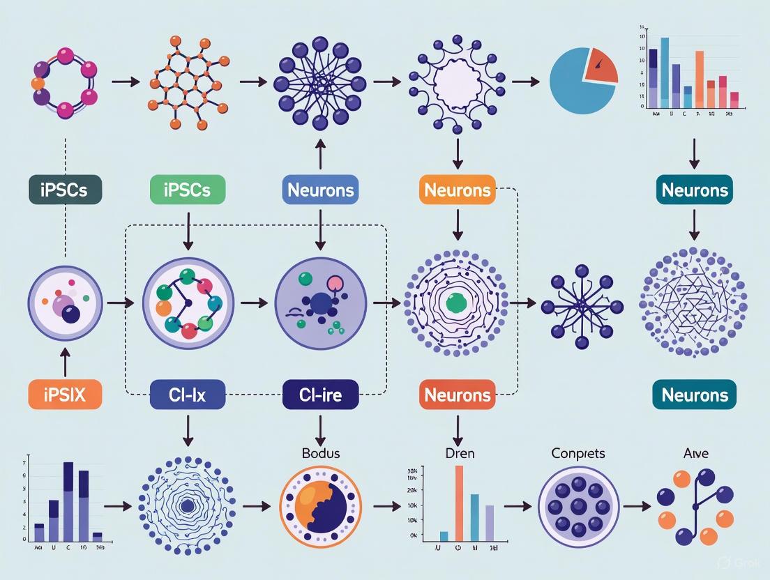
Experimental Workflow & Data Integration
The following diagram outlines a comprehensive workflow for differentiating iPSCs into mature neurons and validating their maturation status using multi-omics and functional approaches.
Signaling Pathways in Neuronal Maturation
The maturation process is regulated by intricate signaling pathways that are reflected in both transcriptional and proteomic data. The following diagram summarizes key pathways and their functional outcomes.
Frequently Asked Questions
Q1: What is the most critical period for metabolic remodeling during neuronal differentiation, and how can I confirm it in my cultures?
The first week of differentiation is a critical window for metabolic specialization in human iPSCs differentiating into cortical neurons [10]. During this period, cells undergo a essential metabolic shift from glycolysis towards oxidative phosphorylation. You can confirm this transition in your cultures using Fluorescence Lifetime Imaging Microscopy (FLIM) to detect a progressive increase in enzyme-bound NAD(P)H, which is a hallmark of a shift toward oxidative metabolism [10]. Additionally, 13C₆-glucose metabolic flux analysis can reveal enhanced labeling of pentose phosphate pathway intermediates and glutathione, indicating a shift toward biosynthetic and antioxidant glucose utilization [10].
Q2: My iPSC-derived neurons show inconsistent maturation after 4 weeks. What functional benchmarks should I use to track their development?
A multifaceted assessment strategy is essential. The table below outlines key functional parameters that evolve during neuronal maturation, based on a comprehensive 10-week study [6].
Table: Functional Maturation Benchmarks for Human iPSC-Derived Neurons
| Maturation Week | Membrane Resistance | Firing Profile | Synaptic Activity | Network Activity |
|---|---|---|---|---|
| 1-4 | High | Immature, inconsistent | Limited | None |
| 5 | Decreasing | Mature, regular firing pattern emerges | Developing | None |
| 6-7 | Lower | Consistent regular firing | Abundant fast glutamatergic and depolarizing GABAergic | Synchronized network bursts appear |
| 8-10 | Adult-like | Stable, mature | Mature, balanced inhibition/excitation | Robust, synchronized |
Q3: Does 3D culture improve the maturation of neural progenitor cells (NPCs), and when should I transition to this system?
Yes, transitioning NPCs to 3D floating neurosphere cultures significantly improves homogeneity and differentiation potential [11]. This method promotes a more homogenous expression of the NPC marker PAX6 and increases the subsequent differentiation potential toward astrocytes, as evidenced by higher expression of markers like GFAP and aquaporin 4 (AQP4) [11]. You should implement this system during the NPC expansion phase, after initial neural induction but before terminal differentiation [11] [12].
Q4: I am working with cardiomyocyte differentiation. What combined stimuli most effectively define a maturation window for electrophysiological maturity?
For iPSC-derived cardiomyocytes (iPSC-CMs), a combination of metabolic, structural, and electrical stimuli applied during the post-differentiation phase drives advanced maturity. A 2025 study systematically tested these factors and found that electrostimulation (ES) was the key driver for mitochondrial development and metabolic maturation [13]. When combined with a lipid-enriched maturation medium (MM) with high calcium and nanopatterning (NP) for cell alignment, the protocol generated cardiomyocytes with adult-like electrophysiological properties, including a more negative resting membrane potential and a faster action potential upstroke [13].
Experimental Protocols & Workflows
Protocol 1: Tracking Metabolic Maturation in Cortical Neurons
This protocol defines the critical window for metabolic remodeling during the first two weeks of neuronal differentiation [10].
- Key Materials: Human iPSCs with inducible NGN2, neural differentiation media, doxycycline.
- Week 0-1 (Critical Window): Initiate differentiation with doxycycline to induce NGN2 overexpression. Analyze cells at day 7.
- Day 7 Analysis: Perform high-resolution respirometry to detect early enhancements in oxidative phosphorylation. Use FLIM to measure the ratio of enzyme-bound to free NAD(P)H, indicating a shift toward oxidative metabolism [10].
- Week 1-2 (Progression): Continue differentiation and analyze cells at day 14.
- Day 14 Analysis: Conduct 13C₆-glucose metabolic flux analysis. Expect to see delayed labeling of TCA cycle metabolites but enhanced labeling of pentose phosphate pathway intermediates and glutathione, confirming a metabolic shift to support biosynthesis and antioxidant defense [10].
- Troubleshooting: If metabolic shift is not observed, ensure the purity of your neuronal population by checking NGN2 induction efficiency and the health of the starting iPSC culture.
The following diagram illustrates the experimental workflow and the key metabolic shifts to monitor during this critical period.
Protocol 2: A Multifactorial 10-Week Maturation Timeline for Neurons
This protocol provides a week-by-week framework for functional maturation, allowing you to plan experiments based on defined neuronal capabilities [6].
- Key Materials: Human NPCs, Poly-L-Ornithine/Laminin coated plates, BrainPhys Neuronal Medium, BDNF, GDNF, dibutyryl cyclic-AMP, ascorbic acid [6].
- Weeks 1-4 (Baseline Excitability): Plate NPCs and initiate terminal differentiation. During this phase, neurons will exhibit high membrane resistance and developing, inconsistent firing profiles. Focus on immunocytochemistry for morphological markers (e.g., MAP2, TAU) and initial patch-clamp recordings to confirm the presence of action potentials [6].
- Weeks 5-6 (Functional Synapse Formation): This is a critical window for the emergence of mature electrophysiological properties.
- Week 5: Perform patch-clamp to identify the emergence of a mature, regular firing pattern. Membrane resistance will noticeably decrease [6].
- Week 6: Test for the appearance of fast glutamatergic synaptic currents and depolarizing GABAergic responses. Calcium imaging can now be used to detect the first synchronized network bursts [6].
- Weeks 7-10 (Network Stabilization): The culture develops into a stable, interconnected network.
- Focus: Continue monitoring synaptic and network activity. The alterations in GABAA receptor subunit expression occur during this period, leading to more mature inhibitory signaling. Use Sholl analysis on biocytin-filled neurons to quantify complex morphological development [6].
- Troubleshooting: If synchronized network activity fails to develop by week 7, ensure a sufficient density of neurons and consistent, twice-weekly half-medium changes with fresh BDNF/GDNF.
The Scientist's Toolkit: Essential Reagents for Maturation Studies
Table: Key Reagents for Defining Maturation Windows
| Reagent/Condition | Function in Maturation | Key Readouts |
|---|---|---|
| NGN2 Inducible System [10] | Drives rapid, synchronous differentiation of iPSCs to excitatory cortical neurons. | Neuronal purity, expression of cortical markers. |
| BDNF & GDNF [6] | Neurotrophins that support neuronal survival, neurite outgrowth, and synaptic plasticity during long-term maturation. | Increased neurite complexity, emergence of synaptic activity. |
| 3D Neurosphere Culture [11] [12] | Provides a more in vivo-like environment, enhancing NPC homogeneity and astrocyte differentiation potential. | Increased PAX6 homogeneity, higher GFAP expression post-differentiation. |
| Metabolic Maturation Media [14] | Shifts energy metabolism from glycolysis to fatty acid oxidation; critical for cardiomyocyte maturation. | TTX-sensitive action potentials, increased mitochondrial content & respiration. |
| Electrostimulation (ES) [13] | Key driver for mitochondrial development and electrophysiological maturation in cardiomyocytes. | More negative resting membrane potential, faster action potential upstroke, "notch-and-dome" morphology. |
| Nanopatterning (NP) [13] | Provides structural cues that promote sarcomere organization and cell alignment in cardiomyocytes. | Organized sarcomeres, elongated nuclei, improved connexin 43 localization. |
The following diagram summarizes how these different stimuli can be integrated in a combined maturation strategy for specific cell types.
Protocols in Practice: A Guide to Differentiation Methods and Their Target Applications
Transcription factor programming of induced pluripotent stem cells (iPSCs) using Neurogenin-2 (NGN2) has emerged as a powerful method for generating human excitatory neurons. This approach significantly reduces heterogeneity and improves consistency across different stem cell lines compared to extrinsic factor-based differentiation methods [15] [16]. As a master regulator of neurogenesis, NGN2 overexpression can rapidly differentiate functional neurons from hPSCs, neural progenitors, or fibroblasts with high reproducibility, typically within 1-2 weeks [15] [17]. However, researchers often encounter challenges with variable maturation rates, unwanted cell populations, and protocol-dependent heterogeneity that can compromise experimental outcomes. This technical support center addresses these specific issues through troubleshooting guides and FAQs designed for researchers, scientists, and drug development professionals working within the context of optimizing neuronal maturation from iPSCs.
Key Research Reagents and Materials
The table below summarizes essential reagents for successful NGN2 neuronal differentiation:
Table 1: Essential Research Reagents for NGN2 Neuronal Differentiation
| Reagent Category | Specific Examples | Function in Protocol |
|---|---|---|
| Induction System | Doxycycline (Dox)-inducible NGN2 | Controls transgene expression; typically used at 1-3 μg/mL [18] [17] |
| Small Molecule Inhibitors | LDN-193189 (BMP inhibitor), SB431542 (TGF-β inhibitor), DAPT (Notch inhibitor) | Enhances neural induction purity and prevents progenitor contamination [17] [19] |
| Anti-proliferative Agents | Cytarabine (Ara-C), Mitomycin-C, Fluorouracil (5-FU) | Eliminates dividing progenitor cells and reduces clump formation [17] |
| Maturation Enhancers | GENtoniK cocktail (GSK2879552, EPZ-5676, NMDA, Bay K 8644) | Accelerates functional maturation through epigenetic and calcium signaling modulation [4] |
| Cryopreservation Aids | Rho-kinase (ROCK) inhibitor | Improves cell survival after freezing and thawing [17] |
Troubleshooting Common Experimental Issues
Heterogeneous Neuronal Populations
Problem: Resulting cultures contain mixed neuronal subtypes or non-neuronal cells despite NGN2 expression.
Solutions:
- Implement FACS sorting: Isolate iPSCs with homogeneous NGN2 expression levels using linked GFP reporters before differentiation initiation. This addresses heterogeneity caused by variable transgene expression [16].
- Optimize NGN2 induction duration: Extend doxycycline-mediated NGN2 expression beyond standard protocols. Recent studies indicate that prolonged NGN2 dosage dramatically improves neuronal purity by more completely converting progenitors [18].
- Incorporate dual SMAD inhibition: Combine NGN2 programming with SMAD (LDN-193189, SB431542) and WNT inhibitors during early differentiation phases to generate patterned induced neurons (hpiNs) with more consistent excitatory glutamatergic identity [19].
- Use anti-proliferative agents: Include cytarabine (Ara-C) or similar compounds during replating to eliminate actively dividing neural progenitor cells that create contaminating niches [17].
Incomplete Neuronal Maturation
Problem: Neurons exhibit immature electrophysiological properties, simplified morphology, or fetal-like transcriptomic signatures even after extended culture.
Solutions:
- Apply maturation-accelerating cocktails: Implement the GENtoniK cocktail (GSK2879552 [LSD1 inhibitor], EPZ-5676 [DOT1L inhibitor], NMDA, and Bay K 8644 [LTCC agonist]) which targets both chromatin remodeling and calcium-dependent transcription to drive adult-like maturity [4].
- Validate with appropriate markers: Assess multiple maturity dimensions simultaneously:
- Extend culture duration: Maintain neurons for at least 100-150 days with appropriate trophic support, as human neurons follow protracted timelines regardless of induction method [20] [17].
Technical Variability Across Experiments
Problem: Inconsistent differentiation outcomes between replicates, batches, or laboratory sites.
Solutions:
- Use safe-harbor engineered lines: Employ iPSCs with NGN2 stably integrated into the AAVS1 locus rather than lentiviral delivery to minimize copy number variation and position effects [18] [17].
- Standardize plating conditions: Optimize and consistently maintain critical parameters:
- Implement quality controls: Perform rigorous genomic stability assessment post-reprogramming using SNP arrays rather than conventional karyotyping to detect subtle rearrangements that might affect differentiation consistency [16].
Frequently Asked Questions (FAQs)
Q1: What is the typical efficiency and timeline for generating functional neurons using NGN2 programming?
NGN2 programming can generate neurons with neuronal morphology appearing within 2-4 days, with most cells exhibiting neuronal morphology by day 4 [17]. However, achieving functionally mature neurons with robust synaptic activity requires significantly longer culture periods—typically 28-56 days for basic electrophysiological properties, and up to 150 days for complete maturation resembling postnatal states [17] [4]. Efficiency can reach 75-90% glutamatergic neurons when optimized protocols are used [17].
Q2: Why do my NGN2-induced neurons show variable responses to depolarizing stimuli?
Neuronal responses to stimuli like KCl are highly maturation-dependent. Immature neurons (7-10 DIV) exhibit dramatically different electrophysiological properties and immediate early gene responses compared to more developed cultures (21+ DIV) [22]. This reflects natural development of calcium signaling, receptor composition, and synaptic connectivity. Consistently control for and report neuronal age in experiments, and consider using maturation-accelerating compounds like the GENtoniK cocktail for more consistent responses [4].
Q3: How can I minimize the presence of non-neuronal cells in my NGN2 cultures?
Persistent non-neuronal cells (often neural progenitors or astrocytes) typically result from:
- Incomplete NGN2-mediated conversion - address through extended NGN2 expression [18]
- Progenitor proliferation - suppressed using anti-mitotics like cytarabine or 5-fluorouracil [17]
- Insufficient patterning - improved by combining NGN2 with SMAD/WNT inhibition [19] Single-cell RNA sequencing can identify contaminating cell types for targeted troubleshooting [17].
Q4: What are the key differences between NGN2-programmed neurons and those from directed differentiation?
Table 2: Comparison of Neuronal Differentiation Methods
| Parameter | NGN2 Programming | Directed Differentiation |
|---|---|---|
| Timeline | 7-28 days for basic neurons [17] | Weeks to months for similar stages [15] |
| Efficiency | High (≥75% glutamatergic) [17] | Variable, often lower [15] |
| Heterogeneity | Lower with optimization [16] | Higher, multiple cell types [15] |
| Regional Identity | Mixed cortical/thalamic features [17] | More specific regional identities possible [15] |
| Technical Demand | Lower with engineered lines [17] | Higher, requires precise timing [15] |
Q5: Can NGN2 programming generate specific neuronal subtypes beyond generic glutamatergic neurons?
Yes, with additional patterning. While NGN2 alone predominantly produces excitatory neurons with mixed cortical and thalamic features [17], combining NGN2 with regionalizing factors can generate more specific subtypes:
- Cortical neurons: NGN2 + SMAD/WNT inhibition [19]
- Motor neurons: NGN2 with additional patterning factors [15]
- Sensory neurons: NGN2 with specific morphogens [15] The resulting neuronal identity should be validated through combination of marker expression (FOXG1 for forebrain, ISL1 for motor/sensory) and transcriptomic profiling [17].
Experimental Workflows and Signaling Pathways
Optimized NGN2 Neuronal Differentiation Workflow
Maturation Acceleration via GENtoniK Cocktail Mechanism
Successful generation of homogeneous glutamatergic neurons via NGN2 programming requires attention to three critical aspects: (1) starting cell quality with homogeneous NGN2 expression, (2) appropriate patterning during early differentiation, and (3) systematic maturation enhancement. By implementing the troubleshooting strategies and optimized protocols outlined in this technical guide, researchers can significantly improve the reproducibility and translational relevance of their iPSC-derived neuronal models for studying neurological disorders and screening therapeutic compounds.
DUAL SMAD inhibition represents a foundational methodology in human pluripotent stem cell (hPSC) research for efficiently directing cells toward neuronal lineages. By simultaneously blocking transforming growth factor–beta (TGF-β) and bone morphogenetic protein (BMP) signaling pathways, this protocol enables robust and reproducible induction of neuroectoderm, serving as the basis for generating diverse brain region–specific neuronal subtypes [23]. The approach has become indispensable for both basic neuroscience research and translational applications, including its recent use in clinical trials for Parkinson's disease [23] [24].
Within the context of optimizing neuronal maturation from iPSCs, DUAL SMAD inhibition offers a unique advantage: it produces heterogeneous neural cultures that closely mimic developmental processes. Unlike direct differentiation methods that generate homogeneous neuronal populations, this approach yields cultures containing a mix of neurons, neural precursors, and glial cells through a stepwise differentiation process that passes through a neural stem cell stage [5]. This heterogeneity is particularly valuable for modeling complex neural environments and studying the interactions between different neural cell types during maturation.
Mechanism of Action: Core Signaling Pathways
Molecular Foundations of Neural Induction
During embryonic development, the formation of three germ layers—ectoderm, mesoderm, and endoderm—is orchestrated by a complex interplay of signaling pathways, primarily WNT/β-catenin, FGF, TGF-β, and BMP families [23]. Active TGF-β and BMP signaling normally prevent neuronal differentiation by maintaining pluripotency or diverting cells toward mesodermal and endodermal lineages. The DUAL SMAD inhibition protocol induces neuronal fate in hPSCs by simultaneously blocking both pathways, thereby eliminating signals necessary for pluripotency and mesendodermal fate induction [23].
The TGF-β and BMP pathways converge on intracellular SMAD proteins, which transmit extracellular signals to the nucleus upon phosphorylation. Phosphorylated SMAD proteins form complexes with SMAD4 and translocate to the nucleus to regulate gene expression. DUAL SMAD inhibition disrupts this process using specific inhibitors [23]:
- SB431542: A cell-permeable small molecule that inhibits TGF-β signaling by selectively targeting Activin receptor–like kinases ALK4, ALK5, and ALK7, thereby suppressing SMAD2/3 activation
- Noggin: An endogenous BMP antagonist that binds to and sequesters BMP ligands, preventing receptor activation
- LDN193189: A synthetic small-molecule inhibitor that targets ALK2/3/6 receptors, blocking phosphorylation of SMAD1/5/8
The outcome of this coordinated inhibition is the robust and reproducible induction of neural fate, with hPSCs exiting the pluripotent state and defaulting to a neuroectodermal lineage [23].
Figure 1: DUAL SMAD Inhibition Signaling Mechanism. Simultaneous inhibition of TGF-β and BMP pathways prevents SMAD phosphorylation and nuclear translocation, removing barriers to neural differentiation and enabling formation of heterogeneous neural cultures.
Experimental Protocols: Methodology and Workflows
Core DUAL SMAD Inhibition Protocol
The standard DUAL SMAD inhibition protocol for neural differentiation from iPSCs follows a well-established workflow with specific timing and reagent requirements. The process typically begins with undifferentiated iPSCs maintained under standard culture conditions, then transitions through defined stages of neural induction and maturation [5] [25].
Key Methodological Steps:
Initial Cell Plating: Undifferentiated iPSCs are dissociated into single cells and plated onto Matrigel-coated dishes in conditioned medium supplemented with ROCK inhibitor Y-27632 to promote cell survival [25].
Neural Induction: After 72 hours, cells are switched to knock-out serum replacement media (KSR) containing both Noggin (or LDN193189) and SB431542 to initiate neural induction [25].
Neural Precursor Formation: Treatment with DUAL SMAD inhibitors typically continues for 10-14 days, during which cells differentiate into neuroepithelial cells expressing PAX6 and other early neural markers [25].
Terminal Differentiation: Neural precursors can be further differentiated into specific neuronal subtypes through the addition of patterning factors and maturation media [5].
This protocol yields heterogeneous cultures containing a mix of central nervous system (CNS) progenitors and neural crest cells, with the initial plating density influencing the ratio of CNS versus neural crest progeny [25].
Workflow for Synchronized Neuronal Maturation
For researchers focusing specifically on neuronal maturation, a modified DUAL SMAD inhibition approach can generate temporally synchronized cortical neurons:
Figure 2: Synchronized Neuronal Maturation Workflow. This specialized protocol using DUAL SMAD inhibition followed by Notch inhibition enables generation of temporally synchronized cortical neurons for maturation studies.
This synchronized approach produces homogeneous populations of cortical neurons that mature gradually over months in vitro, exhibiting progressive increases in neurite complexity, electrophysiological maturity, and synaptic function [26]. The timeline mirrors the protracted development of human cortical neurons in vivo, providing a valuable model for studying maturation processes.
Research Reagent Solutions: Essential Materials
Table 1: Key Reagents for DUAL SMAD Inhibition Protocol
| Reagent | Function | Application Notes |
|---|---|---|
| SB431542 | TGF-β pathway inhibitor; targets ALK4/5/7 receptors | Used at typical concentrations of 10-20 μM; critical for suppressing mesendodermal differentiation [23] [25] |
| Noggin | Recombinant BMP antagonist; binds and sequesters BMP ligands | Can be replaced by small molecule LDN193189 for more consistent results [23] |
| LDN193189 | Small molecule BMP inhibitor; targets ALK2/3/6 receptors | Often preferred over Noggin for higher purity and reproducibility [23] |
| Y-27632 | ROCK inhibitor; prevents apoptosis in dissociated cells | Essential for single-cell passaging; used at 5-10 μM [5] |
| Doxycycline | Tetracycline analog; induces transgene expression in Tet-ON systems | Used at 1-2 μg/mL for inducible differentiation protocols [5] |
| DAPT | γ-secretase inhibitor; blocks Notch signaling | Used for synchronized neurogenesis at progenitor stage [26] |
Troubleshooting Guide: Common Experimental Challenges
Low Neural Induction Efficiency
Problem: Poor yield of neural progenitor cells despite DUAL SMAD inhibition treatment.
Potential Causes and Solutions:
- Inadequate inhibitor concentration: Validate working concentrations of SB431542 (typically 10-20 μM) and Noggin/LDN193189 through dose-response testing
- Improper initial cell density: Optimize plating density as this significantly affects neural conversion efficiency and the ratio of CNS versus neural crest progeny [25]
- Cell line variability: Different iPSC lines may show varying differentiation efficiencies; consider testing multiple lines or optimizing protocol for specific lines
- Pluripotency maintenance: Ensure iPSCs are in undifferentiated state before initiation of neural induction
Excessive Heterogeneity or Unwanted Cell Types
Problem: Cultures contain unexpected non-neural cell types or incorrect neural subtypes.
Troubleshooting Strategies:
- Validate patterning factors: Ensure appropriate anterior-posterior and dorsal-ventral patterning cues are applied after initial neural induction [23]
- Timing of protocol steps: Precise timing of inhibitor treatment and growth factor addition is critical; extend DUAL SMAD inhibition if persistent pluripotent cells observed
- Cell sorting strategies: Implement fluorescence-activated cell sorting (FACS) using neural surface markers if higher purity is required [27]
- Metabolic selection: Consider using selective media conditions that favor neural cell survival
Poor Neuronal Maturation
Problem: Neurons fail to develop mature electrophysiological properties or synaptic connectivity.
Optimization Approaches:
- Extended culture duration: Human neurons require extended time (months) to mature fully; ensure adequate culture maintenance [26]
- Epigenetic modulation: Consider transient inhibition of EZH2, EHMT1/2, or DOT1L at progenitor stage to precociously enhance maturation [26]
- Activity-dependent stimulation: Implement chronic stimulation protocols to promote functional maturation
- Co-culture systems: Consider astrocyte co-cultures or conditioned media to provide trophic support
Frequently Asked Questions
Q: How does DUAL SMAD inhibition compare to NGN2 overexpression for neuronal differentiation?
A: These methods produce distinctly different neural cultures. DUAL SMAD inhibition generates heterogeneous cultures containing a mix of neurons, neural precursors, and glial cells through a developmental process that includes neural stem cell stages. In contrast, NGN2 overexpression produces more homogeneous cultures composed predominantly of mature neurons without going through extended progenitor stages. Transcriptomic analyses reveal enrichment of neural stem cell and glial markers in DUAL SMAD inhibition cultures, while NGN2 cultures show elevated markers for cholinergic and peripheral sensory neurons [5].
Q: What is the typical efficiency of neural conversion using DUAL SMAD inhibition?
A: When properly optimized, the protocol can achieve very high efficiencies of neural conversion. Original reports demonstrated greater than 80% PAX6+ neural progenitor cells using combined Noggin and SB431542 treatment, compared to less than 10% with either inhibitor alone [25]. Efficiency can be monitored using neural markers like PAX6, SOX1, and SOX2 at early timepoints.
Q: Can DUAL SMAD inhibition generate region-specific neuronal subtypes?
A: Yes, the protocol serves as a foundation for generating diverse brain region-specific neuronal subtypes. By default, DUAL SMAD inhibition produces anterior (forebrain) neural progenitors that predominantly give rise to cortical neurons. Specific neuronal subtypes can be generated by adding appropriate patterning factors – for example, caudalizing agents like WNT activators or retinoic acid can shift identity toward midbrain, hindbrain, or spinal cord fates [23].
Q: How long does complete neuronal maturation take using this method?
A: Neuronal maturation following DUAL SMAD inhibition follows a protracted timeline that mirrors human development. While early neuronal markers appear within 1-2 weeks, full electrophysiological maturation with repetitive action potentials and functional synapses typically requires 50-100 days in vitro, with continued maturation occurring over even longer periods [26]. This slow maturation timeline reflects cell-intrinsic mechanisms that include specific epigenetic barriers [26].
Q: What are the key advantages of DUAL SMAD inhibition for disease modeling?
A: The method offers several advantages: (1) It produces heterogeneous cultures that better mimic the cellular diversity of neural tissue; (2) It follows developmental principles, allowing study of disease processes across different developmental stages; (3) The inclusion of glial precursors enables modeling of neuron-glia interactions; (4) It has been validated across numerous hPSC lines and in clinical-grade applications [23].
Advanced Applications: Integration with Emerging Technologies
CRISPR/Cas9 Engineering in DUAL SMAD Inhibition
The combination of DUAL SMAD inhibition with CRISPR/Cas9 gene editing enables powerful approaches for disease modeling and lineage tracing. For example, researchers have successfully generated knock-in reporter lines where fluorescent proteins are inserted into endogenous neural genes such as tyrosine hydroxylase (TH), allowing specific identification and purification of dopaminergic neurons following differentiation [27]. This integration provides:
- Precise lineage tracking: Endogenous fluorescent reporters enable real-time monitoring of neuronal differentiation and maturation
- Cell sorting capabilities: FACS purification of specific neuronal populations from heterogeneous cultures
- Disease modeling: Introduction of disease-associated mutations into isogenic iPSC lines
- High-throughput screening: Enables quantitative analysis of differentiation outcomes
3D Organoid Development
DUAL SMAD inhibition serves as the foundational step for generating more complex 3D neural organoids that recapitulate aspects of human brain development and disease. These 3D models overcome limitations of 2D cultures by better mimicking cell-cell and cell-extracellular matrix interactions, enabling study of higher-order cellular organization and network dynamics [28] [29]. Recent advances include:
- Disease-specific organoids: Modeling dementia disorders like Alzheimer's disease, Parkinson's disease, and ALS/FTD
- Multi-regional assemblies: Combining organoids representing different brain regions
- Integration of non-neural lineages: Incorporating microglia or vascular cells for more complete models
Table 2: Characterization of Neural Cultures Following DUAL SMAD Inhibition
| Parameter | Typical Outcome | Measurement Method | Reference |
|---|---|---|---|
| Neural Conversion Efficiency | >80% PAX6+ cells | Immunocytochemistry, Flow Cytometry | [25] |
| Culture Composition | Mixed neurons, neural precursors, glial cells | Transcriptomic Analysis, Immunostaining | [5] |
| Action Potential Development | 50-100 days for repetitive firing | Patch Clamp Electrophysiology | [26] |
| Synaptic Function | mEPSCs detectable by 50-75 days | Electrophysiology, Immunostaining | [26] |
| Neurite Complexity | Progressive increase over 100 days | Morphometric Analysis | [26] |
| Cortical Neuron Purity | ~90% TBR1+ with synchronization | scRNA-seq, Immunostaining | [26] |
A critical challenge in iPSC research is selecting a differentiation and screening protocol that aligns with the specific research objective. The choice between building a high-content disease model or a platform for high-throughput screening (HTS) dictates every subsequent experimental parameter. This guide provides troubleshooting and procedural advice to help you match your method to your goal within the context of optimizing neuronal maturation from iPSCs.
Frequently Asked Questions (FAQs)
1. How do I decide between a 2D monolayer and a 3D organoid system for my disease modeling project? The decision hinges on the biological question. Use 2D monolayers for investigating cell-autonomous mechanisms, high-content imaging, and electrophysiological studies at the single-cell level. They offer simplicity and reproducibility. Opt for 3D organoids when your research question involves complex cell-cell interactions, tissue-level architecture, and microenvironmental effects, as they more accurately mimic the cellular complexity of the developing brain [30] [31]. However, be prepared to address higher heterogeneity and technical complexity.
2. What are the key considerations for adapting a neuronal differentiation protocol for high-throughput screening? The primary considerations are scalability, reproducibility, and quantifiability. The protocol must generate highly uniform cultures in multi-well plates (e.g., 96 or 384-well format) with minimal well-to-well variability. The readout, whether it's cell survival, neurite outgrowth, or a simplified transcriptional signature, must be robust and amenable to automation. Reductionist systems that focus on a key, quantifiable phenotype are often more successful than highly complex models for HTS [32].
3. Why does my iPSC-derived motor neuron model fail to show a disease-relevant phenotype, even with a patient-derived line? The absence of a phenotype is a common hurdle. First, verify that your neurons are fully mature, as many disease phenotypes are age-dependent. Consider extending the maturation period. Second, assess whether non-cell-autonomous factors (e.g., from glial cells) are missing from your culture system. Introducing astrocytes or microglia might be necessary. Finally, implement more sensitive phenotypic assays, such as longitudinal live-cell imaging to track subtle changes in neurite health or single-cell RNA sequencing to identify cryptic transcriptional shifts [32] [33].
4. How can I improve the consistency of neuronal differentiation across multiple iPSC lines? Line-to-line variability is a major challenge. To mitigate it, ensure all your iPSC lines have undergone rigorous quality control for karyotype and pluripotency. Use a highly standardized, possibly automated, differentiation protocol. For spinal motor neuron differentiation, leveraging a neuromesodermal progenitor (NMP) intermediate has been shown to enhance the efficiency and posterior identity of the resulting neurons, improving consistency [34]. Additionally, maintain consistent cell seeding densities and batch-test all critical reagents.
Troubleshooting Guides
Issue: Poor Survival of Mature Neurons in Long-Term Culture
Potential Causes and Solutions:
- Cause 1: Toxic metabolite accumulation in the culture medium.
- Solution: Optimize the feeding schedule. For mature neurons, consider half-medium changes every 2-3 days instead of full changes to avoid shocking the cells. Test the addition of antioxidant supplements like N-acetylcysteine.
- Cause 2: Inadequate neurotrophic support.
- Solution: Supplement the maturation medium with a combination of neurotrophic factors. Common choices include BDNF, GDNF, and CNTF, typically in the range of 10-20 ng/mL. Re-evaluate and titrate the concentrations for your specific neuronal subtype.
- Cause 3: Excessive stress from over-dissociation or passaging.
- Solution: Mature post-mitotic neurons are sensitive to passaging. Instead, plate neural progenitors and allow them to differentiate and mature in the same well. Use gentle reagent-based methods for any necessary dissociation.
Issue: High Well-to-Well Variability in a High-Throughput Screening Assay
Potential Causes and Solutions:
- Cause 1: Inconsistent starting cell population.
- Solution: Use a large, master bank of pre-differentiated neural progenitor cells (NPCs) that has been thoroughly quality-controlled. Thaw a single vial for an entire screen to ensure a uniform starting point. Employ automated cell counters and dispensers for precise and consistent plating.
- Cause 2: Edge-effect evaporation in multi-well plates.
- Solution: Use tissue culture-treated plates designed for HTS. Fill the outer wells with PBS only and do not use them for experimental data. Consider using plate seals during incubation periods.
- Cause 3: Inconsistent compound dispensing or dosing.
- Solution: Use automated liquid handlers for compound addition. Include control wells that receive only the vehicle (e.g., DMSO) dispersed by the same system to account for any dispensing effects.
Experimental Protocols for Key Applications
Detailed Protocol 1: Large-Scale Phenotypic Screening for Motor Neuron Survival
This protocol is adapted from a recent large-scale study that successfully modeled sporadic ALS [32].
Workflow Diagram: High-Throughput Screening Pipeline for Motor Neuron Health
Methodology:
- iPSC Culture and Quality Control: Maintain a curated library of iPSC lines from patients and healthy controls. All lines must undergo rigorous quality control, including genomic integrity checks (e.g., karyotyping), pluripotency verification (e.g., expression of OCT3/4, NANOG), and trilineage differentiation potential [32].
- Motor Neuron Differentiation: Employ a optimized five-stage spinal motor neuron differentiation protocol. Adapt a well-established method to generate high-purity cultures [32] [34].
- Key Steps: Induce neuromesodermal progenitors (NMPs) with CHIR99021 (a GSK3β inhibitor) and growth factors. Pattern towards posterior spinal identity. Differentiate into motor neuron progenitors and then mature motor neurons.
- Quality Control: Validate cultures by immunostaining for motor neuron markers (ChAT, MNX1/HB9, ISL1, TUJ1). Aim for >90% purity. Assess functional maturity via patch-clamp electrophysiology to confirm action potential firing [32] [34].
- Phenotypic Screening with Live-Cell Imaging: Plate mature motor neurons in 96 or 384-well plates. Use a motor neuron-specific reporter (e.g., HB9::GFP) for selective tracking. Place plates in an automated live-cell imaging system. Acquire images every 4-24 hours over 7-14 days to track survival and neurite degeneration [32].
- Compound Library Addition: At a pre-defined maturation stage, add drugs from your library. Include positive controls (e.g., 10 µM Riluzole) and vehicle controls (e.g., 0.1% DMSO). The study screening over 100 clinical trial drugs found that a combination of Riluzole, Memantine, and Baricitinib was effective [32].
- Data Analysis: Use automated image analysis software to quantify:
- Motor neuron survival over time.
- Neurite length and branching complexity.
- Correlate in vitro phenotypes with donor clinical data (e.g., survival time).
Quantitative Data from Screening Campaigns
| Screening Parameter | Performance Metric | Notes |
|---|---|---|
| Culture Purity | >92% motor neurons (ChAT+, MNX1+, TUJ1+) [32] | Essential for cell-autonomous effect studies. |
| Phenotype Correlation | Accelerated neurite degeneration correlated with donor survival [32] | Validates clinical relevance of the model. |
| Clinical Trial Drug Re-Creation | ~3% of tested drugs showed efficacy [32] | Reflects actual clinical trial failure rates, validating model predictive power. |
| Key Effective Drugs | Riluzole, Memantine, Baricitinib (combinatorial) [32] | Identified as promising therapeutic combination for SALS. |
Detailed Protocol 2: Establishing a High-Content Model for Hereditary Sensory Neuropathy
This protocol uses 3D organoids to model a complex neurodevelopmental disorder, HSAN IV [33].
Workflow Diagram: Disease Modeling with Isogenic Control iPSC Lines
Methodology:
- Generate Isogenic Control Lines: Use CRISPR-Cas9 genome editing to correct the disease-causing mutation (e.g., in the NTRK1 gene) in the patient-derived iPSCs. This creates a genetically matched control, isolating the mutation as the only variable [33].
- Dorsal Root Ganglion (DRG) Organoid Differentiation: Differentiate both patient and isogenic control iPSCs into 3D DRG organoids, which contain sensory neurons and supporting glial cells.
- Key Steps: Guide iPSCs through a neural crest stem cell pathway. Aggregate cells to form 3D structures and pattern towards a sensory neuron fate using specific morphogens [33].
- Phenotypic Analysis: Compare patient and isogenic organoids across multiple dimensions.
- Lineage Specification: Analyze the balance of neuronal and glial differentiation by immunostaining for markers like ISLET1/BRN3A (sensory neurons) and FABP7 (glia). The HSAN IV model showed premature gliogenesis [33].
- Maturation: Assess expression of mature sensory markers (TRKA, TRPV, CGRP) and evaluate axonal outgrowth.
- Functional Assays: Perform calcium imaging to assess neuronal activity and response to stimuli.
The Scientist's Toolkit: Research Reagent Solutions
Key materials and reagents used in advanced iPSC-based neuronal research.
| Item | Function/Application | Examples/Notes |
|---|---|---|
| Non-Integrating Reprogramming Vectors | Generating clinical-grade iPSCs without genomic integration. | Sendai Virus Vectors: High efficiency, can be diluted out [31] [37]. Episomal Plasmids: Cost-effective, no viral components [32] [31]. |
| Small Molecule Inhibitors/Activators | Directing differentiation by modulating key signaling pathways. | CHIR99021: GSK3β inhibitor, activates Wnt signaling for NMP induction [34]. SMAD Inhibitors (e.g., A-83-01): Promotes neural induction [31] [34]. |
| CRISPR-Cas9 System with High-Fidelity Variants | Precise genome editing for creating isogenic controls. | SpCas9-HF1 / eSpCas9(1.1): Engineered for reduced off-target effects [36]. AI gRNA Design Tools (e.g., DeepHF): Improve on-target efficiency prediction [35] [36]. |
| Tandem Mass Tag (TMT) Labeling | Multiplexed proteomic analysis from limited samples like neurospheres. | Allows comparative analysis of up to 10+ samples simultaneously, maximizing data yield [30]. |
| Longitudinal Live-Cell Imaging Systems | Automated, non-invasive tracking of cell health and phenotype over time. | Essential for quantifying dynamic processes like neurodegeneration in high-throughput screens [32]. |
The choice between two-dimensional (2D) monolayers and three-dimensional (3D) organoid models is a critical decision in experimental design, particularly within the context of optimizing neuronal maturation from induced pluripotent stem cells (iPSCs). While 2D cultures have long been the workhorse for high-throughput assays and straightforward mechanistic studies, 3D organoids are revolutionizing the field by recapitulating the complex architecture and cellular interactions of the human brain [38] [39]. This technical support guide provides a structured comparison, troubleshooting advice, and detailed protocols to help researchers navigate these advanced culture systems effectively.
Comparative Analysis: 2D Monolayers vs. 3D Organoids
The table below summarizes the core characteristics of each model system to inform your experimental planning.
| Feature | 2D Monolayer Cultures | 3D Organoid Models |
|---|---|---|
| Core Structure | Single layer of cells on a flat, rigid plastic/glass surface [38] | Self-organizing 3D structures that mimic organ architecture [38] [39] |
| Cellular Complexity & Microenvironment | Limited cell-cell and cell-matrix interactions; lacks physiological tissue context [38] | Recapitulates complex cell-cell interactions and a more native tissue microenvironment [38] [20] |
| Key Advantages | Simplicity, cost-effectiveness, suitability for high-throughput drug and toxicity screening [38] [39] [40] | Models human-specific developmental processes, patient-specific disease phenotypes, and complex tissue-level functions [38] [20] [40] |
| Primary Limitations | Poor translation to in vivo human physiology; fails to model tissue-level properties [38] | Batch-to-batch variability, hypoxic cores leading to necrosis, extended culture times for maturation (≥6 months) [38] [20] |
| Ideal Applications | High-content imaging, electrophysiology (patch clamp), initial drug efficacy/toxicity screening, genetic manipulation [38] [40] | Disease modeling (e.g., Parkinson's, Alzheimer's), studying cell-cell interactions (e.g., neuro-immune), investigation of complex tissue development [38] [20] [40] |
Troubleshooting Guides
Guide 1: Addressing 3D Organoid Culture Challenges
Problem: Central Necrosis and Hypoxic Cores
- Cause: Limited diffusion of oxygen and nutrients into the organoid's core, especially in larger structures [20].
- Solution: Integrate bioengineering approaches. Use spinning bioreactors or orbital shakers to improve medium convection. Consider co-culturing with endothelial cells to encourage the formation of vascular networks, or use microfluidic devices to enhance nutrient delivery [20].
Problem: Incomplete or Arrested Maturation
- Cause: Organoids often remain at a fetal-to-early postnatal stage, even after extended culture, lacking adult neuronal and glial markers [20].
- Solution: Extend culture periods to ≥6 months with careful monitoring. Incorporate microenvironmental modulators, such as electrical stimulation or specific growth factor regimens, to promote gliogenesis and synaptic refinement. Employ co-culture systems with microglia or astrocytes to support mature network activity [20].
Problem: High Batch-to-Batch Variability
- Cause: Inconsistencies in iPSC line differentiation potential, reagent lots, and manual handling during complex protocols [38] [20].
- Solution: Standardize protocols and implement rigorous quality control. Use defined matrices and growth factors. For colorectal organoids, ensure prompt tissue processing (<6-10 hours) or optimized cryopreservation methods to maintain consistent cell viability [41].
Guide 2: Addressing 2D Monolayer Culture Challenges
Problem: Poor Differentiation Efficiency into Specific Neuronal Subtypes
- Cause: Inadequate or imprecise patterning signals during neural induction and differentiation [38] [40].
- Solution: Employ well-established, precise patterning protocols. For midbrain dopaminergic neurons (critical for Parkinson's disease research), use a floor plate-based strategy with morphogens like Sonic Hedgehog (SHH) and WNT activators, followed by maturation with BDNF and GDNF [40].
Problem: Lack of Physiologically Relevant Cellular Interactions
- Cause: The simplified 2D environment lacks the diverse cell types and spatial organization found in vivo [38].
- Solution: Develop co-culture systems. For blood-brain barrier (BBB) modeling, co-culture iPSC-derived brain microvascular endothelial cells (BMECs) with pericytes, neurons, and astrocytes to create a more functional barrier in a dish [38].
Frequently Asked Questions (FAQs)
Q1: When should I choose a 2D model over a 3D organoid model for my drug screening campaign? Choose a 2D model for primary, high-throughput screening of large compound libraries due to its simplicity, lower cost, and easier data readouts. Reserve 3D organoid models for secondary, more physiologically relevant validation of hits identified in the 2D screen, as they can better predict human-specific drug responses and toxicities [38] [39] [40].
Q2: How can I accurately assess the maturity and functionality of my brain organoids? A multimodal assessment framework is essential [20].
- Structural: Use immunofluorescence for cortical layer markers (SATB2 for upper layers, TBR1/CTIP2 for deep layers) and synaptic proteins (PSD-95, SYB2) [20].
- Cellular Diversity: Employ fluorescence-activated cell sorting (FACS) and single-cell RNA sequencing (scRNA-seq) to quantify neurons, astrocytes (GFAP, S100β), and oligodendrocytes (MBP, O4) [20].
- Functional: Use multielectrode arrays (MEAs) to record synchronized network activity and calcium imaging to visualize dynamic activity across cell populations [20].
Q3: What are the critical steps for successfully generating patient-derived organoids (PDOs) from colorectal tissue?
- Tissue Procurement: Transfer samples immediately in cold antibiotic-supplemented medium to preserve viability [41].
- Processing Time: Process tissue within 6-10 hours. If delayed, use short-term refrigerated storage with antibiotics or cryopreservation, noting a potential 20-30% variability in cell viability between these methods [41].
- Established Culture: Use a matrix like Matrigel and culture in medium supplemented with essential growth factors (e.g., EGF, Noggin, R-spondin1) to support long-term expansion of epithelial cell diversity [41].
Q4: Can 3D organoids model the blood-brain barrier (BBB)? Current brain organoids generally lack a fully functional BBB, which is a major limitation for studying drug penetration and neurovascular interactions. However, rudimentary BBB units with endothelial tubes, pericytes, and astrocytic endfeet have been observed in some advanced models. A more direct approach is to construct a BBB in a dish using iPSC-derived astrocytes, neurons, and endothelial cells in a specialized 2D or 3D setup [38] [20].
Experimental Workflows and Signaling Pathways
Workflow 1: Generating Neural Stem Cells (NSCs) from iPSCs for 2D Culture
This foundational protocol is used to create NSCs, which can then be further differentiated into various neural lineages [38].
Workflow 2: Key Signaling Pathway for Midbrain Patterning
This pathway is critical for directing stem cells to become midbrain dopaminergic neurons, both in 2D and for the generation of midbrain organoids [40].
The Scientist's Toolkit: Key Research Reagent Solutions
| Reagent/Category | Function in 2D/3D Cultures | Example Application |
|---|---|---|
| Growth Factors (FGF-2, EGF, BDNF, GDNF) | Promote NSC self-renewal, proliferation, and neuronal survival/maturation [38] [40]. | Essential in NSC derivation protocol [38]; BDNF/GDNF used for maturation of midbrain dopaminergic neurons [40]. |
| Small Molecule Inhibitors/Activators (Noggin, R-spondin) | Modulate key signaling pathways (e.g., BMP, WNT) to direct cell fate. | Noggin and R-spondin1 are key components in long-term colon organoid culture medium [41]. |
| Defined Extracellular Matrices (e.g., Matrigel) | Provide a 3D scaffold that supports complex tissue morphogenesis and polarization. | Used as a scaffold for embedding and growing colorectal and brain organoids [41] [20]. |
| Chemically Defined Media Supplements (B27, N2) | Provide essential nutrients, hormones, and lipids for the survival and differentiation of neural cells. | Used in the medium for neural rosette and NSC formation from EBs [38]. |
| Cell Type-Specific Markers (SOX2, Nestin, PAX6, TBR1, GFAP) | Enable characterization of undifferentiated stem cells, neural progenitors, and differentiated neurons/glia via immunostaining. | SOX2/Nestin identify NSCs; TBR1 marks deep-layer neurons; GFAP identifies astrocytes [38] [20]. |
Overcoming Immaturity: Strategies to Enhance Reproducibility and Functional Output
The use of Neurogenin-2 (NGN2) programming to generate induced neurons (iNs) from induced pluripotent stem cells (iPSCs) has revolutionized neurological disease modeling and drug screening. This transcription factor-driven approach significantly expedites neuronal production compared to traditional directed differentiation methods, typically yielding functional neurons within weeks rather than months [42]. However, a significant challenge persists: substantial molecular heterogeneity within the resulting neuronal populations. Recent single-cell transcriptomic studies have revealed that NGN2-induced neurons (NGN2-iNs) often comprise multiple transcriptionally distinct populations, including neurons with central nervous system (CNS) features alongside unexpected peripheral nervous system (PNS) lineages [43]. This heterogeneity stems primarily from variable NGN2 expression levels and inconsistent transgene integration across cell populations [43] [16]. For researchers aiming to produce reproducible, clinically relevant models for neurological disorders, controlling this heterogeneity is paramount. This technical guide addresses the key sources of variability in NGN2 programming and provides evidence-based troubleshooting strategies to achieve homogeneous, predictable neuronal differentiation outcomes.
FAQs: Core Concepts and Problem Identification
Q1: Why does significant heterogeneity persist in my NGN2-induced neurons despite using a standardized protocol?
Recent single-cell RNA sequencing studies demonstrate that NGN2-induced neurons (NGN2-iNs) exhibit inherent molecular heterogeneity, containing multiple distinct neuronal subtypes rather than a uniform population. This heterogeneity includes cells expressing markers of both central and peripheral nervous system lineages [43]. The primary drivers identified are:
- Variable NGN2 expression levels: The dosage and duration of NGN2 expression directly influence neuronal fate acquisition [43].
- Inconsistent transgene integration: Conventional lentiviral methods result in random integration with varying copy numbers across cells [16].
- Protocol-dependent factors: The use of different small molecules, plating densities, and astrocyte co-culture conditions can further contribute to heterogeneity [17].
This heterogeneity is observed across multiple iPSC clones and lines from different individuals, confirming it as an intrinsic challenge in NGN2 programming systems [43].
Q2: How does heterogeneity impact the reliability of my disease modeling and drug screening applications?
Neuronal heterogeneity can significantly confound experimental results in several ways:
- Variable electrophysiological properties: Different neuronal subtypes exhibit distinct functional characteristics that can mask or mimic disease phenotypes.
- Inconsistent drug responses: Heterogeneous cultures yield unpredictable compound effects due to varying receptor expression and signaling pathways across neuronal subtypes.
- Reduced reproducibility: Batch-to-batch variability increases experimental noise, requiring larger sample sizes and complicating data interpretation [43] [16].
For high-content screening and precise disease modeling, achieving defined neuronal populations is essential for generating statistically robust, interpretable data.
Q3: What are the most effective strategies to minimize heterogeneity at the genetic engineering stage?
The most impactful approaches focus on controlling NGN2 expression at the integration level:
- Safe-harbor integration: Targeting the AAVS1 locus ensures consistent transgene expression and minimizes position effects [17] [44].
- Fluorescence-activated cell sorting (FACS): Isolating cells with homogeneous NGN2-GFP expression levels using a T2A-linked reporter system [16].
- Clonal selection: Expanding single-cell clones to establish lines with identical integration patterns [16].
These methods directly address the fundamental issue of variable transgene expression that underlies much of the observed heterogeneity.
Troubleshooting Guides: Practical Solutions for Common Scenarios
Problem: Heterogeneous NGN2 Expression Despite Selection
Issue: After antibiotic selection, NGN2 expression remains variable upon induction, leading to inconsistent neuronal differentiation.
Solutions:
Implement FACS sorting for homogeneous expression:
- Use a bicistronic vector with NGN2-T2A-GFP
- Induce with doxycycline for 12 hours before sorting
- Isolate the median GFP-expressing population ("GFPsort" gate) to eliminate both negative and ultra-high expressers [16]
- Expand this sorted pool for consistent differentiation experiments
Apply stringent clonal selection:
- After FACS sorting, plate individual cells into 96-well plates
- Expand single-cell clones and validate consistent NGN2 expression upon induction
- Select clones with minimal expression heterogeneity for long-term use [16]
Utilize commercial engineered lines:
- Consider established lines like iP11N or iNgn2-dCas9-KRAB-CloneG12 with targeted AAVS1 integration for more predictable results [17]
Table 1: Comparison of NGN2 Expression Control Methods
| Method | Key Advantage | Implementation Time | Effectiveness | Technical Demand |
|---|---|---|---|---|
| Random Integration + Antibiotic Selection | Simple, widely used | 2-3 weeks | Low | Low |
| AAVS1 Safe Harbor Targeting | Consistent expression, minimal position effects | 3-4 weeks | High | Medium-High |
| FACS Sorting for Expression | Directly addresses expression heterogeneity | 1-2 weeks (post-transduction) | High | Medium |
| Single-Cell Clonal Expansion | Maximum population uniformity | 4-6 weeks | Very High | High |
Problem: Off-Target Neural and Non-Neural Cell Types
Issue: Differentiation cultures contain peripheral nervous system neurons, mesenchymal cells, or other off-target populations alongside target excitatory neurons.
Solutions:
Combine NGN2 with developmental patterning:
Optimize small molecule combinations:
Implement reporter-based purification:
- Use CAMK2A::GFP or other pan-neuronal reporters to isolate mature neuronal populations from progenitors and off-target cells [19]
The following workflow diagram illustrates an optimized protocol integrating these key strategies:
Problem: Inconsistent Neuronal Maturation and Function
Issue: Neurons show variable maturation rates, with subpopulations displaying immature electrophysiological properties even after extended culture.
Solutions:
Standardize culture conditions:
Implement quality control checkpoints:
Establish cryopreservation workflows:
The Scientist's Toolkit: Essential Research Reagents
Table 2: Key Reagents for Homogeneous NGN2 Neuron Generation
| Reagent Category | Specific Examples | Function | Optimization Notes |
|---|---|---|---|
| NGN2 Expression System | All-in-one Tet-on vector (rtTA + TREtight-NGN2-T2A-GFP) [16] | Controlled, inducible NGN2 expression | Link to fluorescent reporter enables FACS sorting |
| Gene Editing Components | CRISPR/Cas9 RNP + AAVS1 donor plasmid [44] | Safe harbor locus targeting | Reduces positional effects and copy number variation |
| Neural Induction Supplements | Noggin, LDN-193189 (BMP inhibition) [17] [19] | Promotes neural fate, suppresses mesoderm | Noggin may outperform LDN in some contexts [17] |
| Patterning Molecules | DAPT (Notch inhibition), WNT inhibitors [17] [19] | Enhances neuronal differentiation, reduces progenitors | Prevents niche formation and clumping |
| Anti-proliferative Agents | Cytarabine (AraC), Mitomycin-C, 5-Fluorouracil [17] | Eliminates dividing progenitor cells | Critical for long-term monolayer culture |
| Maturation Support | BDNF, NT-3, NT-4 [17] [16] | Promotes neuronal survival and maturation | Standard component in maturation media |
Advanced Workflow: Integrated Protocol for Maximum Homogeneity
The following comprehensive workflow integrates multiple strategies to minimize heterogeneity throughout the NGN2 neuron generation process:
This integrated approach addresses heterogeneity at multiple critical points:
- Pre-differentiation genetic control through standardized integration and expression sorting
- Lineage guidance during differentiation using patterning molecules
- Functional validation via multiparameter quality control assessments
Implementation of this comprehensive strategy supports the generation of highly homogeneous, functionally mature neuronal populations suitable for sensitive disease modeling applications and high-throughput drug screening campaigns.
FAQs & Troubleshooting Guide
Q1: How can I improve the synaptic density and spontaneous activity of my iPSC-derived cortical neurons?
A1: Low synaptic activity and network silence are often due to a suboptimal maturation environment.
- Cause: Conventional neuronal maintenance media (NMM) may not support full synaptic function and network formation, despite supporting neuron survival.
- Solution: Switch to a specialized medium like BrainPhys, which is formulated to enhance synaptic activity and neuronal function.
- Evidence: Studies show that cultures in BrainPhys exhibit a significant increase in synaptic markers (SNAP25, PSD95, SV2), elevated expression of activity-dependent genes (ARC, CAMK2B), and a higher number of spontaneous action potentials measured by Multi-Electrode Array compared to NMM cultures [45].
- Trade-off: Be aware that long-term culture (>35 days) in BrainPhys may lead to an overgrowth of non-neuronal cells, potentially altering culture purity. It is optimal for shorter-term maturation studies [45].
Q2: My neural progenitor cells (NPCs) show poor axonal outgrowth and neuronal yield after differentiation. What strategies can enhance this?
A2: Insufficient neurotrophic support is a primary cause of poor neuronal maturation and outgrowth.
- Cause: Standard differentiation protocols may not provide adequate levels of key neurotrophic factors.
- Solution: Engineer your NPCs to constitutively overexpress Brain-Derived Neurotrophic Factor (BDNF).
- Evidence: Human iPSC-derived NPCs engineered to overexpress BDNF (tBDNF) produce cultures with a significantly higher number of mature (NeuN-positive) neurons and demonstrate markedly greater axonal outgrowth. This effect is comparable to early supplementation with recombinant BDNF protein [46].
- Additional Benefit: This BDNF overexpression can create a chemoattractive gradient, promoting directional axonal growth towards the BDNF source, which is crucial for circuit formation [46].
Q3: I am using BDNF and GDNF in my experiments. What are the key delivery challenges and how can they be addressed?
A3: These proteins have poor blood-brain barrier permeability and can have systemic side effects, making delivery a major hurdle.
- Challenge: Effective delivery to the central nervous system (CNS) with spatiotemporal control is difficult.
- Solution: Consider a combined cell and gene therapy approach.
- Evidence: iPSC-derived Neural Progenitor Cells (iNPCs) can be genetically modified to secrete GDNF (iNPC-GDNFs). When transplanted, these cells act as long-term, localized "bio-pumps," providing neuroprotection in disease models like Amyotrophic Lateral Sclerosis (ALS) and retinal degeneration, without forming tumors [47]. Similar strategies have been used with BDNF-overexpressing cells to reverse the impact of stressor challenges in models [48].
- Alternative: For non-cell-based delivery, adeno-associated viral (AAV) vectors are a promising tool for stable, long-term expression of neurotrophic factors like BDNF and GDNF in the brain [49].
Q4: How do I direct iPSCs specifically toward a neural lineage efficiently?
A4: The default differentiation path for iPSCs is often non-neural. A robust induction method is required.
- Cause: Spontaneous differentiation is inefficient and generates heterogeneous cell populations.
- Solution: Implement dual SMAD inhibition at the initial stage of differentiation.
- Evidence: Simultaneously inhibiting the BMP and TGFβ/Activin branches of the SMAD signaling pathway efficiently directs human iPSCs toward a neuroectodermal fate. This is typically achieved using small molecule inhibitors like Noggin (or LDN193189) for BMP and SB431542 for TGFβ/Activin signaling [50].
- Protocol: This inhibition destabilizes the pluripotency network and suppresses differentiation into mesodermal and endodermal lineages, resulting in highly efficient neural induction.
Experimental Protocols
Protocol: Enhancing Neuronal Maturation and Activity with BrainPhys Medium
This protocol is adapted from studies comparing the effects of BrainPhys and standard neuronal maintenance medium (NMM) on iPSC-derived neurons [45].
Key Reagents:
- Human iPSC-derived Neuroprogenitor Cells (NPCs)
- BrainPhys Medium (StemCell Technologies, Catalog #05790)
- Neuronal Maintenance Medium (NMM): 1:1 mix of Neurobasal-A and DMEM/F-12, supplemented with B-27, N-2, BDNF, GDNF, and ascorbic acid.
- Multi-Electrode Array (MEA) System
Methodology:
- Final Plating: Plate your NPCs at the final differentiation density according to your established cortical neuron differentiation protocol.
- Media Application: At the point of terminal neuronal maturation, divide the cultures into two groups. Replace the medium in one group with BrainPhys medium, and maintain the other group in NMM as a control.
- Maintenance: Culture the cells for up to 35 days, refreshing the respective media every 2-3 days.
- Functional Analysis (Spontaneous Activity):
- Between 20-30 days post-plating, plate cells for MEA analysis.
- Record baseline neuronal activity (spikes and network bursts) from both groups.
- Replace the medium in the test group with BrainPhys, while the control group remains in NMM.
- Record neuronal activity every second day for at least 10 days. A significant increase in the number of spikes and bursts in the BrainPhys group is typically observed after ~6 days [45].
- Endpoint Analysis: At day 35, fix cells for immunocytochemistry to quantify synaptic markers (e.g., vGLUT1, PSD95, SV2) and assess neurite branching.
Troubleshooting: If non-neuronal cell overgrowth occurs in BrainPhys after 35 days, shorten the differentiation period in this medium or implement methods to purify neuronal populations.
Protocol: Genetic Modification of NPCs for BDNF Overexpression
This protocol outlines the generation of BDNF-overexpressing NPCs to enhance neuronal differentiation and axonal growth, based on methods from recent literature [46].
Key Reagents:
- Human iPSC-derived long-term neuroepithelial-like stem (lt-NES) cells or equivalent NPCs.
- Lentiviral vector carrying human BDNF cDNA (e.g., under a constitutive promoter like CAG or EF1α).
- Polybrene or other transduction enhancers.
- Puromycin or other appropriate selection antibiotic.
Methodology:
- Viral Transduction:
- Culture NPCs to 50-70% confluence.
- Incubate cells with the BDNF-lentivirus at the predetermined MOI (e.g., 5-20) in the presence of 4-8 µg/mL polybrene for 12-24 hours.
- Replace the virus-containing medium with fresh NPC medium.
- Selection and Expansion:
- 48-72 hours post-transduction, begin selection with the appropriate antibiotic (e.g., 0.5-2 µg/mL puromycin) for 5-7 days.
- Expand the stable, polyclonal population of BDNF-overexpressing NPCs (tBDNF-NPCs) for subsequent experiments.
- Validation:
- Confirm BDNF overexpression via qPCR (measuring BDNF mRNA levels) and/or immunohistochemistry (staining for BDNF protein in fixed cells) [46].
- Functional Assay - Directional Outgrowth:
- Seed control NPCs in one compartment of a microfluidic device (e.g., Xona Microfluidics).
- Seed tBDNF-NPCs in the opposite compartment.
- After 30 days of differentiation, fix and immunostain for axonal markers (e.g., βIII-tubulin).
- Quantify axonal density projecting through the microchannels. A significant bias towards the tBDNF-containing compartment indicates chemoattractive guidance [46].
- Viral Transduction:
Research Reagent Solutions
The table below summarizes key reagents used in the protocols and research discussed.
Table 1: Essential Reagents for Neurotrophic Factor Research in iPSC-Derived Neurons
| Reagent | Function/Description | Example Application in Protocol |
|---|---|---|
| BrainPhys Medium | A specialized medium formulated to support neuronal synaptic activity and electrophysiological function. | Enhancing synaptic maturation and spontaneous network activity in cortical neurons [45]. |
| BDNF (Brain-Derived Neurotrophic Factor) | A neurotrophin that promotes neuronal survival, differentiation, synaptic plasticity, and axonal growth. | Supplementing media or engineering NPCs to overexpress it to improve neuronal yield and outgrowth [46] [50] [51]. |
| GDNF (Glial Cell Line-Derived Neurotrophic Factor) | A potent neurotrophic factor for midbrain dopaminergic neurons; promotes survival of many neuronal populations. | Delivering via engineered iPSC-derived Neural Progenitor Cells (iNPC-GDNFs) for neuroprotection in disease models [47] [52]. |
| Noggin / LDN193189 | Bone Morphogenetic Protein (BMP) pathway inhibitor; used for dual SMAD inhibition. | Directing iPSCs toward a neuroectodermal fate during initial neural induction [50]. |
| SB431542 | A small molecule inhibitor of the TGF-β and Activin/Nodal pathways; used for dual SMAD inhibition. | Used in conjunction with Noggin/LDN193189 for highly efficient neural induction from iPSCs [50]. |
| Microfluidic Devices | Chips with fluidically isolated compartments connected by microchannels that allow axonal penetration. | Studying directed axonal outgrowth and the chemoattractive effects of BDNF [46]. |
Signaling Pathways & Experimental Workflows
The following diagrams illustrate the key signaling pathways and an experimental workflow for modifying NPCs.
BDNF Signaling Pathway
Neural Induction & Differentiation Workflow
Technical Support Center: FAQs & Troubleshooting Guides
Frequently Asked Questions
FAQ 1: Why is genomic instability a critical issue in iPSC research, particularly for neuronal differentiation? Genomic instability in iPSCs is a major concern because it can significantly alter the cells' fundamental characteristics. This instability can originate from pre-existing variations in the parental somatic cells, mutations induced by the reprogramming procedure itself, or mutations that arise during prolonged cell culture (passage-induced mutations) [53]. For neuronal differentiation, this can lead to an inconsistent ability to differentiate into specific neuronal subtypes, reduces the reliability of experimental data, and increases the risk of aberrant outcomes in disease modeling or drug screening. Furthermore, if used for future therapies, these alterations could increase the risk of malignant transformation [53].
FAQ 2: My iPSC-derived neurons show high heterogeneity in maturation. Could this be linked to the reprogramming method? Yes, the choice of reprogramming method is a significant factor. Studies systematically investigating genomic alterations have found that Sendai virus (SV)-derived iPSCs show a higher frequency of copy number alterations (CNAs) and single-nucleotide variations (SNVs) compared to those generated with non-integrating episomal vectors (Epi) [54]. Specifically, one study observed that all SV-iPS cell lines exhibited CNAs during the reprogramming phase, while only 40% of Epi-iPS cells showed such alterations. Furthermore, SNVs were found exclusively in SV-derived cells during passaging and differentiation [54]. This increased genomic instability in certain lines can directly contribute to variability in downstream differentiation, including neuronal maturation.
FAQ 3: What are the limitations of standard karyotyping for quality control, and what advanced methods are recommended? Standard karyotyping has limited resolution and can miss many genomic rearrangements, including microdeletions [16] [53]. To achieve a comprehensive genomic audit, a multi-method approach is recommended. Array-based methods, such as array Genomic Hybridization (aGH), provide a higher resolution and can identify submicroscopic aberrations [53]. For the most stringent assessment, Single Nucleotide Polymorphism (SNP) array (e.g., with 560,000 probes) can be used to compare iPSC clones directly to their parental somatic cell line, revealing newly acquired genomic aberrations that other methods might miss [16].
FAQ 4: How can I reduce heterogeneity in my final population of iPSC-derived neurons? A key strategy is to ensure a homogeneous starting population. When using transcription factor-driven differentiation (e.g., with NGN2 for glutamatergic neurons), the iPSC population often has variable levels of the transcription factor due to random integration, leading to heterogeneous neurons [16]. You can overcome this by using Flow-Activated Cell Sorting (FACS) to isolate a subpopulation of iPSCs that exhibit a homogeneous and median expression level of the differentiation factor (e.g., GFP linked to NGN2). This results in a highly consistent and reproducible neuronal population [16].
Troubleshooting Guides
Issue: Low reproducibility in neuronal differentiation experiments between different iPSC clones.
| Potential Cause | Diagnostic Steps | Recommended Solution |
|---|---|---|
| Underlying genomic instability in iPSC clone | Perform high-resolution genomic screening (e.g., SNP array) on master cell bank. | Select only clones with a clean genomic profile for differentiation experiments [16]. |
| Variable expression of pro-differentiation transcription factors | Use flow cytometry to assess expression heterogeneity (e.g., GFP signal from an NGN2 vector). | Employ FACS to create a homogeneous iPSC starter population for differentiation [16]. |
| Clone-to-clone variability in differentiation potential | Differentiate multiple clones and rigorously characterize the resulting neurons. | Pre-screen multiple iPSC clones to identify and bank those with high propensity for your target neuronal lineage [55]. |
Issue: Differentiated neuronal cultures contain a high percentage of non-neuronal or immature cells.
| Potential Cause | Diagnostic Steps | Recommended Solution |
|---|---|---|
| Inefficient or incomplete differentiation | Immunostain for pluripotency markers (OCT3/4, NANOG) and neuronal markers (TUJ1, MAP2). | Optimize differentiation protocol; include a puromycin selection step if using an inducible system to eliminate undifferentiated cells [16]. |
| Immature, fetal-like phenotype of neurons | Assess functional maturity via electrophysiology (e.g., patch clamp). | Explore prolonged culture times or use of small molecules to promote adult-like maturation [55]. |
Quantitative Data on Genomic Instability
The following table summarizes quantitative findings on genomic alterations associated with different reprogramming methods, as identified in a systematic study [54].
Table 1: Frequency of Genomic Alterations by Reprogramming Method
| Reprogramming Method | Cell Phase Evaluated | Copy Number Alterations (CNAs) | Single-Nucleotide Variations (SNVs) |
|---|---|---|---|
| Sendai Virus (SV) | Reprogramming | 100% of lines exhibited CNAs [54] | Not specified for this phase [54] |
| Passaging & Differentiation | Not specified | Observed [54] | |
| Episomal Vector (Epi) | Reprogramming | 40% of lines exhibited CNAs [54] | Not detected [54] |
| Passaging & Differentiation | Not specified | Not detected [54] |
Table 2: Recommended Genomic Quality Control Methods and Their Resolution
| Quality Control Method | Typical Resolution | Key Strengths | Key Limitations |
|---|---|---|---|
| Karyotype (G-banding) | ~5-10 Mb [53] | Gold standard, detects balanced translocations and aneuploidy [53]. | Low resolution, misses microdeletions/duplications [16]. |
| Array Genomic Hybridization (aGH) | ~50-100 kb [53] | Higher resolution, detects microdeletions, automated. | Cannot detect balanced translocations. |
| SNP Array | Very High (e.g., 560,000 probes) [16] | Highest resolution for CNV screening, compares to parental genome [16]. | More complex data analysis. |
Detailed Experimental Protocols
Protocol 1: High-Stringency Genomic Quality Control for iPSC Clones
This protocol outlines a rigorous quality control pipeline to identify iPSC clones with genomic integrity, suitable for generating high-quality neuronal models [16].
Key Materials:
- Established iPSC clones
- Parental somatic cell line (e.g., fibroblasts)
- SNP Infinium array kit (e.g., with 560,000 probes)
Methodology:
- Reprogramming and Clone Selection: Generate iPSCs using a non-integrating method (e.g., Sendai virus or episomal vectors). Manually pick 10-15 individual clones and expand them to passage 5 [16].
- Pluripotency Confirmation: Confirm the undifferentiated state of clones via immunostaining for markers such as NANOG, SOX2, OCT3/4, and SSEA4 [16].
- Vector Clearance: For viral methods, perform RT-PCR to ensure the loss of the reprogramming vector [16].
- High-Resolution Genomic Screening:
- Extract genomic DNA from the iPSC clones and the original parental fibroblast line.
- Perform a SNP array analysis.
- Use appropriate software (e.g., ASCAT R package) to compare the iPSC clones to the fibroblast of origin.
- Selection Criterion: Only select clones that show no newly acquired genomic aberrations (CNVs) compared to the parental line [16].
Protocol 2: Generating Homogeneous iPSC-Derived Glutamatergic Neurons via FACS
This optimized protocol reduces heterogeneity in NGN2-driven neuronal differentiation by ensuring uniform transcription factor expression [16].
Key Materials:
- Genomically stable iPSC line (from Protocol 1)
- "All-in-one Tet-on" lentiviral vector (e.g., expressing rtTA and NGN2-T2A-GFP)
- Puromycin
- Doxycycline
- Flow-Activated Cell Sorter (FACS)
Methodology:
- Cell Line Engineering:
- Transduce iPSCs with the "all-in-one Tet-on" lentiviral vector.
- Select a polyclonal population with puromycin for 4 days and expand the cells [16].
- Induction and Sorting:
- Induce the transduced iPSCs with doxycycline for 12 hours to express NGN2-GFP.
- Use FACS to isolate a subpopulation of cells exhibiting a consistent, median level of GFP fluorescence ("GFPsort" gate). This ensures homogeneous NGN2 expression [16].
- Expansion and Banking:
- Plate the sorted cells and expand them in culture without doxycycline to create a bank of homogeneous iPSC-NGN2 cells.
- Confirm the maintenance of pluripotency and the lack of transgene leakage (GFP-negative without induction, uniformly positive with induction) [16].
- Differentiation:
- Initiate neuronal differentiation from this homogeneous starter population by adding doxycycline. This results in a highly synchronized and consistent population of glutamatergic neurons (iGluNeurons) [16].
Signaling Pathways and Experimental Workflows
High-Stringency iPSC to Neuron Workflow
Genomic Instability Origins & QC Mitigation
The Scientist's Toolkit: Research Reagent Solutions
Table 3: Essential Reagents for Stringent iPSC Generation and Neuronal Differentiation
| Item | Function/Description | Example Use Case |
|---|---|---|
| RNA-LNP Reprogramming Cocktail | Non-integrating, ready-to-use lipid nanoparticles containing mRNAs for reprogramming factors (e.g., OCT4, SOX2, KLF4, c-MYC, LIN28). Avoids genomic integration risks [56]. | Highly efficient reprogramming of fibroblasts and PBMCs for clinical-grade iPSC generation [56]. |
| Episomal Vectors | Non-viral, non-integrating plasmid vectors encoding reprogramming factors. A lower-cost alternative to RNA-LNP [54]. | Research-scale generation of iPSCs with a lower frequency of CNAs compared to viral methods [54]. |
| "All-in-one Tet-on" Vector | A single lentiviral vector for inducible expression of a transcription factor (e.g., NGN2) and rtTA. Allows precise temporal control of differentiation [16]. | Driving homogeneous and inducible differentiation of iPSCs into specific neuronal subtypes (e.g., glutamatergic neurons) [16]. |
| SNP Infinium Array | High-resolution genomic screening tool with hundreds of thousands of probes across the genome. | Gold-standard detection of copy number variations (CNVs) by comparing iPSC clones to their parental somatic line [16]. |
| Array Genomic Hybridization (aGH) | A high-resolution array that detects submicroscopic genomic deletions and duplications. | Complementary to karyotyping for comprehensive genomic integrity assessment of iPSC banks [53]. |
Core Concepts: Why Bank Intermediate Progenitors?
What is Intermediate Progenitor Banking and why is it critical for neuronal maturation research?
Intermediate progenitor banking involves the cryopreservation of specific, characterized cell populations at a midpoint in a differentiation protocol, rather than differentiating fully from pluripotent stem cells for every experiment. For neuronal maturation from induced pluripotent stem cells (iPSCs), this typically means banking Neural Precursor Cells (NPCs) or other defined neural progenitors.
Incorporating this step is crucial for reducing experimental variability because it addresses key sources of inconsistency:
- Minimizes line-to-line and batch-to-batch variability: By generating a large, homogeneous progenitor bank from a single differentiation event, you ensure all subsequent experiments start from an identical cellular baseline [57].
- Decouples complex processes: The intensive, multi-week process of differentiating iPSCs to neural lineages is separated from the later stages of neuronal maturation and functional analysis [58].
- Enables quality control checkpoint: The progenitor banking stage provides an opportunity to thoroughly characterize and quality-check the cells before committing to long-term maturation studies [57].
- Facilitates experimental design: Cryopreserved progenitors allow researchers to easily initiate maturation studies at different times or with different treatment conditions using the same starting material, greatly improving reproducibility [57].
What quantitative improvements can I expect from implementing progenitor banking?
Table 1: Impact of Progenitor Banking on Experimental Outcomes
| Experimental Parameter | Without Progenitor Banking | With Progenitor Banking | Reference |
|---|---|---|---|
| Protocol duration for repeat experiments | 4-6 weeks (from iPSC) | 1 week (from cryopreserved stock) | [57] |
| Differentiation efficiency assessment point | Terminal differentiation only | Multiple checkpoints possible | [57] |
| Cell recovery post-thaw | Not applicable | >70% for neural progenitors | [58] |
| Batch-to-batch functional consistency | High variability (30-40% in firing patterns) | Improved consistency (<15% variability) | [6] |
Implementation Protocols
How do I establish a cryopreservable neural progenitor bank from iPSCs?
Materials Required:
- iPSCs maintained in feeder-free conditions (e.g., on Matrigel or Geltrex) [59]
- Neural induction medium with dual SMAD inhibitors [6]
- Cryopreservation medium (e.g., CryoStor CS10) [60]
- Coated vessels for neural culture (Poly-L-Ornithine/Laminin) [6]
Step-by-Step Protocol:
iPSC Quality Control: Begin with high-quality iPSCs at 70-80% confluency. Verify pluripotency markers (OCT-4, NANOG, SSEA) and ensure cultures are free of spontaneous differentiation [59] [60].
Neural Induction:
- Adapt dual SMAD inhibition protocol as previously described [6].
- Culture in neural induction medium for 10-12 days with daily medium changes.
- Monitor morphological changes from compact colonies to rosette structures.
Neural Progenitor Expansion:
Banking Preparation:
- At passage 3-5, harvest NPCs at 80-90% confluency using enzymatic dissociation.
- Perform comprehensive characterization: immunofluorescence for Nestin, SOX1, SOX2, and PAX6 [58].
- Confirm absence of pluripotent contaminants (OCT-4 negative) and proliferative status (Ki67 positive).
Cryopreservation:
- Resuspend characterized NPCs in cryopreservation medium at 1-5×10⁶ cells/mL.
- Use controlled-rate freezing if available, or overnight storage at -80°C in isopropanol chambers before transfer to liquid nitrogen.
- Record viability and recovery rates for each banked vial.
Diagram 1: Neural Progenitor Banking Workflow. This standardized workflow ensures consistent generation of quality-controlled neural progenitor banks from iPSCs, incorporating critical quality checkpoints.
How do I thaw and differentiate banked neural progenitors for maturation studies?
Materials Required:
- Banked NPC vials from characterized bank
- Pre-warmed neuronal maturation medium (e.g., BrainPhys with supplements) [6]
- Coated plates/coverslips (Poly-L-Ornithine/Laminin)
- ROCK inhibitor (Y-27632) for improved viability [59]
Standardized Thawing Protocol:
Rapid Thaw:
- Quickly thaw vial in 37°C water bath until only small ice crystal remains.
- Transfer contents to 15mL tube with 9mL pre-warmed neural basal medium.
- Centrifuge at 300×g for 5 minutes.
Plating for Maturation:
- Resuspend pellet in neuronal maturation medium supplemented with 10μM ROCK inhibitor.
- Plate at optimized density (50,000-100,000 cells/cm²) onto pre-coated surfaces.
- After 24 hours, replace medium with fresh maturation medium without ROCK inhibitor.
Long-term Maturation:
Troubleshooting Guides
Common problems with progenitor banking and their solutions
Table 2: Troubleshooting Neural Progenitor Banking
| Problem | Possible Causes | Solutions | Prevention |
|---|---|---|---|
| Poor recovery post-thaw | Inadequate cryopreservation medium; Improper freezing rate; High DMSO toxicity | Test different cryoprotectants; Use controlled-rate freezing; Gradual DMSO dilution during thaw | Characterize multiple freezing conditions during bank establishment [59] |
| Loss of progenitor identity | Over-passaging before banking; Incomplete characterization; Spontaneous differentiation | Limit passages before banking; Comprehensive marker analysis; Sort for specific surface markers if needed | Establish early passage master banks; Regular quality control [58] |
| Inconsistent differentiation after thaw | Variable thawing protocols; Incorrect plating density; Bank heterogeneity | Standardize thaw protocol; Optimize density for each line; Clone selection before banking | Create detailed SOPs; Test multiple vials from same bank [57] |
| Low viability in maturation phase | Banked cells too differentiated; Extended time in cryopreservation; Contamination | Bank at precise developmental stage; Monitor bank stability over time; Sterility validation | Establish expiration dates for banks; Regular viability testing [6] |
Optimization FAQ for neuronal maturation from banked progenitors
Q: What is the optimal plating density for banked neural progenitors to achieve synchronized neuronal maturation? A: Based on systematic analysis, plating at 100,000 cells/cm² consistently produces synchronized maturation with network activity emerging between 5-6 weeks. Lower densities (≤50,000 cells/cm²) delay synchronization, while higher densities (≥200,000 cells/cm²) accelerate exhaustion of nutrients [6].
Q: How long can I maintain banked neural progenitors in liquid nitrogen without losing differentiation potential? A: Current data suggests properly banked neural progenitors maintain differentiation competence for at least 2 years when stored consistently in liquid nitrogen vapor phase. We recommend annual quality control testing of representative vials to monitor stability of your specific bank [59].
Q: What quality control metrics should I implement for each new vial of banked progenitors? A: For each thawed vial, assess: (1) Viability (>70% by trypan blue exclusion), (2) Progenitor marker expression (Nestin, SOX2 >90% by immunocytochemistry), (3) Absence of pluripotent markers (OCT-4 <1%), and (4) Differentiation competence in a small-scale test (Tuj1+ neurons >60% after 2 weeks) [58] [6].
Q: Can I use the same banking protocol for iPSC lines from different genetic backgrounds? A: While the core protocol remains consistent, some line-to-line optimization may be necessary, particularly for plating densities and exact timing of banking. We recommend establishing line-specific banks and characterizing each independently, as different genetic backgrounds can influence optimal banking parameters [59] [6].
The Scientist's Toolkit: Essential Research Reagents
Table 3: Key Reagents for Successful Neural Progenitor Banking
| Reagent Category | Specific Examples | Function | Protocol Notes |
|---|---|---|---|
| Cryopreservation Media | CryoStor CS10; 90% FBS/10% DMSO | Cell protection during freeze-thaw cycle | CS10 shows superior recovery for sensitive neural progenitors; Prepare fresh DMSO mixtures [59] |
| Extracellular Matrices | Geltrex; Matrigel; Poly-L-Ornithine/Laminin | Surface coating for attachment and signaling | Use consistent batch for banking and differentiation; Test coating efficiency [57] [6] |
| Neural Induction Supplements | Dual SMAD inhibitors (SB431542, LDN193189) | Direct differentiation toward neural lineage | Quality varies by supplier; Test different lots for consistency [6] |
| Progenitor Maintenance Factors | FGF2 (bFGF); EGF | Promote proliferation while maintaining progenitor state | Aliquot to avoid freeze-thaw cycles; Use at consistent concentrations [6] |
| Viability Enhancers | ROCK inhibitor (Y-27632); RevitaCell | Improve survival after thawing and passaging | Essential for single-cell dissociation; Use only first 24h post-thaw [59] |
Validation and Quality Control
How do I validate the success of my progenitor banking approach?
Functional Validation Protocol:
Electrophysiological Maturation Timeline:
- Perform weekly patch-clamp recordings from week 3 to week 10 of maturation
- Expect progressive maturation: Week 3-4: Immature action potentials; Week 5: Regular firing patterns; Week 6+: Mature firing patterns with synaptic activity [6]
- Compare consistency across multiple vials from same bank
Calcium Imaging for Network Development:
- Monitor synchronized network activity development
- Expect emergence of coordinated calcium oscillations between weeks 5-6 [6]
- Quantify synchronization index across different batches
Molecular Marker Progression:
- Perform immunocytochemistry at weeks 2, 4, 6, and 8
- Track progression from early (βIII-tubulin) to intermediate (MAP2) to late (synapsin) neuronal markers
- Assess glial contamination (GFAP) should remain <5% [6]
Diagram 2: Neuronal Maturation Timeline & Validation Checkpoints. This timeline shows expected maturation milestones when working from banked neural progenitors, with critical validation timepoints to assess banking success.
What are the key success metrics for evaluating progenitor banking implementation?
Successful implementation should demonstrate:
- Reduced experimental variability: Coefficient of variation for functional endpoints (e.g., firing frequency, synchronization index) should decrease by ≥40% compared to differentiation from iPSCs [6]
- Time efficiency: Protocol initiation to functional neurons reduced from 6+ weeks to 3-4 weeks
- Resource optimization: 30-50% reduction in reagents and media consumption by eliminating repeated neural induction phases
- Reproducibility: >85% consistency in differentiation outcomes across multiple vials and users
- Experimental flexibility: Ability to initiate complex experimental designs (drug screening, multi-line co-cultures) with synchronized starting material [57]
Benchmarking Success: A Multi-Parameter Framework for Validating Neuronal Maturity
Frequently Asked Questions (FAQs)
Q1: What are the key electrophysiological signals I should measure to benchmark the functional maturity of my iPSC-derived neuronal networks? To assess functional maturation, you should monitor a hierarchy of electrophysiological signals that report on activity from single neurons to synchronized networks. The key signals and their characteristics are summarized in the table below [62].
Table 1: Key Electrophysiological Signals for Functional Benchmarking
| Signal Type | Physiological Significance | Common Analysis Methods | Measurement Tools |
|---|---|---|---|
| Action Potentials (APs, Spikes) | Foundational all-or-nothing impulses for neuronal communication; patterns indicate neuronal maturity [62]. | Band-pass filtering, spike detection via amplitude thresholding, waveform analysis (e.g., Full-Width at Half-Maximum) [62]. | Patch-clamp (single cell), surface & 3D Microelectrode Arrays (MEAs) (population) [62]. |
| Spike Bursts | High-frequency clusters of spikes separated by quiescent periods; a key indicator of emerging network formation and synaptic communication [62]. | Burst detection algorithms (based on Inter-Spike Intervals and spike density), mean firing rate (MFR) calculation [62]. | MEAs (surface, 3D), calcium imaging (as a proxy) [62]. |
| Local Field Potentials (LFPs) & Neural Oscillations | Summed synaptic activity of neuronal populations; complex, rhythmic oscillations are a hallmark of mature network function and information processing [62]. | Low-pass filtering, power spectrum analysis (e.g., Fourier analysis), wavelet analysis, phase-amplitude coupling [62]. | Surface MEAs, implantable 3D MEAs, EEG-like recordings [62]. |
| Functional Connectivity | Temporal relationships between spatially remote neural events; measures how information is integrated across a network [62]. | Cross-correlation, spike time tiling coefficient (STTC), correlated spectral entropy (CorSE) [62]. | MEA (surface, 3D), implantable MEAs [62]. |
Q2: My iPSC-derived neurons show spontaneous activity but lack synchronized network bursting. What could be the issue? A lack of synchronized network activity often points to incomplete maturation. This can be due to several factors [62] [4]:
- Immature Synaptic Connections: The neurons may possess intrinsic excitability but lack the robust, mature synaptic connections required for network-wide synchronization.
- Insufficient Culture Time: Human neurons follow a protracted developmental timeline, and achieving mature network phenotypes can require extended culture periods of several months.
- Absence of Key Cell Types: Pure neuronal cultures may lack glial cells (e.g., astrocytes), which are known to play a critical role in modulating synaptic function and promoting network maturation [63].
Q3: Are there ways to accelerate the functional maturation of iPSC-derived neurons to reduce time in culture? Yes, recent advances have identified small-molecule cocktails that can accelerate the maturation process. One such combination, termed GENtoniK, has been shown to enhance synaptic density, electrophysiological function, and transcriptional maturity across cortical neurons, spinal motoneurons, and even in 3D organoids [4]. The cocktail consists of:
- GSK2879552: An inhibitor of Lysine-Specific Demethylase 1 (LSD1/KDM1A), an epigenetic regulator.
- EPZ-5676: An inhibitor of Disruptor of Telomerase-like 1 (DOT1L), another epigenetic modulator.
- NMDA: An N-methyl-D-aspartate receptor agonist.
- Bay K 8644: An L-type calcium channel (LTCC) agonist. This combination targets both chromatin remodeling and calcium-dependent transcription to drive a coordinated maturation program [4].
Q4: What are the main differences between 2D neuronal cultures and 3D brain organoids for electrophysiological studies? While 2D cultures are excellent for high-throughput screening and easy access for patch-clamp recording, they lack the complex three-dimensional cytoarchitecture of the native brain. Brain organoids better recapitulate the 3D cellular organization and cell-type diversity of the developing brain [62]. However, this complexity presents a challenge for traditional electrophysiological tools like planar microelectrode arrays (MEAs), which have limited access to the depth of the tissue. This has driven the development of next-generation 3D MEAs and flexible, implantable electrodes designed specifically for volumetric neural activity recording within organoids [62].
Troubleshooting Guides
Low or No Detectable Electrophysiological Activity
Table 2: Troubleshooting Low Activity in iPSC-Derived Neurons
| Observed Problem | Potential Causes | Solutions and Checks |
|---|---|---|
| No action potentials or synaptic currents. | - Poor cell health or viability.- Incomplete neuronal differentiation.- Incorrect ionic composition in recording solutions. | - Confirm expression of neuronal markers (e.g., MAP2, TBR1) [4].- Check solution osmolarity, pH (7.3-7.4), and ensure proper gassing with 95% O₂/5% CO₂ for solutions like Artificial Cerebrospinal Fluid (ACSF) [64].- Validate the function of voltage-gated channels using known agonists/antagonists. |
| Spikes are present but inconsistent and of low amplitude. | - Immature neurons with low density of ion channels.- High access resistance during patch-clamp recordings.- Electrical interference or noise. | - Extend culture time to allow for maturation.- During patch-clamp, optimize seal quality and compensate for series resistance [64] [65].- Ensure proper grounding and use of a Faraday cage to minimize electrical noise [64]. |
Poor Synchronization and Network Bursting
Table 3: Troubleshooting Network Synchronization
| Observed Problem | Potential Causes | Solutions and Checks |
|---|---|---|
| Neurons fire independently without coordinated bursting. | - Immature synaptic networks.- Lack of excitatory-inhibitory (E/I) balance.- Inadequate culture density. | - Consider using maturation cocktails (e.g., GENtoniK) to promote synaptic maturation [4].- Incorporate inhibitory neurons into the culture to establish a more realistic E/I balance.- Ensure optimal plating density to encourage neurite outgrowth and connection formation. |
| Network bursts are infrequent or weak. | - General network immaturity.- Low synaptic strength.- Presence of metabolic stress. | - Co-culture with astrocytes or glial precursors to provide trophic support [63].- Extend the culture time, as synchronized bursting can take months to emerge in human iPSC-derived models [62].- Add antioxidant supplements (e.g., ascorbic acid) to the culture medium to reduce oxidative stress [64]. |
The Scientist's Toolkit: Essential Reagents and Materials
Table 4: Key Research Reagent Solutions for Electrophysiology
| Item | Function / Role | Example & Notes |
|---|---|---|
| Maturation Cocktail | Accelerates functional and transcriptional maturation of neurons. | GENtoniK: Combination of GSK2879552 (LSD1i), EPZ-5676 (DOT1Li), NMDA, and Bay K 8644 (LTCC agonist) [4]. |
| Artificial Cerebrospinal Fluid (ACSF) | Oxygenated physiological solution to maintain tissue viability during recording. | Contains salts, pH buffers (e.g., sodium bicarbonate), and an energy source (e.g., myo-inositol). Must be bubbled with carbogen (95% O₂/5% CO₂) [64]. |
| Internal/Pipette Solution | Conducts ionic current and mimics the intracellular environment during patch-clamp. | Varies by experiment but typically contains potassium gluconate or KCl, pH buffers, and ATP. Must be filtered and have hypo-osmotic osmolarity compared to the bath solution [64]. |
| NGN2 Transcription Factor | Enables rapid, consistent, and homogeneous differentiation of iPSCs into glutamatergic neurons. | Inducible overexpression of Neurogenin-2 (NGN2) reduces heterogeneity and improves consistency across different iPSC lines [8]. |
| 3D Microelectrode Arrays (MEAs) | Records extracellular electrophysiological signals from the 3D volume of brain organoids. | Next-generation platforms like flexible and fully implantable electrodes overcome the limitations of planar MEAs for 3D tissues [62]. |
Experimental Workflow & Protocol for Functional Benchmarking
The following diagram outlines a comprehensive workflow for the differentiation, maturation, and functional benchmarking of iPSC-derived neurons, incorporating key protocols and troubleshooting points.
Detailed Protocol: Acute Slice Preparation for Ex Vivo Validation
While this FAQ focuses on in vitro iPSC models, validation against ex vivo tissue is sometimes necessary. Below is a summarized protocol for preparing acute brain slices, a gold-standard preparation for patch-clamp electrophysiology [65].
- Solution Preparation: Prepare and thoroughly bubble the cutting solution (e.g., containing low Ca²⁺, high Mg²⁺, and often sucrose, NMDG, or choline to reduce excitotoxicity) with carbogen (95% O₂/5% CO₂). Keep solutions cold [64] [65].
- Rapid Dissection: Quickly dissect the brain region of interest and submerge it in the ice-cold, oxygenated cutting solution [65].
- Sectioning: Using a vibratome (e.g., Compresstome), cut 250-350 µm thick sections in the optimal orientation to preserve circuits of interest. The cutting solution should remain cold and oxygenated [65].
- Incubation and Recovery: Transfer slices to a recovery chamber containing standard ACSF at 32-34°C for 30-60 minutes. Afterwards, maintain slices at room temperature until recording [64] [65].
- Patch-Clamp Recording: Visualize slices under a microscope on a stable, vibration-damped air table. Use filtered internal solution in pulled glass micropipettes to achieve a high-resistance seal (>1 GΩ) on a healthy neuron for whole-cell recording [64] [65].
Frequently Asked Questions (FAQs)
1. What is the primary goal of integrating multiple omics data types in neuronal maturation studies? Integrating multi-omics data aims to provide a comprehensive, systems-level overview of the molecular mechanisms governing neuron differentiation and function. Since biological processes involve more than one class of biomolecule, combining data from different 'omics' layers (e.g., transcriptome, proteome, metabolome) helps uncover the complex interactions and regulatory networks that operate from gene expression to functional phenotype, which single-omic analyses cannot capture [66]. This is crucial for understanding the cascade of events from genetic instruction to functional outcome in iPSC-derived neurons.
2. We often see a poor correlation between transcriptomic and proteomic data from our iPSC-derived neurons. Is this normal and what could be the cause? Yes, this is a common and expected challenge. A disconnect often exists because the most abundant protein may not correlate with high gene expression due to post-transcriptional regulation, differences in protein and mRNA half-lives, and the temporal lag between transcription and translation [67]. Furthermore, the proteome's complexity, influenced by post-translational modifications and protein turnover, means that transcript levels are not always direct proxies for protein abundance [66].
3. What are the main computational strategies for integrating matched versus unmatched multi-omics data? The strategy depends entirely on how your samples were processed.
- Matched (Vertical) Integration: This applies when different omics data (e.g., RNA and protein) are profiled from the same cell or sample. The cell itself is used as an anchor for integration. Common tools for this include Seurat v4, MOFA+ (factor analysis), and neural network-based methods like totalVI and scMVAE [67].
- Unmatched (Diagonal) Integration: This is used when omics data are drawn from different cells or samples. This is more challenging as there is no direct cellular anchor. Tools like GLUE (Graph-Linked Unified Embedding) use prior biological knowledge to link omic data and project cells into a common embedded space to find commonality [67].
4. How can we integrate metabolic flux data with other omics layers, given it is a functional readout? Integrating metabolic fluxes is an advanced step. One promising method is the use of hybrid models like the Metabolic-Informed Neural Network (MINN), which integrates multi-omics data into Genome-Scale Metabolic Models (GEMs) to predict metabolic fluxes [68]. This approach combines the mechanistic, structured framework of GEMs with the pattern-finding power of machine learning, allowing you to connect gene expression and proteomics data to functional metabolic outcomes.
5. Our multi-omics study on GRN-deficient neurons revealed changes across all layers. How do we pinpoint the most relevant pathways? An integrated multi-omics analysis should move beyond simple lists of differentially expressed molecules. Use pathway and network-based integration tools to identify commonly altered biological processes across your omics layers. For example, in a study of GRN-deficient neurons, proteomics, lipidomics, and metabolomics data all converged on pathways related to neuron projection, synaptic dysfunction, and brain metabolism, highlighting these as critical, core mechanisms affected by the deficiency [69].
Troubleshooting Guides
Issue 1: Low Sample Throughput and Quantitative Precision in Proteomics
Problem: Proteomic data generation is a bottleneck, with lower sample throughput and quantitative precision compared to transcriptomics or metabolomics, making it difficult to achieve robust statistics [66].
Solutions:
- Optimized Sample Preparation: For an integrated multi-omics extraction from a single sample, use a methanol/water/chloroform mixture (Folch method). This allows for the sequential isolation of proteins, lipids, and metabolites from one cell pellet, ensuring matched samples across omics layers and improving reproducibility [69].
- Advanced LC-MS/MS: Employ data-independent acquisition (DIA) on a high-resolution Orbitrap Mass Spectrometer. A 210-minute gradient with a C18 column can improve protein identification and quantification [69].
- Prioritize Depth vs. Breadth: Decide whether to quantify a low number of proteins at high precision or a larger number at lower precision, based on your specific biological question [66].
Issue 2: Disconnect Between Transcriptomic and Proteomic Measurements
Problem: As highlighted in FAQ #2, mRNA levels and protein abundance do not correlate well, leading to difficulties in interpretation.
Solutions:
- Conduct Integrative Analysis: Do not analyze each dataset in isolation. Statistically identify features that show the same expression trend (e.g., upregulation or restoration) at both omics levels. For example, one study on salt stress in plants found 86 upregulated and 58 downregulated features that were consistent at both the transcript and protein level, pinpointing the most robust molecular candidates [70].
- Look for Conserved Pathways: Even if individual molecules don't correlate, the biological pathways they belong to might be consistently altered. Use pathway enrichment analysis (e.g., on MAPK or inositol signaling pathways) on both datasets to find common regulatory mechanisms [70].
- Consider Temporal Dynamics: The lack of correlation may be due to different turnover rates. Consider a time-course experiment to track the dynamics of transcript and protein expression throughout the neuronal maturation process.
Issue 3: Integrating Heterogeneous and Unmatched Data from Different Experiments
Problem: Combining omics data from different labs, cells, or experimental batches is challenging due to technical noise and biological variability.
Solutions:
- Leverage Mosaic Integration Tools: If your experimental design has various combinations of omics that create sufficient overlap (e.g., some samples have RNA+protein, others have RNA+epigenomics), use tools like COBOLT or StabMap. These are designed to integrate such mosaic data into a single representation [67].
- Use Bridge Integration: Tools like Seurat v5 offer "bridge integration" to integrate different cells from different samples, even across different omics modalities like mRNA, chromatin accessibility, and protein [67].
- Apply Multi-Layer Network Theory: This emerging computational method can help dissect the complex interactions between heterogeneous datasets, though it requires large and systematic datasets to be most effective [66].
Experimental Protocols for Key Multi-Omic Analyses
Protocol 1: Concurrent Proteomics, Lipidomics, and Metabolomics from a Single iPSC/Neuron Sample
This protocol, adapted from a published multi-omics study, ensures matched molecular data from a single cell pellet [69].
Key Reagent Solutions:
- Harvesting Solvent: Ice-cold methanol/water mixture (5/2, v/v, HPLC grade).
- Extraction Solvent: Methanol/water/chloroform mixture (Folch method).
- Internal Standards: 13C515N folic acid for metabolomics; a mix of deuterated lipid standards (e.g., EquiSplash from Avanti Polar Lipids) for lipidomics.
- Protein Digestion Buffers: Lysis buffer (8 M urea, 50 mM ammonium bicarbonate, 150 mM sodium chloride); reduction/alkylation agents (TCEP and IAA); Trypsin/Lys-C mix for digestion.
Methodology:
- Harvesting: Wash cells with PBS twice. Quench and harvest cells by adding 350 µL of ice-cold methanol/water directly onto the culture plate and scraping.
- Simultaneous Extraction: Add 1 mL of chloroform to the homogenate to achieve phase separation. Incubate on ice for 1 hour with frequent vortexing.
- Phase Separation: Centrifuge at 12,700 rpm at 4°C for 15 minutes.
- Transfer the bottom chloroform layer (lipids) to a new tube.
- Transfer the top aqueous layer (metabolites) to another tube.
- Retain the protein pellet in the original tube.
- Sample Preparation for MS:
- Proteins: Reconstitute the pellet in lysis buffer, then reduce, alkylate, and digest with Trypsin/Lys-C. Desalt peptides before LC-MS/MS.
- Lipids: Reconstitute in methanol/chloroform/water (18/1/1, v/v/v).
- Metabolites: Reconstitute in water with 0.1% formic acid.
Protocol 2: Integrative Analysis of Transcriptomic and Proteomic Data for Pathway Discovery
This workflow is designed to identify core regulatory mechanisms from paired transcript-protein data [70].
Methodology:
- Differential Expression: Identify significantly altered transcripts and proteins in your experimental condition (e.g., GRN-deficient neurons) compared to controls (e.g., WT neurons).
- Trend Analysis: Cross-reference the differential expression lists to find molecules that show the same direction of change (e.g., upregulated or downregulated) at both the transcript and protein level. This creates a high-confidence list of key players.
- Pathway Enrichment: Perform functional enrichment analysis (e.g., GO, KEGG) on this high-confidence molecular list.
- Validation: Prioritize pathways that are statistically enriched and biologically plausible (e.g., synaptic function in neurons) for further functional validation.
Data Presentation
| Integration Scenario | Recommended Tool | Methodology | Key Application |
|---|---|---|---|
| Matched Integration (Data from same cell) | Seurat v4 [67] | Weighted Nearest-Neighbour | Integrating mRNA, protein, and chromatin accessibility from the same sample. |
| MOFA+ [67] | Factor Analysis | Identifying major sources of variation across multiple omics layers in a single set of samples. | |
| totalVI [67] | Deep Generative Model | Integrating transcriptomic and proteomic data from CITE-seq experiments. | |
| Unmatched Integration (Data from different cells) | GLUE [67] | Variational Autoencoder | Integrating triple-omic data (e.g., chromatin accessibility, DNA methylation, mRNA) using prior knowledge. |
| Mosaic Integration (Overlapping omics combinations) | StabMap [67] | Mosaic Data Integration | Mapping datasets with unique and shared features onto a common reference. |
| Metabolic Flux Integration | MINN [68] | Metabolic-Informed Neural Network | Integrating multi-omics data into genome-scale metabolic models to predict fluxes. |
Table 2: Essential Research Reagent Solutions for Multi-Omic Studies
| Reagent / Material | Function in Multi-Omic Experiments |
|---|---|
| Methanol/Water/Chloroform | Enables simultaneous extraction of proteins, lipids, and metabolites from a single sample (Folch method) [69]. |
| Deuterated Lipid Standards | Internal standards added before lipid extraction for accurate quantification in lipidomics [69]. |
| Isobaric Label Tags (e.g., TMT) | Allows for multiplexed proteomic analysis, increasing throughput and quantitative accuracy across samples. |
| Trypsin/Lys-C Mix | Protease used to digest proteins into peptides for downstream LC-MS/MS analysis [69]. |
| Doxycycline-inducible NGN2 | Critical for rapid, synchronous, and pure differentiation of iPSCs into cortical glutamatergic neurons (i3Neuron system) [69]. |
| High-Resolution Mass Spectrometer | Essential equipment for precise identification and quantification of proteins, lipids, and metabolites. |
Workflow and Pathway Diagrams
Multi-Omic iPSC Neuron Workflow
Multi-Omic Data Integration Logic
The differentiation of induced pluripotent stem cells (iPSCs) into neurons is a cornerstone of modern biomedical research, enabling the study of neural development, disease modeling, and drug discovery [71] [72]. The choice of differentiation protocol profoundly influences the resulting cellular composition, marker expression, and functional characteristics of the neural cultures, making protocol selection critical for experimental design and outcomes [72] [73]. This technical support center addresses the key challenges researchers face when working with different neural differentiation methods, focusing on the two predominant approaches: differentiation through neural stem cells using DUAL SMAD inhibition and direct neuronal programming via NGN2 overexpression [72]. Understanding the inherent strengths, limitations, and technical considerations of each method is essential for optimizing neuronal maturation and ensuring reproducible, high-quality results for specific research applications.
Table: Core Characteristics of Major Neural Differentiation Protocols
| Protocol Characteristic | DUAL SMAD Inhibition | NGN2 Overexpression |
|---|---|---|
| Overall Approach | Stepwise differentiation through neural stem cell stage [72] | Direct programming of iPSCs into neurons [72] |
| Differentiation Time | Longer (e.g., 14 days to neural stem cells) [74] | Shorter (e.g., 5-7 days to neurons) [72] [75] |
| Cellular Heterogeneity | High (mix of neurons, neural precursors, glial cells) [72] [73] | Low (predominantly homogeneous neuronal populations) [72] [73] |
| Primary Applications | Developmental studies, complex disease modeling [74] | Reductionist disease modeling, high-throughput screening [32] |
| Technical Complexity | Moderate, requires morphological selection [74] | High initial genetic modification, simpler subsequent differentiation [72] |
Troubleshooting Guides
Cellular Heterogeneity and Purity Issues
Problem: Unexpected cellular heterogeneity in NGN2-derived cultures Despite the reputation of NGN2 programming for generating homogeneous neuronal cultures, researchers often encounter unexpected cellular heterogeneity, including persistent proliferative cells and non-neuronal cell types [18].
Solutions:
- Extend NGN2 induction period: Research indicates that extended NGN2 dosage substantially improves neuron purity by ensuring complete conversion of iPSCs to neurons [18]. Standard 3-5 day induction may be insufficient for some lines.
- Optimize timing of antimitotics: Incorporate cytosine-β-d-arabinofuranoside (Ara-C) or other antimitotic agents on days 2-3 of differentiation to eliminate proliferating undifferentiated iPSCs [72].
- Implement FACS purification: For critical applications requiring maximum purity, use fluorescence-activated cell sorting with neuronal markers (e.g., TUJ1) to isolate pure neuronal populations [32].
- Validate with multiple markers: Characterize cultures with markers for neural progenitors (SOX2), astrocytes (GFAP), and microglia (CD11B) to quantify contamination [32].
Problem: Inconsistent neural stem cell populations with DUAL SMAD inhibition DUAL SMAD inhibition protocols can yield variable proportions of neural stem cells, neurons, and glial cells, affecting experimental reproducibility [72] [74].
Solutions:
- Implement rosette selection: Manually pick neural rosettes to enrich for true neural stem cells, improving population uniformity [74].
- Standardize passage timing: Passage cells at consistent density and timing (e.g., day 10 of differentiation) to maintain reproducible neural stem cell characteristics [74].
- Utilize quality control markers: Regularly assess PAX6, NESTIN, and FOXG1 expression via immunocytochemistry to verify neural stem cell identity and OCT4 absence to confirm loss of pluripotency [74].
- Consider 3D aggregate culture: For neural progenitor expansion, use 3D suspension culture to improve yield and reproducibility while minimizing spontaneous differentiation [76].
Protocol Efficiency and Optimization
Problem: Low neuronal differentiation efficiency Inefficient conversion of iPSCs to neurons remains a common challenge across protocols, resulting in poor yields and persistent undifferentiated cells.
Solutions:
- Optimize small molecule combinations: For DUAL SMAD inhibition, ensure proper concentrations of SB431542 (TGF-β inhibitor), LDN193189 (BMP inhibitor), and where appropriate, XAV939 (Wnt inhibitor) for cortical patterning [74].
- Implement accelerated protocols: Consider rapid differentiation approaches that generate neuromesoderm progenitors within 2 days and neural progenitor cells in 6 days through optimized morphogen signaling [77].
- Pre-test iPSC line differentiation potential: Different iPSC lines exhibit variable neural differentiation efficiency; pre-screen lines or use well-characterized lines like KOLF2.1J with known differentiation performance [18] [32].
- Monitor cellular viability during differentiation: Use viability stains (acridine orange/propidium iodide) to track cell health and adjust conditions accordingly [76].
Problem: Inconsistent neuronal maturation and functional properties Even with successful initial differentiation, neurons may exhibit immature electrophysiological properties or insufficient synaptic activity, limiting their utility for disease modeling and drug screening.
Solutions:
- Extend maturation periods: Allow 4-8 weeks post-differentiation for complete maturation, with regular medium changes and appropriate neurotrophic support (BDNF, GDNF, NT-3) [32] [77].
- Implement co-culture systems: Culture with primary astrocytes or other supporting cells to enhance maturation and synaptic development [77].
- Validate functional properties: Perform calcium imaging, patch clamp electrophysiology, or microelectrode array recordings to confirm neuronal functionality [32] [77].
- Optimize substrate coating: Test various extracellular matrix components (laminin, poly-D-lysine, matrigel) to improve neuronal attachment, neurite outgrowth, and network formation [72].
Frequently Asked Questions (FAQs)
Q1: What are the fundamental differences in cellular composition between DUAL SMAD inhibition and NGN2 overexpression protocols?
DUAL SMAD inhibition produces heterogeneous cultures containing a mix of neurons, neural precursors, and glial cells, more closely mimicking developing neural tissue [72] [73]. In contrast, NGN2 overexpression generates more homogeneous cultures composed predominantly of mature neurons with minimal glial contamination [72] [73]. Transcriptomic analyses confirm that DUAL SMAD inhibition cultures are enriched in neural stem cell and glial markers, while NGN2 cultures show elevated markers for cholinergic and peripheral sensory neurons [72] [73].
Q2: How does protocol selection impact disease modeling applications?
Protocol choice should align with research objectives. NGN2-derived homogeneous neuronal cultures are ideal for reductionist studies of cell-autonomous disease mechanisms and high-throughput drug screening where uniformity is critical [32]. DUAL SMAD inhibition produces more complex, developmentally relevant systems better suited for studying neurodevelopmental processes, cell-cell interactions, and diseases involving multiple neural cell types [74]. For ALS modeling, highly purified motor neurons (92-97% purity) have proven effective for identifying disease-specific phenotypes and drug screening [32].
Q3: What are the key molecular markers to validate differentiation success for each protocol?
For DUAL SMAD inhibition-derived neural stem cells, key markers include PAX6, NESTIN, and FOXG1, with absence of pluripotency marker OCT4 [74]. For mature neurons from either protocol, validate with TUJ1 (neurons), MAP2 (mature neurons), and synaptic markers (SYNAPSIN, PSD95) [32]. NGN2-derived cultures should exhibit high percentages of TUJ1+ neurons (>95%) with minimal GFAP+ astrocytes (<1%) and CD11B+ microglia (<0.1%) [32]. Motor neuron-specific protocols should demonstrate MNX1/HB9 and ChAT expression [32].
Q4: How can I address reporter gene silencing in genetically engineered iPSC lines?
Reporter silencing (e.g., mCherry loss under EF1α promoter) is common in iPSCs despite stable transgene integration [18]. This silencing often doesn't impact transgene function (e.g., NGN2-induced differentiation) but complicates tracking. Solutions include: using CAG promoters which may be more resistant to silencing [18], performing regular FACS sorting to maintain positive populations, validating results with antibody staining rather than relying solely on fluorescence, and using pharmacological agents like vitamin C to reduce epigenetic silencing [18].
Q5: What are the best practices for ensuring protocol reproducibility across different iPSC lines?
To maximize reproducibility: (1) Use well-characterized, karyotypically normal iPSC lines with demonstrated neural differentiation potential [32] [76]; (2) Implement rigorous quality control at each differentiation stage, including pluripotency assessment before differentiation [32] [74]; (3) Maintain consistent passage numbers and culture conditions; (4) Include positive control lines in each differentiation batch; (5) Use defined, serum-free media formulations and batch-tested components [74]; (6) For 3D differentiations, control aggregate size and agitation rates to ensure uniform nutrient exchange [76].
Table: Marker Expression Profiles Across Differentiation Protocols
| Cell Type/Marker | DUAL SMAD Inhibition | NGN2 Overexpression | Functional Significance |
|---|---|---|---|
| Neural Stem Cells | |||
| PAX1 | High [74] | Low/Absent [72] | Forebrain patterning |
| NESTIN | High [74] | Low/Absent [72] | Neural progenitor identity |
| SOX2 | High [72] | Low [72] | Stem cell maintenance |
| Neuronal Markers | |||
| TUJ1 (β-III-tubulin) | Moderate-High [74] | Very High (>95%) [32] | Immature neurons |
| MAP2 | Variable [74] | High [32] | Mature neuronal dendrites |
| Regional Identity | |||
| FOXG1 | High (cortical) [74] | Variable [72] | Forebrain identity |
| MNX1/HB9 | Low (unless patterned) [32] | Can be engineered [32] | Motor neuron identity |
| Glial Cells | |||
| GFAP (astrocytes) | Present [72] [73] | Minimal (<1%) [32] | Astrocyte contamination |
| CD11B (microglia) | Present [32] | Minimal (<0.1%) [32] | Microglial contamination |
The Scientist's Toolkit
Research Reagent Solutions
Table: Essential Reagents for iPSC Neural Differentiation
| Reagent/Category | Specific Examples | Function/Application |
|---|---|---|
| Small Molecule Inhibitors | SB431542, LDN193189, XAV939, DORSOMORPHIN [77] [74] | Dual SMAD inhibition (TGF-β/BMP pathway inhibition) for neural induction |
| Growth Factors | bFGF, EGF, BDNF, GDNF, NT-3 [77] [76] [74] | Neural progenitor expansion (bFGF/EGF) and neuronal maturation (BDNF/GDNF/NT-3) |
| Patterning Molecules | CHIR99021 (Wnt activator), SAG (Shh agonist), BMP4, Retinoic Acid [77] | Regional specification (dorsal vs. ventral spinal cord identities) |
| Genetic Tools | TetON-NGN2 system, rtTA, Puromycin resistance cassette [18] [72] | Inducible neuronal programming and selection of transfected cells |
| Extracellular Matrix | Matrigel, Laminin-521, Vitronectin, Poly-D-Lysine [72] [76] | Substrate for cell attachment and differentiation |
| Cell Culture Media | Essential 6, N2B27, Neural Induction Medium, Neural Maintenance Medium [77] [74] | Defined culture environments for specific differentiation stages |
| Selection Agents | Puromycin, Hygromycin B, Cytosine β-d-arabinofuranoside (Ara-C) [18] [72] | Elimination of undifferentiated cells or selection of genetically modified cells |
| Viability Assays | Acridine Orange/Propidium Iodide, Calcein AM/Ethidium Homodimer [76] | Assessment of cell viability and quantification of cell death |
Experimental Workflows and Signaling Pathways
Troubleshooting Guide: FAQs for Neuronal Maturation and Screening Models
How can I standardize the assessment of neuronal maturation in my iPSC-derived cultures?
A standardized, multifactorial approach is essential for accurately assessing the maturation phases of human neurons derived from induced pluripotent stem cells (h-iPSC-Ns). Due to significant functional and epigenetic diversity in these models, relying on a single parameter is insufficient [6].
Implement this comprehensive assessment protocol:
- Temporal Framework: Conduct analyses over a defined in vitro period, such as a 10-week maturation timeline. The sequence of differentiation events is consistent and provides a robust framework for experiments [6].
- Single-Cell Electrophysiology: Use whole-cell patch clamp techniques to track key indicators of functional maturation. Look for a gradual decrease in membrane resistance alongside improved excitability. By the fifth week of maturation, firing profiles should be consistent with those of mature, regular-firing neurons [6].
- Network-Level Analysis: Employ calcium imaging to monitor the development of synaptic activity and network communication. Fast glutamatergic and depolarizing GABAergic synaptic connections, along with synchronized network activity, typically become abundant from the sixth week of maturation [6].
- Morphological Assessment: Perform morphometric analysis (e.g., Sholl analysis) and immunocytochemistry to quantify neuronal complexity and the presence of key neuronal markers [6].
My compound screening results are inconsistent. What could be the cause?
Inconsistencies in compound screening often stem from issues with the screening model itself or the data quality used to build predictive models.
- Validate Your Disease Model: Ensure your cellular model truthfully reflects the disease pathology. For neuronal disorders, this means confirming that your h-iPSC-Ns have reached a relevant maturation stage before screening, as immature neurons may not exhibit key pathological features [78] [6]. Leverage more complex 3D models like patient-derived organoids (PDOs) when possible, as they better mimic cell-cell interactions and the tissue microenvironment than traditional 2D cultures [79].
- Audit Data Quality for Predictive Models: If you are using machine learning models to predict compound activity, poor data quality is a primary culprit for poor performance [80].
- Identify and Impute Missing Values: Do not simply drop records with missing data. Use strategic imputation techniques (e.g., median/mode imputation or K-nearest neighbors) to fill gaps [80].
- Standardize Data Formats: Inconsistent formats for categorical labels or measurements create noise. Standardize these across your entire dataset [80].
- Detect and Handle Outliers: Extreme values can skew model performance. Use Z-scores or winsorization techniques to identify and manage outliers that might dominate the learning process [80].
A computational model for predicting binding affinity is underperforming. How can I improve it?
Improving an underperforming machine learning model requires a systematic approach to troubleshooting.
- Evaluate Beyond Accuracy: First, use a broader set of metrics to diagnose the problem. For classification, examine precision, recall, and F1-score, especially if your data has imbalanced classes. The AUC-ROC score is also valuable for evaluating performance across all thresholds [81].
- Check for Overfitting or Underfitting:
- Overfitting: If your model performs well on training data but poorly on validation data, it has likely overfitted. Apply regularization (L1/L2), reduce model complexity, or use dropout in neural networks [81].
- Underfitting: If performance is poor on both sets, the model may be too simple. Increase model complexity or train for more epochs [81].
- Engineer Better Features: Feature quality is often more critical than the algorithm choice [80].
- Remove Irrelevant Features: Use techniques like Recursive Feature Elimination (RFE) or correlation analysis to eliminate features that add noise [81].
- Create New Features: Leverage domain expertise to create powerful new features, such as ratios or interaction terms, that capture complex relationships [80].
- Tune Hyperparameters Systematically: Do not rely on default settings. Use GridSearchCV or RandomizedSearchCV for a systematic search of hyperparameters like learning rate, number of layers, or tree depth [81].
What are the key differences between classic and material design charts for data visualization?
When creating line charts to present experimental time-course data (e.g., neuronal activity over a maturation timeline), you can choose between Classic and Material Line Charts. The following table outlines their key differences:
| Feature | Classic Line Chart | Material Line Chart |
|---|---|---|
| Package | corechart |
line |
| Browser Support | Supports older browsers (uses SVG/VML) | Does not support old IE (requires SVG) [82] |
| Visual Design | Standard color palette, sharper corners | Improved color palette, rounded corners, clearer labels, softer gridlines [82] |
| Option Availability | Full set of options is available | Some classic options are not yet supported (still in beta) [82] |
| Code for Options | chart.draw(data, options); |
chart.draw(data, google.charts.Line.convertOptions(options)); [82] |
Experimental Protocols for Key Assessments
Protocol 1: Comprehensive Functional Maturation Analysis of h-iPSC-Ns
This protocol outlines a multifactorial approach to characterize the functional maturation of human iPSC-derived neurons over a 10-week period [6].
Key Materials:
- Cells: Human Neural Precursor Cells (NPCs) differentiated into forebrain cortical glutamatergic neurons using GDNF and BDNF induction [6].
- Coating: Poly-L-Ornithine (PLO) and Laminin.
- Differentiation Medium: BrainPhys Neuronal Medium supplemented with N2, B27, BDNF, GDNF, dibutyryl cyclic-AMP, and ascorbic acid [6].
Methodology:
- Cell Culture and Differentiation: Plate NPCs on PLO/laminin-coated coverslips in a 24-well format and initiate differentiation by switching to the differentiation medium. Perform a half-medium change twice weekly [6].
- Immunocytochemistry and Morphometry:
- Fix cells weekly from DIV8 to DIV51.
- Permeabilize, block, and incubate with primary antibodies (e.g., for neuronal markers), followed by fluorescent secondary antibodies.
- Acquire images using a fluorescent microscope.
- Use the Simple Neurite Tracer plugin in Fiji for neuronal tracing.
- Perform Sholl analysis to quantify dendritic complexity and arborization [6].
- Patch-Clamp Electrophysiology:
- Perform whole-cell patch clamp recordings at room temperature throughout the maturation period.
- Record spontaneous synaptic activity and evoked action potentials.
- Key parameters to track: membrane resistance, action potential kinetics, and excitability [6].
- Calcium Imaging:
- Use calcium-sensitive dyes or indicators to monitor intracellular calcium levels.
- Record spontaneous activity to track the development of synchronized network bursts [6].
Expected Outcomes: A successful experiment will show a progressive decrease in membrane resistance coupled with increased ability to fire action potentials, culminating in mature regular firing profiles by week 5. Synchronized network activity should emerge around week 6 [6].
Protocol 2: High-Throughput Compound Screening Assay
This protocol describes the setup for a high-throughput or high-content screening campaign to identify compounds that modulate a specific target or phenotype.
Key Materials:
- Screening Model: A disease-relevant model, such as a panel of cancer cell lines, iPSC-derived neurons, or 3D organoids [78].
- Compound Libraries: Diverse collections of compounds for screening.
- Detection System: Plate readers or high-content imaging systems equipped with automated microscopy.
Methodology:
- Assay Development:
- Optimize cell seeding density, compound treatment time, and concentration ranges.
- Establish robust positive and negative controls.
- Define a primary readout (e.g., cell viability, neurite outgrowth, calcium flux) and secondary assays for validation.
- Screening Execution:
- Dispense cells and compounds into assay-ready microplates using automation.
- For High-Throughput Screening (HTS), use rapid, standardized experimental methods to process many samples quickly [78].
- For High-Content Screening (HCS), use automated fluorescent microscopy to capture multi-parameter data (e.g., cell growth, differentiation, apoptosis) [78].
- Data Analysis:
- Calculate Z'-factors to confirm assay robustness.
- Normalize data to controls and identify "hits" that exceed a predefined activity threshold.
- Use machine learning-based image segmentation and analysis for HCS data to quantify complex morphological and phenotypic readouts [79].
Quantitative Data Tables for Model Assessment
Table 1: Key Electrophysiological Metrics During Neuronal Maturation
This table summarizes the expected progression of functional properties in maturing h-iPSC-derived cortical neurons over a 10-week period [6].
| Week In Vitro | Membrane Resistance | Action Potential Profile | Synaptic Activity | Network Activity |
|---|---|---|---|---|
| 1-4 | High | Immature, single spikes | Limited or none | None |
| 5 | Decreasing | Mature, regular firing | Evoked postsynaptic currents | Not synchronized |
| 6-10 | Lower, stable | Sustained repetitive firing | Abundant spontaneous currents | Synchronized bursts present |
Table 2: Troubleshooting Machine Learning Model Performance
This table provides a structured approach to diagnosing and resolving common issues with predictive models in drug discovery [81] [80].
| Problem | Diagnostic Check | Corrective Action |
|---|---|---|
| Low Accuracy | Evaluate precision, recall, F1-score, AUC-ROC [81] | Address class imbalance with SMOTE; improve feature engineering [81] |
| Overfitting | Large gap between training and validation score [81] | Apply L1/L2 regularization; reduce model complexity; use dropout [81] |
| Underfitting | Poor performance on both training and test data [81] | Increase model complexity; add more relevant features; train for more epochs [81] |
| Inconsistent Results | Perform k-fold cross-validation [81] | Use cross-validated scores for model selection; ensemble methods (e.g., Random Forest) [81] |
The Scientist's Toolkit: Research Reagent Solutions
| Item | Function/Application |
|---|---|
| BDNF & GDNF | Neurotrophic factors used in differentiation medium to promote maturation and survival of iPSC-derived neurons [6]. |
| BrainPhys Neuronal Medium | A specialized medium optimized for the synaptic function and long-term health of human neurons in culture [6]. |
| Poly-L-Ornithine / Laminin | Common substrate combination for coating culture surfaces to promote attachment and neurite outgrowth of neuronal cells [6]. |
| ROCK Inhibitor (Y-27632) | Increases survival of pluripotent stem cells after passaging and thawing [6]. |
| Patient-Derived Organoids (PDOs) | 3D cell structures from patient tissue that closely mimic the genetics and heterogeneity of the original tumor or organ for more relevant screening [79]. |
Visualized Workflows and Signaling Pathways
Diagram 1: Neuronal Maturation Assessment Workflow
Diagram 2: PEMF-Induced Neuronal Maturation via Cholesterol Pathway
Diagram 3: Compound Screening & Model Optimization Pipeline
Conclusion
Optimizing neuronal maturation from iPSCs is a multi-faceted endeavor that hinges on a deep understanding of developmental biology, careful selection and execution of differentiation protocols, and rigorous multi-parameter validation. The integration of foundational knowledge with advanced methodological optimizations—such as ensuring genomic stability and homogeneous transcription factor expression—is paramount for generating reproducible and physiologically relevant models. Looking ahead, the convergence of these refined differentiation strategies with cutting-edge technologies like CRISPR-Cas9 gene editing, single-cell multi-omics, and complex 3D co-culture systems will further enhance the fidelity of iPSC-derived neurons. This progress promises to accelerate the discovery of pathogenic mechanisms and the development of novel therapeutics for neurodegenerative and neurodevelopmental disorders, ultimately bridging the gap between in vitro models and clinical application.
References
- 1. Development of Cellular Energy Metabolism During ... [https://pmc.ncbi.nlm.nih.gov/articles/PMC12615542/]
- 2. Metabolic reprogramming during neuronal differentiation ... [https://elifesciences.org/articles/13374]
- 3. Tracing mitochondrial marks of neuronal aging in iPSCs- ... [https://www.nature.com/articles/s42003-025-08152-2]
- 4. Combined small-molecule treatment accelerates ... [https://www.nature.com/articles/s41587-023-02031-z]
- 5. Transcriptomic profiling of neural cultures from the KYOU ... [https://pmc.ncbi.nlm.nih.gov/articles/PMC12602485/]
- 6. Multifactorial approach is needed to unravel the maturation ... [https://www.nature.com/articles/s41598-024-81140-4]
- 7. Temporal dynamics of proteome and phosphorproteome ... [https://pubmed.ncbi.nlm.nih.gov/40568166/]
- 8. Optimization of Transcription Factor-Driven Neuronal ... [https://pubmed.ncbi.nlm.nih.gov/39888571/]
- 9. Transcriptomic characterization of maturing neurons from ... [https://www.sciencedirect.com/science/article/pii/S2667242125000612]
- 10. Development of Cellular Energy Metabolism During ... [https://pubmed.ncbi.nlm.nih.gov/41231358/?utm_source=FeedFetcher&utm_medium=rss&utm_campaign=None&utm_content=0MwD5wAf_AD9JQTQpHt2GrDZih97ATDm7nKAp15CPQo&fc=None&ff=20251113231432&v=2.18.0.post22+67771e2]
- 11. Neurosphere Based Differentiation of Human iPSC Improves ... [https://pmc.ncbi.nlm.nih.gov/articles/PMC4699090/]
- 12. Neurosphere Culture from Neural Progenitor Cells [https://www.atcc.org/resources/application-notes/establishment-of-3d-neurospheres]
- 13. Comprehensive promotion of iPSC-CM maturation by ... [https://www.nature.com/articles/s41467-025-58044-6]
- 14. Metabolic Maturation Media Improve Physiological Function of ... [https://pmc.ncbi.nlm.nih.gov/articles/PMC7437654/]
- 15. Making neurons, made easy: The use of Neurogenin-2 in ... [https://pmc.ncbi.nlm.nih.gov/articles/PMC8758946/]
- 16. Optimization of Transcription Factor-Driven Neuronal ... [https://link.springer.com/article/10.1007/s12015-025-10845-4]
- 17. Fully defined NGN2 neuron protocol reveals diverse ... [https://pmc.ncbi.nlm.nih.gov/articles/PMC11440061/]
- 18. Extended NGN2 Expression in iPSCs Dramatically Enhances ... [https://pmc.ncbi.nlm.nih.gov/articles/PMC12262333/]
- 19. Combining NGN2 Programming with Developmental ... [https://www.sciencedirect.com/science/article/pii/S2211124718306259]
- 20. Navigating Brain Organoid Maturation: From Benchmarking ... [https://pmc.ncbi.nlm.nih.gov/articles/PMC12383812/]
- 21. Ngn2 failed to induce astrocyte-to-neuron reprogramming ... [https://www.sciencedirect.com/science/article/pii/S2589238X2500035X]
- 22. Neuronal activity-dependent gene expression is stimulus ... [https://www.frontiersin.org/journals/molecular-neuroscience/articles/10.3389/fnmol.2025.1609772/full]
- 23. Dual SMAD inhibition as a versatile platform in human ... [https://pmc.ncbi.nlm.nih.gov/articles/PMC12581692/]
- 24. Dual SMAD inhibition as a versatile platform in human ... [https://www.sciencedirect.com/science/article/pii/S1016847825001086]
- 25. Highly efficient neural conversion of human ES and iPS ... [https://pmc.ncbi.nlm.nih.gov/articles/PMC2756723/]
- 26. An epigenetic barrier sets the timing of human neuronal ... [https://www.nature.com/articles/s41586-023-06984-8]
- 27. Application of CRISPR/Cas9 editing and digital droplet ... [https://pmc.ncbi.nlm.nih.gov/articles/PMC7322529/]
- 28. Merits and challenges of iPSC-derived organoids for clinical ... [https://pmc.ncbi.nlm.nih.gov/articles/PMC10250752/]
- 29. iPSC-derived neural organoids in dementia research [https://www.sciencedirect.com/science/article/pii/S0168010225001634]
- 30. Optimized pipeline for personalized neurobiological ... [https://pubmed.ncbi.nlm.nih.gov/39657900/]
- 31. Current Development of iPSC-Based Modeling in ... [https://pmc.ncbi.nlm.nih.gov/articles/PMC12027653/]
- 32. Large-scale drug screening in iPSC-derived motor neurons ... [https://www.nature.com/articles/s41593-025-02118-7]
- 33. Modeling Human Neural Development and Disease with ... [https://www.isscr.org/upcoming-programs/poster-award-webinar]
- 34. Human iPSC-derived spinal neural progenitors enhance ... [https://www.nature.com/articles/s41419-025-07961-x]
- 35. Artificial Intelligence for CRISPR Guide RNA Design [https://arxiv.org/abs/2508.20130]
- 36. Optimized CRISPR guide RNA design for two high-fidelity ... [https://www.nature.com/articles/s41467-019-12281-8]
- 37. Pluripotent Stem Cells: Recent Advances and Emerging ... [https://pmc.ncbi.nlm.nih.gov/articles/PMC12025069/]
- 38. Studying Human Neurological Disorders Using Induced Pluripotent... [https://pmc.ncbi.nlm.nih.gov/articles/PMC6705133/]
- 39. Recent progress on the organoids: Techniques ... [https://www.sciencedirect.com/science/article/pii/S0753332225001362]
- 40. -dimensional midbrain Three : a next-generation tool for... organoids [https://stemcellres.biomedcentral.com/articles/10.1186/s13287-025-04660-4]
- 41. A Practical Guide to Developing and Troubleshooting Patient ... [https://pmc.ncbi.nlm.nih.gov/articles/PMC12566426/]
- 42. Diseased, differentiated and difficult: Strategies for ... [https://www.frontiersin.org/journals/cellular-neuroscience/articles/10.3389/fncel.2022.962103/full]
- 43. NGN2 induces diverse neuron types from human pluripotency [https://pmc.ncbi.nlm.nih.gov/articles/PMC8452516/]
- 44. Protocol for generating NGN2 iPSC lines and large-scale ... [https://pmc.ncbi.nlm.nih.gov/articles/PMC11919623/]
- 45. Accelerated neuronal and synaptic maturation by ... - Nature [https://www.nature.com/articles/s41598-020-57516-7]
- 46. BDNF Overexpression Enhances Neuronal Activity and ... [https://pmc.ncbi.nlm.nih.gov/articles/PMC12346939/]
- 47. Human iPSC-derived neural progenitor cells secreting ... [https://www.sciencedirect.com/science/article/pii/S2213671123001042]
- 48. Use of induced pluripotent stem cell derived neurons ... [https://pmc.ncbi.nlm.nih.gov/articles/PMC4196567/]
- 49. The Influence of Neurotrophic Factors BDNF and GDNF ... [https://pmc.ncbi.nlm.nih.gov/articles/PMC9405786/]
- 50. Neuronal Cell Differentiation of iPSCs for the Clinical ... [https://www.mdpi.com/2227-9059/12/6/1350]
- 51. Brain-derived neurotrophic factor and its clinical implications [https://pmc.ncbi.nlm.nih.gov/articles/PMC4697050/]
- 52. A small neuroinductive isoform named djGDNF47 [https://www.sciencedirect.com/science/article/abs/pii/S0141813025054595]
- 53. Genetic instability of iPSCs: Why does it matter? [https://www.cellgs.com/blog/genetic-instability-of-ipscs-why-does-it-matter.html]
- 54. Tracing genomic instability in induced mesenchymal ... [https://www.nature.com/articles/s12276-025-01439-8]
- 55. Key Challenges in Clinical iPSC Applications [https://www.reprocell.com/blog/key-challenges-in-clinical-ipsc-applications-how-reprocell-helps-you-succeed]
- 56. iPSC Reprogramming Cocktail [https://www.ubrigene.com/ipsc-products/ipsc-reprogramming-cocktail/]
- 57. Protocol for generating a human iPSC-derived tri-culture ... [https://pmc.ncbi.nlm.nih.gov/articles/PMC12590421/]
- 58. Functional maturation of human iPSC-derived pyramidal ... [https://pmc.ncbi.nlm.nih.gov/articles/PMC10708947/]
- 59. Tips and tricks for successfully culturing and adapting human ... [https://pmc.ncbi.nlm.nih.gov/articles/PMC8640166/]
- 60. Frequently Asked Questions on iPSCs [https://www.stemcell.com/ipsc-faq.html]
- 61. Troubleshooting Guide for hPSC Culture [https://www.stemcell.com/troubleshooting-tips-for-hpsc-culture.html]
- 62. Beyond Structure: Next-Generation Electrophysiological ... [https://pmc.ncbi.nlm.nih.gov/articles/PMC12394082/]
- 63. Utility of iPSC-Derived Cells for Disease Modeling, Drug ... [https://pmc.ncbi.nlm.nih.gov/articles/PMC9180434/]
- 64. #LabHacks: Tips for improving your electrophysiology ... [https://www.scientifica.uk.com/neurowire/labhacks-tips-for-improving-your-electrophysiology-experiments]
- 65. A Comprehensive Guide to Acute Brain Slices for ... [https://precisionary.com/a-comprehensive-protocol-guide-to-acute-brain-slices-for-electrophysiology/?srsltid=AfmBOord_GGJvzKQ39L6CVXhuHlKyRhzbqPb2YhI8vZBhJJ9rCedon-c]
- 66. Designing and interpreting 'multi-omic' experiments that may ... [https://pmc.ncbi.nlm.nih.gov/articles/PMC7477987/]
- 67. A Guide to Multi-omics Integration Strategies [https://frontlinegenomics.com/a-guide-to-multi-omics-integration-strategies/]
- 68. MINN: A metabolic-informed neural network for integrating ... [https://www.sciencedirect.com/science/article/pii/S2001037025003265]
- 69. Multiomics Evaluation of Human iPSCs and iPSC-Derived ... [https://pmc.ncbi.nlm.nih.gov/articles/PMC11799864/]
- 70. Integration of transcriptomics and proteomics data for ... [https://pubs.rsc.org/en/content/articlehtml/2025/en/d5en00327j]
- 71. Induced pluripotent stem cells (iPSCs) [https://www.nature.com/articles/s41392-024-01809-0]
- 72. Transcriptomic profiling of neural cultures from the KYOU ... [https://www.frontiersin.org/journals/molecular-neuroscience/articles/10.3389/fnmol.2025.1661986/full]
- 73. Transcriptomic profiling of neural cultures from the KYOU ... [https://pubmed.ncbi.nlm.nih.gov/41229557/]
- 74. Differentiation of human induced pluripotent stem cells into ... [https://www.frontiersin.org/journals/cell-and-developmental-biology/articles/10.3389/fcell.2022.1023340/full]
- 75. Optimized protocol for the generation of functional human ... [https://www.sciencedirect.com/science/article/pii/S2666166723004537]
- 76. Human induced neural progenitor cells generated from three ... [https://stemcellres.biomedcentral.com/articles/10.1186/s13287-025-04433-z]
- 77. Accelerated differentiation of human induced pluripotent ... [https://pmc.ncbi.nlm.nih.gov/articles/PMC10540309/]
- 78. Compound Screening Platform [https://www.bocsci.com/compound-screening-platform.html?srsltid=AfmBOopdwWHBc6rg3TustsLzgRUOT_sRduNMX-Qf4gp2bxkuNV-o6POa]
- 79. Disease Modeling [https://www.moleculardevices.com/applications/disease-modeling]
- 80. Why Your Model Isn't Hitting Target Accuracy [https://www.hivelocity.net/blog/model-accuracy-troubleshooting-guide/]
- 81. What to Do When Your Classification Model Isn't ... [https://medium.com/@softwarechasers/what-to-do-when-your-classification-model-isnt-performing-well-8af5c7ac0f72]
- 82. Line Chart | Charts [https://developers.google.com/chart/interactive/docs/gallery/linechart]