Optimizing Slow Freezing Protocols for Adipose-Derived Stem Cells: A Guide to Cryopreservation, Quality Control, and Clinical Translation
This article provides a comprehensive guide to the slow freezing cryopreservation of Adipose-Derived Stem Cells (ADSCs), a critical process for their use in research and clinical therapies.
Optimizing Slow Freezing Protocols for Adipose-Derived Stem Cells: A Guide to Cryopreservation, Quality Control, and Clinical Translation
Abstract
This article provides a comprehensive guide to the slow freezing cryopreservation of Adipose-Derived Stem Cells (ADSCs), a critical process for their use in research and clinical therapies. It covers the fundamental principles of cryobiology, including mechanisms of cryodamage and the role of cryoprotectants. A detailed, step-by-step slow freezing protocol is presented, alongside strategies for troubleshooting and optimizing post-thaw viability and function. The content also addresses essential quality control measures and compares the effects of cryopreservation on ADSC characteristics, emphasizing species-specific requirements and the validation needed for clinical manufacturing. Aimed at researchers and drug development professionals, this review synthesizes current evidence to support the development of robust, standardized cryopreservation methods for regenerative medicine.
Understanding Cryobiology: The Science of Preserving ADSCs
Core Principles of Slow Freezing Cryopreservation
Slow freezing is a cornerstone technique for the long-term preservation of adipose-derived stem cells (ADSCs), which are multipotent cells with significant therapeutic potential in regenerative medicine and drug development [1]. The fundamental objective of this protocol is to maintain high cell viability, purity, and functionality after thawing, enabling the establishment of living cell banks for research and clinical applications [2] [1]. The process relies on controlled-rate cooling, typically at approximately 1 °C per minute, to facilitate gradual cellular dehydration and minimize the lethal formation of intracellular ice crystals [1]. This method is particularly vital for ADSCs, as it provides the necessary time for quality control, supports large-scale production, and ensures a consistent, on-demand supply of cells for experimental and therapeutic use, thereby advancing the field of stem cell-based therapies [2] [3].
Core Principles and Mechanisms
The successful cryopreservation of ADSCs via slow freezing is governed by several interconnected biophysical and biochemical principles. Adherence to these principles is crucial for mitigating the primary sources of cryoinjury.
Gradual Dehydration and Controlled Cooling: During the slow cooling phase, water progressively moves out of the cell due to the increasing solute concentration in the extracellular space. This process reduces the amount of water available to form intracellular ice, which is a primary cause of cell membrane rupture and death [1]. The cooling rate must be carefully controlled, usually kept within -1 °C to -3 °C per minute, to ensure sufficient time for this osmotic dehydration to occur [1].
The Role of Cryoprotective Agents (CPAs): CPAs are integral to protecting cells from freeze-thaw damage. They are categorized as:
- Penetrating (Intracellular) CPAs: Small molecules like Dimethyl Sulfoxide (DMSO) and glycerol permeate the cell membrane. They depress the freezing point of water, reduce the fraction of water that turns to ice, and mitigate electrolyte concentration effects [4] [1].
- Non-Penetrating (Extracellular) CPAs: Larger molecules like trehalose, sucrose, and polyethylene glycol (PEG) remain outside the cell. They create an osmotic gradient that draws water out of the cell, further promoting controlled dehydration and stabilizing the cell membrane [2] [4].
Oxidative Stress Mitigation: The freezing process elevates cellular reactive oxygen species (ROS) levels, leading to oxidative stress, which can cause DNA damage, protein dysfunction, and apoptosis [5]. Incorporating antioxidants like metformin into cryopreservation solutions has been shown to reduce ROS levels and improve post-thaw cell recovery by activating protective pathways such as AMPK and Nrf2 [5].
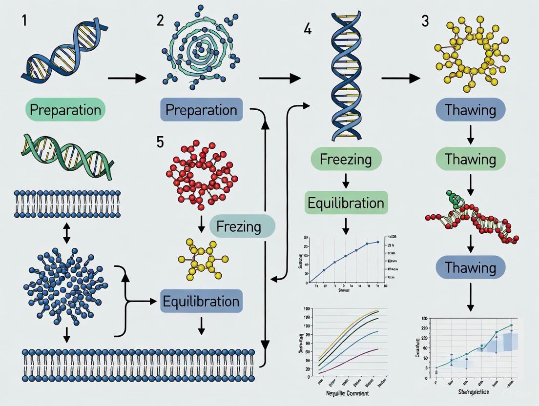
The following diagram illustrates the sequential protective mechanisms and the critical workflow during the slow freezing process.
Quantitative Data on Cryopreservation Solutions
The formulation of the cryopreservation medium is a critical determinant of post-thaw outcomes. Research has evaluated various combinations of penetrating and non-penetrating CPAs, with and without antioxidant supplements, to optimize ADSC recovery.
Table 1: Comparison of Cryopreservation Solution Efficacy on ADSC Recovery
| Cryoprotectant Formulation | Post-Thaw Viability (%) | Key Functional Outcomes | Study Model |
|---|---|---|---|
| 10% DMSO + 90% FBS (Conventional) | ~79% [3] | Maintained immunophenotype (CD73, CD90, CD105) and adipogenic potential after decade-long storage; some reduction in osteogenic gene expression [3]. | Human ADSCs [3] |
| Trehalose + Glycerol (TG) | Not explicitly quantified | Provided a non-toxic, DMSO-free base; required intracellular delivery for maximum efficacy [5] [4]. | Human Adipose Tissue [5] |
| Trehalose + Glycerol + 2mM Metformin (TGM) | Significantly higher than TG and DF groups [5] | Lowest ROS level (29.20 ± 1.73); highest tissue retention rate and structural integrity in vivo; reduced SVF apoptosis [5]. | Human Adipose Tissue / Nude Mouse [5] |
| 5% DMSO + 3% Trehalose + 2% PEG + 2% BSA (FBS-free) | High viability and recovery [2] | Effectively preserved bADSC metabolic activity and clonogenicity while minimizing oxidative stress and apoptosis [2]. | Buffalo ADSCs [2] |
| Bambanker (Serum-Free Commercial Medium) | >90% [6] | Preserved spindle-shaped morphology, surface markers (CD29, CD90), and trilineage differentiation potential, though with slight reduction in cardiomyogenic differentiation [6]. | Rat AD-MSCs [6] |
Table 2: Impact of Cryopreservation Sequence on Genetically Modified ADSCs
| Processing Sequence | Cell Number | BMP-2 Production | Osteogenic Potential (Alizarin Red) |
|---|---|---|---|
| Group 1: No freezing (Transduction without freezing) | Baseline | Baseline | Baseline [7] |
| Group 2: Freeze, then transduce (Cells frozen at P1, thawed & transduced at P3) | Equivalent to Group 1 | Trend similar to Group 1 | Higher than Group 3; No difference from Group 1 [7] |
| Group 3: Transduce, then freeze (Cells transduced at P3, then frozen) | Equivalent to Group 1 | Trend toward decrease | Lower than Group 2 [7] |
Detailed Experimental Protocols
Standardized Slow Freezing Protocol for Human ADSCs
This protocol is adapted from established methodologies used in recent research [5] [3] [1].
I. Pre-freezing: Cell Harvest and CPA Addition
- Cell Harvesting: Culture expand ADSCs to the desired passage (typically P2-P3). Harvest cells using 0.25% trypsin-EDTA digestion. Inactivate trypsin with a complete culture medium (e.g., DMEM/F12 supplemented with 10% FBS) [8] [3].
- Cell Counting and Centrifugation: Count the cells using an automated cell counter or hemocytometer with Trypan Blue exclusion to assess initial viability. Centrifuge the cell suspension at 300-400 g for 5-10 minutes. Discard the supernatant [8] [7].
- CPA Resuspension: Resuspend the cell pellet in the pre-chilled (4°C) cryopreservation solution at a concentration of 1-5 x 10^6 cells/mL. A common and effective solution is 10% DMSO in FBS, though serum-free commercial media like Bambanker or custom formulations (e.g., containing trehalose and glycerol) are valid alternatives [3] [6] [7].
- Aliquoting and Equilibration: Aliquot 1 mL of the cell suspension into sterile cryovials. Keep the vials at 4°C for 30 minutes to allow for CPA equilibration [5].
II. Controlled-Rate Freezing
- Use a Programmable Freezer or "Mr. Frosty": Place the cryovials into a pre-cooled isopropanol freezing chamber (e.g., "Mr. Frosty") or a programmable rate-controlled freezer.
- Slow Freezing Cycle:
III. Long-Term Storage After 24 hours at -80°C, promptly transfer the cryovials to the vapor or liquid phase of a liquid nitrogen tank (-150°C to -196°C) for long-term storage [3] [1].
IV. Thawing and CPA Removal
- Rapid Thawing: Retrieve the vial from liquid nitrogen and immediately place it in a 37°C water bath with gentle agitation until only a small ice crystal remains (approximately 1-2 minutes) [3] [1].
- Decontamination: Wipe the outside of the vial with 70% ethanol before opening.
- Gradual Dilution: Transfer the thawed cell suspension to a centrifuge tube containing 10 mL of pre-warmed complete culture medium. Add the medium drop-wise while gently shaking the tube to gradually reduce the extracellular CPA concentration and prevent osmotic shock [3] [7].
- Centrifugation and Washing: Centrifuge the cell suspension at 300-400 g for 5 minutes. Discard the supernatant containing the diluted CPA.
- Resuspension and Plating: Resuspend the cell pellet in fresh complete culture medium. Plate the cells at the desired density for subsequent experiments or further expansion [7].
Protocol: Evaluating a Novel TGM Cryopreservation Solution
This protocol is based on a 2025 study investigating a solution of Trehalose, Glycerol, and Metformin for adipose tissue cryopreservation [5].
I. Solution Preparation
- Prepare the base cryopreservation solution containing 1 M trehalose and 20% glycerol in a physiological buffer (e.g., PBS).
- Supplement this base solution (TG) with 2 mM metformin to create the TGM solution. This concentration was identified as optimal for minimizing ROS [5].
II. Tissue Processing and Cryopreservation
- Adipose Tissue Harvest: Obtain human adipose tissue from liposuction aspirates. Wash the tissue extensively with PBS and centrifuge to remove blood, oil, and debris.
- Mixing with CPA: Aliquot 1.5 mL of purified adipose tissue into cryovials. Add an equal volume (1.5 mL) of the TGM cryopreservation solution. Mix uniformly and leave at room temperature for 10 minutes for equilibration.
- Slow Freezing: Follow the controlled-rate freezing protocol detailed in Section 4.1 (II), transferring vials to liquid nitrogen after 24 hours at -80°C for storage [5].
III. Thawing and Analysis
- Thawing and Washing: Thaw samples rapidly in a 37°C water bath. Slowly add PBS to the cryopreserved tissue for washing. Centrifuge the mixture at 1000 rpm for 3 min, remove the liquid layer, and retain the middle adipose layer. Repeat 2-3 times.
- Viability and Function Assessment:
- ROS Assay: Isolate the Stromal Vascular Fraction (SVF) and measure intracellular ROS levels using a fluorescent probe like DCFH-DA and a microplate reader.
- Apoptosis Assay: Analyze SVF cells for apoptosis via flow cytometry (e.g., Annexin V/PI staining).
- In Vivo Transplantation: Transplant 1 mL of thawed adipose tissue subcutaneously into immunodeficient mice. Analyze tissue retention rates and histological integrity after one month [5].
The protective mechanism of an optimized solution containing metformin involves the activation of specific cytoprotective signaling pathways, as visualized below.
The Scientist's Toolkit: Essential Research Reagents
A successful slow freezing experiment requires a carefully selected set of reagents and tools. The table below catalogs key solutions and their specific functions in the cryopreservation workflow.
Table 3: Essential Reagents for Slow Freezing Cryopreservation of ADSCs
| Reagent / Solution | Function in Protocol | Example Formulation / Notes |
|---|---|---|
| Intracellular CPA | Penetrates cell, depresses freezing point, reduces intracellular ice formation. | DMSO (10%): Gold standard, but cytotoxic [4]. Glycerol (20%): Less toxic, often used in combination [5]. |
| Extracellular CPA | Stabilizes cell membrane externally, creates osmotic gradient for dehydration. | Trehalose (1-3%): Requires delivery methods for intracellular effect [5] [2]. Sucrose: Common non-penetrating sugar [1]. |
| Antioxidant Supplement | Scavenges ROS, reduces oxidative stress-induced apoptosis during freezing. | Metformin (2mM): Identified as optimal concentration in TGM solution [5]. |
| Serum / Protein Base | Provides undefined growth factors and proteins, enhances membrane stability. | Fetal Bovine Serum (FBS, 90%): Common base; risk of xenogenic reactions [3]. Bovine Serum Albumin (BSA, 2%): Defined protein source, used in FBS-free media [2]. |
| Basal Freezing Medium | The isotonic base solution for the cryopreservation cocktail. | University of Wisconsin (UW) Solution: Used in hypothermic and supercooling preservation [9]. Plasma-Lyte A: Base for some DMSO-free formulations [2]. |
| Commercial Serum-Free Medium | Ready-to-use, defined formulation, eliminates batch variability and safety concerns of serum. | Bambanker: Enables storage at -80°C without programmable freezer [6] [7]. |
| Collagenase Type I | Enzymatically digests adipose tissue to isolate the Stromal Vascular Fraction (SVF) and ADSCs. | 0.075% - 0.1% solution: Standard concentration for tissue digestion [5] [3]. |
Cryopreservation is a cornerstone technique for the long-term storage of adipose-derived stem cells (ASCs), which are vital for regenerative medicine and cell-based therapies [10] [11]. The process enables the creation of cell banks, facilitates transportation, and allows time for quality control testing [12]. However, the slow freezing protocol, a standard method for ASC cryopreservation, exposes cells to significant cryodamage, which can compromise their viability, functionality, and therapeutic potential post-thaw [10] [13]. This damage primarily manifests in three forms: osmotic stress, mechanical injury from ice crystals, and oxidative stress [10]. These interconnected pathways of injury can lead to cell death, reduced proliferation, impaired differentiation capacity, and altered phenotype [12]. Understanding the mechanisms underlying these damage pathways is therefore critical for developing robust cryopreservation protocols. This application note details the sources of cryodamage and provides validated, quantitative methodologies to identify and mitigate these stresses, ensuring the high quality of ASCs following slow freezing.
Pathways of Cryodamage: Mechanisms and Identification
The cryopreservation process, particularly during the freezing and thawing phases, subjects cells to a series of physical and chemical stresses. The following diagram illustrates the three primary interconnected pathways of cryodamage and their impact on ASCs.
Osmotic Damage
During slow freezing, extracellular ice formation occurs first, leaving behind a hypertonic solution of unfrozen cryoprotectants and salts [10] [13]. This creates a steep osmotic gradient that drives water out of the cell, leading to profound cell shrinkage and dehydration [10]. This excessive shrinkage can cause irreversible damage to the cell membrane and cytoskeleton, a process known as "solution effects" damage [13]. The subsequent thawing process, if not controlled, can cause a rapid influx of water, leading to swelling and potential membrane rupture.
Key Quantitative Markers:
- Cell Volume Change: Measured via flow cytometry or cell sizing instruments. A post-thaw recovery of cell volume to >85% of pre-freeze levels within 60 minutes is indicative of good osmotic resilience [12].
- Viability: Osmotic stress directly induces apoptosis and necrosis. Viability can be assessed using trypan blue exclusion or flow cytometry with Annexin V/propidium iodide (PI) staining. A viability of >80% immediately post-thaw is a common benchmark, though this can recover to >90% after a 24-hour culture period [12] [3].
Mechanical Damage
Mechanical damage is primarily inflicted by the formation and growth of ice crystals. During slow freezing, if the cooling rate is too rapid for water to exit the cell, intracellular ice formation (IIF) occurs [10] [13]. These sharp, intracellular ice crystals can physically pierce and disrupt organelles and the plasma membrane, leading to immediate cell lysis [10]. During the thawing phase, small ice crystals can recrystallize into larger, more damaging structures, exacerbating the injury.
Key Quantitative Markers:
- Immediate Post-Thaw Lysis: A sharp decline in viability measured immediately (0-2 hours) post-thaw is often attributable to mechanical damage from IIF. This can be quantified by a high PI-positive population in flow cytometry [12].
- Membrane Integrity: The integrity of the plasma membrane and intracellular organelles can be assessed via lactate dehydrogenase (LDH) release assays or fluorescent probes that detect compromised membranes.
Oxidative Damage
The cryopreservation process itself, combined with the ischemia-reperfusion-like injury during thawing, triggers a burst of reactive oxygen species (ROS) [10]. This oxidative stress can overwhelm the cell's antioxidant defenses, leading to the oxidation of lipids (peroxidation of membrane lipids), proteins (denaturation and loss of enzymatic function), and DNA (strand breaks and mutations) [10]. This damage may not be immediately lethal but can manifest as reduced proliferative capacity, accelerated senescence, and impaired differentiation potential after thawing [12].
Key Quantitative Markers:
- Intracellular ROS Levels: Measured using fluorescent probes like H2DCFDA and analyzed via flow cytometry or fluorescence microscopy. Levels can be 2-3 times higher in cryopreserved cells compared to fresh controls [10].
- Lipid Peroxidation: Quantified by measuring malondialdehyde (MDA) levels using assays like the thiobarbituric acid reactive substances (TBARS) assay.
- Functional Assays: Long-term proliferation assays (e.g., CFU-F efficiency) and differentiation assays (osteogenic and adipogenic potential) are critical for assessing the functional consequences of oxidative stress [12] [3].
Table 1: Key Assays for Quantifying Cryodamage in ASCs
| Damage Type | Key Assays | Measurement Output | Benchmark for Healthy ASCs |
|---|---|---|---|
| Osmotic | Cell Volume Analysis | Volume recovery kinetics & final volume | >85% volume recovery within 60 min [12] |
| Annexin V/PI Staining | Early apoptosis (Annexin V+/PI-) & necrosis (PI+) | Viability >80% post-thaw; >90% after 24h [12] [3] | |
| Mechanical | Propidium Iodide (PI) Uptake | % of cells with ruptured membranes (lysed) | <20% PI+ cells immediately post-thaw [12] |
| LDH Release Assay | Amount of cytosolic enzyme in supernatant | Low LDH release relative to total lysis control | |
| Oxidative | DCFDA ROS Assay | Fluorescence intensity of oxidized probe | <2x increase vs. fresh control [10] |
| TBARS Assay | Concentration of Malondialdehyde (MDA) | Lower MDA levels relative to unprotected controls | |
| Colony-Forming Unit (CFU-F) | Number of colonies formed after 14 days | Minimal reduction compared to fresh ASCs [12] [3] |
Experimental Protocols for Assessment
This section provides detailed, step-by-step protocols for a comprehensive assessment of ASC quality after cryopreservation.
Protocol 1: Post-Thaw Viability and Apoptosis Analysis
This protocol is critical for quantifying immediate osmotic and mechanical damage.
Workflow: Viability and Apoptosis Analysis
Materials:
- Cryopreserved ASC vial
- Water bath (37°C)
- Complete growth medium (e.g., DMEM/F12 + 10% FBS)
- Phosphate Buffered Saline (PBS)
- Trypan Blue solution (0.4%)
- Annexin V-FITC/PI Apoptosis Detection Kit
- Automated cell counter or hemocytometer
- Flow cytometer
Procedure:
- Thawing: Rapidly thaw the cryovial in a 37°C water bath with gentle agitation until only a small ice crystal remains (~1-2 minutes) [14] [12].
- CPA Removal: Transfer the cell suspension to a 15 mL conical tube. Slowly add 9 mL of pre-warmed complete growth medium drop-wise to dilute the cytotoxic DMSO. Centrifuge at 300g for 5 minutes [14] [12].
- Resuspension: Carefully decant the supernatant and resuspend the cell pellet in 1 mL of PBS.
- Cell Counting: Mix 20 µL of cell suspension with 20 µL of Trypan Blue. After 1-2 minutes, load onto a hemocytometer and count live (unstained) and dead (blue) cells using an automated counter or manually. Calculate viability: % Viability = (Live Cells / Total Cells) × 100.
- Apoptosis Staining: According to the kit manufacturer's instructions, stain approximately 1×10^5 cells in 100 µL binding buffer with Annexin V-FITC and PI. Incubate for 15 minutes in the dark. Add 400 µL of binding buffer and analyze within 1 hour using a flow cytometer.
- Analysis: distinguish populations: Viable (Annexin V-/PI-), Early Apoptotic (Annexin V+/PI-), Late Apoptotic/Necrotic (Annexin V+/PI+).
Protocol 2: Functional Recovery: Proliferation and Clonogenicity
This protocol assesses long-term functional recovery from oxidative and other cumulative damage.
Materials:
- Thawed and washed ASCs (from Protocol 1)
- Complete growth medium
- 96-well and 6-well cell culture plates
- Cell Counting Kit-8 (CCK-8) or MTS reagent
- Crystal Violet or Giemsa stain
Procedure:
- Proliferation (CCK-8 Assay):
- Seed cells in a 96-well plate at a density of 5,000 cells/well in 100 µL medium. Include background control wells with medium only [15].
- At 24, 48, and 72 hours, add 10 µL of CCK-8 solution directly to each well. Incubate the plate for 2-4 hours at 37°C.
- Measure the absorbance at 450 nm using a microplate reader. Plot the optical density (OD) values over time to generate a growth curve.
- Clonogenic (CFU-F) Assay:
- Seed low-density cells in 6-well plates (100-500 cells/well, depending on cell line) and culture for 14 days, changing the medium every 3-4 days [12].
- After 14 days, remove the medium, wash with PBS, and fix the cells with 4% paraformaldehyde for 15 minutes.
- Stain with 0.5% Crystal Violet for 30 minutes at room temperature.
- Gently rinse with water to remove excess stain and air-dry the plates.
- Count colonies containing >50 cells. Calculate CFU-F efficiency: (Number of Colonies / Number of Cells Seeded) × 100.
Protocol 3: Multi-Lineage Differentiation Potential
This protocol validates the retention of stemness, which is sensitive to oxidative and other cryodamage.
Materials:
- Thawed ASCs
- Osteogenic Differentiation Medium (e.g., DMEM + 10% FBS, 0.1 µM Dexamethasone, 10 mM β-glycerophosphate, 50 µM Ascorbate-2-phosphate)
- Adipogenic Differentiation Medium (e.g., DMEM + 10% FBS, 1 µM Dexamethasone, 0.5 mM IBMX, 10 µg/mL Insulin, 200 µM Indomethacin)
- 4% Paraformaldehyde (PFA)
- Alizarin Red S solution (for osteogenesis)
- Oil Red O solution (for adipogenesis)
Procedure:
- Differentiation Induction: Culture ASCs in 12-well plates until 100% confluent. Replace the growth medium with either osteogenic or adipogenic induction medium. Maintain cultures for 21 (osteogenesis) or 14 (adipogenesis) days, refreshing the differentiation medium every 3-4 days [14] [3].
- Osteogenic Staining (Alizarin Red S):
- Aspirate medium, wash with PBS, and fix with 4% PFA for 15 minutes.
- Wash with distilled water and incubate with 2% Alizarin Red S solution (pH 4.2) for 30-45 minutes at room temperature, protected from light.
- Wash extensively with distilled water until the background is clear. Image the orange-red mineralized nodules.
- Adipogenic Staining (Oil Red O):
- Aspirate medium, wash with PBS, and fix with 4% PFA for 15 minutes.
- Wash with 60% isopropanol and let air dry completely.
- Incubate with filtered Oil Red O working solution for 30-60 minutes.
- Wash with distilled water to remove excess stain. Image the red lipid droplets. For quantification, elute the stain with 100% isopropanol and measure absorbance at 520 nm [14].
The Scientist's Toolkit: Reagents & Materials
Table 2: Essential Reagents for Cryopreservation and Quality Control of ASCs
| Category | Reagent/Material | Function & Rationale | Example Protocol Usage |
|---|---|---|---|
| Cryoprotectants | Dimethyl Sulfoxide (DMSO) | Permeable CPA; reduces intracellular ice formation but is cytotoxic [10] [16]. | 10% final concentration in FBS [12]. |
| Glycerol | Permeable CPA; less toxic than DMSO, stabilizes cell membrane [15]. | 20% combined with Trehalose [15]. | |
| Trehalose | Non-permeable CPA; stabilizes membranes via water replacement; non-toxic [15]. | 1.0 M combined with Glycerol [15]. | |
| STEM-CELLBANKER | Commercial, defined, xeno-free CPA; reduces DMSO-related toxicity [16]. | Used as a direct replacement for DMSO/FBS [16]. | |
| Culture Media | Fetal Bovine Serum (FBS) | Provides nutrients, growth factors, and proteins that mitigate osmotic shock. | 90% in CPA; 10% in growth medium [12]. |
| Serum-Free Medium | Xeno-free alternative for clinical applications; requires optimized CPA cocktails. | For thawed cell culture post-wash. | |
| Viability Assays | Trypan Blue | Dye exclusion test for membrane integrity; rapid viability assessment. | 1:1 mix with cells for counting [14] [3]. |
| Annexin V/Propidium Iodide (PI) | Distinguishes viable, apoptotic, and necrotic cell populations via flow cytometry. | Staining for 15 min in binding buffer [12]. | |
| Functional Assays | Cell Counting Kit-8 (CCK-8) | Colorimetric assay based on metabolic activity to measure proliferation. | 10 µL added to wells; incubate 2-4h [15]. |
| Crystal Violet | Stains cell nuclei; used for counting colonies in CFU-F assays. | 0.5% solution, stain for 30 min [12]. | |
| Differentiation Kits | Osteogenic Induction Kit | Provides defined components (Dexamethasone, β-glycerophosphate, Ascorbate) for bone differentiation. | Medium changes every 3-4 days for 21 days [3]. |
| Adipogenic Induction Kit | Provides defined components (IBMX, Indomethacin, Insulin, Dexamethasone) for fat differentiation. | Medium changes every 3-4 days for 14 days [14]. |
Advanced Mitigation Strategies
Beyond standard protocols, several advanced strategies can further mitigate cryodamage.
Table 3: Advanced Strategies for Mitigating Specific Cryodamage Pathways
| Strategy | Mechanism of Action | Protocol Application | Evidence of Efficacy |
|---|---|---|---|
| CPA Cocktails (Trehalose + Glycerol) | Combines membrane-stabilizing effects of glycerol with glass-forming and membrane-protecting effects of trehalose [15]. | Replace 10% DMSO with 1.0 M Trehalose + 20% Glycerol in PBS. Slow freeze at -1°C/min [15]. | Post-thaw viability similar to DMSO, but with significantly higher migration capacity and reduced toxicity [15]. |
| Macromolecular Cryoprotectants (e.g., Polyampholytes, PVA) | Mimic antifreeze proteins; inhibit ice recrystallization during thawing and modify ice crystal morphology [17] [13]. | Add 7.5% carboxylated poly-L-lysine (COOH-PLL) to standard freezing medium [13]. | Viability increased from 71.2% to 95.4% for MSCs compared to 10% DMSO alone [13]. |
| Antioxidant Supplementation | Scavenges ROS generated during freezing/thawing, reducing oxidative damage to lipids, proteins, and DNA. | Add antioxidants (e.g., Ascorbic Acid, N-Acetylcysteine) to the pre-freeze culture medium and/or the post-thaw recovery medium. | Mitigates senescence and preserves differentiation potential post-thaw [10]. |
| Controlled Rate Freezing | Ensures optimal, reproducible cooling rate (-1°C/min), allowing water to leave cells before IIF occurs [10] [13]. | Use a programmable freezer or a passive cooling device (e.g., "Mr. Frosty") filled with isopropanol [12]. | Standardizes the process and significantly improves consistency and post-thaw recovery versus uncontrolled freezing [12]. |
The successful cryopreservation of Adipose-Derived Stem Cells via slow freezing is contingent upon a detailed understanding and proactive mitigation of osmotic, mechanical, and oxidative stress. By employing the quantitative assessment protocols outlined in this document—ranging from immediate viability and apoptosis checks to long-term functional assays for proliferation and differentiation—researchers can accurately benchmark the quality of their cryopreserved products. Furthermore, adopting advanced strategies, such as using less toxic CPA cocktails like trehalose-glycerol or incorporating macromolecular ice inhibitors, can significantly elevate post-thaw cell recovery and functionality. Integrating these rigorous assessment and mitigation workflows is essential for ensuring that cryopreserved ASCs meet the stringent quality standards required for both foundational research and clinical applications in regenerative medicine.
Cryopreservation is a cornerstone technology for the long-term storage of biologics, including adipose-derived stem cells (ASCs), which are critical for regenerative medicine and research [18]. The success of slow-freezing protocols is highly dependent on cryoprotectant agents (CPAs) that mitigate freezing-induced damage. For decades, dimethyl sulfoxide (DMSO) has been the predominant penetrating CPA employed due to its efficacy. However, concerns over its toxicity have spurred research into safer alternatives, particularly non-penetrating agents like trehalose [4] [19]. This Application Note details the mechanisms of DMSO and non-penetrating alternatives, providing structured data and protocols framed within slow-freezing protocols for ASCs.
Mechanisms of Action
Penetrating Cryoprotectant: Dimethyl Sulfoxide (DMSO)
DMSO is a small, amphipathic molecule that readily crosses cell membranes. Its primary mechanism of action during slow freezing is to prevent intracellular ice crystal formation, which is lethal to cells [4]. As the extracellular solution freezes, water is sequestered as ice, thereby concentrating the solutes in the remaining liquid. This creates an osmotic gradient that draws water out of the cell, preventing intracellular freezing but risking harmful cell shrinkage. DMSO permeates the cell, equalizing osmotic pressures across the membrane and reducing the extent of dehydration. Furthermore, DMSO interacts with water and membrane phospholipids, stabilizing the cell membrane against the mechanical stresses of freeze-concentration and phase transitions [20] [17].
Despite its effectiveness, DMSO induces concentration- and temperature-dependent toxicities. It can cause mitochondrial damage, alter chromatin conformation in fibroblasts, and induce unwanted differentiation in stem cells [18]. In clinical applications, the administration of DMSO-cryopreserved cell products has been associated with adverse reactions, including cardiac, neurological, and gastrointestinal effects [18] [19]. These drawbacks necessitate post-thaw washing steps, which can lead to significant cell loss and introduce logistical complexities [18].
Non-Penetrating Cryoprotectants: Trehalose and Other Agents
Non-penetrating CPAs are typically large molecules or sugars that cannot cross the lipid bilayer. Their mechanism is extracellular and multifaceted, based on several key principles:
- Osmotic Regulation ("Solution Effects" Damage Mitigation): Like DMSO, extracellular CPAs like trehalose and sucrose increase the solute concentration of the external solution during freezing. This promotes a more controlled, osmotic dehydration of the cell, minimizing intracellular ice formation [20].
- Glass Transition (Vitrification): At high concentrations, these solutes can form an amorphous, glassy state upon cooling instead of crystallizing. This glass is highly viscous, halting all biochemical reactions and mechanical damage from ice crystals [4] [17].
- Water Replacement and Membrane Stabilization: Trehalose, a disaccharide found in extremophiles, has a unique "clam-shaped" conformation that allows it to replace water molecules around phospholipid heads and proteins during dehydration. It forms hydrogen bonds, maintaining the structural integrity of the membrane and proteins in the dry or frozen state [4].
A significant challenge with trehalose is the innate impermeability of the mammalian plasma membrane to it. Therefore, to act as an intracellular CPA, delivery strategies such as electroporation, nanoparticle-mediated delivery, or prolonged incubation are required for maximum efficacy [4].
The diagram below illustrates the collaborative mechanisms of penetrating and non-penetrating CPAs in protecting a cell during the slow-freezing process.
The following tables summarize key quantitative data on the performance of DMSO and non-penetrating alternatives in the cryopreservation of adipose-derived cells and tissues.
Table 1: Efficacy of DMSO and Trehalose in Adipose Tissue Cryopreservation
| Cryoprotectant Solution | Cell/Tissue Type | Post-Thaw Viability / Outcome | Key Findings | Source |
|---|---|---|---|---|
| 10% DMSO + FBS | Human Adipose Tissue | Baseline for comparison | Standard protocol, but requires washing and carries toxicity risks. | [5] |
| 1M Trehalose + 20% Glycerol | Human ADSCs | High preservation efficiency | A non-toxic, serum-free alternative with acceptable outcomes. | [18] [4] |
| Trehalose + Glycerol + Metformin (TGM) | Human Adipose Tissue | Lowest apoptosis; highest in vivo retention | Superior to TG and DMSO+FBS groups; robust structural integrity. | [5] |
| Combined Trehalose & DMSO | Human Adipose Tissue | >80% viability | Combination effective; antigen expression levels close to fresh cells. | [4] |
Table 2: Advanced & Emerging Low-DMSO Cryopreservation Strategies
| Strategy | CPA Composition | Cell Type | Post-Thaw Viability / Recovery | Key Findings | Source |
|---|---|---|---|---|---|
| Polyampholyte Polymer | 2.5% DMSO + 20 mg/mL Polyampholyte | hBM-MSCs | 76% Viability, 30% Recovery | Rescued viability/recovery from <50% and 17% with 2.5% DMSO alone. | [21] |
| Hydrogel Microencapsulation | 2.5% DMSO in Alginate Microcapsule | hUC-MSCs | >70% Viability | 3D encapsulation mitigates cryoinjury, enabling low-DMSO use. | [22] |
| Slow Vitrification | 6.5M EG, 0.5M Sucrose, 10% COOH-PLL | Human MSC Monolayers | Significantly improved viability | High CPA concentrations enable ice-free state with less apoptosis. | [18] |
Detailed Experimental Protocols
Protocol 1: Slow-Freezing Cryopreservation of Adipose-Derived Stem Cells (ASCs) Using Low-DMSO Polyampholyte Solution
This protocol is adapted from a study demonstrating high post-thaw MSC viability with only 2.5% DMSO supplemented with a synthetic polyampholyte [21].
Research Reagent Solutions:
| Reagent/Material | Function / Explanation |
|---|---|
| Polyampholyte Polymer (e.g., synthesized from poly(methyl vinyl ether-alt-maleic anhydride) and dimethylamino ethanol) | Synthetic macromolecular cryoprotectant; believed to stabilize cell membranes and provide cryoprotection synergistically with low DMSO. |
| Dimethyl Sulfoxide (DMSO) | Penetrating cryoprotectant. The objective is to reduce its concentration to 2.5% (v/v). |
| Stromal Medium (e.g., DMEM/F-12 with 10% FBS) | Base medium for cell suspension and post-thaw recovery culture. |
| Programmable Freezer or "Mr. Frosty" | To achieve a controlled cooling rate of -1°C/min. |
Methodology:
- Cell Preparation: Harvest and count ASCs. Use a high cell density of 5 x 10^5 cells/mL for cryopreservation.
- CPA Preparation: Prepare the cryopreservation solution on ice. The final solution should contain:
- 2.5% (v/v) DMSO
- 20 mg/mL Polyampholyte Polymer
- Made up in Stromal Medium
- Loading: Gently resuspend the ASC pellet in the prepared CPA solution. Aliquot the cell suspension into cryovials.
- Slow Freezing: Place the cryovials in a rate-controlled freezer programmed to cool at -1°C/min to -80°C. Alternatively, use a "Mr. Frosty" freezing container filled with isopropanol and place it at -80°C for 24 hours.
- Long-Term Storage: After 24 hours, transfer the cryovials to a liquid nitrogen tank for long-term storage.
- Thawing: Rapidly thaw the cells by placing the cryovial in a 37°C water bath with gentle agitation for 1-2 minutes.
- Post-Thaw Analysis: Immediately after thawing, dilute the CPA 1:10 with pre-warmed stromal medium. Centrifuge at 300 g for 5 minutes to remove the CPA. Resuspend the cell pellet in fresh medium and plate for analysis. Note: A 24-hour recovery period is recommended before assessing true post-thaw viability and functionality [21].
Protocol 2: DMSO-Free Cryopreservation of Adipose Tissue Using Trehalose-Glycerol-Metformin (TGM)
This protocol is derived from a 2025 study investigating a novel, non-toxic cryopreservation solution for intact adipose tissue [5].
Research Reagent Solutions:
| Reagent/Material | Function / Explanation |
|---|---|
| Trehalose (1M) | Non-penetrating cryoprotectant; provides extracellular protection via osmotic regulation and glass formation. |
| Glycerol (20%) | Penetrating cryoprotectant; works synergistically with trehalose. Safe for humans at low concentrations. |
| Metformin (2mM) | Antioxidant; reduces freezing-induced oxidative stress and apoptosis by activating the AMPK/Nrf2 pathway. |
| Phosphate Buffered Saline (PBS) | Solvent for preparing the TGM cryopreservation solution. |
| Gradient Cooling Cassette | Device filled with isopropanol to ensure a reproducible, controlled cooling rate. |
Methodology:
- Tissue Harvesting and Preparation: Purify human lipoaspirate by washing with PBS and centrifugation at 1200 g for 3 minutes. Repeat 3-5 times to remove oil and debris. Retain the purified middle layer of adipose tissue.
- CPA Preparation: Prepare the TGM cryopreservation solution in PBS, containing:
- 1 M Trehalose
- 20% Glycerol
- 2 mM Metformin (identified as the optimal concentration for reducing ROS)
- Loading and Equilibration: Aliquot 1.5 mL of purified adipose tissue into a cryovial. Add an equal volume (1.5 mL) of TGM solution, mix uniformly, and leave at room temperature for 10 minutes for equilibration.
- Controlled Slow Freezing:
- Place cryovials in a gradient cooling cassette at 4°C for 30 minutes.
- Transfer to -20°C for 4 hours.
- Transfer to -80°C for 24 hours.
- Long-Term Storage: Transfer the vials to liquid nitrogen for long-term storage (e.g., 2 weeks or more).
- Thawing and Washing: Retrieve vials from LN2 and thaw rapidly in a 37°C water bath for 5 minutes. Wash the thawed tissue 2-3 times with PBS to remove the CPAs by gentle centrifugation (1000 rpm for 3 min). The tissue is now ready for transplantation or downstream analysis.
The following workflow diagram summarizes the key steps of this TGM-based protocol.
Implementation in Research
Integrating these CPA strategies requires careful consideration. For research where minimizing DMSO is critical, Polyampholyte-supplemented low-DMSO protocols are highly effective for ASCs in suspension [21]. For applications involving intact adipose tissue fragments, the TGM (Trehalose-Glycerol-Metformin) solution provides a potent, DMSO-free option that mitigates oxidative stress [5]. Hydrogel microencapsulation presents another versatile strategy, physically protecting cells and enabling a reduction of DMSO to 2.5% while maintaining viability above the 70% clinical threshold [22].
Long-term cryopreservation studies (over 10 years) using standard DMSO protocols show that ASCs largely retain their immunophenotype and adipogenic potential, though some negative impact on osteogenic potential has been observed [3]. This underscores the importance of not only viability but also functional recovery as key metrics for evaluating any new CPA formulation. The field is moving towards a DMSO-free preservation era, supported by commercially available solutions, though these require further independent validation across a wider range of biologics [18].
The Critical Role of Cooling Rates and the 'Gold Standard' of -1°C/min
For researchers in regenerative medicine and drug development, the cryopreservation of adipose-derived stem cells (ASCs) presents a critical challenge: balancing high post-thaw viability with the retention of essential biological functions. The cooling rate during freezing represents one of the most fundamental process parameters influencing cryopreservation success. While the rate of -1°C/min is frequently described as a "gold standard" in cryopreservation protocols, its applicability must be validated against specific cell types and experimental conditions. This Application Note examines the scientific basis for controlled cooling rates in ASC cryopreservation, provides validated protocols, and presents comparative data to guide research and development workflows.
Scientific Rationale: The Thermodynamic Basis of Controlled Cooling
The fundamental challenge in cryopreservation lies in navigating the physical transitions of water as temperatures fall below freezing. During slow cooling, the extracellular solution freezes first, increasing solute concentration outside the cell and creating an osmotic gradient that draws water out through the membrane. This gradual cellular dehydration minimizes the lethal formation of intracellular ice crystals [23] [1]. If cooling occurs too rapidly, water cannot exit the cell quickly enough, leading to intracellular ice formation (IIF), which is typically fatal to cells [1]. Conversely, excessively slow cooling prolongs exposure to hypertonic conditions, causing solution effects injury from concentrated solutes and excessive cell volume reduction [23].
The cooling rate of -1°C/min has emerged as a benchmark for many cell types because it optimally balances these competing risks. This rate allows sufficient time for cellular dehydration while minimizing both IIF and toxic solute exposure. A recent study on sheep spermatogonial stem cells confirmed that a cooling profile beginning at 1°C/min from 0°C to -10°C resulted in significantly greater post-thaw viability (79.64%) and stemness activity compared to faster cooling profiles [23].
Table 1: Comparison of Cooling Rate Impacts on Stem Cell Cryopreservation Outcomes
| Cooling Rate | Post-Thaw Viability | Key Advantages | Primary Risks | Recommended Cell Types |
|---|---|---|---|---|
| -1°C/min (Slow freezing) | 70-80% [1] | Minimizes intracellular ice formation; preserves differentiation potential [3] | Cellular dehydration; osmotic stress | ASCs, MSCs, Spermatogonial Stem Cells [3] [23] |
| > -50°C/min (Vitrification) | Variable | Ultra-rapid cooling prevents ice crystallization | CPA toxicity; requires high CPA concentrations; sample volume limitations [1] | Oocytes, Embryos |
| Uncontrolled (Passive freezing) | Lower than controlled-rate | Simple, low-cost, easy to scale [24] | No control over critical process parameters; inconsistent outcomes [24] | Early-stage clinical products |
Establishing the -1°C/min Protocol for Adipose-Derived Stem Cells
Experimental Evidence for ASC Cryopreservation
The viability of the -1°C/min standard is supported by a decade-long study on human ASCs cryopreserved using this exact cooling rate. Researchers reported mean post-thaw viability of 78-79% even after 10+ years of storage, with immunophenotype characterization and adipogenic differentiation potential remaining largely intact compared to fresh ASCs [3]. While some variations in osteogenic gene expression were observed, the core stem cell properties were effectively preserved, demonstrating the protocol's effectiveness for long-term biobanking [3].
Detailed Step-by-Step Protocol
Materials and Equipment:
- Cultured ASCs (Passage 2-4)
- Cryoprotective Agent: 10% DMSO in FBS
- Cryovials
- Programmable controlled-rate freezer OR isopropanol-based freezing container
- Liquid nitrogen storage tank
Procedure:
- Cell Preparation: Harvest ASCs at 80-90% confluence using standard trypsin/EDTA digestion. Centrifuge at 300g for 5 minutes and resuspend in stromal medium for counting [3].
- CPA Addition: Prepare cryopreservation medium (10% DMSO in FBS). Suspend cells at 1×10⁶ cells/mL in cryopreservation medium and aliquot 1 mL into each cryovial [3] [8].
- Equilibration: Incubate filled cryovials on ice for 15-30 minutes to allow CPA permeation.
- Controlled-Rate Freezing: Place cryovials in a programmed freezing apparatus. Implement the following cooling profile:
- Liquid Nitrogen Storage: Transfer cryovials to liquid nitrogen vapor phase (-135°C to -196°C) for long-term storage.
- Thawing Protocol: Rapidly warm cryovials in a 37°C water bath with gentle agitation until only a small ice crystal remains (approximately 1-2 minutes) [3]. Immediately dilute the CPA by adding pre-warmed stromal medium dropwise (1:10 dilution ratio). Centrifuge at 300g for 5 minutes to remove CPA and resuspend in fresh culture medium [3].
Workflow Visualization
Comparative Analysis of Cooling Methodologies
Alternative Cooling Profiles
While -1°C/min serves as a general standard, species-specific and cell-type-specific optimizations may be necessary. A 2025 study on goat and buffalo ADSCs demonstrated that optimized cryomedium formulations combined with controlled cooling significantly improved post-thaw recovery, metabolic activity, and clonogenicity while reducing oxidative stress and apoptosis [2]. Furthermore, research on sheep spermatogonial stem cells compared three cooling profiles, finding that a multi-stage protocol beginning at 1°C/min through critical temperature zones yielded superior viability (79.64%) and stemness preservation compared to programmable or uncontrolled rapid freezing methods [23].
Table 2: Species-Specific Cryopreservation Optimization Requirements
| Species/Cell Type | Optimal Cryomedium | Cooling Rate | Post-Thaw Viability | Key Functional Metrics Preserved |
|---|---|---|---|---|
| Human ASCs | 10% DMSO in FBS [3] | -1°C/min [3] | 78-79% [3] | Immunophenotype, adipogenic differentiation [3] |
| Goat ADSCs | 5% DMSO, 3% FBS, 2% PEG, 3% trehalose, 2% BSA [2] | Controlled rate | Significantly improved vs. basic protocol [2] | Recovery, metabolic activity, clonogenicity [2] |
| Buffalo ADSCs | 5% DMSO, 2% PEG, 3% trehalose, 2% BSA (FBS-free) [2] | Controlled rate | Significantly improved vs. basic protocol [2] | Recovery, metabolic activity, reduced oxidative stress [2] |
| Sheep Spermatogonial Stem Cells | Standard DMSO-based | Multi-stage beginning at 1°C/min [23] | 79.64% [23] | Stemness activity, proliferation rate [23] |
The Scientist's Toolkit: Essential Research Reagent Solutions
Table 3: Key Reagents for ASC Cryopreservation Research
| Reagent/Material | Function | Example Application | Considerations |
|---|---|---|---|
| DMSO (Dimethyl Sulfoxide) | Penetrating cryoprotectant; reduces ice crystal formation [25] [1] | Standard cryopreservation at 10% concentration [3] | Cytotoxic at room temperature; requires post-thaw removal [25] |
| Trehalose | Non-penetrating cryoprotectant; stabilizes membranes and proteins [4] | Combined with DMSO for enhanced cryoprotection [4] | Requires delivery methods for intracellular activity [4] |
| Fetal Bovine Serum (FBS) | Provides extracellular protection; supports cell membrane integrity [2] | Standard component of cryopreservation medium [3] | Batch variability; potential immunogenic concerns [2] |
| Polyethylene Glycol (PEG) | Macromolecular cryoprotectant; modulates ice crystal growth [2] | Species-specific optimized protocols [2] | Molecular weight-dependent efficacy |
| Alginate Hydrogel | 3D microencapsulation matrix; provides physical protection [26] | Enables DMSO reduction to 2.5% while maintaining >70% viability [26] | Requires specialized equipment for encapsulation |
Technological Implementation and Method Selection
Equipment Considerations
The implementation of the -1°C/min cooling rate can be achieved through several technological approaches. Programmable controlled-rate freezers (CRFs) offer precise control over cooling parameters and provide comprehensive documentation capabilities valuable for GMP manufacturing [24]. Isopropanol-based freezing containers provide an accessible alternative for laboratories with limited resources, offering approximately -1°C/min cooling through passive heat transfer [23]. Recent survey data indicates that 87% of cell therapy developers use controlled-rate freezing, while only 13% rely on passive freezing methods, primarily for early-stage clinical development [24].
Decision Framework for Protocol Selection
The following decision pathway illustrates the critical factors in determining the appropriate cooling strategy for ASC cryopreservation:
The cooling rate of -1°C/min remains a scientifically validated standard for adipose-derived stem cell cryopreservation, supported by evidence of maintained viability, immunophenotype, and differentiation potential over extended storage periods. However, researchers should consider this rate as a starting point for optimization rather than a universal solution. Successful implementation requires integration with appropriate cryoprotectant formulations, standardized thawing methodologies, and quality control measures. As the field advances toward more complex cell-based therapeutics, further refinement of cooling parameters tailored to specific ASC subpopulations and clinical applications will be essential for maximizing therapeutic efficacy.
A Step-by-Step Guide to the ADSC Slow Freezing Protocol
This application note provides detailed methodologies for two critical upstream processes in the slow freezing of adipose-derived stem cells (ASCs): the formulation of cryoprotective medium (cryomedium) and the harvesting of cells from culture. Standardizing these initial steps is fundamental to ensuring high post-thaw viability, functionality, and phenotypic stability of ASCs for research and drug development applications. The protocols are designed to be integrated into a comprehensive slow-freezing workflow for ASCs, ensuring the reliability and reproducibility required for scientific and pre-clinical studies.
Formulating Cryomedium for Adipose-Derived Stem Cells
The cryomedium is designed to protect cells from the physical and chemical stresses of the freezing process, primarily by preventing the formation of intracellular ice crystals. The composition must be optimized for ASCs to preserve their differentiation potential, immunophenotype, and secretory functions post-thaw.
Core Components and Standard Formulations
Cryomedium typically consists of a base medium, a cryoprotective agent (CPA), and a protein source [27]. The table below summarizes the standard formulations used for ASC cryopreservation.
Table 1: Standard Cryomedium Formulations for Adipose-Derived Stem Cells
| Component | Serum-Containing Formulation | Chemically-Defined/Serum-Free Formulation | Function & Notes |
|---|---|---|---|
| Base Medium | Complete growth medium (e.g., DMEM/F-12) | Serum-free medium or commercially available specialized cryomedium (e.g., Synth-a-Freeze) | Provides a physiological pH and osmotic environment. |
| Cryoprotectant | 10% Dimethyl Sulfoxide (DMSO) or 10% Glycerol | 7.5% - 10% DMSO | Penetrates the cell to depress the freezing point and minimize ice crystal formation. DMSO is the most common [27] [28]. |
| Protein Source | 90% Fetal Bovine Serum (FBS) | 10% Cell Culture-Grade Bovine Serum Albumin (BSA) or protein-free alternatives | Stabilizes the cell membrane and mitigates CPA toxicity. Serum-free options reduce batch-to-batch variability and regulatory concerns [27]. |
| Other Constituents | - | 50% cell-conditioned medium (optional) | May enhance cell survival by providing familiar growth factors and secretomes [27]. |
Safety Note: DMSO is a known facilitator for the transportation of organic molecules into tissues. Reagents containing DMSO must be handled with equipment and practices appropriate for the hazards posed by such materials, including the use of proper personal protective equipment (PPE) and sterile technique within a laminar flow hood [27].
Preparation Protocol
Title: Preparation of Cryomedium Application: Formulating a sterile, cold cryoprotective solution for the slow freezing of ASCs. Principle: A pre-cooled, homogenous cryomedium is essential to minimize osmotic shock and ensure even distribution of cryoprotectants around the cells prior to the freezing process.
Materials:
- Complete growth medium or selected serum-free base
- Cryoprotectant (e.g., DMSO, cell culture grade)
- Protein source (e.g., FBS or BSA)
- Sterile 50 mL conical tubes
- Pipettes and sterile pipette tips
- Refrigerator (2°C to 8°C) or ice bucket
Procedure:
- Aseptic Preparation: Perform all steps under a laminar flow hood using sterile technique.
- Prepare Base Mixture: In a sterile 50 mL conical tube, combine the base medium and the protein source according to the chosen formulation from Table 1. For example, for a standard serum-containing formulation, mix 90% FBS with the base medium.
- Chill the Mixture: Cap the tube and place it in a refrigerator (2°C to 8°C) or on ice for at least 15-30 minutes. The final cryomedium must be cold at the time of use.
- Add Cryoprotectant: Immediately before use, add the required volume of cryoprotectant (e.g., 10% DMSO) drop-wise to the cold base mixture while gently swirling the tube to ensure rapid dilution and mixing. This prevents localized high concentrations of DMSO, which can be toxic to cells.
- Maintain Cold Temperature: Keep the prepared cryomedium on ice or in the refrigerator until contact with the cell pellet. Use the cryomedium promptly.
Harvesting Adipose-Derived Stem Cells
Harvesting involves detaching adherent ASCs from the culture substrate while maintaining high viability and a healthy, undifferentiated state. The goal is to obtain a single-cell suspension of log-phase cells for cryopreservation.
Pre-Harvest Considerations and Cell Quality Control
The physiological state of the cells at the time of harvest is a critical determinant of post-thaw success.
- Culture Phase: Cells must be harvested during the logarithmic (log) phase of growth, when they are most robust and exhibit the highest viability [27]. Over-confluent cultures should be avoided.
- Passage Number: Use cells at as low a passage number as possible to prevent senescence and phenotypic drift [27].
- Quality Control: Prior to harvesting, cells should be characterized and checked for microbial contamination [27]. Viability should be at least 90% [27].
Table 2: Key Surface Marker Expression in ASCs Pre- and Post-Cryopreservation
| Surface Marker | Pre-Freeze Expression (Typical) | Post-Thaw Expression (Typical) | Notes |
|---|---|---|---|
| CD73, CD90, CD44 | >95% [28] | >95% [28] | Positive markers; generally stable post-thaw. |
| CD105 | >95% [29] | May decrease significantly (e.g., to ~75% in TCP-expanded cells) [29] | A positive marker that can be sensitive to freeze-thaw, depending on expansion method. |
| CD29, CD201 | ~100% [29] | ~100% [29] | Positive markers; highly stable. |
| CD31, CD45, CD34 | <2% [28] | <2% [28] | Negative markers (hematopoietic/endothelial); remain low post-thaw. |
Protocol for Harvesting Adherent ASCs
Title: Harvesting of Adherent ASC Cultures for Cryopreservation Application: Gentle detachment and preparation of a single-cell suspension from adherent ASC cultures. Principle: Using enzymatic reagents to disrupt cellular adhesion to the substrate while minimizing damage to surface proteins and cell integrity.
Materials:
- Log-phase cultured ASCs
- Balanced salt solution (e.g., DPBS, without calcium or magnesium)
- Dissociation reagent (e.g., 0.25% Trypsin-EDTA or TrypLE Express)
- Complete growth medium (containing serum to inactivate trypsin)
- Sterile centrifuge tubes (15 mL or 50 mL)
- Centrifuge
- Hemocytometer or automated cell counter (e.g., Countess) with Trypan Blue
Procedure:
- Aspirate Medium: Aspirate and discard the spent culture medium from the tissue culture vessel.
- Wash Cells: Gently rinse the cell layer with a balanced salt solution (e.g., DPBS) to remove any residual serum and dead cells. Aspirate the wash solution.
- Add Dissociation Reagent: Add a sufficient volume of the pre-warmed dissociation reagent to cover the cell layer (e.g., 2-3 mL for a T175 flask).
- Incubate: Place the culture vessel in a 37°C incubator for 2-5 minutes. Monitor the cells under a microscope until they round up and begin to detach. Gently tap the flask to facilitate complete detachment.
- Neutralize Enzymatic Activity: Once the majority of cells are detached, add a volume of complete growth medium that is at least equal to the volume of dissociation reagent used. The serum in the medium will inactivate the trypsin. Gently pipette the solution over the surface to dislodge any remaining cells and create a homogeneous single-cell suspension.
- Transfer and Centrifuge: Transfer the cell suspension to a sterile centrifuge tube. Centrifuge at approximately 100–400 × g for 5 to 10 minutes to form a cell pellet [27].
- Resuspend and Count: Aspirate the supernatant carefully without disturbing the pellet. Resuspend the cell pellet in a small, known volume of complete growth medium. Determine the total cell count and percent viability using a hemocytometer or automated cell counter with Trypan Blue exclusion [27].
Integrated Workflow and Reagent Toolkit
The processes of harvesting and cryomedium formulation converge at the step of preparing the final cell suspension for aliquoting into cryovials. The following diagram and toolkit outline this integrated workflow and the essential materials required.
Diagram Title: Workflow for ASC Harvest and Cryomedium Preparation
The Scientist's Toolkit: Research Reagent Solutions
Table 3: Essential Materials for ASC Harvesting and Cryopreservation
| Item Category | Specific Examples | Function & Application Note |
|---|---|---|
| Dissociation Reagents | Trypsin-EDTA (0.25%), TrypLE Express | Enzymatically cleaves proteins to detach adherent cells. TrypLE is a gentler, xeno-free alternative. |
| Cryoprotectants | Dimethyl Sulfoxide (DMSO), Glycerol | Penetrating agents that protect cells from intracellular ice crystal formation. DMSO is the industry standard [30]. |
| Specialized Cryomedia | Gibco Synth-a-Freeze, Recovery Cell Culture Freezing Medium | Chemically-defined, ready-to-use formulations that ensure consistency and are suitable for clinical-grade applications [27]. |
| Cell Counting Solutions | Trypan Blue dye, Automated Cell Counters (e.g., Countess) | Differentiates between live (unstained) and dead (blue) cells to assess viability before freezing [27]. |
| Controlled-Rate Freezing Apparatus | "Mr. Frosty" isopropanol chambers, Controlled-rate freezers | Achieves the critical slow cooling rate of approximately 1°C per minute, which is essential for high viability [27] [28]. |
| Cryogenic Storage Vials | Sterile cryovials (e.g., Nunc, Corning) | Designed to withstand extreme low temperatures and seal securely to prevent liquid nitrogen ingress during storage. |
In the field of adipose-derived stem cell (ADSC) research, the cryopreservation process is indispensable for creating readily available, therapeutically viable cell banks. The slow freezing protocol has emerged as the predominant method for long-term preservation of these cells, which are typically cooled at a controlled rate of approximately 1°C/min to -80°C before transfer to liquid nitrogen for storage [31] [3]. At the heart of this process lies a critical compromise: the use of dimethyl sulfoxide (DMSO) as a cryoprotective agent (CPA). While DMSO effectively prevents intracellular ice formation and ensures post-thaw viability, its inherent cellular toxicity poses significant challenges for both research integrity and clinical translation [31] [18]. This application note examines the precise balancing act required when incorporating DMSO into slow freezing protocols for ADSCs, providing researchers with evidence-based strategies to optimize this critical step.
The toxicity profile of DMSO is well-documented and multifaceted. Studies have demonstrated that DMSO can cause mitochondrial damage to astrocytes, negatively impact cellular membrane and cytoskeleton integrity, and alter chromatin conformation in fibroblasts [18]. Perhaps more concerning for therapeutic applications, adverse reactions from cardiac, neurological, and gastrointestinal systems have been reported in patients receiving DMSO-containing cellular products [18]. These concerns are particularly acute in vitrification protocols, where higher concentrations (4-8 M) of cryoprotectants are typically required, making the reduction of DMSO concentration an even more pressing priority [18].
Quantitative Analysis of DMSO Efficacy and Toxicity
Concentration-Dependent Effects on Cell Viability
The relationship between DMSO concentration and cell viability follows a predictable yet complex pattern. Conventional cryopreservation protocols typically utilize 10% DMSO (v/v) in combination with serum, which has demonstrated post-thaw viability of approximately 80% in ADSCs [3]. However, recent innovations have challenged this standard, demonstrating that significant reductions in DMSO concentration can be achieved while maintaining acceptable viability thresholds.
Table 1: DMSO Concentration Effects on Post-Thaw Cell Viability and Function
| DMSO Concentration | Additional CPA Components | Post-Thaw Viability | Functional Outcomes | Reference |
|---|---|---|---|---|
| 10% (conventional) | Fetal Bovine Serum | 78-80% | Maintained differentiation potential and immunophenotype after decade-long storage | [3] |
| 5% | 3% FBS, 2% PEG, 3% trehalose, 2% BSA | Optimal for goat ADSCs | High recovery, metabolic activity, with reduced oxidative stress and apoptosis | [2] |
| 2.5% | Hydrogel microencapsulation | >70% (clinical threshold) | Retained multidifferentiation potential and stemness gene expression | [26] |
| 0.5% | 0.2M trehalose | Significantly higher than cryopreservation without CPA | Superior cellular function and graft retention in vivo | [31] |
Toxicological Thresholds in Clinical Applications
The safety profile of DMSO becomes particularly relevant when considering clinical translation of ADSC therapies. A 2025 toxicology study demonstrated that cryopreserved MSCs containing 5% DMSO, when administered to septic mice and immunocompromised rats, showed no DMSO-related adverse effects on mortality, body weight loss, body temperature, or organ injury markers [32]. This suggests that for many research applications, DMSO concentrations at or below 5% may offer an acceptable balance between efficacy and safety.
Analysis of intravenous DMSO administration in humans has established that a maximum dose of 1 g DMSO per kg body weight per infusion is considered acceptable for hematopoietic stem cell transplantation [19]. Fortunately, the doses of DMSO delivered via intravenous administration of MSC products are typically 2.5–30 times lower than this established threshold [19]. When adequate premedication is provided, only isolated infusion-related reactions, if any, are typically reported at these reduced exposure levels [19].
Strategic Approaches to DMSO Optimization
Combination Strategies with Non-Toxic CPAs
One of the most promising approaches to reducing DMSO dependence involves combining it with non-permeating cryoprotectants that act through complementary mechanisms. Trehalose, a disaccharide synthesized by organisms prone to dehydration and extreme cold, has emerged as a particularly effective partner for DMSO in cryopreservation protocols [31]. The proposed mechanism of trehalose includes water replacement, glass transition, and chemical stability, which helps stabilize the phospholipid bilayer when in its "clam-shaped" conformation [31].
Research has consistently demonstrated that combination approaches yield superior results compared to DMSO alone. A 2021 systematic review of trehalose in human adipose tissue cryopreservation concluded that all seven studies examining DMSO and trehalose together showed they could be combined effectively to cryopreserve adipocytes [31]. Importantly, the review noted that while trehalose alone was inferior to DMSO when used extracellularly, studies that devised methods to deliver trehalose into the cell found it comparable to DMSO [31].
Table 2: Advanced CPA Formulations for ADSC Cryopreservation
| Formulation Type | Key Components | Reported Efficacy | Advantages | Citations |
|---|---|---|---|---|
| DMSO-Trehalose Combination | 0.5M DMSO + 0.2M trehalose | Significantly higher adipocyte viability vs. simple cryopreservation | Superior cellular function and graft retention | [31] |
| Polymer-Enhanced | 5% DMSO, 3% FBS, 2% PEG, 3% trehalose, 2% BSA | Optimal for goat ADSCs | Reduced oxidative stress and apoptosis | [2] |
| Xeno-Free Defined Medium | 5% DMSO, 5% ethylene glycol, antioxidants, polymers | Plating efficiency equivalent to unfrozen controls | Chemically defined, clinically suitable | [33] |
| Hydrogel Microencapsulation | 2.5% DMSO with alginate microcapsules | >70% viability, retained differentiation potential | Significant DMSO reduction, 3D structure preservation | [26] |
Technological Enablers for DMSO Reduction
Beyond chemical combinations, technological innovations have played a crucial role in enabling DMSO reduction. Hydrogel microencapsulation represents a particularly promising approach, with a 2025 study demonstrating that alginate-based microcapsules enable effective cryopreservation of mesenchymal stem cells with as low as 2.5% DMSO while sustaining cell viability above the 70% clinical threshold [26]. The mechanism of protection appears to involve the unique chemical composition and physical state of alginate-based hydrogels, where extracellular ice crystals within microspheres do not damage the encapsulated cells and can protect against devitrification damage during rewarming [26].
Other advanced strategies include the use of synthetic polymers like SuperCool X-1000, a polyvinyl alcohol copolymer that functions analogously to antifreeze glycoproteins [34]. When combined with DMSO and trehalose, this polymer has shown promise in reducing the required DMSO concentration while maintaining post-thaw viability and differentiation capacity in equine ADSCs [34].
Detailed Experimental Protocols
Standardized Slow Freezing Protocol with Optimized CPA Addition
The following protocol outlines the optimized procedure for slow freezing of adipose-derived stem cells with reduced DMSO concentration, based on current best practices from the literature:
Materials Required:
- Cryomedium: DMEM/F12 base supplemented with 5% DMSO, 3% trehalose, and 2% polyethylene glycol (PEG)
- Sterile cryovials
- Controlled-rate freezer or "Mr. Frosty" isopropanol chamber
- Water bath maintained at 37°C
- Centrifuge
- Cell culture reagents including complete growth medium
Procedure:
- Cell Preparation: Harvest ADSCs at 80-90% confluence using standard trypsinization procedures. Perform cell counting and viability assessment using trypan blue exclusion or automated cell counters.
- CPA Addition: Centrifuge cell suspension at 300 × g for 5 minutes and resuspend in pre-chilled cryomedium at a concentration of 1-2 × 10^6 cells/mL. The cryomedium should contain the optimized CPA combination of 5% DMSO with trehalose and PEG.
- Aliquoting: Distribute 1 mL of cell suspension into each cryovial and seal tightly.
- Slow Freezing: Place cryovials in a controlled-rate freezer or isopropanol chamber and maintain at -80°C for 24 hours. The cooling rate should be maintained at approximately 1°C/min.
- Long-term Storage: Transfer cryovials to liquid nitrogen storage (-196°C) for long-term preservation.
- Thawing Protocol: When needed, rapidly thaw cryovials in a 37°C water bath with gentle agitation until only a small ice crystal remains (approximately 1-2 minutes).
- DMSO Dilution: Immediately transfer cell suspension to a centrifuge tube containing 10 mL of pre-warmed complete medium added dropwise with gentle mixing to minimize osmotic shock.
- Cell Recovery: Centrifuge at 300 × g for 5 minutes, discard supernatant, and resuspend cell pellet in fresh complete medium for subsequent experiments.
Post-Thaw Assessment Protocol
Comprehensive evaluation of post-thaw cell quality is essential for validating any modified cryopreservation protocol:
Viability Assessment:
- Utilize dual fluorescent staining with calcein AM (2 µM) for live cells and propidium iodide (3 µM) for dead cells
- Count using hemocytometer or automated cell counter
- Acceptable viability threshold: >70% for clinical applications, >80% for research applications [26] [3]
Functional Assays:
- Clonogenic Assay: Seed cells at low density (100 cells/cm²) and culture for 10-14 days. Fix with 4% PFA and stain with 0.5% crystal violet. Count colonies containing >50 cells.
- Differentiation Potential: Assess multilineage differentiation capacity using established adipogenic, osteogenic, and chondrogenic induction media with appropriate staining protocols (Oil Red O for adipocytes, Alizarin Red for osteocytes, Alcian Blue for chondrocytes).
- Metabolic Activity: Measure using MTT assay or similar metabolic indicators at 24, 48, and 72 hours post-thaw to assess recovery kinetics.
- Apoptosis Assay: Perform annexin V/propidium iodide staining with flow cytometry analysis at 6 and 24 hours post-thaw to quantify early and late apoptotic populations [32].
The Scientist's Toolkit: Essential Research Reagents
Table 3: Key Reagent Solutions for Optimized ADSC Cryopreservation
| Reagent | Function | Concentration Range | Notes & Considerations |
|---|---|---|---|
| DMSO (Dimethyl Sulfoxide) | Penetrating cryoprotectant | 2.5%-10% | Concentration-dependent toxicity; lower ranges preferred with complementary CPAs |
| Trehalose | Non-penetrating cryoprotectant | 0.2-0.3M | Requires intracellular delivery for maximum efficacy; stabilizes cell membranes |
| Polyethylene Glycol (PEG) | Polymer, reduces ice crystal formation | 1-3% | Enhances glass formation during freezing |
| SuperCool X-1000 | Synthetic ice recrystallization inhibitor | Manufacturer's recommendation | Mimics antifreeze glycoproteins; reduces ice crystal damage |
| Alginate Hydrogel | Microencapsulation matrix | Varies by protocol | Enables significant DMSO reduction; provides 3D protection |
| Antioxidants (Glutathione, Ascorbic acid 2-phosphate) | Red oxidative stress during freeze-thaw | 1-5mM | Particularly beneficial in xeno-free formulations |
| Fetal Bovine Serum (FBS) | Extracellular cryoprotectant | 10-90% | Associated with batch variability and safety concerns; trending toward reduction |
| Polyvinyl Alcohol (PVA) | Synthetic polymer | 0.5-1.5% | Shear stress reduction; chemically defined alternative to serum |
The optimization of CPA addition for ADSC cryopreservation represents an ongoing balance between cryoprotective efficacy and cellular toxicity. Current evidence strongly supports the strategic reduction of DMSO through combination with non-permeating cryoprotectants like trehalose, technological enablers such as hydrogel microencapsulation, and the use of synthetic polymers that inhibit ice recrystallization. The research community continues to move toward chemically defined, xeno-free cryopreservation solutions that minimize DMSO concentration while maintaining post-thaw viability and functionality.
Future directions in this field will likely focus on further refinement of DMSO-free formulations, standardization of protocols across different ADSC sources, and enhanced understanding of the molecular mechanisms underlying cryoprotection. As these advances continue, researchers should prioritize comprehensive post-thaw assessment that includes not only viability metrics but also functional potency, differentiation capacity, and long-term culture performance to ensure that optimized cryopreservation protocols truly meet the needs of both basic research and clinical translation.
Within the field of regenerative medicine and tissue engineering, adipose-derived stem cells (ASCs) represent a multipotent cell source with significant therapeutic potential. A critical component for their clinical and research application is a reliable cryopreservation protocol that maintains high cell viability, immunophenotype, and differentiation capacity post-thaw. Controlled-rate freezing is a cornerstone technique for the long-term storage of ASCs, providing a systematic method to transition cells from room temperature to -80°C before final storage in liquid nitrogen. This application note details a standardized protocol for the controlled-rate freezing of human ASCs, framing it within a broader thesis on optimizing slow freezing protocols for adipose-derived stem cell research. The methodologies and data presented are designed for researchers, scientists, and drug development professionals requiring robust and reproducible cell banking procedures.
The Scientific Basis of Controlled-Rate Freezing
Controlled-rate freezing, or slow freezing, is designed to mitigate the two primary sources of cell damage during cryopreservation: intracellular ice formation and excessive cell dehydration [1] [35]. The process involves cooling cells at a defined, slow rate, typically around -1 °C/min. This gradual temperature reduction allows water to slowly exit the cell, thereby minimizing the lethal formation of intracellular ice crystals. The extracellular environment, containing a cryoprotective agent (CPA), becomes progressively more concentrated, creating an osmotic gradient that draws water out of the cell [1]. Success in this process is a delicate balance; a cooling rate that is too rapid does not allow sufficient time for dehydration, leading to intracellular ice formation, while a rate that is too slow exposes cells to prolonged hypertonic stress and CPA toxicity [35].
The process can be conceptually divided into key temperature zones. Research on human induced pluripotent stem cells (hiPSCs), which share sensitivity to freezing damage with ASCs, suggests that an optimal cooling profile may not be constant. A model proposed by Hayashi et al. indicates that a "fast-slow-fast" pattern—faster cooling in the initial dehydration zone, slower cooling in the nucleation zone where ice crystal formation is most probable, and faster cooling again in the final stage—may yield the highest survival rates [35]. While the protocol herein utilizes a constant rate for simplicity and reproducibility, advanced users may explore such complex profiles with specialized equipment.
Quantitative Evidence for ASC Cryopreservation
Extensive research demonstrates that ASCs cryopreserved using controlled-rate freezing retain their critical characteristics over both short and long terms.
Table 1: Post-Thaw Viability and Immunophenotype of Cryopreserved ASCs
| Storage Duration | Post-Thaw Viability | Expression of Positive Markers (CD73, CD90, CD105) | Expression of Negative Markers (CD34, CD45) | Source |
|---|---|---|---|---|
| Short-Term (3-7 years) | 79% | >95% | <2% | [3] |
| Long-Term (≥10 years) | 78% | >95% | <2% | [3] |
| 3 Weeks | High (No precise % given) | Unchanged (CD44, CD73, CD90, CD105) | Not specified | [7] |
Table 2: Differentiation Potential of Cryopreserved ASCs Post-Thaw
| Storage Duration | Adipogenic Potential | Osteogenic Potential | Notes |
|---|---|---|---|
| Short-Term (3-7 years) | Remained intact | One donor group showed remarkably higher gene expression vs. fresh ASCs | [3] |
| Long-Term (≥10 years) | Remained virtually unchanged | Generally negative impact; decreased osteopontin expression | RUNX2 and osteonectin expressions not significantly changed [3] |
| 3 Weeks | Not specified | Retained, though influenced by transduction timing | Freezing prior to transduction showed better osteogenic potential [7] |
Detailed Experimental Protocols
Protocol 1: Isolation and Culture of Human ASCs
This foundational protocol is based on a peer-reviewed method for isolating ASCs from lipoaspirate tissue [36].
Reagents and Materials:
- Human adipose tissue (lipoaspirate)
- Phosphate-buffered saline (PBS)
- Digestion Solution: 0.1% Collagenase Type I, 1% Bovine Serum Albumin (BSA) in PBS.
- Resuspension Solution: Dulbecco’s Modified Eagle Medium (DMEM)/F-12 supplemented with 10% Fetal Bovine Serum (FBS).
- Complete Culture Medium (CCM): α-Minimal Essential Medium (α-MEM), 10% FBS, 1% Penicillin-Streptomycin.
- 50 mL centrifuge tubes, sterile pipettes, cell culture dishes (145 cm²).
Procedure:
- Transport: Collect lipoaspirate in a sterile container and maintain at room temperature.
- Separation and Wash: Transfer ~15 mL of lipoaspirate to a 50 mL tube. Allow layers to separate, then aspirate the infranatant blood layer. Wash the remaining adipose layer with PBS until the wash solution is clear, centrifuging at 300 × g for 5 minutes between washes.
- Digestion: Incubate the adipose tissue with an equal volume of pre-warmed (37°C) digestion solution for 60 minutes at 37°C with intermittent vigorous shaking.
- Neutralization and Centrifugation: Neutralize the collagenase by adding an equal volume of resuspension solution. Centrifuge at 300 × g for 5 minutes. This will yield a pellet—the stromal vascular fraction (SVF).
- SVF Washing: Discard the supernatant and resuspend the SVF pellet in PBS. Repeat centrifugation and resuspension until the pellet appears white.
- Plating and Expansion: Resuspend the final SVF pellet in resuspension solution, count the cells, and plate them on culture dishes at a density of 5 × 10⁶ cells per 10 cm plate in CCM. Culture in a humidified incubator at 37°C and 5% CO₂, changing the medium every 3-4 days until cells reach confluence.
Protocol 2: Controlled-Rate Freezing of ASCs
This protocol is optimized for cryopreserving cultured ASCs, synthesizing methods from multiple studies [3] [7] [36].
Reagents and Materials:
- Cultured ASCs (Passage 3-4 recommended)
- Trypsin-EDTA (0.25%)
- Freezing Medium: Culture medium (e.g., DMEM + 10% FBS) supplemented with 10% Dimethyl Sulfoxide (DMSO) [3] [7]. Commercial serum-free freezing media (e.g., Bambanker) are also effective [7].
- Cryogenic vials
- Isopropanol freezing container (e.g., "Mr. Frosty," Nalgene) or a controlled-rate freezer [3] [37].
Procedure:
- Harvesting: Wash the ASC culture with PBS and detach the cells using 0.25% trypsin-EDTA. Neutralize the trypsin with complete culture medium.
- Centrifugation and Counting: Centrifuge the cell suspension at 300 × g for 5 minutes. Aspirate the supernatant and resuspend the cell pellet in a small volume of medium. Perform a cell count.
- Freezing Medium Preparation: Pellet the required number of cells and resuspend them in cold freezing medium at a concentration of 1 × 10⁶ cells/mL [7]. Keep the suspension on ice.
- Aliquoting: Dispense 1 mL of the cell suspension into each cryogenic vial. Seal the vials tightly.
- Controlled-Rate Freezing:
- Using an isopropanol freezer: Place the cryovials into the pre-cooled (4°C) isopropanol chamber and immediately transfer the entire container to a -80°C freezer. This apparatus achieves an approximate cooling rate of -1°C/min [3].
- Using a controlled-rate freezer: For greater precision, use a programmable freezer to cool the vials at a rate of -1°C/min [37].
- Long-Term Storage: After 18-24 hours, promptly transfer the vials to a liquid nitrogen tank for long-term storage, either in the vapor phase (approx. -150°C to -160°C) or the liquid phase (-196°C) [35].
Workflow Visualization
The following diagram illustrates the complete controlled-rate freezing process for ASCs from culture to long-term storage.
The Scientist's Toolkit: Essential Research Reagents
Table 3: Key Reagents for ASC Isolation and Cryopreservation
| Reagent/Solution | Function | Key Consideration |
|---|---|---|
| Collagenase Type I | Enzymatically digests the extracellular matrix of adipose tissue to release the Stromal Vascular Fraction (SVF). | Concentration and incubation time must be optimized to avoid damaging ASCs [36]. |
| Dimethyl Sulfoxide (DMSO) | A permeable cryoprotectant agent (CPA) that penetrates the cell, reduces ice crystal formation, and prevents dehydration damage. | Toxic to cells at room temperature; must be washed off post-thaw. Use at a final concentration of 10% [3] [1] [7]. |
| Serum (FBS) | Component of freezing medium; provides proteins and other molecules that stabilize the cell membrane during freeze-thaw cycles. | Batch-to-batch variability can affect outcomes; serum-free commercial alternatives are available [7] [36]. |
| Trehalose | A non-permeable disaccharide that acts as a stabilizing CPA. It functions via water replacement and vitrification of the extracellular solution. | Requires special methods (e.g., electroporation) for intracellular delivery to be fully effective as a DMSO alternative [31]. |
| Isopropanol Freezing Container | A simple and cost-effective device that ensures a consistent, slow cooling rate (approx. -1°C/min) when placed in a -80°C freezer. | Essential for labs without access to a programmable controlled-rate freezer [3]. |
The controlled-rate freezing protocol detailed herein, transitioning ASCs systematically from room temperature to -80°C, provides a robust and reliable method for the long-term preservation of cellular functionality. The quantitative data confirms that ASCs processed in this manner maintain high viability, characteristic immunophenotype, and multi-lineage differentiation potential for many years. Adherence to these standardized protocols ensures the generation of high-quality, reproducible cell banks, which is a fundamental prerequisite for advancing both basic research and clinical applications in regenerative medicine.
Long-Term Storage in Liquid Nitrogen and Best Practices
Within the broader scope of optimizing a slow freezing protocol for adipose-derived stem cell (ASC) research, establishing robust practices for long-term storage in liquid nitrogen is a critical determinant of experimental reproducibility and therapeutic efficacy. ASCs, multipotent cells readily isolated from adipose tissue, have emerged as a cornerstone for regenerative medicine and cell-based therapies due to their self-renewal capacity and differentiation potential [29] [38]. The successful translation of laboratory findings to clinical applications often necessitates the use of cryopreserved cells as "off-the-shelf" products, making the storage phase a pivotal link in the chain from cell expansion to application [29] [1]. This protocol outlines best practices for the long-term storage of ASCs, focusing on maintaining cell viability, phenotypic identity, and functional potency post-thaw, thereby ensuring a reliable and high-quality cell source for research and drug development.
Key Principles of Cryopreservation
Cryopreservation aims to place cells in a state of suspended animation at ultra-low temperatures, typically in liquid nitrogen at -196°C, where all biochemical and metabolic processes are effectively halted [1] [39] [40]. The slow freezing method, which is the focus of this protocol, involves a controlled cooling rate to facilitate gradual cellular dehydration, minimizing the lethal formation of intracellular ice crystals [1] [40]. The process relies on cryoprotective agents (CPAs) such as dimethyl sulfoxide (DMSO), which penetrate the cell membrane, depress the freezing point, and reduce ice crystal formation [1] [39]. However, CPAs can exert cytotoxic effects and must be used under controlled conditions [1]. The overarching goal of long-term storage is not merely to ensure post-thaw cell survival but to preserve the critical attributes of ASCs, including their immunophenotype, clonogenicity, and multipotent differentiation capacity [29] [7].
Quantitative Data on Post-Thaw ASC Characteristics
A comparative analysis of ASCs expanded in different systems revealed key quantitative changes in cell characteristics following cryopreservation and thawing. The data below summarize the viability, immunophenotype, and functional properties post-thaw.
Table 1: Viability and Growth Kinetics of Cryopreserved ASCs Post-Thaw
| Parameter | TCP-Expanded ASCs | HFB-Expanded ASCs | Notes |
|---|---|---|---|
| Cell Survival Rate | >90% [29] | >90% [29] | Measured immediately post-thaw |
| Proliferation/Growth Kinetics | No significant difference from HFB-cells [29] | No significant difference from TCP-cells [29] | Assessed over multiple days in culture |
| Colony-Forming Unit (CFU) Potential | Demonstrated, but not quantitatively superior [29] | Higher trend, but not statistically significant [29] | Indicator of stemness |
Table 2: Immunophenotypic Changes in ASCs After Freeze-Thaw Cycle
| Surface Marker | Pre-Freeze Expression | Post-Thaw Expression | Significance |
|---|---|---|---|
| CD73, CD90 | >95% (both systems) [29] | >95% (both systems) [29] | Consistently high, unaffected by freeze-thaw |
| CD105 | >95% (both systems) [29] | ~75% (TCP); remained high (HFB) [29] | Significant decrease in TCP cells post-thaw |
| CD274 (PD-L1) | Lower on HFB-cells [29] | Comparable between systems (~48% increase in TCP) [29] | Freeze-thaw balanced initial inter-system difference |
| CD29, CD201 | ~100% [29] | ~100% [29] | Unaffected by cryopreservation |
| Stro-1 | ~10% [29] | ~10% [29] | Unaffected by cryopreservation |
Step-by-Step Experimental Protocols
ASC Isolation and Expansion
The initial quality of the cell population is fundamental to successful cryopreservation. The following protocol for isolating ASCs from lipoaspirate is adapted from established methods [36] [38].
Materials & Reagents:
- Human lipoaspirate tissue
- Phosphate-buffered saline (PBS)
- Collagenase Type I solution (0.075% in PBS) [38]
- Digestion neutralization solution (e.g., α-MEM with 20% FBS)
- Red Blood Cell (RBC) Lysis Buffer (e.g., Ammonium-Chloride-Potassium (ACK) buffer) [7]
- Stromal Medium: α-MEM, 20% Fetal Bovine Serum (FBS), 1% L-glutamine, 1% penicillin/streptomycin [38]
- Cell culture plates and sterile filters (70µm, 100µm, 40µm)
Procedure:
- Transport and Wash: Transport lipoaspirate at room temperature and process within 24 hours [36]. Transfer the tissue to a centrifuge tube and wash extensively with PBS to remove blood cells and debris [36] [38].
- Enzymatic Digestion: Incubate the washed adipose tissue with an equal volume of pre-warmed Collagenase Type I solution for 30-60 minutes at 37°C with intermittent agitation [36] [38].
- Neutralization and Centrifugation: Neutralize the collagenase by adding an equal volume of neutralization solution or complete medium. Centrifuge the mixture at 300-400 x g for 5-10 minutes. This will separate the contents into three layers: a floating mature adipocyte layer, a middle aqueous layer, and a pelleted stromal vascular fraction (SVF) [36] [38].
- SVF Processing: Discard the upper adipocyte and middle aqueous layers. Resuspend the SVF pellet in PBS and incubate with RBC lysis buffer for 10 minutes on ice to remove contaminating erythrocytes [7]. Wash the pellet with PBS and filter the cell suspension through 100µm and 40µm filters to remove tissue aggregates [41].
- Plating and Expansion: Resuspend the final SVF pellet in stromal medium and plate the cells on culture dishes. Incubate at 37°C with 5% CO₂. Change the medium after 72 hours to remove non-adherent cells, and subsequently every 2-3 days until cells reach 80-90% confluence [36] [38].
Cryopreservation and Long-Term Storage Protocol
This protocol for slow freezing is designed for ASCs at early passages (e.g., P2-P4) to prevent senescence [7].
Materials & Reagents:
- Cultured ASCs at 80-90% confluence
- Trypsin-EDTA (0.25%)
- Cryopreservation Medium: Culture medium (e.g., serum-free commercial medium) supplemented with 10% DMSO [7]. Alternatives with lower DMSO (e.g., 2.5%) can be used with hydrogel microcapsules [26].
- Controlled-rate freezing device (e.g., Mr. Frosty) or programmable freezer
- Cryogenic vials
- Liquid nitrogen storage tank
Procedure:
- Cell Harvest: Wash the ASC culture with PBS and detach the cells using Trypsin-EDTA. Neutralize the trypsin with complete medium and collect the cell suspension.
- Centrifugation and Counting: Centrifuge the cell suspension at 300 x g for 5 minutes. Aspirate the supernatant and resuspend the cell pellet in an appropriate volume of cold complete medium. Perform a cell count and viability assessment.
- CPA Addition and Vialing: Adjust the cell concentration to a target of (1 \times 10^6) cells/mL in cold cryopreservation medium [7]. Gently mix the cell suspension. Aliquot 1 mL of the cell suspension into each labeled cryogenic vial.
- Controlled-Rate Freezing:
- Long-Term Storage: After 24 hours, promptly transfer the cryovials to the vapor phase of a liquid nitrogen storage tank (-150°C to -196°C) for long-term preservation [1]. Note: Storage in the liquid phase carries a risk of vial rupture and cross-contamination.
The workflow for the entire process, from isolation to storage, is summarized in the diagram below.
Thawing and Post-Thaw Assessment
Materials & Reagents:
- Water bath (37°C)
- Complete culture medium
- Centrifuge
Procedure:
- Rapid Thawing: Retrieve the cryovial from liquid nitrogen and immediately thaw it quickly by gentle agitation in a 37°C water bath until only a small ice crystal remains [1].
- CPA Removal: Decontaminate the vial and carefully transfer the contents to a tube containing 10-15 mL of pre-warmed complete medium. This step dilutes the cytotoxic DMSO.
- Centrifugation and Washing: Centrifuge the cell suspension at 300 x g for 5 minutes. Discard the supernatant containing the DMSO and resuspend the cell pellet in fresh complete medium.
- Assessment: Count the cells and assess viability using Trypan Blue exclusion or similar methods. Plate the cells for subsequent experiments. Functional assessments, including trilineage differentiation (adirogenic, osteogenic, chondrogenic) and flow cytometry for surface markers (CD73, CD90, CD105), should be performed to confirm the retention of stem cell properties post-thaw [29] [7].
The Scientist's Toolkit: Research Reagent Solutions
Table 3: Essential Reagents for ASC Isolation and Cryopreservation
| Reagent / Material | Function / Application | Example & Notes |
|---|---|---|
| Collagenase Type I | Enzymatic digestion of adipose tissue to liberate the SVF. | Worthington Biochemical; typically used at 0.075% concentration [36] [38]. |
| Dimethyl Sulfoxide (DMSO) | Penetrating cryoprotectant agent (CPA) for slow freezing. | Protects against intracellular ice formation. Use at 10% in standard protocols; lower concentrations (2.5%) possible with hydrogel encapsulation [26] [1] [7]. |
| Fetal Bovine Serum (FBS) | Component of culture and cryopreservation media; promotes cell attachment and growth. | Atlanta Biologicals; should be pre-screened for its ability to support ASC proliferation and differentiation [38]. |
| Serum-Free Cryopreservation Medium | Ready-to-use, defined formulation for clinical-grade applications. | Bambanker; reduces variability and safety concerns associated with FBS [7]. |
| Algorithmate Hydrogel | 3D biomaterial for cell microencapsulation. | Provides a physical barrier that reduces cryoinjury, enabling a significant reduction in required DMSO concentration [26]. |
| Controlled-Rate Freezing Device | Ensures a consistent, optimal cooling rate of ~-1°C/min. | Nalgene "Mr. Frosty" or programmable freezers (Ice-Cube); critical for maximizing cell survival during the freezing process [36] [7]. |
The long-term storage of ASCs in liquid nitrogen is a critical juncture in the pathway from basic research to clinical application. Adherence to standardized protocols for slow freezing—emphasizing controlled cooling, appropriate cryoprotectant use, and validated post-thaw assessment—ensures the preservation of a functionally competent cell population. While challenges such as CPA toxicity and subtle immunophenotypic shifts persist [29] [1], the methodologies outlined herein provide a robust framework for maintaining the viability and multipotency of ASCs. As the field advances, the integration of novel technologies like hydrogel microencapsulation promises to further refine these practices, enhancing the safety and efficacy of cryopreserved ASCs for regenerative medicine and drug development [26].
Within the framework of a broader thesis investigating slow-freezing protocols for adipose-derived stem cells (ADSCs), optimizing post-thaw recovery is paramount. Cryopreservation is a traumatic process, and the steps taken during thawing and cryoprotectant agent (CPA) removal are decisive for cell survival, functionality, and downstream experimental validity [42] [43]. ADSCs, with their significant regenerative potential for musculoskeletal disorders and other applications, are a valuable resource that necessitates precise handling [44] [45]. This application note provides detailed protocols and critical insights into executing the thawing and CPA removal phases to maximize the recovery of viable and functional ADSCs, ensuring they are ready for research and therapeutic development.
Theoretical Background: Cellular Stress During Thawing
The thawing process subjects cells to two primary stressors: ice crystal formation and osmotic shock.
During freezing, controlled-rate cooling aims to minimize intracellular ice crystal formation, which can mechanically damage membranes and organelles [42] [43]. Upon thawing, rapid warming is critical to prevent the growth of small, innocuous ice crystals into larger, damaging structures through a process called recrystallization [46].
The second major stressor, osmotic shock, occurs during CPA removal. CPAs like Dimethyl Sulfoxide (DMSO) penetrate cells to prevent ice formation. When thawed cells are introduced into a standard culture medium, the sudden extracellular dilution creates a large osmotic gradient. This causes water to rush into the cells faster than DMSO can diffuse out, potentially leading to excessive cell swelling and rupture [42]. Therefore, a controlled, gradual dilution is essential to equilibrate osmotic pressures safely and prevent this volume excursion.
Critical Steps in the Thawing and CPA Removal Protocol
The following protocol is designed for ADSCs frozen as a cell suspension using a standard slow-freezing method (e.g., -1°C/min) in a cryopreservation medium containing 10% DMSO.
Materials and Reagents
Table 1: Essential Reagents and Materials for Thawing and CPA Removal
| Item | Function | Notes/Specifications |
|---|---|---|
| Cryovial of Frozen ADSCs | Source of cells | Frozen in 10% DMSO-containing medium; stored in liquid nitrogen vapor phase [47]. |
| Water Bath or Bead Bath | For rapid thawing | Maintained at 37°C; ensure vial is protected from water contamination [48]. |
| Complete Growth Medium | For cell dilution & culture | Pre-warmed to 37°C; contains serum or proteins to help stabilize cells [47]. |
| Sterile Centrifuge Tubes | For dilution and washing | 15 mL or 50 mL conical tubes. |
| Centrifuge | For pelleting cells | Capable of ~200-400 × g [47]. |
| Cell Culture Vessel | For seeding cells | Tissue-culture treated flasks or plates, pre-coated if required. |
Step-by-Step Protocol
- Preparation: Pre-warm a sufficient volume of complete growth medium to 37°C. Ensure the centrifuge and laminar flow hood are ready.
- Rapid Thawing: Retrieve the cryovial from liquid nitrogen storage and immediately place it in a 37°C water bath. Submerge only the bottom half of the vial. Gently swirl the vial until only a small ice crystal remains (typically less than 1 minute) [48]. The goal is to transition the cells through the dangerous temperature zone (-60°C to 0°C) as quickly as possible.
- Decontamination and Transfer: Wipe the exterior of the cryovial thoroughly with 70% ethanol and transfer it to the laminar flow hood. Using a pipette, gently transfer the thawed cell suspension drop-wise into a sterile centrifuge tube containing 10 mL of pre-warmed complete growth medium. This initial, slow dilution is the first step in mitigating osmotic shock [46].
- Centrifugation and CPA Removal: Centrifuge the cell suspension at approximately 200 × g for 5-10 minutes to pellet the cells [47] [48].
- Supernatant Removal: Carefully decant or aspirate the supernatant, which contains the diluted DMSO. Take care not to disturb the cell pellet.
- Cell Resuspension and Seeding: Gently resuspend the cell pellet in a fresh, pre-warmed complete growth medium. Plate the cells at a high density in an appropriate culture vessel to optimize recovery and support cell-cell contact [48]. For example, a T25 flask or a 6-well plate can be used, depending on the initial cell count in the vial.
Diagram 1: Workflow for Thawing and CPA Removal
The Scientist's Toolkit: Key Research Reagent Solutions
Table 2: Essential Reagent Solutions for ADSC Thawing and Recovery
| Reagent/Material | Critical Function | Application Notes |
|---|---|---|
| DMSO (Cell Culture Grade) | Standard intracellular CPA. | Use a dedicated bottle opened only in a sterile hood. Final concentration of ~10% is common for freezing [47] [46]. |
| Serum (FBS) or HSA | Protein source in freezing medium. | Provides extracellular protection, stabilizes cell membranes, and can reduce CPA toxicity [47]. |
| Serum-Free Cryopreservation Media | Chemically defined, xeno-free CPA medium. | Alternative to FBS-containing media; often includes 10% DMSO and other non-penetrating CPAs like sugars [47]. |
| Complete Growth Medium | Provides nutrients for post-thaw recovery. | Must be pre-warmed to 37°C to avoid thermal shock. Supports cell attachment and proliferation. |
| DPBS (without Ca²⁺/Mg²⁺) | Balanced salt solution for washing. | Can be used for a more controlled, stepwise dilution of DMSO if needed, instead of direct medium addition. |
Troubleshooting and Optimization Strategies
Even with a standardized protocol, challenges can arise. The table below outlines common problems and evidence-based solutions.
Table 3: Troubleshooting Guide for Thawing and CPA Removal
| Observed Problem | Potential Cause | Recommended Solution |
|---|---|---|
| Low Cell Viability Post-Thaw | Intracellular ice crystal damage from slow thawing. | Ensure a rapid and consistent thaw in a 37°C water bath until only a tiny ice crystal remains [48]. |
| Low Cell Attachment & Survival | Osmotic shock during DMSO removal. | Ensure slow, drop-wise dilution of the thawed cell suspension into pre-warmed medium [42] [46]. |
| Poor Cell Attachment & Spreading | Cells were plated at too low a density. | Plate thawed cells at a high density to optimize recovery and support cell-cell contact [48]. |
| Low Yield or Slow Growth | Cells were in poor health or over-confluent before freezing. | Freeze cells during the logarithmic growth phase at a high viability (>90%) and use low-passage stocks [42] [47]. |
| Contamination | Breach in sterile technique during thawing or handling. | Review aseptic techniques, especially when wiping the vial and working in the hood. |
The journey of resuscitating ADSCs from a frozen state does not end at the freezer; it culminates in the meticulous execution of the thawing and CPA removal protocols. By understanding the underlying cellular stresses and adhering to the detailed steps outlined herein—rapid thawing, controlled dilution, and gentle handling—researchers can significantly enhance post-thaw cell recovery. This ensures that the high-quality ADSCs necessary for advanced research and robust, reproducible therapeutic development are consistently available.
Solving Common Challenges and Enhancing Protocol Performance
Strategies for Reducing DMSO Toxicity and Serum-Free Formulations
The slow freezing cryopreservation of Adipose-Derived Stem Cells (ASCs) is a cornerstone technique for their application in regenerative medicine and drug development. Dimethyl sulfoxide (DMSO) has been the predominant cryoprotectant agent (CPA) in slow freezing protocols due to its ability to penetrate cells and suppress ice crystal formation [18] [1]. However, its application is associated with significant drawbacks, including dose-dependent cellular toxicity, induction of unwanted differentiation, and risks of adverse patient reactions upon administration [18] [19]. Furthermore, the use of fetal bovine serum (FBS) in traditional cryomedia raises concerns regarding xenogeneic immune responses and batch-to-batch variability [2]. This necessitates the development of safer, defined strategies. This Application Note details evidence-based protocols and formulations aimed at mitigating DMSO-related toxicity and achieving effective, serum-free cryopreservation of ASCs, providing researchers with robust tools for clinical and biobanking applications.
Understanding DMSO Toxicity and the Rationale for Alternatives
While DMSO's efficacy as a CPA is well-established, its cytotoxic effects pose significant challenges for the translational use of ASCs. A comprehensive understanding of these toxicities is essential for developing mitigation strategies.
Table 1: Documented Adverse Effects of DMSO on Cells and Patients
| Affected System | Specific Effects | Key Evidence |
|---|---|---|
| Cellular Toxicity | - Induces mitochondrial damage and apoptosis [18]- Alters cell membrane and cytoskeleton integrity [18]- Causes epigenetic variations, reducing pluripotency in stem cells [18]- Disrupts DNA methyltransferases and histone modification enzymes [18] | |
| Impact on ASC Function | - Can impair differentiation potential [16]- May decrease post-thaw survival and proliferation rates [16] | |
| Clinical Adverse Effects | - Mild to severe adverse reactions (cardiac, neurological, gastrointestinal) [18]- Hemolysis and hemoglobinuria at high infusion concentrations [19]- Characteristic "garlic-like" odor from dimethyl sulfide exhalation [19] | Patient infusion [18] |
The post-thaw washing step, required to remove DMSO before patient administration, introduces additional operational complexity and can lead to significant cell loss due to the fragility of thawed cells and osmotic/mechanical stresses [18]. These factors collectively drive the pursuit of DMSO-reduced and DMSO-free cryopreservation strategies.
Strategic Approaches and Comparative Efficacy
Research has converged on three primary strategic approaches to overcome the limitations of DMSO. The choice of strategy depends on the specific application, regulatory considerations, and required post-thaw cell functionality.
Table 2: Strategic Approaches for Reducing DMSO Toxicity in ASC Cryopreservation
| Strategy | Rationale | Key Formulations/Products | Post-Thaw Performance |
|---|---|---|---|
| DMSO Reduction with Adjunct CPAs | Lower DMSO concentration reduces toxicity; non-penetrating CPAs provide extracellular stabilization [2]. | - 5% DMSO + 3% Trehalose + 2% PEG + 2% BSA [2]- 10% DMSO + 1% Sericin + 0.1M Maltose [16] | Comparable or improved viability, metabolic activity, and clonogenicity compared to 10% DMSO [16] [2]. |
| DMSO-Free Commercial Media | Fully replace DMSO with proprietary, often non-toxic, chemically defined mixtures [18] [49]. | - CryoProtectPureSTEM (CPP-STEM) [49]- STEM-CELLBANKER [16]- CryoScarless (CSL) [49] | Viability and CD34+ cell recovery comparable to DMSO controls for CPP-STEM; performance varies by product [18] [16] [49]. |
| Intracellular Sugar Delivery | Enable non-penetrating sugars (e.g., trehalose) to act as intracellular CPAs, mimicking freeze-tolerant organisms [4]. | - Trehalose delivered via electroporation [18] or nanoparticle carriers [18] | Improved cryopreservation efficiency; eliminates need for post-thaw washing if non-toxic CPAs are used [18]. |
The following workflow diagram illustrates the decision-making process for selecting and implementing these strategies.
Cryopreservation Strategy Workflow
Detailed Experimental Protocols
Protocol 1: Slow Freezing with DMSO-Reduced Cryomedium
This protocol is adapted from species-specific optimization studies for ADSCs and demonstrates the effective integration of DMSO with exocellular cryoprotectants [2].
Research Reagent Solutions:
- Basal Medium: DMEM/F12
- Intracellular CPA: DMSO (cell culture grade)
- Exocellular CPAs: Trehalose, Polyethylene Glycol (PEG, MW 8000), Bovine Serum Albumin (BSA, Fraction V)
- Supplement: Fetal Bovine Serum (FBS), if required
Procedure:
- CPA Preparation: Prepare the cryomedium fresh before use. For goat ADSCs, use a formulation containing 5% (v/v) DMSO, 3% (v/v) FBS, 2% (w/v) PEG, 3% (w/v) trehalose, and 2% (w/v) BSA in DMEM/F12 basal medium. For buffalo ADSCs or when aiming for serum-free conditions, use a formulation of 5% (v/v) DMSO, 2% (w/v) PEG, 3% (w/v) trehalose, and 2% (w/v) BSA [2].
- Cell Harvesting: Culture ASCs to 80-90% confluence. Wash with PBS and dissociate using 0.25% trypsin-EDTA. Neutralize the trypsin with complete medium containing serum.
- Cell Counting and Centrifugation: Count the cells using a hemocytometer or automated counter. Centrifuge the cell suspension at 300 × g for 5 minutes to pellet the cells.
- Resuspension in Cryomedium: Aspirate the supernatant and resuspend the cell pellet in the prepared cryomedium to a final concentration of 1-2 × 10^6 cells/mL. Gently mix to ensure a homogeneous suspension.
- Aliquoting and Equilibration: Aliquot the cell suspension into cryogenic vials (e.g., 1 mL per vial). Keep the vials at 4°C for 10-30 minutes to allow for CPA equilibration.
- Controlled-Rate Freezing: Place the vials in a pre-cooled (4°C) isopropanol freezing container (e.g., "Mr. Frosty") and transfer immediately to a -80°C freezer. Alternatively, use a controlled-rate freezer programmed to cool at -1°C/min from 4°C to -80°C [3] [1].
- Long-Term Storage: After 24 hours, promptly transfer the cryovials to the vapor or liquid phase of a liquid nitrogen storage tank for long-term preservation.
Protocol 2: DMSO-Free Cryopreservation Using Commercial Media
This protocol utilizes commercially available, chemically defined DMSO-free media, simplifying the process and enhancing clinical safety [16] [49].
Research Reagent Solutions:
- DMSO-Free Cryomedium: CryoProtectPureSTEM (CPP-STEM), STEM-CELLBANKER, or CryoScarless (CSL)
- Wash Medium: DPBS or serum-free culture medium
Procedure:
- Cell Harvesting: Follow Steps 2 and 3 from Protocol 4.1 to obtain a cell pellet.
- Resuspension in Cryomedium: Resuspend the cell pellet directly in the chosen DMSO-free cryomedium according to the manufacturer's recommended cell concentration.
- Aliquoting and Freezing: Aliquot the suspension into cryovials. Proceed with controlled-rate freezing as described in Protocol 4.1, Step 6. While some media are validated for passive freezing in -80°C mechanical freezers, controlled-rate freezing is recommended for maximum reproducibility.
- Thawing and Post-Thaw Handling: Rapidly thaw the cryovial in a 37°C water bath with gentle agitation. Immediately upon thawing, transfer the cell suspension to a centrifuge tube containing a pre-warmed wash medium. Centrifuge at 300 × g for 5 minutes to remove the cryomedium. Resuspend the cell pellet in fresh culture medium for viability assessment or downstream applications. Note: Some DMSO-free media may allow for direct dilution without washing; refer to specific product instructions [49].
Post-Thaw Viability and Functional Analysis
Accurate assessment of post-thaw cells is critical for validating any cryopreservation protocol.
- Viability and Recovery: Use the Trypan Blue exclusion assay immediately after thawing. Mix cell suspension with 0.4% Trypan Blue solution at a 1:1 ratio and count viable (unstained) and dead (blue) cells on a hemocytometer [2]. Calculate viability and recovery rates.
- Functional Potency Assays:
- Clonogenicity: Seed a low density of post-thaw ASCs (e.g., 100-1000 cells per well in a 6-well plate) and culture for 10-14 days. Fix, stain with crystal violet, and count colonies containing >50 cells to assess clonogenic potential [2].
- Multilineage Differentiation: Culture post-thaw ASCs in adipogenic, osteogenic, and chondrogenic induction media for 2-3 weeks. Differentiated lineages can be confirmed by staining with Oil Red O (lipids), Alizarin Red S (calcium deposits), and Alcian Blue (proteoglycans), respectively [16] [3].
- Flow Cytometry: Analyze the expression of standard MSC surface markers (CD73, CD90, CD105) and absence of hematopoietic markers (CD34, CD45) to confirm immunophenotype post-thaw [3].
The Scientist's Toolkit: Essential Research Reagents
Table 3: Key Reagents for DMSO-Reduced and Serum-Free Cryopreservation
| Reagent Category | Specific Example | Function in Cryopreservation |
|---|---|---|
| Penetrating CPAs | DMSO | Penetrates cell, reduces intracellular ice formation, lowers freezing point. |
| Non-Penetrating CPAs | Trehalose, Sucrose | Stabilizes cell membrane, creates hypertonic environment for gentle dehydration, inhibits ice recrystallization [4] [2]. |
| Polymers & Proteins | Polyvinylpyrrolidone (PVP), Polyethylene Glycol (PEG), Bovine Serum Albumin (BSA) | Modulates ice crystal growth, stabilizes cell membranes, reduces mechanical damage [16] [2]. |
| Serum Substitutes | Sericin, STEM-CELLBANKER | Provides macromolecular and protein support in a defined, xeno-free formulation [16]. |
| Commercial Media | CryoProtectPureSTEM (CPP-STEM) | Pre-optimized, DMSO-free balanced salt formulation with glycol derivatives and proteins for clinical-grade cryopreservation [49]. |
The move towards DMSO-reduced and serum-free cryopreservation protocols is imperative for the advancement of clinically robust and safe ASC-based therapies. The strategies and detailed protocols outlined herein provide a clear roadmap for researchers to enhance post-thaw cell viability and functionality while mitigating the risks associated with traditional cryopreservation methods. By adopting these optimized formulations and validation techniques, the field can improve the reliability and translational potential of adipose-derived stem cells in regenerative medicine and drug development.
The cryopreservation of adipose-derived stem cells (ADSCs) is a critical process in regenerative medicine and clinical research, enabling the long-term storage of these valuable cells for therapeutic applications. Traditional cryopreservation protocols rely heavily on penetrating cryoprotective agents (CPAs) such as dimethyl sulfoxide (DMSO), which despite their effectiveness, pose significant challenges including cytotoxicity, adverse effects upon transplantation, and potential alteration of cell differentiation potential [26] [50]. These limitations have driven research toward safer alternatives, particularly non-penetrating CPAs that function extracellularly to protect cells during the freezing process.
Non-penetrating CPAs offer distinct advantages for ADSC cryopreservation, primarily through their ability to mitigate cryoinjury without entering cells, thereby eliminating concerns about intracellular toxicity. This application note focuses on three prominent non-penetrating CPAs—trehalose, polyethylene glycol (PEG), and dextran—within the context of slow freezing protocols for ADSC research. We provide a comprehensive analysis of their protective mechanisms, optimized concentrations, detailed application protocols, and performance metrics to facilitate their implementation in research and potential clinical applications.
Mechanisms of Action
Protective Mechanisms of Non-Penetrating CPAs
Non-penetrating CPAs employ multiple complementary mechanisms to protect cells during the cryopreservation process. Understanding these mechanisms is essential for optimizing their application in ADSC preservation.
Trehalose, a non-reducing disaccharide, provides protection through two primary hypotheses. The vitrification hypothesis proposes that trehalose forms a high-viscosity glassy state during freezing, preventing the formation of damaging ice crystals by increasing the glass transition temperature (Tg) of the solution [51]. As a kosmotrope, trehalose orders water molecules in its immediate vicinity, altering the hydrogen bond network and inhibiting ice formation across multiple hydration shells [51]. The water replacement hypothesis suggests that trehalose stabilizes cellular components by replacing bound water molecules, thereby hydrogen-bonding to phospholipids and proteins to maintain structural integrity during dehydration [51]. This dual-action mechanism protects cell membranes from phase transitions and proteins from cold denaturation.
Polyethylene Glycol (PEG) demonstrates molecular weight-dependent cryoprotective mechanisms. Low-molecular-weight PEGs (400-600 Da) can permeate cells during pre-incubation and provide intracellular protection by suppressing osmotic pressure development and inhibiting intracellular ice formation [50] [52]. Higher molecular weight PEGs (1K-5K Da) primarily function extracellularly through ice recrystallization inhibition (IRI) and ice nucleation inhibition (INI) activities [52]. PEG also contributes to membrane stabilization during subzero preservation, potentially through suppression of lipid peroxidation [50].
Dextran, a complex polysaccharide, functions primarily as an extracellular CPA by modifying ice crystal growth and morphology. Its large molecular size creates osmotic gradients that promote gentle cellular dehydration before freezing, thereby reducing the likelihood of intracellular ice formation. While less extensively studied for ADSC cryopreservation specifically, dextran has demonstrated effectiveness in preserving structural integrity in various cell types through its colligative properties and ability to modify the extracellular environment [53].
Figure 1: Mechanisms of Cryoprotection. This diagram illustrates how non-penetrating CPAs mitigate various cellular stress pathways during freezing through multiple protective mechanisms, ultimately leading to improved cell recovery.
Comparative Performance Data
Quantitative Analysis of Non-Penetrating CPA Efficacy
The following tables summarize key performance metrics for trehalose, PEG, and dextran in stem cell cryopreservation, based on current research findings. These data provide guidance for selecting appropriate CPAs and concentrations for ADSC cryopreservation.
Table 1: Performance Comparison of Non-Penetrating CPAs for Stem Cell Cryopreservation
| CPA | Optimal Concentration | Cell Type Tested | Post-Thaw Viability | Key Advantages | Reference |
|---|---|---|---|---|---|
| Trehalose | 0.1-0.4 M (as supplement) | Human ADSCs | ~90% (with 20% glycerol) | Non-toxic, maintains differentiation potential | [15] |
| Trehalose | 1.0 M (with 20% glycerol) | Human ADSCs | Similar to 10% DMSO | Xeno-free, preserves migration capability | [15] |
| PEG 200 | 10 wt.% | Tonsil MSCs | Significantly enhanced vs. control | Biocompatible, requires 2h pre-incubation | [50] |
| PEG 400 | 10 wt.% | Tonsil MSCs | Significantly enhanced vs. control | Intracellular uptake, IRI activity | [52] |
| PEG 1K-5K | 10 wt.% | Tonsil MSCs | Moderate enhancement | Extracellular protection, no pre-incubation needed | [52] |
| Alginate Microcapsules | N/A | hUC-MSCs | >70% (with 2.5% DMSO) | Enables DMSO reduction to 2.5% | [26] |
Table 2: Impact of CPA Combinations on ADSC Functionality Post-Thaw
| CPA Combination | Proliferation Capacity | Migration Capability | Multilineage Differentiation | Stemness Markers | Reference |
|---|---|---|---|---|---|
| 1.0 M Tre + 20% Gly | Similar to fresh cells | Higher than 10% DMSO | Preserved (osteogenic, adipogenic, chondrogenic) | Maintained | [15] |
| PEG200 (10 wt.%) | Comparable to DMSO control | Not specified | Osteo/chondro/adipogenic potential maintained | Not specified | [50] |
| 0.1-0.4 M Tre + 10% DMSO | Improved vs. DMSO alone | Not specified | Varies by cell type | Enhanced in some cases | [51] |
The data indicate that trehalose-glycerol combinations can achieve post-thaw viability and functionality comparable to conventional DMSO-based protocols while eliminating DMSO toxicity concerns. PEGs, particularly low-molecular-weight variants with proper pre-incubation, demonstrate remarkable cryoprotective efficiency. Importantly, several non-penetrating CPA strategies not only maintain cell viability but also preserve critical ADSC functionalities including proliferation capacity, migration capability, and multilineage differentiation potential—essential characteristics for their therapeutic application.
Experimental Protocols
Detailed Methodologies for CPA Implementation
Trehalose-Glycerol Protocol for ADSC Cryopreservation
This protocol describes the optimized procedure for cryopreserving human ADSCs using a combination of 1.0 M trehalose and 20% glycerol, based on established methodology [15].
Reagents and Materials:
- Trehalose dihydrate powder
- Glycerol (cell culture grade)
- Phosphate Buffered Saline (PBS)
- DMEM/F12 culture medium
- Fetal Bovine Serum (FBS)
- Penicillin/Streptomycin
- Cryovials
- Controlled-rate freezing container
- Water bath
CPA Preparation:
- Prepare 1.0 M trehalose solution by dissolving trehalose powder in PBS and filter sterilize using a 0.22-μm filter.
- Prepare 20% glycerol solution by diluting glycerol in PBS.
- Combine equal volumes of 2.0 M trehalose and 40% glycerol to achieve final concentrations of 1.0 M trehalose and 20% glycerol.
- Cool the CPA solution to 4°C before use.
Cell Cryopreservation:
- Harvest ADSCs at 80-90% confluency using standard trypsinization procedure.
- Centrifuge cell suspension at 1500 rpm for 5 minutes and discard supernatant.
- Resuspend cell pellet in cold CPA solution at a density of 1 × 10^6 cells/mL.
- Transfer 1 mL aliquots to cryovials and place immediately in a controlled-rate freezing container.
- Freeze cells at -1°C/min to -80°C and hold for minimum 4 hours (or overnight).
- Transfer cryovials to liquid nitrogen for long-term storage.
Thawing and Recovery:
- Rapidly thaw cryovials in a 37°C water bath with gentle agitation.
- Transfer cell suspension to 10 mL of pre-warmed culture medium.
- Centrifuge at 1500 rpm for 5 minutes to remove CPA.
- Resuspend cell pellet in fresh culture medium and plate at desired density.
- Assess cell viability via trypan blue exclusion or live/dead staining.
Low-Molecular-Weight PEG Protocol for MSC Cryopreservation
This protocol adapts the optimized procedure for PEG-based cryopreservation of mesenchymal stem cells [50] [52].
Reagents and Materials:
- PEG 200, 400, or 600 Da
- DMEM culture medium
- Cryovials
- Controlled-rate freezing container
- Water bath
CPA Preparation:
- Prepare 10 wt.% PEG solution in complete culture medium.
- Filter sterilize using a 0.22-μm filter.
Cell Cryopreservation with Pre-incubation:
- Harvest MSCs at 80-90% confluency using standard procedure.
- Centrifuge cell suspension and resuspend in PEG solution at 1 × 10^6 cells/mL.
- Transfer to cryovials and pre-incubate for 2 hours at 37°C with 5% CO₂.
- After pre-incubation, transfer cryovials to controlled-rate freezing container.
- Freeze at -1°C/min to -80°C and hold for 12 hours.
- Transfer to liquid nitrogen for long-term storage.
Thawing and Assessment:
- Rapidly thaw cryovials in 37°C water bath.
- Dilute cell suspension 10-fold with pre-warmed culture medium.
- Centrifuge at 1500 rpm for 5 minutes.
- Resuspend in fresh medium and plate for subsequent analysis.
- Assess cell recovery using live/dead assay or trypan blue exclusion.
Figure 2: ADSC Cryopreservation Workflow. This diagram outlines the general workflow for cryopreserving ADSCs using non-penetrating CPAs, highlighting key variables that require optimization for specific applications.
The Scientist's Toolkit
Essential Research Reagent Solutions
Table 3: Key Reagents for Non-Penetrating CPA Research
| Reagent | Function/Application | Specifications | Supplier Examples |
|---|---|---|---|
| Trehalose Dihydrate | Non-penetrating CPA | Cell culture grade, >98% purity | Solarbio, Sigma-Aldrich |
| Polyethylene Glycol | Non-penetrating CPA | Various molecular weights (200-20,000 Da) | TCI, Sigma-Aldrich, Alfa Aesar |
| Dextran | Non-penetrating CPA | Low molecular weight fractions | Sigma-Aldrich, Pharmacia |
| Sodium Alginate | Hydrogel microencapsulation | High purity, biomedical grade | Sigma-Aldrich, NovaMatrix |
| Glycerol | Penetrating CPA | Cell culture grade, sterile | Hercules, Sigma-Aldrich |
| Calcium Chloride | Crosslinking agent for alginate | Anhydrous, cell culture tested | Gibco, Sigma-Aldrich |
| DMSO | Control penetrating CPA | Cell culture grade, sterile | Sigma-Aldrich, HyClone |
| Fetal Bovine Serum | Culture medium supplement | Qualified for stem cell culture | Gibco, Corning |
| DMEM/F12 | Basal culture medium | With L-glutamine, HEPES | Gibco, Corning |
| Trypsin-EDTA | Cell dissociation | 0.25%, phenol red | HyClone, Sigma-Aldrich |
| Live/Dead Assay Kit | Viability assessment | Dual fluorescence staining | Invitrogen, Thermo Fisher |
| CCK-8 Kit | Proliferation assessment | Colorimetric measurement | Dojindo, Beyotime |
The incorporation of non-penetrating CPAs represents a significant advancement in ADSC cryopreservation methodology, addressing critical limitations associated with traditional DMSO-based approaches. Trehalose, PEG, and dextran each offer distinct mechanisms of cryoprotection that can be leveraged to maintain high post-thaw viability while preserving essential stem cell functionalities.
Trehalose-based formulations, particularly when combined with glycerol, demonstrate exceptional potential for clinical translation due to their non-toxic profile and ability to maintain ADSC migration capacity—a crucial property for regenerative applications. PEGs, especially low-molecular-weight variants with optimized pre-incubation protocols, provide versatile cryoprotection through multiple mechanisms including intracellular ice inhibition and membrane stabilization.
The successful implementation of these non-penetrating CPAs requires careful attention to protocol details including concentration optimization, pre-incubation conditions, and controlled-rate freezing parameters. As research in this field advances, further refinement of these protocols will likely enhance their efficacy and reproducibility, ultimately supporting the development of safer, more effective cell-based therapies.
Addressing Species-Specific Cryopreservation Needs
Cryopreservation is an enabling technology for the widespread distribution and application of mammalian cells in research and therapy [54]. For adipose-derived stem cells (ASCs), which are abundant and have significant potential in regenerative medicine, effective preservation is critical for building cell banks for future use [16] [4]. The process is complicated by inherent species-specific biological differences and the varying sensitivity of cell types to cryoinjury. Standard slow-freezing protocols, while foundational, require precise adaptation to mitigate the primary cryoinjuries of intracellular ice formation, osmotic shock, and excessive cellular dehydration [55]. This document outlines detailed, actionable protocols and analytical tools to address these species-specific needs, with a focus on optimizing the slow-freezing of human and other mammalian ASCs.
Quantitative Data on Cryopreservation Outcomes
The following tables summarize key quantitative findings from recent research on ASC cryopreservation, providing a basis for protocol selection and optimization.
Table 1: Post-Thaw Viability and Functionality of Cryopreserved ASCs
| Storage Duration / Method | Cryoprotectant Formulation | Post-Thaw Viability | Key Functional Retention | Source/Model |
|---|---|---|---|---|
| Long-term (≥10 years) | 10% DMSO in FBS | ~78% | Adipogenic potential intact; slight reduction in osteogenic potential [28]. | Human ASCs |
| Short-term (3-7 years) | 10% DMSO in FBS | ~79% | Adipogenic and osteogenic potential largely intact [28]. | Human ASCs |
| 7-day Deep-Supercooling | UW solution + 5% PEG + 0.2M 3-OMG (oil-sealed) | High viability reported | Retention of stemness, attachment, and multilineage differentiation [54]. | Human ADSCs |
| Standard Cryopreservation | Cell Banker 2 | >90% | Superior proliferation and multilineage potential vs. DMSO [16]. | Human ASCs |
| Standard Cryopreservation | 10% DMSO + 10% Serum | ~80% | Preservation of differentiation potency [16]. | Human ASCs |
| Standard Cryopreservation | 10% PVP | ~70% | Maintained differentiation potency, though inferior to DMSO [16]. | Human ASCs |
Table 2: Efficacy of Alternative Cryoprotectant Agents (CPAs)
| Cryoprotectant Agent | Type | Key Advantages | Key Disadvantages | Reported Efficacy |
|---|---|---|---|---|
| Dimethyl Sulfoxide (DMSO) | Penetrating | Cheap, effective, widely used [16]. | Cytotoxic, requires post-thaw removal, can induce differentiation [16] [4]. | High viability and function with serum [16] [28]. |
| Trehalose | Non-Penetrating (typically) | Low toxicity, non-antigenic, chemically inert [4]. | Poor cell membrane permeability; requires delivery strategies [4]. | Comparable to DMSO when delivered intracellularly [4]. |
| Polyvinylpyrrolidone (PVP) | Non-Penetrating | Polymer, stabilizes cell membrane [16]. | Less effective than DMSO alone [16]. | ~70% viability at 10% concentration [16]. |
| Cell Banker Series | Defined Formulation | Chemically defined, xeno-free options, high performance [16]. | Commercial product cost. | >90% viability, excellent function retention [16]. |
| Sericin + Maltose | Natural Polymer | Biocompatible, effective proliferation and multilineage potential [16]. | Requires validation for clinical use. | >95% viability, superior to DMSO alone [16]. |
Experimental Protocols
Standard Slow-Freezing Protocol for Human ASCs
This protocol is adapted for the cryopreservation of human adipose-derived stem cells (ASCs) and is validated for long-term storage [28].
Materials:
- Cells: Cultured human ASCs (Passage 3-5 recommended).
- Basal Medium: DMEM/F-12.
- Cryoprotectant Solution: 10% (v/v) DMSO in Fetal Bovine Serum (FBS). Note: DMSO should be handled with care due to cytotoxicity.
- Equipment: Programmable freezing chamber or "Mr. Frosty" isopropanol container, -80°C freezer, liquid nitrogen tank, 37°C water bath, centrifuge.
Method:
- Cell Harvesting: Culture ASCs to 80% confluence. Detach cells using 0.25% trypsin-EDTA and inactivate with complete medium. Centrifuge the cell suspension at 300 g for 5 minutes [28].
- CPA Loading and Vialing: Resuspend the cell pellet in the pre-chilled 10% DMSO/FBS solution at a concentration of 1 x 10^6 cells/mL. Aliquot 1 mL of the cell suspension into labeled cryogenic vials [28].
- Controlled Cooling: Place the cryovials into a Mr. Frosty freezing container pre-cooled at 4°C. Immediately transfer the container to a -80°C freezer for 24 hours. This apparatus ensures an approximate cooling rate of -1°C/min, which is critical for slow dehydration and minimizing intracellular ice formation [28].
- Long-Term Storage: After 24 hours, swiftly transfer the vials to the vapor phase of a liquid nitrogen tank for long-term storage [28].
- Thawing and CPA Removal: When needed, retrieve a vial and thaw it rapidly in a 37°C water bath with gentle agitation for 1-2 minutes. Immediately dilute the cryoprotectant by adding 10 mL of pre-warmed stromal medium drop-wise to the vial. Centrifuge at 300 g for 5 minutes to remove the CPA-containing supernatant. Resuspend the cell pellet in fresh culture medium for subsequent experiments [28].
Deep-Supercooling Preservation for Short-Term Storage
This protocol enables liquid-state storage of cell suspensions at deep subzero temperatures (-16°C) for up to 7 days without ice formation, avoiding traditional CPAs [54].
Materials:
- Cells: Human ADSCs.
- Storage Solution: University of Wisconsin (UW) solution supplemented with 5% (w/v) 35 KDa Polyethylene Glycol (PEG) and 0.2 M 3-O-Methyl-D-Glucose (3-OMG).
- Sealing Agent: Paraffin oil.
- Equipment: 5-ml round-bottomed polystyrene tubes, portable temperature-controlled freezers.
Method:
- Sample Preparation: Centrifuge cultured hADSCs and resuspend in the supplemented UW solution (UW + 5% PEG + 0.2M 3-OMG) at a density of 1 x 10^6 cells/mL. Aliquot 1 mL of the suspension into a 5-ml tube [54].
- Surface Sealing: Gently add 0.5 mL of paraffin oil onto the surface of the cell suspension by trickling it down the inner wall of the tube. This step is critical to prevent air bubble entrapment and eliminate primary ice nucleation sites, enabling stable deep-supercooling [54].
- Supercooled Storage: Transfer the sealed tubes to a temperature-controlled freezer set to the target deep-supercooling temperature (e.g., -13°C or -16°C). Maintain for the desired storage period (up to 7 days) [54].
- Recovery: After storage, remove the tubes and warm them in a 4°C cold room for 10 minutes. Carefully aspirate the paraffin oil layer. Add 3 mL of warm culture media, mix gently, and centrifuge at 300 g for 5 minutes. Resuspend the cell pellet in complete media for further culture and analysis [54].
Signaling Pathways and Workflows
The following diagrams illustrate the critical decision pathways in cryopreservation strategy and the specific workflow for the deep-supercooling protocol.
Diagram 1: Cryopreservation Strategy Decision Pathway
Diagram 2: Deep-Supercooling Experimental Workflow
The Scientist's Toolkit: Research Reagent Solutions
Table 3: Essential Reagents and Materials for ASC Cryopreservation
| Reagent/Material | Function/Principle | Application Notes |
|---|---|---|
| Dimethyl Sulfoxide (DMSO) | Penetrating CPA; reduces intracellular ice formation by colligative action [16] [55]. | Cytotoxic at room temperature; requires slow addition and prompt post-thaw removal. Standard concentration is 10% v/v with serum [16] [28]. |
| University of Wisconsin (UW) Solution | Hypothermic storage solution; designed to suppress metabolic activity and cell swelling during cold ischemia [54]. | Serves as the base solution for deep-supercooling preservation. Requires supplementation with PEG and 3-OMG for optimal results [54]. |
| Polyethylene Glycol (PEG) 35 kDa | Macromolecular crowding agent; stabilizes the cell membrane and suppresses ice nucleation in supercooling [54]. | Used at 5% w/v in UW solution for deep-supercooling protocol. Its large size prevents cellular uptake [54]. |
| 3-O-Methyl-D-Glucose (3-OMG) | Non-metabolizable sugar; acts as an osmotic balancer and may offer membrane stabilization [54]. | Used at 0.2 M in the UW-based deep-supercooling solution to help mitigate chilling injury [54]. |
| Trehalose | Non-penetrating disaccharide CPA; stabilizes membranes and proteins via "water replacement" theory and vitrification [4]. | Ineffective extracellularly for mammalian cells; requires specialized techniques (e.g., electroporation, engineered pores) for intracellular delivery to be effective [4]. |
| Cell Banker 2/3 | Commercial, defined, serum-free/serum-containing cryopreservation solutions [16]. | Ready-to-use solutions that often yield high post-thaw viability and function, reducing protocol development time. Ideal for standardizing cell banking [16]. |
| Paraffin Oil | Immiscible, inert fluid; physically seals the aqueous solution from air to eliminate heterogeneous ice nucleation [54]. | Critical for achieving stable deep-supercooling. Must be layered gently without introducing air bubbles at the interface [54]. |
Combating Oxidative Stress with Antioxidants like Metformin
Adipose-derived stem cells (ASCs) play a crucial role in tissue regeneration and metabolic homeostasis, but their therapeutic potential is significantly compromised by oxidative stress. Reactive oxygen species (ROS) can irreversibly damage biological molecules, leading to cellular dysfunction, impaired differentiation capacity, and reduced viability [56]. During slow freezing protocols for ASC cryopreservation, cells experience substantial oxidative stress that can diminish post-thaw recovery and functionality [57] [58]. This application note explores the integration of metformin as an antioxidant agent within slow freezing protocols to enhance ASC resilience and preserve stemness properties against oxidative damage.
The accumulation of oxidative stress and mitochondrial dysfunction-related cell damage is particularly detrimental to aged ASCs, which show decreased stemness and regenerative potential [59]. Furthermore, sustained oxidative stress during cryopreservation and thawing processes can activate inflammatory pathways, compromise membrane integrity, and reduce the therapeutic efficacy of ASCs [56] [58]. Metformin, a well-established anti-diabetic drug with demonstrated antioxidant properties, offers a promising pharmacological approach to mitigate these challenges and improve long-term storage outcomes for ASCs [59] [60] [61].
Quantitative Analysis of Oxidative Stress Parameters and Metformin Effects
Table 1: Effects of Oxidative Stress on Adipose-Derived Stem Cell Functions
| Cellular Function | Experimental Conditions | Key Findings | Impact Level |
|---|---|---|---|
| Viability | H₂O₂ exposure (0.05-0.25 mM) for 7 days | Significant reduction in cell number from 0.05 mM H₂O₂ (p<0.001) | High [56] |
| Metabolic Activity | H₂O₂ exposure (0.05-0.25 mM) for 7 days | Significant reduction from 0.05 mM H₂O₂ concentration | High [56] |
| Intracellular ROS | H₂O₂ (0.25 mM) vs. GOx-induced sustained H₂O₂ | Higher ROS accumulation with direct H₂O₂ vs. GOx-induced exposure | Moderate [56] |
| Stemness Properties | Aged ASCs without intervention | Decreased stemness and regenerative potential due to oxidative stress accumulation | High [59] |
Table 2: Metformin's Protective Effects on ASCs and Redox Parameters
| Parameter | Metformin Effect | Experimental Context | Significance |
|---|---|---|---|
| Stemness Improvement | Reduced proliferation, enhanced self-renewal | Human ASC culture, 2D and whole tissue models | High [59] |
| mTOR Signaling | Decreased mTOR and ERK activity | ASC culture with metformin treatment | High [59] |
| Autophagy | Increased autophagy activity | Mechanism for stemness improvement | Moderate [59] |
| MPO Activity | Reduced myeloperoxidase activity | Type 2 diabetes patients on metformin | High [61] |
| Antioxidant Defenses | Increased PSH and vitamin C levels | Clinical study of diabetic patients | Moderate [61] |
Experimental Protocol: Integrating Metformin into ASC Slow Freezing Procedures
Pre-Cryopreservation Metformin Priming of ASCs
Objective: To enhance ASC oxidative stress resistance through metformin priming before initiation of freezing protocols.
Materials:
- Human ASCs (passage 3-4) isolated from adipose tissue [59]
- ASC medium: DMEM/F-12 with HEPES and L-glutamine, supplemented with 33 μM biotin, 17 μM pantothenate, 10 ng/mL EGF, 1 ng/mL bFGF, 500 ng/mL insulin, 2.5% fetal bovine serum, and 12.5 μM/mL gentamicin [59]
- Metformin hydrochloride (Sigma-Aldrich) [59]
- Tissue culture flasks/plates
- CO₂ incubator (37°C, 5% CO₂)
Procedure:
- Isolate ASCs from human adipose tissue biopsies using collagenase digestion protocol (200 U/mL collagenase in HBSS with 2% w/v BSA, 60 min at 37°C with stirring) [59]
- Plate ASCs at a density of 50,000 cells/cm² in appropriate culture vessels
- At 70% confluence, treat cells with metformin at concentrations ranging from 1-10 mM for 48-72 hours [59]
- Confirm priming efficacy by assessing reduction in mTOR and ERK signaling pathways via Western blot
- Proceed to cryopreservation protocol with primed cells
Optimized Slow Freezing Protocol with Antioxidant Supplementation
Objective: To preserve ASC viability and functionality during long-term storage while minimizing oxidative damage.
Materials:
- Metformin-primed ASCs
- Cryoprotective Agent (CPA): 10% DMSO in 90% fetal bovine serum [62] OR 250-400 mM trehalose with reversible electroporation [62]
- Programmable controlled-rate freezer
- Cryogenic vials
- Liquid nitrogen storage system
Freezing Protocol:
- Harvest metformin-primed ASCs using standard trypsinization procedure
- Resuspend cells at 1-2 × 10⁶ cells/mL in cryopreservation medium Option A: Conventional CPA
- Aliquot 1 mL cell suspension into cryogenic vials
- Place vials in controlled-rate freezer and execute slow freezing protocol:
- For thawing: Rapidly warm in 37°C water bath for 2-3 minutes
- Dilute thawed cell suspension 1:10 with pre-warmed ASC medium
- Centrifuge at 300× g for 5 minutes to remove CPA
- Resuspend in fresh ASC medium for subsequent experiments
Signaling Pathways in Metformin-Mediated Oxidative Stress Protection
Diagram Title: Metformin Protects ASCs from Oxidative Stress
Research Reagent Solutions for Oxidative Stress Management
Table 3: Essential Reagents for Combating Oxidative Stress in ASC Research
| Reagent/Category | Specific Examples | Function & Application | Experimental Notes |
|---|---|---|---|
| Antioxidant Compounds | Metformin hydrochloride | AMPK activation, mTOR inhibition, autophagy enhancement | Use 1-10 mM for in vitro studies [59] |
| Cryoprotective Agents | DMSO, Trehalose | Membrane stabilization, ice crystal inhibition | 10% DMSO standard; 250-400 mM trehalose with electroporation [62] |
| Metabolic Modulators | Rapamycin, U0126 inhibitor | mTOR and ERK pathway inhibition | Validation compounds for mechanism studies [59] |
| Oxidative Stress Inducers | H₂O₂, Glucose Oxidase (GOx) | Experimental oxidative stress induction | GOx provides sustained H₂O₂ production [56] |
| Detection Assays | Amplex UltraRed, CM-H₂DCFDA | Quantification of H₂O₂ and intracellular ROS | Critical for protocol validation [56] |
| Cell Viability Assays | MTS conversion, Trypan blue exclusion | Assessment of metabolic activity and membrane integrity | Post-thaw viability assessment [56] [63] |
Technical Considerations and Protocol Optimization
Metformin Concentration Optimization: Experimental evidence suggests that metformin concentration significantly influences biological outcomes. While clinical plasma concentrations rarely exceed 40 μM, in vitro studies often require higher concentrations (1-10 mM) to observe significant effects on stemness and oxidative stress pathways [59] [64]. Researchers should conduct dose-response studies to identify optimal concentrations for their specific ASC isolates and experimental conditions.
Cryopreservation Duration Impact: Studies indicate that initial viable cell isolation is significantly higher from adipose tissue cryopreserved for <1 year compared to >2 years, but this difference neutralizes with continued cell growth [63]. No significant differences in cell viability or growth persist at subsequent time points with respect to cryopreservation duration, supporting the feasibility of long-term biobanking when proper protocols are followed [63].
Integration with Other Antioxidant Strategies: Combinatorial approaches incorporating photobiomodulation and additional antioxidants alongside metformin treatment may provide enhanced protection against oxidative stress during cryopreservation [58]. These multimodal strategies target oxidative stress through complementary mechanisms, potentially offering synergistic benefits for ASC preservation.
The integration of metformin as an antioxidant intervention within slow freezing protocols represents a promising strategy to enhance the post-preservation viability and functionality of ASCs. Through modulation of key signaling pathways including AMPK, mTOR, and ERK, metformin priming strengthens cellular defense mechanisms against oxidative stress encountered during cryopreservation. The detailed protocols provided herein enable researchers to systematically implement this approach, potentially improving outcomes in regenerative medicine applications that rely on banked ASC populations.
Analyzing the Impact of Expansion Systems (Bioreactor vs. Flask) on Freeze-Thaw Outcomes
The transition from laboratory-scale research to clinical-grade manufacturing of Adipose-Derived Stem Cells (ASCs) necessitates scalable expansion systems and effective cryopreservation protocols. While traditional tissue culture polystyrene (TCP) flasks are widely used for research, automated bioreactor systems like the hollow fiber bioreactor (HFB) are critical for large-scale, clinical-grade production [29] [65]. A pivotal, yet often underexplored, question is how these distinct expansion environments influence the phenotypic and functional characteristics of ASCs after they undergo a freeze-thaw cycle—a universal step in creating "off-the-shelf" cell therapies [29]. This application note, framed within a broader thesis on slow-freezing protocols for ASCs, presents a comparative analysis of TCP and HFB systems on post-thaw ASC outcomes, providing structured data, detailed protocols, and key reagents to guide researchers and drug development professionals.
Comparative Analysis of Post-Thaw Cell Characteristics
The expansion system can impart specific attributes to ASCs that persist through cryopreservation. The data below summarize key differences observed in cells expanded in TCP versus HFB systems after thawing.
Table 1: Impact of Expansion System on Post-Thaw ASC Immunophenotype
| Surface Marker | Function / Significance | Post-Thaw Expression (TCP) | Post-Thaw Expression (HFB) | Significance of Change |
|---|---|---|---|---|
| CD105 | Mesenchymal marker (ISCT criteria) | Significantly decreased (~75% positive) [29] | Maintained high expression (>95%) [29] | Significant difference between systems [29] |
| CD73 & CD90 | Mesenchymal markers (ISCT criteria) | Highly expressed (>95%) [29] | Highly expressed (>95%) [29] | No significant difference [29] |
| CD274 (PD-L1) | Immunomodulatory protein | Proportion of positive cells increased post-thaw [29] | Proportion of positive cells increased post-thaw, matching TCP levels [29] | Pre-freeze difference balanced post-thaw [29] |
| CD34 | Progenitor/hematopoietic marker | Differential change pattern with freezing [29] | Differential change pattern with freezing [29] | Freezing increased difference between systems [29] |
| CD29 & CD201 | Adhesion/Progenitor markers | Maintained high expression (~100%) [29] | Maintained high expression (~100%) [29] | No significant effect from system or cryopreservation [29] |
Table 2: Impact of Expansion System on Post-Thaw ASC Functionality
| Functional Attribute | Post-Thaw Performance (TCP) | Post-Thaw Performance (HFB) | Significance of Change |
|---|---|---|---|
| Trilineage Differentiation | Preserved (Adipogenic, Osteogenic, Chondrogenic) [29] | Preserved (Adipogenic, Osteogenic, Chondrogenic) [29] | No statistical difference between systems [29] |
| Cell Viability | High robustness (>90% survival) [29] | >90% survival [29] | TCP demonstrated greater robustness [29] |
| Proliferation/Growth Kinetics | No significant difference from HFB [29] | No significant difference from TCP [29] | No statistical difference [29] |
| Clonogenicity (CFU-F) | Maintained [29] | Appeared higher, but not statistically significant [29] | No statistical difference [29] |
| Effect on Fibroblast Migration | Supported fibroblast migration in wound scratch assay [29] | Supported fibroblast migration in wound scratch assay [29] | No statistical difference [29] |
Detailed Experimental Protocols
Protocol A: Parallel Expansion of ASCs in Flask and Bioreactor Systems
This protocol is designed to enable a direct comparison between TCP and HFB-expanded ASCs by ensuring equivalent population doublings [29].
- Objective: To expand ASCs under different conditions while controlling for total population doublings.
Materials:
- Cryopreserved ASCs (P0 or P1)
- Stromal Medium (e.g., DMEM/F-12 with 10% FBS or human platelet lysate and antibiotics)
- Trypsin-EDTA (0.25%)
- TCP Flasks (T175)
- Hollow Fiber Bioreactor (HFB) System (e.g., Quantum Cell Expansion System)
- Coating substrate for HFB (e.g., Fibronectin)
Method:
- Thaw and Seed Initiator Culture: Thaw a vial of ASCs and expand in TCP flasks until sufficient cell numbers are obtained for both systems.
- System Seeding:
- HFB Arm: Seed one-fifth of the total ASCs into a single HFB unit (e.g., 1.7 m² surface area). Culture the cells for a single passage until target confluence is reached [29].
- TCP Arm: Seed the remaining four-fifths of the ASCs. In the presented study, this arm was seeded at a higher density and passaged 1:3 repeatedly until passage 4 (P4) to theoretically achieve a total yield equivalent to a quarter of the HFB output, accounting for differences in surface area and passaging schedules [29].
- Harvest and Cryopreserve: Harvest ASCs from both systems at their respective endpoints (e.g., HFB at P1, TCP at P4). Pool cells, perform cell counting and viability assessment, and cryopreserve using a standardized slow-freezing protocol (see Protocol B).
Protocol B: Standardized Slow-Freezing and Thawing of ASCs
A controlled-rate freezing protocol is essential for maintaining high cell viability and functionality post-thaw [3] [8] [65].
- Objective: To cryopreserve and thaw expanded ASCs with minimal loss of viability or function.
Materials:
Freezing Method:
- Prepare Cells: Resuspend the harvested and counted ASCs in an appropriate volume of cold cryopreservation medium at a typical concentration of 1-5 x 10^6 cells/mL [8].
- Aliquot: Dispense 1 mL of cell suspension into each cryovial.
- Slow Freezing: Place cryovials in a pre-cooled isopropanol freezing container (e.g., Mr. Frosty) and immediately transfer to a -80°C freezer for a minimum of 24 hours to achieve a cooling rate of approximately -1°C/min [3] [8].
- Long-Term Storage: After 24 hours, promptly transfer the cryovials to a liquid nitrogen tank for long-term storage.
Thawing Method:
- Rapid Thaw: Retrieve a cryovial from liquid nitrogen and immediately place it in a 37°C water bath with gentle agitation until only a small ice crystal remains (1-2 minutes) [3].
- Dilute CPA: Transfer the cell suspension to a sterile tube. Slowly dilute the cryoprotectant agent (CPA) by adding 10 mL of pre-warmed stromal medium drop-wise over 1-2 minutes to mitigate osmotic shock [3].
- Wash Cells: Centrifuge the cell suspension at 300-400 x g for 5-10 minutes. Discard the supernatant containing the CPA [8].
- Resuspend and Culture: Resuspend the cell pellet in fresh stromal medium, perform a cell count and viability check (e.g., using Trypan Blue exclusion), and seed the cells for subsequent experiments.
The Scientist's Toolkit: Essential Research Reagents & Platforms
Table 3: Key Reagents and Platforms for ASC Expansion and Cryopreservation Research
| Item | Function / Application | Examples / Key Components |
|---|---|---|
| Hollow Fiber Bioreactor (HFB) | Automated, closed-system for large-scale adherent cell expansion; provides high surface area in a small footprint. | Quantum Cell Expansion System (Terumo BCT) [29] [65] |
| Stirred-Tank Bioreactor (STR) | Scalable suspension culture using microcarriers for adherent cell growth; enables high cell density harvests. | Systems utilizing novel impellers (e.g., Bach impeller) [68] |
| Serum-Free / Xeno-Free Media | GMP-compliant cell culture media; eliminates variability and safety concerns associated with animal sera. | MSC-Brew GMP (Miltenyi Biotec); formulations with Human Platelet Lysate (hPL) [65] |
| DMSO-Reduced Cryomedium | Cryoprotectant solutions aiming to minimize the toxicity of DMSO while maintaining cell viability and function. | Formulations with 5% DMSO, 3% trehalose, 2% BSA, 2% PEG [66] |
| Antioxidant Supplements | Additives to cryopreservation solutions that reduce oxidative stress and apoptosis during freeze-thaw. | Metformin (e.g., in TGM solution: Trehalose, Glycerol, Metformin) [67] |
| Automated Cell Processing System | Integrated platforms for automated cell isolation, expansion, and harvest within a closed GMP-compliant system. | CliniMACS Prodigy (Miltenyi Biotec) [65] |
Experimental and Analytical Workflows
The following diagrams outline the core experimental workflow and the subsequent analytical process for comparing expansion systems.
Experimental Workflow for Comparison
Post-Thaw Analytical Pathway
Quality Control and Functional Assessment of Cryopreserved ADSCs
Post-Thaw Viability, Recovery, and Metabolic Activity Assays
Within the broader context of optimizing slow freezing protocols for adipose-derived stem cells (ASCs), the accurate assessment of post-thaw cell quality is paramount. Cryopreservation imposes significant stress on cellular systems, and viability measurements immediately following thawing provide only a preliminary indication of successful preservation. A comprehensive functional assessment that includes recovery, metabolic activity, and differentiation potential is essential for evaluating the true therapeutic capacity of preserved ASCs. These assays collectively inform researchers about the structural integrity, functional competence, and long-term regenerative potential of ASCs following cryopreservation, enabling critical optimization of freezing protocols for clinical and research applications in regenerative medicine and drug development.
Quantitative Assessment of Post-Thaw Cell Quality
The following assays provide complementary data on different aspects of cellular health after cryopreservation, forming a complete picture of cryopreservation efficacy.
Table 1: Core Post-Thaw Viability and Metabolic Assays for Adipose-Derived Stem Cells
| Assay Category | Specific Assay | Measured Parameter | Typical Output | Significance in Cryopreservation |
|---|---|---|---|---|
| Viability & Membrane Integrity | Live/Dead Staining (Calcein-AM/PI) | Plasma membrane integrity | Percentage of viable cells (%) | Quantifies acute cryoinjury; immediate post-thaw assessment [3] [7] |
| Flow Cytometry with Annexin V/PI | Apoptosis vs. necrosis | Early/late apoptosis and necrotic populations | Identifies mode of cell death triggered by freeze-thaw stress [67] | |
| Metabolic Activity | MTT/XTT Assay | Mitochondrial reductase activity | Optical Density (OD) units | Measures metabolic competence; indicator of recovery potential [23] [66] |
| Intracellular ROS Detection | Oxidative stress levels | Mean Fluorescence Intensity (MFI) | Assesses oxidative damage from cryopreservation [67] | |
| ATP Assay | Cellular ATP content | Luminescence/Relative Light Units (RLU) | Direct measure of energetic status post-thaw | |
| Proliferation & Recovery | Population Doubling Time | Expansion capacity | Time (hours/days) | Evaluates long-term recovery and growth potential after thawing [23] |
| Clonogenic Assay (CFU-F) | Progenitor frequency | Colony Count and Size | Measures stemness retention and self-renewal capacity [23] [66] | |
| Functional Capacity | Trilineage Differentiation | Adipogenic, Osteogenic, Chondrogenic potential | Staining quantification, Gene Expression | Confirms retention of multipotency, a key MSC property [3] [6] |
| Surface Marker Expression (Flow Cytometry) | Immunophenotype (CD73, CD90, CD105, CD44) | Percentage of Positive Cells (%) | Verifies identity and purity; ensures cryopreservation doesn't alter phenotype [3] [7] |
Table 2: Representative Post-Thaw Data from Cryopreserved Stem Cell Studies
| Cell Type | Freezing Protocol / Medium | Viability (%) | Metabolic Activity (Relative to Fresh) | Proliferation/Recovery | Key Functional Outcome | Source |
|---|---|---|---|---|---|---|
| Sheep SSCs | Isopropanol-based slow freezing (1°C/min) | 79.64 ± 4.1% | Stemness Activity: 0.456 ± 0.044 OD | Proliferation Rate: 0.849 ± 0.019 OD | Best preservation of stemness vs. other profiles [23] | |
| Human ASCs | 10% DMSO/FBS, Mr. Frosty, 1°C/min | ~78-79% (after 10+ years) | N/A | N/A | Maintained immunophenotype and adipogenic potential [3] | |
| Human ASCs | Bambanker (Serum-free + 10% DMSO) | >90% | N/A | Successful culture expansion post-thaw | Osteogenic potential maintained post-transduction [7] | |
| Rat AD-MSCs | Bambanker, -80°C | >90% | N/A | Preserved spindle morphology | Cardiomyogenic differentiation potential was diminished [6] | |
| Goat AD-MSCs | 5% DMSO, 3% FBS, 2% PEG, 3% Trehalose, 2% BSA | Optimal results | Optimal results | Optimal results | Species-specific optimized formulation [66] |
Experimental Protocols for Key Assays
Post-Thaw Viability and Apoptosis Assay
This protocol quantifies immediate cell survival and distinguishes between apoptosis and necrosis following the freeze-thaw cycle.
Materials:
- Phosphate Buffered Saline (PBS), calcium-enriched: For washing and dye preparation.
- Fluorescent Dyes: Calcein-AM (2 µM final concentration) and Propidium Iodide (PI, 3 µM final concentration). Alternatively, Annexin V-FITC and PI for apoptosis detection.
- Staining Buffer: 10mM HEPES/NaOH (pH 7.4), 140mM NaCl, 2.5mM CaCl₂ for Annexin V staining.
- Equipment: Hemocytometer, fluorescent microscope with FITC and TRITC filters, flow cytometer (optional).
Procedure:
- Thawing and Washing: Rapidly thaw cryovials in a 37°C water bath (1-2 minutes). Gently transfer cell suspension to a centrifuge tube containing pre-warmed culture medium. Centrifuge at 300 × g for 5 minutes to remove cryoprotectants like DMSO [3] [7].
- Cell Suspension: Resuspend the cell pellet in PBS or staining buffer at a density of 1 × 10⁶ cells/mL.
- Staining: Incubate 100 µL of cell suspension with Calcein-AM and PI (or Annexin V-FITC and PI) for 20 minutes at room temperature in the dark.
- Analysis:
- Microscopy: Place 10-20 µL on a hemocytometer. Count live (green, Calcein-AM⁺) and dead (red, PI⁺) cells under appropriate fluorescence filters. Calculate viability: % Viability = (Live Cells / Total Cells) × 100 [3].
- Flow Cytometry: For Annexin V/PI, analyze at least 10,000 events on a flow cytometer. Identify populations: Viable (Annexin V⁻/PI⁻), Early Apoptotic (Annexin V⁺/PI⁻), Late Apoptotic (Annexin V⁺/PI⁺), Necrotic (Annexin V⁻/PI⁺) [67].
Metabolic Activity Assay (MTT)
This colorimetric assay measures the metabolic activity of post-thaw cells based on the reduction of MTT to purple formazan by mitochondrial reductases.
Materials:
- MTT Reagent: 3-(4,5-Dimethylthiazol-2-yl)-2,5-diphenyltetrazolium bromide, prepared as 5 mg/mL stock in PBS.
- Culture Medium: Phenol-red free to avoid interference.
- Solubilization Solution: Acidified isopropanol (0.1N HCl) or DMSO.
- Equipment: Microplate reader, 96-well cell culture plate.
Procedure:
- Seeding: Seed post-thaw cells in a 96-well plate at a standardized density (e.g., 5 × 10³ - 1 × 10⁴ cells/well) in culture medium. Include wells without cells as blanks.
- Recovery: Allow cells to recover for 24-48 hours in a 37°C, 5% CO₂ incubator.
- MTT Incubation: Add MTT stock solution to each well (10% of total media volume). Incubate for 2-4 hours at 37°C.
- Solubilization: Carefully remove the medium. Add solubilization solution (e.g., 100 µL DMSO) to each well to dissolve the formed formazan crystals.
- Absorbance Measurement: Gently shake the plate and measure the absorbance at 570 nm with a reference wavelength of 630-650 nm on a microplate reader.
- Data Analysis: Subtract blank absorbance. Express results as raw OD values or normalize to a pre-freeze control to determine the percentage of metabolic activity recovery [23] [66].
Clonogenic Assay (Colony-Forming Unit - Fibroblast, CFU-F)
This assay evaluates the self-renewal capacity and stemness of a population of ASCs by quantifying their ability to form colonies from single progenitor cells.
Materials:
- Culture Medium: Standard ASC growth medium (e.g., DMEM/F12 with 10% FBS).
- Staining Solution: 0.5% Crystal Violet in methanol.
- Equipment: 6-well or 10 cm cell culture dishes, inverted microscope.
Procedure:
- Low-Density Seeding: Seed post-thaw cells at a very low density (100-1,000 cells) in a culture dish large enough to allow colony formation without merging (e.g., 100 cells per 10 cm dish).
- Culture: Incubate cells for 10-14 days, changing the medium every 3-4 days.
- Fixation and Staining: Aspirate medium. Rinse with PBS and fix cells with 4% paraformaldehyde for 15 minutes. Stain with Crystal Violet solution for 30 minutes.
- Rinse and Count: Gently rinse with water until the background is clear. Air dry the plates.
- Analysis: Count colonies (typically defined as aggregates of >50 cells) manually or with imaging software. Calculate the colony-forming efficiency: % CFU-F = (Number of Colonies / Number of Cells Seeded) × 100 [23] [66].
The Scientist's Toolkit: Essential Research Reagents
Table 3: Key Reagent Solutions for Post-Thaw Analysis of ASCs
| Reagent / Solution | Function / Application | Example Usage in Protocols |
|---|---|---|
| Dulbecco's Phosphate Buffered Saline (DPBS) | Washing cells, diluting dyes, and preparing reagent solutions. | Washing cell pellets after thawing to remove cryoprotectants like DMSO [7]. |
| Dimethyl Sulfoxide (DMSO) | Standard penetrating cryoprotectant agent (CPA). Serves as a control or baseline in CPA studies. | 10% DMSO in FBS is a common cryopreservation medium control [3] [7]. |
| Trehalose | Non-toxic, non-penetrating cryoprotectant that stabilizes membranes and proteins. | Used in combination with glycerol and metformin in novel, serum-free cryomedium [67]. |
| Fetal Bovine Serum (FBS) | Provides extracellular cryoprotection, proteins, and growth factors in freezing media. | Component of traditional freezing media (e.g., 90% FBS + 10% DMSO) [67]. |
| Bovine Serum Albumin (BSA) | Extracellular cryoprotectant; reduces osmotic stress and membrane damage. | Key component in serum-free commercial media like Bambanker [6] [7]. |
| Polyethylene Glycol (PEG) | Macromolecular cryoprotectant that modulates ice crystal formation and reduces osmotic shock. | Included in species-specific optimized cryomedium for goat ADSCs [66]. |
| Metformin | Antioxidant additive to cryomedium; reduces freezing-induced oxidative stress and apoptosis. | Added at 2mM to trehalose-glycerol cryomedium to improve post-thaw outcomes [67]. |
| Collagenase Type I | Enzymatic digestion of thawed adipose tissue to isolate Stromal Vascular Fraction (SVF) for analysis. | 0.075%-0.1% solution used to digest adipose tissue post-thaw to isolate SVF cells [67] [7]. |
| Stromal Medium (DMEM/F12 + 10% FBS) | Standard culture medium for recovering and expanding ASCs post-thaw. | Used for diluting cryoprotectants post-thaw and for subsequent cell culture [3] [7]. |
A multi-parametric approach combining viability, metabolic, and functional assays is critical for accurately evaluating the success of slow-freezing protocols for adipose-derived stem cells. The data obtained from these post-thaw analyses provide researchers with the necessary evidence to refine cryopreservation formulations and cooling profiles, ultimately ensuring that cryopreserved ASCs retain their therapeutic potential for clinical applications in regenerative medicine and drug development.
Validating stemness—the fundamental property that defines a stem cell—is a critical prerequisite for any research or therapeutic application involving adipose-derived stem cells (ADSCs). This validation primarily involves confirming two core functional competencies: clonogenicity (the ability of a single cell to proliferate and form a colony, demonstrating self-renewal potential) and trilineage differentiation potential (the capacity to differentiate into adipocytes, osteoblasts, and chondrocytes in vitro) [1] [69]. The International Society for Cellular Therapy (ISCT) has established these as minimal criteria for defining human mesenchymal stem cells, requiring plastic-adherence, specific surface marker expression (CD105+, CD73+, CD90+; CD45-, CD34-, CD14- or CD11b-, CD79α- or CD19-), and multipotent differentiation capability [1] [70].
For ADSCs preserved via slow freezing protocols, functional validation is paramount. While studies show that cryopreserved ADSCs largely retain their immunophenotype and adipogenic potential even after long-term storage (over 10 years), some research indicates a potential negative impact on osteogenic gene expression, underscoring the need for post-thaw verification [3]. This document provides detailed Application Notes and Protocols for standardizing the assessment of clonogenicity and trilineage differentiation, ensuring that ADSCs maintain their therapeutic potential following cryopreservation.
Pre-Assessment: ADSC Cryopreservation and Thawing Protocol
The following slow freezing protocol is recommended for the preservation of ADSCs prior to stemness validation [1] [3].
Reagent Preparation: Cryoprotectant Agents (CPAs)
CPA selection is crucial for maintaining post-thaw viability and functionality. While conventional CPAs use DMSO and serum, novel, less-toxic formulations are emerging.
Table 1: Cryoprotectant Agent (CPA) Formulations for ADSC Slow Freezing
| CPA Formulation | Composition | Remarks |
|---|---|---|
| Conventional CPA [3] | 10% (v/v) Dimethyl Sulfoxide (DMSO) in Fetal Bovine Serum (FBS) | Common but carries risks of DMSO toxicity and FBS-related batch variability and immunogenicity. |
| Novel TGM Solution [5] | 1 M Trehalose + 20% Glycerol + 2 mM Metformin | A non-toxic, serum-free alternative. Shows superior post-thaw tissue retention, reduced apoptosis, and mitigates oxidative stress. |
Slow Freezing and Thawing Workflow
The following diagram outlines the key steps for the slow freezing and thawing of ADSCs.
- Protocol Steps:
- Harvest and Count: Harvest cultured ADSCs using 0.25% trypsin-EDTA and perform a viable cell count [3].
- CPA Addition: Resuspend cell pellets in the selected pre-cooled CPA at a density of approximately 1 x 10^6 cells per cryovial [3].
- Slow Freezing: Place cryovials in an isopropanol freezing chamber or a controlled-rate freezer and hold at -80°C for a minimum of 24 hours to achieve a cooling rate of approximately -1°C/min [1] [3].
- Long-Term Storage: Transfer cryovials to liquid nitrogen (-196°C) for long-term storage [1].
- Thawing and CPA Removal: Rapidly thaw cryovials in a 37°C water bath. Immediately dilute the cell suspension drop-wise with pre-warmed culture medium. Centrifuge at 300-500 x g for 5 minutes to remove the CPA. Resuspend the cell pellet in fresh complete medium for subsequent assays [1] [3].
Core Stemness Validation Assays
Colony-Forming Unit (CFU) Assay for Clonogenicity
The CFU assay quantifies the self-renewal capacity of a stem cell population by measuring the ability of single cells to form colonies.
Table 2: Key Parameters for Clonogenicity (CFU) Assay
| Parameter | Specification | Interpretation |
|---|---|---|
| Seeding Density | 100 - 1,000 cells in a 10 cm culture dish [2] | Low density ensures isolated colony growth. |
| Culture Duration | 10 - 14 days | Allows for sufficient colony formation. |
| Staining Method | 0.5% Crystal Violet or Giemsa stain | Visualizes and quantifies cell colonies. |
| Quantification | Colonies with >50 cells are counted as CFUs. | Standardizes the definition of a viable colony. |
| Calculation | (Number of Colonies / Number of Cells Seeded) x 100 | Determines the CFU-Frequency (%), a key potency indicator. |
Trilineage Differentiation Assay
This functional assay confirms the multipotency of ADSCs by inducing differentiation down three mesenchymal lineages. The success of differentiation is assessed through histochemical staining and gene expression analysis.
Table 3: Trilineage Differentiation Protocol and Assessment
| Lineage | Induction Protocol & Key Components | Assessment Methods & Markers |
|---|---|---|
| Adipogenesis | Induction Medium: DMEM, 10% FBS, 1 µM Dexamethasone, 0.5 mM IBMX, 10 µM Insulin, 200 µM Indomethacin.Duration: 14-21 days. [70] [3] | Staining: Oil Red O for intracellular lipid droplets.Gene Markers: Upregulation of PPARγ, FABP4 (aP2), LEP (Leptin). |
| Osteogenesis | Induction Medium: DMEM, 10% FBS, 0.1 µM Dexamethasone, 10 mM β-glycerophosphate, 50 µM Ascorbate-2-phosphate.Duration: 21-28 days. [70] [3] | Staining: Alizarin Red S for calcium deposits.Gene Markers: Upregulation of RUNX2, SPP1 (Osteopontin), BGLAP (Osteocalcin). |
| Chondrogenesis | Induction Medium: High-glucose DMEM, 1% ITS+ Premix, 0.1 µM Dexamethasone, 50 µM Ascorbate-2-phosphate, 40 µg/mL Proline, 10 ng/mL TGF-β1 or TGF-β3.Culture: Pellet or micromass culture.Duration: 21-28 days. [70] | Staining: Alcian Blue or Toluidine Blue for sulfated proteoglycans.Gene Markers: Upregulation of SOX9, ACAN (Aggrecan), COL2A1 (Collagen Type II). |
The following diagram summarizes the workflow from thawed cells to validated trilineage potential.
The Scientist's Toolkit: Essential Reagents and Materials
Table 4: Research Reagent Solutions for ADSC Stemness Validation
| Reagent / Material | Function / Application | Examples / Specifications |
|---|---|---|
| Collagenase, Type I | Enzymatic digestion of adipose tissue to isolate the Stromal Vascular Fraction (SVF). [3] | 0.075% concentration in PBS with 1% BSA; digestion at 37°C for 50-60 min. [5] |
| Cell Culture Medium | Basal medium for expansion and maintenance of ADSCs. | DMEM/F12 or α-MEM, supplemented with 10% FBS or clinical-grade Human Platelet Lysate. [3] [69] |
| Cryoprotectant Agents (CPAs) | Protect cells from freeze-thaw damage during cryopreservation. | Penetrating: DMSO, Glycerol.Non-Penetrating: Trehalose, Polyethylene Glycol (PEG), Dextran, FBS. [5] [1] [2] |
| Flow Cytometry Antibodies | Immunophenotyping to confirm MSC surface marker profile. | Positive Panel: CD73, CD90, CD105, CD44.Negative Panel: CD34, CD45, CD31, CD11b, HLA-DR. [70] [3] |
| Differentiation Inducers | Key components in trilineage differentiation media. | Adipo: IBMX, Indomethacin, Insulin.Osteo: β-glycerophosphate, Ascorbate-2-phosphate.Chondro: TGF-β, ITS+ Premix. [70] [3] |
| Histochemical Stains | Visualization of differentiation endpoints. | Lipids: Oil Red O.Calcium: Alizarin Red S.Proteoglycans: Alcian Blue. [70] [3] |
Data Interpretation and Quality Control
Successful validation requires adherence to established quality control benchmarks.
- Immunophenotype: A valid ADSC population must show >95% expression of CD73, CD90, and CD105, and <2% expression of hematopoietic markers (CD45, CD34, CD14, HLA-DR) [70] [69].
- Differentiation Potential: Positive differentiation is confirmed by clear, positive histochemical staining (Oil Red O+, Alizarin Red S+, Alcian Blue+) in induced samples compared to undifferentiated controls, corroborated by the upregulation of lineage-specific genes via qPCR [3].
- Impact of Cryopreservation: Post-thaw ADSCs should be evaluated against these benchmarks. While adipogenic potential often remains robust, monitor osteogenic markers like osteopontin (SPP1), as some studies note a decrease after long-term cryopreservation [3].
Functional validation through these standardized protocols ensures that ADSC populations, especially those recovered from slow freezing, meet the essential criteria of stemness, guaranteeing their suitability and reliability for downstream research and clinical applications.
For research involving human adipose-derived stem cells (ASCs), the ability to cryopreserve and subsequently thaw cells without compromising their biological identity is paramount. A critical aspect of this identity is defined by the panel of cell surface markers expressed by the cells, which is used for immunophenotyping to confirm stem cell purity and potency. The slow freezing protocol is a cornerstone technique for the long-term storage of ASCs, essential for creating cell banks for drug development and regenerative medicine applications. However, the freezing and thawing process itself poses a significant risk to cell integrity. This application note provides detailed methodologies and data to help researchers ensure that the immunophenotypic profile of ASCs remains consistent and reliable after thawing, thereby safeguarding experimental validity and reproducibility.
Cryopreservation Media Optimization for ASCs
The choice of cryoprotective agent (CPA) is a critical determinant of post-thaw cell viability, recovery, and importantly, the stability of surface markers. Research indicates that standard cryomedium containing 10% DMSO and fetal bovine serum (FBS) can be optimized to mitigate CPA toxicity while maintaining cell functionality.
Quantitative Comparison of CPA Formulations
A key study systematically evaluated different CPA combinations for their efficacy in long-term (3 months) cryopreservation of human ASCs. The post-thaw assessment included cell viability, phenotype (via flow cytometry), proliferation, and differentiation potential [71].
Table 1: Performance of Different Cryoprotective Agent (CPA) Formulations for Human ASCs after 3-Month Cryopreservation [71]
| CPA Formulation | Cell Viability | Phenotype (Immunophenotyping) | Proliferation Rate | Multilineage Differentiation Potential |
|---|---|---|---|---|
| 10% DMSO + 90% FBS (Standard) | High | Normal (CD73+, CD90+, CD105+, CD14-, CD19-, CD34-, CD45-) | Normal | Maintained (Adipogenic, Osteogenic, Chondrogenic) |
| 5% DMSO (without FBS) | High (Comparable to Standard) | Normal | Normal | Maintained; Enhanced expression of stemness markers (NANOG, OCT-4, SOX-2, REX-1) |
| 0.25 M Trehalose | Not Reported | Normal | Not Reported | Maintained |
The data demonstrates that reducing DMSO to 5% without FBS is not only sufficient but potentially superior for long-term ASC cryopreservation, as it maintains high viability, normal phenotype, and function while aligning with xeno-free principles for clinical applications [71].
Synergistic Effects of Combined CPAs
Further evidence supports the combined use of intracellular and extracellular cryoprotectants. A systematic review highlighted that formulations combining DMSO with the disaccharide trehalose could effectively cryopreserve adipocytes and ASCs [4]. Trehalose, a non-toxic sugar, acts through water replacement and glass transition mechanisms to stabilize biomembranes but requires delivery into the cell for maximum efficacy [4]. Studies on goat and buffalo adipose-derived mesenchymal stem cells further underscore that cryopreservation requirements can be species-specific, and optimized, FBS-free media incorporating DMSO, PEG, trehalose, and BSA can effectively maintain post-thaw functionality while minimizing oxidative stress and apoptosis [66].
Experimental Protocol: Post-Thaw Immunophenotyping of ASCs
The following protocol details the steps from thawing ASCs to analyzing their surface marker expression via flow cytometry.
Materials and Reagents
- Cryopreserved ASCs: Frozen in a controlled-rate freezer in cryovials, stored in liquid nitrogen.
- Water Bath: Set to 37°C.
- Culture Medium: DMEM/Ham F-12, supplemented as required.
- Wash Buffer: Phosphate-Buffered Saline (PBS) or HHBS buffer.
- Assay Buffer: HHBS or PBS with 1% Bovine Serum Albumin (BSA).
- Viability Stain: e.g., LIVE/DEAD dye or Cell-ID Cisplatin.
- Fc Receptor Blocking Solution: e.g., Human TruStain FcX.
- Antibody Panel: Fluorophore-conjugated antibodies against standard MSC markers (e.g., CD90-FITC, CD73-PE, CD105-APC) and hematopoietic lineage markers (e.g., CD14-PE, CD19-PE, CD34-FITC, CD45-FITC) [72] [71].
- Flow Cytometer: Equipped with appropriate lasers and filters.
Step-by-Step Procedure
Rapid Thawing:
- Retrieve a cryovial of ASCs from liquid nitrogen.
- Immediately place it in a 37°C water bath with gentle agitation until only a small ice crystal remains (approximately 1-2 minutes) [72].
- Ensure the vial cap remains above the water level to prevent contamination.
Cell Transfer and Washing:
- Gently pipette the cell suspension from the cryovial into a 15 mL conical tube containing 10 mL of pre-warmed culture medium. This dilutes the cytotoxic DMSO.
- Centrifuge the tube at 220 × g for 5 minutes at room temperature [72].
- Carefully decant the supernatant.
Viability and Cell Count:
- Resuspend the cell pellet in an appropriate volume of buffer.
- Perform a cell count and viability assessment using the Trypan Blue exclusion test or an automated cell counter.
Cell Staining for Flow Cytometry:
- Aliquot 1 × 10^6 cells per flow cytometry tube and wash with assay buffer.
- Fc Receptor Blocking: Resuspend the cell pellet in assay buffer containing Fc blocking reagent (e.g., 5 µL/100 µL) and incubate on ice for 10 minutes to reduce non-specific antibody binding [72].
- Surface Antibody Staining: Add pre-titrated antibodies to the cells. Incubate on ice for 20-30 minutes, protected from light [72] [71].
- Wash and Resuspend: Wash the cells twice with assay buffer to remove unbound antibody. Finally, resuspend the cells in a suitable volume of buffer for acquisition.
Flow Cytometry Acquisition and Analysis:
- Acquire data on a flow cytometer, collecting a minimum of 20,000 events per sample.
- Use forward and side scatter to gate on the intact lymphocyte population and exclude debris and dead cells.
- Analyze fluorescence intensity using histogram and dot plot visualizations to determine the percentage of cells positive for each marker [73].
The workflow below summarizes the key steps from thawing to data analysis.
Validation and Advanced Analysis Techniques
Gating Strategy for Data Integrity
A rigorous gating strategy is essential for accurate quantification of cell populations and ensuring surface marker data is derived from viable, single cells.
- Viable Cell Gate: Use a viability dye (e.g., LIVE/DEAD stain) to exclude dead cells and debris, which can exhibit non-specific antibody binding [74].
- Singlets Gate: Plot forward scatter height (FSC-H) versus area (FSC-A) to gate on single cells and exclude doublets or cell aggregates that can distort fluorescence measurements [73].
- Lymphocyte Gate: On a FSC-A vs. SSC-A dot plot, draw a gate around the target lymphocyte population [72].
- Fluorescence Analysis: Analyze the gated population using histogram overlays (for single markers) or quadrant gates on dot plots (for multiple markers) to determine the percentage of positive cells for each surface marker [73].
Leveraging Advanced Computational Tools
While conventional manual gating is the gold standard, high-dimensional data can benefit from computational approaches. Dimensionality reduction algorithms like t-Distributed Stochastic Neighbor Embedding (t-SNE) provide an intuitive visualization of all cell subsets within a sample [75]. This can be used to qualitatively verify that the post-thaw ASC population clusters similarly to its fresh counterpart, providing an additional layer of validation that the freezing process has not induced aberrant subpopulations or altered the overall immunophenotypic landscape [75]. For diagnostic classification in hematological malignancies, automated pattern-based approaches using machine learning algorithms have shown high accuracy, underscoring the power of standardized, quantitative analysis over subjective gating [76].
The Scientist's Toolkit: Essential Research Reagents
Table 2: Key Reagents for ASC Immunophenotyping after Thawing
| Reagent / Material | Function / Application | Example |
|---|---|---|
| DMSO (5-10%) | Intracellular cryoprotective agent (CPA) | Prevents intracellular ice crystal formation during freezing [71]. |
| Trehalose | Extracellular (and with delivery, intracellular) CPA | Stabilizes cell membranes and proteins via water replacement; low toxicity [4]. |
| Fetal Bovine Serum (FBS) | Serum supplement in cryomedium | Can provide protective proteins; use is minimized in xeno-free protocols [71]. |
| LIVE/DEAD Viability Dye | Flow cytometry stain | Distinguishes viable from non-viable cells for accurate gating [74]. |
| Fc Receptor Blocking Reagent | Immunostaining additive | Reduces non-specific antibody binding, lowering background signal [72]. |
| CD73, CD90, CD105 Antibodies | Positive marker identification | Fluorophore-conjugated antibodies to define the ASC population via immunophenotyping [71]. |
| CD14, CD19, CD34, CD45 Antibodies | Negative marker identification | Antibodies to exclude hematopoietic cell contaminants from the ASC analysis [71]. |
Within the broader thesis on optimizing slow-freezing protocols for Adipose-Derived Stem Cells (ASCs), a critical and often translational question arises: what is the functional fate of these cells after extended cryostorage? For clinical applications in regenerative medicine and drug development, ASCs are often required for use at a later time relative to their harvest, making cryopreservation an indispensable process [77]. While short-term stability (up to one or two years) is well-documented, data on decade-long cryostorage is essential for validating biobanking strategies for both autologous and allogeneic therapies [78] [79]. This application note synthesizes recent findings on the long-term (≥10 years) stability of cryopreserved ASCs, providing researchers and scientists with quantitative data, detailed protocols for assessment, and essential reagents for ensuring cell functionality after prolonged storage.
Quantitative Data on Long-Term Cryostorage Impact
The following tables summarize key quantitative findings from studies investigating ASCs after decade-long cryostorage, comparing them with short-term frozen and fresh counterparts.
Table 1: Post-Thaw Viability and Phenotypic Characterization
| Parameter | Long-Term (≥10 years) Cryostorage | Short-Term (3-7 years) Cryostorage | Fresh ASCs | Citation |
|---|---|---|---|---|
| Mean Post-Thaw Viability | 78% | 79% | Not Applicable | [78] |
| Expression of CD73, CD90, CD105, CD44 | >95% (Positive) | >95% (Positive) | >95% (Positive) | [78] [80] |
| Expression of CD31, CD34, CD45 | <2% (Negative) | <2% (Negative) | <2% (Negative) | [78] |
| Expression of Stemness Markers (OCT4, KLF4, STRO-1) | Maintained | Maintained | Maintained | [80] |
| Colony and Spheroid Forming Potential | Retained | Retained | Retained | [80] |
Table 2: Multilineage Differentiation Potential Post-Long-Term Cryostorage
| Lineage | Assessment Method | Findings in Long-Term Cryopreserved ASCs | Citation |
|---|---|---|---|
| Adipogenic | Oil Red O Staining / PPARγ2 expression | Potential remained virtually unchanged compared to fresh ASCs. | [78] [80] |
| Osteogenic | Alizarin Red S Staining / Osteocalcin & Osteopontin expression | Maintained, but with a noted decrease in osteopontin gene expression in one study. | [78] [80] |
| Chondrogenic | Histochemical Staining / qPCR | Maintained differentiation capacity. | [80] |
| Neural | β-III tubulin, GFAP staining / Nestin expression | Demonstrated differentiation into neural cell lineages. | [80] |
| Ocular (Corneal Keratocytes, Trabecular Meshwork) | Lineage-specific marker expression (e.g., Keratocan, AQP1) | Successfully differentiated into specialized ocular lineages. | [80] |
| In Vivo Wound Healing | Mouse dorsal wound model | Significantly improved wound healing vs. control, though effect was inferior to short-term cryopreserved SVF. | [81] |
Experimental Protocols for Assessing Long-Term Stability
To validate the impact of long-term cryostorage within a slow-freezing protocol framework, the following key experiments and their detailed methodologies are provided.
Protocol: Thawing and Initial Viability Assessment
This protocol is critical for the initial evaluation of cryopreserved ASCs [78] [80].
- Rapid Thawing: Remove cryovial from liquid nitrogen and immediately transfer it to a 37°C water bath. Gently agitate until only a small ice crystal remains (1-2 minutes).
- Cryoprotectant Dilution: Decontaminate the vial with 70% ethanol. Transfer the cell suspension to a sterile centrifuge tube. Dilute the cryoprotectant (e.g., DMSO) drop-by-drop, slowly over 30 seconds, with a pre-warmed stromal medium (e.g., DMEM/F-12 with 10% FBS) at a 1:10 ratio. This slow dilution minimizes osmotic shock.
- Centrifugation: Centrifuge the cell suspension at 300 × g for 5 minutes at room temperature.
- Resuspension and Counting: Carefully remove the supernatant and resuspend the cell pellet in fresh stromal medium.
- Viability Staining: Mix 10 µL of cell suspension with an equal volume of Trypan Blue or stain with fluorescent dyes (e.g., 2 µM Calcein AM for live cells and 3 µM Propidium Iodide for dead cells). Count live (green) and dead (red) cells using a hemocytometer or automated cell counter.
Protocol: Flow Cytometry for Immunophenotyping
This protocol confirms the preservation of ASC surface markers after long-term storage [78] [80].
- Harvesting: Harvest the thawed and cultured ASCs (at passages P1-P4) at 80% confluency using 0.25% trypsin-EDTA.
- Antibody Staining: Resuspend approximately 1 × 10^5 cells per tube in staining buffer. Add antibody-fluorophore conjugates against standard mesenchymal markers (e.g., CD29-PE, CD44-FITC, CD73-FITC, CD90-FITC, CD105-FITC) and hematopoietic markers (e.g., CD31-Alexa Fluor 647, CD34-PE, CD45-PE). Include appropriate isotype controls. Incubate for 20-30 minutes at 4°C.
- Washing and Fixation: Centrifuge to remove unbound antibodies and resuspend the cell pellet in 1% paraformaldehyde or staining buffer for immediate analysis.
- Acquisition and Analysis: Acquire a minimum of 10,000 events per sample on a flow cytometer (e.g., BD FACSAria). Analyze the data using software like FlowJo_V10, gating on the live cell population and comparing fluorescence to isotype controls.
Protocol: Trilineage Differentiation Potential
This functional assay is crucial for verifying stemness according to ISCT criteria [78] [80].
- Adipogenic Differentiation: Culture ASCs in adipogenic induction media (typically containing insulin, indomethacin, IBMX, and dexamethasone) for 14-21 days. Confirm differentiation by fixing cells and staining intracellular lipid droplets with Oil Red O.
- Osteogenic Differentiation: Culture ASCs in osteogenic induction media (typically containing ascorbic acid, β-glycerophosphate, and dexamethasone) for 21-28 days. Confirm differentiation by fixing cells and staining calcium deposits with Alizarin Red S.
- Chondrogenic Differentiation: Pellet ASCs and culture in chondrogenic induction media (typically containing TGF-β, ascorbic acid, and proline) for 21-28 days. Analyze pellets for sulfated glycosaminoglycan production by Alcian Blue or Safranin O staining.
For molecular confirmation, perform qPCR analysis on differentiated cells for lineage-specific genes (e.g., PPARγ2 for adipogenesis; Osteocalcin and Osteopontin for osteogenesis).
Assessment Workflow for Post-Thaw ASCs
The Scientist's Toolkit: Essential Reagents & Materials
Table 3: Key Research Reagent Solutions for Cryopreservation and Assessment
| Reagent/Material | Function/Application | Examples & Notes |
|---|---|---|
| Cryopreservation Medium | Protects cells from freeze-thaw injury. | Standard Home-made: 70% Basal Medium (DMEM), 20% FBS, 10% DMSO [80]. Commercial GMP-grade: CryoStor CS10 (serum-free, defined composition) [82] [83]. |
| Basal Culture Medium | For cell expansion and post-thaw culture. | DMEM/F-12 Ham's, supplemented with 10% FBS and antibiotics [78]. |
| Viability Stains | Distinguishing live and dead cells. | Trypan Blue (manual counting), Calcein AM (live, green), Propidium Iodide (dead, red) for fluorescence [78]. |
| Flow Cytometry Antibodies | Confirming MSC immunophenotype. | Conjugates against CD29, CD44, CD73, CD90, CD105 (positive) and CD31, CD34, CD45 (negative) [78] [80]. |
| Differentiation Kits | Assessing trilineage differentiation potential. | Commercially available, serum-free induction and maintenance media for adipogenic, osteogenic, and chondrogenic lineages ensure reproducibility. |
| Histochemical Stains | Visualizing differentiation outcomes. | Oil Red O (lipids), Alizarin Red S (calcium), Alcian Blue (proteoglycans) [80]. |
| Controlled-Rate Freezer | Ensuring consistent, optimal cooling rate. | Alternative: Use of isopropanol freezing containers (e.g., Nalgene Mr. Frosty) placed at -80°C overnight to achieve ~-1°C/min [78] [82]. |
Cryopreservation Optimization Logic
Decade-long cryostorage of ASCs using slow-freezing protocols is a viable strategy for biobanking, preserving critical attributes including high post-thaw viability, immunophenotype, and multipotent differentiation capacity. The consistency in adipogenic potential and the retention of ability to differentiate into specialized lineages like neural and ocular cells underscores their resilience [78] [80]. However, a noted, albeit variable, decline in aspects of osteogenic potential and in vivo wound-healing efficacy compared to short-term counterparts highlights the need for continued optimization of cryopreservation formulations [78] [81]. For researchers and drug developers, these findings validate the long-term storage of ASCs while emphasizing the necessity of rigorous post-thaw quality control, including functional potency assays, to ensure cell functionality and consistency in clinical and translational applications.
Good Manufacturing Practice (GMP) and Biosafety Considerations for Clinical Use
The application of Adipose-Derived Stem Cells (ASCs) in regenerative medicine has witnessed exponential growth, with nearly 200 clinical trials currently underway worldwide to prove their efficacy in treating diverse diseases and pathological conditions [84]. To transition these promising cellular therapies from research tools to clinically approved drugs, strict adherence to Good Manufacturing Practice (GMP) and comprehensive biosafety protocols is imperative. ASCs, a subset of mesenchymal stem cells isolated from the stromal vascular fraction (SVF) of adipose tissue, possess multipotent differentiation capacity, immunomodulatory properties, and relative ease of procurement [85]. The manufacturing of these cells as Advanced Therapy Medicinal Products (ATMPs) demands standardized, validated processes that ensure product safety, identity, purity, and potency, while minimizing inherent biological variability and risks of contamination [84] [69]. This document outlines the essential GMP and biosafety considerations for the clinical application of ASCs, with a specific focus on integration within a slow-freezing cryopreservation research framework.
GMP-Compliant Pre-Processing: From Tissue Harvest to SVF Isolation
The foundation of a safe and effective ASC product is laid during the initial tissue handling and processing stages. GMP compliance must be maintained from the moment of tissue harvest.
Tissue Sourcing and Donor Eligibility
Adipose tissue should be procured from eligible donors under informed consent. Donor screening is crucial to exclude transmissible infectious diseases. One documented protocol involves testing donors for HIV, HCV, and HBV to ensure tissue safety [84]. Lipoaspiration is typically performed by qualified surgeons in an operating room under total or local anesthesia. The choice of anesthetic agents is important, as some, like lidocaine, have been reported to negatively impact preadipocyte viability and chondrocyte cytotoxicity [69]. The collected tissue (e.g., 150 mL) is then transported to the GMP facility in pre-validated, temperature-controlled transportation systems (e.g., 20 ± 10 °C) and should ideally be processed within 24 hours of collection [84]. Overnight storage at +4 °C is possible, though it may reduce subsequent SVF cell viability by approximately 11.6% [86].
Isolation of the Stromal Vascular Fraction (SVF)
The isolation of SVF, the heterogeneous cell population containing ASCs, must be performed in a cleanroom environment using closed or semi-closed systems to minimize contamination. A common GMP-compliant method involves a series of washing and enzymatic digestion steps [84] [86].
Detailed SVF Isolation Protocol:
- Washing: Adipose tissue is washed with warm (37°C) Dulbecco's Phosphate-Buffered Saline (DPBS) with calcium and magnesium (
DPBS +/+) or Ringer's Lactate solution. The aqueous phase is discarded after phase separation [84] [86]. - Enzymatic Digestion: The washed tissue is digested with a GMP-grade enzyme blend, such as Liberase or Celase, at a defined concentration (e.g.,
0.28 Wünsch U/mLor0.15 U/ml collagenase) for45-70 minutesat37 °Cunder gentle, constant agitation [84] [86]. - Reaction Termination & Fraction Separation: The enzymatic reaction is stopped by adding DPBS without calcium and magnesium (
DPBS -/-) supplemented with1% human albumin solutionor by using the albumin solution directly. The mixture is allowed to separate, and the lower aqueous phase, containing the SVF cells, is collected [84]. - Centrifugation and Filtration: The SVF-containing suspension is centrifuged (e.g.,
400 RCFfor5 minutes). The resulting pellet is resuspended, then filtered through100 μmand40 μmsieves to remove debris and tissue fragments [84]. - Final Pellet and Resuspension: The filtered suspension is centrifuged again, and the final SVF pellet is resuspended in a clinical-grade solution like
5% human albumin[84].
This process typically yields a consistent number of cells (approximately 185 x 10^3 cells/mL of lipoaspirate) with viability around 82% [86]. The diagram below illustrates the workflow from tissue harvest to the final SVF product.
The Scientist's Toolkit: Key Reagents for GMP-Compliant SVF Isolation
Table 1: Essential Reagents for GMP-Compliant SVF Isolation and Culture
| Reagent/Material | GMP-Grade Example | Function in the Process |
|---|---|---|
| Digestive Enzyme | Liberase, Celase, Collagenase-NB 6 (SERVA) [84] [69] | Enzymatic breakdown of the extracellular matrix to release SVF cells. |
| Buffered Salt Solution | Dulbecco's Phosphate-Buffered Saline (DPBS), Ringer's Lactate [84] [86] | Washing tissue and diluting enzymes; provides an isotonic environment. |
| Protein Supplement | Human Albumin Solution (Albital) [84] [86] | Acts as a stabilizer, enzyme neutralizer, and component of cryopreservation solutions. |
| Serum Alternative | Supernatant Rich in Growth Factors (SRGF), Platelet Lysates (e.g., PLT-Max) [87] [86] [69] | Xeno-free supplement for cell culture media, providing growth factors for ASC expansion. |
| Cell Detachment Agent | TrypLE Select, TrypZean [69] | Enzymatic detachment of adherent cells (e.g., ASCs) during sub-culturing. |
GMP-Compliant Cryopreservation: A Slow-Freezing Protocol for ASCs and SVF
Cryopreservation is critical for creating "off-the-shelf" allogeneic products or for managing autologous treatment timelines. The slow-freezing method is the most widely validated approach for ASCs and SVF.
Cryopreservation Protocol and Formulation
The objective is to cool cells at a controlled rate of 1 °C/min to minimize the formation of damaging intracellular ice crystals [3] [4].
Detailed Slow-Freezing Protocol:
- Post-Processing Cell Preparation: After isolation or expansion, harvest ASCs/SVF and centrifuge to form a pellet. Determine total viable cell count and viability using an automated cell counter (e.g., NucleoCounter NC-100) [84].
- Cryomedium Formulation: Resuspend the cell pellet in a pre-chilled, validated cryopreservation solution. A common and effective formulation for ASCs is 10% Dimethyl Sulfoxide (DMSO) in a carrier solution. DMSO is a penetrating cryoprotectant that reduces ice crystal formation [16] [4]. While fetal bovine serum (FBS) has been used, GMP-compliant alternatives are mandated. These include:
- Human Serum (HS) or Autologous Serum:
80% HS + 10% DMSOhas shown post-thaw viability >80%[16]. - Chemically Defined, Xeno-Free Solutions: Commercial options like STEM-CELLBANKER (Cell banker 3) have demonstrated superior post-thaw viability (
90.4 ± 4.5%) and maintained differentiation potency compared to10% DMSO[16]. - Other Formulations: Solutions containing
1% Methyl Cellulose (MC) + 10% DMSOor sericin-based solutions have also been investigated [16].
- Human Serum (HS) or Autologous Serum:
- Packaging and Freezing: Aliquot the cell suspension into controlled-rate freezing vials (e.g.,
1 x 10^6 cells/vial[3]). Place vials in an isopropanol-based freezing container (e.g., Mr. Frosty) and transfer immediately to a-80 °Cfreezer. The isopropanol chamber ensures an approximate cooling rate of1 °C/min[3]. - Long-Term Storage: After
18-24 hoursat-80 °C, transfer the vials to the vapor or liquid phase of a liquid nitrogen tank (≤ -150 °C) for long-term storage [3].
Impact of Long-Term Cryostorage on ASCs
Studies on ASCs cryopreserved for over a decade (long-term: ≥10 years) show that key characteristics remain largely intact compared to short-term (3-7 years) frozen and fresh cells [3].
- Viability and Phenotype: Post-thaw viability for long-term frozen groups was a mean of
78%(vs.79%for short-term). The expression of stromal markers (CD29, CD90, CD105, CD44, CD73) remained high (>95%), while hematopoietic markers (CD31, CD34, CD45, CD146) were low (<2%), consistent with fresh ASCs [3]. - Differentiation Potential: The adipogenic potential of ASCs was virtually unchanged even after decade-long storage. However, the osteogenic potential may experience a modest, but not always significant, decrease, particularly in the expression of genes like osteopontin [3].
Quantitative Analysis of Cryopreservation Solutions
The choice of cryoprotectant significantly influences post-thaw cell recovery and functionality. The table below compares different solutions as reported in the literature.
Table 2: Comparison of GMP-Compliant Cryopreservation Solutions for ASCs/SVF
| Cryopreservation Solution | Reported Post-Thaw Viability | Impact on Differentiation Potential | Key Advantages / Disadvantages |
|---|---|---|---|
| 10% DMSO in FBS | 79.9 ± 3.8% [16] |
Maintained, though some studies report reduced osteogenic potential [3] [16]. | Adv: Well-established, cost-effective. Dis: Xeno-containing; DMSO toxicity concerns. |
| 10% DMSO in Human Serum | >80% [16] |
Maintained adipogenic and osteogenic potential [16]. | Adv: Xeno-free, clinically relevant. Dis: Lot-to-lot variability of human serum. |
| STEM-CELLBANKER | 90.4 ± 4.5% [16] |
Maintained adipogenic and osteogenic potential; one study showed superior proliferation and multilineage potential vs. DMSO [16]. | Adv: Chemically defined, xeno-free, known ingredients, high viability. |
| Sericin + Maltose + DMSO | >95% [16] |
More effective than 10% DMSO alone in preserving proliferation and multilineage potential [16]. |
Adv: Potential alternative to animal-derived components. Dis: Less established in GMP settings. |
| 5% DMSO in Autologous Serum | High viability post-SVF freezing, minimal impact on clonogenic/differentiation potential after 2 months [86]. | Clonogenic and differentiation potentials minimally affected after 2 months of cryostorage [86]. | Adv: Fully autologous, minimizes immunogenic risk. |
Biosafety, Quality Control, and Product Release
Rigorous quality control (QC) testing is mandatory throughout the manufacturing process to ensure biosafety and product quality. The final cell product must meet predefined release criteria before administration to patients.
In-Process and Release Assays
The following tests are essential components of a QC strategy for an ASC product:
- Cell Count and Viability: Must be performed pre-cryopreservation and post-thaw. Viability should typically exceed
70-80%, with higher thresholds (>90%) often set for release [84] [3] [86]. - Immunophenotype: Flow cytometric analysis must confirm the presence of MSC markers (e.g., CD73, CD90, CD105
>95%) and the absence of hematopoietic markers (e.g., CD45, CD34, CD14, CD19<2%) as per International Society for Cellular Therapy (ISCT) guidelines [3] [4] [69]. - Sterility Testing: Tests for bacterial and fungal contamination are mandatory. This is often performed using automated culture systems like BacT/ALERT [87].
- Endotoxin Testing: The level of bacterial endotoxins must be below the regulatory threshold (e.g.,
<5.0 EU/kg/hr), typically measured using the Limulus Amebocyte Lysate (LAL) assay [87]. - Potency Assays: These are critical lot-release tests that demonstrate the biological function of the product. For ASCs, this includes in vitro trilineage differentiation (adiopogenic, osteogenic, chondrogenic) assays, often validated with histological staining (Oil Red O for fat, Alizarin Red for calcium, Alcian Blue for cartilage) [87] [88].
- Karyotyping: Performed on expanded cells to ensure genetic stability after multiple passages in culture [88].
- Mycoplasma Testing: Essential to confirm the absence of this common cell culture contaminant.
The Role of Automation and Closed Systems
Implementing automated, closed-system bioreactors represents a significant advancement in GMP-compliant manufacturing. Systems like the NANT 001 bioreactor automate cell seeding, media exchange, and harvesting based on real-time confluence monitoring [87]. This approach minimizes operator-dependent variability, reduces contamination risk, ensures full process traceability, and provides significant economic advantages in terms of reduced labor commitment [87]. The use of such technologies is highly aligned with EU and FDA GMP guidelines for ATMPs.
The following diagram summarizes the entire journey from tissue harvest to the final cryopreserved product, integrating the critical quality control checkpoints required for product release.
The successful clinical translation of ASC therapies is entirely dependent on the establishment of robust, reproducible, and well-documented GMP processes. This encompasses every step from donor selection and tissue procurement to the final cryopreservation of the cell product. Standardization of SVF isolation, the use of xeno-free reagents, adherence to a controlled slow-freezing protocol with defined cryoprotectants like GMP-grade DMSO or commercial substitutes, and the implementation of a comprehensive quality control system are all non-negotiable elements. Furthermore, the adoption of automated closed systems can significantly enhance process control and compliance. By rigorously applying these GMP and biosafety considerations, researchers and manufacturers can advance the field of adipose-derived stem cell therapy, ensuring that these promising ATMPs are both safe and efficacious for patients.
Conclusion
The slow freezing cryopreservation of ADSCs is a sophisticated but manageable process vital for making cell therapies widely available. Success hinges on a deep understanding of cryobiology, meticulous protocol execution, and rigorous post-thaw quality control. Key takeaways include the necessity of optimizing cryoprotectant cocktails to minimize DMSO toxicity, the importance of recognizing species-specific and even donor-specific responses, and the proven long-term stability of properly preserved ADSCs. Future directions should focus on standardizing serum-free and xeno-free protocols for clinical compliance, further exploring the therapeutic potential of novel cryoprotectants like trehalose-metformin combinations, and integrating advanced biomaterials for ambient temperature transport. As the field of cell therapy advances, refined and reliable cryopreservation strategies will be the cornerstone of effective 'off-the-shelf' regenerative treatments.
References
- 1. Effects, methods and limits of the cryopreservation on ... [https://stemcellres.biomedcentral.com/articles/10.1186/s13287-024-03954-3]
- 2. Species-specific optimisation of cryopreservation media for ... [https://www.sciencedirect.com/science/article/abs/pii/S0011224025000173]
- 3. Effects of Decade Long Freezing Storage on Adipose ... [https://www.nature.com/articles/s41598-018-26546-7]
- 4. Cryopreservation of Human Adipose Tissues and ... [https://www.mdpi.com/2073-4409/10/7/1837]
- 5. A novel cryopreservation solution for adipose tissue based ... [https://pmc.ncbi.nlm.nih.gov/articles/PMC11756080/]
- 6. Exploring the Potential Effects of Cryopreservation on the ... [https://www.mdpi.com/1422-0067/25/18/9908]
- 7. Cryopreservation of Human Adipose-Derived Stem Cells for ... [https://pmc.ncbi.nlm.nih.gov/articles/PMC6310697/]
- 8. Human Adipose-Derived Mesenchymal Stem Cells ... [https://pmc.ncbi.nlm.nih.gov/articles/PMC4770156/]
- 9. Deep-supercooling for extended preservation of adipose ... [https://www.sciencedirect.com/science/article/abs/pii/S0011224019302056]
- 10. Principles and Protocols For Post-Cryopreservation Quality ... [https://pmc.ncbi.nlm.nih.gov/articles/PMC9113563/]
- 11. Cryopreservation and its clinical applications [https://www.sciencedirect.com/science/article/pii/S221342201630155X]
- 12. Quantitative assessment of the impact of cryopreservation on ... [https://stemcellres.biomedcentral.com/articles/10.1186/s13287-020-02054-2]
- 13. Advanced cryopreservation engineering strategies: the critical ... [https://cellregeneration.springeropen.com/articles/10.1186/s13619-023-00173-8]
- 14. Successful isolation of viable stem cells from ... [https://pmc.ncbi.nlm.nih.gov/articles/PMC10036689/]
- 15. The combination of trehalose and glycerol: an effective and ... [https://stemcellres.biomedcentral.com/articles/10.1186/s13287-020-01969-0]
- 16. Cryopreservation of Adipose-Derived Mesenchymal Stem ... [https://pmc.ncbi.nlm.nih.gov/articles/PMC4733906/]
- 17. Chemical approaches to cryopreservation [https://www.nature.com/articles/s41570-022-00407-4]
- 18. Strategies in developing dimethyl sulfoxide (DMSO) [https://pmc.ncbi.nlm.nih.gov/articles/PMC9579275/]
- 19. Dimethyl sulfoxide in cryopreserved mesenchymal stromal cell ... [https://translational-medicine.biomedcentral.com/articles/10.1186/s12967-025-06807-6]
- 20. Orthobiologic Products: Preservation Options for ... [https://www.mdpi.com/2077-0383/13/21/6577]
- 21. Low DMSO Cryopreservation of Stem Cells Enabled by ... [https://pmc.ncbi.nlm.nih.gov/articles/PMC7509910/]
- 22. Hydrogel microcapsulation technology reduces the DMSO ... [https://www.sciencedirect.com/science/article/pii/S2352320425002123]
- 23. Comparison of different freezing rates on post-thaw viability ... [https://www.sciencedirect.com/science/article/abs/pii/S0011224025000094]
- 24. Cryopreservation - Key Survey Findings from the ISCT ... [https://www.isctglobal.org/telegrafthub/blogs/ken-ip1/2025/05/28/cryopreservation-key-survey-findings-from-the-isct]
- 25. From cold chain to ambient: Benefits, risks, and evidence ... [https://pmc.ncbi.nlm.nih.gov/articles/PMC12593556/]
- 26. Hydrogel microcapsulation technology reduces the DMSO ... [https://pmc.ncbi.nlm.nih.gov/articles/PMC12639263/]
- 27. | Thermo Fisher Scientific - TR Cell Freezing Protocols [https://www.thermofisher.com/tr/en/home/references/gibco-cell-culture-basics/cell-culture-protocols/freezing-cells.html]
- 28. Effects of Decade Long Freezing Storage on Adipose ... Derived Stem [https://www.nature.com/articles/s41598-018-26546-7?error=cookies_not_supported&code=ff20ece0-486c-410b-90b8-ecb8ea6fbfba]
- 29. Comparative analysis of cryopreserved adipose stem cells ... [https://www.nature.com/articles/s41598-024-83255-0]
- 30. Cell Freezing Media Market | Global Market Analysis Report [https://www.futuremarketinsights.com/reports/cell-freezing-media-market]
- 31. Cryopreservation of Human Adipose Tissues and ... [https://pmc.ncbi.nlm.nih.gov/articles/PMC8307030/]
- 32. A potency and toxicology study on tolerability of cryopreserved ... [https://pmc.ncbi.nlm.nih.gov/articles/PMC12590272/]
- 33. Chemically Defined and Xeno-Free Cryopreservation of ... [https://journals.plos.org/plosone/article?id=10.1371/journal.pone.0152161]
- 34. Addition of synthetic polymer in the freezing solution ... [https://pmc.ncbi.nlm.nih.gov/articles/PMC10756151/]
- 35. Improving Cell Recovery: Freezing and Thawing ... [https://www.mdpi.com/2073-4409/11/5/799]
- 36. Isolation and Primary Culture of Adult Human Adipose ... [https://bio-protocol.org/en/bpdetail?id=2161&type=0]
- 37. Controlled-Rate Freezer Publications [https://strexcell.com/freezer-system-publications/]
- 38. Adipose-derived Stem Cells: Isolation, Expansion and ... [https://pmc.ncbi.nlm.nih.gov/articles/PMC3668445/]
- 39. Cryopreservation of biological materials: applications and ... [https://link.springer.com/article/10.1007/s11626-025-01027-0]
- 40. Fundamental principles of cryopreservation and stem cell ... [https://journaltxdbu.com/full-text/73]
- 41. Good Manufacturing Practice-Compliant Cryopreserved ... [https://www.mdpi.com/2077-0383/13/11/3028]
- 42. Improving Cell Recovery: Freezing and Thawing ... [https://pmc.ncbi.nlm.nih.gov/articles/PMC8909336/]
- 43. Cell Cryopreservation: Common Protocols, Thawing Tips ... [https://www.genfollower.com/cell-cryopreservation-thawing-guide/]
- 44. Adipose-derived regenerative cells (ADRCs) in ... [https://www.sciencedirect.com/science/article/pii/S2950575525000346]
- 45. Adipose-derived stem cell-based optimization strategies for ... [https://stemcellres.biomedcentral.com/articles/10.1186/s13287-024-03703-6]
- 46. Cryopreservation of Cells: Dos and Don'ts [https://www.corning.com/worldwide/en/products/life-sciences/resources/stories/at-the-bench/the-dos-and-donts-of-cryopreservation.html]
- 47. Cell Freezing Protocols | Thermo Fisher Scientific - US [https://www.thermofisher.com/us/en/home/references/gibco-cell-culture-basics/cell-culture-protocols/freezing-cells.html]
- 48. Thawing Cells | Thermo Fisher Scientific - US [https://www.thermofisher.com/us/en/home/references/gibco-cell-culture-basics/cell-culture-protocols/thawing-cells.html]
- 49. Dimethyl sulfoxide-free cryopreservation solutions for ... [https://www.sciencedirect.com/science/article/abs/pii/S1465324921007805]
- 50. Low-Molecular-Weight PEGs for Cryopreservation of Stem ... [https://pmc.ncbi.nlm.nih.gov/articles/PMC11156479/]
- 51. Trehalose in cryopreservation. Applications, mechanisms ... [https://pmc.ncbi.nlm.nih.gov/articles/PMC11411628/]
- 52. Rediscovery of poly(ethylene glycol)s as a cryoprotectant for ... [https://biomaterialsres.biomedcentral.com/articles/10.1186/s40824-023-00356-z]
- 53. Iron Dextran - StatPearls - NCBI Bookshelf [https://www.ncbi.nlm.nih.gov/books/NBK557747/]
- 54. Deep-supercooling for extended preservation of adipose - derived ... stem [https://pmc.ncbi.nlm.nih.gov/articles/PMC7195234/]
- 55. Ice Inhibition for Cryopreservation: Materials, Strategies, ... [https://pmc.ncbi.nlm.nih.gov/articles/PMC7967093/]
- 56. Oxidative Stress Response in Adipose Tissue-Derived ... [https://pmc.ncbi.nlm.nih.gov/articles/PMC9654835/]
- 57. Preservation protocols for human adipose tissue-derived adult stem cells - PubMed [https://pubmed.ncbi.nlm.nih.gov/21082416/]
- 58. Management of oxidative stress for cell therapy through ... [https://www.sciencedirect.com/science/article/pii/S0928098724000265]
- 59. Metformin Improves Stemness of Human Adipose-Derived ... [https://pmc.ncbi.nlm.nih.gov/articles/PMC8698459/]
- 60. Examining the clinical relevance of metformin as an ... [https://pmc.ncbi.nlm.nih.gov/articles/PMC10867198/]
- 61. Evaluation of the effects of metformin on antioxidant ... [https://www.sciencedirect.com/science/article/abs/pii/S1056872723000958]
- 62. Cryopreservation of Human Adipose-Derived Stem Cells in Combination with Trehalose and Reversible Electroporation - PubMed [https://pubmed.ncbi.nlm.nih.gov/27383230/]
- 63. Successful Isolation of Viable Adipose-Derived Stem Cells ... [https://pmc.ncbi.nlm.nih.gov/articles/PMC4402176/]
- 64. Metformin and Adipose Tissue: A Multifaceted Regulator in ... [https://www.e-enm.org/journal/view.php?doi=10.3803/EnM.2025.2371]
- 65. Automated Manufacturing Processes and Platforms for ... [https://link.springer.com/article/10.1007/s12015-024-10812-5]
- 66. Species-specific optimisation of cryopreservation media for ... [https://pubmed.ncbi.nlm.nih.gov/39921190/]
- 67. A novel cryopreservation solution for adipose tissue based on ... [https://stemcellres.biomedcentral.com/articles/10.1186/s13287-025-04142-7]
- 68. Scalable, High‐Density Expansion of Human Mesenchymal ... [https://pmc.ncbi.nlm.nih.gov/articles/PMC12417783/]
- 69. Overview of current adipose-derived stem cell (ADSCs ... [https://stemcellres.biomedcentral.com/articles/10.1186/s13287-020-02006-w]
- 70. Optimization of Human Adipose-Derived Mesenchymal ... [https://bmrat.org/index.php/BMRAT/article/view/988]
- 71. Phenotypic and Functional Characterization of Long-Term ... [https://www.nature.com/articles/srep09596]
- 72. PBMC Immunophenotyping by Flow Cytometry | AAT Bioquest [https://www.aatbio.com/resources/application-notes/pbmc-immunophenotyping-by-flow-cytometry]
- 73. Data analysis in flow cytometry [https://www.abcam.com/en-us/technical-resources/guides/flow-cytometry-guide/data-analysis-in-flow-cytometry]
- 74. Kits for Flow... | Thermo Fisher Scientific - PH Immunophenotyping [https://www.thermofisher.com/ph/en/home/life-science/cell-analysis/flow-cytometry/flow-cytometry-assays-reagents/immunophenotyping-kits-flow-cytometry.html]
- 75. Quantitative Comparison of Conventional and t-SNE-guided ... [https://pmc.ncbi.nlm.nih.gov/articles/PMC6560168/]
- 76. Comparison between five pattern-based approaches for ... [https://www.sciencedirect.com/science/article/abs/pii/S0010482525005451]
- 77. and Thawing of Freezing - Adipose Stromal/ Derived (ASCs) Stem Cells [https://pubmed.ncbi.nlm.nih.gov/40445286/]
- 78. Effects of Decade Long Freezing Storage on Adipose ... Derived Stem [https://www.nature.com/articles/s41598-018-26546-7?error=cookies_not_supported&code=b2de6182-6820-4017-9626-5436bc88bbe8]
- 79. Cryopreservation Engineering Strategies for Mass ... [https://link.springer.com/article/10.1007/s12257-019-1359-9]
- 80. Fidelity of Long-Term Cryopreserved Adipose-Derived Stem ... [https://pmc.ncbi.nlm.nih.gov/articles/PMC6938680/]
- 81. Investigation of the stemness and wound-healing potential ... [https://www.sciencedirect.com/science/article/pii/S2352320425000306]
- 82. Protocols and Best Practices for Freezing Cells [https://www.stemcell.com/cryopreservation-basics-protocols-and-best-practices-for-freezing-cells]
- 83. Cryopreservation: A Review Article - PMC [https://pmc.ncbi.nlm.nih.gov/articles/PMC9756256/]
- 84. Processing Adipose Tissue Samples in a GMP ... [https://pmc.ncbi.nlm.nih.gov/articles/PMC10525825/]
- 85. Adipose-derived stem cells: Sources, potency, and ... [https://www.sciencedirect.com/science/article/pii/S0753332219307346]
- 86. Improved GMP compliant approach to manipulate ... [https://stemcellres.biomedcentral.com/articles/10.1186/s13287-018-0886-1]
- 87. GMP-Compliant Production of Autologous Adipose ... [https://www.frontiersin.org/journals/bioengineering-and-biotechnology/articles/10.3389/fbioe.2022.834267/full]
- 88. Good manufacturing practice-compliant isolation and ... [https://link.springer.com/article/10.7603/s40730-014-0021-6]