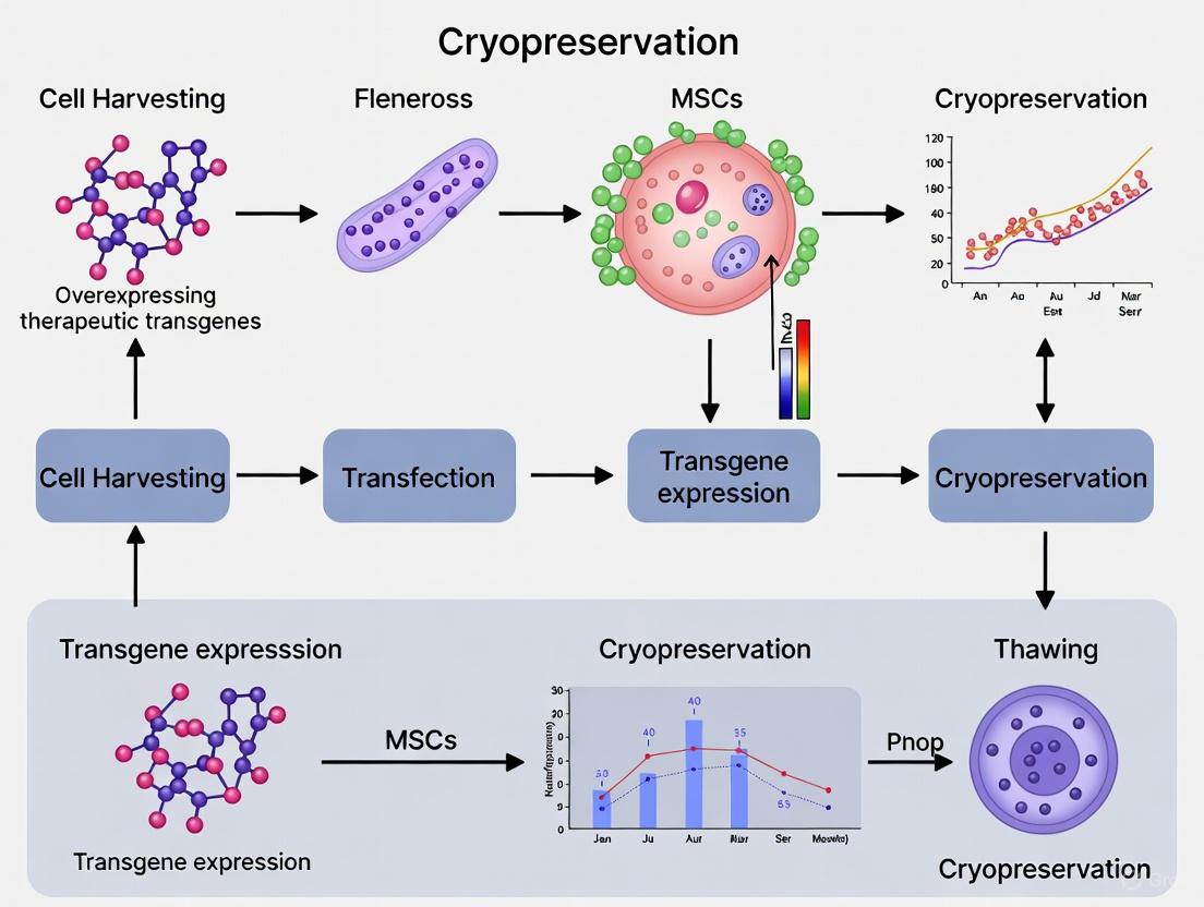
Preserving Potency: A Comprehensive Guide to Cryopreserving Engineered MSCs for Advanced Therapies
The development of mesenchymal stem cells (MSCs) overexpressing therapeutic transgenes represents a frontier in regenerative medicine and oncology.
Preserving Potency: A Comprehensive Guide to Cryopreserving Engineered MSCs for Advanced Therapies
Abstract
The development of mesenchymal stem cells (MSCs) overexpressing therapeutic transgenes represents a frontier in regenerative medicine and oncology. However, cryopreservation of these engineered cells presents unique challenges for ensuring post-thaw viability, functionality, and transgene expression. This article provides a detailed examination of the foundational principles, methodological protocols, and optimization strategies critical for the successful cryopreservation of modified MSCs. Drawing on recent preclinical evidence and industry surveys, we outline key considerations for maintaining anti-cancer potency, migratory potential, and phenotypic stability after thawing. This resource is tailored for researchers, scientists, and drug development professionals navigating the path from laboratory development to off-the-shelf, clinically viable cell therapy products.
The Science Behind Engineered MSCs and Cryopreservation Fundamentals
Mesenchymal stem/stromal cells (MSCs) have emerged as highly promising vehicles for therapeutic transgenes in regenerative medicine and cell-based gene therapy applications. These multipotent cells possess unique biological properties that make them particularly suitable for therapeutic delivery, including their capacity for self-renewal, multilineage differentiation, immunomodulatory functions, and tropism to sites of injury [1] [2]. Originally identified in bone marrow, MSCs have since been isolated from various tissues including adipose tissue, umbilical cord, dental pulp, and placental tissue [1] [2]. According to the International Society for Cell & Gene Therapy (ISCT), MSCs are defined by three minimal criteria: (1) adherence to plastic under standard culture conditions; (2) expression of specific surface markers (CD73, CD90, CD105 ≥95%) while lacking expression of hematopoietic markers (CD34, CD45, CD14/CD11b, CD79α/CD19, HLA-DR ≤2%); and (3) capacity to differentiate into osteoblasts, adipocytes, and chondrocytes in vitro [1] [2].
The therapeutic potential of MSCs has expanded beyond their native regenerative capabilities to include their use as delivery vehicles for therapeutic transgenes. This approach leverages the natural biological properties of MSCs while enhancing their therapeutic efficacy through genetic engineering to overexpress factors that promote tissue repair, modulate immune responses, or combat pathological processes [3]. When engineered to overexpress therapeutic transgenes, MSCs can serve as sustained, localized bioreactors that secrete desired factors at disease sites, offering significant advantages over conventional drug delivery systems [4].
MSC Biology and Characterization
MSCs can be isolated from various tissue sources, each with distinct advantages and characteristics relevant to their use in therapeutic transgene delivery. The source selection impacts critical parameters including cell yield, proliferative capacity, differentiation potential, and immunomodulatory properties [2] [5].
Table 1: Comparison of Primary MSC Sources for Therapeutic Transgene Applications
| Source Tissue | Key Advantages | Limitations | Therapeutic Strengths |
|---|---|---|---|
| Bone Marrow (BM-MSCs) | Most extensively studied; Strong immunomodulatory effects [1] | Invasive harvest; Limited cell number (0.001-0.01%) [2] | Gold standard for research; Strong scientific foundation [5] |
| Adipose Tissue (AD-MSCs) | Abundant tissue source; Easier harvest [1] [2] | Donor age and health influence quality [2] | High yield; Rapid proliferation [2] |
| Umbilical Cord (UC-MSCs) | Enhanced proliferation; Low immunogenicity [1] | Allogeneic source only [1] | Suitable for allogeneic transplantation; "Younger" cells [1] [2] |
| Dental Pulp (DP-MSCs) | Accessible from medical waste [1] | Limited tissue volume [1] | Dental and craniofacial applications [1] |
| Placenta (P-MSCs) | Emerging source with unique properties [1] | Complex composition challenges isolation [2] | Enhanced immunosuppressive effects [2] |
Characterization Methods and Release Criteria
Proper characterization of MSCs is essential for quality control and reproducibility in therapeutic transgene applications. The ISCT-established criteria provide the foundation for MSC identification, though additional characterization is often employed for genetically engineered MSCs [2] [5].
Diagram 1: Comprehensive MSC characterization workflow for therapeutic transgene applications
Standardized isolation techniques vary by tissue source but generally involve enzymatic digestion, density gradient centrifugation, or adherence-based separation [5]. For umbilical cord-derived MSCs, two primary approaches exist: explant culture and enzymatic digestion methods [5]. Quality assessment should include evaluation of senescence markers (p53, p21, p16) as these impact the long-term functionality of engineered MSCs [2] [6].
Table 2: Essential Characterization Techniques for MSCs
| Characterization Category | Specific Methods | Acceptance Criteria |
|---|---|---|
| Morphological | Plastic adherence; Fibroblast-like morphology [2] | ≥95% adherent with characteristic morphology |
| Immunophenotyping | Flow cytometry for CD73, CD90, CD105 [2] [5] | ≥95% positive for markers |
| Negative Markers | Flow cytometry for CD34, CD45, CD14/CD11b, CD79α/CD19, HLA-DR [2] [5] | ≤2% positive for markers |
| Differentiation Potential | Osteogenic: Alizarin Red; Adipogenic: Oil Red O; Chondrogenic: Alcian Blue [2] | Positive staining for specific lineages |
| Functional Potency | Colony-forming unit (CFU-F) assay [5]; Paracrine factor secretion | Donor-specific baseline establishment |
Genetic Engineering of MSCs
Transgene Delivery Methods
Multiple genetic engineering approaches can be employed to introduce therapeutic transgenes into MSCs, each with distinct advantages and limitations for clinical translation.
Diagram 2: Genetic engineering methods for MSC transgene delivery
Experimental Protocol: Lentiviral Transduction of MSCs
This protocol outlines a standardized approach for engineering MSCs to overexpress therapeutic transgenes using lentiviral vectors, which provide efficient transduction and stable transgene expression.
Materials:
- Early passage MSCs (P3-P5)
- Lentiviral vector carrying therapeutic transgene and selection marker
- Polybrene (hexadimethrine bromide, 4-8 μg/mL working concentration)
- Growth medium: αMEM supplemented with 20% FBS, 1% Penicillin/Streptomycin, and 10 ng/mL FGF-2 [7]
- Selection antibiotic appropriate for resistance marker (e.g., puromycin, G418)
- Phosphate-buffered saline (PBS)
- Trypsin/EDTA solution
- Flow cytometry analysis reagents
Procedure:
Cell Preparation:
- Culture MSCs in growth medium until 70-80% confluent
- Harvest cells using trypsin/EDTA and seed at 5,000-10,000 cells/cm² in tissue culture plates
- Incubate overnight at 37°C with 5% CO₂ to allow cell attachment
Transduction:
- Replace medium with fresh growth medium containing polybrene (4-8 μg/mL)
- Add lentiviral vector at appropriate multiplicity of infection (MOI, typically 5-50)
- Include untransduced controls in polybrene-containing medium
- Incubate for 24 hours at 37°C with 5% CO₂
Post-Transduction Processing:
- Remove virus-containing medium and replace with fresh growth medium
- Culture for additional 24-48 hours to allow transgene expression
Selection (if applicable):
- Begin antibiotic selection 48-72 hours post-transduction
- Determine optimal antibiotic concentration by kill curve analysis on untransduced cells
- Maintain selection for 5-7 days or until all control cells are eliminated
- Culture surviving cells for expansion and characterization
Validation:
- Assess transduction efficiency via flow cytometry for reporter genes
- Quantify therapeutic transgene expression using ELISA, Western blot, or qRT-PCR
- Verify MSC phenotype retention post-transduction (surface marker expression)
- Confirm multilineage differentiation potential
Critical Parameters:
- MSC passage number significantly impacts transduction efficiency (use early passages P3-P5)
- Viral titer must be predetermined for each vector batch
- Polybrene concentration may require optimization for different MSC sources
- Always include untransduced controls throughout the process
- Monitor cell morphology and proliferation rates post-transduction
Cryopreservation of Engineered MSCs
Cryopreservation Protocol for Transgene-Overexpressing MSCs
Maintaining the viability, functionality, and transgene expression of engineered MSCs through cryopreservation is essential for clinical translation and banking. Recent evidence confirms that properly cryopreserved MSCs retain their therapeutic properties post-thaw [7].
Materials:
- Genetically engineered MSCs at 70-80% confluence
- Cryopreservation medium: 10% DMSO in 90% autologous plasma or FBS [7]
- Controlled-rate freezing container
- Cryogenic vials
- Water bath set at 37°C
- Centrifuge
Procedure:
Cell Preparation:
- Harvest engineered MSCs at optimal density (70-80% confluence)
- Centrifuge at 300× g for 5 minutes and resuspend in growth medium
- Perform cell count and viability assessment
Cryopreservation:
- Centrifuge cells and resuspend in cold cryopreservation medium at 1×10⁶ to 1×10⁷ cells/mL
- Aliquot 1 mL cell suspension into cryogenic vials
- Place vials in controlled-rate freezing container
- Store at -80°C for 24 hours, then transfer to liquid nitrogen vapor phase
Thawing and Recovery:
- Rapidly thaw vials in 37°C water bath with gentle agitation
- Transfer cell suspension to pre-warmed growth medium (1:10 dilution)
- Centrifuge at 300× g for 5 minutes to remove DMSO
- Resuspend in fresh growth medium and plate at 5,000-10,000 cells/cm²
- Replace medium after 24 hours to remove non-adherent cells
Quality Control Post-Thaw:
- Assess viability using trypan blue exclusion (target ≥80%)
- Verify adherence capacity (≥70% within 24 hours)
- Confirm retention of transgene expression
- Validate immunophenotype and differentiation potential
- Evaluate proliferation rates compared to pre-freeze controls
Effects of Cryopreservation on MSC Function
Recent studies demonstrate that properly executed cryopreservation maintains MSC functionality. Research shows that frozen bone marrow aspirate concentrate (BMAC) retains equivalent cartilage repair capacity to fresh BMAC in osteoarthritis models [7]. Additionally, MSCs cryopreserved for four weeks maintained normal proliferation and multilineage differentiation potential [7].
Table 3: Functional Assessment of Cryopreserved Engineered MSCs
| Functional Attribute | Assessment Method | Acceptance Criteria Post-Thaw |
|---|---|---|
| Viability | Trypan blue exclusion | ≥80% viability |
| Adherence Capacity | Microscopic evaluation at 24 hours | ≥70% adherence |
| Proliferation | Population doubling time | Within 20% of pre-freeze values |
| Multilineage Differentiation | Osteogenic, adipogenic, chondrogenic induction | Retention of differentiation capacity |
| Transgene Expression | ELISA, Western blot, functional assay | ≥70% of pre-freeze expression |
| Immunomodulatory Function | Mixed lymphocyte reaction; cytokine secretion | Significant suppression of immune activation |
| Senescence Markers | β-galactosidase staining; p53, p21, p16 expression | Comparable to pre-freeze levels |
The Scientist's Toolkit: Research Reagent Solutions
Table 4: Essential Research Reagents for MSC Transgene Engineering
| Reagent/Category | Specific Examples | Function/Application |
|---|---|---|
| Cell Culture Media | αMEM with 20% FBS, 1% Penicillin/Streptomycin, 10 ng/mL FGF-2 [7] | Optimal MSC expansion and maintenance |
| Characterization Antibodies | CD73, CD90, CD105, CD34, CD45, CD14/CD11b, CD79α/CD19, HLA-DR [2] | Flow cytometry immunophenotyping |
| Differentiation Kits | Osteogenic: Dexamethasone, β-glycerophosphate, ascorbate; Adipogenic: IBMX, indomethacin, insulin; Chondrogenic: TGF-β, ascorbate [2] | Trilineage differentiation induction |
| Transduction Enhancers | Polybrene (4-8 μg/mL) | Increases viral transduction efficiency |
| Selection Antibiotics | Puromycin, G418/Geneticin | Selection of successfully transduced cells |
| Cryopreservation Media | 10% DMSO in 90% autologous plasma/FBS [7] | Maintains viability and function during freezing |
| Senescence Assay Kits | β-galactosidase staining; p53, p21, p16 analysis [6] | Detection of replicative senescence |
| Vector Systems | Lentiviral, adenoviral, transposon systems | Therapeutic transgene delivery |
MSCs represent versatile and powerful vehicles for therapeutic transgenes, combining intrinsic biological properties with engineerable functionality. The successful implementation of MSC-based gene therapy requires careful attention to cell source selection, characterization, genetic engineering methods, and cryopreservation protocols. Standardized approaches as outlined in these Application Notes and Protocols ensure reproducible and clinically relevant outcomes. As the field advances, the integration of improved vector systems, cryopreservation techniques, and potency assays will further enhance the therapeutic potential of engineered MSCs for diverse clinical applications.
The Critical Need for Cryopreservation in Off-the-Shelf Therapies
The development of "off-the-shelf" allogeneic cell therapies represents a paradigm shift in regenerative medicine, offering the potential for scalable, cost-effective treatments for a broad patient population. For Mesenchymal Stromal Cells (MSCs) overexpressing therapeutic transgenes, cryopreservation is not merely a storage method but a critical enabling technology that facilitates the decoupling of manufacturing from treatment administration. This application note details the necessity, challenges, and optimized protocols for cryopreserving genetically engineered MSCs to ensure the successful translation of these advanced therapies from research to clinical application. Effective cryopreservation allows for comprehensive quality control testing, logistical flexibility for "just-in-time" delivery to clinical sites, and the establishment of cell banks that ensure batch-to-batch consistency—all essential requirements for commercially viable and regulatory-approved therapies [8] [9].
The transition from autologous to allogeneic MSC therapies is a key market trend, with allogeneic products expected to dominate due to their potential for batch production and commercial scalability [10]. This transition is fundamentally dependent on robust cryopreservation protocols that maintain the viability, functionality, and therapeutic potency of the cells throughout their shelf life. For genetically modified MSCs, this challenge is compounded by the need to preserve not only basic cellular functions but also the expression and functionality of the introduced transgenes post-thaw. Industry surveys indicate that 87% of cell therapy developers currently use controlled-rate freezing for cryopreservation, with particular emphasis on its necessity for late-stage clinical and commercial products [11].
Quantitative Landscape of MSC Therapeutics and Storage
The growing prominence of MSC-based therapies underscores the critical importance of optimized cryopreservation protocols. The following data illustrates the market context and storage parameters essential for the successful commercialization of these advanced therapies.
Table 1: Global Mesenchymal Stem Cells Market Landscape (2024-2035)
| Parameter | 2024 Value | 2035 Projection | CAGR (2025-2035) |
|---|---|---|---|
| Market Value | USD 3.82 Billion | USD 9.08 Billion | 8.20% |
| Dominant Product Segment | Products (75.20% share) | - | - |
| Dominant Workflow Segment | Culture & Cryopreservation (31.8% share) | - | - |
| Therapy Type Trend | Shift from Autologous (54.4% share) to Allogeneic | - | - |
| Clinical Trial Activity | 1,100+ trials registered globally [10] | - | - |
Table 2: Standardized Storage Conditions for MSC-Based Biologics
| Biological Material | Recommended Storage Temperature | Key Stability Concerns | Supporting Evidence |
|---|---|---|---|
| MSC Cells | ≤ -150°C (liquid nitrogen vapor phase) | Cryopreservation-induced delayed-onset cell death, loss of functionality [8] | Industry standard for clinical-stage therapies [11] |
| MSC-Derived Extracellular Vesicles (EVs) | -80°C | Vesicle rupture, cargo loss, aggregation [12] | Better preservation of particle concentration, RNA content, and bioactivity vs. -20°C [12] |
| Cryopreserved Starting Materials | ≤ -150°C | Maintaining proliferative capacity and differentiation potential | Essential for ensuring manufacturing consistency [11] |
Critical Challenges in Cryopreservation of Therapeutic MSCs
Cryopreservation-Induced Cellular Damage
The freezing and thawing processes pose significant stresses to MSCs, which can compromise their therapeutic efficacy. Key mechanisms of damage include:
- Intracellular Ice Formation: Rapid cooling can lead to the formation of intracellular ice crystals, which are invariably lethal to cells, causing physical damage to intracellular organelles and membranes [8].
- Solution Effects and Osmotic Stress: During slow cooling, cells are exposed to increasingly hypertonic extracellular solutions as pure water freezes out. This leads to deleterious changes in pH, electrolyte concentrations, and cell volume excursions that can damage the plasma membrane [8] [9].
- Cryoprotectant Toxicity: While cryoprotectants like dimethyl sulfoxide (DMSO) are essential for successful cryopreservation, they can exert toxic effects on cells, particularly during the addition and removal steps or during prolonged exposure at suboptimal temperatures [8].
- Cryopreservation-Induced Delayed-Onset Cell Death: A particularly insidious challenge is the phenomenon where cells appear viable immediately post-thaw but undergo apoptosis hours or days later. This delayed cell death can significantly impact therapeutic efficacy, as it reduces the actual functional cell dose administered to the patient [8].
Specialized Challenges for Genetically Engineered MSCs
For MSCs overexpressing therapeutic transgenes, additional challenges emerge:
- Maintenance of Transgene Expression and Function: The cryopreservation process must preserve not only cell viability but also the constitutive or inducible expression of the therapeutic transgene. Stress from freezing and thawing can potentially alter epigenetic regulation or disrupt signaling pathways necessary for transgene expression.
- Scale-Up Hurdles: Industry surveys identify the "ability to process at a large scale" as the single biggest hurdle for cryopreservation in cell and gene therapy (cited by 22% of respondents) [11]. Scaling cryopreservation protocols from research-scale to industrial-scale batch processing while maintaining critical quality attributes is a significant challenge, particularly for sensitive, engineered cell lines.
Experimental Protocols for Cryopreservation Optimization
Protocol: Controlled-Rate Freezing of Transgenic MSCs
This protocol is designed for the preservation of MSCs overexpressing therapeutic transgenes, with emphasis on maintaining post-thaw viability and functionality.
Materials and Equipment:
- Cultured MSCs (passage 3-6, 80-90% confluence)
- Cryoprotective Agent (CPA): 1M DMSO in complete culture medium
- Controlled-Rate Freezer (CRF)
- Programmable water bath or validated thawing device
- Cryogenic vials
- Liquid nitrogen storage system
Procedure:
- Pre-Freeze Assessment: Confirm transgene expression and functionality via appropriate assays (e.g., flow cytometry, ELISA, functional activity assays). Ensure cells are in logarithmic growth phase and >90% viable by trypan blue exclusion.
- Harvesting and CPA Addition:
- Detach cells using a gentle dissociation reagent.
- Centrifuge and resuspend cells in cold complete medium at a concentration of 5-10 × 10^6 cells/mL.
- Slowly add an equal volume of pre-chilled CPA solution to achieve a final concentration of 10% DMSO and 5-10 × 10^6 cells/mL, with continuous gentle mixing.
- Dispense 1 mL aliquots into cryogenic vials and place on ice (≤4°C). Complete the entire process from CPA addition to initiation of freezing within 30-60 minutes to minimize cryoprotectant toxicity.
- Controlled-Rate Freezing:
- Transfer vials to the pre-cooled chamber of the CRF.
- Initiate the following freezing profile:
- Hold at 4°C for 5 minutes.
- Cool at -1°C/min to -40°C.
- Cool at -5°C/min to -80°C.
- Hold at -80°C for 10 minutes.
- Note: The optimal cooling rate should be empirically determined for specific MSC lines and transgene constructs. Sensitive cells (e.g., iPSC-derived MSCs) may require slower rates (-0.5°C to -1.5°C/min) [11].
- Transfer to Long-Term Storage: Immediately transfer cryovials to a liquid nitrogen vapor-phase storage system (-135°C to -150°C) for long-term preservation.
Protocol: Post-Thaw Viability and Potency Assessment
Comprehensive post-thaw analysis is critical for evaluating the success of the cryopreservation protocol, especially for genetically engineered MSCs.
Immediate Post-Thaw Analysis (0-2 hours):
- Rapid Thawing: Thaw cryovials in a 37°C water bath with gentle agitation until only a small ice crystal remains (approximately 2-3 minutes).
- CPA Removal: Gently transfer cell suspension to a pre-warmed tube containing 10mL of complete medium. Centrifuge at 300 × g for 5 minutes to remove DMSO. Resuspend in fresh medium.
- Viability Assessment: Determine cell viability using trypan blue exclusion and/or flow cytometry with Annexin V/PI staining. Acceptable viability should exceed 80% for clinical applications.
- Cell Recovery Calculation: Calculate percentage recovery: (Post-thaw viable cell count / Pre-freeze viable cell count) × 100.
Extended Functional Analysis (24-72 hours):
- Delayed-Onset Apoptosis Assessment: Re-analyze viability and apoptosis markers (Annexin V/PI) at 24 and 72 hours post-thaw to detect delayed-onset cell death [8].
- Transgene Expression Verification: Quantify transgene expression at the protein (flow cytometry, ELISA) and/or functional level using appropriate bioassays. Compare to pre-freeze levels to ensure preservation of therapeutic potential.
- Functional Potency Assays: Perform standardized potency assays relevant to the therapeutic mechanism of action (e.g., immunomodulation, differentiation capacity, secretion of therapeutic factors).
The Scientist's Toolkit: Essential Research Reagent Solutions
Table 3: Key Reagents for Cryopreservation Research
| Reagent / Material | Function | Application Notes |
|---|---|---|
| Controlled-Rate Freezer (CRF) | Precisely controls cooling rate during freezing; critical process parameter [11] | Default profiles often require optimization for sensitive/engineered MSCs [11] |
| DMSO (Cryoprotectant) | Penetrating cryoprotectant; reduces intracellular ice formation [8] | Concentration typically 5-10%; associated with toxicity; requires controlled addition/removal [8] |
| Serum-Free Cryopreservation Media | Defined formulation; avoids bovine serum albumin (BSA) and animal components [8] | Redances batch variability and regulatory concerns; may include non-penetrating CPAs (e.g., trehalose, sucrose) |
| Programmable Thawing Device | Provides consistent, controlled thawing at ~45°C/min [11] | Replaces non-GMP water baths; reduces contamination risk and improves reproducibility [11] |
| Annexin V / PI Apoptosis Kit | Detects early (Annexin V+) and late (PI+) apoptotic/necrotic cells post-thaw | Essential for identifying cryopreservation-induced delayed-onset cell death [8] |
Visualizing the Cryopreservation Workflow and Critical Quality Attributes
The following diagram illustrates the complete workflow for cryopreserving transgenic MSCs, highlighting critical process parameters and their impact on the critical quality attributes of the final product.
Cryopreservation Workflow for Transgenic MSCs
The successful development of off-the-shelf therapies based on genetically engineered MSCs is fundamentally dependent on robust, scalable cryopreservation protocols. As evidenced by industry data, controlled-rate freezing has become the standard for clinical-stage products, but requires careful optimization to address the unique challenges posed by sensitive cell types and the imperative to maintain transgene expression and functionality. By implementing the detailed protocols and quality control measures outlined in this application note, researchers can enhance the viability, functionality, and consistency of their cryopreserved transgenic MSC products, thereby accelerating the path to clinical application and commercial success. Future advancements will likely focus on further reducing cryoprotectant toxicity, improving scalability, and developing more predictive potency assays to ensure that the therapeutic potential of these innovative cells is fully realized upon administration [8] [11] [3].
The development of "off-the-shelf" mesenchymal stem cell (MSC) therapies, particularly those involving MSCs engineered to overexpress therapeutic transgenes, represents a frontier in regenerative medicine and oncology. A critical step in the commercialization and widespread distribution of these products is reliable cryopreservation. However, the process of freezing and thawing presents unique challenges for gene-modified MSCs that are not as pronounced in their native counterparts. The stability of the therapeutic transgene, the integrity of the cell membrane compromised by transfection, and the metabolic burden imposed by high levels of transgene expression are three interlinked hurdles that can compromise the efficacy and safety of the final product. This application note synthesizes recent research to detail these challenges and provide validated protocols to ensure that cryopreserved, engineered MSCs retain their critical quality attributes (CQAs) and therapeutic potential post-thaw.
The following tables consolidate quantitative data from pivotal studies, providing a clear overview of the impacts of cryopreservation and the efficacy of proposed solutions.
Table 1: Impact of Cryopreservation on Key Attributes of Engineered MSCs
| Key Attribute | Impact of Cryopreservation (Freshly Thawed) | Recovery Post-24h Acclimation | Supporting Evidence |
|---|---|---|---|
| Transgene Expression | No significant change in expression level or therapeutic potency reported [13] [14]. | Not Required | Cytoplasmic CD::UPRT::GFP transgene expression and cancer cell killing efficacy maintained post-thaw [13]. |
| Membrane Integrity / Phenotype | Decrease in surface markers CD44 and CD105 [15]. | Marker expression recovers [15]. | Flow cytometry analysis showed significant reduction in FT cells, restored after 24h [15]. |
| Metabolic Activity | Significantly increased apoptosis and metabolic activity [15]. | Apoptosis reduced; metabolic profile improves [15]. | Annexin V/PI staining and metabolic activity assays (e.g., Resazurin) confirmed recovery [15]. |
| Proliferation & Clonogenicity | Decreased cell proliferation and clonogenic capacity [15]. | Functional capacity regained [15]. | Colony-forming unit assays and proliferation metrics showed significant improvement after acclimation [15]. |
| Immunomodulatory Function | Maintained ability to arrest T-cell proliferation [15]. | Significantly more potent [15]. | T-cell proliferation assays demonstrated enhanced function in TT group versus FT group [15]. |
Table 2: Efficacy of Cryopreserved CD::UPRT-Expressing MSCs in Cancer Models
| Cancer Model | Type of Study | Key Efficacy Finding | Reference |
|---|---|---|---|
| Spontaneous Canine Cancers | In Vivo (Veterinary Patients) | Patients showed a progression-free interval of >20 months after treatment with cryopreserved MSCs and 5FC [13]. | [13] |
| Human Hepatocellular Carcinoma (Huh-7, HepG2) | In Vitro Coculture | With only 10% engineered MSCs, over 70% killing efficiency of cancer cell lines was achieved [14]. | [14] |
| Human Hepatocellular Carcinoma | In Vivo (Mouse Model) | Tumour mass growth was inhibited by >80% in the treated group [14]. | [14] |
| Multiple Human Cancer Cell Lines | In Vitro Coculture | Thawed and freshly modified MSCs showed comparable cytotoxicity in the presence of the prodrug 5-flucytosine (5FC) [13]. | [13] |
Detailed Experimental Protocols
Below are detailed methodologies for key experiments cited in this note, which can be adapted for quality control (QC) testing of cryopreserved, gene-modified MSC batches.
Protocol: Assessment of Transgene Stability and Potency Post-Thaw
This protocol is adapted from studies demonstrating stable transgene expression and function after cryopreservation [13] [14].
1.0 Objective: To verify that cryopreservation does not diminish the expression or therapeutic efficacy of the cytoplasmic transgene (e.g., CD::UPRT::GFP) in engineered MSCs.
2.0 Materials:
- Thawed, gene-modified MSCs (e.g., cryopreserved in CryoStor10)
- Appropriate culture medium (e.g., MEM alpha with supplements)
- Phosphate Buffered Saline (PBS), Plasma-Lyte A, or similar
- Flow cytometer (e.g., CytoFLEX LX)
- GFP fluorescence filter set
- Target cancer cell lines (e.g., Huh-7, HepG2, A549)
- Prodrug (e.g., 5-Flucytosine, 5FC)
- Cell viability assay kit (e.g., based on resazurin or similar)
3.0 Procedure: 1. Cell Thawing & Plating: Thaw the cryopreserved, gene-modified MSCs rapidly in a 37°C water bath. Dilute the cell suspension in pre-warmed culture medium and centrifuge to remove the cryoprotectant. Plate the cells for analysis and for the co-culture assay. 2. Transgene Expression Analysis (Flow Cytometry): - Harvest a sample of cells 24 hours post-thaw. - Create a single-cell suspension using a gentle dissociation reagent and pass through a 100 µm cell strainer. - Analyze at least 10,000 events on a flow cytometer. Use non-modified MSCs as a negative control to set the GFP-positive gate. The percentage of GFP-positive cells in the thawed sample should be comparable to historical data from pre-freeze or freshly transfected cells [13]. 3. Functional Potency Assay (Co-culture): - Plate target cancer cells in a multi-well plate. - After the cancer cells have adhered, add the thawed, gene-modified MSCs at a defined ratio (e.g., 10:1 cancer cells to MSCs) [14]. - Add the prodrug 5FC to the culture medium. - Incubate for 48-72 hours. - Measure the viability of the cancer cells using a standardized viability assay. The cytotoxicity (cancer cell kill) achieved by the thawed MSCs should be comparable to that of freshly prepared engineered MSCs [13].
4.0 Data Analysis: Compare the %GFP-positive cells and the IC50 of cancer cell kill from the thawed batch against pre-established specifications or control data.
Protocol: Evaluating Post-Thaw Membrane Integrity and Functional Recovery
This protocol is based on research highlighting the transient negative impact of cryopreservation on MSC surface markers and function, and their recovery after acclimation [15].
1.0 Objective: To assess the recovery of MSC phenotype and critical functions following a 24-hour acclimation period post-thaw.
2.0 Materials:
- Thawed MSCs (split for immediate and 24h analysis)
- Complete culture medium
- Antibodies for flow cytometry: CD73, CD90, CD105, CD44, CD34, CD45, HLA-DR
- Flow cytometer with appropriate configuration
- Apoptosis detection kit (Annexin V / Propidium Iodide)
- T-cell proliferation assay kit (e.g., CFSE-based)
3.0 Procedure: 1. Experimental Groups: - FT (Freshly Thawed): Analyze cells immediately after thawing and washing. - TT (Thawed + Time): Plate thawed cells at a standard density and culture for 24 hours before analysis [15]. 2. Phenotypic Characterization (Flow Cytometry): - Harvest cells from both FT and TT groups. - Follow standard staining procedures for MSC positive (CD73, CD90, CD105) and negative (CD34, CD45, HLA-DR) markers, including CD44. - Analyze expression levels. A significant recovery of CD44 and CD105 in the TT group compared to the FT group is indicative of membrane and phenotypic recovery [15]. 3. Apoptosis Assay: - Label cells from both groups with Annexin V and PI according to kit instructions. - Analyze by flow cytometry. A significant reduction in the percentage of early (Annexin V+/PI-) and late (Annexin V+/PI+) apoptotic cells in the TT group is expected [15]. 4. Functional Immunomodulation Assay (T-cell Proliferation): - Co-culture peripheral blood mononuclear cells (PBMCs) or isolated T-cells (activated with mitogens like PHA or anti-CD3/CD28 beads) with MSCs from the FT and TT groups. - Measure T-cell proliferation using a CFSE dilution assay or similar. - MSCs from the TT group are expected to demonstrate a significantly greater suppression of T-cell proliferation compared to the FT group [15].
4.0 Data Analysis: Statistical comparison (e.g., t-test) between FT and TT groups should confirm significant recovery in phenotype, reduced apoptosis, and enhanced immunomodulatory function after 24 hours.
Visualizing Workflows and Functional Relationships
Experimental Workflow for Post-Thaw Validation
The following diagram illustrates the key steps and decision points in the post-thaw validation of engineered MSCs.
Functional Recovery Pathway Post-Thaw
This diagram outlines the molecular and cellular events during the post-thaw acclimation period that lead to the recovery of MSC functionality.
The Scientist's Toolkit: Essential Research Reagents
Table 3: Key Reagents for Cryopreservation and QC of Engineered MSCs
| Reagent / Material | Function / Application | Example & Notes |
|---|---|---|
| Cryopreservation Medium | Protects cells from freezing damage; often contains permeating CPAs and proteins. | CryoStor10 (GMP-grade, defined formulation) [13]. Alternative: 10% DMSO in FBS, though serum-free, GMP alternatives are preferred for clinical use [15]. |
| Non-Viral Transfection System | Engineered to introduce therapeutic transgenes into MSCs with high efficiency and a favorable safety profile. | Polyethylenimine (PEI) combined with a Fusogenic Lipid (DOPE/CHEMS) and HDAC Inhibitor (Bufexamac) for high transgene expression [13]. |
| Controlled-Rate Freezer | Ensures a consistent, optimal cooling rate during cryopreservation to minimize intracellular ice crystal formation. | If unavailable, a passive freezing device like Mr. Frosty can be used, which provides an approximate cooling rate of -1°C/min in a -80°C freezer [13]. |
| Automated Thawing System | Standardizes the thawing process for improved viability and reproducibility. | ThawSTAR Automated Thawing System [13]. Manual thawing in a 37°C water bath is common but introduces more variability. |
| Hypothermic Holding Medium | Stabilizes cells post-thaw during transport or before administration, minimizing additional stress. | HypoThermosol [13]. Used to resuspend cells after thawing and washing, prior to in vivo administration. |
| Exocellular Cryoprotectant | Non-penetrating CPA that protects cells osmotically and stabilizes membranes. | Sucrose (0.2M). Often used in combination with DMSO to reduce the required concentration of the latter and improve overall cryoprotection [16]. |
Core Principles of Cryoinjury and Cryoprotectant Mechanisms
The cryopreservation of mesenchymal stromal cells (MSCs) is a critical step in the development of off-the-shelf cellular therapeutics, particularly for engineered cells overexpressing therapeutic transgenes. The process enables the creation of cell banks, facilitates quality control testing, and allows for the widespread distribution of living medicines [17] [18]. However, the freezing and thawing procedures impose severe physical and chemical stresses that can impair cell viability, motility, and functionality—a phenomenon known as cryoinjury [19] [17]. For MSCs engineered to carry therapeutic transgenes, these injuries present a substantial risk to clinical efficacy, as they can compromise the very functions these cells are designed to execute. A deep understanding of the core principles governing cryoinjury and the protective mechanisms of cryoprotectants is therefore fundamental to advancing cell-based therapies from the bench to the clinic. This document details these principles and provides actionable protocols within the context of a broader thesis on preserving gene-modified MSCs.
Core Principles of Cryoinjury
Cryoinjury refers to the structural and functional damage sustained by cells during the cryopreservation and thawing processes. The injury mechanisms are multifaceted and can be broadly categorized into physical, chemical, and biological insults.
Physical Insults: Intracellular Ice Formation and Osmotic Stress
The formation of ice crystals is a primary driver of physical cryoinjury. During slow freezing, as the temperature falls below the freezing point, ice forms first in the extracellular solution. This extracellular ice formation increases the solute concentration in the remaining unfrozen liquid, creating a hypertonic environment. Consequently, water osmotically flows out of the cell, leading to cellular dehydration and shrinkage [20]. If the cooling rate is too rapid, water does not have sufficient time to exit the cell, resulting in the lethal formation of intracellular ice crystals that can pierce and disrupt organelles and the plasma membrane [20] [21]. The phase transition of membrane lipids from a fluid to a gel state as temperatures drop further reduces membrane fluidity and is associated with lower cell survival [19].
Biological Insults: Programmed Cell Death and Loss of Function
Beyond immediate physical damage, the freeze-thaw process can trigger delayed biological responses, most notably apoptosis (programmed cell death). Studies show that cryopreservation reduces cell viability and increases apoptosis levels, which can manifest hours after thawing [18]. A fundamental mechanism of this delayed cell death has been linked to the cell cycle. Research indicates that MSCs in the S phase (DNA replication phase) are exquisitely sensitive to cryoinjury, demonstrating heightened levels of delayed-onset apoptosis post-thaw [22]. The cryopreservation and thawing processes induce double-stranded breaks in the labile replicating DNA of S-phase cells, leading to post-thaw dysfunction and death. This loss of specific cell populations can skew the functionality of the entire therapeutic product. Quantitative assessments confirm that cryopreservation not only reduces immediate viability but also impairs metabolic activity and adhesion potential for at least 24 hours post-thaw, indicating a prolonged recovery period is necessary for functional restoration [18].
Diagram 1: Key Mechanisms of Cryoinjury in MSCs. The diagram illustrates how the freezing process inflicts physical and biological damage, leading to cell death and functional loss. DSBs: Double-Stranded Breaks.
Core Principles of Cryoprotectants
Cryoprotectants (CPAs) are hyperosmotic additives designed to mitigate cryoinjury. They function by stabilizing intracellular proteins, reducing intracellular ice formation, and counteracting the damaging effects of concentrated electrolytes [19].
Permeating vs. Non-Permeating Cryoprotectants
CPAs are classified based on their ability to cross the cell membrane. Permeating CPAs, such as dimethyl sulfoxide (DMSO) and glycerol, are small molecules that enter the cell. They reduce the freezing point of water intracellularly and decrease the amount of ice formed at any given temperature by increasing the total solute concentration inside the cell. This action minimizes the osmotic differential across the membrane during freezing, thereby reducing the extent of cell dehydration [20]. DMSO is the most widely used permeating CPA for MSCs, though its intrinsic toxicity and potential to trigger allergic responses in patients are significant concerns [20] [17]. Non-permeating CPAs, such as sucrose and trehalose, remain outside the cell. They work by increasing the extracellular osmolarity, which promotes gentle cell dehydration before freezing, further reducing the potential for intracellular ice formation. They also help stabilize the cell membrane [20].
The Role of CPA Addition and Removal
The protocols for adding CPAs before freezing and removing them after thawing are critical. The addition of CPAs must be performed in a dropwise manner with gentle mixing to allow for proper equilibration and to prevent osmotic shock [19]. Conversely, the removal process post-thaw, typically involving centrifugation and rinsing, must be carefully controlled. Rapidly reducing the external CPA concentration can cause excessive cell swelling and lysis due to the osmotic influx of water [20]. This is a key vulnerability point for post-thaw cell recovery.
Quantitative Impact of Cryopreservation on MSCs
The quantitative effects of cryopreservation on MSCs are variable but follow consistent trends. The table below summarizes key cellular attributes affected during the first 24 hours post-thaw, a critical window for clinical infusion.
Table 1: Quantitative Post-Thaw Recovery of Human Bone Marrow-Derived MSCs
| Cell Attribute | 0-4 Hours Post-Thaw | 24 Hours Post-Thaw | Long-Term Impact (Beyond 24h) |
|---|---|---|---|
| Viability | Significantly reduced [18] | Recovers to near-baseline levels [18] | N/A |
| Apoptosis Level | Significantly increased [18] | Decreased but may remain elevated [18] | N/A |
| Metabolic Activity | Significantly impaired [18] | Remains lower than fresh cells [18] | N/A |
| Adhesion Potential | Significantly impaired [18] | Remains lower than fresh cells [18] | N/A |
| Phenotype (Surface Markers) | Largely unchanged (CD73, CD90, CD105 positive) [23] [18] | Largely unchanged [23] [18] | Largely unchanged [23] |
| Transgene Expression | Preserved in engineered MSCs [23] | Preserved in engineered MSCs [23] | Preserved after 11 months of storage [23] |
| Proliferation Rate | N/A | N/A | Comparable to fresh cells [18] |
| Clonogenic Potential (CFU-F) | N/A | N/A | Variable; can be reduced in some cell lines [18] |
| Differentiation Potential | N/A | N/A | Variably affected (osteogenic & adipogenic) [18] |
| Migratory & Tumor Tropism | N/A | N/A | Preserved in engineered MSCs [23] |
Experimental Protocols for Cryopreservation of Engineered MSCs
The following protocol is adapted from studies demonstrating the successful cryopreservation of MSCs transiently transfected to overexpress a therapeutic transgene (CD::UPRT::GFP), with viability and function maintained after up to 11 months in storage [23].
Protocol: Cryopreservation of Transfected MSCs
Objective: To preserve transfected MSCs with high viability and retained transgene expression post-thaw.
The Scientist's Toolkit: Table 2: Essential Reagents and Materials
| Item | Function/Description | Example/Note |
|---|---|---|
| Cryopreservation Medium | GMP-grade solution to protect cells during freezing. | CryoStor10 (CS10) [23]. Alternatively, culture medium with 10% DMSO [18]. |
| Permeating Cryoprotectant | Reduces intracellular ice formation. | Dimethyl Sulfoxide (DMSO) at 10% final concentration [18]. |
| Wash Solution | To remove serum and CPAs post-thaw. | Plasma-Lyte A or similar isotonic solution [23]. |
| Controlled-Rate Freezer | Provides consistent, slow cooling. | "Mr. Frosty" freezing container (-1°C/min) [23] [18] or programmable freezer. |
| Liquid Nitrogen Storage | For long-term storage at -135°C to -196°C. | Vapor phase is preferred to minimize contamination risk [20] [17]. |
| Automated Thawing System | Ensures consistent and rapid thawing. | ThawSTAR system or 37°C water bath [23]. |
Methodology:
- Post-Transfection Harvest: After the transfection procedure (e.g., 24 hours post-transfection with PEI-Max and enhancers), wash the engineered MSCs twice with Plasma-Lyte A or PBS. Harvest the cells using a gentle enzyme like TrypLE Express [23].
- Centrifugation and Resuspension: Centrifuge the cell suspension (e.g., at 300-352 ×g for 5-10 minutes). Discard the supernatant and resuspend the cell pellet in the pre-chilled cryopreservation medium (e.g., CS10) at a concentration of 1-3 x 10^6 cells/mL [23].
- Aliquoting and Equilibration: Transfer 1 mL of the cell suspension into each cryovial. Place the cryovials at room temperature for 10 minutes to allow for equilibration between the cells and the CPA [19] [23].
- Controlled-Rate Freezing: Place the cryovials into a controlled-rate freezing device, such as a "Mr. Frosty" container. Store the container at -80°C for 24 hours to achieve a cooling rate of approximately -1°C/min [23] [18].
- Long-Term Storage: After 24 hours, promptly transfer the cryovials to a long-term storage location, either in the vapor phase of liquid nitrogen (-135°C to -196°C) or in an ultralow-temperature freezer (-150°C or below) for up to 11 months or longer [23] [17].
Diagram 2: Experimental Workflow for Cryopreserving Engineered MSCs. The protocol from transfection to post-thaw assessment ensures functional preservation.
Protocol: Thawing and Post-Thaw Assessment
Objective: To rapidly recover cryopreserved transfected MSCs and quantify their viability and functionality.
Methodology:
- Rapid Thawing: Remove the cryovial from liquid nitrogen storage and thaw quickly using an automated thawing system or by placing it in a 37°C water bath until only a small ice crystal remains (approximately 1 minute) [23] [18].
- CPA Removal and Washing: Transfer the thawed cell suspension into a tube containing 4 mL of pre-warmed wash solution (e.g., Plasma-Lyte A or culture medium). Centrifuge at 300 ×g for 5 minutes to pellet the cells and remove the CPA-containing supernatant [19] [23].
- Resuspension and Plating: Resuspend the cell pellet in fresh pre-warmed culture medium. Count the cells using an automated cell counter and assess viability using dyes like acridine orange (AO) and DAPI, or trypan blue exclusion [23] [18].
- Functional Assessment:
- Viability & Apoptosis: Assess at 0, 2, 4, and 24 hours post-thaw to capture immediate and delayed-onset apoptosis [18].
- Transgene Expression: Use flow cytometry to quantify the percentage of GFP-positive cells and confirm sustained transgene expression [23].
- Potency Assay: Co-culture thawed, engineered MSCs with relevant cancer cell lines in the presence of the prodrug 5-flucytosine (5FC). Measure cancer cell death to confirm the functional activity of the expressed enzyme (CD::UPRT) [23].
- Migration Assay: Use a matrigel invasion chamber to verify that the tumor-homing capacity of the MSCs is retained post-thaw [23].
Advanced Strategies for Mitigating Cryoinjury in MSCs
Standard cryopreservation protocols can be optimized to specifically enhance the recovery of functional MSCs.
Cell Cycle Synchronization
A pivotal strategy involves synchronizing the cell cycle prior to freezing. Since S-phase MSCs are highly vulnerable to cryopreservation-induced DNA damage and apoptosis, blocking cell cycle progression at the G0/G1 phase can dramatically improve post-thaw outcomes. This can be achieved through growth factor deprivation, commonly known as serum starvation. This simple pre-freezing manipulation has been shown to preserve viability, clonal growth, and immunomodulatory function at pre-cryopreservation levels, performing as well as priming with interferon-gamma (IFNγ) but without its pleiotropic effects [22].
Optimized Cryopreservation Formulations
Moving beyond standard DMSO-containing media, the use of GMP-grade, defined cryopreservation solutions like CryoStor10 (CS10) has shown excellent results for engineered MSCs [23]. These commercial formulations are designed to minimize osmotic stress and CPA toxicity. Research into DMSO-free alternatives is also ongoing, utilizing combinations of non-permeating CPAs like sucrose and trehalose to reduce the risks associated with DMSO, such as its cytotoxicity and potential to provoke allergic reactions in patients [20] [17].
Protocols and Best Practices for Cryopreserving Modified MSCs
The cryopreservation of mesenchymal stem cells (MSCs), particularly those engineered to overexpress therapeutic transgenes, represents a critical step in the development of off-the-shelf cell therapies. Maintaining high cell viability, recovery, and—most importantly—therapeutic potency post-thaw is essential for clinical and commercial success [24] [13]. The choice of cryoprotectant is pivotal in this process, balancing cell protection against potential toxicity and functional impairment. This application note provides a detailed comparison between conventional dimethyl sulfoxide (DMSO) formulations and a current Good Manufacturing Practice (cGMP)-grade solution, CryoStor CS10, within the specific context of preserving MSCs engineered for therapeutic transgene expression. We summarize quantitative performance data and provide standardized protocols to support robust, reproducible cryopreservation strategies in advanced therapeutic medicinal product (ATMP) development.
Cryoprotectant Mechanisms and Classification
Cryoprotective Agents (CPAs) function by mitigating the two primary mechanisms of freezing-induced cell death: the formation of intracellular ice crystals that mechanically damage cellular structures, and "solution effects," where the concentration of solutes in the unfrozen fraction leads to osmotic stress and protein denaturation [25]. CPAs are broadly classified into two categories based on their cellular permeability:
- Penetrating (Endocellular) Cryoprotectants: Low molecular weight compounds that cross the cell membrane. They depress the freezing point of water both inside and outside the cell and reduce the fraction of water that forms ice, thereby minimizing intracellular ice crystal formation. DMSO is the most common penetrating CPA [25].
- Non-Penetrating (Exocellular) Cryoprotectants: High molecular weight compounds that remain outside the cell. They protect the cell by stabilizing the cell membrane, inhibiting the growth of extracellular ice crystals, and reducing the osmotic imbalance across the cell membrane during freezing. Common examples include sucrose, trehalose, and synthetic polymers like hydroxyethyl starch [25].
The following diagram illustrates the logical relationship between cryopreservation goals, the mechanisms of cell damage, and the protective functions of different cryoprotectant classes.
Quantitative Comparison of Cryoprotectant Performance
Post-Thaw Viability and Cell Recovery
A 2024 study directly compared several clinical-ready cryopreservation formulations for MSCs, including an in-house formulation (Plasmalyte-A with 5% HA and 10% DMSO, PHD10), NutriFreez (10% DMSO), and CryoStor solutions (CS5 and CS10) [24]. The results demonstrated that the specific formulation, not just the DMSO concentration, significantly impacts post-thaw outcomes.
Table 1: Post-Thaw Viability and Recovery of Cryopreserved MSCs
| Cryopreservation Solution | DMSO Concentration | Immediate Post-Thaw Viability | Cell Recovery after 6h | Proliferative Capacity after 6 Days |
|---|---|---|---|---|
| PHD10 | 10% | High and comparable to other 10% DMSO solutions | Maintained with a decreasing trend | Similar to NutriFreez; No significant difference |
| NutriFreez | 10% | High and comparable to other 10% DMSO solutions | Maintained with a decreasing trend | Similar to PHD10; No significant difference |
| CryoStor CS10 | 10% | High and comparable to other 10% DMSO solutions | Maintained | 10-fold less compared to NutriFreez/PHD10 at 3-6 M/mL |
| CryoStor CS5 | 5% | Lower than 10% DMSO solutions | Decreasing trend | 10-fold less compared to NutriFreez/PHD10 at 3-6 M/mL |
Source: Adapted from [24]
Furthermore, a study on MSC spheroids found that CryoStor CS10 outperformed other cGMP-grade media and a conventional control medium (DMEM with 20% FBS and 10% DMSO), resulting in higher viability and better preservation of spheroid morphology and stem cell marker expression after thawing [26].
Impact on MSC Phenotype and Potency
A critical requirement for therapeutic MSCs is the retention of their immunomodulatory potency post-thaw. The 2024 study found that MSCs cryopreserved in NutriFreez and PHD10 showed comparable potency in inhibiting T-cell proliferation and improving monocytic phagocytosis, with no significant differences between them [24]. This indicates that the core immunomodulatory functions can be preserved with optimized formulations.
For MSCs engineered to overexpress a therapeutic transgene, cryopreservation must also maintain transgene expression and function. A pivotal 2022 study demonstrated that MSCs highly overexpressing a cytoplasmic therapeutic transgene (CD::UPRT::GFP) could be successfully cryopreserved in CryoStor CS10 for up to 11 months [13]. Post-thaw, these cells retained their transgene expression, viability, phenotypic profile, migratory potential, and, crucially, their cancer-killing potency in the presence of the prodrug 5-flucytosine. This confirms that CryoStor CS10 is compatible with complex, genetically modified MSC therapies [13].
Detailed Experimental Protocols
Protocol: Cryopreservation of Therapeutic Transgene-Expressing MSCs
This protocol is adapted from the methodology used to successfully cryopreserve MSCs overexpressing CD::UPRT::GFP, as documented by Tan et al. (2022) [13].
Workflow Overview:
Materials:
- Cells: MSCs (passage 4-6), post-transfection with therapeutic transgene.
- Cryopreservation Medium: CryoStor CS10 (cGMP-grade, serum-free, animal component-free) [27] [13].
- Wash Buffer: Plasma-Lyte A or other physiological balanced salt solution [13] [28].
- Equipment: Controlled-rate freezing chamber (e.g., "Mr. Frosty" filled with isopropanol) or programmable freezer, cryogenic vials, liquid nitrogen storage system, 37°C water bath, centrifuge.
Step-by-Step Procedure:
- Cell Preparation: Harvest MSCs 24 hours post-transfection, ensuring they are in a logarithmic growth phase and have been confirmed for high transgene expression [13].
- Harvesting: Wash the cell monolayer twice with Wash Buffer (e.g., Plasma-Lyte A) and detach using a gentle enzyme solution like TrypLE Express [13] [28].
- Neutralization & Counting: Neutralize the enzyme with a 10x volume of culture medium supplemented with serum. Centrifuge the cell suspension at 100-300 × g for 5 minutes. Aspirate the supernatant and resuspend the cell pellet in a small volume of Wash Buffer for accurate cell counting [13] [28].
- Formulation: Centrifuge the cell suspension again and thoroughly resuspend the cell pellet in cold CryoStor CS10 to achieve a final concentration of 1-3 x 10^6 cells/mL [13]. Gently mix to ensure a homogeneous suspension.
- Aliquoting: Dispense 0.5 - 1.0 mL of the cell suspension into pre-labeled cryogenic vials. Keep the vials on ice or at 4°C during this process to maintain a uniform temperature.
- Freezing: Immediately transfer the cryovials into a pre-chilled isopropyl alcohol freezing chamber. Place the chamber in a -80°C freezer for a minimum of 3 hours (or overnight) to achieve a consistent cooling rate of approximately -1°C/min [13].
- Storage: After 3-24 hours, promptly transfer the cryovials to the vapor phase of liquid nitrogen for long-term storage (< -150°C) [13].
Protocol: Comparative Testing of Cryoprotectant Formulations
This protocol outlines a method for directly comparing the performance of different cryoprotectants, such as research-grade DMSO solutions versus cGMP-grade formulations, on a specific MSC line.
Materials:
- Test Formulations:
- Assessment Tools: Trypan blue exclusion assay; Flow cytometer with Annexin V/PI staining; Cell proliferation assay (e.g., alamarBlue); Functional potency assays (e.g., T-cell suppression assay) [24] [28].
Step-by-Step Procedure:
- Cell Culture: Use a single, well-characterized batch of MSCs (or transgene-expressing MSCs) at a consistent passage number. Split cells to ensure they are 70-80% confluent and in an active growth phase at the time of cryopreservation [24] [28].
- Sample Preparation: Harvest and count the cells as described in Section 4.1. Divide the cell pool into three equal aliquots. Pellet each aliquot and resuspend in one of the three test cryopreservation solutions (A, B, C). Use multiple cryopreservation densities (e.g., 3, 6, and 9 million cells/mL) to assess concentration effects [24].
- Cryopreservation: Cryopreserve all vials using an identical, standardized controlled-rate freezing method as in Section 4.1. Store all vials in the same liquid nitrogen tank to ensure identical storage conditions.
- Thawing and Analysis: After a minimum storage period (e.g., 1 week), thaw the vials rapidly in a 37°C water bath. For cells cryopreserved at high density, implement a dilution step (e.g., 1:1 or 1:2 with Plasma-Lyte A/5% HA) to achieve a standard concentration for testing [24].
- Time-Course Assessment: Assess cell viability and recovery immediately post-thaw (0h) and then maintain the cells in the post-thaw solution at room temperature for 2, 4, and 6 hours, measuring viability at each interval to determine stability [24] [28].
- Functional Assays: Plate the thawed cells and allow them to recover in culture for the required duration before conducting phenotypic characterization (flow cytometry for CD73, CD90, CD105), proliferation assays, and transgene-specific potency assays [24] [13].
The Scientist's Toolkit: Essential Research Reagents
Table 2: Key Reagents for MSC Cryopreservation Research
| Reagent / Solution | Function / Application | Key Characteristics |
|---|---|---|
| CryoStor CS10 | Defined, cGMP-grade cryopreservation medium [27] [13]. | Serum-free, animal component-free, contains 10% DMSO, pre-formulated and ready-to-use. |
| Dimethyl Sulfoxide (DMSO) | Penetrating cryoprotectant for in-house formulation [24] [25]. | USP-grade recommended for clinical relevance; used at 5-10% (v/v) concentration. |
| Plasma-Lyte A / Normosol-R | Isotonic, extracellular-like carrier solution [24] [28]. | Used as a base for in-house CPA formulation or as a wash/dilution buffer post-thaw. |
| Human Serum Albumin (HSA) | Macromolecular additive for in-house CPA [24]. | Provides colloidal osmotic support and can stabilize cell membranes; used at 5%. |
| HypoThermosol FRS | Intracellular-like preservation solution [28]. | Designed for hypothermic storage and shipment of thawed cells to minimize cold stress. |
| TrypLE Express | Enzyme for cell detachment [13] [28]. | Gentle, animal-origin-free alternative to trypsin for harvesting cells pre-cryopreservation. |
| Annexin V / PI Staining | Flow cytometry-based apoptosis and necrosis detection [24] [26]. | Provides a more accurate assessment of post-thaw cell health than dye exclusion alone. |
| alamarBlue Assay | Metabolic activity assay for cell viability and proliferation [28]. | Quantitative method to assess functional recovery and proliferative capacity post-thaw. |
The selection of a cryoprotectant for MSCs, especially those engineered with therapeutic transgenes, extends beyond merely preventing immediate cell death. While conventional 10% DMSO formulations can maintain baseline viability, evidence indicates that cGMP-grade, optimized solutions like CryoStor CS10 offer significant advantages for commercial therapy development. These advantages include enhanced preservation of cell recovery over time and, critically, the demonstrated ability to maintain complex therapeutic functions, such as high-level transgene expression and targeted potency, post-thaw [24] [13]. For researchers navigating the path from discovery to clinical application, adopting a defined, cGMP-compliant cryopreservation platform from an early stage can de-risk development, streamline regulatory approval, and ultimately ensure that the therapeutic potential of engineered MSCs is reliably delivered to the patient.
Application Note AN-001 | Version 1.0
Within advanced therapeutic medicinal products (ATMPs), the cryopreservation of Mesenchymal Stromal Cells (MSCs) overexpressing therapeutic transgenes is a critical unit operation. The choice of freezing methodology can significantly impact post-thaw viability, recovery, and, most importantly, the functional potency of the final cell product. This Application Note provides a comparative analysis of Controlled-Rate Freezing (CRF) and Passive Freezing (PF) techniques, summarizing quantitative data and providing detailed protocols to support researchers and scientists in optimizing their cryopreservation workflows for transgenic MSCs.
Quantitative Comparison of Freezing Methods
The following tables summarize key comparative data from recent studies on various cell types, illustrating the impact of different freezing methodologies.
Table 1: Comparison of Post-Thaw Cell Viability and Recovery
| Cell Type / Tissue | Controlled-Rate Freezing (CRF) | Passive Freezing (PF) | Reference / Model |
|---|---|---|---|
| Hematopoietic Progenitor Cells (HPCs) | CD34+ viability: 77.1% ± 11.3% [29] | CD34+ viability: 78.5% ± 8.0% [29] | Clinical-scale retrospective study [29] |
| Bovine Ovarian Tissue | High viability, follicular morphology, and TAC; Low fibrosis and ROS [30] | Significantly lower viability, morphology, and TAC; Higher fibrosis and ROS [30] | Pre-clinical tissue model [30] |
| General MSC Population (Slow Freezing) | ~70-80% cell survival [20] | Information not specified in search results | Standard laboratory practice [20] |
Table 2: Comparison of Functional Engraftment and In-Vivo Outcomes
| Outcome Measure | Controlled-Rate Freezing (CRF) | Passive Freezing (PF) | Notes |
|---|---|---|---|
| Neutrophil Engraftment (Days) | 12.4 ± 5.0 [29] | 15.0 ± 7.7 [29] | No statistically significant difference (p=0.324) [29] |
| Platelet Engraftment (Days) | 21.5 ± 9.1 [29] | 22.3 ± 22.8 [29] | No statistically significant difference (p=0.915) [29] |
| 2-Cell Mouse Embryo Development | Good implantation rate (22.1%); Higher glucose uptake [31] | Significantly lower implantation rate (10.2%); Reduced glucose uptake [31] | Indicates potential for functional impairment with PF [31] |
Table 3: Technical and Practical Method Considerations
| Parameter | Controlled-Rate Freezing | Passive Freezing |
|---|---|---|
| Process Control | High. Programmable, precise control over cooling rate, especially during phase transition [32] [33]. | Low to None. Uncontrolled process; cooling rate depends on equipment and ambient conditions [32] [30]. |
| Reproducibility | High and repeatable. Validatable process suitable for GMP [34] [33]. | Low. Variable and less reproducible outcomes [34]. |
| Cost & Infrastructure | High initial investment; requires specialized equipment [34]. | Low cost; utilizes standard -80°C mechanical freezers [29] [34]. |
| Ideal Application | GMP manufacturing; Sensitive cell types (MSCs, iPSCs); Critical R&D protocols [32] [17]. | Research settings with robust cell types; Logistics where cost is a primary driver [29]. |
Experimental Protocols for Transgenic MSC Cryopreservation
Protocol: Controlled-Rate Freezing of MSCs
This protocol is adapted from established clinical-grade MSC manufacturing processes [35] and principles of controlled-rate freezing [33].
Key Reagent Solutions:
- Cryopreservation Medium: 10% Dimethylsulfoxide (DMSO, GMP-grade) in 90% Human Serum Albumin (HSA) or the patient's preferred cryoprotectant agent (CPA) formulation [35].
- Basal Medium: D-MEM low glucose.
- Detachment Reagent: TrypLE Select or other GMP-compatible enzyme.
Procedure:
- Cell Harvesting: Culture MSCs to the desired passage (e.g., passage 2). Detach cells using a GMP-compatible detachment reagent. Perform a cell count and assess viability, ensuring it is >90% pre-cryopreservation [35].
- CPA Introduction: Centrifuge the cell suspension. Gently resuspend the cell pellet in pre-chilled cryopreservation medium. The final cell concentration should be optimized (e.g., 10-50 x 10^6 cells/mL). Keep the cell suspension on ice or at 4°C to minimize CPA toxicity [36] [20]. Note: Exposure to DMSO should typically be limited to <30 minutes before freezing. [36]
- Aliquoting: Aseptically aliquot the cell suspension into appropriate cryogenic vials or bags.
- Controlled-Rate Freezing:
- Place the samples in a controlled-rate freezer (CRF).
- Initiate a programmed freeze cycle. A standard protocol for MSCs often includes [33]: a. Start Temperature: +4°C. b. Phase 1: Cool from +4°C to -5°C at a rate of -2°C/min. c. Phase 2 (Seeding): Hold at -5°C for 5-10 minutes and induce ice nucleation (seeding) to prevent supercooling and manage the latent heat of fusion [33]. d. Phase 3: Cool from -5°C to -50°C at a critical rate of -1°C/min [32] [33]. e. Phase 4: Rapidly cool from -50°C to -100°C at a rate of -10°C/min or higher. f. Transfer: Immediately transfer samples to a long-term storage vessel (liquid or vapor phase nitrogen, below -135°C) [33].
Protocol: Passive Freezing of MSCs
This protocol utilizes a -80°C mechanical freezer and an isopropanol (IPA) freezing container [34].
Key Reagent Solutions:
- Cryopreservation Medium: As per Section 3.1.
- Passive Freezing Device: Isopropanol-filled freezing chamber (e.g., "Mr. Frosty").
Procedure:
- Cell Preparation: Follow Steps 1-3 from the Controlled-Rate Freezing protocol (Section 3.1).
- Aliquoting and Loading: Place the filled cryogenic vials into the IPA freezing container at room temperature. Ensure the container is filled to the indicated level with isopropanol.
- Passive Freezing: Place the entire container directly into a -80°C mechanical freezer. The isopropanol ensures a quasi-linear cooling rate of approximately -1°C/min [34].
- Long-Term Storage: After 18-24 hours, promptly remove the vials from the freezing container and transfer them to a long-term storage freezer or liquid nitrogen tank [34].
Workflow and Decision Pathway
The following diagram illustrates the experimental workflow for a comparative study of cryopreservation methods for transgenic MSCs.
Figure 1: Experimental Workflow for Comparative Cryopreservation Study.
The Scientist's Toolkit: Essential Research Reagents and Materials
Table 4: Key Reagent Solutions for MSC Cryopreservation
| Item | Function / Application | Example / Note |
|---|---|---|
| Cryoprotectant Agent (CPA) | Protects cells from ice crystal damage and osmotic stress during freeze-thaw [36] [20]. | DMSO (GMP-grade): Common permeating CPA. HSA/Sucrose: Non-permeating CPAs for osmotic balance [35]. |
| Basal Freezing Medium | Base solution for CPA formulation. | Dulbecco's Modified Eagle Medium (DMEM) low glucose, often supplemented with proteins (HSA) [35]. |
| Cell Detachment Reagent | Non-enzymatic or enzymatic dissociation of adherent MSCs. | TrypLE Select (GMP-compatible, animal-origin-free) [35]. |
| Controlled-Rate Freezer | Provides precise, programmable control over cooling rates. | Essential for CRF protocol validation and GMP compliance [34] [33]. |
| Passive Freezing Device | Provides an approximate -1°C/min cooling rate in a -80°C freezer. | Isopropanol freezing container. A cost-effective alternative for research use [34]. |
| Cryogenic Storage Vials/Bags | Containers for sterile, secure long-term storage. | Must be validated for liquid nitrogen exposure (vapor phase recommended) [33]. |
For the cryopreservation of transgenic MSCs intended as ATMPs, the choice between Controlled-Rate and Passive Freezing is multifaceted. While passive freezing offers a cost-effective and simple solution that may be sufficient for robust cell types or specific research contexts [29], controlled-rate freezing provides superior process control, reproducibility, and validation support. This makes CRF the more suitable and lower-risk option for GMP manufacturing and for preserving the critical quality attributes—including viability, phenotype, and, most importantly, the functional potency of the therapeutic transgene—of these advanced cell therapies [17] [20].
Within advanced therapeutic medicinal product development, the cryopreservation of mesenchymal stem cells (MSCs) overexpressing therapeutic transgenes is a critical unit operation. This process ensures the stability, viability, and functional potency of these valuable cell-based products from the manufacturing facility to the patient bedside. A robust, standardized protocol is essential to minimize ice crystal formation, cryoprotectant (CPA) toxicity, and osmotic stress, which can compromise critical quality attributes (CQAs) such as post-thaw viability, phenotypic identity, and transgene expression [37] [18] [11]. This application note provides a detailed, step-by-step protocol for the cryopreservation of MSCs, with special considerations for genetically modified cells, supported by quantitative data and workflow visualizations to aid researchers and drug development professionals.
Pre-Freezing Preparations
Proper preparation is fundamental to successful cryopreservation. Key considerations include cell quality, reagent selection, and material preparation.
- Cell Health and Confluency: Cells must be harvested during the logarithmic phase of growth, typically at 80-90% confluency for adherent MSCs, to ensure maximum health and recovery potential [38] [39]. It is recommended to change the culture medium 24 hours before freezing to remove waste products and refresh nutrients [39].
- Characterization and Contamination Testing: Prior to freezing, cells should be characterized and checked for microbial contamination, including mycoplasma [38]. For MSCs overexpressing transgenes, confirm transgene expression and function pre-freeze to establish a baseline.
- Cryopreservation Medium (CPM): While home-made formulations like 90% Fetal Bovine Serum (FBS) with 10% DMSO are common, undefined serum components introduce variability and regulatory concerns [38] [25]. For genetically modified MSCs in a therapeutic context, use a serum-free, GMP-manufactured cryopreservation medium such as CryoStor CS10, which is pre-formulated with optimized DMSO levels and protective agents [38] [40]. Pre-cool the CPM to 2-8°C before use [41].
Research Reagent Solutions
Table 1: Essential materials and reagents for MSC cryopreservation.
| Item | Function | Example & Notes |
|---|---|---|
| GMP-Grade Cryopreservation Medium | Protects cells from freezing damage; reduces ice crystal formation. | CryoStor CS10 [38]; A defined, serum-free alternative to FBS/DMSO mixtures. |
| Dimethyl Sulfoxide (DMSO) | Penetrating cryoprotectant. | Cryopreservation-grade DMSO [41]. Handle with care; cytotoxic at room temperature [37] [40]. |
| Sterile Cryogenic Vials | Long-term storage of cell suspension. | Use internally-threaded vials to prevent contamination [38]. |
| Controlled-Rate Freezer | Achieves optimal, reproducible cooling rate. | Critical for process consistency. Passive coolers (e.g., "Mr. Frosty") are an alternative [41] [38]. |
| Liquid Nitrogen Storage Tank | Provides long-term storage at <-135°C. | Store cells in the gas phase to prevent explosion risks from liquid nitrogen ingress [41]. |
Step-by-Step Freezing Protocol
This protocol assumes MSCs are cultured as an adherent monolayer and are genetically modified to overexpress a therapeutic transgene.
Step 1: Cell Harvest
- Assess Cultures: Visually confirm cells are 80-90% confluent and free from contamination using an inverted microscope [39].
- Detach Cells:
- Wash the cell monolayer with a balanced salt solution (e.g., DPBS without Ca2+/Mg2+) to remove serum [41].
- Add a sufficient volume of a cell dissociation reagent (e.g., trypsin or TrypLE Express) to cover the monolayer and incubate at 37°C until cells detach [41].
- Gently tap the vessel to aid detachment and neutralize the enzyme using a volume of complete growth medium at least equivalent to the volume of dissociation reagent used [39].
- Collect Suspension: Transfer the cell suspension to a sterile centrifuge tube.
Step 2: Cell Counting and Centrifugation
- Determine Cell Count and Viability: Remove a 100-200 µL aliquot of the cell suspension. Mix with Trypan Blue and determine the total cell count and percent viability using a hemocytometer or an automated cell counter [41] [39]. Cell viability should exceed 90% prior to freezing for optimal recovery [39].
- Pellet Cells: Centrifuge the remaining cell suspension at approximately 200 × g for 5-10 minutes at room temperature [41] [18]. Speed and duration may vary based on MSC type.
- Aspirate Supernatant: Carefully decant or aspirate the supernatant without disturbing the cell pellet.
Step 3: Resuspension in Freezing Medium
- Calculate Volume: Based on the cell count, calculate the volume of cold cryopreservation medium required to resuspend the cells at a final concentration of 1-5 x 10^6 cells/mL [38] [39]. This high concentration is recommended for MSCs [18].
- Resuspend Pellet: Gently resuspend the cell pellet in the pre-cooled cryopreservation medium to achieve a homogeneous cell suspension. Avoid vigorous pipetting to prevent mechanical damage.
- Special Consideration for Transgene-MSCs: If the therapeutic transgene confers sensitivity to DMSO, testing DMSO-free or low-DMSO CPA formulations is necessary during process development [40].
Table 2: Recommended cell concentrations for cryopreservation.
| Cell Type | Recommended Concentration | Cryopreservation Medium | Key Reference |
|---|---|---|---|
| MSCs (General) | 1-5 x 10^6 cells/mL | Serum-free, GMP-grade medium (e.g., 10% DMSO) | [18] [38] [25] |
| MSCs (Bone Marrow) | 1 x 10^6 cells/mL | FBS + 10% DMSO | [18] |
| Other Stem Cells (e.g., iPSCs) | Manufacturer's recommendation | Specialty media (e.g., mFreSR) | [38] |
Step 4: Aliquoting and Packaging
- Dispense into Vials: Pipette 1.0-1.8 mL aliquots of the cell suspension into pre-labeled, sterile cryogenic vials [41] [39]. Mix the suspension often to maintain a uniform cell density.
- Package Vials: Immediately place the cryovials into a controlled-rate freezing apparatus.
Step 5: Controlled-Rate Freezing
The cooling rate is a Critical Process Parameter (CPP). For most MSCs, a slow cooling rate of -1°C/minute is optimal until the temperature reaches at least -40°C to -90°C, after which vials can be transferred to long-term storage [41] [38] [11]. This controlled cooling minimizes intracellular ice crystallization by allowing water to gradually leave the cell, thus preventing osmotic shock and mechanical damage [37] [25].
Step 6: Long-Term Storage
- After 18-24 hours at -80°C (or once the controlled freezing program is complete), promptly transfer the vials to a liquid nitrogen storage tank.
- For long-term storage, maintain vials in the vapor phase of liquid nitrogen (typically -135°C to -196°C) to prevent risks associated with liquid phase storage, such as vial explosion [41] [38]. Storage at -80°C is not recommended for long-term preservation as cell viability degrades over time [38].
Post-Thaw Assessment and Recovery
Quantitative assessment of post-thaw cells is crucial. Studies show that while viability can recover 24 hours post-thaw, other attributes like metabolic activity and adhesion may remain impaired immediately after thawing [18].
Table 3: Quantitative impact of cryopreservation on MSCs over time (based on [18]).
| Post-Thaw Timepoint | Viability | Apoptosis | Metabolic Activity | Adhesion Potential |
|---|---|---|---|---|
| Immediately (0 h) | Reduced | Increased | Impaired | Impaired |
| 4 Hours | Reduced | Increased | Impaired | Impaired |
| 24 Hours | Recovered | Dropped, but may be elevated | Remains Lower than Fresh | Remains Lower than Fresh |
| Beyond 24 Hours | Variable by cell line | Variable by cell line | Proliferation rate may be unaffected; Colony-forming and differentiation potential variably affected. |
Workflow Visualization
The following diagram illustrates the complete cryopreservation journey from cell culture to storage, highlighting key decision points and quality checks.
The cryopreservation protocol detailed herein, emphasizing controlled-rate freezing and the use of defined cryopreservation media, provides a robust framework for preserving the viability and critical quality attributes of mesenchymal stem cells, including those engineered to overexpress therapeutic transgenes. Adherence to this protocol, coupled with rigorous post-thaw analytics, is essential for ensuring the consistency, efficacy, and safety of MSC-based therapies throughout the drug development pipeline. As the field advances, further optimization of CPA formulations and scaling strategies will be necessary to meet the growing demands of the cell and gene therapy industry [11] [40].
In the context of cryopreservation research involving mesenchymal stem cells (MSCs) overexpressing therapeutic transgenes, post-thaw processing is a critical phase that significantly influences experimental outcomes and therapeutic efficacy. The process of removing cryoprotective agents (CPAs) and recovering viable cells post-thaw is particularly crucial for genetically engineered MSCs, where preserving both cell viability and transgene function is paramount. These processing steps directly impact the success of downstream applications in drug development and clinical translation [20] [42].
Post-thaw processing aims to achieve two primary objectives: effectively remove toxic CPAs like dimethyl sulfoxide (DMSO) while minimizing osmotic stress, and maximize the recovery of functional, viable cells capable of expressing their therapeutic transgenes. For research scientists and drug development professionals working with engineered MSCs, optimizing these protocols ensures that the investment in genetic modification is not compromised during the final stages of cell preparation [42].
Critical Challenges in Post-Thaw Processing
Cryoprotectant Toxicity
DMSO remains the most widely used CPA for MSC cryopreservation due to its proven effectiveness, with conventional protocols typically utilizing 10% (v/v) DMSO concentration [42]. Although DMSO is associated with potential in vivo toxicity, current evidence suggests that the amounts typically administered with cryopreserved MSC products may not pose significant safety concerns when properly managed [42]. However, the intrinsic toxicity of DMSO to cells necessitates its removal post-thaw. The challenge is particularly acute for genetically engineered MSCs, where maintaining transcriptional and translational fidelity is essential for consistent transgene expression.
Osmotic Stress During CPA Removal
The process of removing CPAs introduces significant osmotic stress that can compromise cell recovery. When the external CPA concentration is rapidly reduced during washing steps, water rushes into cells causing excessive volumetric expansion that can lead to membrane damage and cell lysis [20]. This phenomenon is especially problematic for sensitive engineered cell lines where maintenance of membrane integrity is crucial for preserving cellular function. Research indicates that improper addition or removal of CPAs represents a major source of cell damage and loss during the post-thaw workflow [20].
Established Methods and Protocols
Centrifugation-Based Washing
The most common method for CPA removal involves centrifugation to pellet cells followed by resuspension in CPA-free medium. This approach typically requires multiple wash cycles to effectively reduce DMSO concentrations to acceptable levels. The standard protocol involves:
- Thawing cryopreserved cells rapidly in a 37°C water bath until ice crystals are completely dissolved [20]
- Diluting the thawed cell suspension with pre-warmed culture medium to gradually reduce DMSO concentration
- Centrifuging at 300-400 × g for 5-10 minutes to pellet cells [7]
- Carefully removing the supernatant containing DMSO
- Resuspending the cell pellet in fresh culture medium
This method, while straightforward, results in significant cell loss at each step, with studies reporting approximately 20-30% cell loss during post-thaw processing [20]. For engineered MSCs, this not only reduces yield but may potentially enrich for subpopulations with diminished transgene expression.
Alternative CPA Removal Strategies
Recent research has explored several alternative approaches to mitigate the limitations of conventional centrifugation:
Dilution-Only Methods: Some protocols omit washing steps entirely and rely on substantial dilution of the cryopreservation mixture upon administration. While this minimizes processing-related cell loss, it results in higher residual DMSO levels in the final product [42].
Filtration Techniques: Specialized filtration systems that minimize mechanical shear forces have been investigated as alternatives to centrifugation, though these approaches are not yet widely adopted for MSC processing [42].
Novel CPA Formulations: Research into DMSO-free cryopreservation media utilizing combinations of non-permeating CPAs like trehalose, sucrose, and various polymers aims to eliminate the need for CPA removal altogether [42]. While promising, none of these approaches has yet demonstrated consistent efficacy matching DMSO-based preservation for engineered MSCs.
Table 1: Comparison of CPA Removal Methods for Cryopreserved MSCs
| Method | Procedure | Residual DMSO | Cell Recovery Rate | Advantages | Limitations |
|---|---|---|---|---|---|
| Centrifugation Washing | Multiple wash cycles (300-400 × g, 5-10 min) | <1% after 2-3 washes | 70-80% | Effective DMSO removal, standard equipment | Significant cell loss, osmotic stress |
| Dilution Only | Direct dilution in culture medium or administration vehicle | 1-5% depending on dilution factor | >90% | Minimal cell loss, simple procedure | Higher residual DMSO, potential toxicity concerns |
| Filtration Systems | Membrane filtration with gentle pressure | ~2% | 75-85% | Reduced mechanical stress, closed systems | Specialized equipment required, membrane clogging issues |
| DMSO-Free Media | No removal needed | 0% | Varies with formulation | Eliminates DMSO toxicity concerns | Inconsistent efficacy across MSC sources |
Assessment of Cell Recovery and Function
Viability and Recovery Metrics
Evaluating the success of post-thaw processing requires multiple assessment parameters. Basic viability measurements using dye exclusion methods (e.g., trypan blue) provide initial recovery rates, with studies reporting approximately 70-80% cell survival when using proper slow freezing protocols followed by standardized thawing and washing [20]. However, these basic metrics are insufficient for engineered MSCs, where functional capacity and transgene stability are equally important.
Functional Assays for Engineered MSCs
For MSCs overexpressing therapeutic transgenes, post-thaw assessment must include:
- Transgene Expression Analysis: Flow cytometry or immunostaining for reporter genes, quantification of therapeutic protein secretion
- Proliferation Capacity: Colony-forming unit (CFU-f) assays demonstrating preserved self-renewal capability [7]
- Differentiation Potential: Multilineage differentiation assays (osteogenic, chondrogenic, adipogenic) confirming retained stemness [7]
- Immunomodulatory Function: T-cell suppression assays to verify maintenance of therapeutic properties
Recent research on bone marrow aspirate concentrate (BMAC) demonstrated that properly executed freezing and thawing protocols preserved MSC proliferation and multilineage differentiation capacity, with no significant differences observed between fresh and frozen preparations in their ability to repair articular cartilage in an OA rat model [7].
Research Reagent Solutions for Post-Thaw Processing
Table 2: Essential Materials for Post-Thaw Processing of Cryopreserved MSCs
| Reagent/Equipment | Function | Application Notes |
|---|---|---|
| DMSO (Pharmaceutical Grade) | Cryoprotective agent | Use at 10% (v/v) concentration; ensure pharmaceutical grade for clinical applications |
| Sucrose/Trehalose | Non-penetrating CPA | Osmotic stabilizer; often combined with DMSO at 0.1-0.2M concentration [42] [43] |
| Plasmalyte Solution | Dilution/washing medium | Isotonic solution for initial dilution to reduce osmotic shock |
| Complete Culture Medium | Resuspension medium | Contains serum or defined supplements for post-thaw recovery |
| Automated Cell Counter | Viability assessment | Trypan blue exclusion for viability counts post-processing |
| Programmable Centrifuge | CPA removal | Standardized centrifugation protocols (300-400 × g, 5-10 min) |
| Water Bath | Thawing | Precise temperature control at 37°C for rapid thawing |
Experimental Workflow for Post-Thaw Assessment
The following workflow diagram illustrates the complete post-thaw processing and assessment protocol for engineered MSCs:
Post-Thaw Processing Workflow
Advanced Considerations for Engineered MSCs
Impact on Transgene Stability
The stresses of freezing, thawing, and CPA removal may potentially impact the stability and expression of therapeutic transgenes in engineered MSCs. Research suggests that cryopreservation and recovery processes can induce epigenetic modifications that alter gene expression patterns [20]. For CRISPR-engineered MSCs, maintaining consistent expression of edited genes post-thaw is essential for therapeutic efficacy [44]. Regular assessment of transgene expression between passages post-thaw is recommended to monitor for potential silencing or variegation.
Process Standardization
The inherent functional heterogeneity of MSCs presents significant challenges in therapeutic applications [45]. For genetically engineered lines, standardized post-thaw protocols are essential to minimize batch-to-batch variability in both viability and transgene expression. The development of international standards for MSC processing, including specific technical specifications for umbilical cord MSCs (ISO/TS 22859-1:2022) and bone marrow MSCs (ISO/TS 24651:2022), provides important frameworks for standardizing these critical processes [2].
Optimized post-thaw processing is essential for maximizing the recovery and functionality of cryopreserved MSCs overexpressing therapeutic transgenes. The balance between effective CPA removal and minimization of processing-induced cell stress requires careful protocol optimization. As research advances, the development of novel cryopreservation formulations and processing methods that eliminate the need for toxic CPAs represents the future direction for the field. For drug development professionals working with engineered MSC therapies, standardized and validated post-thaw processing protocols are indispensable tools for ensuring consistent, reproducible results in both preclinical studies and clinical applications.
Overcoming Critical Hurdles in Process Scalability and Quality Control
Optimizing Cooling Rates for Sensite Engineered Cell Types
The successful translation of mesenchymal stem cell (MSC)-based therapies from research to clinical application is heavily dependent on robust cryopreservation protocols. For MSCs engineered to overexpress therapeutic transgenes, cryopreservation ensures stability during storage and transport from manufacturing facilities to point-of-care administration [13]. However, the cryopreservation process itself can significantly impact cell viability, functionality, and transgene expression, necessitating optimized protocols tailored to these sensitive engineered cell types.
Cooling rate represents one of the most critical parameters in cryopreservation protocols, directly influencing intracellular ice formation and osmotic stress [20]. Suboptimal cooling rates can compromise the therapeutic efficacy of engineered MSCs, potentially altering their immunomodulatory properties, differentiation potential, and migratory capacity [46]. This application note provides detailed methodologies for determining and validating optimal cooling rates for engineered MSCs, with specific focus on maintaining therapeutic transgene expression and cellular functionality post-thaw.
Quantitative Analysis of Cooling Rate Optimization
Table 1: Experimentally Determined Optimal Cooling Rates for Various MSC Formats
| Cell Type/Format | Optimal Cooling Rate | Cryoprotectant Solution | Post-Thaw Viability | Key Findings | Reference |
|---|---|---|---|---|---|
| MSC Spheroids (3-day culture) | 0.75-1.0°C/min | Me₂SO (concentration not specified) | Not specified | Slow freezing to -40°C before LN₂ immersion preserved cells better than to -80°C | [47] |
| MSCs (2D monolayer) | 1°C/min | SEGA solution (300 mM ethylene glycol, 1 mM taurine, 1% ectoine) | Significantly higher recovery vs. DMSO control | Algorithm-optimized cooling rate and CPA composition | [48] |
| Engineered MSCs (CD::UPRT::GFP) | Not specified | CryoStor10 (10% DMSO) | High viability maintained | No adverse effect on transgene expression or cancer-killing potency after cryopreservation | [13] |
| Fucosylated MSCs | Not specified | Saline + 10% DMSO + 2% HSA | >90% (post-exofucosylation) | Optimized protocol maintained immunomodulatory properties post-thaw | [46] |
Table 2: Impact of Cryopreservation on Engineered MSC Therapeutic Properties
| Therapeutic Property | Impact of Cryopreservation | Experimental Evidence | Significance for Clinical Translation |
|---|---|---|---|
| Transgene Expression | Maintained | Equivalent transgene expression in freshly modified and cryopreserved MSCs highly overexpressing CD::UPRT::GFP [13] | Enables "off-the-shelf" availability of engineered MSC therapies |
| Immunomodulatory Function | Protocol-dependent | Fucosylated MSCs retained immunosuppressive properties with optimized cryopreservation conditions [46] | Ensures consistent therapeutic efficacy after thawing |
| Tumor Tropism / Migratory Capacity | Maintained | Cryopreserved MSCs retained migratory potential toward tumor sites [13] | Critical for targeted delivery of therapeutic transgenes |
| Anti-cancer Potency | Preserved | Comparable cytotoxicity toward cancer cell lines between freshly modified and cryopreserved MSCs [13] | Maintains therapeutic mechanism of action post-thaw |
| Phenotypic Profile | Unchanged | No significant alteration in MSC surface markers post-cryopreservation [13] [46] | Maintains product consistency and quality |
Theoretical Framework and Experimental Protocols
Theoretical Basis for Cooling Rate Optimization
The optimization of cooling rates for multicellular structures like spheroids requires consideration of their integrated physical properties. These structures exhibit generalized characteristics analogous to the membrane permeability coefficients of individual cells, including osmotically inactive volume and permeability coefficients for water and cryoprotectant molecules [47]. The probabilistic model based on the two-factor theory of cryodamage provides a mathematical framework for determining optimal cooling rates that balance the risks of intracellular ice formation and solute effects [47].
For MSC spheroids, the osmotically inactive volume (αsph = 0.684) significantly exceeds that of individual MSCs (αcell = 0.367), reflecting the additional structural constraints in three-dimensional architectures [47]. This fundamental difference necessitates customized cooling protocols rather than directly applying parameters optimized for monolayer cultures.
Figure 1: Theoretical Framework for Cooling Rate Optimization. This diagram illustrates the integrated theoretical and experimental approach to determining optimal cooling conditions for MSC spheroids, combining fundamental biophysical properties with validation workflows.
Differential Evolution Algorithm Protocol for Cooling Rate Optimization
Purpose: To simultaneously optimize cryopreservation solution composition and cooling rate for specific cell types using a systematic, high-throughput approach [48].
Materials:
- Jurkat cells (ATCC TIB-1522) or MSCs
- RPMI 1640 medium (for Jurkat cells) or αMEM (for MSCs)
- Fetal bovine serum (FBS)
- Cryoprotectant agents: trehalose, glycerol, ectoine, sucrose, ethylene glycol, alanine, taurine
- 96-well plates
- Controlled-rate freezing equipment
Method:
- Algorithm Setup: Implement the differential evolution (DE) algorithm using MATLAB with modified Storn and Price code to accept discrete parameters.
- Parameter Space Definition: Define the search space including six concentration levels (0, 1/100, 1/50, 1/10, 1/2, 1) for each cryoprotectant component and cooling rates ranging from 0.5-10°C/min.
- Initial Population Generation: Randomly generate an initial population (generation 0) spanning the entire parameter space.
- Experimental Iteration:
- Combine cells with non-DMSO solutions at DE algorithm-dictated concentrations.
- Freeze cells in 96-well plates at algorithm-dictated cooling rates.
- Measure post-thaw live cell recovery.
- Algorithm Iteration: Feed experimental recovery data back into the DE algorithm to generate subsequent test populations.
- Convergence Detection: Continue iterations until convergence is achieved (typically 6-9 generations), identified by stabilization of the emergent population and live cell recovery metrics.
Validation:
- Confirm optimal conditions via high-throughput concentration studies.
- Validate optimal protocol in vial freezing experiments comparing against standard DMSO protocols.
MSC Spheroid Cryopreservation Protocol Using Optimized Cooling Rates
Purpose: To cryopreserve MSC-based spheroids using theoretically determined optimal cooling parameters that maintain viability and functionality [47].
Materials:
- Human MSCs (bone marrow or adipose tissue-derived)
- Spheroid formation plates or culture systems
- Cryoprotectant solution containing Me₂SO
- Controlled-rate freezing apparatus
- Liquid nitrogen storage system
Method:
- Spheroid Formation:
- Culture MSCs to form spheroids using appropriate 3D culture systems.
- Maintain spheroids for 3 days to ensure proper structure development.
Cryoprotectant Equilibration:
- Prepare cryopreservation solution containing appropriate permeating (Me₂SO) and non-permeating cryoprotectants.
- Gradually expose spheroids to cryoprotectant solutions to achieve equilibration while minimizing osmotic stress.
Cooling Process:
- Transfer spheroids in cryoprotectant solution to cryovials.
- Initiate controlled-rate cooling at 0.75-1.0°C/min.
- Continue slow freezing to -40°C.
- Immediately transfer to liquid nitrogen for long-term storage.
Thawing and Recovery:
- Rapidly thaw spheroids in a 37°C water bath with gentle agitation.
- Gradually dilute cryoprotectants using stepwise addition of isotonic solution.
- Assess viability and functionality through appropriate assays.
Theoretical Basis: This protocol applies the calculated optimal cooling rate of 0.75°C/min determined through probabilistic modeling of the osmotic behavior of MSC spheroids during cooling [47].
The Scientist's Toolkit: Essential Research Reagents
Table 3: Key Reagents for Cryopreservation Optimization Studies
| Reagent/Category | Specific Examples | Function/Application | Considerations for Engineered MSCs |
|---|---|---|---|
| Algorithm Software | MATLAB with DE algorithm code | Optimizes multiple parameters (composition, cooling rate) simultaneously | Dramatically reduces experimental burden; enables high-throughput optimization [48] |
| Controlled-Rate Freezer | Mr. Frosty Freezing Container | Provides consistent, reproducible cooling rates | Essential for validating algorithm-derived cooling rates [48] |
| Cryoprotectant Solutions | CryoStor10, SEGA solution (300 mM ethylene glycol, 1 mM taurine, 1% ectoine) | Protect cells from freezing damage | DMSO-free alternatives can reduce toxicity while maintaining efficacy [48] [13] |
| Therapeutic Transgene Vectors | CD::UPRT::GFP plasmid, PEI MAX transfection reagent | Engineer MSCs for therapeutic applications | Cryopreservation post-transfection must maintain transgene expression and function [13] |
| Viability Assessment Tools | NucleoCounter NC-3000, Flow cytometry with HECA-452 antibody | Quantify post-thaw cell recovery and function | Critical for validating optimized protocols maintain therapeutic properties [13] [46] |
| Cryopreservation Media | Chemically defined, animal component-free media (e.g., MSC-Brew GMP Medium) | Support MSC expansion and maintenance under GMP conditions | Reduces variability and enhances clinical translation potential [49] [50] |
Optimizing cooling rates for engineered MSCs requires an integrated approach combining theoretical modeling, algorithmic screening, and experimental validation. The protocols detailed in this application note provide researchers with robust methodologies for determining cell-type-specific cooling parameters that maintain therapeutic efficacy post-thaw. As the field advances toward increasingly complex engineered cell products, these optimization strategies will be essential for ensuring reliable clinical performance of cryopreserved MSC-based therapies.
Addressing Scale-Up Challenges in Manufacturing and Batch Consistency
This application note provides a detailed framework for overcoming scale-up challenges in the manufacturing of mesenchymal stem cells (MSCs) overexpressing therapeutic transgenes, with a focus on achieving rigorous batch consistency from research to clinical production.
Scale-Up vs. Scale-Out: Strategic Selection for MSC Production
The first critical decision in scaling MSC production is choosing between a scale-up or scale-out strategy. The choice is pivotal and depends primarily on the nature of the therapeutic application [51].
Scale-Up involves increasing the production volume by using a single, larger bioreactor. This approach is suited for allogeneic therapies, where a single, high-volume batch is intended for a large patient population. Its advantages include leveraging economies of scale and centralized production. However, it introduces significant engineering challenges, such as maintaining homogeneous conditions, efficient oxygen transfer, and consistent nutrient distribution in larger volumes, which can affect cell viability and product consistency [51].
Scale-Out involves increasing capacity by running multiple small-scale bioreactors in parallel. This strategy is ideal for autologous or patient-specific therapies, such as those involving genetically modified MSCs for personalized treatments. Scale-out is also crucial for therapies with short shelf lives, as it enables decentralized manufacturing closer to the point of care, reducing logistical challenges and delivery times [51]. While it avoids the engineering hurdles of large bioreactors, it introduces operational complexities, including higher labor demands, a larger facility footprint, and the need for sophisticated batch tracking and process control systems to ensure consistency across multiple parallel runs [51].
Table 1: Strategic Decision Framework: Scale-Up vs. Scale-Out
| Factor | Scale-Up | Scale-Out |
|---|---|---|
| Therapy Type | Allogeneic (one donor, many patients) | Autologous (one patient, one batch) [51] |
| Production Model | Centralized | Modular, Distributed [51] |
| Key Challenge | Engineering (mixing, gas transfer, shear stress) [51] | Operational (batch tracking, logistics, facility space) [51] |
| Best For | High-volume, off-the-shelf products | Patient-specific, personalized medicines [51] |
The following workflow outlines the key decision points and processes for scaling MSC-based therapies:
Quantitative Evidence of Variability and Process Impact
Achieving batch consistency requires understanding the impact of both process parameters and raw materials. A multicenter study demonstrated that the local manufacturing process is a major source of variability in MSC expansion [52].
Table 2: Impact of Manufacturing Center and Growth Media on MSC Characteristics [52]
| Parameter | Primary Source of Variability | Key Findings |
|---|---|---|
| Expansion (Doubling Time) | Manufacturing Center [52] | Varied significantly across different production facilities using the same source material. |
| Gene Expression | Growth Media Supplement [52] | 15 genes were differentially expressed (9 decreased, 6 increased) in HPL-grown vs. FBS-grown MSCs. |
| Immunosuppressive Function | Growth Media Supplement [52] | MSCs cultured in FBS showed greater T-cell inhibition potential compared to those in HPL. |
| Cell Surface Markers (CD105, CD73, CD90) | Not Significant (when protocols were appropriate) | Two centers confirmed all cultures were positive for these markers, regardless of media [52]. |
Furthermore, the choice of culture medium directly impacts critical quality attributes. A 2025 study comparing animal component-free media for fat pad-derived MSCs (FPMSCs) found that MSC-Brew GMP Medium supported superior cell growth and potency compared to other formulations [53].
Table 3: Performance of Animal Component-Free Media on FPMSCs [53]
| Media Formulation | Average Doubling Time | Colony Forming Unit (CFU) Capacity | Viability Post-Thaw (at Passage 3) |
|---|---|---|---|
| MSC-Brew GMP Medium | Lower across passages | Higher | >95% |
| MesenCult-ACF Plus Medium | Higher than MSC-Brew | Lower than MSC-Brew | Data not specified |
| Standard MSC Media (with FBS) | Highest | Lowest | Data not specified |
Protocols for GMP-Compliant MSC Expansion and Cryopreservation
GMP-Compliant Protocol for MSC Expansion
The following protocol is adapted from a 2025 study demonstrating successful GMP-compliant expansion of MSCs [53].
- 1. Starting Material: Infrapatellar fat pad (IFP) tissue acquired as waste tissue from surgery, with donor informed consent and ethical approval [53].
- 2. Isolation:
- Mince IFP tissue into ~1 mm³ pieces.
- Digest with 0.1% collagenase in serum-free media for 2 hours at 37°C.
- Centrifuge digested tissue at 300 × g for 10 minutes. Remove supernatant.
- Wash cell pellet with PBS and filter through a 100 μm filter.
- Resuspend the final cell pellet in a suitable animal component-free expansion medium [53].
- 3. Expansion & Subculture:
- Seed cells at a density of 5 × 10³ cells/cm².
- Use GMP-grade, animal component-free media such as MSC-Brew GMP Medium.
- Passage cells at 80-90% confluency [53].
- Maintain detailed records of passage numbers, seeding densities, and feeding regimens.
- 4. In-Process Quality Control:
- Cell Doubling Time: Calculate at each passage using the formula:
Doubling Time = (duration * ln(2)) / ln(final concentration / initial concentration)[53]. - Cell Phenotype: Confirm expression of MSC surface markers (CD73+, CD90+, CD105+, CD45-) via flow cytometry at passage 3 [53].
- Potency: Assess colony-forming unit (CFU) capacity by seeding low densities (e.g., 20-500 cells/dish) and counting colonies after 10 days [53].
- Sterility: Perform routine tests for mycoplasma, endotoxin, and bacteriological contamination (e.g., using Bact/Alert system) [53].
- Cell Doubling Time: Calculate at each passage using the formula:
Optimized Protocol for Cryopreservation of Therapeutic MSCs
A robust cryopreservation protocol is essential for maintaining the viability, phenotype, and function of therapeutic MSCs throughout the supply chain.
- 1. Cryopreservation Medium Selection:
- Avoid "Homebrew" Media: Do not use lab-prepared 10% DMSO/FBS mixtures due to risks of serum variability, contamination, and inconsistent post-thaw recovery [54] [55].
- Use Chemically Defined, GMP-Grade Media: Employ commercial, ready-to-use, serum-free solutions like CELLBANKER or STEM-CELLBANKER. These lots are manufactured under strict quality control, ensuring batch-to-batch consistency and regulatory compliance [54] [56]. They are designed for direct injection, enhancing patient safety [54].
- 2. Cryopreservation Procedure:
- Harvest and concentrate cells according to standard methods.
- Gently resuspend the cell pellet in the selected GMP-grade cryopreservation medium at the recommended density.
- Dispense into appropriate cryogenic vials.
- Use a controlled-rate freezer, following a optimized cooling ramp (e.g., -1°C/min). Studies show that GMP-grade media like STEM-CELLBANKER can outperform other commercial options, especially at faster freezing rates, supporting storage convenience [54].
- Transfer vials to a ≤ -150°C liquid nitrogen vapor phase storage tank for long-term preservation.
- 3. Post-Thaw Assessment:
- Rapidly thaw cells in a 37°C water bath.
- Immediately assess viability (e.g., via Trypan Blue exclusion, target >95% [53]).
- Verify phenotype retention through flow cytometry.
- Conduct functional assays (e.g., immunosuppression, differentiation) to confirm potency is maintained post-thaw.
The path from research-scale to GMP-compliant clinical manufacturing for transgene-overexpressing MSCs is outlined below:
The Scientist's Toolkit: Essential Reagents for Consistent MSC Manufacturing
The following table lists key reagents critical for ensuring batch consistency and regulatory compliance in the production of therapeutic MSCs.
Table 4: Essential Reagents for GMP-Compliant MSC Manufacturing
| Reagent / Material | Function & Importance | GMP/Clinical Grade Consideration |
|---|---|---|
| Animal-Free Expansion Media (e.g., MSC-Brew GMP Medium) | Supports cell growth and maintains stemness without the risk of xenogeneic contaminants and variability associated with FBS [53]. | Essential. Eliminates immunogenicity risks and batch-to-batch variability, ensuring safety and efficacy [53]. |
| Human Platelet Lysate (HPL) | A human-derived alternative to FBS for media supplementation. Can enhance MSC proliferation [52]. | Requires careful sourcing and qualification, as different HPL preparations can significantly impact MSC gene expression and function [52]. |
| GMP-Grade Cryopreservation Media (e.g., CELLBANKER) | Protects cell viability and functionality during freeze-thaw cycles. Chemically defined and ready-to-use [54] [56]. | Critical for ensuring high, consistent post-thaw recovery and compliance. Avoids variability and contamination risks of "homebrew" DMSO/FBS mixes [54] [55]. |
| Defined Matrix / Cell Culture Surfaces | Provides a consistent substrate for cell adhesion and growth. | Moving away from poorly defined coatings towards GMP-compliant, recombinant, or synthetic matrices enhances process consistency. |
| GMP-Grade Enzymes (e.g., Collagenase) | Used for tissue dissociation and cell harvesting. | High-purity, non-animal origin enzymes reduce the risk of introducing contaminants and ensure reproducible cell yields. |
Qualification of Controlled-Rate Freezers and Temperature Mapping
The successful cryopreservation of mesenchymal stromal cells (MSCs), particularly those engineered to overexpress therapeutic transgenes, is a critical determinant in maintaining cell viability, potency, and therapeutic efficacy for clinical applications [11] [20]. As these advanced therapies progress toward commercialization, robust and standardized cryopreservation protocols are essential. Controlled-rate freezers (CRFs) provide precise manipulation of cooling rates, a key factor in minimizing cryo-injury and preserving the critical quality attributes (CQAs) of sensitive cell products [11]. This document outlines detailed application notes and protocols for the qualification of CRFs and the execution of temperature mapping studies, specifically within the context of research and development for genetically modified MSCs.
Key Survey Findings and Industry Challenges
Recent insights from the ISCT Cold Chain Management and Logistics Working Group survey highlight several critical challenges and current practices in the cell and gene therapy industry [11].
- Lack of Qualification Consensus: There is little consensus on how to qualify controlled-rate freezers, with nearly 30% of respondents relying solely on vendor qualifications, which may not represent the final user-specific conditions and configurations [11].
- Underutilization of Process Data: A significant number of practitioners do not use freeze curves as part of the product release process, relying instead on post-thaw analytics alone. This overlooks valuable process performance data that can predict system failures [11].
- Prevalence of CRF Use: The adoption of controlled-rate freezing is high, with 87% of survey participants using it for cell-based products. Of those, 60% use the CRF's default freezing profile, while others working with more challenging cell types (e.g., iPSCs, CAR-T cells) require optimized conditions [11].
Protocol: Controlled-Rate Freezer Qualification
A comprehensive qualification strategy for a CRF is required to ensure it operates consistently within specified parameters for your specific MSC product and container system.
Stages of Qualification
The qualification process consists of three primary stages, which should be executed sequentially [57].
- Installation Qualification (IQ): Verifies that the CRF is received as designed and specified, and is installed correctly in the chosen environment according to the manufacturer's requirements.
- Operational Qualification (OQ): Demonstrates that the installed CRF operates according to its specifications across its intended operating ranges.
- Performance Qualification (PQ): Also known as temperature mapping, this stage confirms that the CRF consistently provides uniform and controlled temperature conditions throughout the entire chamber volume during a simulated production process.
Performance Qualification (Temperature Mapping) Protocol
The PQ is the most critical phase for ensuring process consistency [11] [57] [58].
Objective: To identify and document temperature variations within the CRF chamber during a controlled freezing run and to establish a validated operating envelope.
Materials:
- Qualified Controlled-Rate Freezer
- Calibrated temperature mapping system with multiple sensors (e.g., 8 to 16 points, depending on chamber size) [58]
- Empty cryogenic vials filled with a solution that mimics the thermal properties of the MSC product
- Vial racks and packaging configurations intended for routine use
Method:
- Sensor Placement: Distribute the calibrated temperature sensors throughout the entire CRF chamber volume. Place sensors in the geometric corners and center of the chamber, as well as in locations identified as potential high-risk zones (e.g., near vents, doors, and within loaded product vials) [11] [58]. A typical mapping strategy is illustrated below.
CRF Chamber Mapping Strategy
- Load Configuration: Perform mapping studies with both empty and fully loaded chambers. The loaded configuration should represent the maximum and most challenging load intended for production, including different vial types (e.g., cryogenic straws) and sizes [57].
- Freezing Cycle Execution: Execute the specific freezing profile intended for the MSC product, typically involving a slow cooling rate (e.g., -1°C/min to -3°C/min) through the critical phase change temperature zone where the latent heat of fusion is released [57] [20].
- Data Collection and Analysis: Record temperature data from all sensors throughout the entire freezing cycle. The analysis should focus on the maximum temperature range observed across all sensors at any point in time, not the averaged data, to ensure no location in the chamber falls outside the acceptance criteria [58].
Acceptance Criteria: The temperature difference between any two points in the chamber should not exceed a predefined threshold (e.g., ±3°C) during the critical freezing phase. All temperature readings must remain within the specified profile limits.
Application Notes for MSC Cryopreservation
The Impact of Cryopreservation on MSCs
Cryopreservation can significantly impact MSC function and viability. Understanding these effects is crucial for developing protective strategies.
Table 1: Effects of Cryopreservation on MSCs and Mitigating Strategies
| Effect on MSCs | Impact on Cell Quality | Mitigation Strategy |
|---|---|---|
| Intracellular Ice Crystal Formation [20] | Physical damage to organelles and membrane; reduced viability. | Control cooling rate to facilitate gradual dehydration [20]. |
| Osmotic Stress [11] [20] | Cell shrinkage/swelling; membrane damage; apoptosis. | Use optimized Cryoprotective Agent (CPA) cocktails; control addition/removal [20] [59]. |
| Cryoprotectant (DMSO) Toxicity [20] [59] | Altered gene expression; impaired cell function; patient side effects. | Use lower DMSO concentrations; explore DMSO-free solutions (e.g., SGI) [59]. |
| Post-thaw Viability & Function Loss [7] [20] | Reduced engraftment, potency, and therapeutic efficacy. | Optimize thawing rate (e.g., ~45°C/min [11]); use animal-free media for post-thaw culture [49]. |
Optimizing the Freezing Profile
While 60% of the industry uses default CRF profiles, researchers working with specialized MSCs (e.g., iPSC-derived or genetically modified cells) often require optimized protocols [11]. The cooling rate is a critical process parameter. A study on bone marrow aspirate concentrate (BMAC), which contains MSCs, demonstrated that a passive freezing method (approximating -1°C/min) in a -80°C freezer preserved MSC proliferation and chondrogenic differentiation capacity post-thaw, confirming functional equivalence to fresh cells [7]. The optimal rate must be determined empirically for your specific MSC line and transgene load.
The Scientist's Toolkit: Research Reagent Solutions
Table 2: Essential Materials for CRF Qualification and MSC Cryopreservation
| Item | Function & Application | Example / Specification |
|---|---|---|
| Controlled-Rate Freezer (CRF) | Precisely controls cooling rate to minimize intracellular ice formation and osmotic stress during freezing [11] [20]. | Various commercial systems with programmable profiles. |
| Calibrated Temperature Mapping System | Measures and records temperature gradients within the CRF chamber during PQ studies [57] [58]. | Multi-sensor data loggers (e.g., 8-16 points), range: -196°C to +400°C [60]. |
| Cryoprotective Agents (CPAs) | Protect cells from freezing damage. Penetrating (e.g., DMSO) and non-penetrating (e.g., sugars) agents are used in combination [20] [59]. | 5-10% DMSO; or DMSO-free solutions (e.g., SGI: Sucrose, Glycerol, Isoleucine in Plasmalyte A) [59]. |
| Animal-Free Culture Media | For post-thaw expansion and washing of MSCs, ensuring GMP-compliance and reducing batch-to-batch variability [49]. | MSC-Brew GMP Medium, MesenCult-ACF Plus Medium [49]. |
| Cryogenic Storage Vials/Bags | Primary container for final MSC product during freezing and storage. Material and fill volume impact heat transfer [11] [57]. | 2 mL cryovials; cryogenic bags. |
| Controlled Thawing Device | Provides rapid and uniform warming (~45°C/min) to minimize DMSO exposure and ice crystal recrystallization, ensuring reproducible post-thaw outcomes [11]. | GMP-compliant dry thawing systems. |
Integrated Workflow for MSC Cryopreservation
The entire process, from cell preparation to final storage, must be carefully controlled and validated. The following workflow integrates CRF operation and qualification within the broader context of preserving therapeutic MSCs.
A rigorous and science-driven approach to controlled-rate freezer qualification and temperature mapping is non-negotiable for the successful development and commercialization of MSC-based therapies. By implementing the detailed protocols and application notes outlined herein, researchers and drug development professionals can establish a robust and reliable cryopreservation process. This ensures that the critical quality attributes of these living medicines, including the expression and function of therapeutic transgenes, are consistently maintained from the laboratory to the clinic.
The cryopreservation of mesenchymal stromal cells (MSCs) overexpressing therapeutic transgenes presents a critical bottleneck in the pathway from laboratory discovery to clinical application. While cryopreservation enables the essential banking and off-the-shelf availability of these advanced therapeutic products, the process introduces significant risks that can compromise cell viability, functionality, and ultimately, therapeutic efficacy [42] [37]. The conventional cryopreservation agent dimethyl sulfoxide (DMSO), while effective, carries inherent toxicity concerns for both the cellular product and the patient recipient [42] [59]. Simultaneously, the physical stresses of the freeze-thaw cycle—namely osmotic shock and ice crystal formation—threaten cellular integrity. For genetically modified MSCs, these risks are compounded, as the preservation process must safeguard not only basic cell viability but also the expression and function of the therapeutic transgene. This application note synthesizes current research to provide detailed protocols and strategic frameworks for mitigating these interconnected risks, ensuring that the critical attributes of engineered MSCs are maintained from cryopreservation to administration.
Understanding the Triad of Cryogenic Risks
DMSO Toxicity: Mechanisms and Clinical Concerns
Dimethyl sulfoxide (DMSO) remains the predominant cryoprotectant for MSCs due to its proven ability to penetrate cell membranes and suppress ice crystal formation. However, its application is a balance between efficacy and toxicity. The risks associated with DMSO can be categorized into cellular and patient-specific effects.
Cellular Toxicity: DMSO can disrupt the actin cytoskeleton and cell membrane integrity during the freeze-thaw cycle [59]. Furthermore, upon thawing and administration, residual DMSO is associated with adverse effects on cell function. Studies indicate that even with post-thaw washing, residual DMSO can potentially diminish the immunomodulatory and differentiation capacities of MSCs, which is particularly critical for cells engineered with therapeutic transgenes [42] [21].
Clinical Toxicity: In patients, the infusion of DMSO-preserved cell products is linked to dose-dependent adverse reactions. These can range from transient nausea and hypertension to more severe complications such as respiratory distress and cardiac arrhythmias [42] [59]. Hematopoietic stem cell transplantation has established a typical accepted dose of approximately 1 g DMSO/kg. Reassuringly, analyses show that the doses delivered via intravenous administration of MSC products are typically 2.5–30 times lower than this benchmark [42]. With adequate premedication, only isolated infusion-related reactions are reported, suggesting that the DMSO burden from MSC therapies is often manageable [42].
Osmotic Stress: The Unseen Damage
During cryopreservation, cells are subjected to severe osmotic imbalances. As the extracellular solution freezes, water freezes out, leading to a dramatic increase in the concentration of solutes outside the cell. This creates an osmotic gradient that draws water out of the cell, causing detrimental dehydration [25] [37]. Conversely, during thawing and the removal of penetrating cryoprotectants like DMSO, the process is reversed. A rapid decrease in external solute concentration causes water to rush back into the cells, leading to excessive cell swelling and potential lysis [37]. This osmotic damage is a primary cause of the significant cell loss (often 20-30%) observed after thawing and washing [37].
Ice Crystal Formation: A Physical Threat
The formation of ice crystals is arguably the most visually apparent cryogenic injury. The damage is twofold:
- Extracellular Ice: The formation of ice in the extracellular space concentrates solutes, exacerbating osmotic stress, and can mechanically crush or shear cells [61].
- Intracellular Ice: This is almost always lethal to cells. Intracellular ice crystals puncture critical organelles and the plasma membrane, leading to immediate cell death [37] [61]. The propensity for intracellular ice formation is highly dependent on the cooling rate. Slow cooling promotes cellular dehydration, minimizing intracellular ice but increasing osmotic stress. Rapid cooling traps water inside the cell, leading to intracellular crystallization [61].
The following diagram illustrates the decision-making workflow for navigating these risks in the context of a research project.
Quantitative Comparison of Cryoprotective Agents and Strategies
The selection of a cryoprotective strategy involves trade-offs between cell viability, recovery, and safety. The table below summarizes quantitative data on the performance of various cryoprotective agents (CPAs) from recent studies.
Table 1: Performance Metrics of Selected Cryoprotective Agents and Strategies
| Cryoprotective Strategy | Post-Thaw Viability (%) | Viable Cell Recovery (%) | Key Advantages | Key Limitations |
|---|---|---|---|---|
| 10% DMSO (Standard) | ~70-80 [37] | Not Specified | High efficacy, protocol well-established [37] | Cellular and systemic toxicity [42] [59] |
| SGI Solution (DMSO-Free) | >80 [59] | 92.9 [59] | Avoids DMSO toxicity; comparable immunophenotype and gene expression [59] | Slightly lower viability vs. standard DMSO [59] |
| Trehalose (250 mM) with Electroporation | 72 [42] | 84 [42] | Biocompatible, stabilizes membranes [62] | Requires physical method for intracellular delivery [42] [62] |
| Ultrasound + Microbubble Trehalose Delivery | Preserved viability & multipotency [62] | Not Specified | Non-toxic delivery; effective intracellular trehalose concentration [62] | Requires specialized equipment; protocol optimization needed [62] |
| Synergistic CPA (30 mM Sucrose + 5% Glycerol + 7.5 mM Isoleucine) | 83 [42] | 93 [42] | Reduced penetrating CPA concentration; high recovery | Requires formulation of multiple components |
Beyond the choice of CPA, the physical method of freezing plays a decisive role in mitigating ice crystal damage. The two primary techniques, slow freezing and vitrification, offer distinct pathways to preservation.
Detailed Experimental Protocols
Protocol 1: Cryopreservation of MSCs Using a DMSO-Free SGI Solution
This protocol is adapted from an international multicenter study that demonstrated the efficacy of a sucrose-glycerol-isoleucine (SGI) solution [59].
Research Reagent Solutions
- Base Medium: Plasmalyte A
- Sucrose: A non-penetrating CPA that provides extracellular protection and mitigates osmotic shock.
- Glycerol: A penetrating CPA with lower toxicity than DMSO; requires membrane permeation.
- L-Isoleucine: An amino acid that may enhance membrane stability during freezing.
- SGI Cryoprotectant Formulation: The final solution consists of sucrose, glycerol, and isoleucine in a base of Plasmalyte A. The exact concentrations are proprietary but proven to be clinically acceptable [59].
Step-by-Step Methodology
- Preparation of SGI Solution: Prepare the DMSO-free cryoprotectant solution by dissolving sucrose, glycerol, and isoleucine in Plasmalyte A. Sterile-filter the solution (0.22 µm) prior to use.
- Cell Harvesting and Suspension: Harvest MSCs (passage 3-5) using standard trypsinization. Terminate trypsin activity with serum-containing medium. Perform a cell count and centrifuge the cell suspension to form a pellet.
- Resuspension in SGI: Resuspend the cell pellet in the pre-chilled SGI solution to a final concentration of 5-10 x 10^6 cells/mL. Gently mix to ensure a homogeneous suspension.
- Aliquoting and Freezing: Aliquot the cell suspension into cryovials or cryobags. Transfer the sealed vials/bags to a controlled-rate freezer.
- Controlled-Rate Freezing: Initiate the freezing program. A standard protocol involves:
- Start at 4°C.
- Cool at a rate of -1°C/min to -40°C.
- Cool at a rate of -5°C/min to -100°C.
- After the program completes, immediately transfer the vials to the vapor phase of liquid nitrogen for long-term storage.
- Thawing and Assessment: Rapidly thaw the cryovial in a 37°C water bath until only a small ice crystal remains. Immediately transfer the cell suspension to a pre-warmed culture medium. Centrifuge to remove the SGI solution and resuspend the cell pellet in fresh growth medium for subsequent viability and functionality assays.
Protocol 2: Intracellular Trehalose Delivery via Ultrasound and Microbubbles
This advanced protocol leverages physical forces to deliver the non-penetrating cryoprotectant trehalose into the cell interior, offering a potentially superior alternative to chemical CPAs [62].
Research Reagent Solutions
- Trehalose Solution: A biocompatible disaccharide that stabilizes membranes and proteins during dehydration; requires intracellular presence for maximal efficacy.
- SonoVue Microbubbles: Lipid-shelled microbubbles that act as cavitation nuclei upon ultrasound exposure, temporarily increasing membrane permeability.
- DMEM without Phenol Red: Used as the trehalose vehicle to avoid interference with ultrasound and imaging.
Step-by-Step Methodology
- Cell and Trehalose Preparation: Harvest and count MSCs. Prepare a cell suspension at a density of 1 × 10^6 cells/mL in DMEM without phenol red containing a defined concentration of trehalose (e.g., 250 mM) [62].
- Microbubble Addition: Add 1% (v/v) SonoVue microbubbles to the cell-trehalose suspension. Gently mix to ensure uniform distribution of microbubbles.
- Ultrasound Exposure Setup: Place the sample in a custom exposure chamber. Position a focused ultrasound source (e.g., 500 kHz) and a passive cavitation detector (PCD) to monitor the procedure.
- Ultrasonication Parameters: Expose the sample to ultrasound under optimized conditions [62]:
- Frequency: 0.5 MHz
- Peak Negative Pressure: 0.25 MPa
- Pulse Length: 100 ms
- Pulse Repetition Period: 2 s
- Total Exposure Time: 5 minutes The PCD should be used to monitor cavitation signals and ensure consistent bioeffects.
- Post-Sonation Processing: After exposure, centrifuge the cell suspension to remove extracellular trehalose and microbubble debris. Resuspend the cell pellet in a standard cryopreservation medium or culture medium for immediate cryopreservation.
- Cryopreservation and Thawing: Cryopreserve the trehalose-loaded MSCs using a standard slow-freezing protocol. Upon thawing, assess cell viability, recovery, and critically, the retention of multipotency to confirm the success of the cryopreservation.
The Scientist's Toolkit: Essential Reagents and Materials
Table 2: Key Research Reagent Solutions for Advanced MSC Cryopreservation
| Item | Function/Description | Application Note |
|---|---|---|
| SGI Solution | A DMSO-free cryoprotectant containing Sucrose, Glycerol, and Isoleucine in Plasmalyte A [59]. | Provides a clinically relevant, non-toxic alternative to DMSO with high cell recovery. |
| Trehalose | A natural disaccharide that acts as a osmoprotectant and membrane stabilizer [62]. | Requires intracellular delivery (e.g., via ultrasound) for maximal cryoprotective effect. |
| SonoVue Microbubbles | Lipid-shelled, FDA-approved ultrasound contrast agents [62]. | Serve as cavitation nuclei in ultrasound-mediated delivery protocols for intracellular trehalose loading. |
| Hydroxyethyl Starch (HES) | A non-penetrating polymer CPA [59]. | Often used in combination with penetrating CPAs to augment extracellular protection. |
| Ectoine | A compatible solute acting as a chemical chaperone [42]. | Used in synergistic CPA cocktails to improve post-thaw outcomes. |
| Controlled-Rate Freezer | A device that programs a precise, user-defined cooling rate [37]. | Critical for implementing reproducible slow-freezing protocols and minimizing intracellular ice formation. |
| Passive Cavitation Detector (PCD) | An acoustic receiver used to monitor microbubble activity during ultrasound exposure [62]. | Essential for standardizing and optimizing ultrasound-mediated delivery protocols. |
Concluding Recommendations
Mitigating the triad of risks in the cryopreservation of therapeutic MSCs requires a strategic and often integrated approach. For research applications where patient infusion is not immediate, the well-established protocol of slow freezing with 10% DMSO offers reliability, but researchers must be cognizant of its potential impacts on cell function. For clinical translation, the move toward DMSO-free or DMSO-reduced strategies is paramount. The SGI solution presents a robust, validated, and ready-to-implement alternative [59]. For the future of the field, advanced physical methods such as ultrasound-mediated delivery of non-toxic cryoprotectants like trehalose represent a promising frontier for completely eliminating chemical toxicity while maximizing post-thaw function [62]. The optimal protocol will ultimately depend on the specific MSC source, the nature of the therapeutic transgene, and the final clinical application.
Assessing Post-Thaw Potency, Efficacy, and Clinical Translation
Validating Post-Thaw Viability, Phenotype, and Transgene Expression
For mesenchymal stem cells (MSCs) engineered to overexpress therapeutic transgenes, the cryopreservation process represents a critical juncture that can significantly impact their clinical utility. Ensuring that these cells retain their viability, characteristic phenotype, and therapeutic gene expression post-thaw is paramount for the success of cell-based therapies. This application note provides a standardized framework for validating these essential parameters, enabling researchers to confidently utilize cryopreserved, genetically modified MSCs in both basic research and clinical drug development.
Core Validation Parameters & Quantitative Benchmarks
A comprehensive post-thaw assessment should evaluate three fundamental areas: viability, phenotypic identity, and transgene expression. The following table summarizes key benchmarks and methodological approaches for each parameter.
Table 1: Core Post-Thaw Validation Parameters and Benchmarks
| Validation Parameter | Key Assessment Metrics | Common Methodologies | Typical Acceptance Benchmarks |
|---|---|---|---|
| Viability & Recovery | - Cell viability (%)- Recovery of viable cells (%)- Apoptosis rate | - Trypan Blue exclusion- Flow cytometry with AV/PI- Automated cell counters | - Viability >70-80% [59] [63]- Recovery >80% [59] |
| Phenotype | - Expression of positive markers (CD73, CD90, CD105)- Lack of expression of negative markers (CD45, CD34, CD14, CD19, HLA-DR) | - Multicolor flow cytometry- Immunofluorescence | - >95% positive for CD73, CD90, CD105 [20]- <2% positive for hematopoietic markers [20] |
| Transgene Expression & Function | - Relative transgene expression level- Functional protein production- Potency in relevant bioassays | - qRT-PCR- Western blot/ELISA- Cell-based functional assays | - No significant change vs. pre-freeze control [63]- Maintenance of expected therapeutic function |
Detailed Experimental Protocols
Post-Thaw Viability and Recovery Assessment
Principle: Determine the proportion of live cells and the total yield of viable cells after the freeze-thaw cycle to assess immediate cryopreservation-induced stress and damage.
Materials:
- DMSO-containing cryoprotectant: 5-10% DMSO in culture medium or PlasmaLyte A [59]
- DMSO-free cryoprotectant: Solutions containing sucrose, glycerol, and isoleucine (SGI) are clinically favorable alternatives [59]
- Staining Solution: Trypan Blue (0.4%) or SYTOX Green, Propidium Iodide (PI), and Annexin V (AV) in appropriate binding buffer [63] [64]
- Equipment: Controlled-rate freezer, 37°C water bath or dry heater, automated cell counter or flow cytometer
Workflow:
Procedure:
- Thawing: Rapidly thaw cryovials in a 37°C water bath until only a small ice crystal remains (approximately 2-3 minutes). For enhanced safety, consider using a dry heater to avoid potential contamination from water baths [20].
- Cryoprotectant Removal: Gently transfer the cell suspension to a centrifuge tube containing pre-warmed complete medium. Centrifuge at 300-400 x g for 5-10 minutes. Carefully aspirate the supernatant.
- Resuspension: Gently resuspend the cell pellet in an appropriate volume of fresh, pre-warmed culture medium.
- Viability Staining:
- Option A (Trypan Blue): Mix 10-20 µL of cell suspension with an equal volume of 0.4% Trypan Blue. Load onto an automated cell counter or hemocytometer to count viable (unstained) and non-viable (blue) cells [59] [64].
- Option B (Flow Cytometry): Stain 1x10^5 cells with Annexin V and Propidium Iodide (PI) according to kit instructions. Analyze via flow cytometry to distinguish live (AV-/PI-), early apoptotic (AV+/PI-), late apoptotic (AV+/PI+), and necrotic (AV-/PI+) populations [63].
- Calculation:
- Viability (%) = (Number of viable cells / Total number of cells) x 100
- Recovery of Viable Cells (%) = (Total viable cells post-thaw / Total viable cells pre-freeze) x 100
Interpretation: While viability thresholds can be context-dependent, a post-thaw viability of >80% is often considered acceptable for clinical applications [59]. Note that viability may decrease slightly over the first 6 hours post-thaw due to apoptosis; therefore, assessment should occur promptly (within 0-2 hours) after thawing [63].
Phenotypic Characterization by Flow Cytometry
Principle: Confirm that cryopreserved MSCs retain their defining surface marker profile, ensuring their identity and purity for downstream applications.
Materials:
- Antibody Panel: Fluorescently conjugated antibodies against CD73, CD90, CD105, CD45, CD34, CD14, CD19, and HLA-DR.
- Staining Buffer: PBS containing 1-2% FBS or BSA.
- Isotype Controls: Corresponding fluorochrome-conjugated isotype control antibodies.
- Equipment: Flow cytometer, centrifuge.
Procedure:
- Cell Preparation: Aliquot 1-5x10^5 post-thaw, viable cells into FACS tubes. Include one tube for unstained cells and one tube per isotype control.
- Staining: Wash cells with staining buffer and centrifuge. Resuspend cell pellets in 100 µL of staining buffer containing the pre-titrated antibody cocktail. Vortex gently and incubate for 20-30 minutes at 4°C in the dark.
- Washing: Add 2 mL of staining buffer to each tube, centrifuge, and aspirate the supernatant. Repeat this wash step once more.
- Resuspension and Analysis: Resuspend the final cell pellet in 200-300 µL of staining buffer or 1% paraformaldehyde. Analyze on a flow cytometer, collecting a minimum of 10,000 events per sample.
- Gating Strategy: Gate on live cells based on forward and side scatter, excluding debris. Then, apply a gate for single cells. Analyze fluorescence in the relevant channels compared to isotype controls.
Interpretation: Post-thaw MSCs should demonstrate ≥95% positivity for CD73, CD90, and CD105, and ≤2% positivity for the hematopoietic lineage markers (CD45, CD34, CD14, CD19) and HLA-DR [20]. The global immunophenotype should be comparable to that of pre-freeze or freshly cultured cells [59].
Transgene Expression Analysis
Principle: Verify that the therapeutic transgene is stably expressed at the mRNA and/or protein level after cryopreservation, confirming that the freezing process has not silenced or altered its expression.
Materials:
- RNA Extraction Kit: Suitable for cell cultures (e.g., spin-column based).
- cDNA Synthesis Kit: Including reverse transcriptase and random hexamers/oligo-dT primers.
- qPCR Reagents: SYBR Green or TaqMan Master Mix, primers and probes specific for the transgene.
- Reference Genes: Validated housekeeping genes (e.g., GAPDH, PCNA, 18S rRNA). It is critical to use reference genes whose expression is stable under cryopreservation conditions [65].
- Protein Analysis: Lysis buffer, SDS-PAGE equipment, antibodies for Western Blot, or ELISA kits for the transgenic protein.
Table 2: Key Research Reagent Solutions for Post-Thaw Validation
| Reagent/Category | Specific Examples | Function & Application Notes |
|---|---|---|
| Cryopreservation Media | CryoStor CS10, Synth-a-Freeze, 90% FBS + 10% DMSO, DMSO-free SGI solution [59] [64] | Protects cells from cryo-injury. DMSO-free options (e.g., SGI: sucrose, glycerol, isoleucine) reduce toxicity [59]. |
| Viability Assay Kits | Annexin V/Propidium Iodide Apoptosis Kit, SYTOX Green, Trypan Blue | Distinguishes live, apoptotic, and dead cells. Flow cytometry allows for more detailed analysis of cell death stages [63] [64]. |
| Flow Cytometry Antibodies | Anti-human CD73, CD90, CD105, CD45, CD34, CD14 | Confirms MSC phenotypic identity post-thaw (positive and negative markers) [20]. |
| qPCR Reagents & Validated Reference Genes | SYBR Green Master Mix, Primers for GAPDH, PCNA, 18S rRNA, PS1 | Quantifies transgene mRNA expression. Stable reference genes (e.g., PCNA, PS1) are essential for accurate normalization in cryopreservation studies [65]. |
Workflow:
Procedure:
- Sample Preparation: Culture post-thaw MSCs for a short period (e.g., 24-48 hours) to allow for recovery and transgene expression. Harvest cells for RNA or protein extraction.
- RNA Extraction & cDNA Synthesis: Extract total RNA using a commercial kit, ensuring high purity (A260/A280 ratio ~2.0). Synthesize cDNA from equal amounts of RNA (e.g., 500 ng - 1 µg).
- Quantitative PCR:
- Prepare reactions containing cDNA, master mix, and primers for both the transgene and validated reference genes.
- Run qPCR using standard cycling conditions.
- Analyze data using the comparative ΔΔCt method, normalizing transgene expression to stable reference genes and relative to a control sample (e.g., pre-freeze cells).
- Protein Analysis (Optional):
- Lyse cells to extract total protein.
- Perform Western Blot analysis or ELISA specific for the transgenic protein, following standard protocols.
- Normalize the signal to a housekeeping protein or total protein content.
Interpretation: Successful cryopreservation should result in no significant difference in transgene expression (mRNA and/or protein) between pre-freeze and post-thaw MSCs. The expression level should be consistent with the intended therapeutic dose.
Integrated Validation Workflow and Concluding Remarks
A robust validation strategy integrates these protocols into a cohesive workflow. Begin with viability and recovery assessment immediately post-thaw (0-2 hours). Once viability is confirmed, proceed with phenotypic analysis. Finally, after a short recovery culture period (e.g., 24 hours), assess transgene expression and functionality. This sequential approach ensures that only cells passing the initial quality checks are used for more time-consuming and costly molecular analyses.
The data generated from this comprehensive validation protocol provides critical evidence that cryopreserved, genetically modified MSCs maintain their therapeutic potential. This is essential for ensuring the reliability and reproducibility of research findings and for building a strong safety and efficacy profile for clinical-grade cell therapies. As cryopreservation science advances, particularly with the development of novel DMSO-free cryoprotectants [59] and optimized freezing protocols, these validation methods will be indispensable for benchmarking and adopting new technologies in the field.
Mesenchymal stem cells (MSCs) engineered to overexpress therapeutic transgenes represent a promising platform for targeted cancer therapy, leveraging their innate tumor-tropic properties to deliver cytotoxic agents directly to tumor sites [13]. The transition of this technology from research to clinically viable "off-the-shelf" therapeutics critically depends on reliable cryopreservation protocols that maintain consistent post-thaw cell performance [66] [13]. Functional assays that quantitatively measure core therapeutic competencies—specifically, cancer cell killing and migratory potential—are therefore essential for quality control and potency assessment throughout product development.
This application note provides detailed protocols for the key functional assays used to validate cryopreserved, genetically modified MSCs, framed within a comprehensive thesis on therapeutic MSC development. We detail methodologies for assessing cancer cell cytotoxicity and migratory/invasive potential, providing structured quantitative data and standardized workflows to ensure assay reproducibility and reliability for researchers and drug development professionals.
Table 1: Key Characteristics of Cryopreserved Engineered MSCs
| Characteristic | Pre-Cryopreservation | Post-Cryopreservation | Assessment Method |
|---|---|---|---|
| Cell Viability | High | No significant change observed [13] | NucleoCounter NC-3000 / Acridine Orange & DAPI staining [13] |
| Therapeutic Transgene Expression | High (CD::UPRT::GFP) | Maintained (No significant reduction) [13] | Flow Cytometry [13] |
| Phenotypic Profile (CD73, CD90, CD105) | Positive | Maintained [13] | Flow Cytometry with immunostaining [13] |
| In Vitro Cancer Cell Killing Potency | Effective | Comparable to fresh equivalents [13] | Co-culture cytotoxicity assays [13] |
| Migratory / Invasive Potential | High | Maintained [13] | Matrigel Invasion Assay; CXCR4 expression [13] |
The Scientist's Toolkit: Research Reagent Solutions
The following table catalogues essential reagents and materials required for the execution of these functional assays, drawing from validated protocols.
Table 2: Essential Research Reagents and Materials
| Item | Function / Application | Examples / Notes |
|---|---|---|
| Cryopreservation Medium | Preserves cell viability and functionality during freeze-thaw cycles. | GMP-grade CryoStor10 (CS10) [13] |
| Transfection Reagent | Enables non-viral genetic modification of MSCs. | Polyethylenimine MAX (PEI) [13] |
| Therapeutic Transgene Plasmid | Encodes the therapeutic protein and a reporter gene. | CD::UPRT::GFP [13] |
| Prodrug | Non-toxic precursor converted to a cytotoxic agent by the therapeutic enzyme. | 5-flucytosine (5FC) [13] |
| Matrigel | Simulates the extracellular matrix for assessing cell invasion. | Used in Transwell migration/invasion assays [13] |
| Cell Viability Stains | Differentiate between live and dead cells for viability quantification. | Acridine Orange (AO) and DAPI [13] |
| Flow Cytometry Antibodies | Characterize MSC surface marker phenotype and receptor expression. | Antibodies against CD73, CD90, CD105, CXCR4 [13] |
Quantifying Cancer Cell Killing Potency
Protocol: Co-culture Cytotoxicity Assay
This assay quantifies the ability of cryopreserved, engineered MSCs to kill cancer cells in the presence of a prodrug.
Cell Preparation:
- Thaw cryopreserved CD::UPRT::GFP MSCs (frozen in CryoStor10) using a controlled-rate thawing system (e.g., ThawSTAR) and resuspend in complete culture medium (e.g., MEM alpha + 20% FBS) [13].
- Culture target cancer cell lines (e.g., human A549 lung adenocarcinoma, canine CLAC lung adenocarcinoma) in their recommended media [13].
Co-culture Setup:
- Seed cancer cells in a multi-well plate (e.g., 96-well) at a density of (5 \times 10^3) to (1 \times 10^4) cells per well and allow to adhere overnight.
- Add engineered MSCs to the wells at a predetermined effector-to-target ratio (e.g., 1:1). Include control wells with cancer cells alone and non-modified MSCs.
Prodrug Application:
- Add the prodrug 5-flucytosine (5FC) to the treatment wells. A typical working concentration is 100 µM [13]. Include negative control wells without 5FC.
Incubation and Viability Assessment:
- Incubate the co-culture for 48-72 hours.
- Quantify cancer cell viability using a validated cytotoxicity assay, such as:
Data Analysis:
- Calculate the percentage of cancer cell killing normalized to the control wells without 5FC.
Quantitative Data and Application
The efficacy of this approach is demonstrated by studies where cryopreserved, transfected MSCs showed potent cytotoxicity against multiple cancer cell lines in the presence of 5FC, an effect that was absent without the prodrug [13]. This confirms that the therapeutic function is specifically tied to transgene expression and its retention post-thaw.
Assessing Migratory and Invasive Potential
Protocol: Matrigel Invasion Assay
This assay evaluates the tumor-tropic homing capacity of MSCs, a critical property for their therapeutic application.
Transwell Setup:
- Coat the membrane of the upper chamber of a Transwell insert (e.g., 8 µm pore size) with a thin layer of Matrigel (e.g., 100 µL at 1 mg/mL) to simulate a basement membrane. Allow it to solidify at 37°C for 1-4 hours [13].
Cell Preparation and Seeding:
- Harvest and wash post-thaw engineered MSCs. Resuspend them in serum-free medium.
- Seed (5 \times 10^4) cells into the Matrigel-coated upper chamber.
Chemoattractant Application:
- Add complete medium containing 10% FBS, or a specific chemoattractant relevant to the target cancer (e.g., conditioned medium from cancer cells), to the lower chamber.
Incubation and Cell Fixation/Staining:
- Incubate the plate for 24-48 hours at 37°C to allow cell migration/invasion.
- After incubation, remove non-invading cells from the upper surface of the membrane with a cotton swab.
- Fix and stain the cells that have invaded through the Matrigel and adhered to the lower surface. Common stains include crystal violet or DAPI.
Quantification:
- Count the stained cells manually under a microscope or extract the stain and measure its absorbance. Alternatively, use high-content imaging systems for automated quantification [67].
Quantitative Data and Application
The migratory potential of MSCs can be further characterized by flow cytometric analysis of surface receptors like CXCR4 (a receptor for SDF-1, a key chemokine in tumor homing) to provide a mechanistic correlate to the functional invasion data [13]. Studies confirm that cryopreservation does not adversely affect the migratory potential or the expression of such homing-related receptors in MSCs [13].
The functional assays detailed herein—cytotoxicity and migration/invasion—provide critical, quantitative metrics for validating the therapeutic potency of cryopreserved, genetically modified MSCs. The provided data demonstrates that with optimized cryopreservation protocols, key therapeutic attributes such as transgene expression, cancer cell killing efficacy, and tumor-homing capacity can be effectively preserved. These standardized application notes and protocols offer a foundational framework for researchers in academia and industry to ensure product consistency, quality control, and regulatory compliance throughout the development of MSC-based cancer therapies.
Within the field of regenerative medicine and cancer therapy, Mesenchymal Stem Cells (MSCs) have emerged as promising therapeutic delivery vehicles due to their innate tumor-tropic properties and hypo-immunogenicity [13] [68]. The development of effective "off-the-shelf" cellular therapies requires robust cryopreservation protocols that maintain consistent cell performance post-thaw. This case study examines the cryopreservation of allogenic canine adipose-derived MSCs (cAD-MSCs) highly overexpressing a cytoplasmic therapeutic transgene—a fusion of cytosine deaminase (CD), uracil phosphoribosyl transferase (UPRT), and green fluorescent protein (GFP)—for cancer treatment in canine models [13] [69]. The research is framed within a broader thesis investigating the impact of cryopreservation on MSCs overexpressing therapeutic transgenes, a critical step toward industrializing and democratizing such therapies through reliable biobanking and supply chains [13].
Post-Thaw Cell Characteristics and Viability
The study demonstrated that cryopreservation in GMP-grade CryoStor10 (CS10) for up to 11 months did not significantly alter the fundamental characteristics or therapeutic potential of the engineered MSCs [13].
Table 1: Post-Thaw Characterization of Cryopreserved CD::UPRT::GFP cAD-MSCs
| Parameter Assessed | Freshly Modified MSCs | Cryopreserved MSCs (Thawed) | Impact of Cryopreservation |
|---|---|---|---|
| Transgene Expression (GFP+) | High | High | No significant change [13] |
| Cell Viability | >80% | >80% | No significant change [13] |
| Phenotypic Profile | Maintained | Maintained | No significant change [13] |
| Migratory Potential | High | High | No significant change; confirmed via matrigel invasion assay and CXCR4 expression [13] |
| Adhesion Properties | Normal | Normal | No significant change [13] |
| Stability in Suspension | N/A | >90% viability after 4h at RT/4°C | Suitable for transport & administration post-thaw [13] |
In Vitro Anti-Cancer Efficacy
The anti-cancer potency of the cryopreserved cells was quantitatively assessed by co-culturing them with various cancer cell lines in the presence of the prodrug 5-flucytosine (5FC). The thawed MSCs demonstrated cytotoxicity equivalent to their freshly modified counterparts [13].
Table 2: In Vitro Anti-Cancer Efficacy of CD::UPRT::GFP MSCs with 5FC
| Cancer Cell Line | Species / Type | Co-culture Ratio (MSC:Cancer) | Prodrug (5FC) Concentration | Observed Cytotoxicity |
|---|---|---|---|---|
| CLAC | Canine / Lung Adenocarcinoma | Various ratios tested | 100 µg/mL | Comparable cell death between fresh and frozen MSCs [13] |
| A549 | Human / Lung Adenocarcinoma | Various ratios tested | 100 µg/mL | Comparable cell death between fresh and frozen MSCs [13] |
| Hs 888.T | Human / Osteosarcoma | Various ratios tested | 100 µg/mL | Comparable cell death between fresh and frozen MSCs [13] |
| RPMI 2650 | Human / Squamous Cell Carcinoma | Various ratios tested | 100 µg/mL | Comparable cell death between fresh and frozen MSCs [13] |
Clinical Outcomes in Canine Patients
Cryopreserved CD::UPRT::GFP cAD-MSCs stored for approximately one year were used to treat "no-option-left" canine patients with spontaneously occurring cancers. The patients received the cells via intra-tumoral injections or intravenous infusion, followed by a daily dose of 5FC [13]. Notably, the treated patients showed a progression-free interval of more than 20 months, providing compelling evidence of the therapy's effectiveness and the viability of the cryopreserved product in a clinical setting [13].
Experimental Protocols
Protocol: Engineering cAD-MSCs via Non-Viral Transfection
This protocol details the procedure for transiently transfecting canine adipose-derived MSCs to highly overexpress the CD::UPRT::GFP transgene, as performed in the cited study [13].
- Key Materials: Canine adipose tissue-derived MSCs (e.g., from Cellider Biotech S.L.), CD::UPRT::GFP plasmid, Polyethylenimine MAX (PEI MAX), DOPE/CHEMS fusogenic lipid, Bufexamac (HDAC inhibitor), MEM alpha culture medium supplemented with 20% FBS [13].
- Procedure:
- Cell Seeding: Seed 5.5 × 10^6 MSCs (Passage 4) into a 500 cm² dish 24 hours prior to transfection.
- Complex Formation: In a sterile tube, add PEI MAX to serum-free MEM alpha at a ratio of 4 µL of PEI (1 mg/mL) per 1 µg of plasmid DNA. Incubate the mixture at room temperature for 15 minutes.
- Enhancer Addition: Supplement the transfection mixture with a transfection enhancer containing DOPE/CHEMS (9:2 molar ratio) and 1 µM Bufexamac.
- Transfection: Add the complete transfection mixture to the cells.
- Incubation: Incubate the cells for 24 hours at 37°C in a humidified atmosphere with 5% CO₂ before analysis or subsequent cryopreservation [13].
Protocol: Cryopreservation and Thawing of Engineered MSCs
This protocol describes the method used for the long-term cryopreservation of transfected MSCs and their subsequent thawing for clinical or experimental use [13].
- Key Materials: CryoStor10 (CS10) cryopreservation medium, Plasma-Lyte A, TrypLE Express, Mr. Frosty Freezing Container, liquid nitrogen storage system, ThawSTAR Automated Thawing System (or 37°C water bath), HypoThermosol Preservation Solution [13].
- Freezing Procedure:
- Harvesting: Wash transfected MSCs twice with Plasma-Lyte A and harvest using TrypLE Express.
- Centrifugation: Centrifuge the cell suspension to form a pellet.
- Resuspension: Resuspend the cell pellet in CS10 cryopreservation medium at a concentration of 1-3 × 10^6 cells/mL and transfer into cryovials.
- Controlled Freezing: Place the cryovials into a Mr. Frosty Freezing Container and store at -80°C overnight.
- Long-Term Storage: The following day, transfer the vials to a vapor phase liquid nitrogen storage system for long-term preservation (up to 11 months was validated) [13].
- Thawing Procedure:
- Rapid Thaw: Thaw the cryovial using an automated thawing system or by gentle agitation in a 37°C water bath until only a small ice crystal remains.
- Dilution & Wash: Gently transfer the cell suspension to a tube containing 4 mL of pre-warmed Plasma-Lyte A.
- Centrifugation: Centrifuge the cell suspension at 300 × g for 5 minutes.
- Resuspension: Resuspend the cell pellet in an appropriate medium (e.g., HypoThermosol for administration or culture medium for in vitro analysis) [13].
The Therapeutic Mechanism
The anti-cancer strategy employed is a stem cell-driven Gene-Directed Enzyme Prodrug Therapy (GDEPT). Engineered MSCs are used as vehicles to deliver the therapeutic transgene, CD::UPRT, directly to tumor sites. The CD enzyme converts the non-toxic prodrug 5-flucytosine (5FC) into the chemotherapeutic agent 5-fluorouracil (5FU). The UPRT enzyme then further converts 5FU into its active metabolites, which disrupt RNA and DNA synthesis, leading to cancer cell death [13] [68]. This localized conversion minimizes systemic exposure and toxicity.
Diagram 1: MSC-driven GDEPT mechanism for targeted cancer therapy.
Enhanced Strategy: Engaging the cGAS-STING Pathway
Subsequent research has explored arming MSCs with additional therapeutic agents to enhance efficacy. A multi-transgene approach involves engineering MSCs to co-express CD::UPRT and Interferon-beta (IFNβ). This strategy aims to synergize with the cGAS-STING signaling pathway, potentially converting immunologically "cold" tumors into "hot" ones and achieving a more robust anti-tumor immune response [68].
Diagram 2: Enhanced anti-tumor mechanism via cGAS-STING pathway activation.
The Scientist's Toolkit: Research Reagent Solutions
Table 3: Essential Reagents and Materials for CD::UPRT::GFP MSC Therapy
| Reagent / Solution | Function / Application | Example / Specification |
|---|---|---|
| CryoStor10 (CS10) | A GMP-grade, serum-free cryopreservation medium. Designed to mitigate freezing-associated cell damage and improve post-thaw viability [13]. | BioLife Solutions |
| Polyethylenimine MAX (PEI MAX) | A cationic polymer used for non-viral transfection. Forms complexes with plasmid DNA, facilitating its entry into MSCs [13]. | Polysciences |
| DOPE/CHEMS Lipids | A fusogenic lipid mixture that enhances endosomal escape, increasing transfection efficiency [13]. | Avanti Polar Lipids |
| Histone Deacetylase Inhibitor (HDACi) | Increases transgene expression by modifying chromatin structure. Bufexamac and Vorinostat were used [13] [68]. | Sigma-Aldrich, BioVision |
| 5-Flucytosine (5FC) | The non-toxic prodrug that is converted into the active chemotherapeutic 5-fluorouracil (5FU) by the CD enzyme [13] [68]. | Research chemical suppliers |
| HypoThermosol | A preservation solution designed to maintain cell viability during hypothermic storage and transport post-thaw [13]. | Sigma-Aldrich |
The advancement of Mesenchymal Stem Cell (MSC)-based therapies, particularly those utilizing genetically engineered cells, represents a frontier in regenerative medicine and cancer treatment. MSCs possess inherent tumor-trophic properties and immunomodulatory capabilities, making them promising vehicles for delivering therapeutic agents [1] [13]. However, the transition from preclinical proof-of-concept to clinically viable treatments necessitates addressing critical logistical challenges, primarily concerning reliable cell storage and immediate availability for therapeutic use [25] [17].
Cryopreservation enables the creation of "off-the-shelf" MSC products, which is an essential step for the industrialization and widespread distribution of these living medicines [13] [17]. For engineered MSCs overexpressing therapeutic transgenes—achieved through either viral or non-viral transfection—ensuring that post-thaw viability, transgene expression, and functional potency are maintained is paramount. This application note synthesizes recent comparative data on the performance of freshly cultured versus cryopreserved engineered MSCs, providing structured protocols and analytical frameworks to support their use in research and drug development.
The following tables consolidate key quantitative findings from recent studies investigating the impact of cryopreservation on engineered MSCs.
Table 1: Impact of Cryopreservation on Viability, Recovery, and Transgene Expression of Engineered MSCs.
| Cell Type / Engineering Method | Viability (Pre-Cryo) | Viability (Post-Thaw) | Cell Recovery | Transgene Expression (Post-Thaw) | Citation |
|---|---|---|---|---|---|
| Canine AD-MSC (Non-viral, CD::UPRT::GFP) | Not Specified | Not Specified | Not Specified | Sustained (Comparable to fresh) | [13] |
| Human BM-MSC (Non-viral, Protocol 1) | 87% ± 3% | 71% ± 4% | 72% ± 9% | 78% ± 6.7% of pre-cryo levels | [70] |
| Human BM-MSC (Non-viral, Protocol 2) | 93% ± 1.2% | 82% ± 5% | 95% ± 2% | 93% ± 1.7% of pre-cryo levels | [70] |
Table 2: Functional Potency of Cryopreserved Engineered MSCs in Disease Models.
| Functional Assay | Cell Type | Therapeutic Transgene | Key Finding (Fresh vs. Thawed) | Citation |
|---|---|---|---|---|
| In vitro Cancer Cell Killing | Canine AD-MSC | CD::UPRT::GFP | Comparable cytotoxicity towards human and canine cancer cell lines in presence of 5-FC prodrug. | [13] |
| In vivo Tumor Homing | Canine AD-MSC | CD::UPRT::GFP | No significant difference in migratory potential assessed via matrigel invasion assay and CXCR4 expression. | [13] |
| In vivo Clinical Outcome | Canine AD-MSC | CD::UPRT::GFP | Progression-free interval >20 months in canine cancer patients treated with thawed cells. | [13] |
| Systematic Review (In vivo outcomes) | Various MSCs | N/A | 257 experiments: 97.7% (251/257) showed no significant difference in efficacy. | [71] |
Experimental Protocols for Evaluating Cryopreserved Engineered MSCs
Protocol: Cryopreservation and Post-Thaw Analysis of Transfected MSCs
This protocol is adapted from studies evaluating non-virally modified MSCs and is suitable for quality control assessment [13] [70].
1. Transfection and Pre-Freeze Handling
- Transfection: Seed MSCs and transfert using your preferred method (e.g., cationic polymer-based non-viral transfection with a plasmid of interest, optionally enhanced with fusogenic lipids and histone deacetylase inhibitors) [13].
- Culture Post-Transfection: Incubate transfected cells for 24 hours before cryopreservation. Note: One study found that a longer pre-culture period (6 days) post-transfection before freezing yielded superior post-thaw viability and transgene expression [70].
2. Cryopreservation
- Harvesting: Wash cells with a balanced salt solution (e.g., Plasma-Lyte A) and harvest using a gentle dissociation enzyme.
- Cryomedium Formulation: Resuspend cell pellet in a GMP-grade cryopreservation medium, such as CryoStor10 (10% DMSO).
- Freezing: Aliquot cell suspension into cryovials at a density of 1-3 x 10^6 cells/mL. Use a controlled-rate freezing device (e.g., Mr. Frosty freezing container) to cool cells at approximately -1°C/min to -80°C overnight.
- Storage: Transfer cryovials to long-term storage in liquid nitrogen vapor phase (-135°C to -190°C) [13] [17].
3. Thawing and Assessment
- Rapid Thawing: Thaw cells quickly in a 37°C water bath with gentle agitation until only a small ice crystal remains.
- Dilution and Wash: Dilute the cell suspension drop-wise in a balanced salt solution or culture medium. Centrifuge to remove the cryoprotectant-containing supernatant.
- Viability and Recovery Analysis: Assess post-thaw viability using acridine orange (AO) and DAPI staining with an automated cell counter. Calculate cell recovery as a percentage of the total viable cells frozen [13] [70].
- Transgene Expression Check: Analyze transgene expression 24-48 hours post-thaw using flow cytometry (for fluorescent reporters like GFP) or Western blot (for therapeutic proteins) and compare to pre-freeze levels [13] [70].
Workflow Diagram: Engineered MSC Cryopreservation and Validation
The following diagram illustrates the complete experimental workflow for the cryopreservation and functional validation of engineered MSCs.
The Scientist's Toolkit: Essential Research Reagents
Table 3: Key Reagents for Cryopreservation and Analysis of Engineered MSCs.
| Reagent / Material | Function / Application | Example & Notes |
|---|---|---|
| Cryopreservation Medium | Protects cells from ice crystal damage and osmotic stress during freeze-thaw. | CryoStor10 (GMP-grade, 10% DMSO). DMSO is the most common penetrating cryoprotectant [25] [13]. |
| Non-Penetrating Cryoprotectants | Extracellular protection, stabilizes cell membranes, reduces required DMSO concentration. | Sucrose, Trehalose. Often used in combination with DMSO [25]. |
| Controlled-Rate Freezer | Ensures reproducible, optimal cooling rate (-1°C/min) to maximize cell survival. | Mr. Frosty container (passive) or automated programmable freezers (active) [13]. |
| Transfection Reagents | Introduces plasmid DNA encoding the therapeutic transgene into MSCs. | Polyethylenimine (PEI MAX) for non-viral transfection. Can be enhanced with Fusogenic lipids (DOPE/CHEMS) [13]. |
| Viability Stains | Differentiates live and dead cells for post-thaw counts. | Acridine Orange (AO) / DAPI used with automated counters (e.g., NucleoCounter) [13]. |
| Flow Cytometry Antibodies | Confirms MSC phenotype (positive: CD73, CD90, CD105; negative: CD14, CD34, CD45) and transgene expression. | Essential for quality control pre- and post-cryopreservation [1] [72]. |
The consolidated data from recent preclinical and clinical studies provide compelling evidence that cryopreservation, when optimized, does not substantially alter the viability, phenotypic profile, migratory capacity, or therapeutic efficacy of engineered MSCs [71] [13] [72]. This holds significant implications for the development of "off-the-shelf" MSC-based drugs, enabling biobanking, rigorous quality control, and timely distribution [25] [17].
Future research should focus on standardizing cryopreservation protocols specifically tailored for different types of engineered MSCs (e.g., virally vs. non-virally modified, with different transgene loads). Further investigation is also warranted to fully understand the subtle molecular changes that may occur during freeze-thaw cycles and their long-term functional consequences. Nevertheless, the current evidence strongly supports cryopreservation as a viable and essential strategy for the commercialization and widespread clinical application of engineered mesenchymal stem cell therapies.
Conclusion
The successful cryopreservation of MSCs overexpressing therapeutic transgenes is a pivotal enabling technology for the widespread clinical application of advanced cell therapies. Robust protocols utilizing defined cryoprotectants and controlled-rate freezing can effectively maintain critical quality attributes, including transgene expression, viability, and anti-cancer potency, as demonstrated in both in vitro and preclinical models. Future efforts must focus on standardizing scalable manufacturing processes, integrating advanced process analytics like freeze curve monitoring, and conducting rigorous clinical trials to validate the long-term efficacy of these 'off-the-shelf' products. By addressing these key areas, the field can fully unlock the potential of engineered MSCs to revolutionize the treatment of cancer and other complex diseases.
References
- 1. Mesenchymal stem cells in treating human diseases [https://www.nature.com/articles/s41392-025-02313-9]
- 2. Mesenchymal stem cells: opening a new chapter in the ... [https://pmc.ncbi.nlm.nih.gov/articles/PMC12465317/]
- 3. Mesenchymal stromal cell therapy: Progress to date and ... [https://www.sciencedirect.com/science/article/pii/S1525001625000930]
- 4. Mesenchymal stem cell perspective: cell biology to clinical ... [https://www.nature.com/articles/s41536-019-0083-6]
- 5. Validated methods for isolation and qualification of ... [https://translational-medicine.biomedcentral.com/articles/10.1186/s12967-025-06972-8]
- 6. Comparative analysis of transcriptomic profiles ... [https://www.sciencedirect.com/science/article/pii/S0006291X25002207]
- 7. Short-Term Cryopreservation Preserved the Function of ... [https://pmc.ncbi.nlm.nih.gov/articles/PMC12524150/]
- 8. Barriers to Effective Cryopreservation of Cell Therapies [https://pubmed.ncbi.nlm.nih.gov/41136846/?utm_source=FeedFetcher&utm_medium=rss&utm_campaign=None&utm_content=1pSbSzklLaREgqCOci_fXvUSH1I3RNQUmrls55CL_iyROxxEU-&fc=None&ff=20251028044018&v=2.18.0.post22+67771e2]
- 9. In cell therapy, cost is a hot issue. The industry might need ... [https://www.pharmavoice.com/news/cell-therapy-cryopreservation-cost-biolife/758018/]
- 10. Mesenchymal Stem Cells Market Report 2025–2035 [https://www.marketbusinessinsights.com/mesenchymal-stem-cells-market]
- 11. Cryopreservation - Key Survey Findings from the ISCT ... [https://www.isctglobal.org/telegrafthub/blogs/ken-ip1/2025/05/28/cryopreservation-key-survey-findings-from-the-isct]
- 12. Different storage and freezing protocols for extracellular vesicles [https://stemcellres.biomedcentral.com/articles/10.1186/s13287-024-04005-7]
- 13. Cryopreservation does not change the performance and ... [https://stemcellres.biomedcentral.com/articles/10.1186/s13287-022-03198-z]
- 14. Evaluating the potential of off-the-shelf engineered ... [https://www.sciencedirect.com/science/article/pii/S0753332224015622]
- 15. Cryopreserved mesenchymal stem cells regain functional ... [https://pmc.ncbi.nlm.nih.gov/articles/PMC6716839/]
- 16. Cryopreservation of Human Mesenchymal Stem Cells in an ... [https://www.nature.com/articles/s41598-017-16134-6]
- 17. Impact of Cryopreservation and Freeze-Thawing on ... [https://link.springer.com/article/10.1007/s40778-022-00212-1]
- 18. Quantitative assessment of the impact of cryopreservation on ... [https://stemcellres.biomedcentral.com/articles/10.1186/s13287-020-02054-2]
- 19. Evaluation of the Efficiency of Two Different Freezing ... [https://pmc.ncbi.nlm.nih.gov/articles/PMC4977390/]
- 20. Effects, methods and limits of the cryopreservation on ... [https://stemcellres.biomedcentral.com/articles/10.1186/s13287-024-03954-3]
- 21. Mesenchymal stromal cells derived from various tissues [https://www.sciencedirect.com/science/article/pii/S0011224015002047]
- 22. Identification of a fundamental cryoinjury mechanism in MSCs ... [https://pubmed.ncbi.nlm.nih.gov/37827209/]
- 23. Cryopreservation does not change the performance and ... [https://pmc.ncbi.nlm.nih.gov/articles/PMC9663191/]
- 24. Key quality parameter comparison of mesenchymal stem ... [https://pmc.ncbi.nlm.nih.gov/articles/PMC11324487/]
- 25. Cryostorage of Mesenchymal Stem Cells and Biomedical Cell ... [https://pmc.ncbi.nlm.nih.gov/articles/PMC9454587/]
- 26. Comparison of Cryopreservation Media for Mesenchymal ... [https://pubmed.ncbi.nlm.nih.gov/38011543/]
- 27. CryoStor® CS10 Cell Freezing Medium [https://www.stemcell.com/products/cryostor-cs10.html]
- 28. A Cornerstone in the Supply Chain of Cell-based Therapies [https://www.insights.bio/cell-and-gene-therapy-insights/journal/article/362/biopreservation-best-practices-a-cornerstone-in-the-supply-chain-of-cellbased-therapies-msc-model-case-study]
- 29. Passive freezing is equivalent to controlled-rate ... [https://pubmed.ncbi.nlm.nih.gov/40323255/]
- 30. Comparison of liquid nitrogen-free slow freezing protocols ... [https://www.sciencedirect.com/science/article/abs/pii/S0011224023003358]
- 31. Comparison of the effects of controlled-rate ... [https://pubmed.ncbi.nlm.nih.gov/10548631/]
- 32. Advancements in Small Volumes Drug Freezing Techniques [https://www.susupport.com/blogs/knowledge/advancements-in-small-volumes-drug-freezing-techniques]
- 33. The Importance of Controlling the Rate of Freezing [https://www.thermofisher.com/blog/life-in-the-lab/the-importance-of-controlling-the-rate-of-freezing/]
- 34. Freeze Down Methods featuring Controlled Rate Freezers [https://strexcell.com/blog/freeze-down-methods-for-cryopreservation/]
- 35. The Utilization of Freezing Steps in Mesenchymal Stromal ... [https://pmc.ncbi.nlm.nih.gov/articles/PMC6646664/]
- 36. The role of preservation in the variability of regenerative ... [https://pmc.ncbi.nlm.nih.gov/articles/PMC7677879/]
- 37. Effects, methods and limits of the cryopreservation on ... [https://pmc.ncbi.nlm.nih.gov/articles/PMC11441116/]
- 38. Protocols and Best Practices for Freezing Cells [https://www.stemcell.com/cryopreservation-basics-protocols-and-best-practices-for-freezing-cells]
- 39. Cryopreservation of Cell Lines [https://www.merckmillipore.com/VE/es/technical-documents/protocol/cell-culture-and-cell-culture-analysis/mammalian-cell-culture/cryopreservation-of-cell-lines]
- 40. From cold chain to ambient: Benefits, risks, and evidence ... [https://pmc.ncbi.nlm.nih.gov/articles/PMC12593556/]
- 41. Cell Freezing Protocols | Thermo Fisher Scientific - US [https://www.thermofisher.com/us/en/home/references/gibco-cell-culture-basics/cell-culture-protocols/freezing-cells.html]
- 42. Dimethyl sulfoxide in cryopreserved mesenchymal stromal cell ... [https://pmc.ncbi.nlm.nih.gov/articles/PMC12362920/]
- 43. Optimization of freezing and thawing protocols for human ... [https://www.sciencedirect.com/science/article/pii/S0011224025000513]
- 44. CRISPR-mediated engineering of mesenchymal stromal/stem ... [https://stemcellres.biomedcentral.com/articles/10.1186/s13287-025-04723-6]
- 45. Mesenchymal Stem Cell-Based Therapies: Challenges ... [https://link.springer.com/article/10.1007/s12013-025-01895-z]
- 46. Optimizing cryopreservation conditions for use of ... [https://www.frontiersin.org/journals/immunology/articles/10.3389/fimmu.2024.1385691/full]
- 47. Theory-based cryopreservation mode of mesenchymal ... [https://www.sciencedirect.com/science/article/abs/pii/S0011224024000610]
- 48. Algorithm-driven optimization of cryopreservation protocols ... [https://pmc.ncbi.nlm.nih.gov/articles/PMC5769925/]
- 49. Optimizing mesenchymal stem cell therapy: from isolation ... [https://pmc.ncbi.nlm.nih.gov/articles/PMC12054297/]
- 50. High throughput morphological screening identifies ... [https://stemcellres.biomedcentral.com/articles/10.1186/s13287-025-04206-8]
- 51. Scale-up vs. scale-out in bioprocessing [https://greenelephantbiotech.com/blog/scale-up-vs-scale-out-in-bioprocessing/]
- 52. Local manufacturing processes contribute to variability in ... [https://www.sciencedirect.com/science/article/abs/pii/S1465324923011015]
- 53. Optimizing mesenchymal stem cell therapy: from isolation to ... [https://bmcmolcellbiol.biomedcentral.com/articles/10.1186/s12860-025-00539-7]
- 54. CELLBANKER®: cost-effective, reliable cryopreservation ... [https://www.amsbio.com/news/cellbanker]
- 55. Cost-Effective Cryopreservation Without Compromise [https://www.technologynetworks.com/cell-science/product-news/cost-effective-cryopreservation-without-compromise-406310]
- 56. Cost-effective cryopreservation without compromise - OBN [https://obn.org.uk/news/cost-effective-cryopreservation-without-compromise/]
- 57. Controlled Rate Freezer Qualification of a Cell Culture ... [https://perfval.com/case-study/controlled-rate-freezer-qualification-of-a-cell-culture-laboratory/]
- 58. Temperature ranges in cold storage: precision mapping & ... [https://www.biovaulttechnical.co.uk/insights-all/temperature-ranges-in-cold-storage]
- 59. Cryopreservation of mesenchymal stem/stromal cells using ... [https://www.sciencedirect.com/science/article/abs/pii/S1465324924007734]
- 60. Temperature Monitoring for Cryo Containers & LN2 ... [https://www.elpro.com/en/cryo-container-monitoring]
- 61. Ice Inhibition for Cryopreservation: Materials, Strategies, ... [https://pmc.ncbi.nlm.nih.gov/articles/PMC7967093/]
- 62. Mesenchymal stem cell cryopreservation with cavitation- ... [https://www.nature.com/articles/s44172-024-00265-6]
- 63. Thawed Mesenchymal Stem Cell Product Shows ... [https://www.nature.com/articles/s41598-019-54462-x]
- 64. Quantitative and Functional Assessment of the Influence ... [https://pmc.ncbi.nlm.nih.gov/articles/PMC8837123/]
- 65. Validation of reference genes for cryopreservation studies ... [https://www.nature.com/articles/srep39396]
- 66. Pharmacokinetic characteristics of mesenchymal stem cells ... [https://www.nature.com/articles/s41392-024-01936-8]
- 67. Assay Guidance Manual - NCBI Bookshelf - NIH [https://www.ncbi.nlm.nih.gov/books/NBK53196/]
- 68. Enhanced anti-tumor efficacy with multi-transgene armed ... [https://pmc.ncbi.nlm.nih.gov/articles/PMC11097589/]
- 69. Cryopreservation does not change the performance and ... [https://pubmed.ncbi.nlm.nih.gov/36376945/]
- 70. Effect of cryopreservation of mesenchymal stromal cells ... [https://www.sciencedirect.com/science/article/abs/pii/S1465324920302437]
- 71. Comparison of freshly cultured versus cryopreserved ... [https://pmc.ncbi.nlm.nih.gov/articles/PMC9286731/]
- 72. Comparative Analysis of Biological Signatures between ... [https://www.mdpi.com/2073-4409/12/19/2355]