Preventing Osmotic Shock in Cell Transplantation: A Comprehensive Guide for Maximizing Viability and Therapeutic Efficacy
This article provides a detailed examination of osmotic shock, a critical yet often overlooked challenge in cell transplantation that can severely compromise cell viability and therapeutic outcomes.
Preventing Osmotic Shock in Cell Transplantation: A Comprehensive Guide for Maximizing Viability and Therapeutic Efficacy
Abstract
This article provides a detailed examination of osmotic shock, a critical yet often overlooked challenge in cell transplantation that can severely compromise cell viability and therapeutic outcomes. Tailored for researchers, scientists, and drug development professionals, the content explores the fundamental biophysical principles of osmotic stress, presents established and emerging methodological strategies for its mitigation, and offers troubleshooting protocols for process optimization. By integrating validation frameworks and comparative analyses of current techniques, this guide aims to equip practitioners with the knowledge to enhance cell survival rates, improve the consistency of regenerative medicine and cell therapy applications, and ultimately accelerate clinical translation.
Understanding Osmotic Shock: The Biophysical Threat to Cell Viability in Transplantation
Core Principles and Troubleshooting
What is Osmotic Shock?
Osmotic shock is a physiological dysfunction caused by a sudden change in the solute concentration around a cell, leading to a rapid, and often damaging, movement of water across the cell membrane [1]. This movement is driven by osmosis, the net movement of water across a semipermeable membrane from an area of high water concentration (low solute concentration) to an area of low water concentration (high solute concentration) [2].
The direction of water flow, and thus the type of stress inflicted on the cell, depends on the osmolarity of the external solution relative to the cell's interior.
- Hypertonic Shock: Occurs when a cell is placed in a hypertonic solution (higher solute concentration than the cell's interior). Water is drawn out of the cell, causing it to shrink and undergo plasmolysis [1] [2].
- Hypotonic Shock: Occurs when a cell is placed in a hypotonic solution (lower solute concentration than the cell's interior). Water enters the cell in large amounts, causing it to swell and potentially burst (cytolysis) or undergo apoptosis [1] [2].
FAQ & Troubleshooting Guide
The table below addresses common issues researchers encounter regarding osmotic shock in experimental settings.
Table: Troubleshooting Common Osmotic Shock Issues
| Question / Issue | Possible Cause & Underlying Principle | Prevention & Solution |
|---|---|---|
| My cells are shrinking and dying after adding a drug solution. | The drug solution is hypertonic. Water exit causes lethal cell shrinkage and inhibits transport of substrates [1] [3]. | Adjust the osmolarity of the drug stock or working solution to be isotonic with your culture medium using a non-penetrating solute. Verify final osmolarity. |
| My cell transplantation yields are low due to lysis during washing/preparation. | Washing steps use hypotonic buffers. Water influx causes cells to swell and burst (cytolysis) [2] [3]. | Use isotonic buffers (e.g., PBS, PBS) for all washing and centrifugation steps. Always check buffer osmolarity. |
| How can I improve intracellular delivery of (nano)cargos? | Standard endocytic pathways may be inefficient. A controlled hypotonic shock can temporarily increase membrane permeability and enhance uptake [4]. | Apply a short-lived hypotonic shock (e.g., by diluting medium with deionized water). This method operates independently of active endocytosis and can increase nanoparticle uptake by 3-5 fold [4]. |
| My cells form abnormal membrane invaginations after solution changes. | Rapid changes in osmolarity prevent gradual membrane adaptation. Confined water expelled during a hypertonic shift generates hydrostatic pressure, forming vacuole-like structures [5]. | Perform solution changes gradually rather than abruptly. This gives the cell time to evacuate water and release membrane tension at the cell edge, preventing forced invagination [5]. |
| Why are cells with cell walls more resistant to osmotic shock? | The rigid cell wall can withstand internal pressure, preventing lysis under hypotonic conditions. It enables the cell to maintain its shape [1]. | N/A – This is a fundamental biological difference. Consider model organism selection if osmotic fragility is a key research variable. |
Essential Experimental Protocols
Detailed Methodology: Osmotic Shock for Subcellular Fractionation
This protocol is used to isolate periplasmic proteins from bacteria by first providing osmotic support with sucrose, then suddenly removing it [6].
Table: Key Reagents for Osmotic Shock Protocol
| Reagent | Function & Rationale |
|---|---|
| Tris-HCl Buffer | Provides a stable physiological pH. |
| EDTA (Ethylenediaminetetraacetic acid) | A chelating agent that binds metal ions, helping to destabilize the outer membrane. |
| Sucrose | A non-penetrating solute that creates a hypertonic environment, shrinking the cell and pulling the plasma membrane away from the cell wall. |
| BugBuster Master Mix | A commercial reagent containing detergents to lyse cells and release cytoplasmic contents. |
Procedure:
- Pellet cells from a 2-mL culture by centrifugation (16,000 × g, 10 min, 4°C).
- Resuspend the pellet in 1 mL of ice-cold Osmotic Shock Buffer 1 (20 mM Tris-HCl, 0.25 mM EDTA, 200 g/L sucrose, pH 8.0).
- Incubate on ice for 10 minutes.
- Centrifuge (16,000 × g, 10 min, 4°C) and discard the supernatant.
- Induce Shock: Rapidly resuspend the pellet in 1 mL of ice-cold Osmotic Shock Buffer 2 (20 mM Tris-HCl, 0.25 mM EDTA, no sucrose, pH 8.0).
- Incubate on ice for 10 minutes.
- Centrifuge (16,000 × g, 10 min, 4°C).
- Collect the supernatant, which now contains the periplasmic protein fraction.
- Resuspend the remaining pellet in 100 µL BugBuster Master Mix to lyse the cells and isolate the cytoplasmic (supernatant) and membrane (pellet) fractions [6].
Cellular Response Pathways & Mechanisms
Cells are not passive during osmotic stress; they activate sophisticated molecular pathways to recover and maintain homeostasis. Calcium (Ca²⁺) is a primary regulator, with its intracellular levels rising during both hypo-osmotic and hyper-osmotic stress [1].
The following diagrams illustrate the key signaling pathways activated in response to osmotic stress.
Diagram 1: Hyper-osmotic Stress Recovery. Increased intracellular calcium activates the MAP kinase Hog1, which signals for glycerol production to balance external osmolarity [1].
Diagram 2: Hypo-osmotic Stress Recovery. Calcium influx and ATP release trigger pathways that regulate ion transport to restore cell volume [1].
The Scientist's Toolkit: Research Reagent Solutions
Table: Essential Reagents for Osmotic Shock Research
| Reagent / Material | Function in Osmotic Shock Context |
|---|---|
| Sucrose | A common, non-penetrating solute used to create precise hypertonic conditions for shock protocols or to control osmolarity [6]. |
| Dimethyl Sulfoxide (DMSO) | A permeable cryoprotectant (CPA). Used in slow freezing to protect cells from ice crystal formation and excessive solute effects during freezing [3]. |
| Trehalose | A non-permeable sugar. Used as a CPA in vitrification to help form a glassy state without ice crystallization, reducing osmotic stress during CPA loading [7]. |
| Phenothiazines | A class of compounds that can inhibit the efflux of amino acids associated with hypo-osmotic stress, useful for studying volume regulation mechanisms [1]. |
| EDTA (Ethylenediaminetetraacetic acid) | A chelating agent used in osmotic shock buffers to destabilize the outer membrane of gram-negative bacteria by binding metal ions [6]. |
| Calcium Ionophores / Blockers | Chemical tools to manipulate intracellular calcium levels, crucial for investigating the role of Ca²⁺ as a primary regulator of osmotic stress [1]. |
FAQs: Osmotic Stress in Cell Transplantation
Q1: What are the primary cell death pathways activated by osmotic stress? Osmotic stress can trigger multiple, distinct programmed cell death pathways. The main ones are:
- Apoptosis: A highly regulated, caspase-dependent process that typically does not trigger inflammation. It can be initiated via the intrinsic (mitochondrial) pathway due to cellular stress or the extrinsic pathway via death receptors [8] [9].
- Necroptosis: A form of programmed necrosis that is caspase-independent but regulated. It often serves as a backup cell death pathway when apoptosis is blocked and is characterized by cell swelling and plasma membrane rupture, leading to a strong inflammatory response [10] [8].
- Other Pathways: Osmotic and related stresses (like ischemia-reperfusion) can also contribute to pyroptosis (inflammation-associated cell death) and dysregulated autophagy, which can lead to cell death under severe conditions [9] [11].
Q2: Why is preventing osmotic shock critical in cell transplantation? During transplantation procedures, such as stem cell delivery, cells are subjected to significant mechanical stress from shear forces and fluid stretching. This can compromise plasma membrane integrity, leading to oncosis (cell swelling) and necrosis [12]. The resulting cell death significantly reduces transplant efficiency, can trigger local immune responses, and increases the number of cells required for a successful therapy, thereby raising costs [12]. Protecting cells from osmotic and mechanical stress is therefore essential for improving cell survival and therapeutic outcomes.
Q3: What are the key morphological differences between apoptosis and necrosis? The following table summarizes the distinct characteristics of each process [8].
| Feature | Apoptosis | Necrosis |
|---|---|---|
| Cell Morphology | Shrinkage; loss of cell contacts | Swelling (oncosis); cell lysis |
| Plasma Membrane | Blebbing but integrity maintained; formation of apoptotic bodies | Loss of integrity; increased permeability |
| Nucleus | Chromatin condensation and fragmentation | Condensation and disintegration |
| Mitochondria | Decrease in membrane potential; swelling | Swelling and fragmentation |
| Inflammatory Response | Typically none | Prominent |
| Post-death Clearance | Phagocytosis by neighboring cells | Cell lysis |
Q4: What experimental strategies can mitigate osmotic stress during transplantation? Emerging strategies focus on physical protection and activating endogenous cellular repair mechanisms:
- Optimize Delivery Parameters: Adjusting needle diameter, injection speed, and medium viscosity can reduce shear stress [12].
- "Electrical Protection" Strategy: Using piezoelectric hydrogels that convert mechanical stress during injection into protective electrical signals. This stimulates calcium influx through channels like Piezo1, rapidly activating the cell's own membrane repair machinery and strengthening the actin cortex to resist deformation [12].
- Cryoprotectant Engineering: In ovarian tissue cryopreservation and transplantation, using advanced cryoprotectants like antifreeze proteins can minimize ice crystal formation and osmotic shock during freezing and thawing [11].
Troubleshooting Guide: Osmotic Stress in Experiments
Problem: Low cell survival post-transplantation or after freeze-thaw cycles.
| Symptom | Possible Cause | Investigation & Solution |
|---|---|---|
| High levels of cell swelling and rupture | Acute osmotic imbalance leading to necrosis | Investigate: Osmolality of preservation and culture media.Solution: Use controlled, multi-step protocols for adding/removing cryoprotectants and transitioning cells between media to minimize osmotic shock [11]. |
| Increased caspase activation and DNA fragmentation | Activation of apoptotic pathways | Investigate: Measure markers like cleaved caspase-3. Check for excessive reactive oxygen species (ROS).Solution: Supplement media with caspase inhibitors (e.g., Z-VAD) or antioxidants to scavenge ROS [10] [11]. |
| Cell death despite caspase inhibition | Activation of alternative pathways like necroptosis | Investigate: Check for phosphorylation of key necroptosis markers RIPK3 and MLKL [10].Solution: Use specific necroptosis inhibitors such as Necrostatin-1 (targets RIPK1) [10]. |
| Poor engraftment and viability after injection | Membrane damage from shear and osmotic stress during delivery | Investigate: Use viability dyes that stain cells with compromised membranes.Solution: Utilize protective carrier hydrogels with shear-thinning properties and consider strategies that activate immediate membrane repair, like piezoelectric nanoparticles [12]. |
Quantitative Data on Osmotic Stressors
Table: Comparative Impact of Different Osmolytes on Seed Germination and Seedling Growth This table summarizes experimental data from a study on Nigella sativa, illustrating the quantitative effects of various osmotic stressors. The values are percentage reductions compared to an untreated control [13].
| Osmolyte | Germination Rate | Germination Index | Vigor Index |
|---|---|---|---|
| NaCl | 77.2% | 77.6% | 91.8% |
| Mannitol | 59.1% | 60.8% | 73.7% |
| Sorbitol | 54.5% | 54.9% | 68.7% |
| PEG-6000 | 27.2% | 27.2% | 39.2% |
Key Finding: NaCl, which induces both ionic and osmotic stress, had the most detrimental effects, followed by the penetrating osmolytes mannitol and sorbitol. The non-penetrating osmolyte PEG-6000 showed the least toxicity, highlighting how the nature of the osmotic agent critically determines the severity of the stress response [13].
Key Signaling Pathways in Osmotic Stress-Induced Cell Death
Osmotic stress engages a complex network of interlinked cell death pathways. The diagram below synthesizes the major signaling cascades leading to apoptosis and necroptosis.
Experimental Protocol: Assessing Cell Death Pathways
Aim: To distinguish between apoptotic and necroptotic cell death in a culture model of osmotic stress.
Materials:
- Cell line of interest (e.g., primary stem cells, established cell line)
- Hyperosmotic media (prepared with NaCl, sorbitol, or PEG-6000 at desired concentration)
- Normosmotic control media
- Inhibitors: Pan-caspase inhibitor (e.g., Z-VAD-fmk), Necroptosis inhibitor (e.g., Necrostatin-1, GSK'872)
- Antibodies: Anti-cleaved caspase-3, anti-phospho-RIPK3 (Ser227), anti-phospho-MLKL
- Cell viability assay kit (e.g., based on ATP content)
- Propidium Iodide (PI) and Hoechst stains for microscopy
Methodology:
- Cell Treatment: Seed cells in plates. Once ~70% confluent, pre-treat groups for 1 hour with:
- Vehicle control (DMSO)
- Z-VAD-fmk (20 µM)
- Necrostatin-1 (10 µM)
- Z-VAD-fmk + Necrostatin-1
- Stress Induction: Replace media in treatment groups with hyperosmotic media containing the respective inhibitors. Maintain control groups in normosmotic media.
- Incubation: Incubate cells for a predetermined time (e.g., 6-24 hours) based on pilot experiments.
- Analysis:
- Viability Measurement: Use a viability assay to quantify overall cell death.
- Immunoblotting: Lyse cells and perform Western blotting for cleaved caspase-3, phospho-RIPK3, and phospho-MLKL to identify the active pathways [10].
- Immunofluorescence: Fix and stain cells with antibodies against phospho-MLKL and cleaved caspase-3, along with nuclear stains (Hoechst) and PI. This allows visualization of the specific death pathway engaged in individual cells.
Interpretation:
- Viability rescued by Z-VAD: Death primarily via apoptosis.
- Viability rescued by Necrostatin-1: Death primarily via necroptosis.
- Viability rescued by both inhibitors: Mixed death pathways are active.
- Phospho-MLKL puncta in cells: Confirmation of ongoing necroptosis [10].
The Scientist's Toolkit: Key Reagents for Osmotic Stress Research
Table: Essential Reagents for Investigating Osmotic Stress-Induced Cell Death
| Reagent | Function & Application | Key Consideration |
|---|---|---|
| Osmotic Inducers (NaCl, Sorbitol, Mannitol, PEG-6000) | Used to create hyperosmotic conditions in cell culture. NaCl induces ionic & osmotic stress; PEG is non-penetrating [13]. | Choose the inducer based on the research question. Verify final osmolality. |
| Caspase Inhibitors (e.g., Z-VAD-fmk) | A pan-caspase inhibitor used to block apoptotic cell death and distinguish it from other pathways [10]. | Pre-treatment is often required. Can unmask necroptosis by inhibiting caspase-8 [10] [8]. |
| Necroptosis Inhibitors (Necrostatin-1, GSK'872) | Necrostatin-1 inhibits RIPK1; GSK'872 inhibits RIPK3. Used to confirm necroptosis and probe pathway mechanics [10]. | Specificity and off-target effects should be considered. Use at validated concentrations. |
| Phospho-Specific Antibodies (anti-pRIPK3, anti-pMLKL) | Gold-standard biomarkers for detecting necroptosis via Western Blot or immunofluorescence [10]. | Phosphorylation status is crucial; optimize lysis conditions with phosphatase inhibitors. |
| Viability/Cytotoxicity Assays (MTT, ATP-based, LDH release) | Quantify overall cell health and plasma membrane integrity. LDH release indicates necrosis/necroptosis. | Use multiple assays for a comprehensive view of cell death. |
| Piezoelectric Hydrogels (e.g., BTO/RGD-OSA/HA-ADH) | An advanced delivery matrix that converts injection shear stress into protective electrical signals, enhancing cell survival during transplantation [12]. | Requires synthesis and characterization expertise. Biocompatibility must be confirmed. |
The period between cell harvest and successful engraftment represents the most critical phase in transplantation research. During this window, cells are exceptionally vulnerable to osmotic shock, a phenomenon that can drastically reduce viability and compromise experimental outcomes. This technical support center provides targeted guidance for researchers navigating this delicate process, with a specific focus on the Selective Osmotic Shock (SOS)-based isolation method as a superior alternative to enzymatic digestion for preserving islet integrity [14] [15]. The protocols and troubleshooting guides that follow are designed to help you maximize cell yield and functionality by mitigating osmotic stress throughout the transplantation workflow.
FAQs: Osmotic Shock & Transplantation
Q1: What is the primary mechanism behind Selective Osmotic Shock (SOS) isolation?
SOS isolation leverages the unique presence of glucose transporters (GLUT2) on pancreatic beta cells. When pancreatic tissue is exposed to high glucose concentrations (300-600 mM), beta cells take up glucose via GLUT2 transporters to equilibrate internal and external osmotic pressure. In contrast, acinar cells rapidly lose water and shrink. Subsequent removal of the glucose solution causes beta cells to lose the excess glucose, while acinar cells rapidly take up water, leading to their rupture due to the immense osmotic pressure difference. This selective destruction isolates the intact islets from the surrounding acinar tissue [14].
Q2: Why is SOS isolation considered less damaging than enzymatic digestion?
Enzymatic digestion is non-selective and damages the islets' extracellular matrix (ECM), which is crucial for cell survival, proliferation, and insulin secretion. The SOS method is a nonenzymatic, physical process that leaves the ECM intact. Research shows that islets retaining their native ECM have markedly reduced apoptosis rates and significantly greater function in vitro regarding insulin response [14] [15].
Q3: What are the key signs of osmotic damage during the isolation procedure?
Signs of osmotic stress and damage include:
- Low Cell Viability: A high percentage of non-viable cells post-isolation, as indicated by dye exclusion tests.
- Fragmented Cells: Visible cell debris and ruptured cell membranes in the culture dish.
- Poor Functional Response: Isolated islets show a blunted insulin secretion response when challenged with glucose, indicating functional impairment [14].
Q4: How long does the engraftment process typically take, and what defines its success?
Engraftment kinetics depend on the cell type and transplant model. In clinical stem cell transplantation, neutrophil engraftment is most commonly defined as the first of three consecutive days where the neutrophil count exceeds 500 x 10^6/L. The time to engraftment varies by graft source [16]:
| Graft Source | Median Time to Neutrophil Engraftment (Days) |
|---|---|
| Peripheral Blood Stem Cells (PBSC) | ~12-15 |
| Bone Marrow (BM) | ~15-20 |
| Cord Blood (CB) | ~23-25 |
In a research context, successful engraftment is confirmed by sustained cell viability, normoglycemia in diabetic models, and histopathological confirmation of graft integration and vascularization.
Troubleshooting Guide: Common SOS Isolation Challenges
Problem: Low Islet Yield After SOS Isolation
- Potential Cause 1: Incomplete tissue mincing. Large tissue pieces prevent effective osmotic equilibration.
- Solution: Ensure pancreatic tissue is finely minced to at least <0.25 cm² pieces before osmotic treatment [14].
- Potential Cause 2: Incorrect osmolyte concentration or exposure time.
- Solution: Precisely prepare the 300 mM glucose RPMI medium and strictly adhere to the 20-minute incubation time on ice [14].
- Potential Cause 3: Ineffective tissue disruption.
- Solution: During the dissociation step, use an irrigation syringe to gently but thoroughly suction the tissue up and down for the full 5-10 minutes. Maintain the apparatus at ~4°C to minimize metabolic stress [14].
Problem: Poor Islet Functionality Post-Isolation
- Potential Cause 1: Osmotic shock during media changes.
- Solution: When decanting supernatants and adding new media, do so gently to avoid direct shear force on the islets. Always centrifuge at low g-forces (200-300 x g) [14].
- Potential Cause 2: Loss of protective ECM.
- Solution: The SOS protocol is designed to preserve ECM. Verify that enzymatic agents are not being introduced at any point in the process. Purity islets using density gradients like Optiprep, prepared to a precise density of 1.09-1.10 g/mL [14].
- Potential Cause 3: Bacterial or fungal contamination.
- Solution: Perform all steps using sterile equipment and reagents under aseptic conditions. Filter-sterilize all prepared media and store at 4°C [14].
Problem: Inconsistent Results Between Experiments
- Potential Cause 1: Variability in reagent pH or osmolarity.
- Solution: Always adjust the pH of all media to 7.4 before filter sterilization. Use a calibrated densitometer to verify the density of Optiprep solutions [14].
- Potential Cause 2: Uncontrolled temperature during the procedure.
- Solution: Keep samples on ice or use frozen aluminum beads during the dissociation step to maintain a temperature of ~4°C, which is critical for reducing cellular metabolism and stress [14].
The Scientist's Toolkit: Essential Reagents for SOS-Based Isolation
The following table details key reagents required for the Selective Osmotic Shock isolation protocol [14].
| Research Reagent | Function / Explanation |
|---|---|
| 300 mM Glucose RPMI | Creates the initial hypertonic environment. Glucose enters beta cells via GLUT2 transporters, protecting them from initial shock. |
| 0 mM Glucose RPMI | Creates the hypotonic environment. The rapid efflux of glucose from beta cells and influx of water into acinar cells causes selective acinar cell lysis. |
| HEPES Buffer | Maintains a stable physiological pH (7.4) throughout the isolation process, crucial for cell health. |
| Optiprep (Density Gradient) | Used to purify and separate the intact, dense islets from lighter cellular debris after the osmotic shock steps. |
| CMRL Culture Media | A complex tissue culture medium supplemented with Fetal Bovine Serum (FBS) and antibiotics used to culture and maintain the isolated islets. |
| IGL Cold Storage Solution | A preservation solution used as a base for creating the Optiprep density gradients and for storing tissue, helping to maintain viability. |
Visualizing the Process: SOS Isolation and Osmotic Shock Mechanism
The diagram below illustrates the core workflow of SOS-based islet isolation and the cellular mechanism of selective osmotic shock.
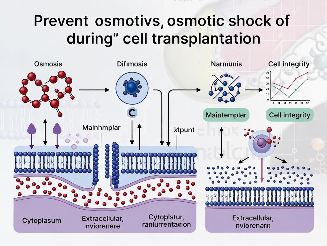
In cell transplantation research, maintaining high cell viability is not merely a preliminary quality check; it is a fundamental determinant of therapeutic success. A critical yet often overlooked factor that directly compromises viability is osmotic shock—the rapid and damaging change in cell volume and integrity that occurs during key procedures like cryopreservation, thawing, and infusion. This technical support center provides targeted guidance to help researchers identify, troubleshoot, and prevent osmotic injury, thereby safeguarding the functional potency of their cellular products and ensuring the efficacy of downstream therapeutic applications.
Troubleshooting Guides
Problem 1: Low Post-Thaw Viability
Observed Symptom: A significant proportion of cells are non-viable immediately after thawing, as indicated by membrane integrity assays.
Potential Causes and Solutions:
- Cause: Suboptimal Cryoprotectant Agent (CPA) Composition
- Solution: Transition from conventional dimethyl sulfoxide (DMSO) to optimized, DMSO-free CPA cocktails. These often combine naturally occurring osmolytes like trehalose (a sugar), glycerol (a sugar alcohol), and isoleucine (an amino acid), which have been shown to significantly improve post-thaw recovery. For hiPSC-derived cardiomyocytes (hiPSC-CMs), such formulations achieved over 90% post-thaw recovery, compared to ~69% with standard DMSO [17].
- Cause: Inadequate Control of Freezing Parameters
- Solution: Implement controlled-rate freezing and optimize key parameters. For hiPSC-CMs, a rapid cooling rate of 5 °C/min and a low nucleation temperature of -8 °C were identified as optimal. Using the wrong cooling rate can cause excessive intracellular ice formation or severe osmotic dehydration, both leading to cell death [17].
- Cause: Anomalous Osmotic Behavior Post-Thaw
- Solution: After thawing, cells may exhibit a sharp, detrimental drop in volume upon resuspension. Understanding and managing this excessive dehydration is crucial. Carefully control the osmolality and composition of the resuspension medium to facilitate a gentler volume recovery [17].
Problem 2: Reduced Cell Functionality After Transplantation
Observed Symptom: Cells survive the transplantation process but show impaired migration, proliferation, or engraftment in vivo.
Potential Causes and Solutions:
- Cause: Prior Exposure to Hyper-Osmotic Stress
- Solution: Minimize exposure to hyper-osmotic conditions during pre-transplant cell processing. Studies show that hyper-osmotic stress triggers a global decrease in cell migration and proliferation. For instance, metastatic cell lines like MDA-MB-231 showed a >40% decrease in migration speed under severe hyper-osmotic stress. This effect was more pronounced in 3D environments, which are more physiologically relevant [18].
- Cause: Disruption of Mechano-Osmotic Signaling
- Solution: Be aware that osmotic stress is not just a volumetric challenge but also a mechanical and signaling one. Osmotically induced cell shrinkage can deform the nucleus, activate the osmosensitive kinase p38 MAPK, and lead to global transcriptional repression. These changes "prime" the chromatin for a fate transition, potentially diverting cells from their intended therapeutic function. Maintaining isotonic conditions helps preserve the native nuclear environment and gene expression programs [19].
Problem 3: Inconsistent Purity of Final Cell Product
Observed Symptom: The final cell product intended for transplantation contains an unacceptable level of undesired or undifferentiated cell types.
Potential Causes and Solutions:
- Cause: Osmotic Sensitivity of Specific Cell Types
- Solution: Leverage differential osmotic sensitivity as a purification strategy. Some cell types may be more resilient to mild osmotic stress than others. Furthermore, biophysical properties like membrane rigidity and adhesive strength, which can be linked to osmotic history, change with differentiation state. Techniques like selective detachment based on adhesion strength can be used to enrich for specific populations [20].
Frequently Asked Questions (FAQs)
Q1: Why is DMSO a problem for cryopreservation, and what are the alternatives? A: While DMSO is a common cryoprotectant, it is associated with several issues: loss of post-thaw cell function and viability, adverse effects in patients (allergic, cardiac, neurological), and epigenetic disruptions. For therapeutic applications, especially with sensitive cells like hiPSC-CMs, DMSO-free alternatives are superior. These are often cocktails of naturally occurring osmolytes (e.g., trehalose, glycerol, isoleucine) that are optimized for specific cell types and have demonstrated significantly higher post-thaw recovery and maintained functionality [17].
Q2: How does hyper-osmotic stress directly impact a cell's therapeutic potential? A: Hyper-osmotic stress initiates a cascade of detrimental effects:
- Impaired Motility: Cell migration speed decreases proportionally to the stress level, critical for cells that need to migrate to engraft [18].
- Reduced Proliferation: The capacity for cells to expand after transplantation is compromised [18].
- Altered Gene Expression: It can induce an osmotic stress response (e.g., via p38 MAPK) and force nuclear deformation, which remodels chromatin and can aberrantly prime cells for differentiation, taking them off-therapy [19].
- Downregulation of Key Transporters: In 3D environments, hyper-osmotic stress can decrease the expression of important channels like aquaporin-5 (AQP5), further disrupting volume regulation [18].
Q3: What are the key parameters to monitor during a freeze-thaw cycle to prevent osmotic shock? A: The most critical parameters are:
- Cooling Rate: This must be optimized for each cell type to balance the risks of intracellular ice formation (too fast) and prolonged osmotic stress (too slow) [17].
- Nucleation Temperature: The temperature at which ice formation is initiated can affect ice crystal size and solute concentration [17].
- CPA Composition and Removal: The type and concentration of CPAs, as well as the protocol for their addition and—just as importantly—their dilution post-thaw, must be carefully designed to prevent damaging osmotic shifts [17].
Q4: My cells are viable post-thaw but don't function properly. What could be wrong? A: High membrane integrity (viability) immediately post-thaw does not guarantee functional potency. The cells may have undergone "metabolic injury" or "apoptotic priming." They might have activated caspase enzymes or suffered mitochondrial damage that commits them to apoptosis hours later. It is essential to perform functional assays (e.g., migration, proliferation, secretion, contraction for cardiomyocytes) 24-48 hours post-thaw and to use multiparametric flow cytometry to detect early apoptotic markers like caspase activation and phosphatidylserine exposure (Annexin V) alongside a viability dye [21] [22].
The following tables consolidate key quantitative findings from research on osmotic stress and cryopreservation, providing a reference for expected outcomes.
Table 1: Impact of Hyper-Osmotic Stress on Cell Migration [18]
| Cell Line | Condition | 125 mM Mannitol (% Change vs. Control) | 250 mM Mannitol (% Change vs. Control) |
|---|---|---|---|
| MDA-MB-231 | 3D | -27.2% | -42.2% |
| 2D | -30.1% | -52.7% | |
| A549 | 3D | +2.6% | -6.8% |
| 2D | -1.3% | -15.7% | |
| T24 | 3D | -20.4% | -26.9% |
| 2D | -1.1% | -17.0% |
Table 2: DMSO-Free vs. DMSO Cryopreservation of hiPSC-CMs [17]
| Parameter | DMSO (10%) | Optimized DMSO-Free CPA |
|---|---|---|
| Post-Thaw Recovery | 69.4% ± 6.4% | > 90% |
| Optimal Cooling Rate | ~1 °C/min (often used) | 5 °C/min |
| Optimal Nucleation Temp. | Not Specified | -8 °C |
| Post-Thaw Function | Preserved (if viable) | Preserved (cardiac markers, calcium transients) |
Experimental Protocols
Protocol 1: Assessing Apoptosis by Flow Cytometry
This multiparametric protocol helps identify cells that are viable but have initiated apoptosis, a common consequence of severe osmotic stress [21] [22].
Method: FLICA (Fluorochrome-Labeled Inhibitors of Caspases) Assay combined with a viability stain.
Step-by-Step Procedure:
- Harvest & Wash: Collect cell suspension (2.5x10^5 - 2x10^6 cells) in a FACS tube. Centrifuge at 1100 rpm for 5 minutes and resuspend the pellet in 1-2 mL of PBS. Repeat centrifugation.
- FLICA Staining: Discard supernatant and resuspend cell pellet in 100 µL PBS. Add 3 µL of FLICA working solution (e.g., FAM-VAD-FMK for pan-caspase detection).
- Incubate: Incubate for 60 minutes at +37°C, protected from light. Gently agitate cells every 20 minutes.
- Wash: Add 2 mL of PBS and centrifuge at 1100 rpm for 5 minutes. Discard supernatant and repeat this wash step once more.
- Viability Staining: Resuspend the final pellet in 100 µL of a propidium iodide (PI) staining mix. Incubate for 3-5 minutes.
- Analysis: Add 500 µL of PBS and analyze immediately on a flow cytometer using 488 nm excitation.
- FLICA-positive/PI-negative: Early Apoptotic
- FLICA-positive/PI-positive: Late Apoptotic/Necrotic
- FLICA-negative/PI-negative: Viable
Protocol 2: Measuring Mitochondrial Transmembrane Potential (Δψm)
Loss of Δψm is a sensitive marker of early apoptosis, which can be triggered by osmotic insult [21].
Method: Staining with Tetramethylrhodamine Methyl Ester (TMRM)
Step-by-Step Procedure:
- Harvest & Wash: Prepare a single-cell suspension as in Protocol 1, steps 1-2.
- Staining: Discard supernatant and add 100 µL of TMRM staining mix (e.g., 150 nM TMRM in PBS).
- Incubate: Incubate for 20 minutes at +37°C, protected from light.
- Analysis: Add 500 µL PBS and analyze on a flow cytometer (488 nm excitation, 575 nm emission).
- TMRM-bright: Healthy, viable cells with energized mitochondria.
- TMRM-dim/dull: Apoptotic or otherwise compromised cells.
Key Signaling Pathways and Workflows
Osmotic Stress Impact on Cell Fate
This diagram illustrates the mechanistic link between osmotic stress and compromised therapeutic efficacy.
Optimized Cryopreservation Workflow
This workflow contrasts standard and optimized protocols to highlight critical steps for preserving viability.
The Scientist's Toolkit: Research Reagent Solutions
Table 3: Essential Reagents for Osmotic Shock and Viability Research
| Reagent / Kit | Function / Application | Key Note |
|---|---|---|
| DMSO-Free CPA Cocktail (e.g., Trehalose, Glycerol, Isoleucine) | Cryopreservation without DMSO toxicity. | Must be optimized for specific cell types; superior for hiPSC-CMs [17]. |
| FLICA Assay Kits (e.g., FAM-VAD-FMK) | Flow cytometry detection of active caspases, marking apoptotic cells. | Critical for identifying early apoptosis post-thaw, before membrane rupture [21] [22]. |
| TMRM Probe | Flow cytometry assessment of mitochondrial membrane potential (Δψm). | A drop in fluorescence indicates early mitochondrial dysfunction, a prelude to apoptosis [21]. |
| Annexin V Conjugates (e.g., FITC, APC) | Detection of phosphatidylserine exposure on the outer leaflet of the plasma membrane. | Used with a viability dye (e.g., PI) to distinguish early apoptotic (Annexin V+/PI-) from late apoptotic/necrotic (Annexin V+/PI+) cells [21]. |
| Propidium Iodide (PI) | DNA intercalating dye that stains cells with compromised membranes. | Standard viability dye; impermeant to live cells. Use in combination with other probes [21]. |
Strategic Protocols for Osmoprotection: From Cryopreservation to Surgical Delivery
Troubleshooting Guides
Common Cryopreservation Issues and Solutions
Table 1: Troubleshooting Common Cell Cryopreservation Problems
| Problem | Possible Causes | Recommended Solutions |
|---|---|---|
| Low post-thaw viability | Suboptimal cooling rate [23], improper cryoprotectant concentration [23], unhealthy pre-freeze cells [24] [25] | - Optimize cooling rate (typically -1°C/min) [24] [25].- Ensure cells are >90% viable and in log phase pre-freeze [24] [25].- Avoid over-exposure to cryoprotectants before freezing [26]. |
| Excessive cell death post-thaw | Intracellular ice formation [23], toxic cryoprotectant exposure [23], improper thawing technique [26] | - Use controlled-rate freezing to prevent ice formation [27].- Rapidly thaw at ~37°C [25] [28] and dilute/DMSO immediately [26] [29].- Use a defined, serum-free freezing medium for consistency [25]. |
| Contamination in frozen stock | Non-sterile technique during freezing [25] | - Wipe all containers with 70% ethanol or isopropanol before opening [25].- Use proper aseptic techniques [25]. |
| Poor cell recovery/functionality | Osmotic shock during thawing [27], improper post-thaw handling [26] | - Thaw rapidly and use pre-warmed media [26] [25].- For sensitive primary cells, consider seeding directly and changing media the next day instead of centrifuging [29]. |
Thawing Process Troubleshooting
Table 2: Thawing-Specific Issues and Corrective Actions
| Observation | Underlying Issue | Corrective Action |
|---|---|---|
| Low viability immediately after thawing | Slow thawing leading to ice recrystallization [25] [27] | Thaw vials rapidly in a 37°C water bath until only a small ice sliver remains [25] [28]. |
| Cells appear healthy post-thaw but fail to attach/grow | Cryoprotectant toxicity (e.g., DMSO) [26] [23], damage from residual dissociation reagents [29] | Remove cryoprotectant promptly post-thaw via centrifugation or media change [26].Use gentle, cell-specific dissociation reagents during pre-freeze harvest [29]. |
| Clumping of cells after thaw | Freezing at an excessively high cell concentration [25] | Freeze cells at the recommended density (e.g., ~1x10^6 cells/mL for many types) and avoid high concentrations [26] [25]. |
Frequently Asked Questions (FAQs)
1. What is the single most critical factor for high cell viability after thawing? While multiple factors are important, controlled cooling rate is fundamental. A slow, controlled rate of approximately -1°C per minute helps prevent lethal intracellular ice crystal formation by allowing water to exit the cell before freezing, thereby minimizing osmotic stress and mechanical damage [24] [25] [23].
2. Why is rapid thawing so strongly recommended? Rapid thawing in a 37°C water bath is crucial for two main reasons:
- It minimizes the time cells are exposed to the toxic effects of cryoprotectants like DMSO [27].
- It prevents damage from ice recrystallization during the warming phase, which can rupture cell membranes [25]. Slow thawing can undo all the careful preparation of the freezing process [23].
3. My cells are not recovering well after thawing, even with rapid thawing. What else should I check? First, verify the quality and health of the cells before freezing; they should be in log phase and over 90% viable [24] [25]. Second, review your post-thaw handling. For some sensitive primary cells, the damage from centrifuging to remove DMSO is harsher than the residual DMSO itself. In these cases, following a recommended seeding density to dilute the DMSO and changing the media the day after seeding can be more effective [29].
4. Can I re-freeze cells that have been thawed? It is strongly discouraged. Each freeze-thaw cycle subjects cells to osmotic stress, ice-crystal formation, and cryoprotectant-related stress. Cells that are thawed, re-frozen, and thawed again will have significantly lower viability than cells thawed only once [26]. It is best to plan experiments and create multiple vials at an appropriate cell count to avoid the need for re-freezing.
5. Are there alternatives to DMSO as a cryoprotectant? Yes, but DMSO remains the most common and effective for many mammalian cell types [23]. Alternatives include:
- Glycerol: Often used for red blood cells, bacteria, and yeasts [26].
- Commercial, defined cryopreservation media: Formulations like CryoStor are serum-free and designed to offer a protective environment [25].
- Polyvinylpyrrolidone (PVP) and Methylcellulose: These are extracellular cryoprotectants that have been studied as alternatives, sometimes in combination with reduced DMSO concentrations [26].
6. What are the key differences between passive and controlled-rate freezing? Controlled-rate freezers (CRFs) actively control the cooling rate, allowing for precise documentation and optimization of critical process parameters. This is preferred for sensitive cells and regulated fields like cell therapy [27]. Passive freezing (using isopropanol chambers like "Mr. Frosty") is a simple, low-cost method that also aims to achieve the -1°C/minute rate and is sufficient for many routine lab cell lines [24] [25] [27].
Experimental Protocols
Detailed Methodology: Standard Slow Freezing and Rapid Thaw
This protocol is adapted from industry standards for freezing suspension cells [24] [25].
Freezing Protocol:
- Harvest: Gently detach and harvest cells that are in the log phase of growth and have >90% viability [24] [25].
- Count: Determine total cell count and viability using Trypan Blue exclusion and a hemocytometer or automated counter [24].
- Centrifuge: Centrifuge the cell suspension at approximately 100–400 × g for 5–10 minutes. Aspirate the supernatant completely [24].
- Resuspend in Freezing Medium: Resuspend the cell pellet in an appropriate, cold freezing medium (e.g., complete growth medium with 10% DMSO or a commercial serum-free alternative) to a final concentration of ~1x10^6 cells/mL [24] [26] [25]. Keep the suspension cold.
- Aliquot: Dispense 1 mL aliquots into sterile cryogenic vials. Mix the cell suspension gently but often during aliquoting to ensure a homogeneous distribution [24].
- Freeze: Place vials in a controlled-rate freezer or an isopropanol freezing chamber and transfer to a -80°C freezer for 18-24 hours to achieve a cooling rate of ~-1°C/minute [24] [25] [28].
- Store: Transfer frozen vials to long-term storage in the vapor phase of liquid nitrogen (< -135°C) [24] [25] [28].
Thawing Protocol:
- Quick Thaw: Retrieve a vial from storage and immediately place it in a 37°C water bath. Gently agitate until only a tiny sliver of ice remains (usually 2-3 minutes). Do not submerge the vial cap [25] [28].
- Decontaminate: Wipe the vial thoroughly with 70% ethanol before opening [25] [28].
- Dilute: Transfer the thawed cell suspension drop-wise into a tube containing 9-10 mL of pre-warmed complete culture medium. This gradual dilution reduces osmotic shock [26].
- Wash (Optional): Centrifuge the cell suspension at a moderate speed for 5-10 minutes to pellet cells and remove the cryoprotectant-containing supernatant. Resuspend the pellet in fresh, pre-warmed culture medium [26].
- Seed: Transfer the cell suspension to a culture vessel at the recommended density. If cells are particularly sensitive, the media can be changed 24 hours after seeding to remove residual DMSO [29].
Workflow Visualization
Cryopreservation and Thawing Process
Osmotic Stress Pathways During Cryopreservation
The Scientist's Toolkit
Table 3: Essential Reagents and Equipment for Cryopreservation
| Item | Function & Rationale | Example Products / Notes |
|---|---|---|
| Cryoprotective Agent (CPA) | Lowers freezing point, reduces ice crystal formation, and protects from osmotic stress [24] [23]. | DMSO (most common for mammalian cells) [24] [23], Glycerol [26], Commercial Serum-Free Media (e.g., CryoStor, Synth-a-Freeze) [24] [25]. |
| Controlled-Rate Freezing Apparatus | Ensures a consistent, optimal cooling rate (typically ~-1°C/min) to prevent intracellular ice formation [24] [25] [27]. | Programmable Controlled-Rate Freezer (CRF) [27], Passive Cooling Chambers (e.g., "Mr. Frosty," CoolCell) [24] [25]. |
| Cryogenic Storage Vials | Designed to withstand extreme temperatures of liquid nitrogen storage without cracking [24]. | Internal-threaded vials are preferred to prevent contamination [25]. |
| Liquid Nitrogen Storage System | Provides long-term storage at <-135°C, halting all metabolic activity to preserve cells indefinitely [24] [26] [25]. | Store in the vapor phase to reduce explosion risks [24] [28]. |
| Water Bath or Thawing Device | Enables rapid thawing at a consistent 37°C to minimize ice recrystallization and cryoprotectant toxicity [25] [27]. | For GMP compliance, use a validated, closed-system thawing device instead of open water baths [27]. |
Formulating Osmotically Balanced Transport and Wash Media
Cellular Mechanisms of Osmotic Stress
What are the fundamental osmotic principles my media must address? During ex vivo handling and storage, cells are removed from their native environment, which disrupts the delicate osmotic balance normally maintained by ATP-driven ion pumps in the cell membrane [30]. Under hypothermic conditions (2-8°C), commonly used for transport, these metabolic pumps are inactivated [30]. In a standard isotonic solution like saline or culture media, which mimics the extracellular ionic balance (high sodium, low potassium), this leads to ion diffusion along concentration gradients and causes cellular swelling and potential lysis [30]. An effective osmotically balanced solution must counteract this by mimicking the intracellular environment (high potassium, low sodium) to prevent water influx and cell swelling during this period of metabolic arrest.
How does osmotic shock specifically damage cells during procedures like islet isolation? Techniques like Selective Osmotic Shock (SOS) leverage osmotic principles to selectively isolate target cells. This method uses hyperosmolar glucose solutions to disrupt exocrine pancreatic cells that lack the GLUT2 glucose transporter, while pancreatic β-cells, which express GLUT2, are protected [31] [32]. In non-transporter cells, exposure to high glucose creates an osmotic gradient that draws water out, causing instantaneous cell shrinkage. Subsequent immersion in a low-glucose solution causes rapid water influx, selectively rupturing these cells [31]. Your media formulation must therefore be tailored to the specific osmolyte transporters expressed by the cell type you wish to preserve.
Experimental Protocols for Validation
Protocol 1: Testing Media Efficacy for Hypothermic Storage
This protocol assesses the performance of a candidate intracellular-like transport medium against a standard extracellular-like solution (e.g., saline) for preserving cell viability during cold storage.
- Step 1: Cell Preparation and Baseline Viability. Obtain the target cells (e.g., follicular unit grafts, islets, or adherent cells). Take a pre-cooled sample and determine baseline viability and cell count using trypan blue exclusion or flow cytometry with Annexin V/PI staining [30].
- Step 2: Experimental Setup. Aliquot the cell samples into two groups. Pellet the cells and resuspend one group in the test intracellular-like preservation solution. Resuspend the other group in a control isotonic saline or culture medium [30].
- Step 3: Hypothermic Storage. Store all samples at 2-8°C for a defined period (e.g., 24 hours), simulating transport conditions [30].
- Step 4: Viability and Function Assessment. After storage, re-warm the samples to 37°C. Measure post-preservation viability. For functional assessment, plate the cells and monitor attachment efficiency, proliferation rates, or conduct cell-specific functional assays (e.g., Glucose-Stimulated Insulin Secretion for islets) [33].
Protocol 2: Selective Osmotic Shock for Cell Isolation
This non-enzymatic method isolates osmotically resistant cells, such as pancreatic islets, and is adapted from published studies in canine and feline models [31] [32].
- Step 1: Tissue Acquisition and Mechanical Disruption. Obtain pancreatic tissue and place it in cold, glucose-free RPMI 1640 medium. Mince the tissue into fragments of <3 mm using a scalpel [31] [32].
- Step 2: Hyperosmolar Incubation. Divide the tissue fragments equally and incubate them in a hyperosmolar glucose solution (e.g., 300 mmol/L or 600 mmol/L glucose in RPMI) for 20-40 minutes at room temperature with periodic agitation [31] [32].
- Step 3: Osmotic Shock and Wash. Centrifuge the samples and completely decant the hyperosmolar solution. Resuspend the pellet in a glucose-free isotonic medium (e.g., RPMI 1640) to induce osmotic shock in exocrine cells. Repeat this wash step three times to ensure complete removal of debris [31] [32].
- Step 4: Islet Collection and Culture. Plate the final pellet in a standard islet culture medium (e.g., RPMI 1640 + 10% FBS) and culture at 37°C. Assess islet yield, purity, and function via GSIS [32].
Troubleshooting Common Osmotic Media Issues
My cells show poor viability after transport in a new balanced salt solution. What is the first parameter I should check? The most common issue is inadvertent intracellular-like composition in an extracellular application, or vice versa. First, verify the sodium-to-potassium (Na+/K+) ratio of your solution against your cell type's requirements. For hypothermic storage, the solution should have a high K+/low Na+ ratio to prevent swelling [30]. For normothermic culture wash steps, the solution must be extracellular-like (high Na+/low K+) to support active pump function. Using the wrong formulation for the temperature context is a frequent error.
I am using DMSO as a cryoprotectant, but my cell therapy product is causing side effects in patients. What are my options? Dimethyl sulfoxide (DMSO) is associated with clinical side effects including cardiovascular, neurological, and allergic reactions [34]. To mitigate this:
- Post-thaw DMSO Reduction: Implement a washing step to reduce DMSO concentration before infusion. Be aware that this process can lead to a significant loss of viable CD34+ cells (median loss of ~48.5%), so it should only be applied to high-risk patients [35].
- Investigate DMSO-Free Formulations: The field is actively developing alternatives using sugars, polymers, amino acids, and other osmolytes. While challenging for sensitive T and NK cells, these infusible, GMP-scalable solutions are the future for safer cell therapies [34].
My isolated islets are unresponsive to glucose challenge after isolation via osmotic shock. Where did my protocol fail? This indicates a loss of β-cell function. Focus your troubleshooting on the SOS parameters:
- Optimize Osmolality and Time: Test different glucose concentrations (e.g., 300 vs. 600 mmol/L) and incubation times (e.g., 20 vs. 40 minutes). Research shows the optimal combination can be cell-source dependent; for feline islets, 600 mmol/L for 20 minutes yielded the best glucose responsiveness [32].
- Control Mechanical Stress: Overly vigorous homogenization or trituration can physically damage cells. Ensure mechanical steps are gentle and brief [32].
- Confirm Function Post-Culture: Islets may need a recovery period in culture post-isolation before robust function is restored. Do not assess function immediately after the isolation shock [33].
Quantitative Data and Formulations
Table 1: Key Research Reagent Solutions for Osmotic Balancing
| Reagent/Solution | Primary Function | Technical Notes & Considerations |
|---|---|---|
| Intracellular-Type Solution | Prevents cell swelling during hypothermia by mimicking the intracellular ionic milieu (high K+/low Na+) [30]. | Essential for transport and cold storage. Do not use for normothermic culture. |
| Hyperosmolar Glucose Solution | Selectively disrupts cells lacking specific glucose transporters (e.g., GLUT2) for purification [31] [32]. | Concentration and exposure time must be optimized for each species and cell type. |
| DMSO (Dimethyl Sulfoxide) | Penetrating cryoprotectant; prevents intracellular ice crystal formation during freezing [34]. | Can cause toxicity and alter cell function. Clinical dose should not exceed 1 g/kg patient weight [34] [35]. |
| Trehalose | Non-penetrating osmolyte and disaccharide; provides membrane stabilization during freezing and hypothermic storage [36]. | Serves as an adjuvant in preservation solutions like UW and ET-Kyoto to improve outcomes [36]. |
| Hydrogel (e.g., Alginate) | Provides a non-toxic, biocompatible physical encapsulation that inhibits ice crystal formation during cryopreservation [36]. | A promising alternative to traditional cytotoxic cryoprotectants for tissues. |
Table 2: Composition and Outcomes of Selective Osmotic Shock (SOS) Protocols
| Species | Glucose Concentration | Incubation Time | Key Outcome Measures | Reference |
|---|---|---|---|---|
| Canine | 300 mOsm / 600 mOsm | 20 min / 40 min | Yield: 428 - 990 islet equivalents/gram. Viability: ~89% across groups. Best Function: Lower glucose (300 mOsm) showed superior stimulation index [31]. | [31] |
| Feline | 300 mmol/L / 600 mmol/L | 20 min / 40 min | Yield: Varied significantly by individual cat. Best Function: 600 mmol/L for 20 min produced the highest glucose stimulation index [32]. | [32] |
Visualizing Osmotic Stress Pathways and Protocols
The following diagram illustrates the key cellular pathways activated by osmotic imbalance during hypothermic storage, and the points targeted by balanced preservation media.
This workflow outlines the key steps for isolating cells using a selective osmotic shock protocol, providing a visual guide for experimental execution.
The Role of Cryoprotectants and Non-Penetrating Osmoprotants
Frequently Asked Questions (FAQs)
Q1: What is the fundamental difference between a cryoprotectant and an osmoprotectant in cell transplantation? Cryoprotectants are primarily used to protect cells from damage during the freezing and thawing processes of cryopreservation. They work by preventing lethal intracellular ice crystal formation and mitigating osmotic stress during temperature changes [37]. Osmoprotectants (or compatible solutes) are a class of compounds that protect cells from osmotic shock—a sudden change in the solute concentration around the cell—by balancing the internal osmotic pressure without disrupting cellular metabolism. They stabilize proteins, maintain membrane integrity, and can scavenge reactive oxygen species (ROS) [38] [39]. In the context of cell transplantation, cryoprotectants are essential for the long-term storage of cells, while osmoprotectants are crucial for maintaining cell viability during the transplantation procedure itself, which can expose cells to osmotic fluctuations.
Q2: Why are non-penetrating agents often preferred or used in combination with penetrating ones? Non-penetrating cryoprotectants are often preferred in certain protocols, or used in combination with penetrating agents, primarily to reduce toxicity. Penetrating agents (e.g., DMSO) can be toxic to cells at high concentrations [37] [40]. Using a combination of both allows for a reduction in the concentration of the penetrating agent required for effective cryopreservation, thereby minimizing its toxic effects while still providing adequate protection. Non-penetrating agents provide extracellular protection by preventing extracellular ice formation and reducing chilling injury [37] [40].
Q3: My cells show low post-thaw viability despite using DMSO. What could be the cause? Low post-thaw viability can be attributed to several factors:
- Inappropriate Cooling/Thawing Rate: The optimal cooling rate (e.g., approximately 1°C/min for many cell types) is critical to prevent intracellular ice formation. Similarly, rapid thawing is generally recommended to avoid recrystallization [37] [41].
- CPA Toxicity & Exposure Time: DMSO can be toxic, especially with prolonged exposure at room temperature [40]. Minimize the time cells are in contact with DMSO before freezing and after thawing.
- Insufficient or Incorrect CPA Concentration: The standard 10% DMSO may not be optimal for all cell types [37]. Hepatocytes, stem cells, and oocytes often require cell-specific optimization.
- Osmotic Shock During CPA Addition/Removal: The addition and, more critically, the removal of CPAs can cause significant osmotic stress. A stepwise or gradual dilution method is recommended to prevent this [37].
Q4: How can I protect my cells from osmotic shock during the washing or dilution steps post-thaw? To protect cells from osmotic shock during washing:
- Use Osmoprotectants: Incorporate non-penetrating osmoprotectants like sucrose, trehalose, or mannitol into the washing solutions. These compounds help stabilize the cell membrane externally and reduce the osmotic gradient [38] [39].
- Apply a Stepwise Dilution: Instead of a single-step dilution, gradually decrease the concentration of the penetrating cryoprotectant by adding the wash medium in a stepwise manner. This allows for a slower, more controlled efflux of the CPA from the cell, preventing rapid water influx and swelling [37].
- Utilize Non-Penetrating CPAs: As part of the freezing solution, non-penetrating CPAs like polyethylene glycol (PEG) can reduce the required concentration of toxic penetrating CPAs, thereby lessening the osmotic stress during their removal [40].
Troubleshooting Guides
Problem 1: Ice Crystal Formation and Cell Membrane Damage
Potential Causes:
- Inadequate or absent cryoprotectant.
- Suboptimal cooling rate that is too slow (leading to excessive dehydration) or too fast (leading to intracellular ice formation) [37].
- Use of a cryoprotectant that is ineffective for the specific cell type.
Solutions:
- Optimize Cryoprotectant Cocktail: Ensure a sufficient concentration and consider using a combination of penetrating (e.g., DMSO, Glycerol) and non-penetrating (e.g., Sucrose, Trehalose, PEG) agents. Vitrification mixtures can be used to achieve a glassy state without ice crystallization [37] [42].
- Control Cooling Rate: Implement a controlled-rate freezer. For many cells, a cooling rate of approximately -1°C/min is effective, though some cells (like oocytes) require rapid cooling [37].
- Use Ice Binding Molecules: Incorporate antifreeze proteins (AFPs) or synthetic polymers that inhibit ice recrystallization (IRI) during the thawing process [42].
Problem 2: Osmotic Shock During Cryoprotectant Removal
Potential Causes:
- Rapid dilution of the extracellular cryoprotectant, causing a sudden influx of water into the cells.
- Lack of osmotic support in the washing or culture medium.
Solutions:
- Stepwise Dilution: Remove the cryoprotectant by sequentially diluting the cell suspension with a medium containing an osmoprotectant (e.g., sucrose). For example, gradually add the wash medium in volumes of 25%, 50%, and 100% of the sample volume over 5-10 minute intervals.
- Incorporate Osmoprotectants: Use washing solutions supplemented with non-penetrating osmoprotectants like sucrose (0.2-0.5 M) or trehalose. These stabilize the cell membrane from the outside and slow down water movement [38] [39].
- Utilize Microencapsulation: Alginate encapsulation can provide a physical barrier that buffers cells against rapid osmotic changes during CPA removal and transplantation [37] [14].
Problem 3: Low Cell Survival Post-Transplantation
Potential Causes:
- Combined stress from cryopreservation and the transplantation microenvironment.
- Inflammatory response and oxidative stress at the transplantation site.
Solutions:
- Pre-conditioning with Osmoprotectants: Incubate cells with osmoprotectants like trehalose, proline, or glycine betaine before cryopreservation. This can enhance their inherent resistance to both freezing and osmotic stress [38].
- Anti-apoptotic Treatment: Pre-incubate cells with anti-oxidants (e.g., Trolox, Ascorbic acid) to reduce reactive oxygen species (ROS) generated during cryopreservation and after transplantation [37].
- Use Bio-inspired CPAs: Explore the use of novel, less toxic cryoprotectants like natural deep eutectic solvents (NADES) or specific saccharides, which show excellent biocompatibility and osmoprotective properties [42].
Quantitative Data for Experimental Design
Table 1: Common Cryoprotectants and Osmoprotectants
| Agent Name | Type (Penetrating/Non-Penetrating) | Common Working Concentration | Key Function & Notes |
|---|---|---|---|
| Dimethyl Sulfoxide (DMSO) | Penetrating [40] | 5-10% (v/v) [37] | Prevents intracellular ice; increases membrane porosity; can be toxic [37]. |
| Glycerol (GLY) | Penetrating [37] | 5-15% (v/v) [37] | Early discovered CPA; protects against ice crystals; often used for red blood cells and spermatozoa [37]. |
| Ethylene Glycol (EG) | Penetrating [37] | 1.5-4 M (for vitrification) [37] | Lower toxicity alternative to DMSO; commonly used in vitrification mixtures [37]. |
| Sucrose | Non-Penetrating [38] | 0.2-0.5 M [38] | Provides osmotic buffering; used in washing solutions to prevent osmotic shock [38]. |
| Trehalose | Non-Penetrating [38] | 0.1-0.4 M [38] | Disaccharide; stabilizes proteins and membranes; acts as an osmoprotectant and cryoprotectant [38] [42]. |
| Polyethylene Glycol (PEG) | Non-Penetrating [40] | Varies by molecular weight [40] | Polymer; inhibits extracellular ice formation; reduces toxicity of penetrating CPAs in combination [40]. |
| Proline | Osmoprotectant [38] | 10-100 mM [38] | Amino acid; accumulates in stressed plants; stabilizes protein structures and scavenges free radicals [38]. |
| Glycine Betaine | Osmoprotectant [38] | 10-100 mM [38] | Quaternary ammonium compound; protects against osmotic stress and stabilizes macromolecular structures [38]. |
Table 2: Cell-Type Specific Cryopreservation Recommendations
| Cell Type | Recommended Cooling Rate | Recommended Cryoprotectant | Special Considerations |
|---|---|---|---|
| Hepatocytes | Slow (~1°C/min) [37] | 10% DMSO [37] | High susceptibility to freezing damage; requires optimized protocols. |
| Pancreatic Islets | Rapid [37] | DMSO or Vitrification Mixtures [37] | Alginate encapsulation can improve post-thaw viability and function [37]. |
| Mesenchymal Stem Cells (MSCs) | Slow (~1°C/min) [37] | 10% DMSO [37] | Pre-incubation with glucose and anti-oxidants can maximize yields [37]. |
| Oocytes | Rapid (Vitrification) [37] | EG + DMSO mixtures common [37] | Extremely sensitive to chilling injury; vitrification is associated with better outcomes [37]. |
| Spermatozoa | Slow or Rapid (protocol dependent) | Glycerol [37] | First successfully cryopreserved cells; relatively robust. |
Experimental Protocol: Selective Osmotic Shock for Cell Isolation
This non-enzymatic method, based on Selective Osmotic Shock (SOS), can be used to isolate specific cell types, such as pancreatic islets, by exploiting differences in cellular osmoprotective mechanisms [14].
Principle: Cells with specific glucose transporters (e.g., Glut-2 in pancreatic beta cells) can rapidly adapt to osmotic changes by transporting solutes. Other cell types (e.g., acinar cells) lack this ability and lyse under controlled osmotic stress [14].
Workflow Diagram:
Materials & Reagents:
- Tissue Sample (e.g., Pancreas)
- Sterile PBS, cold
- RPMI-1640 Medium
- 1 M HEPES Buffer
- D-Glucose
- Betadine (10%)
- Surgical instruments, 50 mL conical tubes, 500 μM strainer, irrigation syringe [14]
Step-by-Step Methodology:
- Tissue Preparation: Harvest the tissue into cold PBS. Remove associated viscera and fat using blunt dissection. Weigh the tissue and rinse with cold PBS. Disinfect by incubating in 10% ice-cold betadine for 5 minutes, followed by three rinses in cold PBS [14].
- Mincing: On a chilled surgical plate, finely mince the tissue into pieces <0.25 cm² and place them in 50 mL conical tubes.
- High-Glucose Exposure (Osmotic Loading): Add a volume of ice-cold 300-600 mM glucose RPMI medium equal to the tissue volume. Incubate on ice for 20 minutes. Centrifuge at 200 × g for 2 minutes at 4°C and decant the supernatant [14].
- Low-Glucose Exposure (Osmotic Shock): Quickly add a volume of 0 mM glucose RPMI medium. Gently invert the tube to mix. This creates a hypotonic environment, causing vulnerable cells to swell and lyse. Centrifuge at 200 × g for 2 minutes at 4°C and decant. Repeat this wash step twice [14].
- Mechanical Dissociation: Transfer the tissue to a sterile beaker with 0 mM RPMI. Using an irrigation syringe, gently dissociate the tissue by repeatedly aspirating and expelling for 5-10 minutes. Keep the beaker on ice. Continue until the tissue is sufficiently dispersed.
- Filtration and Collection: Pass the dissociated cell suspension through a 500 μM strainer into a new collection beaker. Take any retained tissue and repeat the dissociation and filtration steps. Combine the filtered suspensions in 50 mL tubes and centrifuge at 100 × g for 3 minutes at 4°C. The pellet contains the osmotically resistant target cells (e.g., islets) [14].
The Scientist's Toolkit: Essential Research Reagent Solutions
Table 3: Key Reagents for Cryopreservation and Osmoprotection Studies
| Reagent | Function | Example Application in Protocols |
|---|---|---|
| Dimethyl Sulfoxide (DMSO) | Penetrating Cryoprotectant | Standard cryopreservation of cell lines, stem cells; used at 5-10% in culture medium [37] [40]. |
| Sucrose | Non-Penetrating Osmoprotectant | Osmotic buffer in cryoprotectant washing solutions; used at 0.2-0.5 M to prevent osmotic shock [38]. |
| Trehalose | Non-Penetrating Cryo-/Osmoprotectant | Stabilizer in freezing solutions and wash buffers; protects membranes and proteins during desiccation and osmotic stress [38] [39]. |
| Antifreeze Proteins (AFPs) | Ice Recrystallization Inhibitor | Added to cryopreservation media to control ice crystal growth and morphology, reducing mechanical damage [42]. |
| HEPES Buffer | pH Stabilizer | Maintains physiological pH in solutions (e.g., RPMI) during processing steps outside a CO₂ incubator [14]. |
| Alginate | Microencapsulation Polymer | Encapsulates cells or tissues (e.g., islets) to provide a physical buffer against osmotic shock and immune response post-transplantation [37] [14]. |
| Proline / Glycine Betaine | Metabolic Osmoprotectant | Pre-incubation of cells before freezing or transplantation to enhance innate resistance to osmotic stress [38]. |
Mechanism Diagrams
Cell Response to Osmotic Stress and Protection
Cryoprotectant Mechanism of Action
Step-by-Step Guide for Safe Cell Preparation and Resuspension
Frequently Asked Questions (FAQs)
1. What is osmotic shock and why is it a concern in cell preparation? Osmotic shock occurs when cells are exposed to a sudden change in the concentration of solutes, such as salts, outside the cell, causing rapid water movement across the cell membrane. This can lead to cells swelling and bursting (in a hypotonic environment) or shrinking and crumpling (in a hypertonic environment). In cell transplantation research, this is a major concern as it can severely reduce cell viability and compromise the success of the transplant by damaging or destroying the cells before they are even administered [14].
2. How can I tell if my cell suspension has undergone osmotic stress? Signs of osmotic stress can be observed under a microscope. Cells may appear shriveled or, conversely, swollen and burst. You may also notice a significant amount of cell debris in the suspension, and cell viability counts (using dyes like trypan blue) will be lower than expected. A key indicator is a high rate of cell death following the resuspension process [43] [44].
3. What is the single most important factor in preventing osmotic shock? The most critical factor is ensuring that all solutions used during the preparation and resuspension steps are isotonic and properly balanced. This means the osmotic pressure of the solutions should match that of the cell's interior. Using specially formulated buffers like Dulbecco's Phosphate-Buffered Saline (DPBS) or Hank's Balanced Salt Solution (HBSS) is essential. Always pre-warm or cool your media to the correct temperature before adding cells to avoid additional temperature-induced stress [45] [46].
4. My cells are clumping after resuspension. What should I do? Cell clumping is often caused by free DNA and cell debris from lysed cells, which is sticky and causes aggregation [43]. To address this:
- Gently triturate (pipette up and down) the cell suspension using a pipette with a narrow-bore tip.
- Consider adding a small amount of DNase I to your suspension, which will degrade the free DNA and help prevent clumping [47].
- Ensure you are not over-digesting your tissue with proteolytic enzymes during the initial dissociation, as this can lead to more cell lysis [43].
5. Are there non-enzymatic methods to isolate cells that are gentler? Yes, methods like Selective Osmotic Shock (SOS) can be used for specific tissues. This technique exploits the natural properties of certain cells to survive osmotic changes that destroy others. For example, in pancreatic islet isolation, islet cells have glucose transporters (GLUT2) that allow them to adapt to high glucose solutions, while surrounding exocrine cells swell and burst when the osmotic environment is changed, freeing the islets without harsh enzymes [14] [32].
Troubleshooting Guides
Problem: Low Cell Viability After Resuspension
| Possible Cause | Diagnostic Steps | Solution |
|---|---|---|
| Use of Hypotonic or Hypertonic Solutions | Check the osmolarity of all buffers and media used with an osmometer. | Ensure all solutions are isotonic. Use commercial, pre-tested buffers like DPBS or HBSS. |
| Rapid Temperature Change | Review protocol steps for temperature shifts (e.g., moving cells from ice to a 37°C water bath). | Always pre-warm culture media and slowly acclimatize cells to new temperatures when possible. |
| Over-digestion with Enzymes | Assess incubation time and concentration of enzymes like trypsin or collagenase. | Optimize digestion time and enzyme concentration. Use gentler alternatives like TrypLE where appropriate [46]. |
| Harsh Mechanical Force | Observe technique during pipetting and centrifugation. | Use wide-bore pipette tips for resuspension. Centrifuge at the lowest effective speed and duration (e.g., 200 x g for 5 minutes) [45]. |
Problem: Excessive Cell Clumping in Suspension
| Possible Cause | Diagnostic Steps | Solution |
|---|---|---|
| Presence of Free DNA/Debris | Look for stringy, viscous material in the suspension under a microscope. | Add DNase I (1-10 µg/mL) to the suspension to digest free DNA [47]. Filter the suspension through a cell strainer (e.g., 40-70 µm). |
| Incomplete Dissociation | Check for large tissue fragments or clusters in the suspension. | Optimize tissue dissociation protocol (enzyme type, time, temperature). Gentle mechanical homogenization may be needed [46]. |
| High Cell Density | Determine the cell concentration using a hemocytometer. | Dilute the cell suspension to the recommended density for your cell type to reduce cell-cell contact. |
Research Reagent Solutions
The following table lists key reagents essential for safe cell preparation and resuspension.
| Item | Function | Specific Example |
|---|---|---|
| Dulbecco's PBS (DPBS) | An isotonic, balanced salt solution used for washing cells and diluting reagents without inducing osmotic shock. | |
| Hank's Balanced Salt Solution (HBSS) | Another balanced salt solution used for maintaining cellular osmotic balance during tissue dissection and cell washing steps. | |
| TrypLE Express | A gentler, recombinant enzyme alternative to trypsin for dissociating adherent cells. It minimizes damage to cell surface proteins and improves post-digestion viability [47] [46]. | |
| Collagenase | An enzyme blend used to digest the extracellular matrix (particularly collagen) in solid tissues to isolate single cells [47] [46]. | Type I (for intestines, mammary glands), Type II (for cartilage, bone) [46]. |
| DNase I | An enzyme added during or after tissue dissociation to degrade free DNA released from lysed cells, thereby reducing cell clumping and aggregation [47]. | |
| Dispase | A neutral protease effective in breaking down fibronectin and collagen IV in the extracellular matrix. It is considered gentle and good for detaching cell colonies [47] [46]. | |
| Accutase | A ready-to-use blend of proteolytic and collagenolytic enzymes that is effective for dissociating difficult cell lines and primary cells while maintaining good viability [47]. |
Detailed Experimental Protocols
Protocol 1: Standard Safe Preparation of a Single-Cell Suspension from Solid Tissue
This protocol is adapted from general best practices for preparing single-cell suspensions for sensitive downstream applications like flow cytometry or transplantation [47] [46].
Materials:
- Solid tissue sample
- Ice-cold, isotonic buffer (e.g., DPBS or HBSS)
- Appropriate digestion enzyme(s) (e.g., Collagenase, Dispase, TrypLE)
- DNase I
- Cell strainer (70 µm and/or 40 µm)
- Centrifuge tubes
- Culture medium with serum
Steps:
- Tissue Dissection and Mincing: Immediately place the harvested tissue in ice-cold DPBS. Using a scalpel or scissors, finely mince the tissue into pieces smaller than 3 mm³ on a chilled surface. This increases the surface area for enzyme action [47].
- Enzymatic Digestion: Transfer the minced tissue to a tube containing a pre-warmed, pre-mixed enzyme solution (e.g., Collagenase/Dispase with DNase I) in an isotonic buffer. Use a volume that fully submerges the tissue.
- Incubate with Agitation: Incubate the tube at 37°C for 30-60 minutes with gentle agitation (e.g., on a rocking platform or with occasional manual shaking). The time must be optimized for each tissue type to avoid over-digestion [46].
- Mechanical Disruption: After incubation, triturate the tissue mixture vigorously 10-15 times using a 10 mL serological pipette. For tougher tissues, a gentleMACs Dissociator or similar mechanical homogenizer can be used [46].
- Quenching and Filtration: Quench the enzymatic reaction by adding a generous volume of cold culture medium containing serum (e.g., 10% FBS). Pass the entire cell suspension through a 70 µm cell strainer into a new tube to remove undigested fragments. For a cleaner suspension, pass it through a 40 µm strainer.
- Washing and Resuspension: Centrifuge the filtered suspension at 200-400 x g for 5 minutes at 4°C. Carefully decant the supernatant and gently resuspend the cell pellet in an appropriate, isotonic transplantation buffer or culture medium.
- Cell Counting and Viability Assessment: Count the cells and assess viability using trypan blue exclusion or an automated cell counter. Proceed with transplantation only if viability exceeds a predetermined threshold (e.g., >90%).
Protocol 2: Selective Osmotic Shock (SOS) for Islet Cell Isolation
This non-enzymatic method isolates islets by selectively disrupting exocrine pancreatic tissue using osmotic principles, preserving islet viability and function [14] [32].
Materials:
- Pancreatic tissue
- RPMI 1640 zero glucose medium
- Hyperosmolar glucose solution (300 mM or 600 mM glucose in RPMI)
- Ice-cold DPBS
Steps:
- Tissue Preparation: Harvest and finely mince the pancreatic tissue as described in Protocol 1.
- High-Glucose Exposure: Incubate the minced tissue in a hyperosmolar glucose solution (e.g., 600 mM) for 20 minutes on ice. During this step, islet cells with GLUT2 transporters take up glucose, equalizing osmotic pressure. Other cell types shrink and begin to rupture [14] [32].
- Low-Glucose Shock: Centrifuge the tissue (200 x g, 5 min), quickly decant the high-glucose supernatant, and resuspend the pellet in a zero-glucose RPMI medium. This causes water to rush into the shrunken exocrine cells, making them swell and burst, while islet cells lose glucose through their transporters and remain intact [14].
- Washing and Collection: Repeat the low-glucose wash step twice. Gently disrupt the tissue by pipetting to liberate the islets. Filter the suspension through a 500 µm strainer to collect the intact islets [14].
- Culture and Assessment: Culture the isolated islets in standard islet culture media. Assess islet yield, morphology, and function through Glucose-Stimulated Insulin Secretion (GSIS) testing [32].
Workflow Diagrams
Diagram 1: Standard Single-Cell Preparation Workflow
Standard Single-Cell Preparation Workflow
Diagram 2: Selective Osmotic Shock (SOS) Workflow
Selective Osmotic Shock (SOS) Workflow
FAQs: Osmotic Stability in Graft Delivery
1. Why is osmotic stability critical during intraoperative graft delivery? Osmotic stability is fundamental to maintaining proper cellular function and fluid balance. Disruptions in osmotic balance can lead to cell damage, dehydration, or edema, which are detrimental to graft viability [48]. During delivery, cells are exposed to various solutions; if these solutions have a solute concentration different from the cell's internal environment, water will move rapidly across the cell membrane, causing cells to either swell and burst (in a hypotonic solution) or shrink and crumple (in a hypertonic solution) [2].
2. What are the common causes of osmotic shock during graft preparation? A primary cause is the inadequate removal or addition of cryoprotectants like Dimethyl Sulfoxide (DMSO). DMSO is hypertonic, and when cells are introduced to it, water leaves the cells to equilibrate the osmotic pressure [49]. If this process is too rapid during thawing, or if the DMSO is not diluted correctly before infusion, it can cause significant osmotic stress. Similarly, rapid changes in solute concentration during any washing or medium-exchange step can induce shock.
3. How can I minimize osmotic stress during the thawing of cryopreserved cells? The key is a controlled, gradual dilution of the cryoprotectant. A common method is the drop-wise addition of a pre-warmed isotonic solution to the thawed cell suspension. This slowly reduces the concentration of DMSO outside the cells, allowing water to re-enter gradually without causing excessive swelling. Using solutions that are isotonic with body fluids (around 280-300 mOsm) is crucial for this process [50] [49].
4. What role do cryoprotective agents (CPAs) like DMSO play in osmotic balance? CPAs like DMSO are essential for preventing intracellular ice crystal formation during freezing. They are hypertonic solutions, meaning they have a high solute concentration. When cells are exposed to a CPA, water rapidly exits the cells, reducing the chance of intracellular ice formation. Subsequently, the CPA permeates the cells. During thawing, the reverse process must be carefully managed to prevent a massive and damaging influx of water [49].
5. What are the signs that my graft cells have suffered osmotic damage? Signs can include a significant decrease in post-thaw cell viability and recovery rates. Under a microscope, cells may appear swollen and enlarged, or conversely, shrunken. Functionally, this damage can manifest as poor cell attachment, delayed proliferation, and impaired differentiation capacity, ultimately compromising the therapeutic efficacy of the graft [49] [51].
Troubleshooting Guide
| Problem | Potential Cause | Solution |
|---|---|---|
| Low post-thaw cell viability | Intracellular ice crystal formation during freezing; osmotic shock during thawing. | Optimize controlled-rate freezing protocol; ensure slow, drop-wise dilution of cryoprotectant upon thawing [49]. |
| Cells appear swollen and burst after thawing | Rapid dilution of hypertonic cryoprotectant (DMSO), causing a sudden hypotonic environment and water influx. | Slow down the dilution process significantly. Consider using a sucrose or other non-penetrating osmotic buffer in the first dilution step to slow water entry [49]. |
| Poor cell attachment and recovery days after thawing | Sublethal osmotic damage during processing; improper osmolarity of culture/seeding media. | Verify the osmolarity of all post-thaw wash and culture media. Ensure they are isotonic (~280-300 mOsm) [50] [49]. |
| High intracellular sodium levels in assays | Dysregulation of ion pumps (e.g., Na+/K+-ATPase) potentially due to osmotic stress or genetic factors. | Investigate regulatory mechanisms like GPR35 that control Na+/K+-ATPase function; ensure ion homeostasis in culture conditions [51]. |
Quantitative Data on Osmotic Parameters
Table 1: Key Osmotic and Cryopreservation Parameters for Cell Handling
| Parameter | Target / Threshold Value | Significance / Rationale |
|---|---|---|
| Body Fluid Osmolarity | 280 - 300 mOsm [50] | Target for isotonic solutions to prevent net water movement into or out of cells. |
| DMSO (10%) Osmolarity | ~1,400 mOsm [49] | Highly hypertonic; causes rapid cell dehydration. Requires controlled addition/removal. |
| Optimal Freezing Rate (iPSC) | -1 °C / min to -3 °C / min [49] | Balances cell dehydration and intracellular ice formation for best survival. |
| Intracellular Na+ Increase | Indicator of osmotic stress/Na+/K+-ATPase dysfunction [51] | Measured via SBFI fluorescence; high levels indicate ionic imbalance and cell swelling. |
| Microvascular Flow (O2C device) | Threshold: >57 A.U. [52] | Intraoperative measure of graft cortex perfusion; values below threshold predict delayed function. |
Table 2: Research Reagent Solutions for Osmotic Stability
| Reagent / Material | Function in Maintaining Osmotic Stability |
|---|---|
| Dimethyl Sulfoxide (DMSO) | Penetrating cryoprotectant; prevents intracellular ice formation by creating a hypertonic environment during freezing [53] [49]. |
| Isotonic Saline (0.9% NaCl) | Provides an isotonic solution for diluting cell products and washing DMSO post-thaw without causing osmotic shock [2]. |
| Hydroxyapatite / β-Tricalcium Phosphate | Osteoconductive scaffolds used in bone tissue engineering; their composition can support cell attachment and differentiation [54]. |
| SBFI-AM (Na+ fluorescent dye) | Ratiometric fluorescent probe for measuring intracellular sodium concentration, an indicator of osmotic and ionic stress [51]. |
| O2C Spectrometry Device | Measures tissue microperfusion parameters (flow, velocity, oxygen saturation) to assess graft viability intraoperatively [52]. |
Experimental Protocols for Assessing Osmotic Stress
Protocol 1: Assessing Intracellular Sodium Flux
Objective: To quantify changes in intracellular sodium concentration ([Na+]i) in response to osmotic stress or pharmacological intervention.
- Cell Seeding: Culture cells (e.g., SW480, HepG2) on appropriate sterile glass-bottom dishes or plates.
- Dye Loading: Incubate cells with 5-10 µM SBFI-AM, a ratiometric sodium indicator, in a standard loading buffer for 60-90 minutes at 37°C.
- Washing & Equilibration: Replace the dye solution with a fresh, isotonic physiological buffer solution. Allow cells to equilibrate for 15-30 minutes.
- Fluorescence Measurement: Place the dish on a fluorescence microscope or plate reader capable of ratiometric measurements. Excite the dye at 340 nm and 380 nm and record the emission at 505 nm.
- Experimental Intervention:
- Apply the test condition (e.g., a hypertonic shock, a drug that inhibits Na+/K+-ATPase like ouabain, or a solution mimicking transplant media).
- Continuously monitor the 340/380 nm excitation ratio, which is proportional to [Na+]i.
- Data Analysis: Calculate the ratio over time. An increase in the ratio indicates a rise in intracellular sodium, signifying osmotic or ionic stress [51].
Protocol 2: Intraoperative Microperfusion Assessment of Grafts
Objective: To quantitatively evaluate the microperfusion of a graft directly after reperfusion to predict function.
- Device Setup: Use an O2C (Oxygen to see) device, which combines laser Doppler flowmetry and white light spectrometry.
- Sterile Probe: Employ a sterile probe (e.g., LFX-29) approved for intraoperative use.
- Measurement: After graft reperfusion (e.g., 5 minutes post), place the probe directly onto the surface of the graft cortex.
- Standardized Scanning: Measure at three different sites (upper, middle, lower parts of the graft) for a duration of 10 seconds per site.
- Parameter Recording: The device simultaneously records four key parameters: microvascular blood flow (A.U.), flow velocity (A.U.), postcapillary oxygen saturation (SO2, %), and relative hemoglobin amount (rHb, A.U.).
- Analysis and Thresholding: Average the values from the three measurement sites. Compare the "flow" and "velocity" values to established thresholds (e.g., 57 A.U. and 13 A.U., respectively) to stratify the risk of delayed graft function [52].
Signaling and Workflow Diagrams
Troubleshooting Osmotic Damage: QA/QC and Advanced Optimization Strategies
FAQ: Understanding and Identifying Osmotic Shock
What is osmotic shock in the context of cell transplantation? Osmotic shock occurs when cells experience rapid and extreme changes in the concentration of solutes (such as cryoprotectants or salts) outside their membrane. This creates a strong osmotic pressure difference, causing water to rush out of or into the cell too quickly. This rapid water movement can lead to critical cell shrinkage or swelling, potentially causing membrane rupture, internal damage, and cell death [55]. In cell transplantation, this is a significant risk during the addition or removal of cryoprotectants like Dimethyl Sulfoxide (DMSO) before freezing and after thawing [56] [17].
What are the key visual indicators of osmotic shock under a microscope? Researchers should look for immediate and dramatic morphological changes:
- Cell Shrinkage (Crenation): This is a primary indicator during exposure to hyperosmotic conditions (e.g., adding high-concentration DMSO). Water leaves the cell, causing it to shrink and the membrane to become distorted [55].
- Cell Swelling (Lysis): This occurs during a sudden shift to a hypotonic environment (e.g., diluting or washing out cryoprotectants). Water rushes into the cell, which can cause it to swell and potentially burst [17].
- Blebbing: The cell membrane may form irregular, blister-like protrusions. This is a sign of severe membrane stress and impending cell death.
- Loss of Adherence: For adherent cell types, cells experiencing shock may round up and detach from the culture surface.
Which cell functions are most affected by osmotic shock? Osmotic stress directly impacts several critical cellular functions, which can be used as indicators of health:
- Cell Cycle and Proliferation: Hyperosmotic stress can significantly slow down the cell cycle, leading to prolonged G1 and S/G2/M phases. In severe cases, it can induce a reversible growth arrest, where cells stop dividing entirely but remain viable [55].
- Metabolic Activity: A general drop in metabolic activity is a common sign of stress and can be measured with assays like MTT or PrestoBlue.
- Viability and Membrane Integrity: The most direct indicator is a loss of membrane integrity, which can be assessed by dye exclusion tests (e.g., Trypan Blue) [56] [17].
Beyond microscopy, what methods can confirm and quantify osmotic shock?
- Viability Staining: Use dyes like Trypan Blue to identify dead cells with compromised membranes, or employ fluorescent live/dead assays (e.g., Calcein-AM for live cells, propidium iodide for dead cells) for more accurate quantification [56].
- Functional Assays: Measure specific cell functions. For instance, in stem cells, you can assess the retention of multidifferentiation potential [56]. For cardiomyocytes, you can evaluate contractility and calcium transient activity [17].
- Flow Cytometry: This is excellent for quantifying the percentage of dead cells in a population and can also be used to analyze cell cycle arrest [55].
Troubleshooting Guide: Key Indicators and Assessment Methods
The table below summarizes the primary methods for diagnosing osmotic shock and assessing subsequent cell viability.
Table 1: Key Methods for Diagnosing Osmotic Shock and Assessing Viability
| Assessment Method | Key Indicators of Osmotic Shock | Quantitative Readout | Technical Notes |
|---|---|---|---|
| Phase-Contrast Microscopy | Cell shrinkage, swelling, membrane blebbing, detachment. | Morphological description; can be semi-quantified with image analysis. | First-line, rapid assessment. Requires experience to distinguish from other stress types. |
| Trypan Blue Staining | Increase in the percentage of blue-stained cells (dead cells). | Cell viability percentage (Viable Cells / Total Cells × 100). |
Standard, low-cost method. Can be automated with cell counters. [56] |
| Fluorescent Live/Dead Assay | High red fluorescence (dead cell dye) and low green fluorescence (live cell dye). | Viability percentage; can be quantified via fluorescence microscopy or flow cytometry. | More accurate than Trypan Blue; allows for visual confirmation. |
| Flow Cytometry | Population-wide shifts in cell size (forward scatter) and granularity (side scatter); quantifies dead cells. | Precise viability percentage; cell cycle distribution analysis. | Powerful for analyzing heterogeneous cell populations and cell cycle effects. [55] |
| Functional Assays (Cell-type specific) | • Stem Cells: Loss of differentiation potential.• Cardiomyocytes: Reduced calcium transient amplitude.• Secretory Cells (Islets): Impaired Glucose-Stimulated Insulin Secretion (GSIS). | Differentiation efficiency; contraction rate; GSIS index. | Confirms that surviving cells are not just viable but also functional. [56] [17] [33] |
Experimental Protocol: Assessing Osmotic Shock During Cryoprotectant Removal
This protocol provides a detailed methodology to systematically evaluate the impact of different DMSO dilution strategies on cell viability, a common point where osmotic shock occurs.
Objective: To determine the optimal dilution rate for removing DMSO from cryopreserved cells post-thaw to minimize osmotic shock and maximize cell recovery.
Materials:
- Freshly thawed cell suspension (e.g., MSCs or hiPSC-CMs)
- Basal culture medium (e.g., DMEM/F12)
- DMSO-free complete culture medium with serum
- Sterile PBS
- Sucrose or trehalose solution (0.1M - 0.5M)
- Centrifuge tubes
- Serological pipettes
- Hemocytometer or automated cell counter
- Trypan Blue stain or fluorescent live/dead assay kit
- 37°C water bath
Method:
- Thaw Cells: Rapidly thaw the cryovial in a 37°C water bath. Immediately upon thawing, transfer the cell suspension to a centrifuge tube containing 10 mL of pre-warmed basal medium.
- Centrifuge: Gently pellet the cells at a low speed (e.g., 300 x g for 5 minutes).
- Prepare Dilution Strategies: Aspirate the supernatant, which contains the bulk of the DMSO. Resuspend the cell pellet in a small volume of residual medium. Divide this suspension into equal aliquots for the following washing conditions:
- Condition A (Direct Dilution): Resuspend cells directly in 10 mL of complete culture medium.
- Condition B (Step-wise Dilution): Resuspend cells in 5 mL of complete medium with a lower osmolarity. Incubate for 5 minutes. Then, add an additional 5 mL of complete medium.
- Condition C (Osmotic Buffer Wash): Resuspend cells in 10 mL of an isotonic sucrose (or trehalose) solution. Centrifuge, then resuspend the pellet in 10 mL of complete culture medium. [17]
- Incubate and Assess: Incubate all tubes for 15-30 minutes at 37°C. After incubation, mix a sample of each cell suspension with Trypan Blue or a live/dead dye.
- Quantify Viability: Count the number of viable and dead cells for each condition using a hemocytometer or an automated cell counter. Calculate the viability percentage.
Table 2: Example Data Output for DMSO Removal Protocol
| Dilution Condition | Total Cell Count (x10^6) | Viable Cell Count (x10^6) | Viability Percentage (%) | Observations (Morphology) |
|---|---|---|---|---|
| A: Direct Dilution | 1.5 | 0.9 | 60% | Significant swelling observed |
| B: Step-wise Dilution | 1.4 | 1.1 | 79% | Mild swelling in some cells |
| C: Osmotic Buffer Wash | 1.3 | 1.2 | 92% | Normal, healthy morphology |
Visual Workflow: Osmotic Shock Diagnostic Pathway
The following diagram outlines a logical pathway for diagnosing and responding to osmotic shock in an experimental setting.
Diagram: Pathway for diagnosing osmotic shock and guiding subsequent actions based on viability and functional assessments.
The Scientist's Toolkit: Key Reagent Solutions
This table lists essential reagents used in research to prevent and study osmotic shock, particularly in cryopreservation.
Table 3: Research Reagent Solutions for Osmotic Shock Management
| Reagent | Function / Rationale | Example Application |
|---|---|---|
| Hydrogel Microcapsules (Alginate) | Creates a physical 3D barrier that protects cells from rapid solute changes during freezing and thawing. Allows for use of lower, less toxic DMSO concentrations. [56] | Cryopreservation of Mesenchymal Stem Cells (MSCs) with only 2.5% DMSO, maintaining viability above 70%. [56] |
| Non-Permeating Cryoprotectants (Sucrose, Trehalose) | These sugars do not enter the cell. They increase the osmolarity of the external solution, drawing water out gradually and reducing the risk of intracellular ice formation. They also stabilize cell membranes. [57] [17] | Added to cryopreservation solutions to offset the osmotic imbalance during DMSO removal. Used in DMSO-free CPA cocktails. [17] |
| DMSO-Free CPA Cocktails | Mixtures of naturally occurring osmolytes (e.g., sugars, sugar alcohols, amino acids) designed to protect cells without the toxic and osmotic side effects of DMSO. [17] | Cryopreservation of hiPSC-derived cardiomyocytes, achieving >90% post-thaw recovery. [17] |
| Polyethylene Glycol (PEG) | A non-penetrating polymer used in research to induce controlled hyperosmotic stress and study cell responses, such as cell cycle arrest. [55] | Used in vitro at defined osmolalities (e.g., 380-460 mOsm/kg) to study the effects of osmotic pressure on cell cycle dynamics. [55] |
Optimizing Media Composition and Temperature Controls
FAQs: Osmotic Shock Prevention & Cell Culture Optimization
Q1: What is osmotic shock and why is it a critical concern in cell transplantation?
A1: Osmotic shock occurs when cells experience rapid changes in the concentration of solutes in their environment, leading to rapid water movement across the cell membrane. This can cause irreversible cellular damage and death. In transplantation, this is a critical concern because transplanted cells, such as stem cells or pancreatic islets, are abruptly moved from a controlled culture medium into the often harsh and variable in vivo environment. This transition can disrupt intracellular homeostasis, driven by metabolic dysfunction and the accumulation of cytotoxic waste products, ultimately leading to significant cell death—with studies indicating up to 90% of transplanted cells can undergo apoptosis within the initial days post-transplantation [58].
Q2: How can cell culture media be optimized to protect against osmotic stress?
A2: Optimizing media involves careful formulation and the use of protective additives.
- Controlled Cryopreservation: Using cryoprotectants like Dimethyl Sulfoxide (DMSO) is essential. A 10% DMSO solution is hypertonic, which draws water out of cells to reduce lethal intracellular ice crystal formation during freezing. However, the process must be controlled, as DMSO itself can be cytotoxic at high concentrations and during rapid addition/removal [49].
- Algorithm-Driven Formulation: Advanced methods like Bayesian Optimization and other Machine Learning (ML) algorithms can efficiently identify optimal media compositions that support cell health. These approaches can model complex interactions between dozens of media components, finding high-performance formulations with 3–30 times fewer experiments than traditional Design of Experiments (DoE) methods. This is crucial for tailoring media to specific cell types and objectives, such as maintaining the viability of sensitive primary cells [59] [60].
- Strategic Supplementation: For cells in a hostile post-transplantation microenvironment, supplements like perfluorocarbons (PFCs) can be added to media or hydrogels. PFCs have an oxygen solubility 15-20 times greater than water, providing a sustained oxygen supply that mitigates metabolic stress caused by ischemia, a key contributor to osmotic imbalance and cell death [58].
Q3: What role does temperature control play in preventing osmotic shock and improving cell viability?
A3: Precise temperature control is vital at every stage, from culture to cryopreservation to thawing.
- During Cryopreservation: A slow, controlled freezing rate (e.g., -1°C/min for iPSCs) is critical. This slow cooling allows water to gradually leave the cell, minimizing deadly intracellular ice formation while preventing excessive cell dehydration. Research suggests a "fast-slow-fast" cooling profile across different temperature zones may optimize survival for certain sensitive cells [49].
- During Thawing: Rapid thawing is necessary to avoid the dangerous recrystallization of ice. However, the subsequent removal of cryoprotectants must be handled carefully to prevent a sudden influx of water into the cells. This is often managed by using stepwise dilution protocols [49].
- During Culture: Mammalian cells typically require a stable temperature of 37°C. Even small deviations can stress cells and impact metabolism. Some protocols use a temporary hypothermic shift (to 30-35°C) to slow metabolism and extend culture longevity, which can enhance a cell's resilience [61]. Modern incubators use solid-state Peltier devices for precise, vibration-free temperature control, maintaining stability within ±0.13°C, which is essential for consistent results [62].
Q4: Are there specific protocols for isolating sensitive cells that are prone to osmotic damage?
A4: Yes, specialized protocols exist to minimize osmotic damage during isolation. A key example is the Selective Osmotic Shock (SOS) method used for isolating pancreatic islets.
- Principle: This technique exploits the fact that pancreatic β-cells express GLUT2 transporters, allowing glucose to freely enter. When pancreatic tissue is placed in a hyperosmolar glucose solution, glucose enters the β-cells, preventing an osmotic gradient and protecting them. Meanwhile, exocrine cells, which lack these transporters, are subjected to osmotic shock and selectively disrupted [32].
- Protocol: In a study on feline islets, diced pancreatic tissue was subjected to hyperosmolar glucose solutions (300 or 600 mmol/L) for set incubation times (20 or 40 minutes). The treatment of 600 mmol/L glucose for 20 minutes resulted in the highest islet function as measured by a glucose-stimulated insulin secretion (GSIS) test [32]. This demonstrates how controlling the osmolarity and exposure time can selectively isolate and preserve fragile, target cells.
Troubleshooting Guides
Problem: Low Cell Viability After Thawing
| Possible Cause | Diagnostic Steps | Solution |
|---|---|---|
| Intracellular ice formation | Review freezing protocol; was a controlled-rate freezer or isopropanol freezing container used? | Implement a slow-freezing protocol with a cooling rate of -0.3°C to -1.8°C/min for sensitive cells like stem cells [49]. |
| Osmotic shock during DMSO removal | Observe cell swelling and lysis immediately after thawing during dilution/centrifugation. | Use a stepwise dilution method to gradually reduce DMSO concentration instead of a single-step dilution [49]. |
| Suboptimal cryoprotectant concentration | Test different concentrations of DMSO (e.g., 5%, 10%) or combination with other agents. | Optimize cryoprotectant type and concentration; for some cells, adding Ficoll 70 to the freezing solution can improve viability [49] [61]. |
| Incorrect storage temperature | Check the temperature logs of storage tanks. | Ensure long-term storage in vapor-phase liquid nitrogen (below -150°C) or ultra-low freezers to avoid stressful temperature shifts above glass transition points [49]. |
Problem: Poor Cell Yield and Function Post-Transplantation
| Possible Cause | Diagnostic Steps | Solution |
|---|---|---|
| Hostile transplantation microenvironment (Ischemia) | Measure oxygen and nutrient levels at the graft site. | Precondition cells through hypoxic preconditioning (1-5% O₂) to upregulate pro-survival genes [58]. |
| Use oxygen-releasing biomaterials like Calcium Peroxide (CaO₂) or PFC-laden hydrogels to provide sustained local oxygen supply [58]. | ||
| Inadequate media formulation for ex vivo culture | Analyze pre-transplantation viability and phenotype. | Use a data-driven media optimization approach (e.g., Bayesian Optimization) to develop a custom basal media blend that maintains viability and phenotype [59]. |
| Oxidative stress | Measure ROS levels in cultured cells pre-transplantation. | Supplement culture media with antioxidants (e.g., Vitamin C, E) or use genetic modifications to enhance endogenous antioxidant defenses [58]. |
Experimental Protocols
This protocol is designed to isolate functional islets while minimizing mechanical and osmotic damage.
- Tissue Preparation: Immediately submerge harvested pancreatic tissue in cold RPMI 1640 media. Transport on ice and begin isolation within 15 minutes.
- Dicing and Homogenization: Weigh the tissue and dice it into fragments of <3 mm using a surgical blade. Transfer the diced tissue to a 50 mL centrifuge tube with 30 mL of glucose-free RPMI 1640. Homogenize briefly (≈30 sec) with a handheld tissue homogenizer.
- Centrifugation: Centrifuge the tubes at 180 RCF for 5 minutes. Decant the supernatant.
- Selective Osmotic Shock: Resuspend the pellet in 30 mL of a hyperosmolar glucose solution. The tested conditions were:
- Treatment A: 300 mmol/L glucose for 20 minutes.
- Treatment B: 300 mmol/L glucose for 40 minutes.
- Treatment C: 600 mmol/L glucose for 20 minutes.
- Treatment D: 600 mmol/L glucose for 40 minutes.
- Incubate at room temperature with periodic agitation.
- Rinsing: Centrifuge again at 180 RCF for 5 minutes. Decant the glucose solution. Resuspend the pellet in glucose-free RPMI and repeat the rinse for a total of three washes to remove the hyperosmolar solution.
- Culture: Plate the final pellet in a petri dish with standard islet culture media (e.g., RPMI 1640 + 10% FBS) and incubate at 37°C.
This iterative framework efficiently finds optimal media compositions with minimal experiments.
- Problem Definition: Define the media components (factors) and their concentration ranges. Set the optimization objective (e.g., maximize cell viability at 72 hours).
- Initial Experimentation: Run an initial set of experiments (e.g., 6 different media blends) to gather baseline data.
- Model Building & Update: Use the experimental data to build or update a Gaussian Process (GP) surrogate model. This model predicts the objective outcome for any media composition and quantifies its own uncertainty.
- Candidate Selection: The Bayesian Optimizer uses the GP model to select the next batch of media compositions to test. It balances exploring uncertain regions of the design space ("exploration") and refining promising compositions ("exploitation").
- Iteration: Repeat steps 3 and 4—running experiments, updating the model, and selecting new candidates—until the performance goal is met or the experimental budget is exhausted. This approach has been shown to identify improved media conditions using 3–30 times fewer experiments than standard DoE.
Research Reagent Solutions
| Reagent / Material | Function in Optimization & Osmotic Shock Prevention |
|---|---|
| Dimethyl Sulfoxide (DMSO) | A permeating cryoprotectant agent. Creates a hypertonic environment to dehydrate cells before freezing, reducing intracellular ice crystal formation [49]. |
| Perfluorocarbons (PFCs) | Synthetic oxygen carriers with high oxygen solubility. When incorporated into hydrogels, they provide sustained oxygen release to cells in ischemic transplantation sites, mitigating metabolic stress [58]. |
| Calcium Peroxide (CaO₂) | An oxygen-generating compound. Used in solid form within biomaterials to provide a long-term, localized oxygen supply for transplanted cells prior to vascularization [58]. |
| Ficoll 70 | A high-mass polymer. When added to freezing solutions, it can enable viable long-term storage of cells at -80°C by mitigating freezing stress [49] [61]. |
| Hyperosmolar Glucose Solutions | Used in Selective Osmotic Shock protocols to selectively disrupt non-target cells (e.g., exocrine pancreas) while sparing target cells (e.g., insulin-producing β-cells) that express specific glucose transporters [32]. |
| Chemically Defined Media (CDM) | Serum-free media formulations that eliminate variability from animal-derived components. The baseline for further optimization using algorithmic approaches to tailor nutrient levels precisely [61]. |
Diagrams and Workflows
Osmotic Shock Prevention Strategy Map
Experimental Media Optimization Workflow
Addressing Common Workflow Pitfalls in Cell Washing and Volume Transitions
FAQ 1: What are the most common signs of cell damage during or after the washing process?
Recognizing cell damage early is crucial for ensuring the quality of your samples. Common indicators include:
- Reduced Cell Viability: A noticeable drop in the number of living cells after washing, often measured by staining and counting.
- Low Cell Yield: A significant portion of your starting cell population is lost during the washing steps [63].
- Altered Morphology: Cells appear misshapen, swollen, or ruptured when observed under a microscope.
- Increased Hemolysis: For red blood cells, pink or red discoloration in the supernatant indicates hemoglobin release due to ruptured cells [63].
- Impaired Function: The washed cells do not perform as expected in downstream assays or applications, such as failing to respond to stimuli in a glucose-stimulated insulin secretion (GSIS) test [32].
FAQ 2: My cell yields are consistently low after washing. What could be going wrong?
Low cell yield can stem from several points in the workflow:
- Excessive Centrifugal Force: Overly high g-forces during centrifugation can pellet cells so tightly that they are difficult to resuspend without damage, or can directly damage fragile cell types [63].
- Harsh Resuspension: Vigorous pipetting or vortexing to dislodge a cell pellet can physically shear and destroy cells. Always resuspend gently.
- Incorrect Buffer: Using a buffer with the wrong osmolarity or pH can cause osmotic shock or chemical stress, leading to cell death and low yield [14].
- Equipment Calibration: In automated cell washers, inconsistent saline volumes due to poor calibration can lead to inadequate washing or cell loss [64].
FAQ 3: I suspect my culture is contaminated. What should I do?
Contamination requires immediate and decisive action.
- Dispose of Compromised Cultures: To prevent spread, promptly and safely discard contaminated cultures. Attempting to rescue them is generally not recommended unless they are irreplaceable [65].
- Decontaminate Equipment: Thoroughly clean and decontaminate all work surfaces, incubators, water baths, and biosafety cabinets that may have been exposed [65].
- Identify the Source: Review your reagents, media, and aseptic technique. Contamination often arises from non-sterile reagents, poor technique, or a dirty work environment [65].
- Quarantine and Test: Isolate other cultures processed at the same time and consider testing new reagent lots for sterility before use [65].
Troubleshooting Guide: Common Pitfalls and Solutions
The table below outlines specific issues, their potential causes, and corrective actions.
| Pitfall | Primary Cause | Corrective Action |
|---|---|---|
| Low Cell Viability/Yield | High centrifugation speed; harsh resuspension; osmotic shock from incorrect buffer [63] | Optimize centrifuge speed and time; use gentle pipetting for resuspension; ensure buffer osmolarity and pH are physiologically appropriate [14] |
| Inconsistent Washing Results | Improperly calibrated automated cell washer; variable manual technique [64] | Regularly calibrate automated cell washer pumps and sensors; establish and adhere to a standardized manual washing protocol [64] |
| High Contamination Rate | Break in aseptic technique; contaminated reagents or equipment [65] | Re-train staff on aseptic technique; use dedicated reagent aliquots; implement a strict cleaning schedule for incubators and biosafety cabinets [65] |
| Poor Downstream Function | Cell damage during washing; residual contaminants or antibodies [66] [32] | Validate washing protocol does not harm cells; increase number of wash cycles to ensure complete removal of unwanted substances [66] |
Core Protocol: Selective Osmotic Shock (SOS) for Islet Isolation
This non-enzymatic method isolates islets by exploiting the GLUT2 glucose transporter on beta cells, allowing them to survive rapid osmotic changes while exocrine cells are selectively destroyed [14] [32]. The following workflow is adapted from established protocols for feline and human pancreatic tissues [14] [32].
The Scientist's Toolkit: Key Reagents for SOS Protocol
| Reagent | Function | Key Consideration |
|---|---|---|
| RPMI 1640 Zero Glucose Medium | Serves as the base for creating hyper- and hypo-osmotic solutions [14] [32] | The lack of glucose is essential for establishing the initial osmotic gradient. |
| D-Glucose | Used to prepare the high-osmolarity solution (e.g., 300-600 mM) [14] [32] | Concentration and exposure time must be optimized for different tissue types [32]. |
| HEPES Buffer | Maintains a stable pH (7.4) of the solutions throughout the isolation process [14]. | Prevents acidosis or alkalosis, which can compromise cell health. |
| Fetal Bovine Serum (FBS) | Added to culture media after isolation to provide nutrients and promote islet viability [14]. | Use heat-inactivated serum to complement standard culture protocols. |
| OptiPrep / Density Gradient Medium | Used for purifying islets via density gradient centrifugation after osmotic shock [14]. | Helps separate intact, dense islets from debris and damaged cells. |
Detailed Methodology
- Tissue Preparation: Upon harvest, transport the pancreatic tissue in cold buffer. Remove excess fat and connective tissue using blunt dissection and fine mince the tissue into fragments smaller than 3 mm³ [14] [32].
- Initial Rinsing: Homogenize the tissue briefly in a low-glucose or glucose-free solution (e.g., 0 mM Glucose RPMI) and centrifuge at low speed (e.g., 180-200 x g) for several minutes to pellet the tissue. Decant the supernatant [14] [32].
- Selective Osmotic Shock:
- High Glucose Exposure: Resuspend the pellet in a high-glucose hyperosmolar solution (e.g., 300-600 mM Glucose in RPMI). Incubate on ice for a defined period (e.g., 20-40 minutes), agitating periodically [14] [32]. During this step, beta cells equilibrate glucose via GLUT2 transporters, while acinar cells shrink and take up ions.
- Low Glucose Shock: Centrifuge the sample and quickly decant the supernatant. Immediately resuspend the pellet in a large volume of low-glucose solution (0 mM Glucose RPMI). This causes water to rush into the acinar cells, leading to their lysis, while the beta cells safely expel glucose [14].
- Washing and Collection: Repeat the low-glucose wash and centrifugation steps 2-3 times to remove cellular debris [14]. Gently disrupt the remaining tissue clusters using an irrigation syringe and filter the suspension through a 500 μM strainer to separate intact islets from smaller debris [14].
- Post-Isolation Handling: Culture the collected islets in standard culture media (e.g., RPMI 1640 supplemented with 10% FBS and antibiotics) at 37°C for functional assays like glucose-stimulated insulin secretion (GSIS) [14] [32].
Optimizing Volume Transitions to Prevent Osmotic Shock
A core principle in cell washing and isolation is managing volume transitions—the movement of water and solutes across the cell membrane. Abrupt changes in the osmolarity of the extracellular environment cause osmotic shock, leading to cell swelling or shrinkage, membrane rupture, and death [14].
The diagram above illustrates how an abrupt volume transition, such as moving cells to a hypotonic solution, can trigger a damaging chain of events. Water rushes into the cell, causing organelles like mitochondria to swell [67]. This can stretch the inner mitochondrial membrane (IMM) and, under conditions of stress (e.g., calcium overload), trigger the opening of the mitochondrial permeability transition (MPT) pore [67]. This leads to a loss of membrane potential, further swelling, and the release of factors that initiate cell death.
Best Practices for Managing Osmotic Stress
- Gradual Changes: Whenever possible, transition cells between solutions of different osmolarity gradually, using step-wise adjustments.
- Osmolarity Verification: Always check the osmolarity of your washing and culture media to ensure they are appropriate for your cell type.
- Protective Additives: For sensitive cells, consider using media additives that help stabilize cell volume and protect against osmotic stress.
Implementing Quality Control Checkpoints for Consistent Outcomes
FAQs on Osmotic Shock in Cell Transplantation
What is osmotic shock and why is it a critical risk in cell transplantation? Osmotic shock occurs when cells are exposed to a rapid change in the concentration of solutes, such as salts, outside the cell, leading to a sudden influx or efflux of water that can cause cells to swell and burst or shrink and become damaged [51]. This is a critical risk during cell transplantation because the processes of thawing cryopreserved cells and preparing them for infusion involve multiple steps where the extracellular environment changes quickly [68]. Preventing this cellular damage is essential for ensuring high cell viability and therapeutic efficacy post-transplantation.
What are the key checkpoints for preventing osmotic shock during cell thawing and preparation? The key quality control checkpoints focus on controlling the environment during the post-thaw wash and handling stages [68].
- Checkpoint 1: Thawing Medium Addition. After rapid thawing, the cryopreservation medium containing cryoprotectants like DMSO must be diluted gradually. Adding a cell culture medium dropwise to the thawed cell suspension prevents a sudden osmotic shift [69].
- Checkpoint 2: CPA Removal. The procedure for centrifuging and removing the cryoprotectant agent (CPA)-containing supernatant must be optimized. Rapid removal can cause excessive cell expansion and damage from osmotic pressure [70].
- Checkpoint 3: Final Resuspension. The osmolarity and temperature of the final wash and resuspension buffers (e.g., normal saline or infusion media) must be verified before cells are administered to a patient [70].
How can we routinely monitor and control the quality of our thawing process to prevent osmotic stress? Implement a two-tiered quality control system:
- Process Control: Standardize the thawing and washing protocol using detailed, step-by-step Standard Operating Procedures (SOPs) and master checklist templates to ensure nothing is left to interpretation [71].
- Outcome Control: Perform regular cell viability and functional assays post-thaw. Key metrics include measuring post-thaw cell viability (e.g., using trypan blue exclusion), cell recovery rates, and, for specific cell types, functional assays like attachment and proliferation efficiency [68].
Troubleshooting Guides
Problem: Low Post-Thaw Cell Viability
| Possible Cause | Evidence | Corrective Action |
|---|---|---|
| Overly rapid dilution of CPAs | Cell lysis immediately after adding wash medium [70]. | Add the wash medium dropwise to the thawed cell suspension while gently swirling. Increase the volume of medium added slowly over several minutes [69]. |
| Intracellular ice crystal formation | Low viability despite good osmotic control during thawing; damage occurred during freezing [68]. | Optimize the freezing protocol. Use controlled-rate freezing and ensure the correct concentration of CPAs. For some cells, a cooling rate of -1°C/min is optimal [68]. |
| Improper storage temperature | Viability issues even with optimized freeze/thaw protocols. | Store cells at temperatures below the extracellular glass transition temperature (-123°C) in the vapor phase of liquid nitrogen or -150°C freezers to prevent stressful temperature shifts [68]. |
Problem: Poor Cell Attachment and Spreading Post-Thaw
| Possible Cause | Evidence | Corrective Action |
|---|---|---|
| Osmotic stress during CPA removal | Cells appear swollen, rounded, and fail to attach over 24-48 hours [70]. | Review the centrifugation and washing steps. Ensure the osmolarity of all solutions is correct. Consider using a non-permeating osmolyte like sucrose in the wash buffer to protect against osmotic shock [70]. |
| Cryoprotectant (DMSO) toxicity | Altered cell morphology, stunted proliferation, and increased apoptosis after thawing [72]. | Ensure complete removal of DMSO post-thaw through adequate washing steps. Where possible, consider optimizing protocols to use lower DMSO concentrations or exploring DMSO-free cryoprotectant solutions [72]. |
| Suboptimal seeding conditions | Cells attach but do not proliferate or form expected colonies. | For sensitive cells like pluripotent stem cells, use ROCK inhibitor (Y-27632) in the culture medium for the first 24 hours post-thaw to enhance survival and attachment [69]. |
Quantitative Data for Process Optimization
Table 1: Impact of Controlled-Rate Freezing on Cell Recovery
Data adapted from studies on induced pluripotent stem cells (iPSC) and Mesenchymal Stem Cells (MSCs) [68] [70].
| Cooling Rate (°C/min) | Cell Type | Average Post-Thaw Viability | Key Observation |
|---|---|---|---|
| -1 | Human iPSC | 70-80% | Frequently used optimal rate for iPSC; good balance of dehydration and ice crystal prevention [68]. |
| -1 to -3 | Human iPSC | High | Better post-thaw recovery compared to faster rates [68]. |
| -10 | Human iPSC | Low | Increased intracellular ice formation causes significant damage [68]. |
| Slow Freezing (~-1) | MSCs | 70-80% | Standard method for clinical cryopreservation; reliable and easy to operate [70]. |
Table 2: Comparison of Common Cryoprotectant Agents (CPAs)
Data on CPA use and their considerations for preventing osmotic damage and toxicity [70] [72].
| Cryoprotectant | Type | Common Concentration | Considerations for Osmotic Shock & Toxicity |
|---|---|---|---|
| DMSO | Permeating | 5-10% (v/v) | Cytotoxic; must be washed out post-thaw. Rapid addition/removal causes osmotic damage. Infusion can cause patient complications [72]. |
| Glycerol | Permeating | ~10% (v/v) | Lower cell toxicity than DMSO, but can result in worse cryopreservation effect for some cells [70]. |
| Sucrose | Non-Permeating | 0.1-0.5 M | Used as an additive; helps accelerate dehydration, allowing for reduction of DMSO concentration and mitigating osmotic shock [70] [72]. |
| Trehalose | Non-Permeating | 0.1-0.5 M | Similar to sucrose; provides extracellular protection but does not prevent intracellular ice formation [72]. |
Signaling Pathways and Experimental Workflows
Diagram Title: Osmotic Shock Risk Map in Cell Thawing Workflow
Diagram Title: Osmotic Shock Mechanism and QC Intervention Points
The Scientist's Toolkit: Research Reagent Solutions
Table 3: Essential Reagents for Managing Osmotic Stress
| Reagent / Solution | Function in Preventing Osmotic Shock | Example Application |
|---|---|---|
| DMSO-free Cryopreservation Media | Avoids DMSO toxicity and the need for aggressive post-thaw washing, reducing osmotic stress risk [72]. | For research on alternative cryopreservation of clinically relevant cells. |
| ROCK Inhibitor (Y-27632) | Enhances cell survival and attachment post-thaw by inhibiting apoptosis, helping cells withstand osmotic stress [69]. | Added to culture medium for the first 24 hours after thawing pluripotent stem cells. |
| Sucrose / Trehalose | Non-permeating osmolytes that help dehydrate cells before freezing and stabilize the extracellular environment during thawing, permitting lower DMSO use [70] [72]. | Used as an additive (0.1-0.5 M) in freezing or washing media. |
| Controlled-Rate Freezer | Provides a consistent, optimal cooling rate (e.g., -1°C/min) to minimize intracellular ice crystal formation, a primary source of membrane damage that worsens osmotic shock [68]. | Standard protocol for freezing iPSCs and MSCs for banking. |
| Isotonic Wash Buffers | Provides a controlled, physiological osmotic environment for diluting and washing cells post-thaw, preventing sudden volume changes [70]. | Used in all steps after thawing to remove cryoprotectants. |
Benchmarking Success: Validating and Comparing Osmoprotection Methods
Core Concepts in Osmotic Stress
What is the fundamental difference between how cells respond to acute versus gradual osmotic stress? Cells can survive gradual hyperosmotic stress but not acute stress of the same final concentration. During gradual stress (ramp), cells avoid activating caspase and apoptosis signaling pathways and can accumulate protective osmolytes like proline. In contrast, acute stress (step) triggers destructive stress and caspase signaling, leading to significantly reduced cell viability [73].
Which key cellular components does osmotic stress primarily affect? Osmotic stress primarily impacts:
- Cell Volume and Size: Water efflux or influx causes cells to shrink or swell, directly affecting cell size [74] [75].
- Ion Homeostasis: Imbalances in intracellular Na⁺ and K⁺ levels occur, crucial for maintaining osmotic balance [74] [76].
- Cell Cycle Dynamics: Hyperosmotic stress can delay the cell cycle (prolonging G1 and S/G2/M phases) and even induce reversible growth arrest [55].
- Cell Wall/Membrane Properties: In plants, stress alters cell wall mechanical extensibility and biochemical composition [77]. In human cells, it can lead to swelling and membrane damage [75].
Troubleshooting Guide
Problem: Poor Cell Viability After Exposure to Hyperosmotic Conditions
| Possible Cause | Evidence/Symptoms | Recommended Solution | Underlying Principle |
|---|---|---|---|
| Acute application of stressor | Rapid cell death; activation of caspase-3, cPARP; >85% viability loss at 300 mosmol/L [73] | Apply stressor gradually (e.g., linear ramp over 10 hours). | A gradual ramp prevents activation of apoptotic signaling pathways, allowing for protective adaptive responses [73]. |
| Lack of protective osmolyte accumulation | No increase in proline; cells undergo shrinkage and metabolic disruption. | Utilize gradual stress protocols; consider supplementing with compatible solutes like proline. | Gradual stress, unlike acute stress, induces substantial accumulation of proline, which provides osmoprotection [73]. |
| Intracellular ice crystal formation during cryopreservation | Low recovery post-thaw; membrane damage. | Use controlled-rate freezing (~-1°C/min) with cryoprotectants like DMSO; store below -123°C [49]. | Slow freezing balances cell dehydration and intracellular ice formation, preventing mechanical membrane damage [49]. |
| Osmotic shock during thawing | Low cell attachment and survival after plating. | Dilute cryoprotectant-containing medium gradually upon thawing; use pre-warmed culture medium [49]. | Prevents rapid water influx into dehydrated cells, which can cause swelling and rupture [49]. |
Problem: Cell Swelling and Size Increase
| Possible Cause | Evidence/Symptoms | Recommended Solution | Underlying Principle |
|---|---|---|---|
| Dysregulated ion homeostasis (Elevated intracellular Na⁺) | Cells appear large and swollen; increased fluorescent signal from Na⁺ probe SBFI [74]. | Investigate GPR35 expression and Na+/K+-ATPase function. | GPR35 deficiency results in failed Na+/K+-ATPase regulation, leading to Na+ accumulation, osmotic water influx, and swelling [74] [76]. |
| Electroporation-induced osmotic imbalance | Cell swelling in isotonic ionic medium after exposure to pulsed electric fields (PEFs) [75]. | Modulate extracellular medium composition with macromolecules (e.g., sucrose). | PEFs create pores allowing ion influx; extracellular macromolecules counter intracellular colloid osmolality, balancing water flow [75]. |
Problem: Delayed or Arrested Cell Proliferation Under Stress
| Possible Cause | Evidence/Symptoms | Recommended Solution | Underlying Principle |
|---|---|---|---|
| Hyperosmotic stress-induced cell cycle delay/arrest | Emergence of distinct subpopulations: prolonged cell cycle, arrested in G1 or G2; reduced Ki67 marker [73] [55]. | For mild stress, release stress to reactivate proliferation. | Hyperosmotic stress reversibly slows nuclear growth and cycle dynamics; this is reversible for mild stress, allowing re-entry into the cell cycle [55]. |
| Energy reallocation to maintenance | Increased substrate/ATP maintenance coefficients; reduced growth rate [78]. | Ensure adequate energy (glucose) supply during stress. | Cells under stress redistribute metabolic fluxes from growth to energy formation and maintenance of ion gradients [78]. |
Frequently Asked Questions (FAQs)
Q1: What are the most reliable early-stage markers to confirm my cells are experiencing osmotic stress? Early markers include:
- Phosphorylation of SAPK/MAPK pathway proteins (e.g., p38, JNK) indicates activation of stress signaling [73].
- Increase in cell size is a rapid, physical indicator of osmotic imbalance and water influx [74] [75].
- Intracellular Na⁺ accumulation, measurable with fluorescent probes like SBFI, is a direct indicator of ion homeostasis disruption [74].
Q2: How can I experimentally differentiate between NaCl-specific toxicity and general hypertonic stress? Repeat your experiment using a non-ionic osmolyte like mannitol or sorbitol. If the effects on cell viability (e.g., improved survival in ramp vs. step conditions) are similar to those seen with NaCl, the response is likely due to general hypertonicity and not Na⁺-specific toxicity [73] [55].
Q3: My cells are growth-arrested after osmotic stress. Is this reversible? Yes, for mild hyperosmotic stress, the growth arrest is often reversible. Upon returning to iso-osmotic conditions, cells can resume normal cell cycle dynamics, proliferation, and migration. However, prolonged or extreme stress may lead to irreversible arrest and cell death [55].
Q4: Why is the rate of stress application (kinetics) so critical? The kinetics of stress application determine which cellular pathways are activated. Acute stress shocks the system, triggering caspase-dependent apoptosis. Gradual stress allows time for adaptive reprogramming, including the accumulation of protective osmolytes and avoidance of cell death pathways [73].
Quantitative Metrics and Data Tables
Table 1: Cell Viability Under Different Osmotic Stress Protocols
Data based on Jurkat cells exposed to an additional 300 mosmol/liter of NaCl or mannitol [73].
| Stress Type | Ramp Duration | Final Osmolality | Cell Viability (NaCl) | Cell Viability (Mannitol) |
|---|---|---|---|---|
| Acute (Step) | 0 hours | ~580 mosmol/L | ~15% | ~18% |
| Gradual (Ramp) | 10 hours | ~580 mosmol/L | ~40% | ~42% |
Table 2: Single-Cell Cycle Dynamics Under Hyperosmotic Stress
Data based on quantitative time-lapse imaging of MDA-MB-231 FUCCI2 cells [55].
| Osmotic Condition | Median Full Cell Cycle Duration | % of Cells Completing Mitosis | % of Cells Arrested in G1 |
|---|---|---|---|
| Control (320 mOsm/kg) | 30 hours | ~100% | 0% |
| Mild Stress (380 mOsm/kg) | Increased | 67% | 28% |
| High Stress (460 mOsm/kg) | Up to 2x longer | 25% | 62% |
Experimental Protocols for Key Assessments
Protocol 1: Assessing the Kinetics of Osmotic Stress on Cell Viability
This protocol determines if a gradual application of stress improves cell survival compared to an acute challenge.
- Cell Preparation: Seed cells in physiological medium (e.g., ~280 mosmol/L).
- Stress Application:
- Acute (Step) Condition: Directly add a concentrated stock of osmolyte (NaCl or mannitol) to reach the final desired osmolality.
- Gradual (Ramp) Condition: Use a pump to linearly increase the osmolyte concentration in the culture medium over a defined period (e.g., 10 hours).
- Viability Quantification: After treatments, measure cell viability using a standard assay (e.g., trypan blue exclusion, MTT). Ensure the total cumulative stress (Area Under the Curve) is identical for both step and ramp profiles for a fair comparison [73].
Protocol 2: Functional Temporal Screen for Signaling Markers
This multiplexed protocol identifies which specific stress and apoptosis pathways are activated.
- Cell Barcoding: At each time point after stress application, label cell samples with unique concentrations of fluorescent dyes.
- Sample Pooling and Splitting: Combine all barcoded samples, then split into aliquots for staining with specific antibodies.
- Antibody Staining: Stain each aliquot with antibodies against key markers:
- Stress Signaling: p-p38, p-JNK, p-HSP27.
- Apoptosis Signaling: Activated caspase-3, cPARP.
- DNA Damage: γH2AX.
- Proliferation: Ki67.
- Flow Cytometry and Analysis: Analyze by flow cytometry. Demultiplex samples based on barcode fluorescence to reconstruct time-course data for each marker. Calculate the "ON-fraction" (fraction of positive cells) for each marker over time [73].
Essential Research Reagent Solutions
| Reagent/Cell Line | Primary Function in Osmotic Stress Research | Key Insight from Literature |
|---|---|---|
| Mannitol / Sorbitol | Non-ionic osmolytes to induce hypertonic stress without ion-specific toxicity. | Used to confirm that cell viability effects are due to osmolarity, not NaCl-specific toxicity [73] [55]. |
| SBFI-AM (Na⁺ indicator) | Ratiometric fluorescent probe for quantifying intracellular sodium levels. | GPR35 deficiency leads to elevated SBFI fluorescence, indicating Na⁺ accumulation and osmotic imbalance [74]. |
| FUCCI2 Reporter System | Visualizes and quantifies cell cycle phases (G1, S, G2/M) in live cells via fluorescence. | Revealed that hyperosmotic stress induces reversible cell cycle arrest and distinct arrested subpopulations [55]. |
| THP-1 / Jurkat Cells | Human monocyte and T-cell leukemia cell lines, models for immune cell response. | Show robust, cell-type-independent improvement in viability under gradual vs. acute osmotic stress [73]. |
| HepG2 / SW480 Cells | Human liver and colon cancer cell lines, models for epithelial and metabolic studies. | Used to demonstrate GPR35's role in regulating ion flux and preventing osmotic stress-induced swelling [74] [76]. |
| Polyethylene Glycol (PEG) | Polymer used to induce osmotic stress and study water activity in biochemical systems. | PEG-induced osmotic stress in plants triggers cell wall remodeling and changes in mechanical properties [77]. |
Signaling Pathways and Experimental Workflows
Osmoprotection is a critical strategy to prevent osmotic shock, a major cause of cell death during transplantation procedures. Osmotic shock occurs when cells experience rapid changes in the osmotic pressure of their environment, leading to water flux that can cause swelling, rupture, or dehydration. This is particularly problematic in cell transplantation, where cells are transferred from culture media to various physiological solutions or transplantation sites. This technical support center provides troubleshooting guidance and experimental protocols for researchers seeking to optimize osmoprotection in their transplantation workflows.
Understanding Osmoprotection Mechanisms
FAQ: What are the fundamental mechanisms by which osmoprotectants work?
Osmoprotectants, also known as compatible solutes, function through several key mechanisms to protect cells from osmotic stress:
- Osmotic Balance: They accumulate intracellularly to balance internal osmotic pressure with the external environment, preventing water loss or excessive influx [39] [79].
- Membrane Stabilization: Many osmoprotectants interact with phospholipid head groups via hydrogen bonding, replacing water molecules and stabilizing membrane structure during dehydration [80].
- Protein Protection: They act as molecular chaperones, preventing protein denaturation and aggregation under stress conditions [39].
- Oxidative Stress Reduction: Some osmoprotectants indirectly mitigate secondary oxidative stress by maintaining cellular integrity [81].
FAQ: How does the "water replacement hypothesis" explain osmoprotectant function?
The water replacement hypothesis proposes that during dehydration, osmoprotectants form hydrogen bonds with phospholipid head groups in cell membranes, substituting for lost water molecules. This interaction maintains membrane integrity by preventing the transition from liquid crystalline to gel phase, which would otherwise compromise membrane function and lead to cell death [80].
Traditional vs. Novel Osmoprotection Reagents: Comparative Analysis
Table 1: Characteristics of Traditional and Novel Osmoprotection Reagents
| Reagent | Class | Mechanism of Action | Optimal Concentration | Key Applications | Limitations |
|---|---|---|---|---|---|
| Proline [79] | Amino acid | Osmotic adjustment, protein stabilization, redox balance | Varies by cell type (e.g., 10-100 mM) | Plant stress tolerance, microbial preservation | Energy-intensive biosynthesis |
| Trehalose [82] [39] | Disaccharide | Anhydrobiosis, membrane stabilization, protein protection | 10-40 mg/mL (e.g., 10 mg/mL for hiPSC-NS/PCs [82]) | Cell transplantation, cryopreservation, dry eye formulations | Dose-dependent cytotoxicity at high concentrations [82] |
| Glycerol [39] | Polyol | Osmotic balance, rapid cellular uptake | 0.9% in ophthalmic formulations [39] | Cryopreservation, ocular surface protection | Rapid efflux from cells limits duration of protection [39] |
| Glycine Betaine [81] [79] | Quaternary ammonium compound | Osmotic adjustment, enzyme stabilization | Varies by system | Agricultural biostimulants, stress tolerance | Transport system requirements |
| Taurine [83] [39] [84] | Amino acid derivative | Antioxidant, osmolyte, membrane stabilization | Included in liposomal formulations [83] | Ophthalmic formulations, liposome-based delivery systems | Specific transporter requirements |
| L-Carnitine [39] | Amino acid derivative | Osmotic balance, prolonged cellular retention | Varies by application | Ocular surface protection, antioxidant formulations | Active transport dependency |
| GSM Combination [85] | Sugar/osmolyte mixture | Enhanced CPP penetration, osmotic modulation | 200-600 mM components [85] | Intracellular delivery enhancement, primary cell transfection | Requires osmoprotectants (glycerol/glycine) for cell viability [85] |
| Osmoprotective Liposomes [83] | Nanocarrier system | Dual drug delivery and osmoprotection | Phospholipid 10 mg/mL [83] | Glaucoma therapy with ocular surface protection | Complex formulation process |
Table 2: Performance Comparison in Key Application Areas
| Application Area | Traditional Reagents | Novel Reagents/Formulations | Key Advantages of Novel Approaches |
|---|---|---|---|
| Cell Transplantation | Glycerol, proline, trehalose | Trehalose-enhanced differentiation [82], osmoprotective liposomes [83] | Enhanced differentiation (trehalose upregulates VEGFA in neural stem cells [82]), targeted delivery |
| Cryopreservation | Dimethyl sulfoxide (DMSO), glycerol | Combination strategies with sugars and amino acids | Reduced toxicity, enhanced post-thaw viability |
| Ocular Therapies | Saline solutions, simple electrolytes | Multi-component osmoprotective formulations [83] [39] | Simultaneous treatment and protection, enhanced residence time |
| Drug Delivery | Basic buffer systems | GSM-enhanced penetration [85], osmoprotective nanocarriers [83] | Improved intracellular delivery, reduced osmotic stress during transfection |
| Agricultural Biostimulants | Single-component solutions | Complex mixtures with multiple osmoprotectants [79] | Synergistic effects, broader stress protection |
Experimental Protocols and Workflows
Protocol 1: Evaluating Osmoprotectant Efficacy in Cell Transplantation Models
Purpose: To assess the protective effect of osmoprotectants during cell transplantation using human induced pluripotent stem cell-derived neural stem/progenitor cells (hiPSC-NS/PCs).
Reagents and Materials:
- hiPSC-NS/PCs cultured as neurospheres
- Trehalose solutions (10 mg/mL and 40 mg/mL in appropriate medium)
- Control medium without osmoprotectant
- CellTiter-Glo Assay Kit for viability assessment
- qRT-PCR reagents for gene expression analysis (CDKN1A, VEGFA, FGF2, BDNF)
- Immunostaining materials (MAP2, SOX2 antibodies)
- Hyperosmotic stress induction solution (NaCl or mannitol-based)
Procedure:
- Culture hiPSC-NS/PCs as neurospheres according to established protocols [82].
- Treat neurospheres with trehalose solutions (10 mg/mL or 40 mg/mL) or control medium for 7 days.
- Assess cell viability using CellTiter-Glo assay according to manufacturer's instructions.
- Induce osmotic stress by transferring cells to hyperosmotic medium with or without osmoprotectants.
- Evaluate neuronal differentiation via MAP2 immunostaining and quantify neurite outgrowth.
- Analyze gene expression changes using qRT-PCR, focusing on CDKN1A and growth factors (VEGFA, FGF2, BDNF).
- Determine SOX2 protein expression levels via western blotting as a marker of neural stem cell state.
Notes:
- 10 mg/mL trehalose promotes neuronal differentiation and VEGFA upregulation in hiPSC-NS/PCs [82].
- 40 mg/mL trehalose may significantly reduce cell viability, indicating dose-dependent cytotoxicity [82].
- VEGFA upregulation persists after trehalose withdrawal, suggesting long-term effects on growth factor expression.
Protocol 2: Development of Osmoprotective Liposomal Formulations
Purpose: To create liposomal formulations that provide both therapeutic delivery and osmoprotection for ocular applications.
Reagents and Materials:
- Synthetic phospholipids (DOPC, DMPC)
- Cholesterol, α-Tocopherol acetate, Ubiquinol
- Osmoprotectants (taurine, ribitol)
- Active compounds (brimonidine, travoprost)
- HPMC (hydroxypropyl methylcellulose)
- Borate buffer components (sodium tetraborate, boric acid)
Procedure:
- Prepare lipid film using Bangham's method with modifications [83]:
- Combine DOPC (7.5 mg/mL) and DMPC (2.5 mg/mL) with cholesterol and antioxidants (vitamin E, ubiquinol).
- Dissolve in organic solvent and evaporate to form thin lipid film.
- Hydrate lipid film with aqueous phase containing osmoprotectants (taurine, ribitol) and active drugs (brimonidine or travoprost).
- Size reduction through sonication or extrusion to achieve desired liposome size (typically 100-200 nm).
- Characterize liposomes for size, zeta potential, encapsulation efficiency, and osmolarity.
- Incorporate HPMC into external aqueous phase for enhanced residence time in ocular applications.
- Perform in vitro tolerance assays on human corneal and conjunctival cells.
- Evaluate osmoprotective activity in hyperosmolar stress models.
Notes:
- This formulation achieves encapsulation efficiency of 24.78% for brimonidine and ≥99.01% for travoprost [83].
- Liposomes show good tolerance in ocular cells and provide in vitro osmoprotection [83].
- In vivo studies demonstrate faster and longer-lasting reduction of intraocular pressure compared to commercial products [83].
Troubleshooting Common Experimental Issues
FAQ: Cells are still experiencing osmotic shock despite using recommended osmoprotectants. What could be wrong?
Potential Issues and Solutions:
Incorrect Concentration:
- Problem: Dose-dependent effects are common; high concentrations may be toxic (e.g., 40 mg/mL trehalose reduces cell viability [82]).
- Solution: Perform dose-response curves for each cell type and application.
Improper Timing:
- Problem: Osmoprotectants may require pre-incubation to allow cellular uptake.
- Solution: Pre-treat cells with osmoprotectants before inducing osmotic stress.
Combination Strategy Needed:
- Problem: Single osmoprotectants may be insufficient for severe stress.
- Solution: Use combinations with complementary mechanisms (e.g., rapid-acting glycerol with longer-retained L-carnitine [39]).
Cell-Type Specificity:
- Problem: Optimal osmoprotectants vary by cell type due to differences in transport systems.
- Solution: Validate efficacy in your specific cell model before main experiments.
FAQ: How can I enhance intracellular delivery while minimizing osmotic stress?
Solution: Implement the GSM combination approach with osmoprotectants [85]:
- Prepare hypertonic medium with glucose, sucrose, and mannitol (GSM combination).
- Add osmoprotectants (glycerol 30 mM and glycine 15 mM) to counter hypertonic stress.
- Incubate cells with your delivery agent (e.g., cell-penetrating peptides) in this optimized medium.
- This approach enhances penetration efficiency while maintaining cell viability through osmotic protection.
FAQ: What evaluation methods are most reliable for assessing osmoprotectant efficacy?
Comprehensive Assessment Strategy:
- Viability Assays: CellTiter-Glo for metabolic activity [82], membrane integrity dyes.
- Molecular Markers: qRT-PCR for stress response genes and growth factors (VEGFA, FGF2, BDNF) [82].
- Morphological Analysis: Neurite outgrowth quantification for neuronal cells [82].
- Functional Tests: Transepithelial electrical resistance (TEER) for barrier function [39].
- Oxidative Stress Markers: Glutathione ratios, antioxidant enzyme activities [81].
Visualization of Osmoprotection Strategies
Osmoprotection Mechanism Diagram
(This diagram illustrates the mechanisms of traditional versus novel osmoprotection approaches and their collective role in preventing osmotic shock during cell transplantation.)
Experimental Workflow for Osmoprotectant Screening
(This workflow outlines a systematic approach for screening and validating osmoprotectants in cell transplantation research.)
The Scientist's Toolkit: Essential Research Reagents
Table 3: Key Research Reagent Solutions for Osmoprotection Studies
| Reagent/Category | Specific Examples | Primary Function | Research Applications |
|---|---|---|---|
| Traditional Osmoprotectants | Proline, glycerol, trehalose, glycine betaine | Osmotic adjustment, membrane stabilization | Cryopreservation, stress tolerance studies, basic transplantation |
| Novel Formulations | GSM combinations [85], osmoprotective liposomes [83] | Enhanced delivery, combined therapy and protection | Advanced transplantation models, targeted delivery systems |
| Cell Viability Assays | CellTiter-Glo [82], MTT, membrane integrity dyes | Assessment of protective efficacy | Dose optimization, mechanism studies |
| Molecular Biology Tools | qRT-PCR primers (CDKN1A, VEGFA, etc.) [82], antibodies (SOX2, MAP2) | Mechanism elucidation | Pathway analysis, differentiation studies |
| Osmoprotectant Carriers | Liposomes [83], nanoparticles | Enhanced delivery and retention | Therapeutic formulations, in vivo applications |
| Oxidative Stress Markers | Glutathione assay kits, ROS detection dyes | Evaluation of secondary stress | Comprehensive protection assessment |
| Specialized Cell Models | hiPSC-NS/PCs [82], corneal epithelial cells [39] | Transplantation-relevant testing | Preclinical validation |
The field of osmoprotection continues to evolve from simple osmotic balancing agents to sophisticated multifunctional approaches. Traditional reagents like proline and trehalose remain valuable for their well-characterized mechanisms and reliability. However, novel strategies including combination formulations, osmoprotective nanocarriers, and molecules with dual functions (e.g., trehalose enhancing both cell survival and differentiation [82]) represent the future of the field. The optimal approach often involves strategically combining traditional and novel reagents to address the multiple challenges of osmotic stress in cell transplantation. As research advances, we anticipate more cell-type specific osmoprotection strategies and clinically translatable formulations that will significantly improve transplantation outcomes across diverse therapeutic applications.
Within cell transplantation research, a critical yet often overlooked challenge is protecting cells from osmotic shock—the rapid movement of water across cell membranes in response to differences in solute concentration. This phenomenon can occur during key experimental procedures such as the addition or removal of cryoprotective agents (CPAs), thawing of cryopreserved cells, and the preparation of cells for infusion. The resulting cell swelling or shrinkage can cause significant damage, reducing cell viability, functionality, and the overall success of transplantation experiments [86]. This technical support center provides targeted guidance to help you identify, troubleshoot, and prevent the detrimental effects of osmotic stress in your work with diverse cell types.
Troubleshooting Guides
Problem 1: Poor Cell Recovery after Thawing Cryopreserved iPSCs
Observed Issue: Low viability and poor attachment of induced pluripotent stem cells (iPSCs) approximately 24-48 hours after thawing.
| Possible Cause | Detailed Explanation | Recommended Solution |
|---|---|---|
| Osmotic Shock during Thawing | Rapid change in extracellular osmolarity when diluting/removing DMSO-containing freezing medium causes water to rush into cells, leading to lysis [68]. | Thaw cells quickly at 37°C and immediately dilute drop-wise with pre-warmed culture medium while gently mixing. Centrifuge to remove CPA [68]. |
| Improper Cryopreservation Cooling Rate | Suboptimal cooling fails to balance intracellular ice formation and cell dehydration, causing intrinsic damage that manifests upon thawing [68]. | Use a controlled-rate freezer or an isopropanol-based "Mr. Frosty" device to ensure a consistent cooling rate of approximately -1 °C/min [68]. |
| Inadequate Pre-freeze Cell Health | Cells frozen outside their logarithmic growth phase are more vulnerable to all stresses, including osmotic pressure changes during the freeze-thaw cycle [68]. | Ensure cells are in a healthy, logarithmic growth phase and are at the correct confluence (typically 70-80%) before initiating the freezing procedure [68]. |
Problem 2: Low Islet Cell Yield and Viability after Isolation
Observed Issue: Following isolation from pancreatic tissue, the yield of functional islets is low, and a high percentage of cells are non-viable.
| Possible Cause | Detailed Explanation | Recommended Solution |
|---|---|---|
| Enzymatic Digestion Damage | Standard enzymatic (collagenase) methods non-selectively digest the tissue, damaging the islets and their protective extracellular matrix (ECM) [14]. | Consider the Selective Osmotic Shock (SOS) method as a non-enzymatic alternative to preserve islet integrity and ECM [14]. |
| Inefficient Osmotic Shock | In the SOS protocol, incorrect glucose concentrations or exposure times fail to selectively lyse acinar cells while protecting islet cells [14]. | Finely mince tissue and follow the SOS protocol precisely: incubate in 300 mM glucose RPMI on ice for 20 min, then rapidly switch to 0 mM glucose RPMI [14]. |
| Mechanical Disruption Stress | Overly aggressive pipetting or suction during tissue dissociation can physically damage the now-exposed islets after the osmotic steps [14]. | Use gentle, controlled dissociation with an irrigation syringe. Keep the sample on ice or frozen aluminum beads during the process to minimize stress [14]. |
Problem 3: Reduced Viability in Primary Neuronal Cultures after Re-plating
Observed Issue: Primary neurons, particularly dopaminergic (DA) neurons, show high mortality rates following dissociation and re-plating for in vitro studies.
| Possible Cause | Detailed Explanation | Recommended Solution |
|---|---|---|
| Inherent Sensitivity | DA neurons from the substantia nigra are particularly vulnerable to multiple stressors, including osmotic fluctuations, which can trigger apoptotic pathways [87] [88]. | Implement pharmacological protection. Pre-treat cultures with GPR139 agonists (e.g., Compound 1, EC50 39 nM) to bolster resilience, specifically against MPP+ toxicity [88]. |
| Lack of Trophic Support | The removal from their native microenvironment and ECM deprives neurons of critical survival signals, increasing their susceptibility to all forms of stress [86]. | Use culture plates coated with an ECM mimetic (e.g., Geltrex, Matrigel) to provide essential physical and chemical cues that support cell health [89]. |
| Genetic Susceptibility | The absence of specific protective proteins can make cells inherently more sensitive. Knockout studies show hsp70.1-deficient cells are markedly more susceptible to osmotic stress-induced apoptosis [90]. | When possible, assess baseline expression of osmotic stress-related genes like hsp70.1. Consider supplementation with protective osmolytes like trehalose in the culture medium [86]. |
Frequently Asked Questions (FAQs)
Q1: What is the fundamental difference between osmotic shock in cryopreservation versus hypothermic preservation?
The core difference lies in the physical state of water. In cryopreservation, the primary risk is the formation of intracellular ice crystals during freezing and the osmotic stress from concentrated solutes as water freezes. During thawing, the rapid influx of water as the extracellular environment dilutes can cause cells to swell and lyse [70] [86]. In hypothermic preservation (typically 1°C to 35°C), there is no ice formation. The injuries stem from "cold shock" to membrane lipids and ion pumps, leading to ATP depletion, loss of ion homeostasis, and subsequent osmotic swelling and cell death, even in an unfrozen state [86].
Q2: Beyond DMSO, what are some advanced CPA options for reducing osmotic stress?
Research is actively focusing on biocompatible, often biomimetic, alternatives to reduce reliance on toxic organic solvents like DMSO.
- Disaccharides (Trehalose, Sucrose): These are non-penetrating CPAs. They work by stabilizing cell membranes and forming a vitrified, glassy state outside the cell during freezing, reducing mechanical damage from ice [70] [86]. They are often used in combination with lower concentrations of penetrating CPAs.
- Antifreeze Proteins (AFPs): These biomolecules, derived from polar fish or insects, inhibit the growth and recrystallization of ice, thereby minimizing physical piercing of cell membranes [86].
- Hydrogel-based Biomaterials: Advanced synthetic polymers can act as extracellular matrices, mimicking a cell's native environment and providing physical and chemical cues that enhance cell tolerance to osmotic and cold stress during preservation [86].
Q3: My transplanted cells are dying post-infusion. Could in vivo osmotic stress be a factor?
Yes, absolutely. Even if cells survive the in vitro processes, they can experience significant osmotic shock upon infusion into the patient's circulatory system. The osmolarity of the final cell suspension medium must be carefully matched to physiological osmolarity (~290 mOsm/kg). Infusing cells suspended in a significantly hypotonic or hypertonic solution can lead to rapid swelling or shrinkage, respectively, compromising their initial engraftment and survival in vivo [86].
Experimental Protocols
Protocol 1: Selective Osmotic Shock (SOS) for Islet Cell Isolation
This non-enzymatic method leverages differential expression of glucose transporters (Glut2) in beta cells to selectively lyse acinar tissue [14].
Key Reagent Solutions:
- 300 mM Glucose RPMI: Reconstitute RPMI-1640 with 54 g/L Glucose and 10 mL/L of 1 M HEPES; adjust pH to 7.4 and filter sterilize.
- 0 mM Glucose RPMI: Reconstitute RPMI-1640 with 10 mL/L of 1 M HEPES; adjust pH to 7.4 and filter sterilize.
- CMRL Culture Media: 390 mL CMRL-1066, 5 mL 1 M HEPES, 5 mL Penicillin/Streptomycin, 100 mL Fetal Bovine Serum; adjust pH to 7.4.
Workflow:
- Harvest & Prepare: Harvest pancreas into cold PBS. Remove viscera, weigh, and rinse with ice-cold PBS. Finely mince the tissue to pieces <0.25 cm².
- High Glucose Exposure: Incubate minced tissue in 300 mM Glucose RPMI on ice for 20 minutes using a 1:1 tissue-to-medium ratio. Centrifuge at 200 × g for 2 minutes at 4°C.
- Osmotic Shock: Quickly decant the supernatant and resuspend the pellet in 0 mM Glucose RPMI. Gently invert the tube to mix. Centrifuge again at 200 × g for 2 minutes at 4°C. Repeat this low-glucose wash step twice.
- Mechanical Dissociation: Transfer the tissue to a beaker with 50 mL of 0 mM Glucose RPMI. Using an irrigation syringe, gently dissociate the tissue by pipetting up and down for 5-10 minutes while keeping the beaker on ice.
- Filtration & Culture: Filter the cell suspension through a 500 μM strainer. Centrifuge the filtered solution at 100 × g for 3 minutes. Resuspend the pellet (containing the isolated islets) in CMRL Culture Media for further culture or analysis [14].
Diagram Title: Selective Osmotic Shock (SOS) Mechanism for Islet Isolation
Protocol 2: Osmotic Shock-Resistant Thawing for iPSCs
A critical protocol for ensuring high viability of precious, cryopreserved iPSC lines.
Key Reagent Solutions:
- MEF-Conditioned Medium (MEF-CM): Prepared by conditioning Pluripotent Stem Cell Culture Medium with irradiated Mouse Embryonic Fibroblasts (MEFs) for 24 hours. Supplement with bFGF (20 ng/mL) before use.
- Geltrex-Coated Plates: Dilute Geltrex matrix 1:100 in cold DMEM/F-12 and coat culture dishes for 1 hour at 37°C before use.
Workflow:
- Prepare Environment: Pre-warm MEF-CM in a 37°C water bath. Aspirate the Geltrex solution from a coated culture dish.
- Rapid Thaw: Remove the vial from liquid nitrogen and thaw quickly by gently swirling in a 37°C water bath until only a small ice crystal remains.
- Drop-wise Dilution: Transfer the thawed cell suspension to a sterile conical tube. Slowly and drop-wise, add 10 mL of pre-warmed MEF-CM while gently agitating the tube to mix. This gradual dilution prevents a sudden osmotic shock.
- CPA Removal: Centrifuge the cell suspension at 200 × g for 5 minutes to pellet the cells and remove the DMSO-containing supernatant.
- Plate Cells: Resuspend the cell pellet in a sufficient volume of fresh MEF-CM and seed onto the prepared Geltrex-coated dish. Incubate overnight before changing the medium to remove non-adherent debris [68] [89].
Visualization of Key Concepts
Signaling Pathways in Osmotic Stress Response
The cellular response to osmotic stress involves a complex interplay of gene activation and protein expression aimed at restoring homeostasis.
Diagram Title: Differential hsp70 Gene Regulation in Osmotic Stress
The Scientist's Toolkit: Research Reagent Solutions
| Reagent / Material | Function in Osmotic Protection | Example Application |
|---|---|---|
| Dimethyl Sulfoxide (DMSO) | Penetrating cryoprotectant; reduces ice crystal formation by dehydrating cells and modulating freezing point [68] [70]. | Standard slow-freezing cryopreservation of iPSCs and HSPCs (typically at 10% concentration) [68]. |
| Trehalose | Non-penetrating biomimetic CPA; stabilizes membranes and promotes vitrification, reducing osmotic injury [86]. | DMSO-free cryopreservation formulations; hypothermic preservation solutions. |
| HTS (HypoThermosol) | Hypothermic preservation solution; designed to counteract cold-induced ATP loss, ion imbalance, and oxidative stress [86]. | Short-term, non-frozen storage and transport of sensitive primary cells. |
| GPR139 Agonists | Pharmacological agents that activate neuroprotective signaling pathways, enhancing resilience to stress [88]. | Protection of primary dopaminergic midbrain neurons against MPP+ toxicity in culture. |
| Geltrex / Matrigel | ECM-mimetic substrate; provides critical survival and adhesion cues, improving overall cell health and stress tolerance [89]. | Coating culture vessels for plating and recovering sensitive cells like iPSCs and primary neurons. |
Correlating In Vitro Osmoprotection with In Vivo Engraftment Efficiency
FAQs: Osmotic Stress in Cell Transplantation
What is osmotic shock and why is it a critical concern in cell transplantation? Osmotic shock occurs when cells are rapidly exposed to an environment with a different solute concentration, causing water to rush in or out of the cell. This can lead to irreversible cellular damage, including membrane rupture and apoptosis. In transplantation, this is a critical concern because transplanted cells encounter a hostile microenvironment where disruption of cellular homeostasis contributes to substantial cell loss; studies indicate that up to 90% of transplanted stem cells can undergo apoptosis within the initial days, severely compromising therapeutic efficacy [58].
How does osmotic stress specifically impact engraftment efficiency? Osmotic stress directly damages cells, reducing the number of viable cells available to integrate into the host tissue. Furthermore, it can activate stress-induced signaling pathways like p38 MAPK, which detrimentally impact engraftment potential [51]. The Na+/K+-ATPase pump is quintessential for maintaining ion homeostasis and cell volume, and its dysfunction leads to increased intracellular Na+, cell swelling, and impaired cellular function, all of which negatively correlate with successful in vivo engraftment [51].
Which cell types are most vulnerable to osmotic stress during transplantation? All transplanted cells are susceptible, but some are more vulnerable. Human induced pluripotent stem cells (iPSCs) are noted to be more vulnerable to intracellular ice formation—a related cryo-injury—than many other cell types, indicating a general sensitivity to handling processes [49]. Pancreatic β-cells, which express GLUT2 transporters, are notably resistant to certain hyperosmotic glucose solutions, a property exploited in their isolation [31] [32].
What are the key strategies for osmoprotection in vitro? Key strategies include:
- Controlled Cryopreservation: Using slow, controlled-rate freezing (e.g., -1°C/min to -3°C/min for iPSCs) and optimized cryoprotectants like DMSO to prevent lethal intracellular ice crystal formation during freezing and thawing [49].
- Preventing Osmotic Shock During Thawing: Rapidly diluting out cryoprotectants after thawing to minimize exposure to their hypertonic environment [49].
- Metabolic Preconditioning: Preconditioning cells (e.g., with mild hypoxia) to upregulate pro-survival genes and enhance their resilience to the stressful post-transplantation environment, including osmotic challenges [58].
- Non-Enzymatic Dissociation: Using milder enzyme mixtures (e.g., Accutase) or non-enzymatic reagents for cell passaging to preserve membrane integrity and surface proteins, which are crucial for cell signaling and survival [91].
Troubleshooting Guides
Problem: Poor Cell Survival Post-Thaw
| Observation | Potential Cause | Solution |
|---|---|---|
| Low cell viability immediately after thawing | Intracellular ice formation during freezing | Optimize freezing rate; use a controlled-rate freezer. A rate of -1°C/min is frequently used for iPSC [49]. |
| Toxic effects of cryoprotectant (e.g., DMSO) | Ensure rapid and complete dilution of cryoprotectant upon thawing. Consider lower DMSO concentrations if compatible with cell survival. | |
| Cell dehydration during freezing | Verify the osmolarity and composition of the freezing medium. | |
| Cells appear swollen or ruptured after thawing | Osmotic shock during thawing process | Thaw cells quickly and dilute cryoprotectant in a step-wise manner or using a specialized thawing medium to gently restore isotonic conditions [49]. |
Problem: Low In Vivo Engraftment Efficiency
| Observation | Potential Cause | Solution |
|---|---|---|
| High initial cell death at transplantation site | Hostile microenvironment & osmotic stress | Use 3D culture systems (e.g., spheroids, hydrogels) to preserve cell-cell contacts and provide a protective niche, enhancing resilience [91] [58]. |
| Disrupted ion homeostasis in transplanted cells | Explore strategies to support Na+/K+-ATPase function. Research shows GPR35 regulates this pump, and its deficiency leads to osmotic stress and cell damage [51]. | |
| Inadequate vascularization leading to nutrient/oxygen deprivation | Co-transplant with pro-angiogenic factors or use biomaterial scaffolds that promote vascularization [58]. | |
| Poor functional integration | Loss of surface proteins from harsh enzymatic digestion | Use non-enzymatic or milder dissociation reagents during pre-transplantation culture to maintain surface protein integrity [91]. |
Table 1: Engraftment Potential of Different Hematopoietic Cell Sources in NOD/SCID Mice
| Cell Source | Engraftment per Transplanted Cell (Relative to Cord Blood) | Key Finding |
|---|---|---|
| Cord Blood | 1x (Baseline) | High engraftment potential [92]. |
| Adult Bone Marrow | >20-fold lower | Adult sources show significantly lower engraftability [92]. |
| Mobilized Adult Blood | >20-fold lower | Similar to marrow; no difference per CD34+ cell [92]. |
Table 2: Impact of Preconditioning on Stem Cell Survival
| Preconditioning Method | Effect on Cell Survival | Proposed Mechanism |
|---|---|---|
| Hypoxia (1-5% O₂) | Twice the survival rate under serum-deprivation [58]. | Activates HIF-1α, upregulates pro-survival genes (VEGF, GLUT-1) and antioxidant enzymes [58]. |
| Serum Deprivation | Enhanced tolerance to extreme hypoxia and near-anoxia [58]. | Induces autophagy and upregulates protective heat shock proteins (e.g., HSP70) [58]. |
Table 3: Islet Yield Using Selective Osmotic Shock (SOS) vs. Enzymatic Methods
| Species | SOS Protocol (Glucose Concentration & Time) | Islet Yield (Islet Equivalents per Gram) | Reference / Note |
|---|---|---|---|
| Canine | 300 mOsm, 20 min | 428 ± 159 | Purity 37-45% without density gradient [31]. |
| Canine | 600 mOsm, 40 min | 990 ± 394 | Purity 37-45% without density gradient [31]. |
| Porcine | SOS with glucose | ~13,423 | Higher than historical enzymatic yields (~4,210 islets/g) [31]. |
Experimental Protocols
Protocol: Selective Osmotic Shock (SOS) for Islet Isolation
This non-enzymatic protocol exploits the presence of GLUT2 glucose transporters in β-cells to selectively disrupt exocrine tissue [31] [32].
- Tissue Preparation: Mince pancreatic tissue into fragments <3 mm using a scalpel blade.
- Hyperosmolar Incubation: Suspend tissue fragments in a hyperosmolar glucose solution (e.g., 300-600 mmol/L glucose in zero-glucose RPMI 1640). Incubate at room temperature for 20-40 minutes with periodic agitation.
- Hypo-osmolar Shock: Centrifuge tubes and decant the hyperosmolar solution. Wash the pellet 3 times with zero-glucose RPMI to initiate the osmotic shock on exocrine cells.
- Mechanical Disruption: Gently triturate the remaining tissue fragments using a sterile syringe for about 10 minutes to further liberate islets.
- Culture: Plate the isolated islets in standard islet culture media (e.g., RPMI 1640 + 10% FBS) and incubate at 37°C [31] [32].
Protocol: Osmoprotective Cryopreservation and Thawing for iPSCs
Optimized freezing and thawing are crucial to prevent osmotic injury and ensure good cell recovery [49].
- Freezing (Controlled-Rate):
- Harvest cells at the logarithmic growth phase, preferably as evenly-sized aggregates.
- Resuspend in freezing medium containing a cryoprotectant like DMSO.
- Use a controlled-rate freezer, cooling at approximately -1°C/min. Alternatively, use an isopropanol-based "Mr. Frosty" container placed at -80°C for 24 hours before transferring to liquid nitrogen for long-term storage.
- Thawing:
- Thaw cryovials rapidly in a 37°C water bath.
- Immediately upon thawing, transfer the cell suspension to a tube containing pre-warmed culture medium to rapidly dilute the cryoprotectant.
- Centrifuge gently to remove the cryoprotectant-containing supernatant.
- Resuspend the cell pellet in fresh culture medium and plate at the desired density.
Signaling Pathways and Experimental Workflows
GPR35 in Osmotic Stress and Engraftment
Selective Osmotic Shock Workflow
The Scientist's Toolkit: Research Reagent Solutions
Table 4: Essential Reagents for Osmoprotection and Engraftment Research
| Reagent / Material | Function in Context | Example & Notes |
|---|---|---|
| Dimethyl Sulfoxide (DMSO) | Cryoprotectant agent that penetrates cells, reduces ice crystal formation. | Standard concentration is 10%. Hypertonic solution draws water out of cells [49]. |
| Non-Enzymatic Dissociation Reagents | Gently detach adherent cells while preserving surface protein integrity. | Accutase, Accumax, or EDTA/NTA-based solutions are milder than trypsin [91]. |
| Perfluorocarbons (PFCs) | Synthetic oxygen carriers used in hydrogels to alleviate hypoxia at the transplant site. | High oxygen solubility (15-20x that of water). PFC-laden scaffolds enhanced bone formation 2.5-fold [58]. |
| Hydrogel Scaffolds | 3D matrices that provide structural support, mimic ECM, and can be loaded with factors. | Used for 3D cell culture and transplantation. Can be combined with PFCs for oxygen delivery [58]. |
| GPR35 Agonists/Antagonists | Pharmacological tools to study the role of GPR35 in Na+/K+-ATPase function and ion homeostasis. | Lodoxamide (agonist), CID2745687/ML-145 (antagonists). Note: GPR35's pump effect may be ligand-independent [51]. |
| Hyperosmolar Glucose Solutions | Key component for SOS isolation of islets, selectively disrupting GLUT2-negative exocrine cells. | Typically 300-600 mmol/L glucose in zero-glucose base medium like RPMI 1640 [31] [32]. |
Cost-Benefit Analysis of Implementing Advanced Osmoprotection Protocols
Technical Support Center: Foundational Knowledge
What is osmotic shock and why is it a concern in cell transplantation?
Osmotic shock is a stress condition caused by a sudden change in the concentration of solutes, such as salts, across a cell's membrane, leading to rapid water movement into the cell. This can cause cells to swell and rupture (lysis), releasing intracellular components [93]. In the context of cell transplantation, this can occur during the preparation, washing, or administration of cells, potentially compromising cell viability and the success of the procedure.
How do osmoprotection protocols prevent this damage?
Osmoprotection protocols prevent damage by using compounds called osmoprotectants or compatible solutes. These molecules, such as proline and glycine betaine, are accumulated by cells to balance the internal osmotic pressure with the external environment without interfering with cellular functions. This prevents the rapid influx of water that causes lysis [79]. They help stabilize cell structure and function, facilitating water uptake and retention in a controlled manner [79].
Troubleshooting Guides & FAQs
FAQ: My cell viability drops significantly after the final wash before transplantation. What could be going wrong?
This is a classic sign of osmotic shock. The most common cause is a too-rapid change in osmolarity during the buffer exchange steps.
- Solution: Implement a gradual, step-wise dilution of the cryoprotectant or old medium instead of a single-step wash. Ensure that the osmolarity of your washing and resuspension buffers is carefully matched to the intracellular environment. Review the "Osmoprotection Workflow" diagram below for a visual guide.
FAQ: I've added osmoprotectants, but my cells still show poor engraftment efficiency. Why?
The osmoprotectant might be present, but other factors could be affecting its efficacy.
- Solution:
- Check Concentration: The concentration of the osmoprotectant might be sub-optimal. Refer to the table "Quantitative Analysis of Common Osmoprotectants" for guidance on effective ranges.
- Review Timing: The osmoprotectant might need to be present during the pre-conditioning phase to allow cells time to accumulate it. Ensure it is added to the culture medium before the cells are subjected to any stress.
- Assess Overall Health: Underlying issues with cell health or the transplantation protocol itself (e.g., immune rejection) could be the primary cause.
FAQ: Which osmoprotectant should I use for mesenchymal stem/stromal cells (MSCs)?
The choice can depend on your specific cell type and manufacturing process.
- Solution: Proline is a widely studied and effective osmoprotectant for many plant and animal cells [79]. For large-scale GMP production of MSCs, many protocols are shifting from fetal bovine serum to human platelet lysate (hPL), which contains a complex mixture of natural growth factors and potentially beneficial osmoprotective compounds [94]. You may need to empirically test a small panel (e.g., proline, glycine betaine) to determine the best fit for your specific MSC line.
Quantitative Data Presentation
The following table summarizes key quantitative data on commonly used osmoprotectants to aid in selection and cost-benefit analysis.
Table 1: Quantitative Analysis of Common Osmoprotectants
| Osmoprotectant | Effective Concentration Range | Key Functional Benefit | Relative Cost (per gram) | Stability & Storage |
|---|---|---|---|---|
| Proline | 1 - 10 mM | Stabilizes membranes and proteins; serves as nutrient post-stress [79]. | Low | Highly stable, room temp |
| Glycine Betaine | 5 - 20 mM | Highly effective osmotic balancer; protects photosynthetic apparatus [79]. | Medium | Stable, hygroscopic |
| Trehalose | 10 - 50 mM | Protects membrane integrity during desiccation and freezing [79]. | Low | Stable, room temp |
| Human Platelet Lysate (hPL) | 1 - 10% (v/v) | Complex mixture; provides growth factors and undefined osmoprotectants [94]. | High | Frozen; freeze-thaw sensitive |
Detailed Experimental Protocols
Protocol 1: Evaluating Osmoprotectant Efficacy for Cell Transplantation
Objective: To determine the optimal osmoprotectant for maintaining cell viability during a simulated transplantation stressor.
Materials:
- Cell culture of interest
- Basal medium
- Osmoprotectant stock solutions (e.g., 1M Proline, 1M Glycine Betaine, 1M Trehalose)
- Hypotonic shock solution (e.g., 50% diluted PBS)
- Cell viability assay kit (e.g., Trypan blue, MTT)
- Centrifuge
Methodology:
- Pre-conditioning: Split cells into four groups. Culture three groups for 24 hours in medium supplemented with one of the three osmoprotectants (at a concentration from Table 1). Maintain one group in standard medium as a control.
- Stress Induction: Harvest all cells. Subject them to a controlled hypotonic shock by resuspending the pellet in the hypotonic shock solution for 5 minutes.
- Recovery: Return cells to isotonic, osmoprotectant-free medium and culture for 4 hours.
- Viability Assessment: Perform a cell count and viability assay for each group. Compare the percent viability of the osmoprotectant-treated groups against the untreated control.
Protocol 2: Scaling Up an Osmoprotection Protocol for GMP
Objective: To integrate a selected osmoprotectant into a large-scale, automated cell manufacturing process.
Materials:
- Selected osmoprotectant (e.g., Proline)
- GMP-grade basal medium
- Automated bioreactor (e.g., Quantum System, CliniMACS Prodigy) [94]
- GMP-compliant closed system tubing sets
Methodology:
- Media Formulation: Prepare a large batch of GMP-grade culture medium supplemented with the validated concentration of the osmoprotectant.
- System Integration: Load the osmoprotectant-supplemented medium into the automated bioreactor system according to the manufacturer's instructions for a closed-system process [94].
- Process Validation: Execute the standard cell expansion protocol. Monitor key parameters (glucose, lactate) as per the system's capabilities.
- Quality Control: Upon harvest, compare the final cell yield, viability, and critical quality attributes (e.g., phenotype markers, differentiation potential) to historical data from runs without the osmoprotectant to confirm benefit.
Workflow and Pathway Visualization
Osmoprotection Experimental Workflow
Proline Biosynthesis Pathway
The Scientist's Toolkit
Table 2: Essential Research Reagent Solutions
| Item | Function in Osmoprotection Research | Example/Note |
|---|---|---|
| Proline | A primary osmoprotectant; stabilizes proteins and cellular structures during osmotic stress [79]. | Use high-purity, cell culture-grade. |
| Glycine Betaine | A quaternary ammonium compound; highly effective for osmotic adjustment [79]. | Effective in a wide range of organisms. |
| Trehalose | A non-reducing sugar; protects membranes and proteins from desiccation and osmotic damage [79]. | Also used as a cryoprotectant. |
| Human Platelet Lysate (hPL) | A GMP-compliant, xeno-free medium supplement; provides a complex mix of growth factors and potential osmoprotectants [94]. | Used in automated manufacturing systems like the Quantum. |
| Hypotonic Shock Solution | To experimentally simulate osmotic shock and test protocol efficacy. | Typically a 30-50% dilution of standard PBS. |
| Automated Bioreactor | For scalable, GMP-compliant production of cells under controlled conditions, enabling consistent osmoprotectant delivery [94]. | E.g., Quantum Cell Expansion System. |
Conclusion
Preventing osmotic shock is not merely a technical step but a fundamental determinant of success in cell transplantation, directly impacting the viability of costly cellular products and the efficacy of transformative therapies in regenerative medicine and oncology. A holistic approach—integrating a solid understanding of biophysical principles, robust methodological protocols, proactive troubleshooting, and rigorous validation—is essential for standardizing procedures and improving clinical outcomes. Future directions should focus on the development of smart, osmotically balanced delivery systems, the integration of real-time viability sensors during transplantation procedures, and the establishment of universal quality control standards. By systematically addressing the challenge of osmotic shock, the scientific community can significantly enhance the reliability and therapeutic potential of cell-based treatments, paving the way for more predictable and successful clinical applications.
References
- 1. - Wikipedia Osmotic shock [https://en.wikipedia.org/wiki/Osmotic_shock]
- 2. Physiology, Osmosis - StatPearls - NCBI Bookshelf - NIH [https://www.ncbi.nlm.nih.gov/books/NBK557609/]
- 3. Advanced technologies for the preservation of mammalian ... [https://pmc.ncbi.nlm.nih.gov/articles/PMC8765766/]
- 4. Harnessing osmotic shock for enhanced intracellular ... [https://www.sciencedirect.com/science/article/pii/S0378517324012420]
- 5. Physical principles of membrane remodelling during cell ... [https://www.nature.com/articles/ncomms8292]
- 6. Osmotic Shock Procedure [https://bio-protocol.org/exchange/minidetail?id=4741618&type=30]
- 7. Current State and Challenges of Tissue and Organ ... [https://www.mdpi.com/1422-0067/25/20/11124]
- 8. What is the difference between necrosis and apoptosis? [https://www.ptglab.com/news/blog/what-is-the-difference-between-necrosis-and-apoptosis/?srsltid=AfmBOoqFQyBG6kjcUt0gTU2ZPTLcQ0QBKuKMwXzLk9A0UUB6-RX5QmjD]
- 9. Programmed cell death: molecular mechanisms, biological ... [https://pmc.ncbi.nlm.nih.gov/articles/PMC11604731/]
- 10. Mechanisms of Cell Death: Necrosis & Necroptosis [https://blog.cellsignal.com/mechanisms-of-cell-death-necrosis-necroptosis]
- 11. The mechanism and protective strategies of follicle injury after ... [https://ovarianresearch.biomedcentral.com/articles/10.1186/s13048-025-01793-1]
- 12. Endogenous stem cell repair strategy via ... [https://www.sciencedirect.com/science/article/abs/pii/S1385894725013968]
- 13. Comparative study of stress generated by osmolytes on the ... [https://www.sciencedirect.com/science/article/abs/pii/S1878818123002190]
- 14. Selective Osmotic (SOS)-Based Islet Isolation for... Shock [https://pmc.ncbi.nlm.nih.gov/articles/PMC6889061/]
- 15. Selective Osmotic (SOS)-Based Islet Isolation... | SpringerLink Shock [https://link.springer.com/protocol/10.1007/978-1-4939-6364-5_14]
- 16. Engraftment, Graft Failure, and Rejection - NCBI - NIH [https://www.ncbi.nlm.nih.gov/books/NBK543659/]
- 17. DMSO-free cryopreservation of hiPSC-derived cardiomyocytes [https://stemcellres.biomedcentral.com/articles/10.1186/s13287-025-04384-5]
- 18. Quantitative screening of the effects of hyper-osmotic stress on ... [https://pmc.ncbi.nlm.nih.gov/articles/PMC6760113/]
- 19. Mechano-osmotic signals control chromatin state and fate ... [https://www.nature.com/articles/s41556-025-01767-x]
- 20. Downstream bioprocessing of human pluripotent stem cell‐ ... [https://pmc.ncbi.nlm.nih.gov/articles/PMC9635003/]
- 21. Flow cytometry-based apoptosis detection - PMC [https://pmc.ncbi.nlm.nih.gov/articles/PMC3863590/]
- 22. Measurement and Characterization of Apoptosis by Flow ... [https://www.thermofisher.com/us/en/home/references/newsletters-and-journals/bioprobes-journal-of-cell-biology-applications/bioprobes-75/bioprobes-75-journal-club-apoptosis-flow-cytometry.html]
- 23. FAQ Series: The Science Behind Cryopreservation [https://allcells.com/faq-series-the-science-behind-cryopreservation/]
- 24. Cell Freezing Protocols | Thermo Fisher Scientific - US [https://www.thermofisher.com/us/en/home/references/gibco-cell-culture-basics/cell-culture-protocols/freezing-cells.html]
- 25. Protocols and Best Practices for Freezing Cells [https://www.stemcell.com/cryopreservation-basics-protocols-and-best-practices-for-freezing-cells]
- 26. Cell Cryopreservation: Common Protocols, Thawing Tips ... [https://www.genfollower.com/cell-cryopreservation-thawing-guide/]
- 27. Cryopreservation - Key Survey Findings from the ISCT ... [https://www.isctglobal.org/telegrafthub/blogs/ken-ip1/2025/05/28/cryopreservation-key-survey-findings-from-the-isct]
- 28. Cryopreservation best practices - Advancing Cell Culture [https://www.thermofisher.com/blog/cellculture/cryopreservation-best-practices/]
- 29. Tech tips for primary cell culture - common errors [https://bioscience.lonza.com/lonza_bs/CH/en/tech-tips-for-primary-cell-culture-common-errors]
- 30. A review of cellular biopreservation considerations during ... [https://www.ishrs-htforum.org/content/23/1/1]
- 31. Selective Osmotic Shock for Islet Isolation in the Cadaveric ... [https://pmc.ncbi.nlm.nih.gov/articles/PMC6038033/]
- 32. Isolation of feline islets of Langerhans by selective osmotic ... [https://www.frontiersin.org/journals/veterinary-science/articles/10.3389/fvets.2024.1365611/full]
- 33. Long-term storage, cryopreservation, and culture of ... [https://www.frontiersin.org/journals/transplantation/articles/10.3389/frtra.2025.1614849/full]
- 34. Cryopreservation of NK and T cells without DMSO for adoptive ... [https://pmc.ncbi.nlm.nih.gov/articles/PMC12376086/]
- 35. Post-thaw dimethyl sulfoxide reduction in autologous ... [https://www.sciencedirect.com/science/article/pii/S2531137925002330]
- 36. Organ preservation: current limitations and optimization ... [https://pmc.ncbi.nlm.nih.gov/articles/PMC11980443/]
- 37. Cryopreservation: An Overview of Principles and Cell- ... [https://pmc.ncbi.nlm.nih.gov/articles/PMC7995302/]
- 38. Osmoprotectant - an overview [https://www.sciencedirect.com/topics/agricultural-and-biological-sciences/osmoprotectant]
- 39. Osmoprotectant - an overview [https://www.sciencedirect.com/topics/medicine-and-dentistry/osmoprotectant]
- 40. 5 Key Differences Between Cryoprotectant Types [https://allanchem.com/key-differences-cryoprotectant-types/]
- 41. Chemical approaches to cryopreservation [https://www.nature.com/articles/s41570-022-00407-4]
- 42. Bio-based cryoprotectants, mechanisms, and advanced ... [https://www.sciencedirect.com/science/article/pii/S0924224425003000]
- 43. Cell Clumping Troubleshooting [https://www.sigmaaldrich.com/US/en/technical-documents/technical-article/cell-culture-and-cell-culture-analysis/mammalian-cell-culture/cell-clumping-troubleshooting?srsltid=AfmBOorTpGAnOo3sjm7xxtbRy0maW4kKwnf8hNuTxS9E6YGqLrDfVHrr]
- 44. How to Troubleshoot Suspension Cell Culture Problems [https://www.procellsystem.com/resources/cell-culture-academy/how-to-troubleshoot-suspension-cell-culture-problems-62]
- 45. Cell culture and maintenance protocol [https://www.abcam.com/en-us/technical-resources/protocols/cell-culture-and-maintenance-protocol]
- 46. 7 Things to Consider When Preparing a Cell Suspension ... [https://www.parsebiosciences.com/blog/cell-prep-for-single-cell-sequenci/]
- 47. Best Practices for Preparing a Single Cell Suspension from ... [https://pmc.ncbi.nlm.nih.gov/articles/PMC6375754/]
- 48. Osmotic Balance - (Anatomy and Physiology I) [https://fiveable.me/key-terms/anatomy-physiology/osmotic-balance]
- 49. Improving Cell Recovery: Freezing and Thawing ... [https://www.mdpi.com/2073-4409/11/5/799]
- 50. 22.1. Osmoregulation and Osmotic Balance [https://opentextbc.ca/biology/chapter/22-1-osmoregulation-and-osmotic-balance/]
- 51. GPR35 prevents osmotic stress induced cell damage [https://www.nature.com/articles/s42003-025-07848-9]
- 52. Investigation of Different Methods of Intraoperative Graft ... [https://pmc.ncbi.nlm.nih.gov/articles/PMC9605219/]
- 53. Hematopoietic Stem Cell Transplantation with Cryopreserved ... [https://pmc.ncbi.nlm.nih.gov/articles/PMC4420483/]
- 54. Intraoperative Creation of Tissue-Engineered Grafts with ... [https://pmc.ncbi.nlm.nih.gov/articles/PMC9687730/]
- 55. Osmotic pressure modulates single cell cycle dynamics ... [https://www.nature.com/articles/s41598-021-92054-w]
- 56. Hydrogel microcapsulation technology reduces the DMSO ... [https://pmc.ncbi.nlm.nih.gov/articles/PMC12639263/]
- 57. Optimization of freezing and thawing protocols for human ... [https://www.sciencedirect.com/science/article/pii/S0011224025000513]
- 58. Modulation of Stem Cell Survival and Engraftment [https://pmc.ncbi.nlm.nih.gov/articles/PMC12439257/]
- 59. Accelerating cell culture media development using ... [https://www.nature.com/articles/s41467-025-61113-5]
- 60. A data-driven approach for cell culture medium optimization [https://www.sciencedirect.com/science/article/pii/S1369703X24003784]
- 61. How to Optimize Cell Culture Media for High-Yield ... [https://cellculturecompany.com/how-to-optimize-cell-culture-media-for-high-yield-recombinant-protein-production-in-mammalian-cells/]
- 62. Heating and Cooling of Incubator Chambers [https://tark-solutions.com/thermal-technical-library/application-notes/heating-and-cooling-incubator-chambers]
- 63. Traditional and emerging technologies for washing ... [https://pmc.ncbi.nlm.nih.gov/articles/PMC6322496/]
- 64. 3 Criteria for Selecting a Cell Washer [https://blog.helmerinc.com/3-criteria-for-selecting-cell-washer]
- 65. Troubleshooting Common Cell Culture Contamination Issues [https://cellculturecompany.com/troubleshooting-common-cell-culture-contamination-issues/]
- 66. Cell Washing: Procedures, Applications, and Blood ... [https://www.akadeum.com/cell-washing/?srsltid=AfmBOooLvn2wRlxPIo4lyfzlyokQWjOqURjdcw56m-bmeguM90yoEmMM]
- 67. Quantitative Assessment of Mitochondrial Volumetric ... [https://pmc.ncbi.nlm.nih.gov/articles/PMC12134972/]
- 68. Improving Cell Recovery: Freezing and Thawing ... [https://pmc.ncbi.nlm.nih.gov/articles/PMC8909336/]
- 69. Cryopreservation and Thawing of Pluripotent Stem Cells [https://www.stemcell.com/cryopreservation-and-thawing-of-es-ips-cells.html]
- 70. Effects, methods and limits of the cryopreservation on ... [https://stemcellres.biomedcentral.com/articles/10.1186/s13287-024-03954-3]
- 71. How to Create a Consistent Quality Inspection Controls ... - Blog [https://blog.ftq360.com/blog/how-to-create-a-consistent-quality-inspection-process]
- 72. From cold chain to ambient: Benefits, risks, and evidence ... [https://pmc.ncbi.nlm.nih.gov/articles/PMC12593556/]
- 73. Kinetics of osmotic stress regulate a cell fate switch ... [https://pmc.ncbi.nlm.nih.gov/articles/PMC7895434/]
- 74. GPR35 prevents osmotic stress induced cell damage - PMC [https://pmc.ncbi.nlm.nih.gov/articles/PMC11929815/]
- 75. An experimental and theoretical study on cell swelling for ... [https://www.sciencedirect.com/science/article/abs/pii/S1567539423002748]
- 76. GPR35 prevents osmotic stress induced cell damage [https://pubmed.ncbi.nlm.nih.gov/40121360]
- 77. Mechanisms Involved in Cell Wall Remodeling in Etiolated ... [https://www.mdpi.com/2075-1729/15/2/196]
- 78. Osmotic Stress Response: Quantification of Cell ... [https://pmc.ncbi.nlm.nih.gov/articles/PMC444767/]
- 79. A Beginner's Guide to Osmoprotection by Biostimulants - PMC [https://pmc.ncbi.nlm.nih.gov/articles/PMC7917748/]
- 80. A Closer Look at the Potential Mechanisms of Action ... [https://pmc.ncbi.nlm.nih.gov/articles/PMC11858741/]
- 81. Antioxidative and osmoprotecting mechanisms in carrot ... [https://www.nature.com/articles/s41598-022-10835-3]
- 82. Trehalose enhances neuronal differentiation with VEGF ... [https://www.sciencedirect.com/science/article/pii/S2352320425001452]
- 83. Novel Osmoprotective DOPC-DMPC Liposomes Loaded ... [https://pmc.ncbi.nlm.nih.gov/articles/PMC9317418/]
- 84. Monitoring of Allograft Adaptation After Kidney ... [https://www.mdpi.com/1422-0067/26/18/9190]
- 85. Hyperosmotic treatment synergistically boost efficiency of cell ... [https://pmc.ncbi.nlm.nih.gov/articles/PMC5342692/]
- 86. Advanced biomaterials in cell preservation: Hypothermic ... [https://www.sciencedirect.com/science/article/abs/pii/S1742706121004323]
- 87. Dopamine neuronal protection in the mouse Substantia nigra ... [https://pmc.ncbi.nlm.nih.gov/articles/PMC6531791/]
- 88. Protection of Primary Dopaminergic Midbrain Neurons by ... [https://www.frontiersin.org/journals/cellular-neuroscience/articles/10.3389/fncel.2016.00164/full]
- 89. Culturing Induced Pluripotent Stem Cells (iPSCs) on MEF- ... [https://www.thermofisher.com/us/en/home/references/protocols/cell-culture/stem-cell-protocols/ipsc-protocols/culture-ipscs-conditioned-medium.html]
- 90. Targeted disruption of hsp70.1 sensitizes to osmotic stress [https://pmc.ncbi.nlm.nih.gov/articles/PMC1084228/]
- 91. A Beginner's Guide to Cell Culture: Practical Advice for ... [https://pmc.ncbi.nlm.nih.gov/articles/PMC10000895/]
- 92. In vivo engraftment potential of clinical hematopoietic grafts [https://pubmed.ncbi.nlm.nih.gov/9737093/]
- 93. Osmotic Shock - an overview [https://www.sciencedirect.com/topics/agricultural-and-biological-sciences/osmotic-shock]
- 94. Automated Manufacturing Processes and Platforms for ... [https://pmc.ncbi.nlm.nih.gov/articles/PMC11872983/]