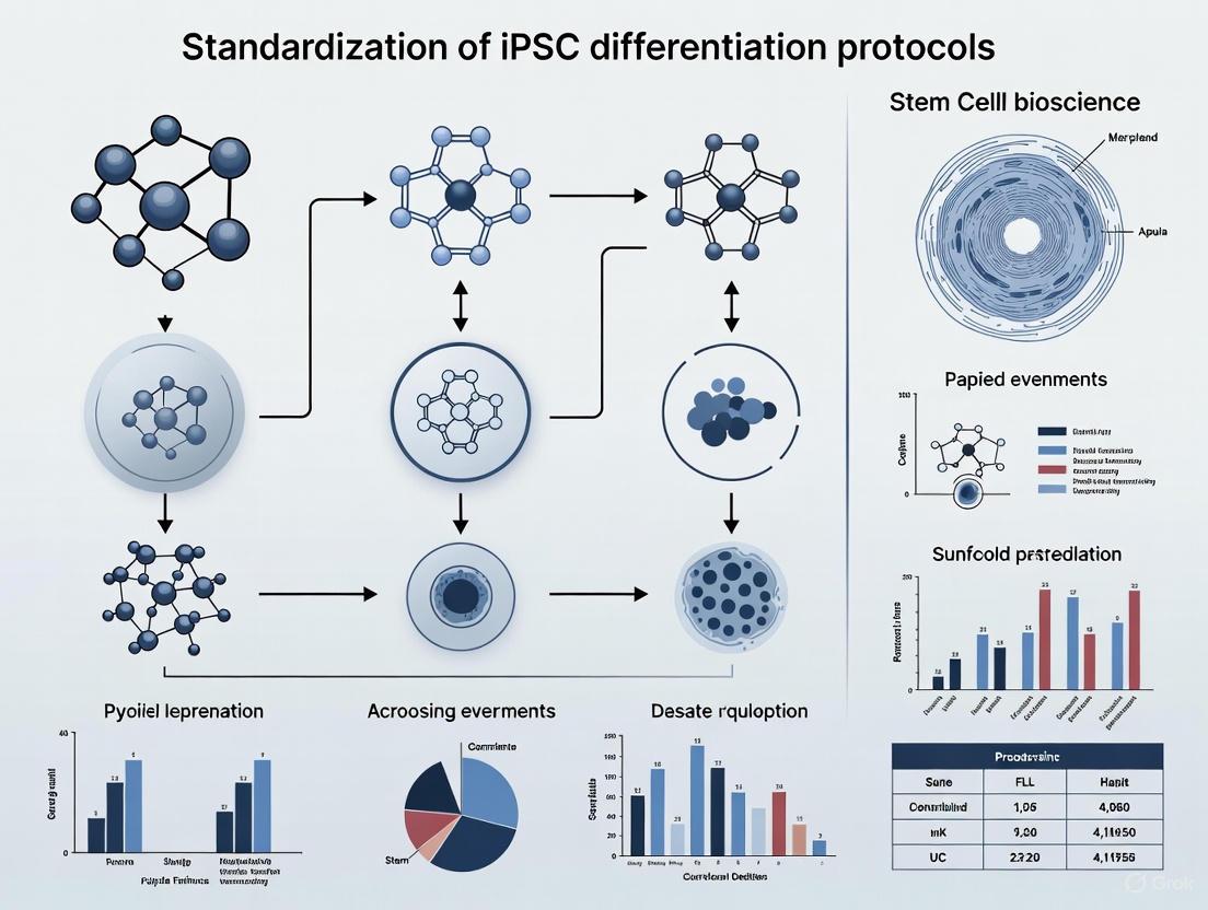
Standardizing iPSC Differentiation Protocols: A Roadmap for Reproducible Research and Clinical Translation
This article addresses the critical challenge of variability in induced pluripotent stem cell (iPSC) differentiation, a major hurdle in research and drug development.
Standardizing iPSC Differentiation Protocols: A Roadmap for Reproducible Research and Clinical Translation
Abstract
This article addresses the critical challenge of variability in induced pluripotent stem cell (iPSC) differentiation, a major hurdle in research and drug development. We explore the scientific and ethical foundations of standardization, detail optimized methodologies for specific lineages like natural killer cells and hepatocytes, provide troubleshooting strategies for common pitfalls, and establish frameworks for rigorous validation. By synthesizing the latest guidelines and research, this resource provides scientists and drug development professionals with a comprehensive strategy to enhance reproducibility, accelerate discovery, and ensure the reliable clinical translation of iPSC-based models and therapies.
The Urgent Need for Standardization: Addressing the Reproducibility Crisis in iPSC Research
Frequently Asked Questions (FAQs)
FAQ 1: What is the greatest source of variability in iPSC differentiation potential? Multiple studies conclude that donor-specific genetic variation is a primary source of functional variability between iPSC lines. Research comparing genetically matched iPSCs from different tissues (fibroblasts and blood) found that the impact of donor genetics exceeds the impact of the original parental cell type. Lines from the same donor were highly similar, while significant differences in transcriptomic, epigenetic, and differentiation profiles were observed between different donors [1].
FAQ 2: How does "epigenetic memory" from the parent somatic cell affect differentiation? iPSCs can retain an epigenetic memory—a gene expression signature and epigenetic profile of the somatic tissue they were derived from. This memory can create a lineage-specific bias, meaning an iPSC derived from blood might differentiate more readily into a blood cell type than into a pancreatic β-cell [2]. This memory is a crucial contributing factor to the variable differentiation efficiency seen between different iPSC lines, even when using the same protocol [2].
FAQ 3: Does variability increase or decrease during differentiation? Epigenetic variation increases as cells differentiate. In iPSCs, epigenetic patterns (like DNA methylation) are strongly associated with the donor's genetic background. However, as iPSCs differentiate into specialized cells (like neural stem cells, motor neurons, or monocytes), the direct relationship with genetic variation weakens, and epigenetic variation becomes more pronounced. The cell type itself becomes a stronger source of epigenetic variation than the original genetic variation [3].
FAQ 4: What are common technical pitfalls that introduce variability in culture? Technical factors are a major source of variability and include:
- Excessive Differentiation in Cultures: Caused by old culture medium, overgrown colonies, or prolonged handling outside the incubator [4].
- Poor Cell Aggregate Size After Passaging: Incorrect incubation times with passaging reagents can create aggregates that are too large or too small, harming differentiation efficiency [4].
- Low Cell Attachment After Plating: This can result from plating too few aggregates, excessive pipetting, or using the wrong culture plate for the coating substrate [4].
Troubleshooting Guides
Problem 1: Excessive Spontaneous Differentiation in iPSC Cultures
Potential Causes and Solutions:
- Cause: Culture medium is expired or degraded.
- Solution: Ensure complete cell culture medium kept at 2-8°C is less than 2 weeks old [4].
- Cause: Colonies are overgrown or unevenly sized.
- Solution: Passage cultures when colonies are large and compact, but before they overgrow. Ensure cell aggregates after passaging are evenly sized. Remove differentiated areas prior to passaging [4].
- Cause: Extended exposure to non-incubator conditions.
- Solution: Avoid having the culture plate out of the incubator for more than 15 minutes at a time [4].
Problem 2: Low Differentiation Efficiency Toward a Target Cell Type
Potential Causes and Solutions:
- Cause: Underlying genetic background of the donor.
- Solution: This is an intrinsic factor. For clinical or standardized applications, generate or source multiple iPSC lines from different donors and select lines with superior differentiation propensity for your target lineage [1].
- Cause: Residual epigenetic memory from the source cell.
- Solution: Implement additional epigenetic modulation steps during the differentiation protocol. For example, targeting epigenetic enzymes like DNA methyltransferases or histone deacetylases could help reset lineage bias and improve the generation of functional mature cells, such as pancreatic β-cells [2].
- Cause: Inconsistent cell state at the start of differentiation.
- Solution: Standardize the "banking" of high-quality, pluripotent iPSCs. Ensure cells are healthy, maintained in log-phase growth, and have a normal karyotype before initiating any differentiation protocol [5].
Table 1: Impact of Genetic Relationship on Epigenetic Variation in iPSCs (DNA Methylation)
| Compared iPSC Lines | Genetic Relationship | Number of Differentially Methylated Regions (DMRs) |
|---|---|---|
| Lines from same donor | Same individual | 10 - 46 DMRs [3] |
| Lines from father-daughter pair | Related donors | ~1,451 - 1,585 DMRs [3] |
| Lines from unrelated donors | Unrelated | ~2,667 - 2,961 DMRs [3] |
Source Data: Nature Communications (2025) [3]
Table 2: Association of Genetic and Epigenetic Variation Across Cell Types
| Cell Type | Strength of Association between Genetic Variation and Epigenetic Variation |
|---|---|
| iPSCs | Strongest association [3] |
| Differentiated Cells (e.g., Neurons, Monocytes) | Weaker association; epigenetic variation increases and is more strongly influenced by cell type [3] |
Source Data: Nature Communications (2025) [3]
Experimental Protocol: Assessing Variability in Differentiation
This protocol outlines key steps for differentiating human pluripotent stem cells (hPSCs) into definitive endoderm (DE), a critical first step for generating liver and pancreatic cells, while highlighting points for variability assessment [6].
1. Resuscitation and Passaging of hPSCs
- Details: Revive frozen hPSC vials using standard procedures with 10 µM Y-27632 (ROCK inhibitor) to enhance survival. Maintain cells in a defined medium like TeSR-E8. For passaging, use a gentle enzyme (e.g., Accutase) and ensure cells are passaged during log-phase growth to maintain pluripotency. Variability Checkpoint: Check for daily spontaneous differentiation and record the passage number and morphology of the starting population [6].
2. Plating Cells for Differentiation
- Details: Plate cells as single cells or small aggregates onto a suitable substrate (e.g., Matrigel, Vitronectin) at a consistent, optimized density. Variability Checkpoint: Note the exact seeding density and the confluency at the start of differentiation. Low or variable attachment can significantly impact outcomes [4] [6].
3. Definitive Endoderm Differentiation
- Details: Induce differentiation using a chemically defined basal medium (e.g., DMEM/F12) supplemented with small molecules. A common method uses 3 µM CHIR99021 (a GSK3 inhibitor that activates Wnt signaling) and 71 µg/mL Vitamin C for 4-6 days. Variability Checkpoint: Use freshly prepared or properly stored small molecule aliquots to ensure consistent potency. Document the exact formulation and source of all components [6].
4. Validation and Analysis
- Details: Validate DE differentiation 4-6 days post-induction.
- Immunofluorescence: Stain for key DE markers like SOX17 and FOXA2.
- Flow Cytometry: Analyze the percentage of cells positive for the surface marker CXCR4 (CD184).
- Variability Assessment: Quantify the efficiency across multiple differentiations and iPSC lines. High variability in the yield of CXCR4+/SOX17+ cells indicates a need to investigate genetic, epigenetic, or technical sources of inconsistency [6].
Diagram 1: Factors influencing differentiation efficiency. Intrinsic (red) and technical (green) factors converge on the starting cell state and directly impact the efficiency and final outcome.
The Scientist's Toolkit: Key Research Reagent Solutions
Table 3: Essential Materials for iPSC Culture and Differentiation
| Item | Function | Example |
|---|---|---|
| Maintenance Medium | Supports the self-renewal and pluripotency of undifferentiated iPSCs. | TeSR-E8, mTeSR Plus [6] |
| Extracellular Matrix (ECM) | Coats culture surfaces to support iPSC attachment and growth. | Matrigel, Vitronectin XF, Synthemax [6] |
| Passaging Reagent | Gently dissocies iPSC colonies for sub-culturing. | ReLeSR, Gentle Cell Dissociation Reagent, Accutase [4] [6] |
| Small Molecule Inhibitors/Activators | Directs cell fate by modulating key signaling pathways during differentiation. | CHIR99021 (Wnt activator), LDN193189 (BMP inhibitor) [6] |
| ROCK Inhibitor | Improves survival of single iPSCs after passaging or thawing. | Y-27632 [6] |
Diagram 2: Simplified definitive endoderm differentiation workflow. A key initial step for generating pancreatic and liver cells, induced by activating Wnt signaling [6].
Technical Support Center: Standardizing iPSC Differentiation Protocols
Irreproducibility in scientific research, particularly in the field of induced pluripotent stem cell (iPSC) studies, carries substantial financial and scientific consequences for drug discovery. Variability in differentiation protocols and characterization methods can lead to flawed disease models and unreliable preclinical data, ultimately wasting research funding and delaying the development of effective therapies. This technical support center provides standardized troubleshooting guidance and FAQs to help researchers enhance the reproducibility of their iPSC work, supporting more efficient and translatable drug discovery efforts.
Troubleshooting Guides for iPSC Differentiation
Problem 1: Excessive Differentiation in Cultures
- Potential Causes and Solutions:
- Old Culture Medium: Ensure complete cell culture medium has been stored at 2-8°C for less than 2 weeks [4].
- Inadequate Maintenance: Remove differentiated areas prior to passaging and avoid keeping culture plates outside the incubator for more than 15 minutes at a time [4].
- Passaging Issues: Ensure cell aggregates after passaging are evenly sized and passage cultures when colonies are large and compact with dense centers [4].
- Colony Density: Decrease colony density by plating fewer cell aggregates during passaging [4].
Problem 2: Low Cell Attachment After Plating
- Potential Causes and Solutions:
- Initial Seeding: Plate 2-3 times more cell aggregates initially and maintain more densely confluent cultures [4].
- Timing: Work quickly after treatment with passaging reagents to minimize duration cell aggregates are in suspension [4].
- Incubation Time: Reduce incubation time with passaging reagents, particularly if cells are passaged prior to multi-layering within colonies [4].
- Proper Handling: Avoid excessive pipetting to break up cell aggregates; instead increase incubation time with passaging reagent by 1-2 minutes [4].
- Plate Selection: Use non-tissue culture-treated plates when coating with Vitronectin XF; use tissue culture-treated plates when coating with Corning Matrigel [4].
Problem 3: Inefficient Differentiation to Target Cell Types
- Potential Causes and Solutions:
- Protocol Variation: Published protocols vary widely in efficiency for generating target cells [7].
- Insufficient Characterization: Inadequate characterization of expression profiles and functionality leads to highly variable outcomes [7].
- Cell Line Variability: Differentiation efficiency varies between stem cell lines, which is particularly problematic for disease-specific research [7].
Table 1: Common iPSC Culture Problems and Solutions
| Problem | Symptoms | Possible Solutions |
|---|---|---|
| Excessive Differentiation | >20% spontaneous differentiation in cultures | Use fresh medium (<2 weeks old); remove differentiated areas before passaging; optimize colony density [4] |
| Poor Cell Attachment | Low attachment after plating cells | Plate more cell aggregates (2-3x); reduce time aggregates spend in suspension; use proper plate type for coating [4] |
| Variable Aggregate Size | Cell aggregates too large or small after passaging | Adjust incubation time (+1-2 min for larger, -1-2 min for smaller); minimize manipulation of aggregates [4] |
| Inefficient Differentiation | Low yield of target cell type; high variability | Follow standardized protocols; implement thorough characterization; account for cell line variability [7] |
Frequently Asked Questions (FAQs)
General iPSC Research Questions
When should I use iPSCs for my experiments? iPSCs are particularly valuable for studying disorders where donor tissue is inaccessible, such as neurodegenerative diseases, cardiomyopathies, and for capturing patient-specific genetic diversity. They enable generation of unlimited quantities of previously inaccessible cell types while maintaining the genetic background of patients with specific mutations or diseases [5].
Why is standardization critical in iPSC research? Advancing human stem cell-based models into preclinical and regulatory testing requires rigorous and reproducible research. Implementing quality standards and reporting best practices ensures reliability and translatability of results, ultimately accelerating adoption in industrial and regulatory contexts [8].
What are the key considerations for successful iPSC differentiation? Efficiently directing iPSC differentiation into desired lineages and preparing sufficient specific cell types represents a major practical challenge. Additionally, ensuring that in vitro outcomes closely represent disease conditions is essential for meaningful results [9].
Technical and Methodology Questions
What methods are available for iPSC reprogramming? Multiple non-integrating reprogramming technologies are available, including RNA-based methods (e.g., StemRNA 3rd Gen containing six reprogramming RNAs), episomal plasmids, and Sendai virus. RNA-based methods offer the advantage of rapid, footprint-free reprogramming without residual vector retention concerns [10].
How should I characterize differentiated neural cells? Thorough characterization of expression profiles and functionality is essential. For neural cells, this includes identifying appropriate markers (e.g., ChAT and vAChT for cholinergic neurons), assessing electrophysiological properties, and verifying morphological characteristics [7].
What factors influence differentiation efficiency? Variations in composition, concentration, and timing of signaling molecules significantly impact results. Recent studies also report variations between different stem cell lines, which is particularly relevant for disease-specific research [7].
Standardized Experimental Protocols
Protocol 1: Basal Forebrain Cholinergic Neuron Differentiation
This protocol summarizes an established method for generating functional basal forebrain cholinergic neurons (BFCNs) from human iPSCs, relevant for Alzheimer's disease research [7].
Key Signaling Molecules and Growth Factors:
- Retinoic Acid (RA): Induces caudalization of neural tube [7]
- Sonic Hedgehog (SHH): Promotes ventralization at high concentrations [7]
- Fibroblast Growth Factor 8 (FGF8): Works with SHH in anterior/posterior patterning [7]
- Brain-Derived Neurotrophic Factor (BDNF): Stimulates cholinergic differentiation [7]
- Nerve Growth Factor (NGF): Controls maturation and survival processes [7]
Protocol 2: Quality Control Assessment for Differentiated Cells
Implementing comprehensive quality control is essential for reproducibility. The following workflow outlines key characterization steps:
Characterization Parameters:
- Identity Markers: Cell-type specific protein expression (e.g., ChAT for cholinergic neurons) [7]
- Functional Capacity: Appropriate physiological responses and signaling [7]
- Morphological Features: Characteristic cellular structures and organization [7]
- Genetic Stability: Maintenance of genomic integrity throughout differentiation [10]
The Scientist's Toolkit: Essential Research Reagents
Table 2: Key Research Reagent Solutions for iPSC Differentiation
| Reagent Category | Specific Examples | Function in Differentiation |
|---|---|---|
| Reprogramming Technologies | StemRNA 3rd Gen (OCT4, SOX2, KLF4, c-MYC, NANOG, LIN28) | Footprint-free somatic cell reprogramming to iPSCs [10] |
| Culture Media | mTeSR Plus, mTeSR1 | Maintenance of pluripotent stem cells in undifferentiated state [4] [5] |
| Passaging Reagents | ReLeSR, Gentle Cell Dissociation Reagent | Non-enzymatic cell dissociation for maintaining cell aggregates [4] |
| Signaling Molecules | SHH, RA, FGF8, BMP9, BDNF, NGF | Directing differentiation toward specific neural lineages [7] |
| Culture Substrates | Vitronectin XF, Corning Matrigel | Providing appropriate surface for cell attachment and growth [4] |
Addressing the high cost of irreproducibility in iPSC research requires systematic implementation of standardized methods, comprehensive characterization, and detailed reporting. By adopting the troubleshooting guides, standardized protocols, and quality control measures outlined in this technical support center, researchers can significantly enhance the reliability and translational potential of their iPSC-based disease models, ultimately contributing to more efficient and successful drug discovery programs.
The International Society for Stem Cell Research (ISSCR) provides comprehensive guidelines and standards to promote an ethical, practical, and sustainable approach to stem cell research and the development of cell therapies [11]. These guidelines address the international diversity of cultural, political, legal, and ethical issues associated with stem cell research and its translation to medicine [11]. The fundamental mission is to alleviate and prevent human suffering caused by illness and injury through rigorous, transparent, and reproducible scientific practices [11].
The ISSCR's framework is built upon widely shared ethical principles in science that call for rigor, oversight, and transparency in all areas of practice [11]. Adherence to these principles provides assurance that stem cell research is conducted with scientific and ethical integrity and that new therapies are evidence-based [11]. For researchers working with induced pluripotent stem cells (iPSCs), implementing these standards is crucial for addressing the reproducibility crisis that has hampered progress in the field [12] [13].
Fundamental Ethical Principles for Stem Cell Research
Core Ethical Commitments
The ISSCR Guidelines establish several fundamental ethical principles that form the foundation for all stem cell research [11]:
Integrity of the Research Enterprise: Research must ensure information is trustworthy, reliable, and accessible through independent peer review, oversight, replication, and accountability at each research stage [11].
Primacy of Patient/Participant Welfare: Physicians and researchersshould never excessively place vulnerable patients or research subjects at risk. The welfare of current research subjects must not be overridden by promise for future patients [11].
Respect for Patients and Research Subjects: Researchers must empower potential human research participants to exercise valid informed consent and provide accurate information about risks and current state of evidence for novel interventions [11].
Transparency: Researchers should promote timely exchange of accurate scientific information and communicate with various public groups, including patient communities [11].
Social and Distributive Justice: Benefits of clinical translation should be distributed justly and globally, with particular emphasis on addressing unmet medical and public health needs [11].
Special Considerations for Embryonic Research
For human embryonic stem cell research, the ISSCR provides specific ethical guidance, noting that such research is ethically permissible in many countries when performed under rigorous scientific and ethical oversight [11]. This position is consistent with policy statements of other professional organizations, including the American Society for Reproductive Medicine and the European Society of Human Reproduction and Embryology [11].
The 2025 update to the ISSCR Guidelines specifically addresses stem cell-based embryo models (SCBEMs), retiring the classification of models as "integrated" or "non-integrated" and replacing it with the inclusive term "SCBEMs" [11]. The guidelines reiterate that human SCBEMs are in vitro models and must not be transplanted to the uterus of a living animal or human host, and include a new recommendation that prohibits ex vivo culture of SCBEMS to the point of potential viability [11].
Troubleshooting Guide: Common Experimental Challenges in iPSC Research
Frequently Asked Questions
Q: Our iPSC differentiation efficiency varies significantly between experiments, even when using the same cell line and protocol. What could be causing this inconsistency?
A: Differentiation variability often stems from inconsistencies in the undifferentiated state of your starting population. According to ISSCR Standards, the undifferentiated status of cells should be monitored by quantitative marker analysis before initiating differentiation [14]. Ensure your pre-culture conditions are consistent, as recent research demonstrates that the composition of pre-culture medium significantly affects cardiac differentiation potential, with different media yielding troponin T positivity rates ranging from 84% to 95% [15].
Q: How can we confirm that our iPSC line is truly pluripotent, especially since we cannot perform teratoma assays due to animal welfare concerns?
A: The ISSCR explicitly states that xenograft (teratoma) assays are not required to indicate pluripotency [14]. Instead, pluripotency should be demonstrated through in vitro differentiation assays that assess capacity to form all three germ layers. Evidence should include quantitative measurements of marker combinations representative of ectoderm, endoderm, and mesoderm lineages, alongside loss of markers of the undifferentiated state [14].
Q: We're establishing a new iPSC line in our lab. What are the essential characterization steps we should perform before beginning experiments?
*A: The ISSCR recommends establishing a Master Cell Bank (MCB) prior to any experimental use, with comprehensive characterization [16]. Essential steps include:
- Authentication using Short Tandem Repeat (STR) analysis [16]
- Pluripotency assessment via in vitro differentiation to three germ layers [14]
- Genomic characterization to identify acquired genetic variations [13]
- Sterility testing including mycoplasma screening [13]
- Assessment of undifferentiated state markers (OCT4, NANOG, etc.) [14]*
Q: How should we handle cell line misidentification issues that we've discovered in our laboratory?
A: Cell line authentication is critical to avoid misidentification and cross-contamination, which are well-documented issues that can lead to erroneous conclusions [16]. The ISSCR recommends authenticating cells at the point of entry into the laboratory, at reasonable time points throughout experimentation, and prior to publication [16]. When authenticating cells, a reference sample from the original donor should be used for confirmation of origin where possible [16].
Q: What are the minimal reporting criteria we should include in our publications to ensure reproducibility?
*A: The ISSCR Standards emphasize that published papers must include detailed information on cell line provenance, characterization methods, culture conditions, and differentiation protocols to ensure reproducibility [13]. This includes specific information about:
- Cell line origin and authentication method [16]
- Culture conditions and passage number [13]
- Characterization data for undifferentiated state [14]
- Genomic stability assessment [13]
- Differentiation protocol details and efficiency metrics [15]*
Technical Standards and Characterization Requirements
The ISSCR Standards for Human Stem Cell Use in Research establish minimum characterization and reporting criteria to enhance reproducibility [17]. The table below summarizes the key characterization requirements for iPSCs:
Table 1: Essential Characterization Requirements for iPSC Research
| Characterization Category | Specific Requirements | Recommended Methods | Frequency |
|---|---|---|---|
| Cell Line Authentication | Confirm unique identity and detect cross-contamination | STR analysis, SNP profiling | Upon acquisition, when establishing MCB, and periodically during extended culture [16] |
| Assessment of Undifferentiated State | Verify expression of markers associated with pluripotency | Flow cytometry, immunocytochemistry, qPCR for OCT4, NANOG, etc. | Regularly during maintenance culture [14] |
| Pluripotency Assessment | Demonstrate differentiation capacity to three germ layers | In vitro differentiation with quantitative analysis of germ layer markers | For new lines, novel reprogramming techniques, or new culture systems [14] |
| Genomic Characterization | Monitor genetic integrity and detect acquired variations | Karyotyping, SNP arrays, whole genome sequencing | At baseline and periodically during extended culture [13] |
| Sterility Testing | Ensure absence of microbial contamination | Mycoplasma testing, sterility assays | Regularly during culture [13] |
Experimental Design and Protocol Standardization
Implementing a Rigorous Cell Banking System
A foundational element of reproducible iPSC research is the establishment of a systematic cell banking strategy. The ISSCR recommends a two-tier biobanking system to ensure consistent, well-characterized cells are available for all experimental use [16].
Diagram 1: Two-Tiered Cell Biobanking Strategy
This systematic approach ensures that all researchers start with the same validated materials capable of delivering reliable data [16]. The Master Cell Bank (MCB) should be created from the earliest possible passage of the established cell line and thoroughly characterized before any experimental use [16]. Working Cell Banks (WCBs) can then be generated from the MCB for routine experimental work [16].
Assessing Pluripotency and Developmental State
A critical challenge in iPSC research is appropriately characterizing the developmental state and differentiation capacity of cells. The ISSCR provides clear guidance on distinguishing between the undifferentiated state and true pluripotency:
Diagram 2: Relationship Between Marker Expression and Functional Pluripotency
The ISSCR emphasizes that no markers present on undifferentiated cells are uniquely expressed in pluripotent cells, and these markers should not be called "pluripotency markers" as pluripotency cannot be defined by marker expression alone [14]. Instead, pluripotency must be demonstrated experimentally by assays that assess differentiation capacity through quantitative measurements of marker combinations representative of all three embryonic germ layers [14].
Research Reagent Solutions for Standardized iPSC Research
Table 2: Essential Research Reagents and Their Functions in iPSC Research
| Reagent Category | Specific Examples | Function in Research | Quality Considerations |
|---|---|---|---|
| Culture Media | StemFit AK03, Essential 8, mTeSR Plus | Maintain pluripotent state; composition affects subsequent differentiation efficiency [15] | Use consistent lots; document complete composition; avoid frequent switching between formulations |
| Extracellular Matrices | iMatrix-511, Biolaminin 521, Recombinant Laminin | Provide substrate for cell attachment and signaling; influence cell behavior and differentiation | Standardize coating concentrations and procedures; validate each new lot |
| Differentiation Inducers | CHIR99021 (GSK-3 inhibitor), XAV939 (Wnt inhibitor) | Direct lineage specification; efficiency varies between cell lines and culture conditions [15] | Titrate concentrations for specific cell lines; use consistent sources; prepare fresh aliquots |
| Cell Dissociation Reagents | TrypLE Select, Accutase, EDTA solutions | Passage cells while maintaining viability and pluripotency; impact recovery and genetic stability | Standardize incubation times and temperatures; quantify recovery rates |
| Characterization Antibodies | Cardiac troponin T, ANP, ProBNP, OCT4, NANOG | Assess differentiation efficiency and pluripotent state [14] [15] | Validate specificity; use appropriate isotype controls; document lot numbers |
Quality Control and Regulatory Compliance
Addressing the Reproducibility Crisis
The stem cell field faces significant challenges with reproducibility, estimated to waste tens of billions of dollars annually and flood the literature with misleading data [12]. Major causes of irreproducibility in iPSC research include:
Cell line variability: hiPS cell lines from different donors or even different clones from the same donor can respond differently due to genetic background or epigenetic idiosyncrasies [12]
Cell authentication and culture contamination issues: Misidentification of hiPS cell lines or undetected contamination remains surprisingly common [12]
Cell handling and protocol complexities: Even when following the same published differentiation protocol, subtle differences in reagents, operator technique, or cell passaging schedule can yield different outcomes [12]
Protocol drift: Standard operating procedures that are not rigorously maintained tend to evolve ("drift") as they are handed off between staff or scaled up [12]
The ISSCR Standards are designed specifically to address these challenges through implementation of systematic characterization practices and comprehensive reporting requirements [13] [17].
Regulatory Framework for Clinical Translation
For researchers moving toward clinical applications, the ISSCR Guidelines emphasize that stem cell-based interventions should only be applied outside formal research settings after products have been authorized by regulators and proven safe and efficacious [11]. The guidelines specifically state that it is a "breach of professional medical ethics and responsible scientific practices to market or provide stem cell-based interventions prior to rigorous and independent expert review of safety and efficacy and appropriate regulatory approval" [11].
Recent analyses of regulatory requirements for clinical-grade iPSC banks highlight the need for harmonization in several key areas: expression vectors authorized for iPSC generation, minimum identity testing, minimum purity testing, and stability testing [18]. Current ICH guidelines for biotechnological/biological products should be extended to cover cell banks used for cell therapies [18].
Adherence to the ISSCR Guidelines and Standards provides a comprehensive framework for ensuring ethical integrity and scientific rigor in stem cell research. By implementing systematic characterization protocols, establishing robust cell banking practices, maintaining detailed documentation, and adhering to ethical principles, researchers can significantly enhance the reproducibility and reliability of their iPSC research.
The consistent application of these standards across laboratories will accelerate progress in the field by ensuring that research findings are accurate, meaningful, and durable [17]. Furthermore, compliance with these guidelines strengthens the pipeline of therapies for patients by ensuring rigor in preclinical research [17]. As the field continues to evolve, commitment to these fundamental principles will remain essential for realizing the full potential of iPSC technologies in both basic research and clinical applications.
Technical Support Center
Frequently Asked Questions (FAQs)
Q1: What are the core principles of responsible stem cell research according to the ISSCR? The International Society for Stem Cell Research (ISSCR) outlines fundamental ethical principles for stem cell research. These include integrity of the research enterprise to ensure trustworthy and reliable science, primacy of patient/participant welfare to protect vulnerable individuals, respect for patients and research subjects through valid informed consent, transparency in the timely sharing of data and methods, and social and distributive justice to ensure the fair global distribution of benefits [11].
Q2: My human Pluripotent Stem Cell (hPSC) cultures are showing excessive differentiation (>20%). What should I check? Excessive differentiation can often be traced to culture conditions and handling. Focus on these key areas:
- Culture Medium: Ensure your complete culture medium is fresh (e.g., less than 2 weeks old when stored at 2-8°C) [4].
- Handling Time: Minimize the time culture plates are outside the incubator to less than 15 minutes [4].
- Passaging Technique: Remove differentiated areas before passaging and ensure the cell aggregates generated are evenly sized. Do not allow cultures to overgrow [4].
- Colony Density: Decrease the colony density by plating fewer cell aggregates during passaging [4].
Q3: What are the minimum characterization standards for human stem cells used in research? The ISSCR Standards establish minimum criteria for characterizing human stem cells to ensure reproducibility [19] [20]. The key tenets are summarized in the table below.
Table 1: Key Characterization Standards for Human Stem Cells in Research
| Characterization Area | Key Requirements |
|---|---|
| Basic Characterization | Consistent generation and accurate characterization of starting research materials [20]. |
| Pluripotency | Rigorous demonstration of undifferentiated state and potential to give rise to all somatic lineages via morphology, gene expression, and functional assays [20]. |
| Genomic Characterization | Monitoring for culture-acquired genetic changes that can alter cell phenotype and impact reproducibility [20]. |
| Stem Cell-Based Models | Confirmation of reproducibility between developers, end-users, and laboratories for models like organoids [20]. |
| Reporting | Inclusion of detailed information on all parameters in published papers to ensure reproducibility [20]. |
Q4: During differentiation into neural lineages, my cells show high variability in efficiency. What could be the cause? Variability in differentiation efficiency is a common challenge. A 2016 review highlighted several potential pitfalls [7]:
- Protocol Inconsistency: Published protocols for the same cell type can vary widely in the composition, concentration, and timing of signaling molecules.
- Insufficient Characterization: Differentiated cells are often inadequately characterized for expression profile and functionality.
- Cell Line Differences: Differentiation efficiency can vary between different stem cell lines, which is critical when using patient-specific iPSCs for disease modeling.
Q5: What are the common cell culture problems that affect attachment and growth? Common issues often relate to technique, incubation, and media [21].
- Technique: Insufficient mixing of the cell inoculum can cause bubbles that hinder attachment. Static electricity on plastic vessels can also disrupt attachment, especially in low-humidity environments [21].
- Incubation: Temperature variations from frequently opening the incubator or improper stacking of vessels can affect growth rates. Evaporation and vibration are other contributing factors [21].
- Media: Defects may not be visible. Testing your media against a batch from another manufacturer can help isolate the problem [21].
Troubleshooting Guides
Problem: Low Cell Attachment After Passaging
Table 2: Troubleshooting Low Cell Attachment
| Possible Cause | Recommended Solution | Rationale |
|---|---|---|
| Low initial cell density | Plate 2-3 times more cell aggregates; maintain a more densely confluent culture [4]. | Provides sufficient cell-cell contact and signaling for survival and proliferation. |
| Prolonged time in suspension | Work quickly after treating cells with passaging reagents [4]. | Minimizes stress and anoikis (cell death due to detachment). |
| Overly sensitive cell line | Reduce incubation time with passaging reagents [4]. | Prevents excessive damage to cell surface proteins needed for attachment. |
| Use of incorrect cultureware | Use non-tissue culture-treated plates with Vitronectin XF; use tissue culture-treated plates with Corning Matrigel [4]. | Ensures the coating matrix can properly bind to the surface for cell attachment. |
Problem: Suboptimal Cell Aggregate Size After Passaging
The size of cell aggregates during passaging is critical for successful hPSC culture. The table below guides how to adjust your technique.
Table 3: Troubleshooting Cell Aggregate Size
| Problem | Solution | Action |
|---|---|---|
| Aggregates too large (>200 µm) | Increase dissociation. | Pipette the mixture up and down (avoid single cells) and increase incubation time by 1-2 minutes [4]. |
| Aggregates too small (<50 µm) | Minimize dissociation. | Reduce pipetting and decrease incubation time by 1-2 minutes [4]. |
| Differentiated cells detaching with colonies | Make dissociation more selective. | Decrease incubation time by 1-2 minutes and lower the incubation temperature to room temperature [4]. |
The Scientist's Toolkit
Table 4: Essential Research Reagent Solutions for iPSC Research
| Reagent / Tool Category | Example | Primary Function |
|---|---|---|
| Culture Medium | mTeSR Plus, mTeSR1 | Defined medium to support the maintenance and growth of undifferentiated hPSCs [4]. |
| Passaging Reagents | ReLeSR, Gentle Cell Dissociation Reagent | Non-enzymatic reagents used to gently dissociate hPSC colonies into small aggregates for subculturing [4]. |
| Attachment Substrates | Vitronectin XF, Corning Matrigel | Extracellular matrix proteins used to coat culture vessels to facilitate cell attachment and growth [4]. |
| Differentiation Kits | STEMdiff Midbrain Organoid Kit | Guided, standardized system to generate specific 3D cell models like organoids from hPSCs [20]. |
| Characterization Tools | Forebrain Neuron Precursor Cells | Ready-to-use, high-quality cell populations to start neural workflows and serve as a reference [20]. |
Experimental Workflows & Signaling Pathways
hPSC Quality Control Workflow
Adhering to standards requires a systematic workflow for quality control. The diagram below outlines the key stages.
Signaling Pathways in Neural Differentiation
The differentiation of stem cells into specific neural lineages recapitulates developmental signaling. This diagram shows the pathway for generating basal forebrain cholinergic neurons (BFCNs), a model for Alzheimer's disease research [7].
Optimized Differentiation Workflows: From Pluripotency to Functional Cell Types
Troubleshooting Guide: Common Issues in iPSC Culture
This guide addresses frequent challenges researchers face when maintaining human pluripotent stem cells (hPSCs), with solutions to ensure optimal culture health and differentiation potential.
Problem 1: Excessive Spontaneous Differentiation in Cultures
Spontaneous differentiation exceeding 20% can compromise the pluripotent cell pool and reduce the efficiency of directed differentiation protocols [4].
- Solution: Several factors can be adjusted to minimize differentiation [4]:
- Medium Quality: Ensure complete culture medium (e.g., mTeSR Plus) stored at 2-8°C is used while fresh (less than 2 weeks old).
- Handling Time: Avoid having culture plates outside the incubator for extended periods; limit to 15 minutes at a time.
- Passaging Technique: Manually remove differentiated areas before passaging. Ensure cell aggregates generated during passaging are evenly sized.
- Culture Density: Do not allow colonies to overgrow. Passage when colonies are large and compact with dense centers. Decreasing the colony density by plating fewer aggregates can also help.
- Reagent Sensitivity: For cultures treated with ReLeSR, reduce incubation time if the cell line is particularly sensitive.
Problem 2: Low Cell Attachment After Passaging
Poor attachment after plating can lead to significant cell loss and experimental delays [4].
- Solution: Consider the following adjustments [4]:
- Initial Seeding Density: Plate 2-3 times the usual number of cell aggregates initially to maintain a more densely confluent culture.
- Handling Speed: Work quickly after cells are treated with passaging reagents to minimize the time cell aggregates spend in suspension.
- Incubation Time: Reduce incubation time with passaging reagents, especially if cells are passaged before multi-layering occurs within the colony.
- Aggregate Manipulation: Do not excessively pipette to break up aggregates. If colonies are dense, a slight increase in incubation time (1-2 minutes) can help.
- Plate Selection: Verify that non-tissue culture-treated plates are used with specific coatings like Vitronectin XF, while tissue culture-treated plates are used with others like Corning Matrigel.
Problem 3: Suboptimal Differentiation Potential
The culture medium used to maintain iPSCs can significantly influence their subsequent ability to differentiate into target cells [22] [15].
- Solution: Optimization strategies include [22]:
- Medium Selection: Culture in medium that supports the glycolytic pathway to help maintain high differentiation potential.
- Biomarker Monitoring: Use CHD7 expression levels as a biomarker for differentiation potential.
- Substrate Adhesion: Culture cells on "less sticky" or less potent cell-binding materials to minimize the inadvertent inclusion of differentiated cells, which often have reduced adhesive properties.
Frequently Asked Questions (FAQs)
FAQ 1: How does the pre-culture medium affect the efficiency of directed differentiation?
The medium used to culture iPSCs immediately before initiating differentiation (pre-culture medium) is critical. Switching to a medium that approximates the composition of the subsequent differentiation medium can reduce "culture adaptation stress" on the cells, leading to higher differentiation efficiency. For example, in cardiac differentiation, using a pre-culture medium similar to EB formation medium increased the yield of cardiac troponin T (cTnT) positive cells to 95%, compared to 84% with a standard pluripotency maintenance medium [15].
FAQ 2: What are the key advantages of 3D organoid models over 2D cultures?
3D organoid models better replicate the cellular complexity, spatial architecture, and microenvironmental dynamics of human tissues compared to traditional 2D cultures [23] [24]. They are particularly valuable for studying disease mechanisms, drug efficacy, and personalized therapies because they retain the histological and genetic composition of their tissue of origin. This makes them excellent for modeling diseases that lack reliable animal models and for high-throughput drug screening in a more physiologically relevant system [23] [24].
FAQ 3: What methods are effective for genetic manipulation of iPSC-derived progenitor cells?
Both viral and non-viral methods can be highly effective. In a study on liver progenitor cells (LPCs) derived from hiPSCs, recombinant adeno-associated virus (rAAV) serotype 2/2 achieved a high transduction efficiency of 93.6%. As a non-viral alternative, electroporation demonstrated a plasmid delivery efficiency of 54.3% [23]. The choice of method depends on the required efficiency, safety considerations, and experimental goals.
The table below consolidates key quantitative findings from recent studies to aid in experimental design and benchmarking.
Table 1: Differentiation Efficiencies and Protocol Metrics
| Cell Type / Process | Key Marker/Parameter | Efficiency/Result | Citation |
|---|---|---|---|
| Cardiac Differentiation | Cardiac Troponin T (cTnT) positivity | 84% - 95% (varies with pre-culture medium) [15] | |
| Endothelial Differentiation | Expression of CD31, VE-cadherin, vWF | >98% cell purity [25] | |
| Transduction (LPCs) | rAAV2/2 (MOI 100,000) | 93.6% [23] | |
| Transfection (LPCs) | Electroporation | 54.3% [23] |
Table 2: Troubleshooting Metrics for hPSC Culture
| Problem Area | Key Parameter | Recommended Adjustment | Citation |
|---|---|---|---|
| Cell Aggregate Size | Mean size >200 µm | Increase incubation time 1-2 min [4] | |
| Cell Aggregate Size | Mean size <50 µm | Decrease incubation time 1-2 min [4] | |
| Differentiated Cell Detachment | --- | Decrease ReLeSR incubation time or lower temp to 15-25°C [4] | |
| Sample Preservation | Cell viability (refrigerated vs. cryo) | 20-30% variability; choose method based on processing delay [24] |
Experimental Protocol: Directed Differentiation of hiPSCs to Liver Progenitor Cells (LPCs)
This optimized protocol generates LPCs with high efficiency for disease modeling and gene therapy studies [23].
1. Materials and Resources
- hiPSC Line: Pre-characterized and mycoplasma-free.
- Basal Medium: RPMI 1640, 1% B-27 Supplement (without Vitamin A), 1% GlutaMAX, 1% sodium pyruvate.
- Small Molecules & Growth Factors:
- CHIR99021 (GSK-3 inhibitor)
- Activin A
- FGFβ
- FGF10
- SB431542 (TGF-β receptor inhibitor)
- Retinoic Acid
- BMP4
- Coating: Matrigel-coated plates.
- Passaging Reagent: Versen solution.
2. Step-by-Step Methodology
- Day -1: Harvest hiPSCs using Versen and seed at a high density of 100,000 cells per cm² on a Matrigel-coated plate [23].
- Definitive Endoderm (Days 0-4):
- Anteroposterior Foregut (Days 4-7):
- Change to basal medium supplemented with 50 ng/mL FGF10, 10 µM SB431542, and 10 µM retinoic acid. Change medium daily [23].
- Liver Progenitor Cells (LPCs) (Days 7-10):
- Culture cells in basal medium with 50 ng/mL FGF10 and 10 µM BMP4. Change medium daily. The resulting LPCs can be used for 2D culture or for generating 3D organoids [23].
The Scientist's Toolkit: Essential Research Reagents
Table 3: Key Reagents for iPSC Culture and Differentiation
| Reagent | Function / Purpose | Example Use Case |
|---|---|---|
| mTeSR Plus / Essential 8 | Serum-free, defined medium for feeder-free maintenance of pluripotent stem cells [23] [22]. | Routine culture of hiPSCs. |
| Matrigel / Laminin-521 | Extracellular matrix proteins that provide a substrate for cell attachment and growth in feeder-free systems [23] [22]. | Coating culture vessels for pluripotent stem cells. |
| CHIR99021 | A GSK-3 inhibitor that activates the Wnt/β-catenin signaling pathway, crucial for initiating differentiation [23]. | Directed differentiation into definitive endoderm. |
| Activin A | A TGF-β family growth factor that directs cells toward a definitive endoderm fate [23]. | Directed differentiation into definitive endoderm. |
| Y-27632 (ROCK inhibitor) | Improves cell survival after passaging by inhibiting apoptosis, especially in single-cell suspensions [15]. | Added to medium for 24 hours after cell dissociation. |
| B-27 Supplement | A defined serum-free supplement optimized for the survival and growth of neuronal and other post-mitotic cells. | Used in basal medium for endodermal and hepatic differentiation [23]. |
| ReLeSR / Gentle Cell Dissociation Reagent | Enzyme-free, defined solutions for the gentle passaging of hPSCs as clumps, minimizing damage to cell surface proteins [4]. | Routine passaging of hPSC colonies. |
Signaling Pathways in Directed Differentiation
The differentiation of iPSCs into specific lineages is controlled by the sequential activation and inhibition of key signaling pathways, mimicking embryonic development [23] [25].
The development of robust and reproducible protocols for differentiating induced pluripotent stem cells (iPSCs) into liver progenitor cells (LPCs) is a critical frontier in regenerative medicine, disease modeling, and drug development. Primary human hepatocytes, the workhorse of liver research, rapidly lose their functional properties in conventional two-dimensional (2D) cultures, making their use as a reliable cell model challenging [23]. Furthermore, many liver diseases lack reliable animal models, necessitating the creation of advanced in vitro systems that accurately recapitulate human liver physiology [23].
Standardized iPSC differentiation protocols aim to address these limitations by generating consistent, high-quality LPCs. These bipotent cells can self-renew and differentiate into the two main epithelial cell types of the liver: hepatocytes and cholangiocytes [26]. The optimization of these protocols is not merely a technical exercise; it is fundamental to ensuring that experimental results are reproducible, comparable across laboratories, and ultimately, translatable to clinical applications. This case study establishes a technical support center to guide researchers through the common challenges encountered in this process, providing troubleshooting guides, detailed protocols, and reagent solutions to foster reliability and efficiency in the generation of iPSC-derived LPCs.
Troubleshooting Guide: Common Problems and Solutions
Researchers often encounter specific technical challenges when cultivating iPSCs and differentiating them into LPCs. The following table addresses these common issues with evidence-based solutions.
Table 1: Troubleshooting Guide for iPSC Culture and LPC Differentiation
| Problem | Possible Cause | Recommended Solution |
|---|---|---|
| Excessive differentiation in iPSC cultures | Old culture medium; overgrown colonies; prolonged time outside incubator [4]. | Use fresh medium (<2 weeks old); passage cultures before over-confluence; limit plate handling to <15 minutes [4]. |
| Poor cell survival after passaging | Over-confluence at passaging; insensitive dissociation method [27]. | Passage cells at ~85% confluency; use EDTA or a gentle dissociation reagent for sensitive lines; employ a ROCK inhibitor (e.g., Y-27632) to improve survival [27]. |
| Low differentiation efficiency | Low-quality iPSCs; incorrect cell density at induction [27]. | Use high-quality, pluripotent iPSCs; remove differentiated areas before passaging; optimize seeding density for differentiation (e.g., 100,000 cells/cm² for LPCs [23]); use a control cell line (e.g., H9) to benchmark performance [27]. |
| Inefficient transgene delivery to LPCs | Suboptimal delivery method or parameters. | For viral delivery: Test different serotypes; for rAAV2/2, use an MOI of 100,000 (93.6% efficiency). For non-viral delivery: Optimize electroporation parameters (54.3% efficiency achieved) [23]. |
Frequently Asked Questions (FAQs)
General Protocol Questions
Q: What are the key stages in a standardized directed differentiation protocol from iPSCs to LPCs? A standardized, multi-stage protocol closely mimics embryonic liver development [23] [28]:
- Definitive Endoderm (DE): Culture iPSCs in a basal medium (e.g., RPMI 1640) supplemented with Activin A and CHIR99021 for the first 24 hours, followed by Activin A and FGFβ for three more days [23].
- Anteroposterior Foregut: Differentiate DE cells in a basal medium with FGF10, SB431542, and retinoic acid [23].
- Liver Progenitor Cells (LPCs): Generate LPCs by culturing in a basal medium with FGF10 and BMP4 [23]. The resulting LPCs can be used for 2D culture or embedded in Matrigel to establish 3D organoid cultures.
Q: Why is a 3D organoid model sometimes preferred over a 2D culture? 3D liver organoids better reproduce liver physiology and cellular characteristics, making them crucial for studying pathogenesis, drug efficacy, and personalized therapies [23]. They help achieve higher expression of metabolically crucial enzymes like cytochrome P450 and create a more tissue-like context for disease modeling [23] [28].
Technical and Analytical Questions
Q: What markers should I use to characterize iPSC-derived LPCs? LPCs are heterogeneous and lack a single unique marker. Identification relies on a panel of markers [26] [29]:
- Hepatocyte Markers: Albumin (ALB), Keratin 8 (KRT8), Keratin 18 (KRT18)
- Cholangiocyte/Biliary Markers: Keratin 19 (KRT19), Keratin 7 (KRT7), EpCAM, SOX9
- Hepatoblast/Progenitor Markers: Alpha-fetoprotein (AFP)
- Stemness Markers: LGR5, CD44
Q: Which signaling pathways are critical for LPC activation and differentiation? The following diagram summarizes the key signaling pathways involved in LPC biology:
Key Signaling Pathways Regulating LPC Activation and Growth
The Hippo signaling pathway is a key regulator. Inactivation of Large Tumor Suppressor kinases (LATS1/2) leads to overactivation of YAP/TAZ, which promotes the dedifferentiation of hepatocytes into LPCs and drives LPC expansion [26] [29]. Other critical pathways include TNFα, IL-6, and growth factor signaling (HGF, FGF) [29]. Macrophages also contribute by secreting TWEAK, which stimulates LPC proliferation [29].
Transduction Efficiency
A critical step in genetic engineering and disease modeling is the efficient delivery of transgenes into LPCs. The following table compares the performance of two common methods as quantified in a recent optimization study.
Table 2: Transgene Delivery Efficiency into Liver Progenitor Cells
| Delivery Method | Specific Parameters | Efficiency | Reference |
|---|---|---|---|
| Viral (rAAV) | Serotype 2/2, MOI 100,000 | 93.6% | [23] |
| Non-Viral (Electroporation) | Not specified | 54.3% | [23] |
Organoid Generation Efficiency
Protocol refinements can significantly impact the yield and scalability of 3D liver models. The data below demonstrate how modifying the differentiation stage for 3D culture initiation and using different media can affect organoid generation.
Table 3: Impact of Protocol Modifications on Liver Organoid Generation
| Protocol Modification | Comparison | Result / Fold Change | Reference |
|---|---|---|---|
| Timing of 3D Culture Initiation | New protocol (HE stage) vs. Previous protocol (IH stage) | Reduced time to organoid generation from >2 weeks to 1 week | [28] |
| Culture Medium | Hepatic Medium (HM) vs. 2D Control | 2.6-fold increase in organoid number | [28] |
| Culture Medium | Expansion Medium (EM) vs. 2D Control | 3.3-fold increase in organoid number | [28] |
Experimental Protocols
Standardized 2D Differentiation to LPCs
This optimized protocol generates LPCs from hiPSCs with high efficiency [23].
Materials:
- Basal Medium: RPMI 1640, 1% B-27 Supplement (without Vitamin A), 1% Glutamax, 1% sodium pyruvate.
- Key Factors: Activin A, CHIR99021, FGFβ, FGF10, SB431542, Retinoic Acid, BMP4.
- Matrix: Matrigel-coated plates.
Procedure:
- Definitive Endoderm (4 days): Harvest hiPSCs using Versen and seed at 100,000 cells/cm² on Matrigel. For the first 24 hours, use Basal Medium supplemented with 100 ng/mL Activin A and 3 µM CHIR99021. For the next three days, use Basal Medium with 100 ng/mL Activin A and 10 ng/mL FGFβ. Change medium daily.
- Anteroposterior Foregut (specified duration): Culture the DE cells in Basal Medium supplemented with 50 ng/mL FGF10, 10 µM SB431542, and 10 µM retinoic acid. Change medium daily.
- Liver Progenitor Cells (LPCs): Culture the foregut cells in Basal Medium supplemented with 50 ng/mL FGF10 and 10 µM BMP4. Change medium daily. The resulting LPCs can be characterized by the expression of markers like SOX9, CK19, and EpCAM.
3D Liver Organoid Generation from LPCs
This protocol describes how to transition from a 2D LPC culture to a 3D organoid model [23].
Materials:
- Cells: LPCs from 2D differentiation.
- Matrix: Matrigel.
- Medium: HepatiCult Organoid Kit medium or a defined hepatic medium (HM) [28].
Procedure:
- Harvest the 2D LPCs using Versen and create a single-cell suspension.
- Centrifuge the cell suspension and remove the supernatant.
- Resuspend the cell pellet in cold Matrigel (20 µL per 20,000 cells).
- Plate the cell-Matrigel suspension as droplets in a cell culture plate and incubate at 37°C for 40-60 minutes to allow the Matrigel to polymerize.
- Carefully add pre-warmed organoid culture medium to the wells without disrupting the Matrigel droplets.
- Culture the organoids, passaging them mechanically or enzymatically every 1-2 weeks to maintain expansion.
The Scientist's Toolkit: Research Reagent Solutions
Selecting the appropriate reagents is fundamental to the success of the differentiation protocol. The following table details essential materials and their functions.
Table 4: Essential Reagents for iPSC to LPC Differentiation
| Reagent Category | Specific Example | Function in Protocol |
|---|---|---|
| Culture Medium | TeSR-E8 / mTeSR Plus [23] [5] | Maintains hiPSC pluripotency and proliferation in feeder-free culture. |
| Extracellular Matrix | Matrigel / Geltrex [23] [27] | Provides a basement membrane matrix that supports cell attachment, growth, and 3D organoid formation. |
| Directed Differentiation Factors | Activin A, CHIR99021, FGF10, BMP4, Retinoic Acid [23] | Guides cell fate through sequential developmental stages: defines endoderm, patterns foregut, and specifies liver lineage. |
| Cell Dissociation Reagents | Versen [23], Gentle Cell Dissociation Reagent [4], EDTA [27] | Passages cells while minimizing damage; Versen is used for harvesting LPCs, while gentler options are for sensitive iPSC passaging. |
| Survival Enhancers | ROCK Inhibitor (Y-27632) / RevitaCell Supplement [27] [30] | Improves cell survival after passaging, thawing, or during single-cell cloning by inhibiting apoptosis. |
Q: What is the core challenge in Natural Killer (NK) cell manufacturing that this case study addresses?
A: The central challenge is selecting an optimal expansion method that balances high cell yield and purity with safety, standardization, and clinical applicability. Traditional methods rely on irradiated feeder cells (often cancer-derived), which pose potential safety risks and batch-to-batch variability. Feeder-free methods, using defined cytokine cocktails or other stimulants, offer a more standardized path but have historically faced hurdles in achieving comparable expansion rates [31] [32]. This analysis is critical for standardizing iPSC differentiation protocols and advancing robust, off-the-shelf NK cell therapies.
Troubleshooting Guides & FAQs
Q: Our feeder-free NK cell cultures are showing poor expansion yields. What are potential causes and solutions?
A: Low expansion in feeder-free systems is a common hurdle. The table below outlines troubleshooting steps.
| Problem | Potential Cause | Recommended Solution |
|---|---|---|
| Poor Expansion Yield | Suboptimal cytokine combination or timing [31] [32]. | Use IL-2 or IL-15 as essential cytokines, supplemented with early-phase IL-21 [31] [32]. Test combinations with IL-18 or IL-27 [32]. |
| Low NK Cell Purity | Overgrowth of non-NK immune cells in culture. | Start with a highly purified NK cell population. For PBMC sources, incorporate antibody stimulation (e.g., minimal dose OKT-3 with anti-CD52) to selectively enhance NK expansion [32]. |
| High Cost of Goods | Use of high concentrations of recombinant cytokines. | Investigate nanoparticle-based delivery systems for cytokines to enhance half-life and reduce the required dosage [31]. |
| Inconsistent Differentiation from iPSCs | Variable efficiency in generating hematopoietic progenitors. | Use a standardized, serum-free, feeder-free differentiation kit and a well-characterized iPSC line like LiPSC-GR1.1 to improve reproducibility [33] [34]. |
Q: When using feeder cells, how can we ensure consistent quality and mitigate safety concerns?
A: Feeder cell quality is paramount. Key steps include:
- Rigorous Quality Control: Tightly manage the culture and storage conditions of feeder cell lines. Use master cell banks and perform regular checks for viability, sterility, and identity [31].
- Genetic Engineering: Utilize genetically engineered feeder cells (e.g., K562-mbIL21) to express specific ligands that enhance NK cell expansion and functionality [31] [32].
- Adequate Irradiation: Ensure feeder cells are properly γ-irradiated to inhibit their proliferation while still allowing them to express stress ligands that stimulate NK cells [31] [32].
Q: From a clinical translation perspective, what are the key considerations for choosing an expansion method?
A: The choice involves trade-offs, summarized in the table below.
| Method | Key Advantage | Major Challenge | Clinical Applicability |
|---|---|---|---|
| Feeder-Based | Very high fold expansion (e.g., 80 to over 12,000-fold) [31] [32]. | Safety concerns (cancer-derived cells), complex quality control, and standardization [31]. | High efficacy in trials but carries regulatory hurdles due to safety profile [31] [35]. |
| Feeder-Free (Cytokine/Antibody) | Defined, xeno-free components enhance safety and standardization [31] [33]. | Historically lower expansion rates; can be costly [31] [32]. | High; essential for creating standardized, off-the-shelf allogeneic products [35]. |
| iPSC-Derived NK Cells | Unlimited, reproducible source; ideal for genetic engineering (e.g., CAR, IL-15) [34]. | Complex and lengthy differentiation protocol (e.g., 28+ days) [33]. | Highly promising; products like FT596 and MSLN.CAR-IL-15 iNKs are in clinical trials [35] [34]. |
Experimental Protocols & Data
Detailed Protocol: Feeder-Free NK Cell Differentiation from iPSCs
This protocol aligns with the thesis goal of standardizing iPSC differentiation [33].
Part I: Differentiate CD34+ Hematopoietic Progenitor Cells from hPSCs
- EB Formation: Harvest hPSCs to create a single-cell suspension. Seed cells in AggreWell plates in
EB Formation Medium(Basal Medium + Supplement A + Y-27632) to form embryoid bodies (EBs). - EB Maturation: On day 2, perform a half-medium change with
EB Medium A. On day 3, switch toEB Medium B(Basal Medium + Supplement B) with half-medium changes on days 7 and 10. - Progenitor Harvest: On day 5, harvest EBs. On day 14, dissociate EBs and isolate
CD34+cells using a positive selection kit [33].
- EB Formation: Harvest hPSCs to create a single-cell suspension. Seed cells in AggreWell plates in
Part II: Differentiate NK Cells from CD34+ Progenitors
- Lymphoid Progenitor Expansion: Seed
CD34+cells on a coated plate inStemSpan Lymphoid Progenitor Expansion Medium. Culture for 14 days, with medium changes, to generateCD5+CD7+lymphoid progenitors. - NK Cell Differentiation: Harvest progenitors and reseed in
StemSpan NK Cell Differentiation Medium(Basal Medium + NK Differentiation Supplement + UM729) on a non-coated plate. - NK Cell Harvest: Harvest cells containing
CD56+NK cells on day 28 for downstream assays [33].
- Lymphoid Progenitor Expansion: Seed
The following table summarizes key performance metrics from the literature for direct comparison [31] [32] [34].
| Method | Specific Approach | Reported Fold Expansion | Purity (CD56+/CD45+) | Key Components & Reagents |
|---|---|---|---|---|
| Feeder-Based | γ-irradiated PBMCs | 80 - 794 | Not Specified | Irradiated PBMCs, IL-2 [31] |
| Feeder-Based | K562-mbIL-21 | ~842 | 91.5% | Engineered K562 cells, IL-15 [31] |
| Feeder-Based | K562-mbIL-18 | ~9,860 | ≥98% | Engineered K562 cells, cytokines [32] |
| Feeder-Free | Cytokine Combination (IL-2, IL-15, IL-18, IL-27) | ~17 | Not Specified | Recombinant human cytokines [32] |
| Feeder-Free | Antibody Stimulation (OKT-3 + anti-CD52) | ~1,000 | ~60% | Agonist antibodies [32] |
| iPSC-Derived | Feeder-Free Spin EB Protocol | Yield: ~1.3x10^5 cells/EB | >98% | APEL medium, SCF, VEGF, BMP-4, cytokines [34] |
The Scientist's Toolkit: Research Reagent Solutions
| Item | Function in NK Cell Differentiation/Expansion |
|---|---|
| IL-2 / IL-15 | Essential cytokines for NK cell survival, proliferation, and activation [31] [32]. |
| IL-21 | A supporting cytokine that, when added early, enhances long-term expansion and function [31] [32]. |
| StemSpan SFEM II | A serum-free medium base optimized for hematopoietic cell expansion [33]. |
| Y-27632 (ROCK inhibitor) | Improves viability of dissociated single cells, such as hPSCs, after passaging [33]. |
| AggreWell Plates | Enable standardized formation of uniform embryoid bodies (EBs) from hPSCs [33]. |
| Lymphoid Progenitor Expansion Supplement | Directs differentiation of CD34+ hematopoietic progenitors toward the lymphoid lineage [33]. |
| Anti-CD52 Antibody | An agonist antibody that, in combination with others, can stimulate robust NK cell expansion in feeder-free systems [32]. |
Signaling Pathways and Workflow Visualizations
Feeder-Based NK Cell Activation Workflow
Feeder-Free iPSC to NK Cell Differentiation
Critical Cytokine Signaling in NK Cell Expansion
Technical Support Center
Troubleshooting Guides
Troubleshooting iPSC Culture Quality
A successful cell therapy manufacturing process begins with high-quality starting material. The table below outlines common issues encountered when maintaining human pluripotent stem cells (hPSCs) and their recommended solutions [4].
| Problem & Observation | Potential Cause | Recommended Action |
|---|---|---|
| Excessive differentiation (>20%) in cultures [4] | • Old culture medium• Overgrown colonies• Excessive time outside incubator | • Use complete medium less than 2 weeks old [4].• Passage colonies when large and compact; remove differentiated areas first [4].• Avoid having culture plate out of incubator for >15 minutes [4]. |
| Low cell attachment after passaging [4] | • Low initial plating density• Over-manipulation of cell aggregates• Incorrect cultureware | • Plate 2-3 times higher number of cell aggregates [4].• Work quickly after passaging; minimize suspension time [4].• Use non-tissue culture-treated plates with Vitronectin XF; use tissue culture-treated plates with Corning Matrigel [4]. |
| Colonies remain attached, require scraping [4] | • Insufficient incubation time with passaging reagent | • Increase incubation time with reagent (e.g., ReLeSR) by 1-2 minutes [4]. |
Troubleshooting Cardiomyocyte Differentiation
Generating cardiomyocytes from iPSCs is a multi-step process. The table below summarizes key challenges during differentiation protocols, such as with the STEMdiff Cardiomyocyte Differentiation Kit [36].
| Problem & Observation | Potential Cause | Recommended Action |
|---|---|---|
| Cultures are <95% confluent on Day 0 of differentiation [36] | • Error in cell counting/seeding• Poor quality starting hPSCs• Insufficient cell dissociation | • Do not start differentiation. Seed a range of densities (e.g., 3.5-8.0 x 10^5 cells/well of a 12-well plate) to achieve >95% confluency within 48 hours [36].• Assess pluripotency (e.g., OCT3/4, TRA-1-60 markers; ensure >90% positive) and karyotype [36].• Use Gentle Cell Dissociation Reagent; avoid suboptimal reagents like Accutase [36]. |
| Cell detachment from cultureware (Days 2-8) [36] | • Inappropriate matrix used• Harsh media handling | • Coat with Corning Matrigel hESC-Qualified Matrix. Vitronectin is not recommended for differentiation [36].• Use a pipettor for media changes; DO NOT aspirate directly [36]. |
| No visible beating by Day 15+ [36] | • Failed to reach critical confluency on Day 0• Poor starting quality of hPSCs | • Repeat experiment, ensuring >95% confluency is achieved within 48 hours prior to differentiation [36].• Restart with high-quality hPSCs (<10% differentiation) from an earlier passage [36]. |
Frequently Asked Questions (FAQs)
? Starting Material and Culture
Q: What are the critical quality attributes for the starting hPSCs to ensure successful differentiation and scaling? [36] A: Key attributes include:
- Morphology and Markers: High-quality, undifferentiated morphology with >90% expression of pluripotency markers like OCT3/4 and TRA-1-60 [36].
- Genetic Integrity: A normal karyotype, assessable with tools like the hPSC Genetic Analysis Kit [36].
- Trilineage Potential: Demonstrated ability to differentiate into all three germ layers (e.g., using the STEMdiff Trilineage Differentiation Kit) [36].
- Culture Purity: Cultures should have minimal spontaneous differentiation (<10%) [36].
Q: How can I improve the survival of hPSCs when passaging as single cells? A: Supplement the plating medium with 10 µM of a ROCK inhibitor (Y-27632) to reduce apoptosis [36].
? Differentiation and Maturation
Q: Beating in my cardiomyocyte cultures disappeared after a media change. Is this normal? [36] A: Yes. Nutrient depletion and acidic pH before feeding can cause cardiomyocytes to slow or stop beating. After the media change, return the culture to the incubator; beating should resume after a few hours or by the next day [36].
Q: If beating is difficult to observe visually, how can I confirm successful cardiomyocyte differentiation? [36] A: You can:
- Use functional assays like microelectrode array (MEA) to measure electrophysiological activity (beat rate) [36].
- Perform immunocytochemistry or flow cytometry for cardiomyocyte-specific markers such as cardiac troponin T (cTNT) [36].
? Scaling and Manufacturing
Q: What is a primary consideration when moving from a research-scale protocol to a clinical manufacturing process? A: A critical step is transitioning from a "bench-to-bedside" mindset to a "patient-backwards" approach. This means defining the requirements of the final cell therapy product first and optimizing each step of the development process to meet those specific clinical needs [37].
Q: Why is standardization critical in iPSC-based therapy development? A: Collaboration among regulatory authorities, researchers, clinicians, and industry partners is essential. Standardized protocols ensure the consistent production of safe, efficacious, and well-characterized cell products, which is a cornerstone of successful clinical application and regulatory approval [38].
Experimental Protocol Data
Key Workflow: Cardiomyocyte Differentiation
The following workflow summarizes a standardized protocol for generating cardiomyocytes from hPSCs [36].
Critical Cell Seeding Densities for Differentiation
Achieving the correct cell density at the start of differentiation is critical for efficiency [36].
| Parameter | Value or Range | Format / Vessel | Notes |
|---|---|---|---|
| Target Confluency on Day 0 | >95% | - | Must be achieved within 48 hours after seeding [36]. |
| Recommended Seeding Density | 3.5 - 8.0 x 10^5 cells/well | 12-well plate | Line-specific optimization is required [36]. |
| Alternative Density | ~9.2 x 10^4 cells/cm² | - | Calculated equivalent surface density [36]. |
The Scientist's Toolkit: Research Reagent Solutions
This table lists essential materials used in iPSC culture and cardiomyocyte differentiation protocols, as cited in the search results [4] [36].
| Item Name | Function / Application |
|---|---|
| mTeSR Plus / mTeSR1 | Complete, feeder-free maintenance medium for hPSCs [4]. |
| TeSR Medium | Feeder-free maintenance medium used prior to differentiation protocols [36]. |
| Gentle Cell Dissociation Reagent | Used to dissociate hPSCs into a uniform single-cell suspension for accurate seeding prior to differentiation [36]. |
| ReLeSR | A non-enzymatic passaging reagent used for the bulk culture of hPSCs as cell aggregates [4]. |
| Y-27632 (ROCK inhibitor) | Small molecule added to plating medium to significantly improve cell survival after single-cell passaging [36]. |
| Corning Matrigel hESC-Qualified Matrix | A substrate used for coating cultureware for both hPSC maintenance and cardiomyocyte differentiation protocols [36]. |
| STEMdiff Ventricular/Atrial Cardiomyocyte Differentiation Kit | A system of basal media and supplements (A, B, C) designed for the staged differentiation of hPSCs into cardiomyocytes [36]. |
| STEMdiff Cardiomyocyte Maintenance Medium | Medium used from Day 8 onwards to promote the maturation and maintenance of differentiated cardiomyocytes [36]. |
| Vitronectin XF | A defined, recombinant substrate used for coating cultureware for hPSC maintenance [4]. |
| STEMdiff Trilineage Differentiation Kit | Used to assess the trilineage differentiation potential of starting hPSCs, a key quality attribute [36]. |
| hPSC Genetic Analysis Kit | A tool used to assess the karyotype and genetic stability of hPSC cultures [36]. |
Solving Common Challenges: Strategies for Enhancing Protocol Robustness and Yield
Preventing Spontaneous Differentiation and Maintaining Pluripotency in Culture
Troubleshooting Guide: Common Problems & Solutions
This guide addresses frequent challenges in maintaining undifferentiated induced pluripotent stem cell (iPSC) cultures, a critical step for standardizing differentiation protocols.
| Problem | Possible Causes | Recommended Solutions |
|---|---|---|
| Excessive Differentiation (>20%) [4] | Old culture medium; overgrown colonies; prolonged time outside incubator; uneven colony size during passaging. | Use fresh medium (<2 weeks old) [4]; remove differentiated areas before passaging [4]; passage when colonies are large and compact [4]; limit plate handling outside incubator to <15 minutes [4]. |
| Low Cell Attachment After Plating [4] | Low initial seeding density; over-dissociation of cell aggregates; sensitive cell line. | Plate 2-3 times more cell aggregates initially [4]; reduce incubation time with passaging reagents [4]; avoid excessive pipetting that breaks up aggregates [4]. |
| Differentiated Cells Detaching with Colonies [4] | Over-incubation with dissociation reagent. | Decrease incubation time with reagent (e.g., ReLeSR) by 1-2 minutes [4]; lower incubation temperature to room temperature [4]. |
| Spontaneous Differentiation in Single-Cell Cultures [39] | Transition phase from aggregate to single-cell passaging; poor initial cell quality. | Seed cells at higher densities for the first 1-2 passages during adaptation [39]; subsequent passaging should resolve minor differentiation [39]. |
| Poor Recovery After Thawing [40] | Suboptimal freezing/thawing process; osmotic shock; incorrect cell growth phase at freezing. | Thaw cells quickly; dilute cryoprotectant drop-wise to prevent osmotic shock [41]; ensure cells are in logarithmic growth phase before freezing [40]. |
Frequently Asked Questions (FAQs)
Q: How much spontaneous differentiation is considered normal in a healthy iPSC culture?
A: A limited amount (5-10%) of spontaneous differentiation is normal and healthy in iPSC cultures. The key is to manually remove these differentiated areas during passaging to prevent them from overgrowing the culture [39].
Q: Should I passage my iPSCs as single cells or as aggregates?
A: For routine maintenance, passaging as aggregates is generally recommended. This method supports long-term expansion and stable karyotypes for many cell lines. Single-cell passaging can place selective pressure on the population, potentially leading to genetic aberrations. However, specific media like eTeSR are formulated for single-cell passaging if required for your application [39].
Q: When is ROCK inhibitor (Y-27632) required?
A: ROCK inhibitor is essential for enhancing cell survival in situations involving significant dissociation, such as single-cell passaging and thawing cryopreserved cells. It prevents dissociation-induced apoptosis. When passaging hPSCs as aggregates, adding ROCK inhibitor is typically not required and may even negatively affect cell morphology [39].
Q: How do defined culture conditions help maintain pluripotency?
A: Defined, feeder-free culture conditions (using media like E8 and substrates like Vitronectin or Laminin-521) significantly reduce batch-to-batch variability and inter-line heterogeneity. Research shows these conditions promote greater uniformity among PSC lines, reduce the expression of somatic cell markers, and better maintain a molecular state close to embryonic stem cells (ESCs) [42].
Q: Can I transition cells from one feeder-free medium to another?
A: Yes, human ES and iPS cells can be transferred between different feeder-free media systems, such as from mTeSR to Essential 8 Medium. The transition is typically smooth with minimal impact on cell morphology, pluripotency, or growth rate. It is often recommended to passage the cells using a gentle method like EDTA when switching systems [39] [27].
The Scientist's Toolkit: Essential Reagents for iPSC Maintenance
| Reagent Category | Key Products & Components | Function |
|---|---|---|
| Defined Culture Media [43] | mTeSR Plus, Essential 8, StemFlex, iPS-Brew | Provides a defined, serum-free environment with essential nutrients and growth factors (e.g., FGF2, TGF-β) to support self-renewal and pluripotency. |
| Coatings/Substrates [44] [43] | Matrigel, Laminin-521, Vitronectin XF, Geltrex | Provides an extracellular matrix (ECM) for cell attachment, spreading, and survival. Critical for feeder-free culture. |
| Passaging Reagents [4] [39] | ReLeSR, Gentle Cell Dissociation Reagent, EDTA (Versene), Accutase | Gently dissociates cells from the culture vessel. Non-enzymatic reagents are often preferred for aggregate passaging to maintain genomic stability. |
| Survival Enhancers [41] [39] | ROCK inhibitor (Y-27632), CEPT/Ready-CEPT cocktail | Small molecules that inhibit apoptosis, significantly improving cell survival after stressful events like single-cell passaging or thawing. |
| Quality Control Tools [44] | Pluripotency Markers (e.g., Nanog, Oct4, Sox2), Karyotyping, Mycoplasma Testing | Used to regularly verify the undifferentiated state, genetic integrity, and sterility of the iPSC culture. |
Standardized Workflows for Quality Control
Diagram: Pluripotency Maintenance & Differentiation Response Workflow
Diagram: Troubleshooting Excessive Differentiation
Optimizing Cell Line Selection and Managing Donor-Specific Epigenetic Variation
Troubleshooting Guides
Troubleshooting Guide for iPSC Culture and Differentiation
Problem: Excessive Spontaneous Differentiation in Cultures
Potential Causes & Recommended Actions [4]:
- Old or Improperly Stored Medium: Ensure complete cell culture medium (e.g., mTeSR Plus) kept at 2-8°C is less than 2 weeks old.
- Inadequate Handling During Passaging:
- Ensure areas of differentiation are removed prior to passaging.
- Avoid having the culture plate out of the incubator for more than 15 minutes at a time.
- Suboptimal Colony Morphology & Density:
- Passage cultures when majority of colonies are large, compact, with dense centers.
- Do not allow cultures to overgrow.
- Decrease colony density by plating fewer cell aggregates during passaging.
- Sensitive Cell Line: Reduce incubation time with passaging reagents (e.g., ReLeSR) if your cell line is more sensitive.
Problem: Low Cell Survival or Attachment After Passaging
Potential Causes & Recommended Actions [4] [36]:
- Initial Seeding Density:
- Plate a higher number of cell aggregates initially (e.g., 2-3 times higher).
- Maintain a more densely confluent culture.
- Timing and Manipulation:
- Work quickly after treatment with passaging reagents to minimize duration cell aggregates are in suspension.
- Do not excessively pipette up and down to break up cell aggregates.
- Matrix and Coating:
- Ensure appropriate matrix is used (e.g., Corning Matrigel for differentiation) [36].
- Verify correct coating procedure for cultureware.
Problem: Poor Cardiomyocyte Differentiation Efficiency
Potential Causes & Recommended Actions [36]:
- Inadequate Starting Confluency:
- Ensure cells reach >95% confluency within 48 hours before starting differentiation protocol.
- Do not continue incubation if cultures are less than 95% confluent on Day 0.
- Poor Quality of Starting iPSCs:
- Assess pluripotency (morphology, markers OCT3/4 and TRA-1-60 >90%).
- Use high-quality iPSCs with <10% differentiated areas from earlier passage numbers.
- Remove areas of differentiation before starting.
- Inadequate Single-Cell Dissociation:
- Use Gentle Cell Dissociation Reagent and incubate at 37°C and 5% CO₂ for 8-10 minutes.
- Avoid using suboptimal reagents like Accutase or TrypLE without further optimization.
Troubleshooting Guide for Managing Donor-Specific Epigenetic Variation
Problem: High Epigenetic Variability Between Donor Cell Lines
Understanding Epigenetic Influences [45] [46]:
- Definition: Epigenetics refers to changes in gene expression that occur without a change in the DNA sequence itself, influenced by environmental factors such as diet, stress, and toxins [45].
- Donor-Specific Factors: Epigenetic modifications can be influenced by the age of the cell donor, method of reprogramming, and culture environment [45].
- Critical Periods: Epigenetic changes are most susceptible during pre-implantation, pregnancy, and early development stages [46].
Standardization Strategies:
- Donor Screening and Selection:
- Implement comprehensive epigenetic profiling during donor selection.
- Establish baseline epigenetic markers for each donor line.
- Consistent Culture Conditions:
- Maintain identical culture conditions across all donor lines.
- Standardize passage numbers and culture duration for experiments.
- Environmental Control:
- Monitor and control factors known to influence epigenetic changes (e.g., nutritional factors, stress exposure) [46].
Frequently Asked Questions (FAQs)
Cell Line Selection Questions
Q: What are the critical quality control parameters for selecting iPSC lines for differentiation protocols?
A: Essential quality control parameters include [36] [30]:
- Pluripotency Markers: >90% expression of OCT3/4 and TRA-1-60.
- Morphology: Large, compact colonies with dense centers and minimal spontaneous differentiation.
- Genetic Integrity: Normal karyotype verified by genetic analysis (e.g., using hPSC Genetic Analysis Kit).
- Trilineage Potential: Demonstrated ability to differentiate into all three germ layers.
- Passage Number: Use earlier passage numbers whenever possible.
Q: How can I improve the consistency of differentiation outcomes across multiple donor iPSC lines?
A: Standardization is key [36] [30]:
- Uniform Confluency: Ensure consistent >95% confluency across all lines before differentiation induction.
- Matrix Consistency: Use the same matrix batch for all experiments (e.g., Corning Matrigel).
- Media Control: Use freshly prepared, age-controlled media from the same lot.
- Passage Protocol: Standardize passage methods and timing across all lines.
- Quality Documentation: Maintain detailed records of culture history and quality assessments.
Epigenetic Variation Questions
Q: What is epigenetics and why is it important for iPSC research and donor cell lines?
A: Epigenetics is the study of changes in gene expression that occur without altering the DNA sequence itself [45]. These modifications can be influenced by environmental factors such as diet, stress, and toxins, and can occur during embryonic development [45]. In donor iPSC lines, epigenetic differences can affect differentiation efficiency, cell behavior, and experimental outcomes, making it crucial to understand and manage these variations.
Q: How does the uterine environment or culture system affect epigenetic outcomes?
A: Research has demonstrated that the environment, whether in utero or in culture, plays a fundamental role in shaping epigenetic profiles [46]. For iPSCs, the culture system acts as the "environment" that can influence epigenetic markers through factors like:
- Culture medium composition and supplements
- Extracellular matrix components
- Metabolic byproducts in the culture
- Physical culture conditions (oxygen tension, pH fluctuations)
Experimental Protocols & Methodologies
Standardized Protocol for Assessing Donor iPSC Line Quality
Purpose: To establish consistent quality assessment across multiple donor iPSC lines before initiating differentiation experiments.
- Undifferentiated iPSC cultures
- Pluripotency markers (OCT3/4, TRA-1-60 antibodies)
- Gentle Cell Dissociation Reagent
- Flow cytometry buffer and equipment
- Trilineage differentiation kit (e.g., STEMdiff Trilineage Differentiation Kit)
- Genetic analysis kit (e.g., hPSC Genetic Analysis Kit)
Procedure:
- Morphological Assessment
- Examine colonies under microscope for typical pluripotent morphology
- Quantify percentage of differentiated areas
- Document colony size and density uniformity
Pluripotency Marker Verification
- Dissociate cells using Gentle Cell Dissociation Reagent
- Perform flow cytometry for OCT3/4 and TRA-1-60
- Confirm >90% positive expression for both markers
Genetic Integrity Check
- Perform karyotype analysis using hPSC Genetic Analysis Kit
- Verify absence of major chromosomal abnormalities
Differentiation Potential Assessment
- Perform trilineage differentiation using standardized kit
- Verify efficiency toward all three germ layers
Protocol for Epigenetic Profile Monitoring During Differentiation
Purpose: To track epigenetic changes during differentiation across multiple donor lines.
Key Methodological Considerations:
- Sample Timing: Collect samples at consistent time points during differentiation
- Analysis Methods: Utilize bisulfite sequencing for DNA methylation analysis
- Data Normalization: Implement cross-sample normalization protocols
- Control Inclusion: Include reference samples in all batches
Data Presentation
Quantitative Assessment Metrics for iPSC Line Selection
Table 1: Essential Quality Control Parameters for iPSC Line Selection
| Parameter | Target Value | Assessment Method | Frequency |
|---|---|---|---|
| Pluripotency Markers | >90% OCT3/4 & TRA-1-60 positive | Flow Cytometry | Every 5 passages |
| Karyotype | Normal, no major abnormalities | Genetic Analysis Kit | Every 10 passages |
| Differentiation Potential | >70% efficiency to target lineage | Immunocytochemistry | Before major experiments |
| Mycoplasma Contamination | Negative | PCR Testing | Monthly |
| Population Doubling Time | Consistent across passages | Growth Curve Analysis | Every 3 passages |
Table 2: Troubleshooting Common iPSC Differentiation Problems
| Problem Observed | Potential Causes | Recommended Actions | Prevention Strategies |
|---|---|---|---|
| Excessive differentiation | Old medium, overgrowth, prolonged outside incubation | Use fresh medium, passage at proper density, limit plate outside time <15 min | Maintain strict feeding schedule, monitor colony size daily [4] |
| Poor differentiation efficiency | Low starting confluency, poor cell quality | Ensure >95% confluency, use high-quality iPSCs, optimize dissociation | Pre-test cell lines, standardize seeding protocols [36] |
| Cell detachment during differentiation | Inappropriate matrix, harsh media changes | Use correct matrix (Matrigel), pipette gently, avoid aspiration | Validate matrix compatibility, train on gentle handling techniques [36] |
| Variable outcomes across donor lines | Epigenetic differences, culture adaptation | Standardize culture conditions, profile epigenetic baseline | Implement early epigenetic screening, maintain consistent passage numbers |
Workflow Visualization
IPSC Line Selection and Standardization Workflow
Factors Influencing Epigenetic Variation in Donor iPSCs
The Scientist's Toolkit: Essential Research Reagent Solutions
Table 3: Key Reagents for iPSC Culture and Differentiation
| Reagent Category | Specific Products | Function & Application | Considerations |
|---|---|---|---|
| Culture Media | mTeSR Plus, TeSR-E8, StemFlex | Maintain pluripotency, support iPSC growth | Check expiration, store properly, use within 2 weeks at 2-8°C [4] |
| Extracellular Matrices | Corning Matrigel, Geltrex, rh-Laminin-521 | Provide surface for cell attachment and signaling | Critical for differentiation; performance varies by matrix type [36] |
| Passaging Reagents | ReLeSR, Gentle Cell Dissociation Reagent | Dissociate cells while maintaining viability | Optimize incubation time for specific cell lines [4] [36] |
| Differentiation Kits | STEMdiff Cardiomyocyte Kit, Trilineage Differentiation Kit | Directed differentiation to specific lineages | Follow precise timing and medium changes [36] |
| Quality Control Assays | hPSC Genetic Analysis Kit, Pluripotency Markers | Assess genetic integrity and pluripotency | Regular monitoring essential for protocol standardization [36] |
| Cryopreservation Solutions | CRYOSTEM, Freezing medium (90% FBS + 10% DMSO) | Long-term storage of cell lines | Use controlled-rate freezing; test recovery efficiency [30] |
Troubleshooting Guide: Common Issues in iPSC Differentiation
Problem 1: Poor Differentiation Efficiency and Low Yield of Target Cells
Potential Causes and Solutions:
- Cause: Inconsistent starting population of pluripotent stem cells (PSCs) due to spontaneous differentiation in culture.
- Solution: Ensure PSCs are cultured in media that supports the glycolytic pathway and maintain high expression of CHD7, a biomarker correlated with strong differentiation potential [47]. Actively remove spontaneously differentiated cells from cultures before initiating differentiation protocols.
- Cause: Suboptimal culture conditions for the specific iPSC line being used.
- Solution: Optimize seeding density and matrix coating. For difficult-to-differentiate iPSC lines, use H9 or H7 ESC lines as controls and adjust cell density or extend induction time as needed [27].
Problem 2: Excessive Spontaneous Differentiation in Maintenance Culture
Potential Causes and Solutions:
- Cause: Culture medium is outdated or improperly stored.
- Solution: Ensure complete culture medium (e.g., mTeSR Plus) kept at 2-8°C is less than 2 weeks old [4].
- Cause: Colonies are overgrown or allowed to become too confluent.
- Solution: Passage cultures when majority of colonies are large and compact with dense centers, before they overgrow. Decrease colony density by plating fewer cell aggregates during passaging [4].
- Cause: Technical handling issues.
- Solution: Avoid having culture plates out of the incubator for more than 15 minutes at a time. Ensure cell aggregates generated after passaging are evenly sized [4].
Problem 3: Low Cell Survival After Passaging
Potential Causes and Solutions:
- Cause: Inadequate protection during single-cell passaging.
- Solution: Include ROCK inhibitor (Y-27632) or RevitaCell Supplement in the medium for 24 hours after passaging to enhance cell survival [48] [27]. Thiazovivin at 2 μM is also effective as a ROCK inhibitor alternative [48].
- Cause: Excessive manipulation of cell aggregates.
- Solution: When using passaging reagents like ReLeSR, minimize pipetting and manipulation of cell aggregates to prevent excessive breakdown [4].
Frequently Asked Questions (FAQs)
General Concepts
Q: What are the main advantages of using small molecules in iPSC differentiation? Small molecules offer several advantages over genetic approaches and even some growth factors: they are typically rapid-acting, reversible, dose-dependent, and allow precise temporal control over specific signaling pathways. Their effects can be fine-tuned by adjusting concentrations and combinations, making them powerful tools for manipulating cell fate [49]. Structural diversity through synthetic chemistry also enables functional optimization.
Q: How do growth factor and small molecule approaches compare for specific differentiation protocols? The optimal approach depends on the target cell type and application. For hepatocyte differentiation, a comparative study across 15 iPSC lines found that growth factor-derived hepatocyte-like cells (HLCs) displayed more mature hepatocyte morphological features and significantly elevated hepatocyte gene/protein expression (AFP, HNF4A, ALBUMIN). These HLCs were better suited for studies of metabolism, biotransformation, and viral infection. In contrast, small molecule-derived HLCs showed a dedifferentiated, proliferative phenotype more akin to liver tumor-derived cell lines [50].
Q: What molecular mechanisms do small molecules target during differentiation? Small molecules can target specific signaling pathways, epigenetic processes, and other cellular mechanisms. Key targets include:
- Signaling Pathways: MEK/ERK, GSK3β, TGF-β, and BMP pathways [49]
- Epigenetic Regulators: HDACs (histone deacetylases), DNMTs (DNA methyltransferases), and HMTs (histone methyltransferases) [49]
- Metabolic Pathways: Shifting between glycolytic and mitochondrial oxidative metabolism [47]
Technical Implementation
Q: How can I improve the consistency of differentiation across different iPSC lines? Genetic variability and parental cell type significantly influence epigenetic and transcriptional profiles, affecting differentiation performance. To minimize variance:
- Implement culture methods that maintain differentiation potential across lines [47] [51]
- Consider using a nanodot platform to provide consistent physical cues that guide differentiation [51]
- Standardize quality control measures using biomarkers like CHD7 expression to assess differentiation potential [47]
Q: What role does the physical environment play in differentiation efficiency? Recent research shows that nanotopography provides critical physical cues that significantly influence differentiation outcomes. Nanodot arrays of specific diameters can modulate gene expression profiles related to extracellular matrix remodeling and cell cycle regulation, ultimately affecting differentiation efficiency. This suggests that combining biochemical cues (small molecules/growth factors) with optimized physical microenvironments can enhance differentiation control [51].
Comparative Analysis: Small Molecules vs. Growth Factors in Differentiation Protocols
Table 1: Characteristics of Small Molecule and Growth Factor Approaches
| Characteristic | Small Molecules | Growth Factors |
|---|---|---|
| Cost | Generally lower cost, especially at scale | Typically more expensive |
| Stability | High chemical stability | Variable stability, may require special handling |
| Temporal Control | Excellent (rapid, reversible effects) | Moderate |
| Mechanistic Precision | Can target specific enzymes/pathways | Broader signaling activation |
| Batch-to-Batch Variation | Low (synthetic origin) | Higher (biological origin) |
| Documented Efficacy for Hepatocyte Differentiation | Lower maturity markers [50] | Higher maturity markers [50] |
Table 2: Representative Small Molecules in Stem Cell Differentiation
| Compound | Target | Function in Differentiation | References |
|---|---|---|---|
| CHIR99021 | GSK3 inhibitor | Activates Wnt signaling, used in mesendoderm induction and cardiomyocyte differentiation | [49] [48] |
| Valproic Acid (VPA) | HDAC inhibitor | Enhances reprogramming efficiency, modulates epigenetic landscape | [49] |
| BIX-01294 | G9a HMT inhibitor | Facilitates reprogramming by epigenetic modulation | [49] |
| Y-27632 | ROCK inhibitor | Improves cell survival after passaging | [48] [27] |
| SB431542 | TGF-β receptor inhibitor | Promotes neural differentiation | [49] |
Essential Research Reagent Solutions
Table 3: Key Reagents for iPSC Differentiation workflows
| Reagent Category | Specific Examples | Function | Application Notes |
|---|---|---|---|
| Culture Media | Essential 8, mTeSR, StemFlex | Maintain pluripotence | Chemically defined media like E8 support high growth rates [48] |
| Extracellular Matrices | Geltrex, Matrigel, Vitronectin, Laminin-521 | Provide adhesion signals | Matrigel at 1:800 dilution is cost-effective; synthetic alternatives available [48] |
| Small Molecules | CHIR99021, VPA, Y-27632 | Direct differentiation pathways | Enable precise temporal control of signaling pathways [49] |
| Growth Factors | Activin A, BMP4, FGF2, HGF | Activate developmental signaling | Critical for germ layer specification and lineage commitment [48] [50] |
| Passaging Reagents | EDTA, ReLeSR, Gentle Cell Dissociation Reagent | Enable subculturing | EDTA passaging avoids enzymatic damage and centrifugation [48] |
Signaling Pathways and Experimental Workflows
Detailed Experimental Protocols
Protocol 1: Assessment of Differentiation Potential via CHD7 Expression
Purpose: To evaluate the differentiation potential of iPSC lines before initiating differentiation experiments [47].
Materials:
- iPSCs cultured in test conditions (e.g., different media)
- RNA isolation kit
- RT-qPCR reagents
- CHD7-specific primers and probes
- Control siRNA and CHD7 siRNA
Method:
- Culture iPSCs in the media conditions to be tested for at least two passages
- Harvest cells and extract total RNA
- Perform RT-qPCR analysis of CHD7 expression levels
- Normalize CHD7 expression to housekeeping genes
- For functional validation, transfert cells with CHD7 siRNA or mRNA and assess impacts on differentiation capacity via embryoid body formation assays
Interpretation: Higher CHD7 expression correlates with better differentiation potential. Use this biomarker to select optimal culture conditions for maintaining differentiation-competent iPSCs [47].
Protocol 2: Nanodot Platform for Enhanced Differentiation Screening
Purpose: To utilize nanotopography to identify gene expression trends and enhance differentiation efficiency [51].
Materials:
- Nanodot arrays (10-200 nm diameters)
- Standard iPSC culture reagents
- Differentiation protocol components
- RNA sequencing or qPCR analysis tools
- Small molecule libraries for screening
Method:
- Culture iPSCs on nanodot arrays of increasing diameters (10, 50, 100, 200 nm)
- Subject cells to standard differentiation protocols (e.g., cardiomyocyte differentiation)
- Analyze gene expression patterns across different nanodot sizes
- Identify gene expression trends correlated with improved differentiation efficiency
- Screen small molecule compounds that modulate identified pathways
- Validate hits in standard culture conditions
Interpretation: The nanodot platform acts as an artificial microenvironment that reveals key gene expression trends difficult to observe with traditional culture. This approach can identify small molecules that enhance differentiation efficiency of difficult lines [51].
For researchers and drug development professionals working with induced pluripotent stem cells (iPSCs), robust quality control (QC) is the foundation of reliable, reproducible science. The transition of iPSC technologies from research tools to clinical applications hinges on implementing comprehensive QC strategies that span from basic cell authentication to sophisticated functional potency assays. Variability in differentiation outcomes, often traced back to inconsistencies in starting cell populations, underscores the necessity for standardized protocols. This technical support center provides actionable troubleshooting guidance and detailed methodologies to help you identify, resolve, and prevent common QC challenges in your iPSC differentiation workflow, ensuring your research meets the highest standards of rigor and reproducibility.
Troubleshooting Guide: Common iPSC Culture & Differentiation Issues
Troubleshooting Undifferentiated iPSC Culture
| Problem Observed | Potential Causes | Recommended Actions |
|---|---|---|
| Excessive differentiation (>20%) in cultures [4] | Old culture medium; overgrown colonies; prolonged time outside incubator. | Ensure complete medium is less than 2 weeks old [4]. Remove differentiated areas before passaging [4]. Passage when colonies are large and compact, avoiding overgrowth [4]. |
| Low cell attachment after plating [4] | Low initial seeding density; over-pipetting of cell aggregates; sensitive cell line. | Plate 2-3 times more cell aggregates initially [4]. Work quickly after passaging to minimize suspension time [4]. Reduce incubation time with passaging reagents [4]. |
| Colonies remain attached, requiring significant scraping [4] | Insufficient incubation with passaging reagent. | Increase incubation time with the passaging reagent by 1-2 minutes [4]. |
| Differentiated cells detach with colonies [4] | Cell line is sensitive to passaging reagent. | Decrease incubation time with reagent (e.g., ReLeSR) by 1-2 minutes or decrease incubation temperature to room temperature [4]. |
Troubleshooting iPSC Differentiation Protocols
Cardiomyocyte Differentiation
| Problem Observed | Potential Causes | Recommended Actions |
|---|---|---|
| No visible beating by Day 15+ [36] | Starting cultures were <95% confluent at differentiation initiation; poor quality hPSCs. | Do not start differentiation unless >95% confluency is achieved within 48 hours of seeding; optimize seeding density [36]. Verify pluripotency marker expression (e.g., OCT3/4, TRA-1-60 >90%) and trilineage potential of starting cells [36]. |
| Cells detaching during differentiation [36] | Use of an inappropriate extracellular matrix; harsh media changes. | Coat cultureware with a qualified matrix (e.g., Corning Matrigel) [36]. Use a pipettor for media changes; do not aspirate directly [36]. |
| Beating observed, then disappears [36] | Normal response to media change or nutrient depletion. | Feed cultures as scheduled; beating typically resumes after a few hours in the incubator [36]. Confirm cardiomyocyte identity via immunostaining for markers like cardiac troponin T (cTNT) [36]. |
Neural Differentiation
| Problem Observed | Potential Causes | Recommended Actions |
|---|---|---|
| Failure of neural induction [52] | Poor quality of starting hPSCs; incorrect plating density. | Remove all differentiated areas from hPSC culture before induction [52]. Plate cells as small clumps (not single cells) at a density of 2–2.5 x 10^4 cells/cm² [52]. |
| Poor neural cell survival after thawing [52] | Incorrect thawing procedure; osmotic shock. | Thaw cells quickly (<2 mins). Do not use PBS to rinse cells; use pre-warmed complete medium. Add medium drop-wise to thawed cells while swirling the tube [52]. |
Frequently Asked Questions (FAQs)
Q1: How often should I perform quality control assays on my iPSC cultures? A comprehensive characterization, including assessment of genomic integrity, pluripotency, and trilineage differentiation potential, is recommended when a new line is established or when moving from a research to a clinical-grade line [53] [54]. For ongoing culture, regular monitoring is key. Karyotyping should be performed regularly (e.g., every 10 passages) [54], while pluripotency marker expression should be confirmed with each batch of cells used for critical differentiations.
Q2: What are the minimal assays required to confirm pluripotency for a regulated application? While the specific assays may vary, a core set includes:
- Identity/Purity: Flow cytometry for a panel of pluripotency surface markers (e.g., TRA-1-60, SSEA4) and intracellular markers (e.g., OCT4, NANOG) [53] [54].
- Pluripotency (Potency): A validated assay demonstrating differentiation into the three germ layers, such as the embryoid body (EB) formation assay or the use of a trilineage differentiation kit [54] [36].
- Safety: Karyotyping to ensure genomic stability, and testing for sterility, mycoplasma, and endotoxin [53].
Q3: My cardiomyocyte differentiation efficiency is low and variable between cell lines. What can I do? This is a common challenge. First, rigorously assess the quality of your starting iPSCs. They should have high expression of OCT3/4 and TRA-1-60 (>90%) and be free of spontaneous differentiation [36]. Second, do not assume optimal densities for one iPSC line will work across all lines. It is critical to seed a range of densities to ensure >95% confluency is reached within 48 hours before initiating differentiation [36]. Finally, ensure you are using the correct extracellular matrix (e.g., Matrigel) as others like Vitronectin may not support differentiation efficiently [36].
Q4: What are the advantages of automated analytical methods for iPSC QC? Automating methods like ELISA or high-content imaging reduces hands-on time, decreases assay variability, and improves reliability and precision through lower coefficients of variation. This is critical for industrializing iPSC-derived therapies and supports more robust and reproducible manufacturing processes [53] [55].
Core Quality Control Assays & Methodologies
Assay Classification for iPSC Banking and Differentiation
The table below categorizes key analytical methods based on their purpose and use in a GMP environment, helping to structure a fit-for-purpose testing strategy [53].
| Test | Purpose | Typical Use | Key Details |
|---|---|---|---|
| Flow Cytometry | Identity/Purity | Release | Quantifies expression of pluripotency (OCT4, NANOG) or differentiation markers. Must demonstrate specificity for positive and negative controls [53]. |
| Karyotype / Genetic Analysis | Safety/Genomic Stability | Release | Detects large-scale chromosomal abnormalities. SNP arrays offer higher resolution for subchromosomal mutations [54] [36]. |
| Trilineage Differentiation | Potency | Characterization/Release | Demonstrates potential to differentiate into ectoderm, mesoderm, and endoderm. Can use EB formation or directed differentiation kits [54] [36]. |
| PluriTest | Identity/Pluripotency | Characterization | A bioinformatic assay based on genome-wide transcriptional profiling. Compares query sample to a large database of confirmed hPSCs, identifying contamination and abnormalities [54]. |
| Alkaline Phosphatase | Identity/Use | Characterization | A simple histochemical stain for an enzyme highly expressed in pluripotent stem cells [53]. |
| Sterility/Mycoplasma | Safety/Sterility | Release | Standard tests to ensure cells are free from bacterial, fungal, and mycoplasma contamination [53]. |
Detailed Experimental Protocol: Embryoid Body (EB) Formation for Trilineage Potency
This protocol provides a method to assess the spontaneous differentiation potential of iPSCs into all three germ layers in vitro.
1. Principle: When iPSCs are aggregated and cultured in suspension without factors to maintain pluripotency, they spontaneously differentiate into a mixture of cell types derived from the ectoderm, mesoderm, and endoderm. The resulting EBs can be analyzed for germ layer-specific markers [54].
2. Materials (Research Reagent Solutions):
- iPSCs: High-quality, undifferentiated culture.
- Basal Medium: DMEM/F12 or Knockout DMEM.
- Supplements: KnockOut Serum Replacement, Non-Essential Amino Acids, 2-Mercaptoethanol [52].
- Passaging Reagent: Gentle Cell Dissociation Reagent or EDTA [4] [36].
- ROCK Inhibitor (Y-27632): To enhance survival of dissociated cells.
- Non-Adherent Petri Dishes: To prevent cell attachment and promote EB formation.
- Fixative: 4% Paraformaldehyde (PFA).
- Antibodies: For immunocytochemistry analysis of germ layer markers (e.g., βIII-tubulin/ECTODERM, α-smooth muscle actin/MESODERM, AFP/ENDODERM).
3. Workflow Diagram for EB Formation Assay
4. Procedure:
- Harvest iPSCs: Culture iPSCs to ~80% confluency. Wash with DPBS and incubate with a gentle dissociation reagent (e.g., Gentle Cell Dissociation Reagent) for 8-10 minutes at 37°C [36].
- Generate Aggregates: Gently dislodge cells to create small clumps. Do not create a single-cell suspension. Add ROCK inhibitor to the cell suspension to improve viability.
- Initiate EB Formation: Transfer the cell clumps to a non-adherent petri dish containing EB medium (e.g., DMEM/F12 supplemented with KnockOut Serum Replacement, NEAA, and 2-Mercaptoethanol) [52] [55].
- Culture EBs: Place the dish on a slow orbital shaker in a CO₂ incubator to prevent aggregation. Culture for 7-10 days, performing a half-media change every 2-3 days.
- Analyze EBs: After 7-10 days, harvest EBs.
- For immunocytochemistry: Transfer EBs to a plate, allow to attach briefly, fix with 4% PFA, and immunostain for germ layer-specific markers.
- For RT-qPCR/Flow Cytometry: Dissociate EBs into a single-cell suspension and analyze gene or protein expression of germ layer markers.
The Scientist's Toolkit: Essential Research Reagents
| Reagent / Kit | Function | Application Note |
|---|---|---|
| Gentle Cell Dissociation Reagent [36] | Passaging cells as small clumps without single-cell dissociation. | Ideal for maintaining colony integrity during routine culture and for seeding cells for EB formation or neural induction [52] [36]. |
| ROCK Inhibitor (Y-27632) [52] [55] | Enhances single-cell survival post-thawing and after passaging. | Critical for improving cloning efficiency and survival when dissociating to single cells. Use in plating medium for 24 hours [52]. |
| STEMdiff Trilineage Differentiation Kit [36] | Directed differentiation of iPSCs into the three germ layers. | Provides a standardized, robust assay to formally demonstrate pluripotency for QC purposes [36]. |
| mTeSR Plus / Essential 8 Medium [4] [52] | Defined, feeder-free culture medium for maintaining iPSCs. | Ensure medium is fresh (<2 weeks old after supplementation) to prevent spontaneous differentiation [4]. |
| Corning Matrigel hESC-Qualified Matrix [52] [36] | Basement membrane matrix for coating tissue culture plastic. | Essential for supporting the attachment and growth of undifferentiated iPSCs and is required for many differentiation protocols, like cardiomyocyte induction [36]. |
| MycoAlert Mycoplasma Detection Kit [55] | Rapidly detects mycoplasma contamination in cell culture. | Regular testing (e.g., monthly) is a critical safety and QC measure for any cell line bank [55]. |
Advanced Functional Assays: Beyond Markers
High-Content Screening in iPSC-Derived Cells
For drug discovery and detailed phenotypic analysis, high-content screening (HCS) in iPSC-derived cells provides multiparametric, physiologically relevant data [55].
Workflow Diagram for High-Content Screening
Key Considerations:
- Cell Quality: The iPSC lines must be fully characterized for pluripotency and genomic integrity prior to differentiation [55].
- Assay Development: A phenotypic assay that robustly distinguishes patient-derived cells from isogenic controls is required before a screen can begin [55].
- Automation: The protocol requires access to automated liquid handlers, dispensers, and a high-content imaging platform [55].
- Data Analysis: Bioinformatics support is essential for managing and interpreting the large, complex datasets generated [55].
Functional Genetic Assays (CRISPR-Based)
Technologies like CRISPR-Select provide powerful, quantitative methods to determine the functional impact of genetic variants (e.g., pathogenicity, drug response) in a physiologically relevant cellular context, such as iPSC-derived lineages [56].
Logical Flow of a CRISPR-Select Assay
Ensuring Reliability: Frameworks for Characterizing and Validating iPSC-Derived Cells
Core Principles of iPSC Characterization
Why is a multi-modal characterization strategy non-negotiable for establishing standardized iPSC differentiation protocols?
Rigorous, multi-parameter characterization is fundamental to ensuring the identity, purity, safety, and reproducibility of induced pluripotent stem cell (iPSC)-derived cell populations. Relying on a single metric is insufficient, as differentiation protocols can yield heterogeneous populations with variable functional capacity. A standardized framework assessing phenotypic (what the cells look like), functional (what the cells do), and transcriptomic (the underlying gene expression profile) attributes is critical for meaningful cross-study comparisons and reliable disease modeling [7] [57].
Adherence to these benchmark criteria allows researchers to:
- Ensure Reproducibility: Mitigate protocol variability and enable reliable replication of results across different laboratories and cell lines [57].
- Validate Disease Models: Confirm that in vitro models accurately recapitulate key pathological features, thereby increasing the translational value of research findings [58] [59].
- Support Clinical Translation: Meet the stringent regulatory requirements for cell therapy products by providing comprehensive evidence of cell quality and safety [60] [57].
Phenotypic Characterization
What are the essential phenotypic benchmarks for characterizing differentiated iPSC-derived cells?
Phenotypic characterization confirms the identity and morphological properties of the target cell type. It involves assessing specific surface and intracellular markers, as well as cellular morphology, typically via immunostaining and flow cytometry.
Table 1: Key Phenotypic Markers for Example Cell Types
| Cell Type | Key Markers | Characterization Method | Reference |
|---|---|---|---|
| iPSC-Derived Macrophages (IPSDM) | CD45, CD18, Phagocytic receptors | Flow Cytometry, Immunofluorescence | [58] |
| Forebrain Cholinergic Neurons (BFCNs) | NKX2.1, LHX8, ISL1, ChAT, vAChT, p75NTR | Immunocytochemistry, qPCR | [7] |
| Midbrain Dopaminergic Neurons | FOXA2, LMX1A, Tyrosine Hydroxylase (TH) | Immunocytochemistry, qPCR | [7] |
| Hepatocyte-Like Cells | Albumin, ASGPR1, AAT | Immunofluorescence, Functional Assays | [59] |
Experimental Protocol: Immunocytochemistry for Neuronal Markers
- Fixation: Wash cells with PBS and fix with 4% paraformaldehyde for 15 minutes at room temperature.
- Permeabilization and Blocking: Incubate cells in a blocking solution (e.g., 5% normal serum, 0.3% Triton X-100 in PBS) for 1 hour.
- Primary Antibody Incubation: Apply antibodies against target proteins (e.g., TUJ1 for neurons, TH for dopaminergic neurons) diluted in blocking solution. Incubate overnight at 4°C.
- Secondary Antibody Incubation: After washing, incubate with fluorophore-conjugated secondary antibodies for 1 hour at room temperature, protected from light.
- Imaging and Analysis: Mount and image using a fluorescence microscope. Quantify the percentage of positive cells across multiple fields of view to assess differentiation efficiency [7].
Functional Characterization
How do we move beyond markers to confirm the functional maturity of iPSC-derived cells?
The presence of markers does not guarantee functionality. Functional assays test the specialized activities of the differentiated cell type, providing critical validation of physiological relevance.
Table 2: Functional Assays for Validating Cell Type-Specific Activity
| Cell Type | Critical Functional Assays | Measured Output | |
|---|---|---|---|
| iPSC-Derived Macrophages | Phagocytosis, Cytokine Secretion (M1/M2 polarization), Cholesterol Efflux | Phagocytic capacity, IL-6/TNF-α (M1) release, IL-10 (M2) release, % cholesterol efflux to ApoA-I/HDL | [58] |
| Neurons (e.g., BFCNs, Dopaminergic) | Electrophysiology (Patch Clamp), Calcium Imaging, Neurotransmitter Release | Action potentials, synaptic activity, spontaneous Ca2+ oscillations, acetylcholine/dopamine quantification | [7] |
| Hepatocyte-Like Cells | Lipid Accumulation (e.g., Oil Red O), Albumin Secretion, LDL Uptake | Lipid droplet accumulation, albumin ELISA, fluorescent LDL uptake | [59] |
| Cardiomyocytes | Calcium Transient Imaging, Contractility Analysis | Calcium flux rhythm, beat rate, and force measurement | [57] |
Experimental Protocol: Cholesterol Efflux Assay for Macrophages This assay is crucial for modeling metabolic diseases like Tangier disease [58].
- Cell Seeding and Labeling: Seed iPSC-derived macrophages (IPSDM) or control primary macrophages (HMDM). Label cellular cholesterol by incubating with 6 μCi/mL [3H]-Cholesterol for 24 hours.
- Equilibration: Replace medium and equilibrate for 14-16 hours. To upregulate cholesterol transporters like ABCA1, treat cells with Liver X Receptor (LXR) agonists (e.g., 10 μM 9-cis-Retinoic Acid and 5 μg/mL 22-hydroxycholesterol).
- Efflux Phase: Incubate cells for 4 hours with efflux acceptors: Apolipoprotein A-I (apoA-I, 10 μg/mL) for ABCA1-specific efflux or high-density lipoprotein-3 (HDL3, 25 μg/mL) for ABCG1/ABCA1 efflux. Include a control well with no acceptor.
- Quantification: Collect media and lyse cells. Measure radioactivity in both fractions. Calculate percent efflux as: (Counts in Medium / (Counts in Medium + Counts in Cells)) × 100%, subtracting the baseline efflux measured in the no-acceptor control [58].
Transcriptomic Characterization
What role does transcriptomic profiling play in benchmarking and discovering novel cell identities?
Transcriptomic analysis provides an unbiased, genome-wide view of the gene expression landscape. It validates the molecular identity of differentiated cells, confirms the silencing of pluripotency genes, and can reveal novel signatures of cell state and disease.
Experimental Protocol: RNA-Sequencing for Differentiated Cell Populations
- RNA Extraction: Extract high-quality total RNA from differentiated cells (e.g., using Qiagen's All Prep DNA/RNA/miRNA Universal Kit). A minimum of 300 ng of input RNA is recommended.
- Library Preparation and Sequencing: Prepare sequencing libraries using a commercial kit (e.g., Illumina's TruSeq RNA Sample Preparation Kit). Sequence using a platform such as Illumina HiSeq (e.g., 100 bp paired-end reads).
- Bioinformatic Analysis:
- Alignment: Align sequence reads to a reference genome (e.g., hg19) using a splice-aware aligner like STAR.
- Quantification: Calculate transcript abundance in FPKM (Fragments Per Kilobase of transcript per Million fragments mapped) using software like Cufflinks.
- Differential Expression: Identify genes that are significantly differentially expressed using a tool like Cuffdiff. A common threshold is a false discovery rate (FDR)-adjusted p-value < 0.01 and a fold change > 2 [58].
- Validation: This approach can robustly recapitulate known gene expression patterns, such as the association between the 1p13 rs12740374 variant and SORT1 expression in iPSC-derived hepatocytes, validating the platform for functional follow-up of GWAS hits [59].
Troubleshooting Common Characterization Challenges
FAQ: Our differentiated cultures show high marker expression but poor functionality. What could be the cause?
This is a common issue often indicating immaturity of the derived cells. Many differentiation protocols yield cells with a fetal-like transcriptome and electrophysiological profile.
- Solution: Implement prolonged culture periods (weeks to months) and consider maturation strategies such as:
- 3D Co-culture: Culturing with other relevant cell types (e.g., astrocytes with neurons) to better mimic the native microenvironment [7] [57].
- Physiological Stimulation: Applying electrical stimulation for neurons/cardiomyocytes or mechanical stress for muscle cells.
- Metabolic Priming: Adjusting culture conditions to promote a more adult-like metabolic state (e.g., shifting from glycolysis to oxidative phosphorylation) [57].
FAQ: We observe significant batch-to-batch variability in our transcriptomic and functional readouts. How can we improve consistency?
Variability often stems from inconsistencies in the starting iPSC lines or differentiation process.
- Solution:
- Standardize the Starting Material: Thoroughly characterize and quality-control your master iPSC bank. Use karyotyping and pluripotency assays to ensure genetic integrity.
- Incorporate Isogenic Controls: Use CRISPR-Cas9 to create genetically matched control lines, which are critical for isolating disease-specific effects from background genetic noise [57].
- Automate Culture: Where possible, use automated cell culture systems to reduce human error and improve reproducibility in feeding, passaging, and differentiation induction [61].
- Benchmark with Primary Cells: Always include primary human cells (e.g., HMDM) as a reference point to assess the fidelity and maturity of your iPSC-derived populations [58].
The Scientist's Toolkit: Essential Research Reagents
Table 3: Key Reagent Solutions for iPSC Differentiation and Characterization
| Reagent / Tool | Function | Example Use Case |
|---|---|---|
| Differentiation Kits | Pre-formulated media and factors to guide lineage-specific differentiation. Streamline workflow but can be costly. | iPSC Differentiation Kits for neural, cardiac, or hepatic lineages [61]. |
| Cytokines & Growth Factors | Signaling molecules (e.g., BMP4, FGF, SHH, M-CSF) that direct cell fate decisions during differentiation. | M-CSF is essential for generating iPSC-derived macrophages [58]. SHH and FGF8 for ventral neuronal patterning [7]. |
| Small Molecule Inhibitors/Activators | Chemically-defined tools for precise temporal control of key signaling pathways (e.g., TGF-β, Wnt). | Enhancing reprogramming efficiency or directing differentiation [62]. |
| Flow Cytometry Antibody Panels | Multiplexed detection of surface and intracellular markers for quantifying population purity and identity. | Confirming >95% CD45+/CD18+ population for macrophages [58]. |
| qPCR Assays | Sensitive and quantitative measurement of gene expression for key lineage markers. | Validating expression of NKX2.1, LHX8 in cholinergic neurons [7]. |
| CRISPR-Cas9 Systems | Genome editing for creating isogenic control lines or introducing disease-associated mutations. | Validating the functional impact of GWAS variants in iPSC-derived hepatocytes and adipocytes [57] [59]. |
Experimental and Quality Control Workflows
The following diagrams outline the logical workflow for establishing benchmark criteria and the key decision points in a quality control pipeline.
Diagram 1: Benchmarking workflow for standardized protocols.
Diagram 2: Quality control decision pipeline.
Troubleshooting Guide: Common Issues in iPSC Differentiation
This guide addresses frequent challenges researchers encounter when differentiating induced pluripotent stem cells (iPSCs) into various lineages, framed within the broader context of standardizing differentiation protocols for reproducible research.
Problem 1: Excessive Spontaneous Differentiation in Cultures (>20%)
- Potential Cause & Solution: Old or improperly stored cell culture medium can lead to unwanted differentiation. Ensure complete medium is kept at 2-8°C and is less than two weeks old [4].
- Potential Cause & Solution: Physical handling and culture timing are critical. Avoid having culture plates out of the incubator for more than 15 minutes and passage cultures when colonies are large and compact, before they overgrow [4].
- Potential Cause & Solution: Protocol parameters may need adjustment for your specific cell line. Decrease colony density by plating fewer aggregates and consider reducing incubation time with passaging reagents like ReLeSR if your cell line is particularly sensitive [4].
Problem 2: Low Efficiency in Directed Differentiation to Liver Progenitor Cells (LPCs)
- Potential Cause & Solution: The quality of the starting iPSCs is paramount. Ensure cells are of high quality, with less than 10% differentiated areas, and remove any differentiated regions before initiating the protocol [23] [36].
- Potential Cause & Solution: Inaccurate cell seeding density can disrupt differentiation. For LPC differentiation, seed iPSCs at a density of 100,000 cells per cm² pre-coated with Matrigel [23]. For cardiomyocyte differentiation, it is critical that cells reach >95% confluency within 48 hours before starting [36].
- Potential Cause & Solution: Growth factor concentrations and timing must be strictly followed. For definitive endoderm specification, use 100 ng/mL Activin A with 3 µM CHIR99021 for the first 24 hours, followed by 100 ng/mL Activin A and 10 ng/mL FGFβ for the next three days [23].
Problem 3: Poor Cell Survival or Detachment During Differentiation
- Potential Cause & Solution: An inappropriate extracellular matrix can cause cells to detach. For cardiomyocyte differentiation, ensure cultureware is coated with Corning Matrigel hESC-Qualified Matrix. Vitronectin has been found to perform poorly for this application [36].
- Potential Cause & Solution: Harsh media changes can disrupt delicate differentiating cells. Always use a pipettor for media changes and avoid aspirating directly on the cell monolayer [36].
- Potential Cause & Solution: Incorrect passaging of starting cells. Use Gentle Cell Dissociation Reagent and incubate at 37°C and 5% CO₂ for 8-10 minutes to achieve a uniform single-cell suspension without excessive damage [36].
Problem 4: Inconsistent Transgene Delivery Efficiency in Liver Progenitor Cells
- Potential Cause & Solution: Choice of delivery method and serotype significantly impacts efficiency. For viral delivery, recombinant adeno-associated virus (rAAV) serotype 2/2 achieved the highest transduction efficiency (93.6%) at an MOI of 100,000 [23].
- Potential Cause & Solution: Non-viral methods offer an alternative with moderate efficiency. Electroporation demonstrated a plasmid delivery efficiency of 54.3% in LPCs [23].
Frequently Asked Questions (FAQs)
Q1: Can I transition my iPSCs from a feeder-dependent culture or a different feeder-free system to Essential 8 Medium on VTN-N? Yes, cells from other systems can be successfully transitioned. The key is to passage the cells either manually or with EDTA prior to culturing them in the new Essential 8 Medium on VTN-N [27].
Q2: What is the recommended confluency for passaging iPSCs to maintain optimal health for subsequent differentiations? For optimal culture health, cells should be passaged upon reaching approximately 85% confluency. Improved cell health is observed when single-cell passaging is performed between 40-85% confluency. Avoid routinely passaging overly confluent cells, as this leads to poor survival [27].
Q3: My differentiating cardiomyocytes showed beating, but it has disappeared. What should I do? Do not panic. It is common for beating to temporarily disappear after a media change or if nutrients become depleted and the pH turns acidic. Feed the cultures as per the protocol, return them to the incubator, and observe again after a few hours or the next day [36].
Q4: How can I improve the reproducibility of differentiation protocols across different iPSC lines? To improve reproducibility, use a standardized protocol that minimizes the need for line-specific optimization. This includes using defined concentrations of small molecules and growth factors. Always include a control cell line (e.g., H9 or H7 ESC line) in your experiments and be prepared to adjust cell density or extend induction times for difficult-to-differentiate iPSC lines [23] [27].
The table below summarizes key quantitative data from optimized differentiation protocols and related experiments, providing a benchmark for researchers.
| Parameter | Value | Context / Cell Type | Citation |
|---|---|---|---|
| LPC Transduction Efficiency (rAAV 2/2) | 93.6% | Liver Progenitor Cells, MOI 100,000 [23] | |
| LPC Transfection Efficiency (Electroporation) | 54.3% | Liver Progenitor Cells [23] | |
| Recommended Seeding Density for LPC Diff. | 100,000 cells/cm² | iPSCs at start of protocol [23] | |
| Recommended Seeding Density for Cardiomyocyte Diff. | 350,000 - 800,000 cells/well | iPSCs in a 12-well plate format [36] | |
| Activin A Concentration | 100 ng/mL | Definitive Endoderm Specification [23] | |
| CHIR99021 Concentration | 3 µM | Definitive Endoderm Specification (first 24h) [23] | |
| FGFβ Concentration | 10 ng/mL | Definitive Endoderm Specification [23] | |
| Typical Appearance of Beating Cardiomyocytes | Day 8 | hPSC-derived Ventricular Cardiomyocytes [36] |
Experimental Protocol: Directed Differentiation of hiPSCs to Liver Progenitor Cells (LPCs)
This detailed methodology is adapted from a recent study optimizing a protocol for rapid, cost-effective, and straightforward generation of LPCs [23].
1. Pre-Culture (Day -2):
- Harvest hiPSC colonies using Versen solution.
- Seed the cells as a single-cell suspension at a density of 100,000 cells per cm² on a Matrigel-coated 12-well plate in TeSR-E8 medium.
- Incubate at 37°C, 5% CO₂.
2. Pre-Culture Medium Change (Day -1):
- Replace the medium with fresh TeSR-E8 medium.
3. Definitive Endoderm (DE) Specification (Days 0-3):
- Day 0: Replace TeSR-E8 with basal medium (RPMI 1640, 1% B-27 without Vitamin A, 1% Glutamax, 1% sodium pyruvate) supplemented with 100 ng/mL Activin A and 3 µM CHIR99021.
- Days 1-3: Change to basal medium supplemented with 100 ng/mL Activin A and 10 ng/mL FGFβ. Change media daily.
4. Anteroposterior Foregut Patterning (Days 4-6):
- Culture cells in basal medium supplemented with 50 ng/mL FGF10, 10 µM SB431542, and 10 µM retinoic acid. Change media daily.
5. Liver Proitor Cell (LPC) Specification (Days 7-9):
- Culture cells in basal medium supplemented with 50 ng/mL FGF10 and 10 µM BMP4. Change media daily.
Signaling Pathway for Definitive Endoderm to Liver Progenitor Cell
The Scientist's Toolkit: Essential Research Reagents
This table details key reagents used in the featured differentiation protocols and their functions.
| Reagent / Kit | Function / Application | Example Use |
|---|---|---|
| Matrigel hESC-Qualified Matrix | Provides a defined, bioactive extracellular matrix substrate for cell attachment and growth. | Coating cultureware for iPSC maintenance and cardiomyocyte differentiation [36]. |
| Gentle Cell Dissociation Reagent | Enzyme-free solution for dissociating adherent cells into single cells with high viability. | Creating single-cell suspensions from hPSC cultures for passaging or differentiation seeding [36]. |
| CytoTune-iPS Sendai Reprogramming Kit | A non-integrating viral vector system for reprogramming somatic cells into iPSCs. | Generating footprint-free iPSC lines from patient fibroblasts [23]. |
| ROCK Inhibitor (Y-27632) | Increases survival of single human pluripotent stem cells by inhibiting apoptosis. | Added to plating media when passaging cells as single cells to improve attachment and survival [27] [36]. |
| STEMdiff Cardiomyocyte Kits | A complete, serum-free medium system for directed differentiation of hPSCs to cardiomyocytes. | Generating functional, beating ventricular or atrial cardiomyocytes from iPSCs [36]. |
| HepatiCult Organoid Kit | A specialized medium for the growth and expansion of hepatic organoids from liver progenitor cells. | Generating 3D liver organoids from differentiated LPCs for disease modeling [23]. |
| ReLeSR | A non-enzymatic solution for the gentle passaging of hPSCs as aggregates. | Routine passaging of pluripotent stem cells while maintaining colony morphology [4]. |
Troubleshooting Guide: Deterministic Reprogramming
| Problem | Potential Cause | Solution |
|---|---|---|
| Excessive differentiation (>20%) in cultures [4] | Old culture medium, overgrown colonies, or prolonged plate handling. | Use fresh medium (<2 weeks old), remove differentiated areas before passaging, and avoid having culture plates out of the incubator for more than 15 minutes [4]. |
| Inefficient reprogramming | Low efficiency in standard (stochastic) systems. | Implement deterministic systems via depletion of core NuRD complex members (e.g., Mbd3 or Gatad2a) to achieve near 100% efficiency [63]. |
| Inconsistent cell aggregate size during passaging [4] | Suboptimal incubation time or pipetting force with passaging reagents. | For larger aggregates (>200 µm): Increase incubation time by 1-2 minutes and pipette more vigorously. For smaller aggregates (<50 µm): Decrease incubation time and minimize post-dissociation manipulation [4]. |
| Low cell attachment after plating [4] | Insufficient initial cell number or over-exposure to passaging reagents. | Plate 2-3 times more cell aggregates initially and work quickly after reagent treatment to minimize suspension time [4]. |
| Presence of differentiated cells in the culture [4] | Colonies were passaged before becoming large and compact. | Ensure cultures are passaged when colonies are large, compact, and have dense centers. Decrease colony density by plating fewer aggregates [4]. |
Frequently Asked Questions (FAQs)
General Technology Questions
Q: What is the core principle behind deterministic reprogramming? A: Deterministic reprogramming involves the targeted depletion of specific repressive complexes, such as the Mbd3/NuRD complex, to create a highly permissive cellular environment. This eliminates the inherent stochasticity, leading to synchronized, high-efficiency reprogramming where a vast majority of somatic cells convert into iPSCs [63].
Q: How does the efficiency of deterministic systems compare to traditional methods? A: Traditional reprogramming is stochastic, with low efficiency (often <1%). In contrast, deterministic systems can achieve reprogramming efficiencies of up to 100%, making the process highly predictable and synchronized [63].
Q: What are the key transcriptional changes during deterministic reprogramming? A: High-resolution mapping reveals a continuous dynamic transition, not just two waves. Key shifts include [63]:
- Day 1: Rapid downregulation of somatic program genes.
- Days 1-4: Transient activation of biosynthetic pathways.
- Day 5 onwards: Gradual establishment of the stable pluripotency signature.
Technical & Protocol Questions
Q: How is deterministic reprogramming experimentally achieved?
A: A common method uses secondary Mouse Embryonic Fibroblast (MEF) systems with a doxycycline-inducible OKSM transgene and genetic depletion of NuRD complex components (e.g., Mbd3f/- or Gatad2a-/-). Adding doxycycline initiates synchronized reprogramming over approximately 8 days [63].
Q: What is the role of OSKM transcription factors in deterministic systems? A: The binding patterns of Oct4, Sox2, and Klf4 (OSK) are highly dynamic, particularly at enhancers, and govern the transition to pluripotency. c-Myc predominantly binds promoters, driving the expression of essential biosynthetic genes [63].
Q: My cultures have low attachment after passaging. What should I check? A: First, plate a higher number of cell aggregates (2-3 times more). Second, work rapidly after using passaging reagents to minimize the time cells are in suspension. Also, ensure you are using the correct plate type for your coating matrix [4].
Standardization & Quality Control
Q: How does deterministic reprogramming support protocol standardization? A: By providing a synchronized and highly efficient system, it drastically reduces technical variability between experiments and different research labs. This enhances the reliability and translatability of results, which is crucial for preclinical and regulatory testing [8].
Q: What quality control (QC) measures are critical for new iPSC lines? A: Key QC measures include [64] [5] [10]:
- Pluripotency Verification: Live-staining for markers and functional tri-lineage differentiation assays.
- Genomic Integrity: Karyotyping and CGH/SNP arrays to check for genomic stability.
- Identity & Sterility: STR analysis and mycoplasma testing.
Experimental Protocol: Deterministic Reprogramming via NuRD Depletion
Objective: To achieve synchronized, high-efficiency reprogramming of somatic cells into induced pluripotent stem cells (iPSCs) by depleting the Mbd3/NuRD repressor complex.
Key Materials:
- Cell Line: Secondary MEFs (Mbd3f/- or Gatad2a-/-) containing a single-copy, doxycycline-inducible OKSM transgene [63].
- Reprogramming Induction: Doxycycline.
- Culture Medium: Standard iPSC maintenance medium (e.g., based on mTeSR) [5].
Methodology:
- Culture Initiation: Plate the secondary MEFs appropriately.
- Reprogramming Induction: Add doxycycline to the culture medium to activate the OKSM transgene. This is considered Day 0.
- Monitoring & Harvesting: Culture the cells for 8 days, harvesting samples every 24 hours for downstream analysis (e.g., RNA-seq, ChIP-seq, ATAC-seq). The system is designed to be highly synchronous, with full reprogramming to naïve iPSCs achieved by Day 8 [63].
- Validation: Confirm successful reprogramming by analyzing the expression of pluripotency markers (e.g., Nanog, Prdm14) and the silencing of the somatic program.
Molecular Mechanism of Deterministic Reprogramming
The high efficiency of deterministic systems is due to a reconfigured epigenetic landscape that permits a direct path to pluripotency.
The Scientist's Toolkit: Research Reagent Solutions
| Item | Function in Deterministic Reprogramming |
|---|---|
| mTeSR Plus Medium [4] [5] | A defined, feeder-free culture medium optimized for the maintenance and expansion of human iPSCs. |
| ReLeSR [4] | A non-enzymatic passaging reagent used to dissociate iPSC colonies into small, uniform aggregates for subculturing. |
| StemRNA 3rd Gen Reprogramming Technology [10] | A non-integrating, footprint-free RNA-based method for generating iPSCs from somatic cells. |
| Vitronectin XF [4] | A defined, recombinant substrate used for coating culture plates to support iPSC attachment and growth in feeder-free conditions. |
| FGF2 DISCs [65] | An additive that releases FGF2 growth factor into the medium over several days, maintaining constant levels and potentially reducing feeding frequency. |
| Doxycycline-inducible OKSM System [63] | The core tool for deterministic reprogramming, allowing precise temporal control over the expression of the reprogramming factors. |
| Gentle Cell Dissociation Reagent [4] | A reagent used for dissociating cells during passaging, often as an alternative to ReLeSR for sensitive cell lines. |
The following table summarizes key quantitative findings from high-resolution mapping of deterministic reprogramming systems [63].
| Parameter | Measurement | Significance |
|---|---|---|
| Reprogramming Efficiency | Up to 100% | Near-total elimination of stochasticity. |
| Reprogramming Timeline | 8 days | Synchronized, rapid progression to naïve pluripotency. |
| Differentially Expressed Genes | 8,705 genes | Highlights the extensive transcriptional rewiring required. |
| Dynamic Enhancers | 40,174 enhancers | Underscores the critical role of enhancer reprogramming. |
| OSK Co-localization (Enhancers) | Probability: 0.61 (Oct4 & Sox2) | Indicates highly collaborative binding at enhancers. |
| c-Myc Binding Preference | Strong preference for promoters | Drives biosynthetic module essential for reprogramming. |
Troubleshooting Guide: Resolving Common Multi-Site iPSC Differentiation Challenges
FAQ 1: Why do the same cell lines show different molecular profiles across different laboratories?
A multi-site study specifically designed to assess reproducibility found that despite each of the five participating laboratories being able to distinguish two iPSC lines internally, the cross-site reproducibility of their molecular signatures was remarkably poor [66]. In a combined dataset, the laboratory site itself was the dominant source of variation, masking genotypic effects [66]. Only 15 differentially expressed genes were common across all five laboratories, highlighting the significant impact of site-specific technical variations [66].
Table 1: Common Sources of Variation in Multi-Site iPSC Studies
| Source of Variation | Impact | Corrective Action |
|---|---|---|
| Laboratory-specific practices | Largest source of variation in combined molecular data [66] | Implement detailed SOPs and cross-site training |
| Cellular heterogeneity | Biases differential gene expression inference [66] | Implement single-cell quality control and monitoring |
| Passaging effects & progenitor storage | Inflates technical variation [66] | Standardize cell passage numbers and freezing protocols |
| Local reagent lots | Affects differentiation efficiency [67] | Centralize critical reagents or implement lot-testing requirements |
FAQ 2: How can we improve cross-site reproducibility in differentiation outcomes?
Solution: Implement a rigorous pre-differentiation quality control workflow. Research shows that differentiation efficiency strongly correlates with pluripotency marker expression; lines with SSEA4 >70% consistently achieved >90% cardiomyocyte differentiation purity, while those below this threshold frequently failed [67]. Furthermore, a multistep QC workflow assessing cell growth, genomic stability, pluripotency, and trilineage differentiation potential significantly improves reproducible cell line generation [68].
Figure 1: A multistep QC workflow for iPSC line evaluation prior to differentiation [68] [67].
FAQ 3: What specific protocol adjustments improve consistency in suspension culture differentiation?
Solution: Optimize embryoid body (EB) size and Wnt signaling timing. Studies show that EB diameter at Wnt activation is critical—EBs smaller than 100µm disintegrate, while those larger than 300µm differentiate less efficiently due to diffusion limits [67]. The optimal protocol activates Wnt with CHIR99021 when EBs reach 100µm (typically 24 hours), followed by inhibition 48 hours later [67]. This optimized suspension protocol yields approximately 1.21 million cells per mL with ~94% cardiomyocyte purity across multiple cell lines [67].
Table 2: Optimized Cardiac Differentiation Protocol Parameters
| Parameter | Suboptimal Condition | Optimized Condition | Impact |
|---|---|---|---|
| EB Size at Induction | <100µm or >300µm [67] | 100µm diameter [67] | Prevents disintegration and improves differentiation efficiency |
| CHIR99021 Duration | 48 hours (monolayer) [67] | 24 hours (suspension) [67] | Appropriate Wnt activation for suspension culture |
| Pre-culture Medium | Standard maintenance medium [15] | EB formation-like medium [15] | Increases cTnT+ cells from 84% to 95% |
| Culture System | Static monolayer [67] | Stirred suspension [67] | Improves nutrient distribution and reduces batch variation |
Figure 2: Optimized suspension culture differentiation workflow [67].
The Scientist's Toolkit: Essential Research Reagent Solutions
Table 3: Key Reagents for Reproducible iPSC Differentiation
| Reagent Category | Specific Examples | Function in Differentiation |
|---|---|---|
| Pluripotency Maintenance | StemFit AK03, mTeSR1, Essential 8 [68] [15] | Maintains iPSCs in undifferentiated state prior to induction |
| Wnt Pathway Modulators | CHIR99021 (activator), IWR-1 (inhibitor) [67] | Sequential activation/inhibition directs cardiac mesoderm specification |
| Extracellular Matrices | Matrigel, iMatrix-511, Laminin-521 [68] [15] | Provides structural support and biochemical signals for cell attachment |
| Cell Dissociation | TrypLE Select, EDTA-based solutions [15] | Enables passaging and harvesting while maintaining cell viability |
| Characterization Antibodies | Cardiac Troponin T (cTnT), SSEA4, ACTN2 [68] [67] | Verifies pluripotency and differentiation efficiency via flow cytometry/IF |
| Culture Supplements | B-27 Supplement, KnockOut Serum Replacement [67] [15] | Provides hormones, proteins and lipids supporting specialized cell types |
FAQ 4: How does independent verification and validation (IV&V) benefit multi-site research?
Independent Verification and Validation (IV&V) provides a crucial "gut check" process performed by third-party organizations not involved in the original development work [69]. In multi-site studies, IV&V helps ensure that user requirements are met, the project is structurally sound, and necessary security components are in place [69]. This independent assessment is particularly valuable for identifying high-risk areas early in the project lifecycle, allowing teams to mitigate known risks and prepare contingencies before they escalate into more significant problems [69].
FAQ 5: What analytical approaches help overcome technical variation in multi-site omics data?
Solution: Implement factor analysis-based normalization. The multi-site reproducibility study found that despite poor raw cross-site reproducibility, factor analysis could identify systematic biases and remove nuisance technical effects [66]. This approach enables robust analysis of combined datasets by accounting for laboratory-specific variation, revealing that cellular heterogeneity is a major confounder that can be addressed through standardization [66].
Conclusion
The path to reliable and impactful iPSC research is paved with rigorous standardization. By integrating ethical guidelines, optimizing differentiation protocols, proactively troubleshooting variability, and implementing robust validation frameworks, the scientific community can overcome the reproducibility crisis. The future of iPSC technology in disease modeling, drug screening, and cell therapy hinges on this collective effort. Widespread adoption of these standards will not only accelerate preclinical research but also build the foundational trust required for successful clinical translation, ultimately delivering on the promise of regenerative medicine for patients.
References
- 1. Genetic Variability Overrides the Impact of Parental Cell ... [https://www.sciencedirect.com/science/article/pii/S2213671115003744]
- 2. Epigenetic memory as crucial contributing factor in ... [https://link.springer.com/article/10.1007/s00441-025-03952-8]
- 3. iPSCs and iPSC-derived cells as a model of human ... [https://www.nature.com/articles/s41467-025-56569-4]
- 4. Troubleshooting Guide for hPSC Culture [https://www.stemcell.com/troubleshooting-tips-for-hpsc-culture.html]
- 5. Frequently Asked Questions on iPSCs [https://www.stemcell.com/ipsc-faq.html]
- 6. Protocol for differentiation of human pluripotent stem cells into ... [https://pmc.ncbi.nlm.nih.gov/articles/PMC12205342/]
- 7. Common pitfalls of stem cell differentiation - PubMed Central [https://pmc.ncbi.nlm.nih.gov/articles/PMC5002043/]
- 8. Promoting the adoption of best practices and standards to ... [https://www.sciencedirect.com/science/article/pii/S2213671125001353]
- 9. A practical guide to induced pluripotent stem cell research ... [https://www.nature.com/articles/labinvest2014104]
- 10. FAQ - Custom iPSC Services [https://www.reprocell.com/research-stem-cell-services/faq-custom-ipsc-services]
- 11. Guidelines — International Society for Stem Cell Research [https://www.isscr.org/guidelines]
- 12. Raising the bar for reproducibility in stem-cell based research [https://www.bit.bio/blog/raising-the-bar-for-reproducibility-in-stem-cell-based-research]
- 13. ISSCR standards for the use of human stem cells in basic ... [https://pmc.ncbi.nlm.nih.gov/articles/PMC10545481/]
- 14. Section 2: Pluripotency and the Undifferentiated State [https://www.isscr.org/basic-research-standards/pluripotency]
- 15. Effect of iPS cell culture medium on the differentiation ... [https://www.nature.com/articles/s41598-025-13259-x]
- 16. Section 1: Basic Characterization [https://www.isscr.org/basic-research-standards/basic-characterization]
- 17. Overview [https://www.isscr.org/basic-research-standards/overview]
- 18. Quality and Regulatory Requirements for the Manufacture ... [https://pubmed.ncbi.nlm.nih.gov/39821060/]
- 19. Basic Stem Cell Research Standards [https://www.isscr.org/basic-research-standards]
- 20. ISSCR Standards for Human Stem Cell Use in Research [https://www.stemcell.com/psc-cell-quality/isscr-standards]
- 21. What's Killing My Cell Cultures? Troubleshooting ... [https://www.corning.com/worldwide/en/products/life-sciences/resources/stories/at-the-bench/what-is-killing-my-cell-cultures-troubleshooting-cell-growth-issues.html]
- 22. Improving the differentiation potential of pluripotent stem ... [https://pmc.ncbi.nlm.nih.gov/articles/PMC9391418/]
- 23. The Optimization of a Protocol for the Directed ... [https://pmc.ncbi.nlm.nih.gov/articles/PMC12189164/]
- 24. A Practical Guide to Developing and Troubleshooting Patient ... [https://pmc.ncbi.nlm.nih.gov/articles/PMC12566426/]
- 25. Efficient and Reproducible Differentiation Protocol for ... [https://www.sciencedirect.com/science/article/pii/S0188440924001930]
- 26. Liver Progenitor Cells: Cellular Origins, Plasticity, and ... [https://pmc.ncbi.nlm.nih.gov/articles/PMC12561536/]
- 27. Stem Cell Culture Support—Troubleshooting [https://www.thermofisher.com/us/en/home/technical-resources/technical-reference-library/cell-culture-support-center/stem-cell-culture-support/stem-cell-culture-support-troubleshooting.html]
- 28. Efficient and reproducible generation of human induced ... [https://www.nature.com/articles/s41598-023-50250-w]
- 29. Liver progenitor cell-driven liver regeneration [https://www.nature.com/articles/s12276-020-0483-0]
- 30. Tips and tricks for successfully culturing and adapting human ... [https://pmc.ncbi.nlm.nih.gov/articles/PMC8640166/]
- 31. Recent advances in feeder-free NK cell expansion as a ... [https://www.frontiersin.org/journals/immunology/articles/10.3389/fimmu.2025.1675353/full]
- 32. Recent advances in feeder-free NK cell expansion as a ... [https://pmc.ncbi.nlm.nih.gov/articles/PMC12515938/]
- 33. Protocol for Differentiation of NK Cells from PSCs [https://www.stemcell.com/how-to-differentiate-nk-cells-from-pluripotent-stem-cells.html]
- 34. Robust differentiation of NK cells from MSLN.CAR-IL-15– ... [https://pmc.ncbi.nlm.nih.gov/articles/PMC12047432/]
- 35. Strategies for the Development of NK Cell-Based ... [https://www.mdpi.com/2073-4409/14/23/1858]
- 36. Troubleshooting Guide for STEMdiff™ Cardiomyocyte Kits [https://www.stemcell.com/troubleshooting-guide-for-human-pluripotent-stem-cell-derived-cardiomyocytes.html]
- 37. PSC-Derived Therapies Symposium in Boston [https://www.isscr.org/upcoming-programs/2025-boston-international-symposium]
- 38. iPSC-based cell replacement therapy: from basic research ... [https://www.sciencedirect.com/science/article/pii/S1465324925000532]
- 39. Frequently Asked Questions on the Maintenance of hPSCs [https://www.stemcell.com/hpsc-maintenance-faqs.html]
- 40. Freezing and Thawing Optimization of Induced Pluripotent ... [https://pubmed.ncbi.nlm.nih.gov/35269421/]
- 41. Protocol for Maintaining iPSC Cultures [https://www.protocols.io/view/protocol-for-maintaining-ipsc-cultures-g788bzrzx.html]
- 42. Defined culture conditions robustly maintain human stem ... [https://www.nature.com/articles/s42003-025-07658-z]
- 43. Guide to Successful iPSC Cell Culture and Best Practices [https://iotasciences.com/insights/induced-pluripotent-stem-cell-culture/]
- 44. A Protocol for Culture and Characterization of Human ... [https://pmc.ncbi.nlm.nih.gov/articles/PMC10506163/]
- 45. Epigenetics and Donor Eggs [https://www.cofertility.com/family-learn/epigenetics-and-donor-eggs]
- 46. Epigenetics: How it influences egg donation treatments [https://www.ivf-spain.com/en/blog/the-role-of-epigenetics-in-egg-donation-treatment/]
- 47. Improving the differentiation potential of pluripotent stem ... [https://www.nature.com/articles/s41598-022-18400-8]
- 48. A review of protocols for human iPSC culture, cardiac ... [https://pmc.ncbi.nlm.nih.gov/articles/PMC9405110/]
- 49. the chemical manipulation of stem cell fate and somatic ... - PMC [https://pmc.ncbi.nlm.nih.gov/articles/PMC4067267/]
- 50. Comparative analysis of small molecule and growth factor ... [https://www.frontiersin.org/journals/cell-and-developmental-biology/articles/10.3389/fcell.2025.1594340/full]
- 51. Improving iPSC Differentiation Using a Nanodot Platform - PMC [https://pmc.ncbi.nlm.nih.gov/articles/PMC11261571/]
- 52. Stem Cell Culture Support—Troubleshooting [https://www.thermofisher.com/hk/en/home/technical-resources/technical-reference-library/cell-culture-support-center/stem-cell-culture-support/stem-cell-culture-support-troubleshooting.html]
- 53. Characterization of human induced pluripotent stems cells [https://pmc.ncbi.nlm.nih.gov/articles/PMC9929203/]
- 54. Assessment of human pluripotent stem cells with PluriTest [https://www.ncbi.nlm.nih.gov/books/NBK133282/]
- 55. Automated high-content imaging in iPSC-derived neuronal ... [https://pmc.ncbi.nlm.nih.gov/articles/PMC10602900/]
- 56. Multiparametric and accurate functional analysis of genetic sequence... [https://pmc.ncbi.nlm.nih.gov/articles/PMC9729100/]
- 57. Human Stem Cells in Disease Modelling and Treatment [https://pmc.ncbi.nlm.nih.gov/articles/PMC12467989/]
- 58. Functional Analysis and Transcriptomic Profiling of iPSC ... [https://pmc.ncbi.nlm.nih.gov/articles/PMC4565503/]
- 59. Induced Pluripotent Stem Cell Differentiation Enables ... [https://www.sciencedirect.com/science/article/pii/S1934590917300322]
- 60. Current Landscape of FDA Stem Cell Approvals and Trials ... [https://www.reprocell.com/blog/current-landscape-of-fda-stem-cell-approvals-and-trials-2023-2025]
- 61. How IPSC Differentiation Kit Works — In One Simple Flow ... [https://www.linkedin.com/pulse/how-ipsc-differentiation-kit-works-one-simple-rgmxf/]
- 62. Induced pluripotent stem cells (iPSCs) [https://www.nature.com/articles/s41392-024-01809-0]
- 63. Deterministic Somatic Cell Reprogramming Involves ... [https://pmc.ncbi.nlm.nih.gov/articles/PMC7116520/]
- 64. EBiSC best practice: How to ensure optimal generation, ... [https://pubmed.ncbi.nlm.nih.gov/34380020/]
- 65. Best Methods for iPSC Cell Culture: Tips and Tricks ... [https://stemcultures.com/best-methods-for-ipsc-cell-culture/]
- 66. A Multi-Site Omics Study - PMC - PubMed Central [https://pmc.ncbi.nlm.nih.gov/articles/PMC6178242/]
- 67. Efficient and reproducible generation of human iPSC ... [https://www.nature.com/articles/s41467-024-50224-0]
- 68. A Multistep Workflow to Evaluate Newly Generated iPSCs ... [https://pmc.ncbi.nlm.nih.gov/articles/PMC8293472/]
- 69. What is Independent Verification and Validation? [https://www.panorama-consulting.com/what-is-independent-verification-and-validation/]