Standardizing MSC Cryopreservation: A Roadmap for Reproducible Research and Clinical Translation
This article addresses the critical challenge of standardizing mesenchymal stromal cell (MSC) cryopreservation protocols across laboratories, a key hurdle in ensuring reproducible research and successful clinical translation.
Standardizing MSC Cryopreservation: A Roadmap for Reproducible Research and Clinical Translation
Abstract
This article addresses the critical challenge of standardizing mesenchymal stromal cell (MSC) cryopreservation protocols across laboratories, a key hurdle in ensuring reproducible research and successful clinical translation. Aimed at researchers, scientists, and drug development professionals, it provides a comprehensive framework covering the foundational need for standardization, detailed methodological approaches, troubleshooting for common optimization challenges, and validation strategies for protocol comparison. By synthesizing current literature, best practices, and industry survey data, this guide aims to equip scientists with the knowledge to develop robust, consistent, and effective cryopreservation processes that maintain MSC critical quality attributes from bench to bedside.
The Critical Need for Standardization in MSC Cryopreservation
The Problem of Protocol Variability and Its Impact on Data Reproducibility
For researchers working with Mesenchymal Stem Cells (MSCs), the inability to reproduce published cryopreservation outcomes represents a significant barrier to progress in regenerative medicine and drug development. Protocol variability across laboratories introduces substantial inconsistencies in post-thaw cell viability, functionality, and ultimately, experimental reliability. This technical support center addresses the specific challenges posed by this variability and provides standardized, actionable guidance to enhance reproducibility in your MSC cryopreservation workflows.
Troubleshooting Guides
FAQ: Addressing Common MSC Cryopreservation Challenges
| Problem Category | Specific Issue | Potential Causes | Recommended Solutions |
|---|---|---|---|
| Post-Thaw Viability | Low cell survival after thawing [1] | Suboptimal cooling rate causing ice crystal formation [1]; Improper cryoprotectant concentration [1] | Implement controlled-rate freezing at -1°C/min [2] [3]; Validate CPA concentration for your specific MSC source [4] |
| Post-Thaw Viability | High rates of apoptosis [1] | Osmotic stress during CPA addition/removal [1] [4]; Toxic CPA exposure [1] | Employ a slow, drop-wise dilution when adding/removing CPAs [4]; Use lower DMSO concentrations combined with non-permeating agents (e.g., sucrose, trehalose) [5] |
| Functionality Loss | Reduced immunomodulatory function [1] | Disruption of cell surface markers and secretory machinery [1] [6] | Assess immunomodulatory markers (e.g., cytokine secretion) pre- and post-cryopreservation as a quality control measure [1] |
| Functionality Loss | Impaired differentiation potential [1] [6] | Cryopreservation-induced alterations to the epigenetic landscape [1] | Perform post-thaw differentiation assays (e.g., Oil Red O for adipogenesis, Alizarin Red for osteogenesis) to confirm retained multipotency [1] |
| Inconsistent Results | High variability between vials [1] | Lack of standardized freezing protocol; Inconsistent cell handling [7] | Use controlled-rate freezing containers (e.g., CoolCell) instead of homemade alternatives [2] [3]; Standardize cell density at freezing (e.g., 5x10^5 to 1x10^6 cells/mL) [1] [3] |
| Inconsistent Results | Irreproducible data across labs [8] [7] | Use of different base media and cryoprotectant formulations [1] [7] | Transition to chemically-defined, xeno-free cryopreservation media to eliminate lot-to-lot variability of serum-containing media [1] [2] |
Critical Parameters for Assessing Cryopreservation Success
Merely measuring cell survival is insufficient for ensuring therapeutic potency. The following table outlines quantitative and functional parameters that must be evaluated to confirm post-thaw MSC quality.
| Parameter | Assessment Method | Acceptable Post-Thaw Benchmark | Significance |
|---|---|---|---|
| Cell Viability | Annexin V-PI staining; Live-Dead Cell Staining [1] | >80% (Varies by cell source) | Measures membrane integrity and early apoptosis; basic viability indicator [1] |
| Immunophenotype | Flow cytometry for CD90, CD105, CD73 (positive) and CD34, CD45 (negative) [1] [4] | >95% expression of positive markers; <5% for negative markers | Confirms MSC identity and purity; ensures cells have correct surface marker profile [4] |
| Proliferation Capacity | Cell counting assays; DNA synthesis measurement [1] | Re-attains log-phase growth within 48-72 hours | Indicates recovery of metabolic activity and self-renewal capability [1] |
| Differentiation Potential | Trilineage differentiation (Adipogenic, Osteogenic, Chondrogenic) with specific staining [1] [4] | Positive staining for lipid droplets, calcium deposits, and glycosaminoglycans | Functional validation of "stemness" and multipotency after cryopreservation [1] [6] |
| Immunomodulatory Ability | Co-culture with PBMCs; T-cell proliferation assay; Cytokine secretion profile [1] | Significant suppression of T-cell proliferation | Critical for predicting therapeutic efficacy in immunomodulatory applications [1] [6] |
Standardized Experimental Protocols
Detailed Methodology: Slow Freezing of MSCs
The slow freezing method is recommended for clinical and laboratory MSC cryopreservation due to its operational simplicity and lower contamination risk [4]. The following workflow details a standardized protocol to minimize inter-lab variability.
Step-by-Step Protocol:
- Cell Harvesting: Use MSCs in the maximum growth phase (log phase) with greater than 80% confluency to ensure healthiest cells are preserved [2].
- Freezing Medium Preparation: Use a pre-formulated, chemically-defined freezing medium such as CryoStor CS10 or MesenCult-ACF Freezing Medium [2]. If preparing in-house, a common formulation is 10% DMSO in culture medium, but note the variability and safety concerns associated with serum-containing media [2].
- Cell Aliquotting: Resuspend the cell pellet in freezing medium to a final concentration of 1x10^6 cells/mL [1] [3]. Aliquot 1 mL into each cryovial.
- Controlled-Rate Freezing: Place cryovials in a controlled-rate freezing container (e.g., CoolCell) and immediately transfer to a -80°C freezer for 24 hours. This achieves a cooling rate of approximately -1°C/minute, which is critical for high viability [2] [3].
- Long-Term Storage: After 24 hours, promptly transfer vials to a liquid nitrogen tank for long-term storage, either in the vapor phase (-135°C to -196°C) or liquid phase (-196°C) [1] [3].
- Thawing and Recovery: Thaw cells rapidly by gently agitating the vial in a 37°C water bath until only a small ice crystal remains [4] [2]. Immediately and slowly, dilute the cell suspension 1:10 with pre-warmed culture medium to reduce CPA toxicity and osmotic shock [1]. Centrifuge to remove the CPA-containing supernatant, resuspend in fresh complete culture medium, and plate at the desired density.
The Scientist's Toolkit: Essential Research Reagents
Using authenticated, high-quality reagents is fundamental to standardizing protocols. This table lists key materials for reproducible MSC cryopreservation.
| Item | Function & Importance | Example Products & Specifications |
|---|---|---|
| Chemically-Defined Freezing Medium | Provides a consistent, xeno-free environment; eliminates variability and safety risks of serum [1] [2] | CryoStor CS10 [2]; MesenCult-ACF Freezing Medium [2] |
| Controlled-Rate Freezing Container | Ensures consistent, reproducible cooling rate of -1°C/min without expensive programmable equipment [2] [3] | Corning CoolCell [2]; Nalgene Mr. Frosty [2] |
| Cryogenic Vials | Secure, leak-proof containment for long-term storage at ultra-low temperatures [2] | Internal or external threaded vials; sterile [3] |
| Cell Authentication Tools | Confirms MSC phenotype and detects contamination; critical for functional reproducibility [1] [7] | Flow cytometry kits for CD90, CD105, CD73, CD34, CD45 [1] [4]; Mycoplasma testing kits [2] |
| Viability & Functional Assays | Assesses post-thaw recovery beyond simple survival; confirms therapeutic potential [1] | Annexin V-PI Apoptosis Kit [1]; Trilineage Differentiation Kits [1] |
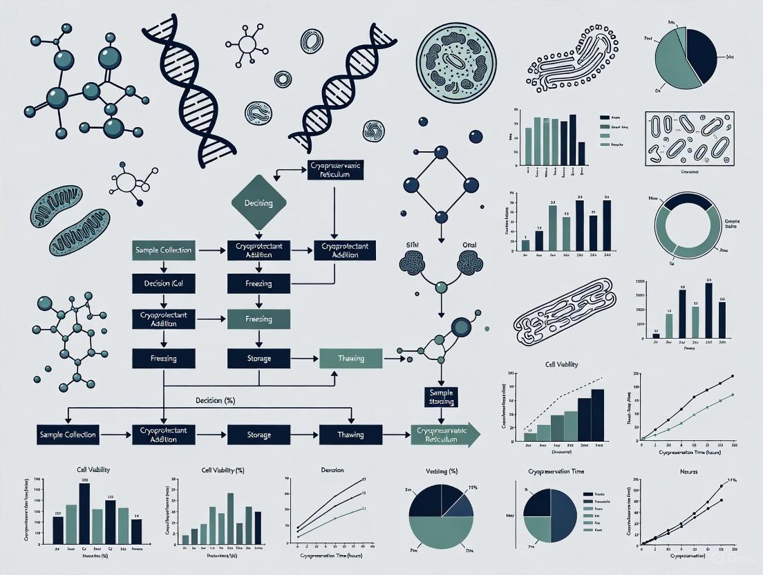
Pathway to Standardization: A Logical Workflow
Implementing a cross-laboratory standardized protocol requires a systematic approach, as visualized below.
The path to overcoming the reproducibility crisis in MSC research is paved with standardized, meticulously documented cryopreservation protocols. By adopting the troubleshooting guides, standardized methodologies, and quality control measures outlined in this resource, researchers and drug development professionals can significantly enhance the reliability and comparability of their data. This commitment to standardization is not merely a technical exercise—it is a fundamental requirement for accelerating the translation of MSC-based therapies from the laboratory bench to the patient bedside.
Fundamental Concepts & FAQs
FAQ 1: Why is standardization in MSC cryopreservation so critical for clinical translation? Standardization is vital to ensure that MSC products are consistent, reproducible, and of high quality across different laboratories and manufacturing facilities. A standardized protocol guarantees that the reader can correctly interpret data and that meta-analyses are generated from comparable datasets [9]. This is a foundational step for the successful transition of MSC-based therapies from research to marketed drug products, addressing significant unmet clinical needs in autoimmunity and other fields [9] [10].
FAQ 2: What are the two primary cryopreservation methods for MSCs, and which is more common? The two main methods are slow freezing and vitrification [11].
- Slow Freezing is the most widely recommended method for clinical and laboratory cryopreservation of MSCs. It involves cooling cells at a controlled rate (approximately -1°C/min), allowing gradual cellular dehydration to minimize intracellular ice crystal formation. It is favored for its ease of operation and lower risk of contamination [2] [11].
- Vitrification uses high concentrations of cryoprotectants and ultra-rapid cooling to solidify cells and their environment into a glassy state without forming ice crystals. While effective, it can be more technically challenging [11].
FAQ 3: What are the core components of a cryopreservation medium? Cryopreservation media typically contain a base culture medium and essential cryoprotective agents (CPAs), which are classified by their mechanism of action [12]:
| Component Type | Function | Common Examples |
|---|---|---|
| Penetrating CPAs | Low molecular weight compounds that enter the cell, bind intracellular water, and reduce ice crystal formation. | Dimethyl sulfoxide (DMSO), Glycerol, Ethylene Glycol [12] [11] |
| Non-Penetrating CPAs | High molecular weight compounds that remain outside the cell, protecting it from osmotic shock and ice crystal growth. | Sucrose, Trehalose, Hydroxyethyl starch, proteins [12] |
For regulated cell and gene therapy fields, it is recommended to use GMP-manufactured, fully-defined cryopreservation media instead of lab-made formulations containing components like fetal bovine serum (FBS), which has undefined components and risks lot-to-lot variability [2].
Troubleshooting Common Cryopreservation Issues
Here is a guide to diagnosing and resolving frequent problems encountered during MSC cryopreservation.
| Problem | Potential Causes | Recommended Solutions |
|---|---|---|
| Low Post-Thaw Viability | • Inappropriate cooling rate: Too slow (excessive dehydration) or too fast (intracellular ice).• High CPA toxicity.• Improper cell concentration.• Suboptimal storage temperature. | • Use a controlled-rate freezer or a validated freezing container to maintain a cooling rate of -1°C/min [2] [11].• Optimize CPA type and concentration; consider combining DMSO with non-penetrating CPAs like sucrose [12].• Test freezing at different cell concentrations (general range: 1x10^3 - 1x10^6 cells/mL) to find the optimum [2].• For long-term storage, use liquid nitrogen (-135°C to -196°C); -80°C is only acceptable for short-term storage (<1 month) [2] [12]. |
| Poor Cell Recovery & Function | • Osmotic shock during CPA addition/removal.• Damage from intracellular ice recrystallization during thawing.• Multiple freeze-thaw cycles. | • Use a slow, stepwise addition and removal of CPAs to minimize osmotic stress [11].• Thaw cells rapidly (e.g., in a 37°C water bath) to minimize ice recrystallization damage [2] [11].• Centrifuge post-thaw to remove CPAs, especially toxic ones like DMSO [11].• Minimize freeze-thaw cycles; one or two freezing steps in early passages is feasible, but exhaustive freezing (≥4 steps) may induce senescence [13]. |
| Inconsistent Experimental Results | • Lack of pre-freezing quality control.• Variable freezing or thawing protocols.• Inadequate record keeping. | • Ensure cells are healthy, free of microbial contamination (e.g., test for mycoplasma), and harvested during their maximum growth phase (log phase, >80% confluency) before freezing [2].• Strictly adhere to a single, validated protocol for all cryopreservation and thawing steps [2].• Maintain detailed inventory records and label vials with all relevant information (passage number, date, cell concentration) to ensure traceability [2]. |
Experimental Protocols for Validation
Protocol: Standard Slow Freezing of MSCs
This is a generalized protocol for the slow freezing of mesenchymal stromal cells.
- Step 1: Harvest and Centrifuge. Harvest the cells using a standard dissociation agent (e.g., TrypLE) and centrifuge them. Carefully remove the supernatant [2].
- Step 2: Resuspend in Freezing Medium. Resuspend the cell pellet in a suitable, pre-cooled freezing medium. For example, use a commercial GMP-grade medium like CryoStor CS10 or a specialized medium such as MesenCult-ACF Freezing Medium for MSCs [2].
- Step 3: Aliquot. Aliquot the cell suspension into sterile, labeled cryogenic vials [2].
- Step 4: Controlled-Rate Freezing. Place the cryogenic vials in an isopropanol-based freezing container (e.g., Nalgene Mr. Frosty) or an isopropanol-free container (e.g., Corning CoolCell) and immediately transfer it to a -80°C freezer for overnight cooling (~-1°C/min). Alternatively, use a controlled-rate freezer [2].
- Step 5: Long-Term Storage. The next day, promptly transfer the vials to a long-term storage liquid nitrogen tank, maintaining a temperature of -135°C to -196°C [2].
Protocol: Thawing Cryopreserved MSCs
- Step 1: Rapid Thaw. Remove the vial from liquid nitrogen and immediately thaw the cells rapidly by gently agitating it in a 37°C water bath until only a small ice crystal remains [2] [11]. To enhance safety and avoid contamination from a water bath, consider using specialized dry thawing equipment [11].
- Step 2: Dilute and Wash. Gently transfer the cell suspension to a tube containing pre-warmed culture medium. This dilution step reduces the concentration of the potentially toxic CPA. Centrifuge the cell suspension to pellet the cells and remove the CPA-containing supernatant [2] [11].
- Step 3: Plate and Culture. Resuspend the cell pellet in fresh, complete culture medium and plate the cells at the desired density. Assess cell viability and confluency after 24 hours [2].
Key Signaling Pathways & Experimental Workflows
Standard MSC Cryopreservation Workflow
The following diagram illustrates the critical decision points and steps in a standardized MSC cryopreservation and thawing process.
Mechanisms of Cryoprotective Agents (CPAs)
This diagram outlines how different types of cryoprotective agents work to protect cells during the freezing process.
The Scientist's Toolkit: Research Reagent Solutions
The table below lists key materials and reagents essential for implementing a standardized MSC cryopreservation protocol.
| Item | Function & Importance |
|---|---|
| Defined Cryopreservation Medium (e.g., CryoStor CS10, MesenCult-ACF) | A ready-to-use, serum-free medium that provides a safe, protective environment during freezing, storage, and thawing. Its use is recommended in regulated fields to ensure consistency and safety [2]. |
| Cryoprotective Agents (CPAs) | DMSO: A penetrating CPA that reduces ice crystal formation but has known toxicity. Sucrose/Trehalose: Non-penetrating CPAs that provide extracellular protection and can help mitigate osmotic shock [12] [11]. |
| Controlled-Rate Freezing Container (e.g., Nalgene Mr. Frosty, Corning CoolCell) | Devices that provide an approximate cooling rate of -1°C/minute when placed in a -80°C freezer, making controlled-rate freezing accessible without expensive equipment [2]. |
| Sterile Cryogenic Vials | Single-use, sterile vials designed for ultra-low temperatures. Internal-threaded vials are preferable to prevent contamination during filling or storage in liquid nitrogen [2]. |
| Liquid Nitrogen Storage System | Essential for long-term storage at -135°C to -196°C. Storage at -80°C is not recommended for the long term, as cell viability will decline over time due to transient warming events [2] [12]. |
Frequently Asked Questions (FAQs)
FAQ 1: Why is there no single, universal cryopreservation protocol for MSCs?
The diversity of MSC sources (e.g., bone marrow, adipose tissue, umbilical cord) and the specific downstream applications (research vs. clinical therapy) necessitate tailored protocols. Furthermore, the selection of cryoprotectants and freezing rates shows significant variability across labs, which complicates the standardization process. The primary goal of standardization is therefore not to find one universal protocol, but to establish a framework of core principles that ensure consistent and reproducible outcomes regardless of the specific application [12].
FAQ 2: What is the core trade-off when using cryoprotectants like DMSO?
Cryoprotectants like Dimethyl Sulfoxide (DMSO) present a fundamental trade-off between protection and toxicity. The same biochemical properties that enable DMSO to protect cells during freezing—such as forming hydrogen bonds with water to prevent ice crystal formation and acting as an antioxidant—are also responsible for its cytotoxic effects. These effects can include altering cellular metabolism and, at higher concentrations or upon post-thaw administration, causing adverse events in patients [14] [15] [16].
FAQ 3: Is controlled-rate freezing always necessary for MSCs?
While controlled-rate freezing at approximately -1°C/min is widely considered the gold standard and is highly recommended for maximizing cell viability and reproducibility, some studies indicate that "straight freeze" methods using isopropanol containers can also be effective for certain cell types. However, uncontrolled freezing carries a higher risk of intracellular ice formation or excessive dehydration, leading to variable and often suboptimal post-thaw outcomes. For the purpose of protocol standardization, the use of a controlled rate is strongly advised [17] [2] [18].
FAQ 4: How does cryopreservation affect the "stemness" and functionality of MSCs?
Cryopreservation can impact MSCs beyond simple viability. The process may disrupt interactions with the extracellular matrix and alter the epigenetic landscape, potentially affecting the cells' ability to self-renew and differentiate. Furthermore, recent research indicates that cells in the S phase of the cell cycle are particularly susceptible to cryoinjury, which can lead to delayed apoptosis and reduced immunomodulatory function post-thaw [15] [19].
Troubleshooting Guides
Issue 1: Low Post-Thaw Viability
| Potential Cause | Diagnostic Steps | Recommended Solution |
|---|---|---|
| Suboptimal Freezing Rate | Review protocol cooling rates in critical zone (0°C to -60°C). | Implement a controlled freezing rate of -1°C/min down to at least -40°C to -80°C before transfer to liquid nitrogen [17] [2]. |
| Intracellular Ice Crystallization | Check viability immediately and 24 hours post-thaw. | Ensure cryopreservation medium contains adequate penetrating cryoprotectants (e.g., 5-10% DMSO) to bind intracellular water [12] [11]. |
| Cryoprotectant Toxicity | Examine protocol for CPA concentration and exposure time at non-frozen temperatures. | Use lower DMSO concentrations (e.g., 5%) combined with non-penetrating CPAs like Hydroxyethyl Starch (HES) or sucrose to reduce toxicity [18]. |
Issue 2: Loss of MSC Stemness and Differentiation Potential Post-Thaw
| Potential Cause | Diagnostic Steps | Recommended Solution |
|---|---|---|
| Disrupted Cell-Matrix Interactions | Perform post-thaw differentiation assays (osteogenic, adipogenic, chondrogenic). | Encapsulate MSCs in protective hydrogels or biomaterials during cryopreservation to mimic a natural niche [15]. |
| Epigenetic Alterations | Analyze expression of key stemness markers and differentiation genes post-recovery. | Validate post-thaw functionality through standardized differentiation capability assays and immunomodulatory assays before use [15]. |
| Cryoinjury to Specific Cell Cycle Phases | Analyze cell cycle distribution pre-freeze and post-thaw. | Synchronize cells in G0/G1 phase prior to freezing via growth factor deprivation (serum starvation) to protect replication-prone cells [19]. |
Issue 3: Inconsistent Results Between Batches or Labs
| Potential Cause | Diagnostic Steps | Recommended Solution |
|---|---|---|
| Variable Cryoprotectant Formulations | Audit and compare the exact composition and concentration of CPAs used. | Adopt chemically defined, xeno-free freezing media to eliminate variability and safety concerns associated with serum-containing media [15] [2]. |
| Inconsistent Cooling Rates | Calibrate and validate freezing equipment (controlled-rate freezers, isopropanol containers). | Standardize the freezing workflow using validated equipment and ensure all users are trained on the same protocol [17] [2]. |
| Lack of Post-Thaw Quality Control | Check if quality control is limited to viability tests only. | Implement a panel of quality control assays post-thaw, including viability, phenotype (flow cytometry), proliferation, and differentiation potential [15] [20]. |
Experimental Protocol Summaries
Protocol 1: Standardized Slow-Freezing for MSC Cryopreservation
This protocol is adapted from established best practices and is suitable for creating Master Cell Banks [2] [20].
Key Reagents and Materials:
- Cryogenic vials
- Controlled-rate freezing device (e.g., programmable freezer or isopropanol-based container like "Mr. Frosty")
- -80°C Freezer
- Liquid nitrogen storage tank
- Defined Cryopreservation Medium (e.g., containing 5-10% DMSO, potentially combined with HES or sucrose)
Methodology:
- Harvesting: Harvest MSCs during their maximum growth phase (typically >80% confluency). Use enzymatic digestion (e.g., trypsin) to create a single-cell suspension [2].
- Centrifugation: Centrifuge the cell suspension to pellet the cells. Carefully remove the supernatant [2].
- Resuspension in Freezing Medium: Resuspend the cell pellet in pre-chilled cryopreservation medium at a specific, validated cell concentration (e.g., between 5 x 10^5 cells/mL and 1 x 10^6 cells/mL) [15] [2].
- Aliquoting: Aliquot the cell suspension into cryogenic vials. Ensure vials are properly labeled [2].
- Controlled-Rate Freezing: Place the cryogenic vials in a controlled-rate freezing device and store them overnight in a -80°C freezer. This achieves a cooling rate of approximately -1°C/minute, which is critical for minimizing intracellular ice formation [17] [2].
- Long-Term Storage: The following day, promptly transfer the vials to the vapor phase of liquid nitrogen (-135°C to -196°C) for long-term storage [15] [2].
Protocol 2: Mitigating Cryoinjury via Cell-Cycle Synchronization
This protocol is based on a 2023 study that identified a fundamental cryoinjury mechanism in S-phase MSCs and a method to mitigate it [19].
Key Reagents and Materials:
- Standard MSC culture media
- Serum-free media or media with reduced growth factors
- Reagents for cell cycle analysis (e.g., propidium iodide)
Methodology:
- Pre-Culture and Expansion: Culture MSCs according to standard protocols until the desired confluence is reached.
- Cell Cycle Synchronization (Serum Starvation): Prior to harvesting for cryopreservation, subject the MSCs to growth factor deprivation. This is typically done by replacing the standard growth medium with serum-free medium or medium containing a reduced serum concentration (e.g., 0.5% FBS) for 24-48 hours. This intervention arrests a majority of the cells in the G0/G1 phase of the cell cycle [19].
- Verification (Optional): A sample of cells can be analyzed by flow cytometry to confirm the increase in the G0/G1 population.
- Cryopreservation: Proceed with the standard cryopreservation protocol (as detailed in Protocol 1) starting from the harvesting step. The study showed that cells treated this way exhibited preserved viability, clonal growth, and T-cell suppression function post-thaw at levels comparable to pre-freeze controls [19].
Standardized Data and Workflow Visualization
Cryopreservation Workflow and Critical Control Points
This diagram outlines the core workflow for MSC cryopreservation, highlighting key steps where standardization is crucial for reproducibility.
Decision Framework for Cryopreservation Protocol Selection
This flowchart provides a logical guide for researchers to select an appropriate cryopreservation strategy based on their specific goals and constraints.
The Scientist's Toolkit: Essential Research Reagents
The following table details key materials and reagents essential for implementing standardized MSC cryopreservation protocols.
| Reagent / Material | Function & Rationale |
|---|---|
| Chemically Defined, Xeno-Free Cryomedium | Pre-formulated, serum-free freezing media (e.g., CryoStor) eliminate batch-to-batch variability and immunogenic risks associated with fetal bovine serum (FBS), ensuring safety and reproducibility for clinical applications [15] [2]. |
| Dimethyl Sulfoxide (DMSO) | The most common penetrating cryoprotectant. It lowers the freezing point of water and minimizes intracellular ice formation. Its concentration must be optimized (often 5-10%) to balance efficacy with inherent cytotoxicity [12] [16]. |
| Hydroxyethyl Starch (HES) | A non-penetrating cryoprotectant. It acts as an extracellular bulking agent, reducing the amount of DMSO required and thus mitigating DMSO-related toxicity. Studies show 5% DMSO/5% HES can be an effective combination [18]. |
| Sucrose / Trehalose | Non-penetrating disaccharides that function as osmotic buffers. They help stabilize cell membranes during freezing and reduce osmotic shock during the addition and removal of CPAs [12]. |
| Controlled-Rate Freezer | Equipment that guarantees a consistent, reproducible cooling rate (typically -1°C/min). This is a cornerstone of protocol standardization, preventing the variable cell death associated with inconsistent cooling [17] [2]. |
| Isopropanol Freezing Container | A cost-effective alternative to programmable freezers. These containers (e.g., Nalgene "Mr. Frosty") provide an approximate cooling rate of -1°C/min when placed in a -80°C freezer, improving standardization over simple placement in a freezer [2]. |
This technical support center is designed to assist researchers and drug development professionals in navigating the critical challenges of standardizing Mesenchymal Stem Cell (MSC) cryopreservation protocols. As Advanced Therapy Medicinal Products (ATMPs) move toward commercial reality, achieving robust, reproducible, and well-defined manufacturing processes is a fundamental regulatory requirement. The variability in current cryopreservation practices represents a significant hurdle to this goal [21] [11]. This resource, structured in a question-and-answer format, provides detailed troubleshooting guides, standardized experimental protocols, and data presentation frameworks to support compliance and enhance the translational success of MSC-based therapies.
Fundamental Concepts in MSC Cryopreservation
FAQ: Why is cryopreservation standardization critical for MSC-based ATMPs?
Answer: Standardization is paramount because cryopreservation is not merely a storage step but a critical unit operation in the manufacturing process of an ATMP. Variations in protocol can directly impact the critical quality attributes (CQAs) of the final product, such as viability, potency, and functionality, thereby affecting clinical safety and efficacy [6] [11]. A 2025 survey of transplant centers revealed significant heterogeneity in practices, including the use of different DMSO concentrations (ranging from 5% to 15%), varying cryopreservation media compositions, and inconsistent post-thaw quality assessment, with 28.6% of patients not undergoing post-thaw testing [21]. This lack of standardization poses a major challenge to ensuring consistent product quality and reliable clinical outcomes.
FAQ: What are the primary mechanisms of cell damage during cryopreservation?
Answer: The primary mechanisms are intracellular ice crystal formation and osmotic stress.
- Ice Crystal Formation: During freezing, intracellular and extracellular water can form ice crystals that physically pierce and damage the cell membrane and internal structures [22].
- Osmotic Stress: As water freezes, the concentration of solutes inside and outside the cell increases dramatically. This can lead to detrimental cell shrinkage during freezing and harmful swelling during thawing, damaging cellular components [11] [22]. The goal of an optimized protocol is to manage these two competing phenomena by controlling cooling rates and using cryoprotective agents (CPAs).
FAQ: What are the two main cryopreservation methods, and which is recommended for MSCs?
Answer: The two primary methods are slow freezing and vitrification.
- Slow Freezing involves a controlled, slow cooling rate (typically around -1°C to -3°C per minute) in the presence of a CPA. This allows water to gradually leave the cell, minimizing deadly intracellular ice formation [11]. It is the most common and recommended method for clinical-grade MSC cryopreservation due to its operational simplicity, scalability, and lower risk of contamination [11].
- Vitrification uses high concentrations of CPAs and ultra-rapid cooling to solidify cells and their environment into a glassy, non-crystalline state [11]. While it avoids ice formation, the high CPA concentrations can be cytotoxic, and the method is more complex and less scalable for large volumes, limiting its current widespread clinical use for MSCs.
The following workflow outlines the key stages in developing an optimized, standardized cryopreservation protocol, integrating both process parameters and quality assessments.
Troubleshooting Common Cryopreservation Challenges
FAQ: We are observing low post-thaw viability in our MSCs. What are the potential causes and solutions?
Answer: Low viability is often linked to suboptimal cooling rates, CPA toxicity, or improper thawing.
| Observed Issue | Potential Causes | Recommended Solutions |
|---|---|---|
| Low Post-Thaw Viability | Intracellular ice crystal damage [22]. | Use a controlled-rate freezer. If unavailable, use an alcohol-free freezing container that provides a rate of ~ -1°C/min [22]. |
| Excessive osmotic stress during CPA addition/removal [11]. | Ensure stepwise or dropwise addition and removal of CPAs. Use non-penetrating CPAs like sucrose to mitigate osmotic shock [11] [23]. | |
| Cryoprotectant (e.g., DMSO) toxicity [11] [22]. | Optimize DMSO concentration (often 5-10%). Consider DMSO-free or lower-DMSO media blends, e.g., with human serum albumin [21] [22]. | |
| Loss of Stemness & Differentiation Potential | Disruption of cell-ECM interactions and epigenetic changes due to freeze-thaw stress [22]. | Ensure optimal pre-freeze cell health and density. Use defined, xeno-free cryomedium. Validate differentiation potential post-thaw with Oil Red O (adipocytes), Alizarin Red (osteocytes), and Alcian Blue (chondrocytes) staining [22]. |
| Inconsistent Results Between Batches | Variable freezing rates or storage conditions [22]. | Strictly control cooling rates and ensure consistent storage below -150°C [21] [22]. |
| Donor-to-donor or passage-level variability. | Use early-passage cells and establish rigorous pre-freeze quality controls for all batches [6]. |
FAQ: How can we effectively remove DMSO post-thaw without causing additional cell loss?
Answer: The process of removing DMSO is critical. A rapid dilution of the external CPA concentration causes a large osmotic gradient, leading to excessive cell swelling and lysis [11]. To mitigate this:
- Gradual Dilution: Thaw cells rapidly and then add pre-warmed culture medium dropwise or in a stepwise manner to the cell suspension, gradually reducing the DMSO concentration.
- Centrifugation and Resuspension: After dilution, centrifuge the cells at a gentle, defined speed and time (e.g., 300-400 x g for 5-7 minutes) to pellet the cells. Carefully aspirate the supernatant and resuspend the pellet in fresh, complete culture medium [22].
- Alternative Methods: Researchers are exploring closed-system washing devices or density gradient centrifugation to improve cell recovery and consistency during this step.
Quality Control and Functional Assay Guidance
A comprehensive quality control strategy must extend beyond simple viability checks to assess the functionality of cryopreserved MSCs, which is a key regulatory expectation for ATMPs. The diagram below maps the essential quality attributes that should be tested and the corresponding assays.
FAQ: What are the mandatory and recommended quality control tests for cryopreserved MSC batches?
Answer: A tiered approach to quality control is essential for ATMPs.
1. Mandatory Release Assays (Basic Quality):
- Cell Viability: Should typically be >70-80% post-thaw. Assess using Trypan Blue exclusion or, more accurately, flow cytometry with Annexin V/Propidium Iodide (PI) to distinguish live, early apoptotic, and necrotic cells [11] [22].
- Cell Phenotype: Confirm expression of positive markers (CD73, CD90, CD105 ≥95%) and lack of negative markers (CD34, CD45, CD14, CD19, HLA-DR ≤2%) via flow cytometry [24] [22].
- Sterility and Mycoplasma: Test for bacterial, fungal, and mycoplasma contamination to ensure product safety.
2. Potency and Functional Assays (Critical for Efficacy):
- Trilineage Differentiation: Demonstrate functional capacity to differentiate into adipocytes (Oil Red O stain), osteocytes (Alizarin Red stain), and chondrocytes (Alcian Blue stain) [22].
- Immunomodulatory Function: Quantify the ability to suppress the proliferation of activated immune cells (e.g., T-cells or PBMCs) in a co-culture assay, often measuring cytokine secretion or CFSE dilution [22].
3. Extended Characterization (For Process Validation):
- Proliferation Capacity: Measure population doubling time post-thaw to ensure recovery.
- Biosafety: Perform karyotyping or other tests to rule out genetic instability after extensive culture and cryopreservation [22].
The Scientist's Toolkit: Research Reagent Solutions
The following table details key materials and reagents essential for standardized MSC cryopreservation protocols.
| Item | Function & Rationale | Standardization Consideration |
|---|---|---|
| Chemically Defined, Xeno-Free Cryomedium | Provides a consistent, serum-free environment for freezing, eliminating batch-to-batch variability and immunogenic risks from animal components [22]. | A GMP-compliant, chemically defined medium is a regulatory expectation for clinical lot production. |
| DMSO (USP Grade) | The most common penetrating CPA. Protects cells from intra-cellular ice formation [11]. | Use high-purity, USP grade. Concentration must be optimized and fixed (e.g., 10% is common); survey data shows variability from 5-15% is problematic [21]. |
| Non-Penetrating CPAs (e.g., Sucrose, Trehalose) | Increase extracellular osmolarity, promoting gentle cell dehydration and reducing osmotic shock during CPA addition/removal [11] [23]. | Their inclusion in cryomedium formulations improves recovery and allows for potential DMSO concentration reduction. |
| GMP-Grade Cryovials/Bags | Medical-grade polypropylene, leak-proof, externally threaded vials with clear labeling patches ensure sample integrity and traceability at ultra-low temperatures [25]. | Consistent, validated container closure systems prevent contamination and cross-sample mix-ups. |
| Controlled-Rate Freezer | Provides a reproducible, linear cooling rate (e.g., -1°C/min), which is critical for maximizing cell viability and minimizing ice crystal damage [21] [22]. | Essential for moving beyond variable "Mr. Frosty" containers to a scalable, validated process. |
| Liquid Nitrogen Storage System | Maintains cells at -150°C to -196°C, ensuring long-term metabolic stasis. Redundant, geographically separate storage is a best practice [26] [22]. | Requires continuous temperature monitoring and alarm systems to ensure product stability. |
Navigating Regulatory Requirements
FAQ: From a regulatory standpoint, what are the key documentation needs for a standardized cryopreservation process?
Answer: Regulatory agencies require detailed and validated information on the entire manufacturing process. For cryopreservation, this includes:
- A Defined and Validated Protocol: A standard operating procedure (SOP) that specifies with precision: cell concentration and volume at freezing, the complete composition of the cryopreservation medium, the type of container, the exact freezing curve (including cooling rates, seeding temperature, and endpoint), storage conditions, and the complete thawing and washing procedure [23].
- Process Validation Data: Data demonstrating that the protocol consistently produces MSCs that meet all pre-defined CQAs (viability, phenotype, potency) across multiple batches.
- Stability Data: Evidence that the cryopreserved product maintains its quality attributes over the proposed storage period.
- Traceability: Full documentation chain from donor to final product, including the storage location and conditions of each cryopreserved vial.
By adopting the principles and practices outlined in this technical support center, researchers can systematically address the sources of variability in MSC cryopreservation, thereby strengthening the scientific and regulatory foundation for the successful commercialization of MSC-based ATMPs.
Frequently Asked Questions (FAQs)
Q1: What is the primary goal of standardizing MSC cryopreservation protocols? The primary goal is to ensure consistent product identity, potency, viability, and stability of Mesenchymal Stromal Cell (MSC)-based therapies across different laboratories and manufacturing sites [27]. Standardization mitigates variability introduced by donors, tissue sources, and cell culture methods, which is essential for obtaining reproducible and reliable outcomes in both research and clinical trials. It directly addresses challenges in comparing clinical trial results and helps position these innovative therapeutics for advancement in regenerative medicine.
Q2: Does cryopreservation negatively impact the therapeutic function of MSCs? Not necessarily. When optimized protocols are used, key therapeutic functions can be preserved. A recent study on bone marrow aspirate concentrate (BMAC) found that freezing at -80°C for four weeks preserved MSC proliferation and multilineage differentiation capacity. Critically, in an osteoarthritis rat model, both fresh and frozen BMAC demonstrated significantly improved cartilage repair compared to a control, with no significant difference between fresh and frozen treatments [28]. This indicates that cryopreservation, when properly executed, can retain functional equivalence.
Q3: Why is DMSO a concern in cryopreservation, and are there alternatives? Dimethyl sulfoxide (DMSO) is a standard penetrating cryoprotectant, but its use is associated with several concerns:
- Cellular Toxicity: DMSO can be toxic to cells, affecting their metabolism and health [11] [29].
- Patient Side Effects: Upon infusion, DMSO can cause adverse reactions in patients, including nausea, vomiting, arrhythmias, and respiratory depression [30] [29]. Promisingly, DMSO-free alternatives are under development. An international multicenter study demonstrated that a novel solution containing sucrose, glycerol, and isoleucine (SGI) provided comparable post-thaw viability, recovery, and immunophenotype to DMSO-containing solutions, with average viability above the clinically acceptable threshold of 80% [29].
Q4: What are the critical control points during the freezing and thawing process? The entire process requires careful control to minimize cell stress and death.
- Freezing: The cooling rate must be tightly controlled, typically using a controlled-rate freezer, to facilitate gradual cellular dehydration and minimize lethal intracellular ice crystal formation [11] [31].
- Thawing: This is a often-underestimated critical step. Thawing must be rapid (e.g., in a 37°C water bath) to avoid devitrification (recrystallization) and to limit the prolonged exposure of cells to concentrated cryoprotectants, which can cause osmotic shock and cell lysis [11] [31]. Non-controlled thawing is a major source of poor cell viability and recovery.
Q5: How can new technologies help reduce reliance on DMSO? Emerging technologies focus on physically protecting cells during cryopreservation. For instance, hydrogel microencapsulation technology has been shown to enable effective cryopreservation of MSCs with DMSO concentrations as low as 2.5%, while maintaining cell viability above the 70% clinical threshold. The hydrogel capsule acts as a protective barrier, mitigating cryo-injury [30]. This approach represents a shift towards using cell-biomaterial constructs for safer and more efficient stem cell storage.
Troubleshooting Common Cryopreservation Challenges
Challenge 1: Low Post-Thaw Viability
| Potential Cause | Investigation | Recommended Solution |
|---|---|---|
| Suboptimal freezing rate | Review controlled-rate freezer profile or passive freezing method validation data. | Use a controlled-rate freezer (CRF). For sensitive cells, avoid default CRF profiles and develop an optimized cooling rate protocol [31]. |
| Improper handling during pre-freeze culture | Check cell culture records for passage number, confluence, and contamination. | Freeze only high-quality, early-passage cells that are in log-phase growth and free from contamination [32]. |
| Toxic effects of DMSO | Test post-thaw viability with different DMSO concentrations or alternative CPAs. | Reduce DMSO concentration or transition to a DMSO-free cryoprotectant solution [29]. Ensure rapid and thorough removal of DMSO post-thaw to limit exposure [11]. |
| Osmotic shock during thawing | Observe thawing procedure for consistency and speed. | Thaw cells rapidly in a 37°C water bath until only a small ice crystal remains. Immediately dilute the cryoprotectant with pre-warmed culture medium [11] [32]. |
Challenge 2: Loss of MSC Functionality After Thaw
| Potential Cause | Investigation | Recommended Solution |
|---|---|---|
| Compromised cell fitness pre-freeze | Perform potency assays (e.g., differentiation, immunomodulation) on pre-freeze cells. | Ensure cells are harvested and processed under conditions that maintain their native phenotype and function before initiating cryopreservation [27]. |
| Inadequate post-thaw recovery time | Assess functionality immediately post-thaw and again after 24-48 hours in culture. | Allow MSCs a recovery period (e.g., 24-48 hours) in culture post-thaw before using them in functional assays or therapies, as some functions are restored after cell repair [28]. |
| Unoptimized cryoprotectant composition | Compare post-thaw functionality using different CPA formulations. | Consider cryoprotectant cocktails that include non-penetrating agents like sucrose or trehalose, which can help stabilize cell membranes [11] [29]. |
Experimental Data & Standardization Protocols
Comparative Analysis of Cryopreservation Methods and Outcomes
The following table summarizes quantitative findings from recent studies on MSC cryopreservation, highlighting key variables and outcomes relevant for protocol standardization.
Table 1: Comparison of Cryopreservation Method Outcomes from Recent Studies
| Study Focus | Cryopreservation Method | Cryoprotectant (CPA) | Key Quantitative Findings | Reference |
|---|---|---|---|---|
| International Multicenter Study | Controlled-rate freezing | 5-10% DMSO (in-house) vs. DMSO-free (SGI) | • Viability: DMSO: ~89.8%; SGI: ~82.9%• Viable Recovery: SGI: 92.9%; DMSO: ~87.3%• Phenotype/Genetics: Comparable | [29] |
| Hydrogel Microencapsulation | Slow freezing | 2.5% DMSO with alginate hydrogel | • Viability: >70% (clinical threshold)• Function: Retained multidifferentiation potential and stemness gene expression. | [30] |
| Short-Term BMAC Preservation | Passive freezing at -80°C | 10% DMSO + 90% autologous plasma | • Viability/Function: Preserved proliferation & multilineage differentiation.• In Vivo: No significant difference in cartilage repair vs. fresh BMAC. | [28] |
| General Slow Freezing | Slow freezing | DMSO-based (various conc.) | • Typical Viability: ~70-80% cell survival.• Key Risk: CPA toxicity and osmotic stress during addition/removal. | [11] |
Standardized Experimental Workflow for Protocol Validation
To ensure consistency across laboratories, the following workflow outlines a core methodology for validating an MSC cryopreservation protocol. The subsequent diagram visualizes this multi-stage process.
Diagram 1: Standardized workflow for validating MSC cryopreservation protocols.
Detailed Methodology:
Pre-freeze MSC Culture & Harvest:
Cryoprotectant Addition & Aliquotting:
- Resuspend the cell pellet in a pre-chilled freezing medium. A common base is Plasmalyte A or autologous plasma supplemented with the chosen CPA [28] [29].
- For DMSO-based protocols, a final concentration of 5-10% is standard, but lower concentrations (2.5-5%) should be evaluated [30] [29].
- Aliquot the cell suspension into cryogenic vials at a consistent cell concentration (e.g., 1-5 million cells/mL) [28].
Controlled-Rate Freezing & Storage:
- Place vials in a controlled-rate freezer. A widely used slow-freezing profile involves a cooling rate of -1°C/min to -3°C/min from 4°C to -40°C or -80°C, before rapid transfer to liquid nitrogen (-196°C) for long-term storage [11] [31].
- Avoid passive freezing in a -80°C freezer for critical or late-stage clinical products due to less control over the cooling rate [31].
Thawing and Cryoprotectant Removal:
- Rapidly thaw vials in a 37°C water bath with gentle agitation until only a small ice crystal remains [11].
- Immediately transfer the contents to a tube containing pre-warmed culture medium to dilute the CPA.
- Centrifuge (e.g., 300-400 x g for 5-10 min) to pellet cells and remove the CPA-containing supernatant [28].
Post-Thaw Analysis:
- Viability & Recovery: Assess using trypan blue exclusion or automated cell counters. Calculate viable cell recovery [29].
- Immunophenotype: Verify surface marker expression (CD73+, CD90+, CD105+; CD45-, CD34-, HLA-DR-) via flow cytometry to confirm identity [11] [27].
- Potency Assays: Perform functional tests relevant to the clinical application, such as:
The Scientist's Toolkit: Essential Reagents & Materials
Table 2: Key Research Reagent Solutions for MSC Cryopreservation
| Reagent / Material | Function / Purpose | Standardization Consideration |
|---|---|---|
| Dimethyl Sulfoxide (DMSO) | Penetrating cryoprotectant; reduces ice crystal formation by binding water molecules. | Pharmaceutical/clinical grade is essential. Concentration (often 5-10%) and exposure time must be standardized due to inherent toxicity [11] [29]. |
| DMSO-Free Cryoprotectants (e.g., SGI) | Alternative CPA cocktail; reduces risk of DMSO-related toxicity to cells and patients. | Formulations like SGI (Sucrose, Glycerol, Isoleucine) require validation but show comparable results to DMSO [29]. |
| Non-Penetrating CPAs (e.g., Sucrose, Trehalose) | Stabilize the cell exterior; increase solution viscosity and mitigate osmotic shock. | Often used in combination with penetrating CPAs to allow for lower DMSO concentrations [11] [29]. |
| Hydrogel (e.g., Alginate) | Biomaterial for microencapsulation; provides a physical 3D barrier against cryo-injury. | Enables significant reduction of DMSO concentration (e.g., to 2.5%); requires specialized equipment for encapsulation [30]. |
| Controlled-Rate Freezer (CRF) | Equipment that provides a precise, user-defined cooling rate during freezing. | Critical for process control and consistency. Default profiles may not be optimal for all cell types; customization may be needed [31]. |
| Cryopreservation Media Base (e.g., Plasmalyte A, Autologous Plasma) | The solution in which CPAs are dissolved; provides ionic and nutrient support. | Using a defined, xeno-free base (vs. FBS-containing media) enhances clinical compatibility and reduces batch variability [28] [29]. |
Core Components of an Effective MSC Cryopreservation Protocol
CPA Classification and Mechanism of Action
What are the fundamental differences between penetrating and non-penetrating cryoprotectants?
Cryoprotective Agents (CPAs) are essential substances that protect biological samples from freezing damage. They are classified into two main categories based on their ability to cross cell membranes, each with distinct protective mechanisms [12] [33].
Penetrating CPAs (also known as endocellular cryoprotectants) are typically low molecular weight compounds that can cross the cell membrane. They function by reducing ice formation inside the cell, minimizing dehydration, and stabilizing intracellular proteins [12]. Their ability to penetrate cells makes them highly effective but also contributes to their potential toxicity [33].
Non-Penetrating CPAs (also known as exocellular cryoprotectants) are typically high molecular weight compounds that remain outside the cell. They protect primarily by inducing osmotic dehydration before freezing, reducing the chance of lethal intracellular ice formation, and stabilizing the cell membrane from the outside [12] [33].
Table 1: Characteristics of Penetrating vs. Non-Penetrating CPAs
| Feature | Penetrating CPAs | Non-Penetrating CPAs |
|---|---|---|
| Molecular Weight | Low [12] | High (polymers and oligosaccharides) [12] |
| Cell Membrane Permeability | Crosses the membrane [12] | Does not cross the membrane [12] |
| Primary Mechanism of Action | Bind intracellular water, lower freezing point, reduce ice crystal formation inside the cell [12] | Create osmotic gradient causing protective dehydration, inhibit ice crystal growth outside the cell [12] |
| Common Examples | DMSO, glycerol, ethylene glycol, propylene glycol [12] [34] | Sucrose, trehalose, ficoll, polyvinylpyrrolidone, hydroxyethyl starch [12] |
| Relative Toxicity | Generally higher [33] | Generally lower [33] |
CPA Mechanism and Classification
Quantitative Performance Data
What is the relative performance of different CPAs in MSC cryopreservation?
Selecting the optimal CPA involves balancing protective efficacy with cellular toxicity. The following table summarizes experimental data on various CPAs and their impact on post-thaw MSC viability.
Table 2: Efficacy of Common and Alternative CPAs in MSC Cryopreservation
| Cryoprotectant | Type | Reported Concentration | Reported Post-Thaw Viability / Effect | Key Findings / Notes |
|---|---|---|---|---|
| DMSO | Penetrating | 5-10% (v/v) | ~55-70% viability [35]; Standard for comparison [11] | Considered the "gold standard" but has known toxicity and can alter gene expression [35]. |
| Sucrose | Non-Penetrating | 12% solution [36] | Improves bacterial survival [36] | Forms stable hydrate shells; low Gibbs free energy of solvation enhances protection [36]. |
| Urea + Glucose | Penetrating + Non-Penetrating | 0.5M Urea + 0.5M Glucose [35] | ~55% viability (comparable to 5% DMSO) [35] | Synergistic effect; urea fluidifies membranes, allowing better glucose penetration [35]. |
| Trehalose (Pre-incubation) | Non-Penetrating | Pre-incubation [35] | Enhances viability when combined with other CPAs [35] | Internalized via endocytosis; acts as a potent cryoprotectant [35]. |
| Glycerol | Penetrating | 10% (v/v) [34] | Improved cell viability in cryobioprinted constructs [34] | Lower cell toxicity than DMSO but may have poorer cryopreservation effect in some contexts [11]. |
| Natural Deep Eutectic Systems (NADES) | Mixed | 50% (w/v) [37] | Varies by formulation and cell line; promising results in vitrification [37] | Composed of natural metabolites; some formulations do not require removal post-thaw, simplifying workflow [37]. |
Troubleshooting Common CPA Issues
FAQ 1: Why is post-thaw viability low even when using standard CPAs like DMSO?
Low viability can stem from multiple factors beyond CPA choice:
- Inadequate Freezing Rate: The slow freezing method requires a carefully controlled cooling rate (typically around -1°C/min to -3°C/min) to allow sufficient cellular dehydration before intracellular ice forms [11]. Using an uncontrolled rate (e.g., placing vials directly in a -80°C freezer) can be detrimental.
- Improper Thawing: Thawing must be rapid (e.g., in a 37°C water bath) to avoid ice recrystallization during the warming process, which can damage cells [11].
- CPA Toxicity Exposure: While necessary, CPAs are toxic. Prolonged exposure to DMSO at room temperature before freezing or after thawing increases cell death [11]. Centrifuging cells immediately after thawing to remove DMSO is crucial [11].
- Suboptimal CPA Formulation: For many cell types, including MSCs, a combination of penetrating and non-penetrating CPAs (e.g., 10% DMSO with sucrose) often provides better protection than a single agent by leveraging synergistic mechanisms [12] [35].
FAQ 2: How can we reduce reliance on potentially toxic CPAs like DMSO and Fetal Bovine Serum (FBS) in clinical applications?
Research is actively focused on developing safer, defined alternatives:
- DMSO-Free Formulations: Combinations of well-tolerated excipients show promise. A key study found that a formulation of 0.5M urea and 0.5M glucose yielded post-thaw MSC viability comparable to 5% DMSO [35]. Pre-incubating cells with trehalose and adding mannitol and sucrose to the freezing medium further enhanced viability [35].
- Natural Deep Eutectic Systems (NADES): These systems, composed of natural primary metabolites like sugars, amino acids, and choline derivatives, are emerging as potent cryoprotectants. Some NADES formulations function at high concentrations for vitrification and may not require removal after thawing, simplifying the process and reducing osmotic stress [37].
- Use of Autologous Plasma: For clinical cell therapies, replacing FBS with the patient's own (autologous) plasma in the cryomedium eliminates the risk of immune reactions and pathogen transmission [38].
FAQ 3: What are the critical steps for a safe and effective thawing process?
Thawing is as critical as freezing. A standardized protocol should be followed:
- Rapid Thaw: Remove the vial from liquid nitrogen and immediately place it in a 37°C water bath with gentle agitation until only a small ice crystal remains [11].
- Quick CPA Dilution: Immediately after thawing, transfer the cell suspension to a larger volume of pre-warmed culture medium. This rapidly dilutes the CPA, reducing its toxic and osmotic effects [11].
- Centrifugation and Wash: Centrifuge the cell suspension to pellet the cells and carefully remove the supernatant containing the CPA [11] [38].
- Resuspension and Assessment: Resuspend the cell pellet in fresh, pre-warmed culture medium and proceed with viability assessment (e.g., trypan blue exclusion) or seeding for expansion.
CPA Workflow and Troubleshooting
The Scientist's Toolkit: Research Reagent Solutions
Table 3: Essential Materials for MSC Cryopreservation Protocols
| Reagent / Material | Function / Application | Example Usage in Protocol |
|---|---|---|
| Dimethyl Sulfoxide (DMSO) | Penetrating CPA; the most common base for cryopreservation solutions [11]. | Used at 5-10% (v/v) in combination with culture medium or serum/plasma for slow freezing [11] [38]. |
| Sucrose / Trehalose | Non-penetrating CPA; adds osmotic support, reduces required DMSO concentration [36] [35]. | Added at 0.1-0.5M to DMSO-based freezing media to improve post-thaw recovery [35]. |
| Autologous Plasma | Biocompatible cryomedium component; avoids use of animal sera (FBS) for clinical applications [38]. | Used as the base solvent (90%) for resuspending the cell pellet with DMSO (10%) before freezing [38]. |
| Urea | Synergistic penetrating CPA; fluidifies cell membranes to facilitate uptake of other protectants like glucose [35]. | Combined at equimolar ratios (e.g., 0.5M) with glucose for DMSO-free cryopreservation formulations [35]. |
| Natural Deep Eutectic Systems (NADES) | Novel, potentially less toxic CPA for vitrification; composed of natural metabolites [37]. | Used at high concentrations (e.g., 50% w/v) as the primary CPA for vitrification protocols, potentially eliminating need for post-thaw removal [37]. |
| Programmable Freezer / Mr. Frosty | Provides controlled cooling rate critical for the slow freezing method [11] [38]. | Cool cells at a rate of -1°C/min from +4°C to -40°C or -80°C before transfer to liquid nitrogen [11] [38]. |
Frequently Asked Questions (FAQs)
Q1: What is the fundamental difference between serum-based and defined, xeno-free cryopreservation media?
A1: The core difference lies in composition, consistency, and regulatory compliance.
- Serum-Based Media: Traditionally, these are "homebrew" solutions often composed of culture medium, 10% Dimethyl Sulfoxide (DMSO), and 10-20% Fetal Bovine Serum (FBS) [2] [39]. FBS is a complex, undefined mixture of growth factors, proteins, and other components, which leads to significant batch-to-batch variability [40] [41]. This variability poses a risk for experimental reproducibility and clinical applications due to the potential introduction of animal-derived pathogens and undefined components [40].
- Defined, Xeno-Free Media: These are commercially prepared, ready-to-use solutions with a chemically defined composition. They are devoid of any animal-derived components (xeno-free), typically using defined cryoprotectants like DMSO, anhydrous dextrose, and polymers [42] [41]. They offer superior lot-to-lot consistency, reduce contamination risks, and are essential for clinical-grade cell banking and manufacturing to meet regulatory standards [42] [2].
Q2: Why is there a strong push to transition to defined, xeno-free media for MSC therapies?
A2: The transition is primarily driven by the requirements of clinical translation and manufacturing standardization.
- Regulatory Compliance: Agencies like the FDA and EMA require well-defined raw materials for cell-based therapies. The undefined nature of FBS is a major regulatory hurdle [40] [43].
- Enhanced Safety: Xeno-free media eliminate the risk of transmitting animal-derived pathogens or eliciting immune responses in patients against bovine antigens [42] [41].
- Process Standardization: To ensure that the therapeutic efficacy of MSCs is consistent across batches, every component, including the cryopreservation medium, must be reproducible and consistent [43]. A study comparing media found that some commercially available "serum-free" media still contained human platelet lysate components, highlighting the need for careful reagent selection and transparent nomenclature [40].
Q3: Does using defined cryopreservation media compromise the post-thaw viability and function of MSCs?
A3: No, when optimized, defined media can perform as well as or better than serum-based formulations. The key is using a medium specifically designed and validated for the cell type.
- Viability: A study on a defined, xeno-free medium showed post-thaw viabilities of 90-96% for pluripotent stem cells, compared to 49% with a standard serum-replacement/DMSO protocol [42].
- Functionality: Research demonstrates that MSCs cryopreserved in defined media maintain their critical attributes. A 2025 study showed that MSCs from bone marrow aspirate concentrate (BMAC) retained their proliferation capacity, multilineage differentiation potential, and ability to repair articular cartilage in a rat model after being frozen at -80°C for four weeks [38]. Another study confirmed that defined media better preserved the expression of key stem cell marker genes post-thaw compared to serum-based options [41].
Q4: What are the "hidden costs" of using in-house prepared serum-based media?
A4: While the upfront reagent cost of "homebrew" serum-based media is low, the total cost of ownership is often higher due to several downstream factors [41]:
- Validation Time: Each new batch of FBS requires testing and validation for its support of cell growth and cryopreservation, consuming significant researcher time [40] [41].
- Experimental Failure: Low or inconsistent post-thaw viability can lead to failed experiments, wasted materials, and the need to repeat cell freezes, costing both time and money [41].
- Lack of Documentation: Homebrew media lack a Certificate of Analysis (CoA) or other quality control documentation, which is a requirement in regulated workflows [41].
Troubleshooting Guides
Problem: Low Post-Thaw Cell Viability
Potential Causes and Solutions:
Cause 1: Suboptimal Freezing Rate.
- Solution: Ensure a controlled cooling rate of approximately -1°C per minute until the sample reaches -80°C. This can be achieved using an isopropanol-based freezing container (e.g., "Mr. Frosty") or an alcohol-free controlled-rate container (e.g., CoolCell) placed in a -80°C freezer [2] [39]. Slower rates can lead to detrimental intracellular ice crystal formation [39].
Cause 2: Inconsistent or Poor-Quality Cryopreservation Media.
Cause 3: Cells Were Not in Optimal Health Before Freezing.
- Solution: Always freeze cells that are in the logarithmic growth phase and have >80% confluency. Avoid freezing cells from over-confluent or stressed cultures [2].
Cause 4: Inappropriate Cell Concentration.
- Solution: Freeze cells at a concentration within the general range of 1x10^3 to 1x10^6 cells/mL. Test multiple concentrations to determine the optimum for your specific MSC source and application [2].
Problem: Loss of MSC Stemness or Differentiation Potential After Thaw
Potential Causes and Solutions:
Cause 1: Serum-Induced Spontaneous Differentiation.
- Solution: Use a defined, xeno-free cryopreservation medium that has been validated to maintain pluripotency markers. Studies show such media can better preserve the expression of key stem cell genes compared to serum-based options [41].
Cause 2: Cellular Damage During Freeze-Thaw.
Problem: High Variability in Experimental Results Across Different Batches of Frozen Cells
Potential Causes and Solutions:
Cause 1: Batch-to-Batch Variability in Serum.
Cause 2: Inconsistent Freezing Protocol.
- Solution: Implement and strictly adhere to a Standard Operating Procedure (SOP) for cryopreservation. This should detail every step from harvest and resuspension to the freezing container used and the transfer time to long-term storage [2].
Quantitative Data Comparison
The table below summarizes key performance data from studies comparing cryopreservation media formulations.
Table 1: Comparative Performance of Cryopreservation Media Formulations
| Media Type | Reported Post-Thaw Viability | Impact on MSC Function | Key Study Findings |
|---|---|---|---|
| Defined, Xeno-Free | 90% - 96% [42] | Preserved | Maintained proliferation, multilineage differentiation, and in vivo cartilage repair capacity; normal karyotype and pluripotency markers after 10 passages [42] [38]. |
| Serum-Based (FBS/DMSO) | ~49% [42] | Variable / Compromised | High tendency for differentiation post-thaw; functionality subject to FBS batch quality [42] [40]. |
| Human Platelet Lysate (hPL) | Supported growth well [40] | Preserved | Supported MSC expansion effectively; however, some "serum-free" commercial media were found to contain hPL components, blurring classification [40]. |
Table 2: Cost and Practicality Comparison of Media Types
| Characteristic | Homebrew (FBS/DMSO) | Ready-Made Defined Media |
|---|---|---|
| Preparation | Requires manual mixing [41] | Ready-to-use, no prep [41] |
| Consistency | Variable (lot-to-lot FBS differences) [40] [41] | High (batch-tested, reproducible) [41] |
| Contamination Risk | Higher (serum-derived, open handling) [41] | Low (sterile, GMP options available) [2] [41] |
| Documentation | None beyond base reagents [41] | Full QC documentation, CoA/CoO [41] |
| Overall Cost | Low upfront, high hidden costs (validation, failures) [41] | Higher upfront, but fewer failed experiments [41] |
Standardized Experimental Protocol: Cryopreservation of MSCs Using Defined Media
This protocol is designed for the cryopreservation of mesenchymal stem cells using a defined, xeno-free medium to ensure maximum viability and functional recovery.
Materials (The Scientist's Toolkit):
- Cells: MSCs at 80-90% confluency, in log-phase growth.
- Defined Cryopreservation Medium: e.g., CryoStor CS10 or CELLBANKER 2 [2] [41].
- DPBS (Dulbecco's Phosphate Buffered Saline): Without calcium and magnesium.
- Detachment Reagent: e.g., Trypsin-EDTA or a non-enzymatic cell dissociation solution.
- Culture Medium: To neutralize detachment reagent.
- Cryogenic Vials: Sterile, internal-threaded recommended.
- Controlled-Rate Freezing Container: e.g., CoolCell or Mr. Frosty.
- -80°C Freezer & Liquid Nitrogen Storage Tank.
Workflow:
- Harvest: Wash cells with DPBS and detach using a suitable dissociation reagent. Neutralize the reaction with culture medium.
- Centrifuge: Transfer the cell suspension to a conical tube and centrifuge (e.g., 300-400g for 5-7 minutes) to pellet the cells. Carefully aspirate the supernatant [2].
- Resuspend: Resuspend the cell pellet in cold (4°C) defined cryopreservation medium to achieve a final concentration of 1-5 x 10^6 cells/mL [2]. Gently mix to ensure a single-cell suspension.
- Aliquot: Dispense 1 mL of the cell suspension into each cryogenic vial. Label vials clearly with a cryo-resistant marker or printed labels.
- Freeze: Place the vials immediately into a controlled-rate freezing container. Transfer the container to a -80°C freezer for 24 hours. This ensures a consistent cooling rate of ~-1°C/min [2] [39].
- Store: After 24 hours, promptly transfer the cryovials to long-term storage in the vapor phase of a liquid nitrogen tank (below -135°C) to ensure long-term stability [2] [39].
Key Research Reagent Solutions
Table 3: Essential Materials for Standardized MSC Cryopreservation
| Reagent / Material | Function | Example Products / Notes |
|---|---|---|
| Defined, Xeno-Free Cryomedium | Protects cells from freezing damage; ensures consistency and safety. | CryoStor [2], CELLBANKER series [41], STEM-CELLBANKER [42]. Select a GMP-grade medium for clinical applications [41]. |
| Controlled-Rate Freezing Device | Ensures optimal cooling rate of -1°C/min, critical for high viability. | CoolCell (alcohol-free) [2], Nalgene "Mr. Frosty" (isopropanol-based) [42] [2]. |
| Cryogenic Vials | Safe containment for long-term storage at ultra-low temperatures. | Use sterile, internal-threaded vials to prevent contamination during storage in liquid nitrogen [2]. |
| Programmed Freezer | Provides the most precise control over cooling rate. | Optional for most labs but essential for sensitive cells or large-scale GMP operations [39]. |
| DMSO (Cell Culture Grade) | Permeable cryoprotectant. | If preparing media in-house, use pre-sterilized, tested DMSO to avoid toxicity [39]. |
This technical support resource is designed to assist researchers in navigating the critical decision between slow freezing and vitrification for Mesenchymal Stem Cell (MSC) cryopreservation. Standardizing these protocols is essential for ensuring the consistent quality, viability, and therapeutic efficacy of MSCs across different laboratories and clinical applications. The following FAQs, troubleshooting guides, and comparative data address the most common challenges faced in the laboratory.
FAQs: Core Principles and Method Selection
What are the fundamental mechanistic differences between slow freezing and vitrification?
The core difference lies in how each method prevents lethal intracellular ice crystal formation.
- Slow Freezing relies on a controlled, slow cooling rate (typically around -1°C/min to -3°C/min). This gradual cooling allows water to leave the cell osmotically before it freezes, minimizing intracellular ice formation. Cells undergo significant dehydration, and ice forms in the extracellular space [11] [44].
- Vitrification uses high concentrations of cryoprotectants and ultra-rapid cooling rates to solidify the entire cellular solution into a glassy, amorphous state. This process avoids the formation of any ice crystals, both inside and outside the cell [44] [11].
The following diagram illustrates the key mechanistic pathways and outcomes for each method.
For standardizing MSC banking, which method is more widely recommended and why?
Slow freezing is currently the more widely established and recommended method for the bulk cryopreservation of MSCs in clinical and biobanking settings [11]. This is primarily due to its:
- Operational Simplicity: The process is easier to control and scale.
- Lower Contamination Risk: It involves fewer manual handling steps compared to some vitrification protocols.
- Proven Track Record: It has a long history of successful use in clinical-grade cell banks [11].
- Adaptability: A modified slow-freezing approach with optimized rehydration has been shown to achieve outcomes comparable to vitrification in other cell types, highlighting the potential for protocol refinement [45].
However, vitrification shows superior performance in preserving specific cell types and structures, particularly in applications like ovarian tissue cryopreservation, where it results in better stromal cell integrity and reduced apoptosis post-thaw [44]. The choice may depend on the specific MSC source and intended application.
What are the key safety considerations regarding cryoprotectant agents (CPAs)?
The most common CPA is Dimethyl Sulfoxide (DMSO), which is effective but poses potential risks.
- Cellular Toxicity: DMSO can be toxic to cells, especially during the addition and removal steps if not performed carefully [11].
- Clinical Concerns: Infusion of stem cell products containing residual DMSO can cause adverse patient reactions, including allergic responses [11].
- Risk Mitigation: Proper washing and removal of CPAs post-thaw are critical. There is active research into developing DMSO-free CPA formulations using alternatives like glycerol, ethylene glycol, and non-permeating agents such as sucrose and trehalose to enhance biosafety [11].
Troubleshooting Guides
Common Challenges in Slow Freezing
| Problem | Potential Cause | Solution |
|---|---|---|
| Low Post-Thaw Viability | - Cooling rate too fast or too slow.- Improper CPA equilibration or removal.- Intracellular ice formation or excessive dehydration. | - Optimize cooling rate (often -1°C/min).- Ensure precise timing for CPA addition/removal.- Use a controlled-rate freezer and validate the protocol. |
| Poor Cell Recovery & Function | - Osmotic shock during thawing.- High DMSO toxicity.- Cell membrane damage from ice crystals. | - Use a stepped CPA removal process with decreasing sucrose concentrations [45].- Reduce DMSO concentration if possible, or explore alternative CPAs.- Ensure rapid and consistent thawing at 37°C. |
Common Challenges in Vitrification
| Problem | Potential Cause | Solution |
|---|---|---|
| Low Survival Rates | - Ice crystal formation due to insufficient cooling rate or low CPA concentration.- CPA toxicity from prolonged exposure.- Devitrification (ice formation during warming). | - Ensure ultra-rapid cooling by directly plunging into LN₂.- Strictly adhere to short, precise exposure times to vitrification solutions.- Use a rapid warming rate to outpace ice crystal formation. |
| Sample Contamination | - Direct contact with liquid nitrogen during plunging. | - Use sealed straws or closed vitrification devices to isolate the sample. |
| Inconsistency Between Batches | - Manual handling and timing inconsistencies. | - Implement rigorous technician training and Standard Operating Procedures (SOPs).- Automate processes where feasible. |
Comparative Experimental Data and Protocols
To inform protocol standardization, the following table summarizes quantitative findings from recent studies comparing the two methods across different biological materials.
Table 1: Comparative Outcomes of Slow Freezing vs. Vitrification
| Biological Material | Key Metric | Slow Freezing Result | Vitrification Result | Citation |
|---|---|---|---|---|
| Human Oocytes | Survival Rate | 65.1% (Traditional)89.8% (Modified Rehydration) | 89.7% | [45] |
| Human Oocytes | Clinical Pregnancy Rate | 33.8% (Modified Rehydration) | 30.1% | [45] |
| Human Ovarian Tissue | Stromal Cell Apoptosis (4 weeks post-transplant) | Higher | Significantly Lower (P < 0.05) | [44] |
| Human Ovarian Tissue | Hormone (E2) Level (6 weeks post-transplant) | Lower | Significantly Higher (P < 0.05) | [44] |
Detailed Protocol: Modified Slow Freezing for Enhanced Recovery
This protocol, adapted from a study on oocytes, highlights how optimizing the thawing phase can bring slow freezing outcomes in line with vitrification [45]. The workflow involves key stages from preparation to storage.
Key Steps:
- CPA Addition: Mix the cell suspension with a cryoprotectant medium containing permeating (e.g., DMSO, EG) and non-permeating (e.g., sucrose) agents.
- Controlled-Rate Freezing: Use a programmable freezer. Cool from room temperature to -6°C at -2°C/min. Hold and induce "seeding" to initiate controlled extracellular ice formation. Continue cooling at a slow rate (e.g., -0.3°C/min to -40°C) before plunging into liquid nitrogen.
- Storage: Store cryovials in liquid nitrogen (-196°C) for long-term preservation.
- Thawing: Rapidly warm cryovials in a 37°C water bath with gentle agitation until fully thawed.
- Modified Rehydration: Critically, do not immediately dilute the CPA. Instead, transfer the thawed cell suspension through a series of solutions with progressively lower concentrations of sucrose (e.g., 0.5M, 0.25M, 0.125M, 0M) to gradually remove the CPA and prevent osmotic shock [45].
- Post-Thaw Analysis: Assess cell survival, proliferation, and differentiation potential to confirm protocol success.
The Scientist's Toolkit: Essential Materials for Cryopreservation
Table 2: Key Reagents and Equipment for Standardized MSC Cryopreservation
| Item | Function & Importance | Example/Note |
|---|---|---|
| Cryoprotectant (CPA) | Prevents ice crystal damage; permeating (DMSO) and non-permeating (sucrose) agents often used in combination. | DMSO concentration typically 5-10%. Sucrose (0.1-0.5M) helps draw water out osmotically [45] [11]. |
| Serum-Free Freezing Medium | Provides a defined, xeno-free environment for clinical-grade MSCs; enhances batch-to-batch consistency. | Chemically defined, GMP-compliant media are essential for regulatory approval [46]. |
| Controlled-Rate Freezer (CRF) | Precisely controls cooling rate for slow freezing, improving reproducibility and viability. | A default profile of -1°C/min is common, but optimization may be needed for specific cell types [31]. |
| Cryogenic Vials | Secure, leak-proof containers for LN₂ storage. Material must withstand ultra-low temperatures. | Use medical-grade polypropylene, DNase/RNase-free, with external threading and clear labeling [25]. |
| Liquid Nitrogen Storage System | Provides long-term storage at -196°C, halting all biological activity. | Ensure proper monitoring and backup systems for sample security. |
| Automated Thawing Device | Provides consistent, controlled thawing, reducing variability and contamination risk vs. water baths. | Preferable for standardizing the critical thawing step at clinical sites [47] [31]. |
This technical support center provides troubleshooting guides and FAQs to help standardize Mesenchymal Stem Cell (MSC) cryopreservation protocols across laboratories, addressing common challenges in cooling rate optimization.
Frequently Asked Questions
1. What is the fundamental difference between controlled-rate freezing (CRF) and passive freezing (PF)? Controlled-rate freezers (CRFs) actively control the cooling rate within a product's tolerance, allowing users to define critical process parameters like the cooling rate before and after ice nucleation, the nucleation temperature itself, and the final sample temperature. This provides automated documentation and control over Critical Quality Attributes (CQAs). In contrast, passive freezing is a simpler, one-step operation using a -80°C mechanical freezer, which offers low-cost infrastructure but lacks control over these critical parameters [31].
2. For standardizing MSC protocols, which freezing method is more widely adopted in the industry? Adoption rates for controlled-rate freezing are high. A recent survey by the ISCT Cold Chain Management & Logistics Working Group found that 87% of respondents use controlled-rate freezing for cryopreserving cell-based products. Of the remaining 13% who use passive freezing, 86% have products exclusively in early stages of clinical development (up to phase II), suggesting a industry trend towards CRF for later-stage and commercial products [31].
3. My post-thaw MSC viability is low. What are the first parameters I should check? Low viability can stem from several points in the protocol. First, verify that your thawing process is controlled. Non-controlled thawing can cause osmotic stress, intracellular ice crystal formation, and prolonged exposure to DMSO, leading to poor cell viability and recovery. Ensure you are using a consistent, rapid thawing method, such as a 37°C water bath with gentle agitation until only a small ice crystal remains [31]. Second, review your freezing curve data if using a CRF, as deviations can indicate system performance issues. Finally, confirm the concentration and temperature of your cryoprotectant (e.g., DMSO) during the pre-freeze preparation, as toxicity can occur if cells are held too long before freezing [48].
4. When scaling up my MSC cryopreservation process, what is the biggest hurdle? The ability to process at a large scale is identified as the single biggest hurdle for cryopreservation in the cell and gene therapy industry [31]. While CRF offers excellent control, it can become a bottleneck for batch scale-up due to limited chamber capacity and resource intensity. Passive freezing offers ease of scaling but may sacrifice consistency. A strategic approach to scaling involves careful planning of whether to cryopreserve an entire manufacturing batch together or in sub-batches, as this impacts process reproducibility [31].
5. Is the default freezing profile on my controlled-rate freezer sufficient for cryopreserving MSCs? Many CRF default profiles are designed to work for a wide variety of products, and 60% of survey respondents use them across all clinical stages. However, sensitive or engineered cells often require optimized profiles. While many standard MSCs may tolerate a default profile, you should validate it for your specific cell type and primary container. Profile optimization is particularly crucial for induced pluripotent stem cells (iPSCs), hepatocytes, cardiomyocytes, and other specialized cell types [31].
Troubleshooting Guide
The following table outlines common issues, their potential causes, and recommended solutions.
| Problem | Possible Causes | Recommended Solutions |
|---|---|---|
| Low Post-Thaw Viability | Uncontrolled thawing process; Excessive supercooling during freezing; Suboptimal cooling rate; Cryoprotectant (CPA) toxicity. | Use a controlled thawing device or validated water-bath protocol; Implement manual ice nucleation (seeding) for CRF; Optimize cooling rate for specific MSC type; Minimize hold time in CPA before freezing. |
| Low Cell Recovery/Yield | Intracellular ice formation; Osmotic shock during CPA addition/removal. | For CRF, optimize cooling rate to minimize intracellular ice; For PF, ensure consistent freezer load and placement; Use a gradual, step-wise addition and removal of CPA. |
| Inconsistent Results Between Batches | Variation in freezer load (PF); Lack of process monitoring; Inconsistent sample volume or container type. | For PF, always fill unused cooler spaces with dummy vials; Use freeze curves as part of process monitoring and control; Standardize sample volume and container across batches. |
| Challenges with Scale-Up | CRF chamber capacity limits; Increased process variance with larger batches. | Plan batch sizes according to freezer capacity; Define strategy for entire batch vs. sub-batch freezing; Consider advanced cryopreservation technologies designed for scale. |
Comparative Data for Protocol Standardization
The table below summarizes key quantitative findings from recent studies to aid in evidence-based protocol selection.
| Study / Source | Cell Type | Key Findings on Viability & Function | Clinical Context / Implication |
|---|---|---|---|
| Pelham-Webb et al. (2025) [49] | Hematopoietic Progenitor Cells (HPCs) | TNC Viability: 74.2% (CRF) vs 68.4% (PF); CD34+ Viability: No significant difference (77.1% CRF vs 78.5% PF); Engraftment: No significant difference in neutrophil/platelet recovery. | Supports PF as an acceptable alternative to CRF for HPC cryopreservation prior to long-term storage. |
| ISCT Survey (2025) [31] | Various Cell-Based Therapies | Adoption: 87% use CRF vs 13% PF; Profile Use: 60% use default CRF profiles; Scale: 22% identified "Ability to process at large scale" as the biggest hurdle. | Highlights industry preference for CRF, especially in late-stage development, and underscores scaling as a major challenge. |
| Cells (2025) [38] [28] | MSCs in Bone Marrow Aspirate Concentrate (BMAC) | In Vitro: MSC proliferation and multilineage differentiation preserved after freezing at -80°C for 4 weeks; In Vivo: No significant difference in cartilage repair between fresh and frozen BMAC in a rat model. | Validates short-term storage at -80°C for BMAC, enabling single harvest for multiple injections and reducing patient burden. |
Detailed Experimental Protocols
Protocol 1: Passive Freezing of MSCs using a "Mr. Frosty"-Type Cooler
This protocol is adapted from a 2025 study that successfully preserved MSC function in Bone Marrow Aspirate Concentrate after freezing at -80°C [38] [28].
- 1. Preparation: Prepare the cryoprotectant solution (e.g., 10% DMSO in autologous plasma or culture medium). Keep the solution and cells chilled (e.g., on ice). Pre-cool the passive freezing device (e.g., "Mr. Frosty") at 4°C for at least one hour before use to ensure a uniform start temperature.
- 2. Cell Harvest and Suspension: Harvest MSCs and centrifuge to form a pellet. Gently resuspend the cell pellet in the pre-cooled cryoprotectant solution to the desired final concentration (e.g., 1-5 x 10^6 cells/mL).
- 3. Aliquotting: Dispense the cell suspension into appropriate cryogenic vials.
- 4. Loading and Freezing: Quickly transfer the filled cryovials to the pre-cooled passive cooler. Fill any empty spaces in the cooler with vials containing cryoprotectant medium only to ensure consistent cooling. Immediately place the loaded cooler into a -80°C mechanical freezer. It is critical that the freezer door remains closed for the duration of the freezing process (typically 24 hours) to ensure a reproducible cooling rate of approximately -1°C/min.
- 5. Long-Term Storage: After 24 hours, promptly transfer the cryovials to long-term storage in a liquid nitrogen freezer (vapor or liquid phase below -150°C) or a -150°C mechanical freezer.
Protocol 2: Controlled-Rate Freezing of MSCs
This general protocol outlines the key steps for using a CRF, based on established cryopreservation methods [48].
- 1. Freezer Preparation: Pre-cool the CRF chamber to the desired start temperature (typically +4°C to -2°C). This temperature must be above the freezing point of the cryoprotective medium.
- 2. Cell Preparation: Prepare the cell suspension in cryoprotectant as described in Protocol 1, steps 1-3. Work quickly to minimize cryoprotectant toxicity.
- 3. Loading and Initiation: Quickly load the cryovials into the pre-cooled CRF chamber and start the programmed freezing cycle.
- 4. Ice Nucleation (Seeding): For many cell types, it is beneficial to induce ice formation (seeding) at a specific supercooled temperature (e.g., -5°C to -10°C). This can be done manually using a "cryopen" or cold forceps to touch the vial, or automatically if the CRF has an optional seeding function. This step releases the latent heat of fusion and helps prevent damaging levels of supercooling.
- 5. Controlled Cooling: The typical cooling rate for many MSCs is -1°C/min. The program should hold the sample after nucleation until the release of the latent heat of fusion is complete, then continue the controlled cooling rate.
- 6. Transfer to Storage: Once the program reaches its end temperature (typically between -40°C and -80°C), transfer the vials immediately to long-term storage. Protect samples from warming during transfer by using dry ice or a pre-cooled container.
Experimental Workflow and Decision Diagram
The following diagram illustrates the logical workflow for selecting and optimizing a cryopreservation method, based on project goals and constraints.
The Scientist's Toolkit: Essential Materials for MSC Cryopreservation
This table lists key reagents and equipment essential for performing standardized MSC cryopreservation.
| Item | Function / Application in Cryopreservation |
|---|---|
| Dimethyl Sulfoxide (DMSO) | A permeating cryoprotective agent (CPA) that reduces ice crystal formation inside cells, but requires controlled use due to potential toxicity. |
| Fetal Bovine Serum (FBS) or Defined Serum Alternatives | Often used as a component of the freezing medium to provide extracellular protection and support post-thaw cell membrane integrity. |
| Programmable Controlled-Rate Freezer | Equipment that provides precise, reproducible control over the cooling rate, which is a critical process parameter for many cell types. |
| Passive Cooling Device (e.g., "Mr. Frosty") | An isopropanol-filled container that provides an approximate -1°C/min cooling rate when placed in a -80°C freezer, offering a simple, low-cost freezing method. |
| Cryogenic Vials | Specially designed containers that can withstand ultra-low temperatures without becoming brittle and cracking. |
| Liquid Nitrogen Storage System | Provides secure long-term storage at temperatures below -150°C (in vapor or liquid phase), which is necessary to maintain long-term cell viability. |
| Water Bath or Controlled Thawing Device | Ensures a rapid, uniform, and consistent thawing process at 37°C, which is critical for high cell recovery and to minimize DMSO exposure time. |
FAQ 1: What is the optimal method for thawing and washing cryopreserved MSCs to maximize cell recovery?
The thawing and initial wash are critical steps that significantly impact mesenchymal stromal cell (MSC) recovery and viability. Incorrect procedures can lead to substantial, immediate cell loss.
Experimental Protocol for Thawing and Washing:
- Rapid Thawing: Thaw cryopreserved MSCs quickly in a 37°C water bath until only a small ice crystal remains [11] [50]. For enhanced safety, consider using a dry heating block instead of a water bath to mitigate contamination risks [11].
- Immediate Dilution: Immediately after thawing, dilute the cell suspension drop-wise with a pre-warmed washing solution. The presence of protein in this solution is essential. Using a protein-free solution can result in a loss of up to 50% of MSCs [51] [52].
- Centrifugation: Centrifuge the diluted cell suspension to remove the cryoprotectant, such as dimethyl sulfoxide (DMSO) [11].
- Resuspension: Resuspend the cell pellet in the desired administration or storage solution for subsequent use.
Table: Impact of Thawing Solution Composition on Immediate MSC Recovery
| Thawing Solution | Protein Additive | Reported Cell Loss | Key Finding |
|---|---|---|---|
| Isotonic Saline, PBS, or Ringer's Acetate [51] [52] | None | Up to 50% [51] [52] | Significant cell loss due to lack of membrane protection. |
| Various Isotonic Solutions [51] [52] | 2% Human Serum Albumin (HSA) | Prevented cell loss [51] [52] | Protein is essential to prevent cell loss during thawing and dilution. |
FAQ 2: What is the best solution for resuspending and storing MSCs after thawing, and how long can they be stored?
The choice of resuspension vehicle and storage concentration is crucial for maintaining MSC viability and stability before administration. Standard solutions like culture medium or phosphate-buffered saline (PBS) may not be optimal for post-thaw storage.
Experimental Protocol for Post-Thaw Stability Assessment: Researchers have evaluated stability by resuspending the washed MSC pellet in different isotonic solutions at a defined concentration (e.g., 5 × 10^6 MSCs/mL). Cell count and viability are then assessed over several hours while stored at room temperature, using flow cytometry with a viability dye like 7-AAD [51] [52].
Table: Stability of MSCs in Different Post-Thaw Storage Solutions
| Resuspension/Storage Solution | Viability After 1-4 Hours | Reported Cell Loss | Recommendation |
|---|---|---|---|
| Culture Medium or PBS [51] [52] | <80% after 1h [51] [52] | >40% after 1h [51] [52] | Poor stability; not recommended for extended storage. |
| Isotonic Saline [51] [52] | >90% for at least 4h [51] [52] | No observed cell loss for 4h [51] [52] | A simple and effective vehicle for short-term storage. |
| Any protein-free vehicle (at low cell concentration) [51] [52] | <80% [51] [52] | >40% instant cell loss [51] [52] | Avoid diluting MSCs to concentrations below 10^5/mL in protein-free solutions. |
FAQ 3: How does post-thaw handling affect the immunomodulatory function of MSCs?
The process of cryopreservation and thawing can influence more than just cell number and viability; it can also affect the potency and functional properties of MSCs, which are critical for their therapeutic effect.
Experimental Protocol for Assessing Immunosuppressive Function: A common method to evaluate MSC immunomodulatory capacity is the in vitro immunosuppression assay. This typically involves co-culturing thawed MSCs with activated immune cells, such as T-cells, and measuring the suppression of T-cell proliferation [13]. It is important to note that this assay often measures specific immunosuppressive pathways, like the indoleamine 2,3-dioxygenase (IDO) pathway [13].
Key Functional Finding: Studies comparing fresh and cryopreserved MSCs have shown that while basic phenotype and differentiation potential are generally unaltered, thawed cells can exhibit a reduced performance in functional assays. One study reported a 50% reduction in the ability of thawed MSCs to suppress T-cell proliferation in an IDO-dependent assay [13]. This highlights the necessity of potency testing after thawing, as viability alone may not fully represent the therapeutic quality of the cell product.
FAQ 4: Can I dilute the DMSO concentration immediately after thawing instead of washing?
Yes, for clinical applications where complete removal of DMSO via centrifugation is not feasible, a dilution-based strategy is a viable alternative. This approach involves cryopreserving MSCs at a very high concentration and then diluting them with an appropriate solution immediately before administration to reduce the final DMSO concentration [50].
Experimental Protocol for Post-Thaw Dilution:
- High-Concentration Freezing: Cryopreserve MSCs at a high density, such as 9 million cells/mL [50].
- Thaw and Dilute: After thawing, immediately dilute the product 1:2 with a solution like Plasmalyte A supplemented with 5% Human Albumin. This achieves a final administration concentration of 3 million cells/mL and reduces the DMSO concentration proportionally [50].
- Stability Window: Cells processed this way have shown improved viability over 6 hours at room temperature compared to no dilution, though there may be a trend of decreased total cell recovery [50].
The Scientist's Toolkit: Essential Reagents for Post-Thaw Handling
Table: Key Reagent Solutions for MSC Post-Thaw Processing
| Reagent/Solution | Function | Clinical-Grade Example |
|---|---|---|
| Human Serum Albumin (HSA) | Prevents cell loss during thawing and dilution; provides membrane protection and osmotic support [51] [52] [50]. | 2% HSA in isotonic saline; 5% HSA in Plasmalyte A [51] [50]. |
| Isotonic Saline | A simple, effective vehicle for post-thaw resuspension and short-term storage, maintaining high viability [51] [52]. | 0.9% Sodium Chloride Injection. |
| Balanced Salt Solutions | Can be used as a base for dilution or washing, but require protein supplementation for optimal results during thawing [51] [50]. | Plasmalyte A [50]. |
| Viability Stains | Critical for assessing membrane integrity and quantifying live/dead cell ratios post-thaw [51] [50]. | 7-Aminoactinomycin D (7-AAD) [51]; Trypan Blue [50]; Annexin V/Propidium Iodide [50]. |
Overcoming Common Challenges in MSC Cryopreservation
Addressing Scale-Up Hurdles for Commercial Cell Therapy Production
Fundamental Concepts in MSC Cryopreservation
Why is cryopreservation critical for scaling MSC therapies?
Cryopreservation is a foundational technology enabling the transition from research-scale to commercial-scale mesenchymal stem cell (MSC) production. It permits the long-term storage of living cells by suspending cellular metabolism at ultralow temperatures (-80°C to -196°C), creating "off-the-shelf" cell banks that are immediately available for therapeutic use [11] [2]. For MSC-based therapies, this is particularly important because therapeutic doses can range from 50 to 400 million cells, quantities impossible to obtain from single donor tissues without extensive ex vivo expansion followed by reliable preservation [12]. Without cryopreservation, cells require continuous passaging, which can lead to epigenetic alterations, telomere shortening, and random genomic losses, ultimately compromising product quality and consistency [11].
What are the core principles of successful cryopreservation?
Successful cryopreservation hinges on balancing two potentially lethal physical phenomena: intracellular ice formation and cellular dehydration [53]. During freezing, the cooling rate must be carefully controlled. If cooling is too rapid, intracellular ice crystals form, mechanically damaging membranes and organelles. If cooling is too slow, prolonged exposure to hypertonic extracellular solutions causes excessive water efflux and cell shrinkage [11] [53]. The optimal cooling rate for most MSCs is approximately -1°C/minute, which allows sufficient cellular dehydration while minimizing intracellular ice formation [3] [2]. The complementary principle of rapid thawing (approximately 100°C/minute) minimizes damage from ice recrystallization and reduces exposure to cryoprotectant toxicity [11] [2].
Table 1: Cryopreservation Methods for MSCs
| Method | Mechanism | Cooling Rate | Key Advantages | Key Limitations |
|---|---|---|---|---|
| Slow Freezing | Gradual dehydration minimizes intracellular ice; uses CPAs [11] | ~ -1°C/min [3] [2] | Simple operation; low contamination risk; well-established for clinical use [11] | Requires optimization of CPAs; potential CPA toxicity [11] |
| Vitrification | High CPA concentration & rapid cooling achieve glassy state without ice [11] | Very rapid (>100°C/min) | Avoids mechanical ice damage entirely [11] | High CPA concentrations increase toxicity risk; technically challenging for large volumes [11] |
Troubleshooting Common Cryopreservation Issues
Why is post-thaw viability low, and how can it be improved?
Low post-thaw viability can stem from multiple factors in the cryopreservation workflow. Key checkpoints for troubleshooting include:
- Cell Health and Density at Freezing: Cells should be harvested during their maximum growth phase (log phase) at >80% confluency and frozen at an optimal density (typically 1x10^6 cells/mL for vials). Too high a density can cause nutrient and CPA insufficiency, while too low a density may result in poor recovery [3] [2].
- Controlled Cooling Rate: Using an uncontrolled method like simply placing vials in a -80°C freezer leads to variable and non-optimal cooling rates. Employ a controlled-rate freezer or an isopropanol-based container (e.g., "Mr. Frosty") or alcohol-free alternative (e.g., Corning CoolCell) to maintain the recommended -1°C/minute rate [3] [2].
- Proper Storage Conditions: Short-term storage at -80°C is acceptable, but cells degrade over time. For long-term storage, vapor-phase liquid nitrogen (-135°C to -180°C) is required to maintain viability [3] [2].
How can we mitigate the toxicity of cryoprotectants like DMSO?
Dimethyl sulfoxide (DMSO) is the most common penetrating cryoprotectant but poses toxicity risks to cells and can trigger allergic reactions in patients [11] [3]. Mitigation strategies include:
- Combination with Non-Penetrating CPAs: Using DMSO in combination with non-penetrating agents like sucrose, trehalose, or hydroxyethyl starch allows for a reduction in DMSO concentration (e.g., from 10% to 5% or lower) while maintaining efficacy [3] [12].
- Optimized CPA Removal: After thawing, centrifuge cells to remove CPAs. To prevent osmotic shock during this step, add fresh medium dropwise to the cell suspension to gradually reduce the CPA concentration [11] [3].
- Alternative Formulations: Consider using commercially available, GMP-manufactured, serum-free, defined cryopreservation media (e.g., CryoStor) that are optimized for safety and efficacy [2].
What are the specific challenges in scaling up cryopreservation?
Scaling cryopreservation from research to commercial production introduces significant hurdles:
- Process Standardization and Variability: Manual processes and research-grade reagents lead to batch-to-batch variability. Implementing automated, closed-system technologies and standardized, qualified reagents is crucial for compliance with Good Manufacturing Practices (GMP) [54] [55].
- Capacity and Logistics: Mass production requires large-scale cryogenic storage capacity and robust cold chain logistics for distribution. This includes real-time temperature monitoring during transport to ensure product viability upon delivery [54] [55].
- Talent Shortage: There is a critical lack of personnel trained in GMP-compliant cell therapy manufacturing. This niche expertise is essential for navigating the transition from development to commercial production [54].
Scale-Up Challenges for Commercial MSC Production
Standardized Protocols for MSC Cryopreservation
What is a standardized slow-freezing protocol for MSC suspensions?
The following protocol, synthesized from current best practices, serves as a foundation for standardizing MSC cryopreservation across laboratories [11] [3] [2]:
- Cell Harvest: Harvest MSCs during the log phase of growth (>80% confluency). Use gentle dissociation reagents and avoid over-exposure. Confirm the absence of microbial contamination (e.g., mycoplasma) before proceeding.
- Preparation: Centrifuge the cell suspension (e.g., 200-300 x g for 5 minutes) and carefully remove the supernatant.
- Freezing Medium Resuspension: Resuspend the cell pellet in a pre-chilled, standardized freezing medium at a concentration of 1-2 x 10^6 cells/mL. An example formulation is:
- Base Medium: Clinical-grade saline or culture medium.
- Penetrating CPA: 10% DMSO.
- Non-Penetrating CPA: 10% serum albumin or 0.1M sucrose.
- Aliquoting: Dispense the cell suspension into labeled, sterile cryogenic vials (internal-threaded vials are preferred to minimize contamination risk).
- Controlled-Rate Freezing:
- Place the cryovials in an isopropanol freezing container (e.g., Nalgene Mr. Frosty) or an alcohol-free controlled-rate container (e.g., Corning CoolCell).
- Immediately transfer the container to a -80°C freezer for 18-24 hours. This setup achieves an approximate cooling rate of -1°C/minute.
- Long-Term Storage: After 24 hours, quickly transfer the vials to a long-term storage system, ideally the vapor phase of liquid nitrogen (-135°C to -196°C) or a -150°C ultra-low freezer.
Standardized MSC Slow-Freezing Workflow
What is the recommended standardized thawing protocol?
Rapid thawing and careful handling are crucial for high cell recovery [3] [2] [53]:
- Rapid Thaw: Remove the vial from long-term storage and immediately place it in a 37°C water bath or bead bath with gentle agitation. Thawing is complete when only a small ice crystal remains (typically 2-3 minutes).
- Decontaminate: Wipe the vial exterior with 70% ethanol before opening.
- Gentle Dilution: Transfer the thawed cell suspension to a sterile tube. Slowly add pre-warmed culture medium dropwise (e.g., over 1-2 minutes) to gently dilute the cytotoxic DMSO and reduce osmotic shock. A 1:10 dilution ratio is common.
- Centrifuge and Resuspend: Centrifuge the cell suspension (200-300 x g for 5 minutes) to pellet the cells and remove the freezing medium containing DMSO.
- Reseed: Resuspend the cell pellet in fresh, pre-warmed culture medium and seed at a high density to support recovery.
Table 2: Troubleshooting Guide for Common Cryopreservation Problems
| Problem | Potential Causes | Recommended Solutions |
|---|---|---|
| Low Post-Thaw Viability | Uncontrolled cooling rate; poor pre-freeze cell health; improper storage [3] [2] | Use controlled-rate freezing container; freeze healthy, log-phase cells; store in vapor-phase LN2 [3] [2] |
| Poor Cell Attachment Post-Thaw | Osmotic shock during thawing; cytotoxic CPA residue; insufficient cell density at seeding [3] [53] | Dilute CPA dropwise post-thaw; ensure proper centrifugation to remove CPA; increase seeding density [3] [2] |
| High Clumping in Post-Thaw Culture | Overly high cell concentration in freezing vial; inadequate removal of CPA [2] | Freeze at optimal cell density (1-2x10^6 cells/mL); ensure gentle but complete resuspension after thawing [2] |
| Inconsistent Results Between Batches | Variable cell passage number/confluency; manual process variability; different freezing media lots [6] [12] | Standardize cell state before freezing; adopt automated systems; use GMP-grade, consistent reagent lots [6] [54] |
The Scientist's Toolkit: Essential Reagents & Materials
Table 3: Research Reagent Solutions for Standardized MSC Cryopreservation
| Item Category | Specific Examples | Function & Importance for Standardization |
|---|---|---|
| Cryopreservation Media | CryoStor CS10 [2]; MesenCult-ACF Freezing Medium [2] | Serum-free, defined, GMP-manufactured media ensure lot-to-lot consistency, enhance safety, and provide a protective environment during freeze-thaw. |
| Cryoprotectants (CPAs) | Dimethyl Sulfoxide (DMSO) [11] [12]; Sucrose [11] [12]; Trehalose [12] | DMSO is a penetrating CPA; Sucrose/Trehalose are non-penetrating CPAs that provide extracellular protection and allow for reduced DMSO concentrations. |
| Controlled-Rate Freezing Containers | Nalgene Mr. Frosty (isopropanol-based) [2]; Corning CoolCell (isopropanol-free) [3] [2] | These passive cooling devices provide the critical -1°C/minute cooling rate in a standard -80°C freezer, ensuring consistent and reproducible freezing. |
| Cryogenic Storage Vials | Internal-threaded cryovials (e.g., Corning) [3] [2] | Internal threads are preferred to minimize contamination risk during filling and storage in liquid nitrogen. Sterile and certified vials prevent leaks. |
| Long-Term Storage Systems | Liquid nitrogen tanks (vapor phase) [3] [2]; -150°C mechanical freezers [3] | Vapor-phase nitrogen (-150°C to -180°C) is the gold standard for long-term storage, preventing stress from temperature fluctuations and ensuring genetic stability. |
Frequently Asked Questions (FAQs)
Can we refreeze MSCs after they have been thawed?
Refreezing previously thawed cells is not recommended. The freeze-thaw process is inherently traumatic for cells. A second freeze-thaw cycle typically results in very low viability and recovery because it compounds cellular stress, including repeated osmotic shocks and potential ice crystal damage [3]. It is best practice to thaw only the number of vials needed for a specific experiment or therapy.
What are the best practices for recording and tracking cryopreserved cells?
Proper record-keeping is essential for traceability and quality control:
- Labeling: Use printed cryo-labels or liquid-nitrogen-resistant markers. Include unique identifiers, passage number, cell density, freeze date, and operator initials.
- Inventory Management: Maintain a digital log (e.g., spreadsheet or database) tracking all vial additions and removals from storage. This ensures adequate stock and helps monitor the age of cell banks [2].
How does MSC source (e.g., bone marrow vs. umbilical cord) affect cryopreservation?
While the fundamental principles of cryopreservation apply to MSCs from all sources, different tissue-derived MSCs may have varying optimal parameters. For instance, umbilical cord-derived MSCs (UC-MSCs) are known for enhanced proliferation and lower immunogenicity, but their specific tolerance to freezing rates or CPA concentrations might differ slightly from bone marrow-derived MSCs (BM-MSCs) [24] [11]. It is advisable to validate and potentially optimize the standardized protocol for each specific MSC source in your laboratory.
What is the role of CDMOs in overcoming scale-up hurdles?
Contract Development and Manufacturing Organizations (CDMOs) are pivotal partners in scaling MSC therapies. They provide:
- Expertise and Capacity: Access to specialized GMP manufacturing expertise, trained personnel, and high-cost infrastructure without the need for capital investment [54].
- Standardized Platforms: Experience in developing and implementing standardized, automated platforms that reduce process variability and enhance scalability [54] [55].
- Regulatory Support: Guidance in navigating complex regulatory pathways and preparing the necessary documentation for clinical trials and commercial approval [54].
Dimethyl sulfoxide (DMSO) has been a cornerstone cryoprotectant in biomedical research and cellular therapies for decades, prized for its ability to penetrate cells and prevent lethal ice crystal formation during freezing. However, its application is a double-edged sword. DMSO is associated with concentration-dependent cellular toxicity, causing mitochondrial damage, altered chromatin conformation, and unwanted differentiation in stem cells [56]. In patients, DMSO can trigger adverse cardiac, neurological, and gastrointestinal reactions [56].
For researchers working on the standardization of Mesenchymal Stromal Cell (MSC) cryopreservation protocols, managing DMSO toxicity is not just a technical hurdle but a critical step toward ensuring product safety, efficacy, and consistency across laboratories. This guide provides actionable strategies and troubleshooting advice for reducing or eliminating DMSO from your cryopreservation workflows.
# FAQ: DMSO Toxicity and Management
1. What are the primary mechanisms of DMSO-induced cellular toxicity? DMSO toxicity is both time- and concentration-dependent. Key mechanisms include:
- Disruption of Membrane Integrity: DMSO interacts with lipid bilayers and proteins, potentially dehydrating lipids and increasing membrane permeability [56].
- Mitochondrial Dysfunction: It can cause damage to mitochondria, impairing cellular energy production [56] [57].
- Epigenetic Alterations: Repeated exposure to even sub-toxic DMSO levels can affect DNA methyltransferases and histone modification enzymes, leading to epigenetic variations and reduced pluripotency in stem cells [56].
- Induction of Differentiation: The presence of DMSO in culture medium can induce unwanted differentiation in stem cells, skewing experimental or therapeutic outcomes [56].
2. Can DMSO be completely removed from cryopreservation protocols? Yes, complete replacement of DMSO is achievable and is an active area of research. Multiple studies have successfully preserved various cell types, including MSCs, using DMSO-free solutions [56] [29]. These approaches often combine non-penetrating cryoprotectants like sugars with other penetrating agents or use novel synthetic polymer solutions. The key is identifying the right combination and protocol for your specific cell type.
3. What is the clinical rationale for reducing DMSO in cell therapy products? Beyond cellular toxicity, the administration of DMSO to patients is linked to adverse reactions ranging from mild (e.g., nausea, allergic reactions) to severe (e.g., cardiovascular issues, neurological symptoms, and respiratory distress) [56] [57]. Removing DMSO eliminates these risks, simplifies the administration process by avoiding complex washing steps post-thaw, and improves the overall safety profile of the cellular therapeutic [56].
# Troubleshooting Guide: Common Challenges in DMSO Reduction
Problem: Poor Post-Thaw Viability After Switching to a DMSO-Free Formulation
- Potential Cause: Inadequate intracellular cryoprotection or improper cooling rate.
- Solutions:
- Combine Cryoprotectants: Use a mixture of non-penetrating agents (e.g., sucrose, trehalose) with a lower-toxicity penetrating agent (e.g., glycerol, ethylene glycol) to provide both intra- and extracellular protection [56] [57] [29].
- Optimize Cooling Rate: The cooling rate must be precisely controlled. For slow freezing, a rate of -1°C to -3°C/min is often used. Vitrification requires much faster rates [11].
- Validate with Functional Assays: Check not just viability but also cell-specific functions post-thaw, as these can be impaired even when viability appears high [13].
Problem: Osmotic Shock During CPA Addition or Removal
- Potential Cause: Rapid changes in solute concentration around the cells.
- Solutions:
- Use a Stepwise Protocol: Add and remove cryoprotectants in a stepwise manner to allow cells to equilibrate osmotically gradually [11].
- Incorporate Osmolytes: Include non-penetrating osmolytes like sucrose or hydroxyethyl starch in the freezing and thawing solutions to help stabilize the cell membrane and control osmotic pressure [12] [11].
Problem: Inconsistent Results with DMSO-Free Protocols Across MSC Donors or Batches
- Potential Cause: Underlying biological variability and lack of protocol standardization.
- Solutions:
- Thorough Pre-Testing: Validate new DMSO-free protocols across multiple donor samples and cell passages before full implementation.
- Standardize Cell State: Freeze cells at a consistent confluence and passage number to minimize variability.
- Adopt a Standardized Formulation: Consider using a pre-validated, multi-component solution like the SGI (Sucrose, Glycerol, Isoleucine) formulation, which has shown consistent results in multi-center studies [29].
# Experimental Protocols for DMSO Reduction
Protocol 1: Implementing a DMSO-Free Cryopreservation Solution
This protocol is based on a multi-center study that validated a DMSO-free solution for MSC cryopreservation [29].
Solution Preparation (SGI Formula):
- Prepare the base solution: Plasmalyte A.
- Add the cryoprotectants to final concentrations of:
- Sucrose: 150 mM (non-penetrating, provides osmotic buffering and membrane stabilization)
- Glycerol: 5% (v/v) (penetrating, provides intracellular protection with lower toxicity than DMSO)
- L-Isoleucine: 5 mM (amino acid, may act as an osmolyte and membrane stabilizer)
- Sterile-filter the solution (0.22 µm) before use.
Cell Freezing:
- Harvest MSCs and centrifuge to obtain a cell pellet.
- Resuspend the cell pellet in the pre-chilled SGI solution at a density of 2-10 x 10^6 cells/mL.
- Aliquot the cell suspension into cryovials or cryobags.
- Freeze using a controlled-rate freezer, following a standard slow-freezing ramp (e.g., -1°C/min to -40°C, then -10°C/min to -100°C, before transfer to liquid nitrogen).
- Alternatively, some protocols place vials at -80°C overnight before liquid nitrogen transfer [29].
Thawing and Assessment:
- Rapidly thaw cells in a 37°C water bath with gentle agitation.
- Immediately dilute the thawed cell suspension in pre-warmed culture medium to reduce CPA concentration.
- Centrifuge to remove the cryoprotectant solution and resuspend in fresh culture medium for analysis.
- Assess post-thaw viability, recovery, immunophenotype (CD73+, CD90+, CD105+, CD45-), and differentiation potential to ensure functionality [29].
Protocol 2: A Systematic Workflow for Transitioning to DMSO-Free Cryopreservation
The following diagram outlines a logical workflow for researchers to follow when developing or adopting a DMSO-free protocol.
Diagram 1: Workflow for Adopting DMSO-Free Cryopreservation. This logic chart guides researchers through the key decision points when transitioning away from DMSO-based cryopreservation.
# Quantitative Data on Alternative Cryoprotectants
The table below summarizes the performance of various cryoprotectants discussed in the literature, providing a comparison for informed decision-making.
Table 1: Comparison of Cryoprotectant Agents for MSC Preservation
| Cryoprotectant | Type | Typical Conc. in Media | Key Advantages | Reported Limitations |
|---|---|---|---|---|
| DMSO [56] [57] | Penetrating | 5-10% (v/v) | High efficacy; gold standard for many cell types. | Cellular & patient toxicity; induces differentiation. |
| Glycerol [57] | Penetrating | 5-15% (v/v) | Lower toxicity than DMSO; good for RBCs, sperm. | Can cause osmotic stress; less effective for some nucleated cells. |
| Sucrose [56] [57] | Non-Penetrating | 0.1-0.5 M | Low toxicity; osmotic buffer; membrane stabilizer. | Provides only extracellular protection; requires combination. |
| Trehalose [56] [57] | Non-Penetrating | 0.1-0.5 M | Natural, low toxicity; forms stable glassy state. | Poor cellular uptake; requires delivery methods. |
| SGI Solution [29] | Combination | 150 mM Sucrose,5% Glycerol,5mM Isoleucine | DMSO-free; clinically relevant; maintains phenotype. | Slightly lower viability vs. DMSO in some studies. |
| Polyampholytes [56] | Macromolecular | Varies | High post-thaw viability; long-term stability shown. | Synthetic polymer; requires further validation. |
# The Scientist's Toolkit: Research Reagent Solutions
Table 2: Essential Materials for DMSO-Free Cryopreservation
| Reagent / Material | Function / Role | Example Use Case |
|---|---|---|
| Sucrose [29] | Non-penetrating cryoprotectant; provides osmotic support and stabilizes cell membranes. | Key component in the SGI DMSO-free freezing solution. |
| Glycerol [29] | Penetrating cryoprotectant with lower cellular toxicity compared to DMSO. | Used in the SGI solution for intracellular protection. |
| L-Isoleucine [29] | Amino acid that may act as an osmoprotectant, helping to stabilize cells against osmotic stress. | Component of the SGI solution to improve overall cryosurvival. |
| Hydroxyethyl Starch (HES) [12] | Non-penetrating macromolecule; adds solution viscosity and controls ice crystal growth. | Used as a supplement in some cryoprotectant cocktails. |
| Polyvinyl Alcohol (PVA) [56] | Synthetic polymer that inhibits ice recrystallization, reducing physical damage to cells. | Shown to significantly improve post-thaw recovery of erythrocytes and other cells. |
| Controlled-Rate Freezer [11] [29] | Equipment that ensures a reproducible and optimal cooling rate during the freezing process. | Critical for the success of both slow-freezing and some vitrification protocols. |
| Platelet Lysate [13] | Xeno-free culture medium supplement; enhances cell growth and may improve cryoresistance. | Used in the expansion and sometimes in the freezing medium of clinical-grade MSCs. |
# Advanced Strategies and Supplementary Techniques
Beyond simple substitution of cryoprotectants, several advanced strategies can enhance the success of DMSO-free cryopreservation. The experimental workflow for one such advanced technique, nano-warming, is illustrated below.
Diagram 2: Experimental Workflow for Nanoparticle-Mediated Warming. This diagram details the steps for using magnetic nanoparticles to enable ultra-rapid and uniform warming of vitrified samples, which is crucial for preventing damage during the thawing phase.
These supplementary techniques include:
- Nano-warming: This technique involves adding synthetic nanoparticles (e.g., Pluronic F127-liquid metal nanoparticles) to the cryopreservation solution. Upon thawing, exposure to an alternating magnetic field induces inductive heating of the nanoparticles, enabling ultra-rapid and uniform warming. This method has been shown to suppress devitrification and recrystallization, resulting in a threefold increase in MSC viability [56].
- Intracellular Delivery of Trehalose: Since trehalose is a non-penetrating sugar, methods like electroporation or nanoparticle-mediated delivery can be used to load it directly into the cell's cytoplasm before freezing. This eliminates the need for toxic penetrating cryoprotectants and the multistep washing to remove them [56].
- Polyampholyte Cryoprotectants: Synthetic polymers with a mix of positive and negative charges have demonstrated excellent cryoprotective abilities. One study reported that human bone marrow-derived MSCs cryopreserved with a polyampholyte CPA maintained high viability and biological properties even after 24 months of storage [56].
The move toward DMSO-reduced and DMSO-free cryopreservation is a critical component in the broader effort to standardize and improve MSC-based therapies and research. While DMSO remains a potent cryoprotectant, the accumulating evidence of its drawbacks and the successful development of viable alternatives make its replacement an achievable goal.
Success hinges on a systematic approach: carefully selecting and combining cryoprotectants, optimizing freezing and thawing kinetics, and rigorously validating post-thaw cell quality and function. By adopting the strategies outlined in this guide, researchers and clinicians can contribute to the development of safer, more consistent, and more effective cellular products.
Frequently Asked Questions (FAQs) on Cryopreservation
FAQ 1: Why is a controlled cooling rate critical for cryopreserving sensitive cells like iPSCs and MSCs?
A controlled cooling rate, typically around -1°C/minute, is essential because it balances two primary causes of cell death: intracellular ice formation and osmotic shock. Slow, controlled cooling allows water to gradually exit the cell before it freezes, minimizing the formation of lethal intracellular ice crystals that can puncture membranes and damage internal structures [53] [58]. For human iPSCs, which are particularly vulnerable to intracellular ice formation, rates between -1°C/min and -3°C/min have been shown to improve post-thaw recovery [53]. This controlled rate can be achieved using a programmable controlled-rate freezer or passive cooling devices like isopropanol-filled containers (e.g., Nalgene Mr. Frosty) or alcohol-free alternatives (e.g., Corning CoolCell) [2] [58].
FAQ 2: What are the key considerations for selecting a cryoprotective agent (CPA) for clinical-grade MSC products?
Selecting a CPA for clinical-grade MSCs involves balancing efficacy with safety and regulatory compliance.
- DMSO Concerns: While DMSO is the most common penetrating CPA, its cytotoxicity and potential to cause adverse reactions in patients (e.g., allergic responses) are significant concerns [4] [12]. Its use necessitates careful post-thaw removal.
- Xeno-Free, Chemically Defined Formulations: For clinical applications, it is recommended to use xeno-free and chemically defined cryopreservation media. These are manufactured under Good Manufacturing Practice (GMP) guidelines, avoid animal-derived components like fetal bovine serum (FBS), and ensure lot-to-lot consistency, safety, and traceability [2] [59].
- CPA Cocktails: Optimal cryopreservation often uses a combination of penetrating (e.g., DMSO) and non-penetrating (e.g., sucrose, trehalose) CPAs. Non-penetrating agents help protect cells from extracellular ice and mitigate osmotic stress [12].
FAQ 3: How does cryopreservation impact the "stemness" and functionality of MSCs?
Cryopreservation can negatively impact MSC functionality beyond simple cell death. The process can:
- Disrupt Cell-Matrix Interactions: Freezing and thawing can damage cell surface receptors and adhesion molecules, disrupting vital interactions with the extracellular matrix (ECM) that are crucial for maintaining stemness and differentiation potential [59].
- Alter the Epigenetic Landscape: The freeze-thaw process may induce changes in gene expression by altering epigenetic modifications, potentially affecting the core pathways that govern self-renewal and multipotency [59].
- Reduce Immunomodulatory Capacity: Some studies indicate that cryopreservation can dampen the anti-inflammatory and immunomodulatory properties of MSCs, which is a key therapeutic mechanism. However, optimized protocols that include a post-thaw "reconditioning" period can help restore this functionality [60].
FAQ 4: What is the recommended long-term storage temperature for preserving cell viability and functionality?
For long-term storage (over one year), cells should be kept at temperatures below -135°C, typically in the vapor phase of liquid nitrogen (approx. -150°C to -160°C) or in liquid nitrogen itself (-196°C) [2] [53]. At these ultra-low temperatures, all metabolic activity ceases, preventing biochemical degradation and ensuring long-term stability. Storage at -80°C is not suitable for long-term preservation, as molecular processes can still slowly occur, and transient warming events from freezer door openings can degrade cell viability over time [2].
Troubleshooting Guides
Table 1: Common Cryopreservation Problems and Solutions
| Problem | Possible Causes | Recommended Solutions |
|---|---|---|
| Low Post-Thaw Viability | • Over-rapid cooling (intracellular ice)• Over-slow cooling (solution effects, dehydration)• High CPA toxicity• Incorrect cell concentration | • Use a controlled-rate freezer or validated passive cooler to maintain -1°C/min [2] [58].• Optimize CPA type and concentration; consider using lower DMSO with non-penetrating agents [12] [59].• Freeze cells at a concentration between 5x10^5 to 1x10^6 cells/mL [2] [59]. |
| Poor Cell Attachment & Recovery Post-Thaw | • Osmotic shock during thawing/CPA removal• Damage to surface adhesion molecules• Low pre-freeze viability or unhealthy cells | • Thaw cells rapidly in a 37°C water bath until a small ice crystal remains [58].• Gently dilute thawed cells in warm medium before centrifugation to dilute CPAs gradually [53].• Harvest cells for freezing during their logarithmic growth phase at >80% confluency [2] [53]. |
| Loss of Stemness or Differentiation Potential | • Disruption of cell-ECM interactions [59]• Epigenetic alterations induced by freeze-thaw stress [59] | • Use a ROCK inhibitor in the post-thaw culture medium to enhance survival and recovery [53].• Validate post-thaw functionality with differentiation assays (osteogenic, adipogenic, chondrogenic) and immunophenotyping (check for CD90, CD105, CD73 expression) [59]. |
| Inconsistent Results Between Batches | • Variable freezing rates• Differences in cell passage number or confluency• Lot-to-lot variability of homemade freezing media | • Strictly adhere to a standardized protocol for all batches [2].• Use commercially available, GMP-manufactured, defined cryopreservation media instead of lab-made FBS-containing media [2] [59].• Maintain detailed records of cell passage, confluency, and all freezing parameters [2]. |
Table 2: Optimized Cryopreservation Parameters for Challenging Cell Types
| Cell Type | Recommended Freezing Medium | Cooling Rate | Optimal Cell Concentration | Key Functional Assays Post-Thaw |
|---|---|---|---|---|
| iPSCs (as aggregates) | Commercial serum-free, defined media (e.g., mFreSR) [2] | -1°C to -3°C/min [53] | Not specified in results; follow vendor recommendations | Pluripotency marker expression (e.g., Oct4, Nanog), karyotyping, directed differentiation [53]. |
| Mesenchymal Stromal Cells (MSCs) | Defined, xeno-free media (e.g., CryoStor CS10) or 5-10% DMSO with HSA [2] [60] | -1°C/min [2] | 1x10^6 cells/mL [59] | Differentiation potential (Oil Red O, Alizarin Red, Alcian Blue staining) [59], immunomodulatory function (T-cell suppression assay) [60]. |
| Engineered Cells (Fucosylated MSCs) | Saline with 10% DMSO + 2% Human Serum Albumin (HSA) [60] | -1°C/min | 1-5x10^6 cells/mL [60] | HCELL/sLeX expression (flow cytometry), in vitro migration to E-selectin, immunomodulatory function [60]. |
Experimental Protocols
Protocol 1: Standard Slow-Freezing Cryopreservation for Cell Suspensions
This is a generalized protocol for cryopreserving mammalian cells, including MSCs and iPSCs. Always refer to cell-specific guidelines for optimal results.
Workflow Overview:
Materials:
- Pre-chilled freezing medium (e.g., commercial GMP-grade or 90% FBS/10% DMSO)
- Cryogenic vials
- Controlled-rate freezing device (e.g., CoolCell or programmable freezer)
- Liquid nitrogen storage tank
Step-by-Step Method:
- Harvest: Harvest cells using standard methods (e.g., trypsin for adherent cells). It is critical to use cells that are healthy, in the logarithmic growth phase, and over 80% confluent [2] [53].
- Count and Centrifuge: Count cells using a hemocytometer or automated counter to ensure viability is >75-90% [58]. Centrifuge the cell suspension (e.g., 300 x g for 5 minutes) and carefully aspirate the supernatant [2] [58].
- Resuspend in Freezing Medium: Gently resuspend the cell pellet in pre-chilled freezing medium to achieve the recommended cell concentration (e.g., 1 x 10^6 cells/mL for many cell types) [2] [59]. Keep the cell suspension on ice and aliquot into cryovials within 10 minutes to minimize CPA exposure at room temperature [58].
- Controlled-Rate Freezing: Immediately transfer the cryovials to a controlled-rate freezing apparatus. Place the apparatus in a -80°C freezer for at least 24 hours to achieve a consistent cooling rate of approximately -1°C per minute [2] [58].
- Long-Term Storage: After 24 hours, promptly transfer the cryovials to a long-term storage system, ideally the vapor phase of liquid nitrogen (below -135°C) [2] [53].
Protocol 2: Post-Thaw Recovery and Assessment for MSCs
Workflow Overview:
Materials:
- Water bath or dry bath at 37°C
- Pre-warmed complete culture medium
- Centrifuge
- Tissue culture vessels
Step-by-Step Method:
- Rapid Thawing: Retrieve a vial from liquid nitrogen storage. Quickly thaw the cells by gently agitating the vial in a 37°C water bath until only a small ice crystal remains (about 1-2 minutes) [2] [58].
- Gradual CPA Dilution: Decontaminate the vial with 70% ethanol. Gently transfer the cell suspension to a sterile tube containing a pre-warmed volume of culture medium that is at least 10 times the volume of the thawed cell suspension. This gradual dilution minimizes osmotic shock [53] [58].
- Centrifuge and Plate: Centrifuge the cell mixture at 300 x g for 5 minutes to pellet the cells and remove the CPA-containing supernatant. Resuspend the cell pellet in fresh, pre-warmed complete culture medium and plate at the desired density [58].
- Post-Thaw Culture: Consider supplementing the medium with a ROCK inhibitor (e.g., Y-27632) for the first 24-48 hours to enhance cell attachment and survival, particularly for sensitive cells like iPSCs [53].
- Viability and Functionality Assessment: Assess cell viability 24 hours post-thaw using a method like trypan blue exclusion or a live/dead assay (e.g., Calcein AM/Ethidium homodimer) [59]. Conduct functional assays, such as differentiation potential or immunomodulatory capacity, once the cells have fully recovered [59] [60].
The Scientist's Toolkit: Research Reagent Solutions
Table 3: Essential Materials for Cryopreservation Workflows
| Item | Function | Example Products & Specifications |
|---|---|---|
| Defined Cryopreservation Media | Provides a xeno-free, GMP-manufactured environment with optimized CPA concentrations to maximize post-thaw viability and function. | CryoStor CS10 [2], mFreSR (for iPSCs) [2], STEMdiff Cardiomyocyte Freezing Medium [2]. |
| Controlled-Rate Freezing Device | Ensures a consistent, optimal cooling rate (typically -1°C/min) to minimize ice crystal formation and cellular damage. | Corning CoolCell (alcohol-free) [2], Nalgene Mr. Frosty (isopropanol-based) [2], programmable controlled-rate freezers. |
| Cryogenic Vials | Sterile, leak-proof containers designed for ultra-low temperature storage. Internal-threaded vials are preferred to prevent contamination [2]. | Corning Cryogenic Vials [2]. |
| Liquid Nitrogen Storage System | Provides long-term storage at temperatures below -135°C (vapor phase) or -196°C (liquid phase) to maintain cellular viability indefinitely. | MVE Taylor Wharton, Chart Industries, custom biobanking solutions. |
| Post-Thaw Recovery Supplements | Small molecule compounds that inhibit apoptosis and enhance cell attachment and survival after the stress of thawing. | ROCK inhibitors (e.g., Y-27632) [53]. |
Thawing Method Comparison: Contamination Risks and GMP Compliance
Question: What are the specific contamination risks associated with traditional water bath thawing, and how do controlled thawing devices address them to meet Good Manufacturing Practice (GMP) standards?
Answer: Traditional water bath thawing presents significant contamination risks that are unacceptable in GMP-compliant manufacturing of cell-based therapies like MSCs. Water baths are a shared resource with a large, warm, moist surface area that serves as an ideal breeding ground for contaminants [61]. The process of submerging and agitating cryovials risks water wicking under the cap, introducing microorganisms directly into the cell product [61]. Furthermore, water baths rely on manual operation and require rigorous cleaning and validation protocols that are difficult to enforce consistently in clinical settings [31].
Controlled thawing devices eliminate these risks by providing a closed, water-free thawing environment. These systems use metallic heating plates or other sealed heating mechanisms to warm cryobags or vials without liquid contact [62]. This design allows the entire thawing process to be performed within a biosafety cabinet, maintaining sterility [61]. Additionally, these devices provide programmable, consistent thawing parameters that remove operator subjectivity and generate documentation for quality control—essential elements for GMP compliance and process standardization [31] [62].
Table: Comparison of Thawing Methods for MSC Cryopreservation
| Parameter | Water Bath Thawing | Controlled Dry Thawing |
|---|---|---|
| Contamination Risk | High (direct water contact, biofilm formation) | Low (closed system, no liquid contact) |
| Process Standardization | Low (operator-dependent agitation, visual endpoint determination) | High (programmable parameters, automated endpoint) |
| GMP Compliance | Challenging (manual documentation, cleaning validation required) | Enhanced (automated data recording, reduced intervention) |
| Thawing Rate | Rapid (excellent thermal conductivity of water) | Slightly slower, but consistent and controlled [61] [62] |
| Post-thaw Viability | Acceptable in experienced hands | Equivalent or superior with proper protocol optimization [62] |
Quantitative Data: Post-Thaw Viability with Controlled Thawing
Question: What quantitative evidence exists regarding post-thaw cell viability and functionality when using controlled thawing devices compared to traditional methods?
Answer: Recent studies provide robust quantitative data demonstrating that controlled dry thawing systems achieve post-thaw outcomes equivalent to, and in some cases more consistent than, traditional water bath methods.
A 2020 study directly comparing thawing methods for cryopreserved haematopoietic stem cells found no significant differences in post-thaw viability between automated dry thawing and conventional wet thawing when assessed by both trypan blue exclusion and flow cytometry [62]. The study analyzed 41 donor samples cryopreserved with the same protocol and stored for up to 17 years, demonstrating the method's reliability across diverse donor profiles and extended storage periods [62].
For MSC therapies specifically, research indicates that the thawing process significantly impacts not only immediate viability but also functional potency. One critical finding is that cryopreserved MSCs may require a recovery period post-thaw to regain full functional capacity. A 2019 study demonstrated that while freshly thawed MSCs maintained immunomodulatory function, they showed reduced metabolic activity, increased apoptosis, and downregulation of key regenerative genes compared to cells allowed a 24-hour acclimation period post-thaw [63].
Table: Post-Thaw MSC Viability and Functional Recovery Data
| Assessment Parameter | Freshly Thawed MSCs (Immediate Use) | MSCs with 24-hour Acclimation |
|---|---|---|
| Viability | Maintained | Maintained or improved |
| Metabolic Activity | Significantly decreased | Recovered to fresh cell levels |
| Apoptosis | Significantly increased | Significantly reduced |
| Immunomodulatory Gene Expression | Downregulated | Upregulated (angiogenic and anti-inflammatory genes) |
| Immunosuppressive Capacity | Maintained, but less potent | Significantly more potent in arresting T-cell proliferation |
| Phenotypic Markers | Decreased CD44 and CD105 expression | Recovered marker expression |
Experimental Protocol: Validating a Controlled Thawing Process for MSCs
Question: What detailed experimental methodology should researchers follow to validate a controlled thawing process for MSC products in their laboratories?
Answer: Validating a controlled thawing process requires a systematic approach assessing both immediate post-thaw metrics and longer-term functional recovery. Below is a comprehensive protocol adapted from recent studies:
Materials and Equipment:
- Controlled-rate thawing device (e.g., VIA Thaw, Cytiva, or equivalent)
- Water bath (for comparative validation)
- Sterile biosafety cabinet
- Pre-warmed complete culture media (α-MEM with supplements)
- Centrifuge and centrifuge tubes
- Viability assessment reagents (trypan blue, Annexin V/PI staining kit)
- Flow cytometer with MSC phenotyping panel (CD73, CD90, CD105, CD44, HLA-DR, hematopoietic markers)
- Immunosuppression assay materials (T-cell proliferation kit)
Step-by-Step Procedure:
Device Preparation: Program the controlled thawing device according to manufacturer specifications. For the VIA Thaw system, set plate temperature to 34°C and input cryobag volume [62].
Sample Transfer: Rapidly transfer frozen MSC cryobags from storage vessel to thawing device using a fully charged dry shipper to maintain cryochain integrity. Continuous temperature monitoring during transfer is essential [62].
Thawing Process: Immediately initiate the thawing cycle. The system will automatically warm the bag between heated metal plates until complete thawing is indicated.
Post-Thaw Processing:
- Immediately transfer bag to biosafety cabinet.
- Aseptically transfer cell suspension to centrifuge tube.
- Gradually dilute contents 1:3-1:5 with pre-warmed culture media to reduce DMSO concentration gradually.
- Centrifuge at 300-400 × g for 5-7 minutes.
- Resuspend in appropriate media for immediate analysis or culture.
Comparative Analysis (for validation):
- Process identical MSC samples using water bath thawing (37°C with gentle agitation until last ice crystal dissolves) [62].
- Analyze both groups using the following assessment panel.
Assessment Timeline and Parameters:
Diagram Title: MSC Post-Thaw Assessment Timeline
Research Reagent Solutions for MSC Thawing Optimization
Question: What essential reagents and materials are required for implementing and optimizing controlled thawing processes for MSC-based therapies?
Answer: Standardizing reagents and materials is crucial for reproducible MSC thawing outcomes across laboratories. The following table details key solutions and their functions:
Table: Essential Research Reagents for MSC Thawing Protocols
| Reagent/Material | Function | Standardization Considerations |
|---|---|---|
| Controlled Thawing Device (e.g., VIA Thaw, Cytiva; SmartBlock, Eppendorf) | Provides consistent, water-free warming of cryopreserved samples | Programmable parameters (temperature, duration); compatibility with container types; data recording capabilities [61] [62] |
| Dilution Medium (Pre-warmed α-MEM with 10% platelet lysate or FBS) | Gradual reduction of cryoprotectant concentration; nutrient replenishment | Consistent composition; pre-warming to 37°C; gradual addition (1:1 initially, then stepwise) to minimize osmotic shock [4] |
| Viability Assessment (Trypan blue; Annexin V/PI staining kit) | Quantification of live/dead cells; apoptosis detection | Standardized staining protocols; timing between thaw and assessment; multiple assessment methods for validation [62] [63] |
| Phenotyping Panel (Anti-CD73, CD90, CD105, CD44, HLA-DR antibodies) | Confirmation of MSC identity and purity post-thaw | Consistent antibody clones and concentrations; validation of staining protocol; appropriate isotype controls [24] [63] |
| Functional Assay Reagents (T-cell proliferation kit; cytokine ELISA panels) | Assessment of immunomodulatory function recovery | Donor-matched T-cells for consistency; standardized stimulation protocols; multiple time point assessments [63] |
Troubleshooting Common Thawing Issues
Question: What specific issues might researchers encounter when implementing controlled thawing devices, and what troubleshooting strategies can address these challenges?
Answer: Issue: Consistently Low Post-Thaw Viability Despite Using Controlled Thawing
Potential Causes and Solutions:
- Osmotic Shock During CPA Removal: Implement a more gradual dilution protocol. Instead of direct 1:10 dilution, use stepwise 1:1, 1:2, and 1:3 dilutions at 3-5 minute intervals to allow cells to adapt to changing osmotic conditions [4].
- Improper Thawing Endpoint: Validate that the automated endpoint correlates with complete ice melting. Conduct parallel validation studies comparing visual endpoint determination with device-indicated completion [62].
- DMSO Toxicity Post-Thaw: Ensure rapid processing after thawing. Centrifuge and remove cryopreservation medium within 15-30 minutes post-thaw, as prolonged DMSO exposure at room temperature increases toxicity [4].
Issue: Inconsistent Functional Recovery Between MSC Batches
Potential Causes and Solutions:
- Insufficient Post-Thaw Recovery Time: Implement a 24-hour acclimation period before functional assessment or administration. Research demonstrates significantly recovered immunomodulatory function after 24 hours of post-thaw culture [63].
- Varied Freezing Protocols Affecting Thawing Optimization: Document and standardize freezing parameters (cooling rate, CPA concentration) as these significantly impact thawing requirements. Optimize thawing protocols for specific freezing methods [31].
- Container-Dependent Performance Variation: Validate thawing protocols with specific container types used in your process. Different cryobags or vials may require protocol adjustments due to varying thermal transfer properties [31].
Integration with Broader MSC Cryopreservation Standardization
Question: How does controlled thawing device implementation contribute to broader standardization of MSC cryopreservation protocols across research and clinical laboratories?
Answer: Controlled thawing represents a critical component in the complete standardization of MSC manufacturing, connecting upstream processes with reliable patient administration. Several key aspects highlight its role in broader standardization:
Data Recording and Process Control: Unlike subjective water bath thawing, controlled devices generate detailed records of thawing parameters (time, temperature profile), creating an auditable trail from manufacturing to administration. This aligns with GMP requirements for process validation and quality control [31].
Reduced Operator Dependency: Automated thawing systems minimize inter-operator variability that plagues traditional methods. A 2020 study demonstrated equivalent post-thaw viability between methods but highlighted the significantly improved consistency of dry thawing by different operators [62]. This consistency is crucial for multi-center trials and commercial scale-up.
Integration with Cold Chain Management: Controlled thawing devices complete the cryopreservation supply chain, providing the final, critical temperature transition point. Standardizing this step ensures that optimization of earlier stages (freezing rate, CPA formulation) is not compromised by variable thawing practices [31].
Enabling Widespread Clinical Application: As MSC therapies move toward broader clinical use, standardized thawing enables reliable bedside preparation by clinical staff without specialized cryopreservation training. This accessibility is essential for realizing the potential of off-the-shelf MSC products while maintaining product quality and patient safety [31] [62].
Frequently Asked Questions (FAQs) on Freeze Curve Data and MSC Cryopreservation
Q1: What are the critical parameters to monitor in a freeze curve for MSC cryopreservation? The most critical parameter is the controlled cooling rate. A standard and well-validated cooling rate for MSC cryopreservation is -1°C per minute until at least -60°C is reached before transfer to long-term storage in liquid nitrogen [64] [2]. This slow, controlled cooling minimizes the formation of damaging intracellular ice crystals by allowing water to gradually exit the cell before it freezes [5] [11]. The freeze curve should be monitored to ensure this rate is consistently maintained throughout the critical phase where water undergoes a phase change.
Q2: Our post-thaw MSC viability is low. How can freeze curve data help us troubleshoot? Deviations in the freeze curve are a primary suspect. Consider these scenarios based on the cooling rate:
- Cooling too rapidly (e.g., > -3°C/min): This can trap water inside the cell, leading to lethal intracellular ice formation [65] [11]. Review your freezing method; using a controlled-rate freezer or a passive freezing device (e.g., "Mr. Frosty") is essential for maintaining the correct rate [2].
- Cooling too slowly: This can cause excessive cellular dehydration and prolonged exposure to concentrated solutes, leading to osmotic stress and "solution effects" damage [5] [65]. Verify that your equipment is calibrated and functioning correctly.
- Transient Warming Events (TWEs): If the freeze curve data or storage monitoring shows temperature spikes above the glass transition temperature (Tg, approximately -50°C to -135°C), this can cause ice recrystallization, where small ice crystals melt and refreeze into larger, more damaging ones [66]. This significantly reduces post-thaw viability and function, even if the initial freeze was optimal [66].
Q3: We observe functional deficits in our MSCs post-thaw, even with good viability. Could the freezing process be the cause? Yes. High viability post-thaw does not guarantee retained function. Key factors include:
- High Senescence: MSCs from later passages or that have undergone extensive ex vivo expansion have higher pre-freeze senescence. Freezing and thawing can exacerbate this, leading to post-thaw growth arrest and an altered, pro-inflammatory secretome [64].
- Cryoprotectant Agent (CPA) Toxicity: While DMSO is necessary, it can be toxic. The concentration and exposure time pre-freeze should be standardized and minimized where possible [12] [65]. Using a combination of permeating (e.g., DMSO) and non-permeating CPAs (e.g., sucrose, trehalose) can help reduce the required DMSO concentration and mitigate toxicity [5] [12] [11].
- Oxidative Damage: The freezing process can generate reactive oxygen species (ROS), causing oxidative damage to lipids, proteins, and DNA [65]. Ensuring the cryopreservation medium contains antioxidants (e.g., in commercial formulations like CryoStor) can help combat this [2].
Q4: What is a Transient Warming Event (TWE) and why is it critical for quality control? A TWE is an unintended warming and re-cooling cycle that a frozen sample experiences during storage or handling [66]. It is critical because:
- It Compromises Product Potency: TWEs above the intracellular glass transition temperature (Tg, ~ -50°C) can lead to a dramatic loss of cell viability and function, such as the colony-forming unit (CFU) potential of stem cells [66].
- The Damage is Mechanistically Understood: TWEs facilitate ice recrystallization, a physical process where small ice crystals melt and re-form into larger, more destructive spikes that physically rupture cell membranes [66].
- It Can Be Mitigated: Using cryopreservation solutions containing ice recrystallization inhibitors (IRIs) has been shown to protect cells from TWE-induced functional loss [66]. Furthermore, strict handling procedures and continuous temperature monitoring during storage are essential quality control measures.
Troubleshooting Guide: Common Issues and Solutions
The following table outlines common problems, their potential root causes related to process monitoring, and data-driven solutions.
| Observed Problem | Potential Root Cause | How to Investigate | Recommended Corrective & Preventive Actions |
|---|---|---|---|
| Low Post-Thaw Viability | 1. Excessive intracellular ice formation (cooling too fast).2. Osmotic damage/over-dehydration (cooling too slow).3. Improper CPA concentration or toxic shock. | 1. Analyze freeze curve data for cooling rate deviations.2. Check CPA type, concentration, and equilibration time.3. Use a dye-exclusion assay (e.g., AO/PI) for viability. | 1. Standardize cooling rate to -1°C/min using a controlled-rate freezer or validated passive cooler [64] [2].2. Adopt a defined, serum-free freezing medium [2].3. Ensure rapid thawing (~2 min at 37°C) to minimize ice recrystallization [11] [2]. |
| High Post-Thaw Senescence & Growth Arrest | 1. High cellular senescence level before freezing.2. Oxidative damage during freeze-thaw.3. Poor recovery culture conditions. | 1. Perform a senescence assay (e.g., SA-β-Gal) on pre-freeze cells.2. Monitor passage number and population doublings. | 1. Freeze MSCs at early passages (e.g., P3-P5) before senescence accumulates [64].2. Use freezing media containing antioxidants [65] [2].3. Include a ROCK inhibitor (e.g., Y-27632) in the post-thaw recovery media [67]. |
| Loss of Immunomodulatory Function | 1. Cryopreservation-induced cellular stress.2. Transient Warming Events (TWEs) during storage.3. Activation of apoptosis pathways. | 1. Review temperature logs of storage tanks for TWEs.2. Perform a functional potency assay (e.g., IDO activity, T-cell suppression) [64]. | 1. Ensure stable storage below -135°C (vapor phase LN₂) or -196°C (liquid phase LN₂) [68] [66].2. Consider cryopreservation solutions with ice recrystallization inhibitors [66].3. Optimize the protocol to use lower DMSO concentrations combined with non-permeating CPAs like trehalose [5] [66]. |
| Poor Standardization Across Labs | 1. Variable freezing protocols (rate, CPA, cell concentration).2. Lack of process monitoring and data recording.3. Different equipment and reagents. | 1. Audit and compare freeze curves and protocols between labs.2. Use a standardized, GMP-grade freezing medium. | 1. Implement a Standard Operating Procedure (SOP) detailing every step from harvest to storage [20].2. Record and archive freeze curves for every batch as part of quality control.3. Create a centralized cell bank with pre-validated protocols to supply all laboratories [20]. |
Experimental Protocol: Validating Your Freeze Curve
This protocol provides a methodology to experimentally correlate freeze curve data with post-thaw outcomes, a critical step for process qualification.
Aim: To validate that the implemented freezing protocol and its recorded freeze curve yield MSCs with acceptable viability, recovery, and functionality.
Materials:
- Cells: A standardized batch of MSCs at a defined passage and confluence (>80%, in log growth phase) [2].
- Freezing Medium: A GMP-compliant, defined formulation (e.g., CryoStor CS10) or a lab-made formulation (e.g., 10% DMSO in FBS, though less defined) [12] [2].
- Equipment: Controlled-rate freezer or passive freezing container (e.g., Nalgene Mr. Frosty, Corning CoolCell), data logging thermometer, liquid nitrogen storage tank, 37°C water bath.
- Consumables: Cryogenic vials.
Methodology:
- Cell Harvest: Harvest the MSCs using a standard method (e.g., TrypLE Select). Perform a cell count and viability check (e.g., AO/PI) to establish a pre-freeze baseline [64].
- CPA Addition: Centrifuge and resuspend the cell pellet in cold freezing medium at the target concentration (e.g., 1-5 x 10^6 cells/mL) [2]. Aliquot into cryovials.
- Instrumentation: Place a temperature probe (from the data logger or controlled-rate freezer) into a "dummy" vial filled with freezing medium to accurately record the product temperature.
- Initiate Freezing: Place the vials (both experimental and dummy) in the controlled-rate freezer or passive freezing container.
- Controlled-Rate Freezer: Set and run the established protocol (e.g., hold at 4°C, then cool at -1°C/min to -60°C, then rapid cool to -100°C) [64].
- Passive Container: Place the container in a -80°C freezer for a minimum of 4 hours (preferably overnight).
- Data Recording: Ensure the freeze curve (temperature vs. time) is recorded and saved for the entire run.
- Storage and Thawing: After freezing, promptly transfer vials to long-term liquid nitrogen storage. After a minimum of 24 hours, thaw one vial rapidly in a 37°C water bath, remove the CPA, and resuspend in culture medium [11] [2].
- Post-Thaw Assessment:
- Immediate Viability: Measure viability using AO/PI or trypan blue exclusion [64].
- Cell Recovery: Calculate the percentage of viable cells recovered relative to the pre-freeze count.
- Functional Potency Assay (24-48 hours post-thaw): Perform an assay relevant to your research, such as:
- Clonogenic Assay: Colony-forming unit fibroblast (CFU-F) assay to assess stemness [64] [20].
- Senescence Assay: SA-β-Gal staining to detect senescent cells [64].
- Differentiation Potential: Induce osteogenic, adipogenic, or chondrogenic differentiation.
- Immunomodulation Assay: Measure suppression of activated T-cell proliferation [68].
Process Monitoring and Validation Workflow
The following diagram illustrates the logical workflow for implementing a quality control system based on freeze curve data, from protocol establishment to continuous monitoring.
Diagram Title: QC Workflow for Cryopreservation Process Monitoring
The Scientist's Toolkit: Essential Reagents and Materials
This table lists key materials used in standardized MSC cryopreservation protocols, as identified in the literature.
| Item | Function & Rationale | Example & Notes |
|---|---|---|
| Defined Freezing Medium | Provides a protective, consistent environment. Superior to lab-made FBS/DMSO mixes due to lot-to-lot consistency, defined composition, and reduced risk of contaminants. | CryoStor CS10 [2] or MesenCult-ACF Freezing Medium [2]. Contains DMSO and often non-permeating CPAs in a balanced salt solution. |
| Controlled-Rate Freezer | The gold standard for ensuring a consistent, reproducible cooling rate (typically -1°C/min), minimizing ice crystal damage [64] [5]. | Various GMP-compliant manufacturers. |
| Passive Freezing Container | A cost-effective alternative to controlled-rate freezers. Uses isopropanol or a conductive polymer to approximate a -1°C/min cooling rate in a -80°C freezer. | Nalgene Mr. Frosty (isopropanol-based) or Corning CoolCell (isopropanol-free) [2]. |
| Ice Recrystallization Inhibitor (IRI) | A supplemental additive that inhibits the growth of ice crystals during Transient Warming Events, protecting cell viability and function [66]. | e.g., N-(2-fluorophenyl)-D-gluconamide (2FA). An emerging tool for enhancing cryopreservation robustness. |
| ROCK Inhibitor | A small molecule (e.g., Y-27632) added to post-thaw culture media. Promotes cell survival and attachment by reducing apoptosis, thereby improving recovery of fragile cells like MSCs [67]. | RevitaCell Supplement [67]. Use in recovery media for 24-48 hours post-thaw. |
| Internal-Threaded Cryovials | Prevents potential contamination during the filling process or when stored in liquid nitrogen, a critical consideration for GMP and biobanking [2]. | e.g., Corning Cryogenic Vials [2]. |
Assessing Protocol Success and Establishing Comparability
For researchers and therapy developers working with mesenchymal stem cells (MSCs), cryopreservation is an essential process that enables storage, transportation, and off-the-shelf availability of cell therapies. Establishing standardized Critical Quality Attributes (CQAs)—viability, recovery, and potency—is fundamental to ensuring that post-thaw MSCs maintain their therapeutic potential. Variability in cryopreservation protocols across laboratories, however, presents a significant challenge to standardization and regulatory approval. This technical support document provides evidence-based troubleshooting guides and FAQs to address specific experimental challenges in evaluating and optimizing these essential CQAs, supporting the broader goal of standardizing MSC cryopreservation protocols.
Core Critical Quality Attributes (CQAs): Definitions and Assessment Methods
The Three Fundamental CQAs
| CQA | Definition | Key Assessment Methods | Clinical/Biological Significance |
|---|---|---|---|
| Viability | The percentage of live cells in the post-thaw population, indicating survival from cryo-injury. | Flow cytometry (Annexin V/PI), fluorescent live/dead staining (calcein-AM/EthD-1), automated cell counters. | Indicates immediate cryopreservation success; high viability is prerequisite for dose accuracy and safety [63] [69]. |
| Recovery | The proportion of viable cells recovered post-thaw compared to the pre-freeze count. | Calculation: (Post-thaw viable cell count / Pre-freeze viable cell count) × 100. | Impacts manufacturing yield and cost; ensures sufficient cell numbers for therapeutic dosing [29] [70]. |
| Potency | The functional capacity of MSCs to exert their intended therapeutic effect. | In vitro functional assays (immunomodulation, differentiation, secretome analysis); gene expression of key markers (IDO, TSG-6). | Most critical CQA; directly links to proposed mechanism of action and clinical efficacy [63] [69] [71]. |
Quantitative Benchmarks for Post-Thaw CQAs
The table below synthesizes quantitative data from recent studies on post-thaw MSC CQAs, providing benchmarks for protocol evaluation.
| Study Context / Key Variable | Post-Thaw Viability | Post-Thaw Recovery | Potency Indicators |
|---|---|---|---|
| Freshly Thawed (FT) MSCs [63] | Significantly decreased | Clonogenic capacity significantly decreased | • Angiogenic/anti-inflammatory gene expression downregulated• IFN-γ secretion diminished• Actin cytoskeleton disrupted [69] |
| 24h Post-Thaw Acclimation (TT MSCs) [63] | Apoptosis significantly reduced | Clonogenic capacity recovered | • Immunomodulatory potency (T-cell suppression) significantly enhanced vs. FT• Angiogenic/anti-inflammatory gene expression upregulated |
| DMSO-Free (SGI) vs. DMSO [29] | SGI: ~82.9% (avg. 11.4% decrease)DMSO: ~89.8% (avg. 4.5% decrease) | SGI: ~92.9% (significantly better than DMSO-based solutions) | Comparable immunophenotype (CD73/90/105) and global gene expression profiles |
| Optimized Cryopreservation [69] | >95% viability maintained | Not specified | Potency maintained in retinal I/R injury model; immunomodulatory potential (IDO activity, T-cell suppression) intact |
CQA Assessment and Troubleshooting Workflow
The Scientist's Toolkit: Essential Research Reagents and Materials
| Item | Function/Application in MSC Cryopreservation | Key Considerations |
|---|---|---|
| Cryoprotective Agents (CPAs) | Protect cells from ice crystal formation and osmotic damage during freeze-thaw. | Penetrating (e.g., DMSO, Glycerol): Protect from intracellular ice. Non-penetrating (e.g., Sucrose, Trehalose): Protect from extracellular ice and osmotic shock [11] [12]. |
| Controlled-Rate Freezer | Enables reproducible, slow cooling at ~1°C/min to -80°C before LN₂ transfer. | Critical to minimize stochastic ice crystal formation; superior to "Mr. Frosty" passive cooling devices for protocol standardization [11] [72]. |
| Serum-Free Cryomedium | Base solution for CPA formulation (e.g., Plasmalyte A). | Avoids animal-derived components (e.g., FBS), enhancing clinical compliance and reducing batch variability [29]. |
| Annexin V / Propidium Iodide (PI) | Flow cytometry reagents to distinguish early apoptotic (Annexin V+/PI-) and late apoptotic/necrotic (Annexin V+/PI+) cells. | Provides detailed viability and apoptosis assessment beyond simple dye exclusion [63]. |
| IFN-γ & TNF-α | Pro-inflammatory cytokines used for in vitro potency assays. | Stimulate MSCs to induce immunomodulatory genes (IDO, TSG-6); validates MSC responsiveness post-thaw [69]. |
Detailed Experimental Protocols for CQA Assessment
Protocol: Flow Cytometry for Viability and Immunophenotype
This protocol assesses viability and confirms MSC surface marker expression post-thaw, addressing CQAs for viability and identity [63] [73].
Step 1: Post-Thaw Cell Preparation Thaw MSCs rapidly at 37°C, dilute drop-wise in pre-warmed culture medium, and centrifuge (300-400 × g for 5 minutes) to remove CPAs. Resuspend the cell pellet in phosphate-buffered saline (PBS) containing 1% bovine serum albumin (BSA) to create a single-cell suspension. Pass the suspension through a 100 µm cell strainer to eliminate clumps. Determine total cell count and adjust concentration to 1 × 10⁶ cells/mL.
Step 2: Antibody Staining Aliquot 100 µL of cell suspension (1 × 10⁵ cells) into flow cytometry tubes. Add fluorochrome-conjugated antibodies against positive MSC markers (CD73, CD90, CD105) and negative markers (CD45, CD34). Include a viability dye (e.g., 7-AAD or PI) in the stain. Incubate for 20-30 minutes in the dark at 4°C. Wash cells twice with PBS/1% BSA to remove unbound antibody.
Step 3: Data Acquisition and Analysis Resuspend cells in a suitable buffer and analyze using a flow cytometer calibrated with appropriate compensation controls. The population of interest should be ≥95% positive for CD73, CD90, and CD105, and ≤2% positive for hematopoietic markers (CD45, CD34). Viability should be calculated as the percentage of cells excluding the viability dye.
Protocol: T-Cell Suppression Assay for Immunomodulatory Potency
This co-culture assay directly tests the functional potency of post-thaw MSCs to suppress immune cell proliferation [63] [69].
Step 1: MSC and PBMC Preparation Seed post-thaw MSCs (after a 24-hour acclimation period or immediately after thawing) in a flat-bottom tissue culture plate and allow to adhere for 4-6 hours. Isolate peripheral blood mononuclear cells (PBMCs) from human blood via density gradient centrifugation. Label the PBMCs with a cell proliferation dye such as CFSE (carboxyfluorescein succinimidyl ester).
Step 2: Co-Culture Setup Activate the labeled PBMCs using CD3/CD28 activation dynabeads or a mitogen like PHA (phytohemagglutinin). Add the activated PBMCs to the adhered MSCs at varying MSC:PBMC ratios (e.g., 1:3, 1:6, 1:12). Include control wells with activated PBMCs alone (maximum proliferation control) and unactivated PBMCs alone (background proliferation control). Culture for 3-5 days.
Step 3: Analysis of Suppression Harvest the PBMCs from the co-culture and analyze CFSE dilution by flow cytometry. The percentage of suppression is calculated by comparing the proliferation of PBMCs co-cultured with MSCs to the proliferation of PBMCs cultured alone. Potent MSCs should show significant, dose-dependent suppression of T-cell proliferation.
Frequently Asked Questions (FAQs) and Troubleshooting
Q1: Our post-thaw MSC viability is >90%, but the cells fail to adhere and expand. What is the cause?
A: High viability with poor adhesion suggests sublethal cryo-injury that is not detected by membrane integrity tests. Key factors to investigate:
- Cryoprotectant Toxicity: DMSO concentration may be too high or exposure time during wash steps too long. Consider reducing DMSO from 10% to 5-7.5% or using a DMSO-free alternative like the SGI solution [29].
- Actin Cytoskeleton Damage: Cryopreservation can disrupt the actin cytoskeleton, impairing adhesion. Implementing a 24-hour post-thaw acclimation period in culture allows the cytoskeleton to reorganize and restores adhesion and proliferative capacity [63] [69].
- Apoptosis: A significant proportion of cells may be undergoing early apoptosis. Use Annexin V/PI staining for a more sensitive assessment. Optimizing the freezing rate and using apoptosis inhibitors in the recovery medium can help.
Q2: Why is there such variability in the reported effect of cryopreservation on MSC immunomodulatory potency?
A: Potency variability stems from multiple sources:
- Post-Thaw Recovery Time: Studies using MSCs immediately post-thaw often report reduced potency (e.g., diminished IDO response), whereas those allowing a 24-hour acclimation period typically show full functional recovery [63] [69]. Always standardize and report the time between thaw and functional assay.
- Donor Heterogeneity: Inherent biological variability between MSC donors affects their resilience to cryopreservation [70]. Use large sample sizes and donor-matched designs for comparative studies.
- Cryopreservation Formulation: The choice of CPA (DMSO vs. DMSO-free) and base medium can influence post-thaw function. While one study found DMSO-free SGI maintained potency [29], another suggests DMSO is required for full functional recovery [63]. Consistency in formulation is key.
Q3: We are developing a DMSO-free protocol. What are the critical points for success?
A: Transitioning to DMSO-free cryopreservation requires careful optimization.
- CPA Combination: Use synergistic combinations of non-penetrating CPAs. The SGI solution (Sucrose, Glycerol, Isoleucine) is a promising candidate, showing good cell recovery and maintained phenotype [29].
- Ice Nucleation Control: The freezing process becomes more critical. Employ a medical-grade Ice Nucleation Device (IND) to induce ice formation at a higher, more consistent temperature (e.g., -5°C to -10°C), reducing the damaging effects of supercooling and ensuring a more reproducible freezing trajectory [72].
- Rigorous Potency Testing: Do not assume equivalence. Thoroughly compare the immunomodulatory and secretory functions of your DMSO-free MSCs against your standard DMSO-preserved cells using validated potency assays [29].
Q4: How can we improve the recovery rate of viable cells post-thaw?
A: Low recovery is often due to intracellular ice formation or osmotic stress.
- Optimize Freezing Rate: A controlled, slow rate of -1°C/min is standard, but the optimal rate can vary by cell type and volume. Use a controlled-rate freezer for reproducibility [11] [72].
- Optimize Thawing Rate: A rapid thaw in a 37°C water bath is crucial to minimize recrystallization. For high-throughput or clinical applications, consider an automated thawing system for consistency [70].
- CPA Removal Technique: Gently dilute the thawed cell suspension (e.g., drop-wise) with warm medium containing a non-penetrating CPA like sucrose to minimize osmotic shock and prevent cell lysis [11].
The cryopreservation of Mesenchymal Stem Cells (MSCs) is a cornerstone of their clinical application, enabling the creation of "off-the-shelf" therapies for conditions ranging from graft-versus-host disease to diabetic nephropathy [24] [6]. However, the freezing and thawing process itself can induce cellular stress, impacting not just viability but also critical therapeutic functions [11] [63]. A simple viability count post-thaw is insufficient to guarantee therapeutic efficacy. Research demonstrates that while a cell may be alive immediately after thawing, key functions like immunomodulation can be significantly impaired [13] [63]. Therefore, validating the immunomodulatory and differentiation potential of MSCs through robust functional assays is a non-negotiable step in standardizing protocols and ensuring reliable, reproducible outcomes across laboratories. This guide provides detailed methodologies and troubleshooting advice to anchor the post-thaw assessment of MSC potency, a crucial component for the advancement of the field.
Validating Immunomodulatory Potential Post-Thaw
The ability of MSCs to suppress immune responses is a primary mechanism behind their therapeutic benefit. Post-thaw validation of this function is essential.
Key Assay: T-Cell Proliferation Suppression
This assay measures the capacity of post-thaw MSCs to inhibit the proliferation of activated immune cells, a gold-standard test for immunomodulatory potency [13].
Detailed Experimental Protocol
- Step 1: Prepare Effector Cells. Isolate Peripheral Blood Mononuclear Cells (PBMCs) from healthy donor blood using density gradient centrifugation. Isolate CD3+ T-cells from the PBMCs using a negative selection kit.
- Step 2: Label T-Cells. Label the isolated T-cells with a fluorescent dye such as CFSE (Carboxyfluorescein succinimidyl ester) or CellTrace Violet according to the manufacturer's instructions. This allows tracking of proliferation through dye dilution in daughter cells.
- Step 3: Activate T-Cells. Stimulate the labeled T-cells with a mitogen like Phytohemagglutinin (PHA) at a concentration of 2-5 µg/mL, or coat the plate with anti-CD3 (e.g., 1 µg/mL) and add soluble anti-CD28 (e.g., 2 µg/mL) to provide a potent activation signal.
- Step 4: Coculture. Plate the post-thaw MSCs (after a 24-hour recovery period, see FAQ 4.1) in a 96-well flat-bottom plate. Seed the activated T-cells onto the MSC monolayer at varying ratios (e.g., 1:1, 1:10, 1:100 MSC:T-cell). Include control wells with activated T-cells alone (maximum proliferation) and unactivated T-cells alone (background proliferation).
- Step 5: Analyze. After 3-5 days of coculture, harvest the cells and analyze T-cell proliferation by flow cytometry based on the dilution of the CFSE/CellTrace signal. Calculate the percentage suppression of proliferation compared to the activated T-cell alone control.
Quantitative Data and Troubleshooting
The table below summarizes common findings and issues related to immunomodulatory assays post-thaw.
Table 1: Immunomodulatory Assay: Data and Troubleshooting
| Observation | Potential Causes | Solutions & Optimization |
|---|---|---|
| Significantly reduced suppression of T-cell proliferation | • Cryopreservation-induced senescence [13].• Disruption of the indoleamine 2,3-dioxygenase (IDO) signaling cascade [74] [13].• Immediate use post-thaw (no recovery period) [63]. | • Allow a 24-hour post-thaw acclimation period before the assay [63].• Validate a freeze-thaw cycle limit for your cell bank (e.g., max 1-2 cycles) [13].• Check IDO activity or expression in post-thaw MSCs. |
| High variability in suppression between replicate wells | • Inconsistent MSC seeding after thawing.• Clumping of cells during thawing, leading to uneven distribution.• Variable T-cell donor response. | • Ensure a single-cell suspension by using a cell strainer during seeding.• Standardize T-cell donor criteria or use a pooled donor source if possible.• Increase the number of technical replicates. |
| Poor MSC viability in coculture | • Cytotoxic effects of residual cryoprotectant (e.g., DMSO) [11] [74].• Overly aggressive activation of T-cells causing feedback damage. | • Ensure thorough centrifugation and washing to remove DMSO post-thaw [11].• Consider using lower DMSO concentrations (e.g., 5%) or DMSO-free cryopreservation media [74] [75]. |
The following workflow diagrams the logical steps for investigating poor immunomodulatory function.
Validating Multilineage Differentiation Potential Post-Thaw
The capacity to differentiate into osteocytes, adipocytes, and chondrocytes is a defining characteristic of MSCs and is crucial for many regenerative applications.
Detailed Differentiation Protocols and Staining
Osteogenic Differentiation
- Protocol: Seed post-thaw MSCs at a high density (20,000-50,000 cells/cm²) in standard growth medium. After 24 hours, replace with osteogenic induction medium containing Dexamethasone (100 nM), Ascorbic Acid (50 µM), and Beta-glycerophosphate (10 mM). Culture for 21-28 days, changing the medium twice weekly [63].
- Staining & Analysis: Fix cells with 4% formaldehyde and stain with 2% Alizarin Red S (pH 4.1-4.3) for 20-30 minutes to detect calcium deposits. Quantify by eluting the stain with 10% cetylpyridinium chloride and measuring absorbance at 562 nm [63] [75].
Adipogenic Differentiation
- Protocol: Seed post-thaw MSCs at 100% confluency. Use a two-cycle induction/maintenance protocol for best results. Induce with a medium containing Dexamethasone (1 µM), IBMX (0.5 mM), Indomethacin (200 µM), and Insulin (10 µg/mL) for 3 days, then maintain in a medium with only Insulin (10 µg/mL) for 1-3 days. Repeat this cycle 2-3 times over 14-21 days [24].
- Staining & Analysis: Fix cells and stain with 0.3-0.5% Oil Red O in 60% isopropanol for 30-60 minutes to visualize lipid droplets. Elute the stain with 100% isopropanol and quantify via absorbance at 520 nm [75].
Chondrogenic Differentiation
- Protocol: Harvest post-thaw MSCs and create a high-density micromass (250,000-500,000 cells) in a 15 mL polypropylene tube or a well of a 96-well plate. Centrifuge gently to form a pellet. Culture the pellet for 21-28 days in a serum-free chondrogenic medium containing TGF-β3 (10 ng/mL), Dexamethasone (100 nM), Ascorbic Acid (50 µM), and ITS+ Premix [63].
- Staining & Analysis: Fix the resulting pellet with 4% formaldehyde, embed in paraffin, and section. Stain sections with 1% Alcian Blue (in 3% acetic acid, pH 2.5) for 30 minutes to detect sulfated proteoglycans in the extracellular matrix [63] [75].
Quantitative Data and Troubleshooting
Table 2: Differentiation Assay: Data and Troubleshooting
| Observation | Potential Causes | Solutions & Optimization |
|---|---|---|
| Weak Alizarin Red staining (poor osteogenesis) | • Epigenetic changes induced by cryopreservation [74].• Inadequate cell density at induction.• Suboptimal activity of post-thaw MSCs. | • Use early passage cells for creating master cell banks.• Ensure 100% confluency at the start of induction.• Test different lots of fetal bovine serum or use defined platelet lysate. |
| Low number of Oil Red O+ droplets (poor adipogenesis) | • Senescence induced by multiple freeze-thaw cycles [13].• Over-passaging before cryopreservation.• Ineffective induction cocktail. | • Limit the number of freeze-thaw cycles [13].• Use a proven two-cycle induction/maintenance protocol.• Ensure IBMX and Indomethacin are fresh and correctly prepared. |
| Small or fragile chondrogenic pellets | • Low post-thaw cell viability and recovery.• Cells not forming a tight pellet during centrifugation.• Loss of pellet during medium changes. | • Centrifuge a higher number of viable cells to form the pellet.• Carefully aspirate media from the side of the tube opposite the pellet.• Use V-bottom plates for micromass culture as an alternative. |
| Successful differentiation but high heterogeneity | • Underlying heterogeneity of the MSC source [6].• Inconsistent cryopreservation leading to a mixed population of functional and impaired cells. | • Use clonal or highly characterized cell lines for standardization.• Improve the uniformity of the freezing process using controlled-rate freezers. |
Frequently Asked Questions (FAQs)
FAQ 1: What is the single most important factor to improve post-thaw MSC function? The data strongly suggests that allowing a 24-hour acclimation period after thawing before using MSCs in functional assays or administration is critical. One study showed that while freshly thawed (FT) MSCs had reduced function, cells that were thawed and given 24 hours to recover (TT) showed regained potency, including improved anti-apoptotic gene expression, clonogenic capacity, and immunomodulatory function [63].
FAQ 2: Our post-thaw viability is >90%, but differentiation is poor. Why? Viability assays (e.g., trypan blue exclusion) primarily measure cell membrane integrity. Poor differentiation despite high viability indicates that cryopreservation has caused sublethal cellular damage that impairs complex functions without immediately killing the cell. This can include disruption of the actin cytoskeleton, metabolic shifts, and epigenetic changes that are not captured by simple viability stains [74] [13] [63]. Always follow viability checks with a functional potency assay.
FAQ 3: How many freeze-thaw cycles can MSCs tolerate? While MSCs can technically survive multiple cycles, their functional properties degrade. Research indicates that one to two freezing steps in early passages is feasible without major impacts on standard quality attributes. However, an exhaustive number of cycles (e.g., ≥4) can induce earlier senescence and alter functionality. It is best practice to minimize freeze-thaw cycles and plan experiments to use vials from the same bank passage [13].
FAQ 4: Is DMSO-free cryopreservation a viable option? Yes, and it is an active area of research driven by concerns over DMSO's cytotoxicity and potential to cause epigenetic changes [74]. Studies have developed successful DMSO-free solutions using combinations of sugars (e.g., sucrose), sugar alcohols (e.g., trehalose, glycerol), and small molecules. These formulations have been shown to improve post-thaw attachment, cytoskeleton alignment, and upregulate cytoprotective genes compared to standard DMSO protocols [74].
The Scientist's Toolkit: Essential Reagents & Materials
Table 3: Key Research Reagent Solutions for Post-Thaw Validation
| Reagent / Material | Function / Application | Examples & Notes |
|---|---|---|
| Defined Cryopreservation Media | Protects cells during freezing; reduces lot-to-lot variability. | CryoStor CS10 (serum-free, defined DMSO concentration). MesenCult-ACF Freezing Medium (specifically formulated for MSCs) [2]. |
| Controlled-Rate Freezer Alternative | Ensures consistent, optimal cooling rate (-1°C/min). | Mr. Frosty (isopropanol-based) or CoolCell (isopropanol-free) freezing containers [2]. |
| Cell Strainers | Removes cell clumps post-thaw for accurate counting and even seeding. | 40µm nylon mesh strainers. Essential for single-cell suspension in flow-based assays. |
| Phytohemagglutinin (PHA) | Mitogen used to activate T-cells in immunomodulation assays. | Validate each new lot for consistent activation potency. |
| CFSE / CellTrace Violet | Fluorescent cell proliferation dyes for tracking T-cell division. | CFSE is cost-effective; CellTrace Violet offers brighter signal and better resolution. |
| Tri-Lineage Differentiation Kits | Provide standardized, optimized media for osteo-, adipo-, and chondrogenesis. | StemPro Differentiation Kits (Thermo Fisher). Reduce protocol variability between labs [63]. |
| Alizarin Red S, Oil Red O, Alcian Blue | Histochemical stains for detecting calcium, lipids, and proteoglycans, respectively. | Ensure correct pH of staining solutions for specificity [63] [75]. |
The following diagram outlines a comprehensive workflow for the full post-thaw validation process, integrating the assays and checks discussed.
Comparative Analysis of Cryopreservation Media and Their Performance Metrics
The standardization of mesenchymal stem cell (MSC) cryopreservation protocols represents a fundamental challenge in regenerative medicine and translational research. Cryopreservation enables the banking of MSC-based therapies and ensures consistent cell quality across experiments, laboratories, and clinical applications. The selection of appropriate cryopreservation media directly influences post-thaw viability, functionality, and therapeutic potential of MSCs, making media comparison essential for protocol optimization.
MSCs are increasingly valuable in treating human diseases due to their self-renewal, pluripotency, and immunomodulatory properties [24]. These nonhematopoietic, multipotent stem cells can differentiate into various mesodermal lineages and modulate the immune system through direct cell-cell interactions and release of immunoregulatory molecules [24]. Maintaining these critical functions after freezing and thawing is paramount for successful research and clinical outcomes.
Performance Metrics of Commercial Cryopreservation Media
Quantitative Comparison of Cryopreservation Media
Table 1: Commercial Cryopreservation Media for MSC Research
| Product Name | Manufacturer | Formulation | Designed Cell Types | Key Features | Reported Performance |
|---|---|---|---|---|---|
| CryoStor CS10 | STEMCELL Technologies | 10% DMSO (USP-grade), cGMP-manufactured | Immune cells, MSCs, ES/iPS cells, sensitive cell types | Mitigates temperature-induced molecular stress | 94.3-97.9% post-thaw viability for B cells; retained T-cell functionality [76] |
| Synth-a-Freeze | Thermo Fisher Scientific | 10% DMSO, defined, animal origin-free | Keratinocytes, ESCs, MSCs, NSCs, primary cells | Protein-free, serum-free, without antibiotics or hormones | Improved recovery vs. conventional media [77] |
| BloodStor 55-5 | STEMCELL Technologies | 55% DMSO, 5% Dextran-40, WFI-quality water | Cord blood, peripheral blood, bone marrow | Compatible with automated stem cell banking systems | Formulated for clinical banking applications [76] |
| mFreSR | STEMCELL Technologies | Serum-free formulation | Human ES and iPS cells | Animal component-free | 5- to 10-fold higher thawing efficiencies vs. serum methods [76] |
| PSC Cryopreservation Kit | Thermo Fisher Scientific | 10% DMSO, xeno-free, with RevitaCell Supplement | Pluripotent stem cells, PBMCs | Includes ROCK inhibitor for improved recovery | Maximizes post-thaw recovery, minimizes differentiation [77] |
| CS-SC-D1 | CellStore | NMPA-approved formulation | Umbilical, bone marrow, adipose-derived MSCs | GMP-manufactured, clinical-grade | >90% MSC viability, 15% improvement in cell yields [78] |
Analysis of Media Formulation Strategies
Cryopreservation media employ different formulation strategies to balance cryoprotection with cellular toxicity:
Permeating vs. Non-permeating Agents: Permeating cryoprotective agents (CPAs) like dimethyl sulfoxide (DMSO) reduce freezing points and enable slower cooling rates, greatly reducing the risk of ice crystal formation that can damage cells [77]. These relatively small molecules (typically less than 100 daltons) penetrate cell membranes and promote vitrification through hydrogen bonding with water [5]. Non-permeating agents like sucrose and trehalose exert protective effects extracellularly and can be combined with permeating agents to reduce CPA-induced toxicity [5].
Serum-Free and Defined Formulations: Traditional laboratory-made freezing media often contain fetal bovine serum (FBS), which introduces undefined components, lot-to-lot variability, and potential infectious risks [76]. Serum-free, defined formulations like Synth-a-Freeze and mFreSR eliminate these concerns while improving post-thaw recovery rates [76] [77].
Clinical-Grade Considerations: cGMP-manufactured media like CryoStor and CS-SC-D1 use USP-grade ingredients and adhere to robust quality management systems, ensuring consistency and reliability for clinical applications [76] [78]. These media are essential for researchers planning to transition from basic research to clinical applications.
Experimental Protocols for Media Validation
Standardized Freezing and Thawing Methodology
Freezing Protocol:
- Cell Preparation: Harvest MSCs at 80-90% confluence during optimal logarithmic growth phase [5].
- CPA Addition: Gently resuspend cell pellet in cryopreservation medium pre-cooled to 2-8°C. Use cell concentration of 1×10^6 to 1×10^7 cells/mL [38].
- Packaging: Aliquot cell suspension into cryovials (e.g., Corning Cryogenic Vials).
- Controlled-Rate Freezing: Use a freezing container (e.g., Corning CoolCell) or programmable freezer at approximately -1°C/min to -80°C [38].
- Transfer to Long-Term Storage: After 24 hours, transfer vials to liquid nitrogen vapor phase (-135°C to -196°C) for long-term preservation [79].
Thawing Protocol:
- Rapid Thawing: Quickly warm cryovials in a 37°C water bath or automated thawing system (e.g., ThawSTAR CFT2) until just thawed (approximately 2 minutes) [76].
- Dilution: Transfer cell suspension to pre-warmed complete culture medium containing 10% FBS or specialized recovery supplements like RevitaCell [77].
- Centrifugation: Pellet cells at 300-400 × g for 5 minutes to remove DMSO-containing medium [38].
- Resuspension and Plating: Resuspend in fresh culture medium and plate at recommended densities (e.g., 40,000 viable cells/cm² for PSCs with RevitaCell supplement) [77].
Functional Validation Assays
Viability and Recovery Assessment:
- Post-Thaw Viability: Measure using trypan blue exclusion or flow cytometry with propidium iodide staining immediately after thawing [76]. High-quality media should maintain >90% viability for MSCs [78].
- Colony Forming Unit (CFU) Assay: Plate mononuclear cells at 300,000 cells/well in six-well plates and culture for 14 days. Fix with 4% PFA and stain with 1% crystal violet solution. Count colonies manually using a pre-specified criterion of over 100 cells [38].
Functional Potency Assays:
- Multilineage Differentiation: Differentiate thawed MSCs toward osteogenic, chondrogenic, and adipogenic lineages under in vitro conditions to verify retained differentiation capacity [24].
- Immunomodulatory Function: Evaluate T-cell suppression capacity by coculturing thawed MSCs with activated T-cells and measuring cytokine secretion (e.g., IL-2) using ELISA [76].
- Proliferation Capacity: Monitor population doubling times and cumulative population doublings through multiple passages to ensure retained growth potential [38].
Table 2: Essential Reagents for MSC Cryopreservation Research
| Reagent / Material | Function | Example Products |
|---|---|---|
| Cryopreservation Media | Protect cells during freezing/thawing, prevent ice crystal formation | CryoStor CS10, Synth-a-Freeze, BloodStor [76] [77] |
| Cryogenic Vials | Secure, sterile containers for cell storage | Corning Cryogenic Vials with Orange Caps [76] |
| Controlled-Rate Freezing Container | Ensure consistent cooling rate (~-1°C/min) | Corning CoolCell LX Cell Freezing Container [76] |
| ROCK Inhibitor | Enhance survival of single cells post-thaw, reduce apoptosis | RevitaCell Supplement [77] |
| Cell Viability Assays | Quantify post-thaw cell integrity and survival | Propidium Iodide staining, CFU-F Assay [76] [38] |
| Differentiation Media | Verify multipotency after cryopreservation | Osteogenic, chondrogenic, adipogenic induction kits [24] |
Troubleshooting Common Cryopreservation Issues
FAQ 1: Why do we observe high viability but poor functionality in thawed MSCs?
Potential Causes:
- Molecular Stress Responses: Even with high viability, cryopreservation induces molecular stress that can impair MSC function. Media like CryoStor are specifically designed to mitigate temperature-induced stress responses [76].
- Apoptosis and Senescence: Post-thaw delayed-onset cell death can occur due to apoptosis, mitochondrial injuries, or cellular senescence [80].
- Surface Protein Damage: Cell surface markers critical for MSC function (CD73, CD90, CD105) are particularly vulnerable to cold stress [80].
Solutions:
- Use specialized media containing additives that address molecular stress responses [76].
- Implement the CFU-f assay to assess functional progenitor cells rather than just viability stains [38].
- Test immunomodulatory capacity through T-cell suppression assays or cytokine secretion profiles [76].
FAQ 2: How can we minimize lot-to-lot variability in cryopreserved MSC batches?
Standardization Strategies:
- Use cGMP-manufactured, pre-formulated media with USP-grade ingredients to ensure consistency [76].
- Implement controlled-rate freezing devices rather than manual -80°C placement for uniform cooling profiles [76].
- Adhere to standardized thawing protocols using automated systems like ThawSTAR CFT2 to eliminate water bath variability [76].
- Bank large master cell banks using consistent media and freezing protocols to serve as internal references [78].
FAQ 3: What are the considerations for transitioning from research-grade to clinical-grade cryopreservation?
Key Requirements:
- Replace research-grade media with cGMP-manufactured alternatives that comply with regulatory standards [76] [78].
- Eliminate animal-derived components (FBS, serum) using defined, xeno-free formulations [76] [77].
- Implement comprehensive documentation and quality control measures, including sterility testing and functional potency assays [78].
- Consider regulatory-approved media like CellStore's CS-SC-D1, which has obtained NMPA certification for clinical applications [78].
MSC Cryopreservation Workflow
FAQ 4: How long can cryopreserved MSCs maintain their functionality?
Evidence from Research:
- Studies demonstrate that BMAC (Bone Marrow Aspirate Concentrate) frozen at -80°C for 4 weeks maintained MSC proliferation and multilineage differentiation capacity equivalent to fresh controls [38].
- In vivo functionality remained intact, with both fresh and frozen BMAC significantly improving cartilage repair in OA rat models [38].
- For long-term storage (years), liquid nitrogen vapor phase (-135°C to -196°C) is recommended to maintain genetic stability and prevent cumulative damage [80] [79].
Cryomedium Selection Strategy
The comparative analysis of cryopreservation media reveals significant differences in formulation strategies, performance metrics, and suitability for specific applications. Serum-free, defined media consistently outperform traditional serum-containing formulations in post-thaw recovery and functionality while eliminating variability and safety concerns. For clinical translation, cGMP-manufactured media with regulatory approvals provide the necessary quality assurance for MSC-based therapies.
Standardized cryopreservation protocols must incorporate appropriate media selection, controlled-rate freezing, rapid thawing with cryoprotectant removal, and comprehensive functional validation beyond simple viability measures. As the field advances toward increasingly sophisticated MSC applications, continued refinement of cryopreservation methodologies will remain essential for realizing the full therapeutic potential of these remarkable cells.
Standardized cryopreservation protocols are critical for maintaining the viability, functionality, and therapeutic potential of Mesenchymal Stem Cells (MSCs) across research and clinical laboratories. The foundation of any reliable MSC biobanking system is properly qualified equipment, particularly controlled-rate freezers (CRFs) that manage the critical freezing process. Validation and temperature mapping of this equipment ensure that MSC samples experience consistent, reproducible freezing conditions regardless of their location within the chamber or the specific unit being used. This technical guide provides detailed protocols and troubleshooting advice to help standardize CRF validation processes, supporting the development of uniform MSC cryopreservation protocols that enhance data comparability and product quality across facilities.
Key Concepts and Terminology
Why Freezer Validation Matters for MSC Cryopreservation
MSCs are highly sensitive to freezing rates, with optimal recovery typically achieved through controlled slow cooling at approximately -1°C/min [16] [11]. Deviations from this rate can cause intracellular ice crystallization (at fast rates) or excessive cellular dehydration (at slow rates), both compromising cell viability and functionality [11]. Validated CRFs ensure that every vial of MSCs, regardless of its position in the freezer, experiences the intended thermal profile, thereby preserving their critical quality attributes for research and clinical applications.
Core Validation Principles
Equipment qualification follows a structured lifecycle approach:
- Installation Qualification (IQ): Verifies proper installation according to specifications
- Operational Qualification (OQ): Confirms equipment operates according to specifications under empty conditions
- Performance Qualification (PQ): Demonstrates consistent performance under actual working conditions with typical loads [81]
FAQs on Controlled-Rate Freezer Validation
Q1: What are the essential components of a controlled-rate freezer validation protocol? A comprehensive validation protocol must include:
- Temperature mapping of the empty chamber to establish baseline performance
- Performance qualification with simulated or actual product loads
- Door opening recovery studies to document return-to-temperature times
- Power failure simulation and recovery testing
- External alarm verification and response time documentation
- Detailed documentation of all procedures, results, and deviations [82]
Q2: How does temperature mapping differ between empty and loaded freezer chambers? Empty chamber mapping identifies the unit's baseline performance and potential hot/cold spots without thermal mass effects. However, a fully loaded freezer typically maintains more stable temperatures due to the thermal mass of its contents [82]. Both empty and loaded studies are essential—empty mapping confirms equipment functionality, while loaded studies replicate real-world conditions where thermal mass from samples provides stability against temperature fluctuations.
Q3: What is the recommended frequency for re-validation of controlled-rate freezers? Re-validation should typically occur:
- Annually as a standard practice
- Following any major repairs or component replacements
- After relocating the equipment to a new position
- When changing the typical load configuration or volume
- If temperature monitoring indicates performance drift [83]
Q4: How many temperature sensors are needed for adequate mapping? The number of sensors depends on chamber size and complexity, but a robust mapping study typically uses a minimum of 9-12 sensors for smaller units, strategically placed to monitor potential variations. Sensors should be positioned to assess top-to-bottom and front-to-back gradients, with particular attention to areas near doors, vents, and compressor connections where temperature fluctuations are most likely [82] [83].
Q5: What temperature recovery performance should I expect after door openings? Recovery times vary by equipment and load, but validated freezers should typically return to setpoint within 30-60 minutes after a brief door opening (10-30 seconds) for access. Specific recovery parameters should be established during validation and monitored during routine use [82].
Step-by-Step Experimental Protocols
Protocol 1: Empty Chamber Temperature Mapping
Purpose: To characterize temperature distribution throughout the empty CRF chamber and identify any hot or cold spots before routine use.
Materials Needed:
- NIST-traceable temperature recording system with sufficient sensors [82]
- Sensor mounting equipment
- Data collection software
- Controlled-rate freezer unit
Methodology:
- Calibrate all temperature sensors against a reference standard
- Strategically position sensors throughout the 3D space of the chamber
- Place additional sensors near the temperature display probe and potential problem areas
- Close the door and initiate the temperature recording system
- Run the study for a minimum of 24 hours to capture normal operational cycles
- Collect and analyze data to identify temperature uniformity and stability
- Document any locations falling outside acceptance criteria (typically ±5°C from setpoint)
Acceptance Criteria: Temperature uniformity within established tolerances with no significant hot/cold spots that could impact product quality.
Protocol 2: Loaded Chamber Performance Qualification
Purpose: To verify temperature distribution and stability when the CRF contains a representative load of MSC samples.
Materials Needed:
- Validated temperature mapping system
- Simulated product load or actual MSC samples
- Data analysis software
Methodology:
- Load the chamber with representative thermal mass simulating typical use
- Position temperature sensors adjacent to product locations
- Program the CRF with standard MSC freezing protocol (typically -1°C/min)
- Initiate freezing cycle and record temperatures throughout the process
- Pay particular attention to the phase change period (latent heat of fusion)
- Document temperature profiles for all sensor locations
- Analyze data for uniformity and identify any problematic locations
Acceptance Criteria: All monitored locations maintain temperatures within validated ranges during critical freezing phases, ensuring consistent MSC cryopreservation.
Protocol 3: Door Opening Recovery Studies
Purpose: To quantify the time required for the CRF to recover to target temperature after routine access door openings.
Methodology:
- Establish stable temperature conditions with typical load
- Open door for defined intervals (simulating routine access)
- Monitor temperature changes at multiple locations
- Record time to return to setpoint temperature after door closure
- Repeat for various door opening durations
- Document maximum temperature excursions and recovery times
Acceptance Criteria: The unit demonstrates predictable recovery performance without excessive temperature excursions that could compromise MSC samples.
Troubleshooting Guides
Temperature Non-Uniformity in Chamber
Problem: Significant temperature variations (>5°C) detected during mapping studies.
| Possible Cause | Investigation Steps | Corrective Actions |
|---|---|---|
| Blocked airflow | Visual inspection of air vents and pathways | Rearrange contents to ensure clear airflow [84] |
| Faulty sensors | Cross-reference with calibrated reference sensor | Replace faulty sensors and repeat testing |
| Door seal leakage | Physical inspection and thermal imaging | Replace door gaskets or adjust door alignment |
| Overloaded chamber | Review loading patterns and density | Reduce load or redistribute contents [84] |
Inconsistent Freezing Rates for MSC Samples
Problem: MSCs in different locations of the chamber demonstrate variable post-thaw viability.
| Possible Cause | Investigation Steps | Corrective Actions |
|---|---|---|
| Inadequate mapping | Review validation data for hot/cold spots | Avoid placing samples in identified problem areas |
| Variable thermal mass | Analyze load configuration and container types | Standardize container types and fill volumes [84] |
| Incorrect shelf loading | Verify loading according to manufacturer specs | Implement standardized loading patterns [84] |
| Equipment performance issues | Review equipment service history and maintenance records | Schedule preventive maintenance and recalibration |
Temperature Mapping Data from Validation Studies
The following table summarizes typical temperature uniformity data from controlled-rate freezer validation studies:
Table 1: Temperature Uniformity Data from CRF Validation Studies
| Study Condition | Temperature Range | Uniformity Acceptance | Recovery Time | Reference |
|---|---|---|---|---|
| Empty Chamber | -79°C to -85°C | ±3°C | N/A | [82] |
| Loaded Chamber (8 bags) | -78°C to -82°C | ±2°C | N/A | [84] |
| Post-Door Opening (30s) | -65°C to -80°C | Return to ±3°C of setpoint | 25-40 minutes | [82] |
| Power Failure Recovery | -70°C to -80°C | Return to ±3°C of setpoint | Equipment-dependent | [82] |
Freezing Rate Validation for MSC Cryopreservation
Different cell types require specific freezing rates. The following table summarizes optimal freezing parameters for MSC cryopreservation:
Table 2: Experimentally Determined Cooling Rates for MSC Cryopreservation
| Cell Type | Target Cooling Rate | Phase Change Management | Final Temperature | Reference |
|---|---|---|---|---|
| MSC Suspensions | -1°C/min to -3°C/min | Extended plateau at freezing point | -35°C to -40°C before transfer to LN₂ | [11] |
| MSC in Bioscaffolds | -1°C/min | Controlled through latent heat | -80°C before storage | [73] |
| General Cell Therapies | -1°C/min | Standard controlled-rate freezing | -80°C before LN₂ storage | [16] |
The Scientist's Toolkit: Essential Research Reagent Solutions
Table 3: Key Equipment and Materials for Controlled-Rate Freezer Validation
| Item | Function | Application Notes |
|---|---|---|
| NIST-Traceable Temperature Sensors | Accurate temperature measurement | Must withstand ultra-low temperatures; wireless preferred for mapping [83] |
| Data Logging System | Records temperature data | Should be FDA 21 CFR Part 11 compliant if for GMP use [83] |
| Thermal Mass Simulants | Represents product load for PQ | Should match thermal properties of actual samples |
| Calibration Equipment | Verifies sensor accuracy | Required for pre- and post-study verification [82] |
| Mapping Software | Analyzes temperature distribution | Generates heat maps and statistical reports [83] |
Validation Workflow and Decision Pathway
The following diagram illustrates the complete validation workflow for controlled-rate freezers in MSC cryopreservation:
Frequently Asked Questions
1. What are the most critical parameters to control when moving from an ad-hoc to a standardized cryopreservation protocol? The most critical parameters are the cooling rate, the composition and concentration of the cryoprotectant agent (CPA), and the post-thaw handling procedures (e.g., thawing rate and CPA removal). Research indicates that a standardized cooling rate of -1°C/minute is widely used and effective for mesenchymal stromal cells (MSCs) using slow freezing methods [85] [2]. For CPA, standardizing the concentration of dimethyl sulfoxide (DMSO)—typically between 5% and 10%—and the base medium (e.g., using xeno-free, chemically defined components instead of serum) is crucial for reducing variability and improving safety [50] [86].
2. What quantitative improvements can I expect in cell viability and recovery by using a standardized protocol? Studies show that standardized protocols can significantly improve post-thaw outcomes. For instance, one comparative study found that MSCs cryopreserved in standardized solutions like NutriFreez (10% DMSO) and PHD10 (10% DMSO) maintained comparable and high viabilities and recoveries for up to 6 hours post-thaw. In contrast, cells frozen in a solution with 5% DMSO (CryoStor CS5) showed a decreasing trend in both viability and recovery [50]. Standardization of cell concentration during freezing also prevents the decreased cell recovery associated with ad-hoc dilution steps post-thaw [50].
3. How does protocol standardization impact the critical functional properties of MSCs, like immunomodulation? Evidence suggests that proper standardization preserves functionality. A 2024 study demonstrated that MSCs cryopreserved using standardized solutions (NutriFreez and PHD10) showed no significant differences in their potency to inhibit T-cell proliferation and improve monocytic phagocytosis compared to their pre-freeze state [50]. This indicates that a well-optimized and consistent protocol can maintain the therapeutic potency of the cells.
4. Can I standardize my protocol using a commercially available cryopreservation medium? Yes, using a commercially available, pre-formulated cryopreservation medium is a highly effective strategy for standardization. These media are xeno-free and chemically defined, eliminating the batch-to-batch variability introduced by using home-made media containing fetal bovine serum (FBS) [86] [2]. They provide a consistent, off-the-shelf solution that ensures cells are frozen in a protective, defined environment, which is critical for clinical applications [86].
Troubleshooting Guides
Problem: Low Post-Thaw Cell Viability
- Potential Cause 1: Inconsistent or suboptimal cooling rate.
- Potential Cause 2: Toxic effects or osmotic shock from cryoprotectant.
- Solution: Standardize the CPA composition and removal process. Test and adhere to a specific DMSO concentration (e.g., 5% vs. 10%). For post-thaw washing, ensure the removal process (e.g., dilution with medium followed by centrifugation) is performed gently and consistently to minimize osmotic stress [11] [85].
- Potential Cause 3: Inconsistent cell concentration at freezing.
- Solution: Define and adhere to an optimal cell concentration range. A general range of 1x10^6 to 9x10^6 cells/mL is common, but the ideal concentration should be experimentally determined for your specific MSC source. Standardizing this prevents issues related to overcrowding or excessive dilution [50] [2].
Problem: Loss of MSC Stemness or Differentiation Potential Post-Thaw
- Potential Cause: Disruption of cell-matrix interactions and epigenetic changes during ad-hoc freezing.
- Solution: Standardize the pre-freeze cell culture conditions and the post-thaw recovery environment. Ensure cells are harvested during their maximum growth phase (log phase, >80% confluency) [2]. After thawing, plate cells in a culture medium enriched with essential growth factors to aid their recovery and help them regain functionality [86].
Problem: High Variability in Experimental Results Between Batches
- Potential Cause: Use of ad-hoc, lab-specific freezing media with undefined components like FBS.
- Solution: Transition to a chemically defined, serum-free commercial cryopreservation medium [86] [2]. This removes the variability and contamination risks associated with FBS. Ensure all other reagents and procedures (freezing container, vial type, storage temperature) are also consistent across all batches.
Comparative Data from Case Studies
The following tables summarize quantitative data from a 2024 study that directly compared different cryopreservation solutions and procedures, highlighting the outcomes of standardized versus variable approaches.
Table 1: Comparison of Post-Thaw Viability and Recovery Over 6 Hours in Different Cryopreservation Solutions [50]
| Cryopreservation Solution | DMSO Concentration | Viability Trend (0-6h) | Cell Recovery Trend (0-6h) | Key Findings |
|---|---|---|---|---|
| NutriFreez | 10% | Comparable & High | Comparable & High | Consistent performance, similar to other 10% DMSO solutions. |
| PHD10 | 10% | Comparable & High | Comparable & High | Consistent performance, similar to other 10% DMSO solutions. |
| CryoStor CS10 | 10% | Comparable & High | Comparable & High | Consistent performance, similar to other 10% DMSO solutions. |
| CryoStor CS5 | 5% | Decreasing | Decreasing | Showed a notable decline over time. |
Table 2: Impact of Cell Concentration and Dilution on Post-Thaw Outcomes [50]
| Freezing Concentration | Post-Thaw Dilution | Final Concentration | Impact on Viability | Impact on Recovery |
|---|---|---|---|---|
| 3 M/mL | None | 3 M/mL | Baseline for comparison | Baseline for comparison |
| 6 M/mL | 1:1 | 3 M/mL | Not specified | Not specified |
| 9 M/mL | 1:2 | 3 M/mL | Improvement over 6h | Trend of decrease |
Table 3: Effect of Cryopreservation Solution on Proliferation and Potency [50]
| Cryopreservation Solution | Proliferation after 6 Days | Immunomodulatory Potency (T-cell Inhibition) |
|---|---|---|
| NutriFreez | Similar cell growth | No significant difference |
| PHD10 | Similar cell growth | No significant difference |
| CryoStor CS5 & CS10 | 10-fold less (at 3 & 6 M/mL) | Not specified |
Experimental Protocols from Key Studies
Protocol 1: Comparative Testing of Clinical-Ready Cryopreservation Solutions [50] This methodology was used to generate the comparative data in the tables above.
- Cell Culture: Culture human bone marrow-derived MSCs in a complete medium like Nutristem XF.
- Freezing Preparation: Harvest MSCs at passage 4. Resuspend cell pellets in the different test solutions:
- NutriFreez (commercial, 10% DMSO)
- PHD10 (in-house: Plasmalyte-A, 5% Human Albumin, 10% DMSO)
- CryoStor CS5 (commercial, 5% DMSO)
- CryoStor CS10 (commercial, 10% DMSO)
- Freezing Conditions: Cryopreserve cells at multiple concentrations (e.g., 3, 6, and 9 million cells/mL) using a controlled freezing rate before storage in liquid nitrogen.
- Thawing and Analysis: Thaw vials in a 37°C water bath. Dilute cells cryopreserved at high concentrations to a uniform final concentration (e.g., 3 M/mL) with a solution like Plasmalyte-A/5% HA.
- Assessment:
- Viability: Measure at 0, 2, 4, and 6 hours post-thaw using Trypan blue exclusion and Annexin V/PI staining.
- Recovery: Calculate viable cell recovery at each time point.
- Phenotype: Confirm MSC surface markers (CD105, CD73, CD90) via flow cytometry.
- Functionality: Assess proliferative capacity over 6 days and immunomodulatory potency (e.g., T-cell proliferation inhibition assay).
Protocol 2: Standardized Slow-Freezing for Creating Cell Banks [2] This is a generalized best-practice protocol for MSC cryopreservation.
- Harvesting: Harvest cells when they are in the log phase of growth and >80% confluent. Centrifuge and remove the supernatant.
- Resuspension: Resuspend the cell pellet in an appropriate, predefined freezing medium (e.g., a commercial, serum-free medium like MesenCult-ACF Freezing Medium or a standardized in-house formulation like PHD10).
- Aliquoting: Aliquot the cell suspension into sterile cryogenic vials at a standardized concentration (e.g., 1x10^6 cells/mL).
- Controlled-Rate Freezing: Place the vials in an isopropanol freezing container or a controlled-rate freezer and transfer them to a -80°C freezer for ~24 hours to ensure a cooling rate of -1°C/minute.
- Long-Term Storage: After 24 hours, promptly transfer the vials to a liquid nitrogen tank (-135°C to -196°C) for long-term storage.
Logical Workflow: Standardized vs. Ad-Hoc Cryopreservation
The diagram below illustrates the key decision points and outcomes in standardized and ad-hoc cryopreservation pathways.
The Scientist's Toolkit: Essential Research Reagents
Table 4: Key Reagents for MSC Cryopreservation Protocols
| Reagent / Solution | Function & Rationale | Examples / Components |
|---|---|---|
| Chemically Defined Freezing Media | Provides a consistent, xeno-free environment with cryoprotectants. Eliminates variability and safety concerns of FBS. Essential for clinical compliance. | CryoStor CS10 [2], MesenCult-ACF Freezing Medium [2], NutriFreez [50] |
| In-House Formulation (PHD10) | A clinically relevant, ad-hoc formulation that can be standardized in-house. Serves as a controlled baseline for comparison against commercial media. | Plasmalyte-A, 5% Human Albumin (HA), 10% DMSO [50] |
| Permeating Cryoprotectant (CPA) | Penetrates the cell, reduces ice crystal formation, and prevents solute imbalance during freezing. DMSO is the most common. | Dimethyl Sulfoxide (DMSO) [50] [12], Glycerol [12] |
| Non-Permeating CPA | Does not enter the cell; protects from extracellular ice damage and osmotic shock. Often used in combination with permeating CPAs. | Sucrose, Trehalose, Hydroxyethyl Starch [11] [12] |
| Controlled-Rate Freezing Device | Ensures a consistent, optimal cooling rate (typically -1°C/min), which is critical for cell survival and protocol reproducibility. | Isopropanol-based containers (e.g., "Mr. Frosty") [2], Programmable freezers [2] |
Conclusion
The path to standardized MSC cryopreservation is complex but essential for advancing the field of regenerative medicine. Success hinges on a multi-faceted approach that integrates consistent methodological application, proactive troubleshooting, and rigorous validation. Future efforts must focus on developing universally accepted criteria for protocol reporting, embracing new technologies that enhance process control, and fostering collaborative initiatives to establish industry-wide benchmarks. By prioritizing standardization, the scientific community can unlock the full therapeutic potential of MSCs, ensuring that these promising cellular products are both effective and reliable for patients. The convergence of improved cryoprotectant formulations, advanced controlled-rate freezing technology, and standardized quality assessment will pave the way for the next generation of reproducible and impactful MSC therapies.
References
- 1. Dealing with Cryopreservation Challenges in Mesenchymal Stem Cell Culture - Atlantis Bioscience Pte Ltd [https://www.atlantisbioscience.com/blog/dealing-with-cryopreservation-challenges-in-mesenchymal-stem-cell-culture/]
- 2. Cryopreservation Basics: Protocols and Best Practices for Freezing Cells [https://www.stemcell.com/cryopreservation-basics-protocols-and-best-practices-for-freezing-cells]
- 3. Cryopreservation of Cells: Dos and Don'ts [https://www.corning.com/worldwide/en/products/life-sciences/resources/stories/at-the-bench/the-dos-and-donts-of-cryopreservation.html]
- 4. Effects, methods and limits of the cryopreservation on ... [https://pmc.ncbi.nlm.nih.gov/articles/PMC11441116/]
- 5. Cryopreservation: An Overview of Principles and Cell-Specific Considerations - PMC [https://pmc.ncbi.nlm.nih.gov/articles/PMC7995302/]
- 6. Challenges and prospects of mesenchymal stem cell therapy [https://www.sciencedirect.com/science/article/abs/pii/S0014299925002973]
- 7. Six factors affecting reproducibility in life science research ... [https://www.nature.com/articles/d42473-019-00004-y]
- 8. Reproducibility and Research Integrity - PMC [https://pmc.ncbi.nlm.nih.gov/articles/PMC5244822/]
- 9. Defining minimal criteria for peer-reviewed reporting of ... [https://www.sciencedirect.com/science/article/pii/S1465324925007443]
- 10. Challenges in Clinical Development of Mesenchymal ... [https://pmc.ncbi.nlm.nih.gov/articles/PMC6811694/]
- 11. Effects, methods and limits of the cryopreservation on ... [https://stemcellres.biomedcentral.com/articles/10.1186/s13287-024-03954-3]
- 12. Cryostorage of Mesenchymal Stem Cells and Biomedical Cell ... [https://pmc.ncbi.nlm.nih.gov/articles/PMC9454587/]
- 13. The Utilization of Freezing Steps in Mesenchymal Stromal ... [https://www.frontiersin.org/journals/immunology/articles/10.3389/fimmu.2019.01627/full]
- 14. Cell freezing and the biology of inexorability - PubMed Central [https://pmc.ncbi.nlm.nih.gov/articles/PMC11564080/]
- 15. Dealing with Cryopreservation Challenges in ... [https://www.atlantisbioscience.com/blog/dealing-with-cryopreservation-challenges-in-mesenchymal-stem-cell-culture/?srsltid=AfmBOoq5-8rxuvQvxleHuZbSzXjy85G0KN9aRqA8OI49QlMuRtodxHI2]
- 16. Optimizing cryopreservation strategies for scalable cell therapies [https://pmc.ncbi.nlm.nih.gov/articles/PMC11659802/]
- 17. Comparison of different freezing rates on post-thaw viability ... [https://www.sciencedirect.com/science/article/abs/pii/S0011224025000094]
- 18. Effect of different freezing rates during cryopreservation of rat ... [https://bmcbiotechnol.biomedcentral.com/articles/10.1186/1472-6750-12-49]
- 19. Identification of a fundamental cryoinjury mechanism in ... [https://www.sciencedirect.com/science/article/pii/S0011224023000913]
- 20. Translation of a standardized manufacturing protocol for ... [https://www.sciencedirect.com/science/article/pii/S1465324919300374]
- 21. Current practices in peripheral blood stem cell processing ... [https://pmc.ncbi.nlm.nih.gov/articles/PMC12283502/]
- 22. Dealing with Cryopreservation Challenges in ... [https://www.atlantisbioscience.com/blog/dealing-with-cryopreservation-challenges-in-mesenchymal-stem-cell-culture/?srsltid=AfmBOopL-uSlXb3GNVDx6rYcpbdCdL7bqLQC-ULEJl0ZYkzDhsmlQ5rw]
- 23. Optimization of freezing and thawing protocols for human ... [https://www.sciencedirect.com/science/article/pii/S0011224025000513]
- 24. Mesenchymal stem cells in treating human diseases [https://www.nature.com/articles/s41392-025-02313-9]
- 25. Guide to Cryopreservation (2023) [https://www.accumaxlab.com/guide-to-cryopreservation/]
- 26. Efficient Mouse Colony Management [https://www.jax.org/news-and-insights/jax-blog/2025/september/mouse-colony-management-faqs]
- 27. Mesenchymal Stromal Cell-Based Products: Challenges ... [https://www.mdpi.com/1422-0067/25/11/6063]
- 28. Short-Term Cryopreservation Preserved the Function of ... [https://www.mdpi.com/2073-4409/14/19/1569]
- 29. Cryopreservation of mesenchymal stem/stromal cells using ... [https://www.sciencedirect.com/science/article/abs/pii/S1465324924007734]
- 30. Hydrogel microcapsulation technology reduces the DMSO ... [https://pmc.ncbi.nlm.nih.gov/articles/PMC12639263/]
- 31. Cryopreservation - Key Survey Findings from the ISCT ... [https://www.isctglobal.org/telegrafthub/blogs/ken-ip1/2025/05/28/cryopreservation-key-survey-findings-from-the-isct]
- 32. How To Overcome The Challenges In Cryopreservation? [https://www.kosheeka.com/overcome-the-challenges-in-cryopreservation/]
- 33. Natural Cryoprotective and Cytoprotective Agents in ... [https://www.mdpi.com/1420-3049/27/10/3254]
- 34. Insights on the role of cryoprotectants in enhancing the ... [https://pmc.ncbi.nlm.nih.gov/articles/PMC11729100/]
- 35. Cryoprotection in Human Mesenchymal Stromal/Stem Cells [https://www.sciencedirect.com/science/article/abs/pii/S0022354923000564]
- 36. DFT-based evaluation of cryoprotectants and their role in ... [https://www.sciencedirect.com/science/article/abs/pii/S0011224025000884]
- 37. Use of natural deep eutectic systems as new cryoprotectant ... [https://www.nature.com/articles/s41598-022-12365-4]
- 38. Short-Term Cryopreservation Preserved the Function of ... [https://pmc.ncbi.nlm.nih.gov/articles/PMC12524150/]
- 39. Best Practices in Cryopreservation [https://www.atcc.org/resources/webinars/2016-webinars/best-practices-in-cryopreservation]
- 40. Comparison of culture media supplements identifies serum ... [https://stemcellres.biomedcentral.com/articles/10.1186/s13287-025-04561-6]
- 41. CELLBANKER®: cost-effective, reliable cryopreservation ... [https://www.amsbio.com/news/cellbanker]
- 42. An effective serum- and xeno-free chemically defined ... [https://pmc.ncbi.nlm.nih.gov/articles/PMC2854046/]
- 43. Mesenchymal stromal cell therapy: Progress to date and ... [https://www.sciencedirect.com/science/article/pii/S1525001625000930]
- 44. Comparative effectiveness of vitrification and slow freezing ... [https://bmcwomenshealth.biomedcentral.com/articles/10.1186/s12905-024-03505-1]
- 45. Equivalent outcomes of human oocytes after vitrification or ... [https://pubmed.ncbi.nlm.nih.gov/40234976/]
- 46. Best practices for maintaining MSC viability during culturing [https://yoconcelltherapy.com/best-practices-for-maintaining-msc-viability-during-culturing/]
- 47. Overcoming Cryopreservation Challenges [https://www.biolifesolutions.com/resource-center/insights-blog/overcoming-cryopreservation-challenges/]
- 48. Control of Freezing, Thawing, and Ice Nucleation [https://link.springer.com/protocol/10.1007/978-1-0716-0783-1_6]
- 49. Passive freezing is equivalent to controlled-rate ... [https://pubmed.ncbi.nlm.nih.gov/40323255/]
- 50. Key quality parameter comparison of mesenchymal stem ... [https://www.frontiersin.org/journals/bioengineering-and-biotechnology/articles/10.3389/fbioe.2024.1412811/full]
- 51. Reconstitution and post-thaw storage of cryopreserved human ... [https://pmc.ncbi.nlm.nih.gov/articles/PMC10154666/]
- 52. Reconstitution and post-thaw storage of cryopreserved ... [https://www.sciencedirect.com/science/article/pii/S2352320423000275]
- 53. Improving Cell Recovery: Freezing and Thawing ... [https://pmc.ncbi.nlm.nih.gov/articles/PMC8909336/]
- 54. Advancing Cell Therapies in Development Through CDMOs [https://www.bioprocessintl.com/cell-therapies/overcoming-scalability-barriers-to-advance-cell-therapies-in-development-cdmos-can-be-the-key-to-success]
- 55. What are the biggest roadblocks still standing in the way ... [https://www.pharmasalmanac.com/articles/what-are-the-biggest-roadblocks-still-standing-in-the-way-of-cell-and-gene-therapy-commercialization]
- 56. Strategies in developing dimethyl sulfoxide (DMSO) [https://pmc.ncbi.nlm.nih.gov/articles/PMC9579275/]
- 57. Cryoprotectant Toxicity: Balancing Safety and Efficacy [https://allanchem.com/cryoprotectant-toxicity-safety-efficacy/]
- 58. Cryopreservation of mammalian cell lines video protocol [https://www.abcam.com/en-us/technical-resources/protocols/cryopreservation-of-mammalian-cell-lines]
- 59. Dealing with Cryopreservation Challenges in ... [https://www.atlantisbioscience.com/blog/dealing-with-cryopreservation-challenges-in-mesenchymal-stem-cell-culture/?srsltid=AfmBOorSxsMd-fngKaFuJHN7drpiX8e0-031s8UtW9sle8zFRj6E8ZnM]
- 60. Optimizing cryopreservation conditions for use of ... [https://www.frontiersin.org/journals/immunology/articles/10.3389/fimmu.2024.1385691/full]
- 61. Cell thawing device – Advantages & Disadvantages [https://www.eppendorf.com/us-en/lab-academy/life-science/molecular-biology/cell-thawing-device-advantages-disadvantages/]
- 62. Automated dry thawing of cryopreserved haematopoietic ... [https://pmc.ncbi.nlm.nih.gov/articles/PMC7588046/]
- 63. Cryopreserved mesenchymal stem cells regain functional ... [https://translational-medicine.biomedcentral.com/articles/10.1186/s12967-019-2038-5]
- 64. Clinical mesenchymal stem cell products experience ... [https://pmc.ncbi.nlm.nih.gov/articles/PMC4274232/]
- 65. Principles and Protocols For Post-Cryopreservation Quality ... [https://pmc.ncbi.nlm.nih.gov/articles/PMC9113563/]
- 66. Very cold but still too warm; functional characterization of ... [https://www.sciencedirect.com/science/article/abs/pii/S146532492500903X]
- 67. Stem Cell Culture Support—Troubleshooting [https://www.thermofisher.com/us/en/home/technical-resources/technical-reference-library/cell-culture-support-center/stem-cell-culture-support/stem-cell-culture-support-troubleshooting.html]
- 68. Impact of Cryopreservation and Freeze-Thawing on ... [https://link.springer.com/article/10.1007/s40778-022-00212-1]
- 69. Cryopreserved Mesenchymal Stromal Cells Maintain ... [https://www.nature.com/articles/srep26463]
- 70. Cryopreservation does not change the performance and ... [https://pmc.ncbi.nlm.nih.gov/articles/PMC9663191/]
- 71. Overcoming challenges in MSC-sEV therapeutics [https://www.sciencedirect.com/science/article/pii/S1465324925005912]
- 72. Cryopreservation of Mesenchymal Stem Cells Using ... [https://www.mdpi.com/1422-0067/21/22/8579]
- 73. Cryopreservation of Human Mesenchymal Stem Cells in an ... [https://www.nature.com/articles/s41598-017-16134-6]
- 74. Improved Post-Thaw Function and Epigenetic Changes in ... [https://pmc.ncbi.nlm.nih.gov/articles/PMC5466057/]
- 75. Dealing with Cryopreservation Challenges in ... [https://www.atlantisbioscience.com/blog/dealing-with-cryopreservation-challenges-in-mesenchymal-stem-cell-culture/?srsltid=AfmBOooglzGrSmITfLBPR4R5TatqbaHn7GBjaF2SaOZUnrhPDTP-QdB2]
- 76. Cell Freezing and Preservation Media | Cell Storage [https://www.stemcell.com/cell-storage-media-overview.html]
- 77. Cell Freezing Media | Thermo Fisher Scientific - NG [https://www.thermofisher.com/sg/en/home/life-science/cell-culture/mammalian-cell-culture/reagents/cell-freezing-media.html]
- 78. Powering China's Stem Cell Therapy Revolution in 2025 [http://www.cellstore-global.com/news/cellstore-stem-cell-cryopreservation-medium-powering-chinas-stem-cell-therapy-revolution-in-2025.html]
- 79. Cryopreservation Media for Cells [https://cet.bio/product/cryopreservation-media-cet/]
- 80. Key Issues Related to Cryopreservation and Storage of ... [https://pubmed.ncbi.nlm.nih.gov/27837550/]
- 81. Controlled Rate Freezer Qualification of a Cell Culture ... [https://perfval.com/case-study/controlled-rate-freezer-qualification-of-a-cell-culture-laboratory/]
- 82. Lab Freezer Validation Protocol [https://techsafety.com/blog/cold-storage-validation]
- 83. Want to Optimize the Efficiency of Your Freezer Validation? [https://www.ellab.com/blog/want-to-optimize-the-efficiency-of-your-freezer-validation/]
- 84. Validation of Controlled Freezing and Thawing Rates [https://www.bioprocessintl.com/formulation/validation-of-controlled-freezing-and-thawing-rates-a-16-l-bag-study]
- 85. Adult multipotent stromal cell cryopreservation - PubMed Central [https://pmc.ncbi.nlm.nih.gov/articles/PMC5813167/]
- 86. Dealing with Cryopreservation Challenges in ... [https://www.atlantisbioscience.com/blog/dealing-with-cryopreservation-challenges-in-mesenchymal-stem-cell-culture/?srsltid=AfmBOopI4JrnpuK8GrIIaGJniz8IkWKXD7L1SId6wVUCtSK1gGL-i5kc]