Stem Cell Fate Mapping: A Comprehensive Guide to Tracking Techniques and Their Applications in Research and Therapy
This article provides a comprehensive analysis of modern stem cell fate mapping techniques, a critical toolkit for researchers and drug development professionals.
Stem Cell Fate Mapping: A Comprehensive Guide to Tracking Techniques and Their Applications in Research and Therapy
Abstract
This article provides a comprehensive analysis of modern stem cell fate mapping techniques, a critical toolkit for researchers and drug development professionals. It explores the foundational principles of cell fate tracking, from historical methods to the latest breakthroughs in single-cell resolution and live imaging. We detail the mechanisms, strengths, and limitations of key methodological families, including genetic barcoding, CRISPR-based editing, and multi-modal imaging. The content further guides troubleshooting and optimization strategies to address common challenges like label dilution and toxicity. A direct, evidence-based comparison of established and emerging technologies equips scientists to select the optimal method for their specific research goals, whether in fundamental developmental biology, regenerative medicine, or clinical transplantation studies.
Defining Cell Fate: Core Principles and the Evolution of Lineage Tracing
What is Cell Fate? Understanding Differentiation, Migration, and Engraftment
Cell fate encompasses the ultimate identity and function a cell acquires through the processes of differentiation, migration, and engraftment. Understanding these mechanisms is paramount in developmental biology and regenerative medicine. This guide objectively compares the predominant experimental techniques used in stem cell fate mapping, detailing their methodologies, applications, and limitations. We provide structured comparisons of quantitative data and essential reagent solutions to inform research and drug development strategies.
Defining Cell Fate in Biology
Cell fate is defined as the ultimate differentiated state to which a cell has become committed [1]. This commitment is the endpoint of a developmental process where a less specialized cell transitions into a distinct, functional cell type, such as a neuron, blood cell, or muscle cell [2]. The determination of cell fate is a tightly regulated process, governed by the interplay of intrinsic factors (e.g., transcription factors and epigenetic regulators within the cell) and extrinsic factors (e.g., signaling molecules from the cell's environment) [3] [2].
Once a cell is determined, its fate is generally stable and irreversible under normal physiological conditions, meaning a cell destined to become a brain cell will not transform into a skin cell [2]. This is crucial for the maintenance of complex multicellular organisms. The process involves not just the commitment but also the subsequent differentiation, which entails the actual biochemical, structural, and functional changes that result in the specific cell type [2]. Furthermore, for stem cells in therapeutic contexts, fate also involves successful migration to the correct anatomical niche and engraftment—the process of settling, surviving, and functioning within a host tissue [4] [5].
Mechanisms Governing Cell Fate
The specification of cell fate occurs through several conserved modes, primarily through autonomous and conditional specification, and is critically maintained by epigenetic regulation.
Modes of Specification
There are three primary mechanisms by which a cell becomes specified for a particular fate [2]:
- Autonomous Specification: This is a cell-intrinsic process where a cell develops based on inherited maternal cytoplasmic determinants (proteins, RNAs) asymmetrically distributed during cell division. The cell's fate is determined by these internal factors, independent of signals from neighboring cells. This leads to mosaic development, where the removal of a specific cell results in a missing structure, as the remaining cells cannot compensate [2].
- Conditional Specification: This is a cell-extrinsic process that relies on signals from neighboring cells or concentration gradients of morphogens. A cell's fate is determined by its interactions and position within the embryo, a concept known as positional value. This mechanism allows for plasticity; if a cell is removed, another can change its fate to compensate, and a cell transplanted to a new location may adopt a new fate based on its local environment [2].
- Syncytial Specification: A hybrid mechanism observed in insects, where morphogen gradients operate within a syncytium—a cell with multiple nuclei—before cellular boundaries form. The nuclei are influenced by these gradients in a concentration-dependent manner [2].
The Role of Epigenetic Regulation
Cell fate determination is profoundly influenced by epigenetic mechanisms that regulate gene expression without altering the DNA sequence itself [2]. These mechanisms create a cellular "memory" that maintains identity and resists changes in fate. Key epigenetic regulators include:
- DNA methylation: Typically adds methyl groups to DNA, repressing gene activity.
- Histone modifications: Acetylation generally loosens chromatin structure to enhance gene transcription, while other modifications can have repressive effects.
- Chromatin remodeling: Dynamic alteration of nucleosome positioning by remodelers makes specific genomic regions accessible or inaccessible to transcription factors [2].
These modifications are orchestrated by enzymes like DNA methyltransferases and histone acetyltransferases, which respond to both intrinsic programs and extrinsic cues, thereby locking in cell fate decisions [2].
Techniques for Cell Fate Mapping: A Comparative Analysis
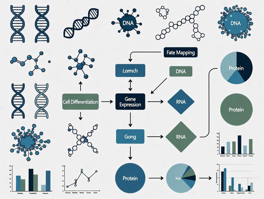
Tracking cell fate—a process known as lineage tracing—is fundamental to understanding normal development and disease. The gold standard for cellular trajectory inference, lineage tracing involves marking a progenitor cell and tracking all its descendants to reveal their fate choices and relationships [6]. The following section compares key technologies used in this field.
Comparison of Fate Mapping Techniques
Table 1: Comparison of Key Cell Fate Mapping Techniques
| Technique | Core Principle | Key Applications | Key Advantages | Primary Limitations |
|---|---|---|---|---|
| Direct Observation [6] | Visual tracking of cells using light microscopy. | Studying transparent embryos (e.g., zebrafish). | Non-invasive, simple, and provides direct visual data. | Limited to transparent organisms with low cell counts; not suitable for complex tissues. |
| Fluorescent Protein Labeling (e.g., Brainbow) [2] [6] | Cre-recombinase-driven stochastic expression of multiple fluorescent proteins. | Mapping neuronal connectivity [6], stem cell proliferation, and organ homeostasis. | Enables visualization of multiple cells and their spatial relationships simultaneously. | Limited number of colors; challenging to control timing/dosage for single-cell resolution [6]. |
| Viral Barcoding [6] [7] | Ex vivo transduction of cells with a retroviral/library containing unique DNA barcode sequences. | Tracking hematopoietic stem cell (HSC) clones after transplantation [6] [7]. | Allows simultaneous tracking of thousands of clones; high information yield from a single experiment [6]. | Limited to dividing cells; potential for viral silencing; non-random integration may affect cell behavior [6]. |
| In Situ Barcoding (e.g., Polylox, CARLIN) [6] [7] | In vivo generation of high-diversity DNA barcodes via Cre-lox recombination [6] or CRISPR/Cas9 editing [7]. | Studying native hematopoiesis [6], clonal dynamics in development and disease. | No transplantation needed; studies fate in unperturbed physiological conditions; very high barcode diversity [6]. | Complexity of generating and breeding engineered mouse models. |
| Natural Barcoding [6] | Using naturally accumulated somatic mutations (nuclear or mitochondrial) as lineage markers. | Retrospective lineage tracing in human tissues; aging studies. | Safe and non-invasive; can be applied to human samples without genetic manipulation. | Low mutation rate requires costly deep sequencing; analysis is complex and retrospective [6]. |
| Single-Cell Multi-Omics [7] | Combining lineage barcodes with single-cell RNA-seq or ATAC-seq. | Reconstructing lineage trajectories and linking clone identity to molecular state. | Reveals transcriptional and epigenetic heterogeneity driving fate decisions. | High cost; computational complexity for data integration. |
Visualizing a Fate Mapping Workflow
The following diagram illustrates a generalized workflow for a DNA barcoding-based fate mapping experiment, integrating both in vivo and ex vivo approaches.
Experimental Protocols in Focus
To provide practical insight, we detail two foundational protocols for studying cell fate in the context of hematopoiesis.
Protocol: Genetic Barcoding of Hematopoietic Stem Cells (HSCs)
This protocol is used to track the clonal output of individual HSCs after transplantation [6] [7].
- Barcode Library Production: Generate a complex library of lentiviral vectors, each containing a unique random DNA sequence (barcode) of 20-30 nucleotides, flanked by universal primer sequences for later amplification.
- HSC Isolation and Transduction: Isolate phenotypically defined HSCs (e.g., Lineage⁻ Sca-1⁺ CD117⁺ CD48⁻/lo CD150⁺) from donor bone marrow. Culture the cells and transduce them with the viral barcode library at a low multiplicity of infection (MOI << 1) to ensure most cells receive a single, unique barcode.
- Transplantation: Transplant the transduced HSCs into lethally irradiated or immunodeficient recipient mice.
- Longitudinal Tracking: Collect peripheral blood and bone marrow samples at various time points post-transplantation (e.g., 4, 8, 16 weeks, 1 year).
- DNA Extraction and Barcode Amplification: Isolve genomic DNA from sorted cell populations (e.g., myeloid cells, T cells, B cells). Amplify the barcode regions using PCR with primers specific to the universal flanking sites.
- High-Throughput Sequencing and Analysis: Sequence the PCR products and map the barcode reads to the original library. Quantify the abundance of each barcode in different cell populations and over time to assess clonal contributions and lineage biases.
Protocol: Enhancing HSC Migration and Engraftment
This functional assay assesses and enhances the homing ability of HSCs, a critical aspect of their fate after transplantation [5].
- HSC Subpopulation Isolation: Isolate distinct HSC subpopulations, such as Short-Term (ST)-HSCs (Flk2⁻CD34⁺) and Long-Term (LT)-HSCs (Flk2⁻CD34⁻), using fluorescence-activated cell sorting (FACS).
- Characterization of Homing Effectors:
- Analyze expression of sialyl Lewis-X (sLex), a ligand for E-selectin critical for the first step of bone marrow homing, via flow cytometry.
- Analyze expression of CXCR4, the receptor for SDF-1α (CXCL12) critical for the second homing step.
- Analyze expression of CD26, a peptidase that deactivates SDF-1α.
- Functional Modulation:
- Fucosylation: Treat HSCs with recombinant human fucosyltransferase VI (rhFTVI) to enhance sLex expression and E-selectin binding.
- CD26 Inhibition: Treat HSCs with a CD26 inhibitor (e.g., Diprotin A) to preserve local SDF-1α gradients.
- In Vitro Migration Assay: Load modulated and control HSCs into a transwell system. Place SDF-1α in the lower chamber. Quantify the number of cells that migrate through the membrane after a set period.
- In Vivo Engraftment Assay: Transplant pretreated HSCs into recipient mice. Analyze bone marrow at early time points to measure homing efficiency, and at later time points (e.g., 4-6 months) to evaluate long-term multi-lineage engraftment in primary and secondary recipients.
Quantitative Data from Fate Mapping and Engraftment Studies
Table 2: Summary of Key Experimental Findings from Cited Research
| Experimental Context | Key Measured Variable | Result / Quantitative Finding | Implication |
|---|---|---|---|
| Native Thrombopoiesis Fate Mapping [8] | Contribution of "short route" vs "long route" to platelet production | The two pathways make comparable contributions in steady state. | Thrombopoiesis is not a single pathway but the sum of functionally distinct routes. |
| HSC Homing Mechanism [5] | sLex expression on Flk2⁻CD34⁺ ST-HSCs | >60% of cells were sLex⁺. | ST-HSCs are intrinsically well-equipped for the initial homing step (E-selectin binding). |
| HSC Homing Mechanism [5] | sLex expression on Flk2⁻CD34⁻ LT-HSCs | <10% of cells were sLex⁺. | LT-HSCs have deficient first-step homing, which can be a target for enhancement. |
| HSC Homing Mechanism [5] | Effect of CD26 inhibition on LT-HSC migration | CD26 inhibition enhanced engraftment in vivo. | Targeting the second homing step (CXCR4/SDF-1) can overcome LT-HSC migration deficits. |
The Scientist's Toolkit: Key Research Reagents
Table 3: Essential Reagents and Tools for Cell Fate Research
| Reagent / Tool | Function in Research | Example Use Case |
|---|---|---|
| Cre-loxP System [2] [6] | Enables cell-type-specific and inducible genetic recombination. | Activating fluorescent reporters (e.g., Brainbow) or generating genetic barcodes (e.g., Polylox) in specific cell lineages. |
| Lentiviral Barcode Libraries [6] [7] | Introduces heritable, unique DNA sequences into cells for clonal tracking. | Massively parallel lineage tracing of hematopoietic stem cells after transplantation. |
| Fluorescent Proteins (e.g., GFP, RFP) [4] [2] | Visual labeling of live cells and their progeny. | Tracking engraftment, migration, and differentiation of transplanted neural stem cells. |
| Recombinant Fucosyltransferase (rhFTVI) [5] | Enzymatically modifies cell surface proteins to enhance E-selectin ligand expression. | Improving the homing efficiency of short-term HSCs for transplantation. |
| CD26 Inhibitors (e.g., Diprotin A) [5] | Protects SDF-1α from degradation by inhibiting the CD26 peptidase. | Enhancing the chemotactic migration and engraftment of long-term HSCs. |
| Marker Enrichment Modeling (MEM) [9] | A computational algorithm that generates quantitative labels for cell populations based on enriched features. | Objectively characterizing and comparing novel cell types identified by single-cell cytometry or transcriptomics. |
The journey from a progenitor to a determined cell involves a sophisticated interplay of autonomous and conditional signals, locked in place by epigenetic mechanisms. Mastery of cell fate is not merely an academic pursuit but a cornerstone of advanced regenerative medicine and therapeutic development. Techniques like genetic barcoding and single-cell multi-omics have moved the field from observing static hierarchies to dynamically mapping fate choices with clonal resolution. Furthermore, functional protocols that enhance migration and engraftment are directly translatable to improving clinical outcomes in areas like hematopoietic stem cell transplantation. As the toolkit evolves, the ability to precisely track, predict, and ultimately direct cell fate will continue to unlock new frontiers in treating degenerative diseases and cancer.
For decades, developmental biologists have sought to reconstruct the intricate lineage trees that trace how a single fertilized egg gives rise to the extraordinary complexity of a complete organism. Traditional methods provided glimpses—static snapshots of cellular relationships that offered limited insight into the dynamic temporal sequences of developmental decisions. The central challenge in stem cell research has been transforming these static observations into comprehensive, dynamic lineage trees that capture not only the "what" and "where" of cell fate, but the "when" and "how" of developmental progression. This comparison guide examines the revolutionary technologies reshaping stem cell tracking and fate mapping, objectively evaluating their performance characteristics, experimental requirements, and applications for research and drug development.
Part 1: The Evolution of Lineage Tracing Technologies
Historical Foundations and Technical Limitations
Classical lineage tracing approaches relied on direct visual observation, dye labeling, and enzymatic reporters, which provided foundational insights but suffered from significant technical constraints. Early methods using Nile Blue staining in amphibian blastula and nucleoside analogues (BrdU, EdU) enabled initial fate mapping but were limited by label dilution through cell divisions and inability to resolve complex lineage relationships [10]. The introduction of fluorescent proteins and Cre-loxP recombinase systems in the late 20th century marked a substantial advancement, allowing heritable genetic labeling of specific cell populations [10]. However, these approaches still faced resolution limitations—homogeneous labeling made distinguishing individual clones within populations difficult, and sparse labeling strategies increased experimental burden while reducing reproducibility [10].
Modern Technology Categories
Contemporary lineage tracing technologies fall into three principal categories, each with distinct mechanisms and applications:
Imaging-Based Approaches leverage advanced microscopy and fluorescent reporter systems for spatial resolution. The Brainbow and Confetti systems utilize stochastic Cre-loxP recombination to generate multicolored fluorescent tags, enabling visual distinction of adjacent clones in tissues [11] [10]. Mosaic Analysis with a Repressible Cell Marker (MARCM) identifies lineage branches through mitotic recombination [10]. More recently, dual recombinase systems (e.g., Cre-loxP/Dre-rox) have enabled simultaneous tracing of multiple cell populations, as demonstrated in studies mapping regenerative bone formation and alveolar epithelial stem cells [10].
DNA Recording Systems utilize genomic edits as heritable lineage marks. CRISPR-based barcoding introduces cumulative insertions/deletions (indels) at specific genomic loci during cell divisions, creating evolving lineage-specific barcodes [11] [12]. Base editing systems generate more predictable mutations, while "DNA typewriter" systems record the sequence of cellular events [12]. The Polylox system employs Cre-loxP recombination to generate diverse DNA barcodes without viral integration [11] [7]. These systems excel at reconstructing complex lineage relationships across extensive cell divisions.
Computational Inference Methods leverage single-cell RNA sequencing (scRNA-seq) data to reconstruct developmental trajectories. Algorithms like CytoTRACE 2 use deep learning to predict developmental potential from transcriptomic data [13]. Pseudotemporal ordering methods reconstruct lineage relationships based on transcriptional similarity, effectively arranging cells along differentiation continua [14]. While powerful for hypothesis generation, these inference-based approaches provide probable rather than definitively demonstrated lineage relationships [12] [14].
Part 2: Comparative Performance Analysis of Leading Technologies
Quantitative Performance Metrics
Table 1: Comprehensive Performance Comparison of Lineage Tracing Technologies
| Technology | Maximum Resolution | Temporal Recording | Throughput | Lineage Tree Accuracy | Key Limitations |
|---|---|---|---|---|---|
| Brainbow/Confetti | Single-cell (spatial) | None (static label) | Moderate (imaging constraints) | High for clone identification | Limited color palette; spectral overlap |
| CRISPR Barcoding | Single-cell (molecular) | Continuous (cumulative edits) | High (sequencing-based) | Very high (empirical recording) | Requires CRISPR delivery; potential toxicity |
| Polylox Barcoding | Single-cell (molecular) | Inducible (Cre-dependent) | High (sequencing-based) | High (diverse barcode library) | Limited to model organisms; Cre toxicity concerns |
| CytoTRACE 2 | Single-cell (computational) | Inferred (pseudotime) | Very high (transcriptomic) | Moderate (inferential) | Computational inference only; no empirical validation |
| scRNA-seq Trajectory | Single-cell (computational) | Inferred (pseudotime) | Very high (transcriptomic) | Moderate (inferential) | Destructive sampling; trajectory inference only |
Experimental Validation Data
Table 2: Experimental Performance Metrics from Validation Studies
| Technology | Clonal Reconstruction Accuracy | Maximum Clones Tracked | Long-term Stability | Cross-platform Compatibility |
|---|---|---|---|---|
| CytoTRACE 2 | 60% higher correlation vs. methods [13] | 406,058 cells in atlas [13] | N/A (computational) | 9 platforms validated [13] |
| CRISPR Barcoding | 84-93% bootstrap support [11] | Several thousand cells [11] | Heritable genomic edits | Requires compatible delivery system |
| Polylox | High (low barcode collision) [7] | >1,000 clones [11] | Stable genomic integration | Limited to engineered mouse models |
| Integration Barcodes | Moderate (retroviral silencing) [11] | Thousands simultaneously [11] | Variable (silencing concerns) | Broad (viral transduction) |
Part 3: Experimental Protocols and Methodologies
CytoTRACE 2 Computational Framework
Objective: Predict absolute developmental potential from scRNA-seq data without experimental perturbation [13].
Methodology Details:
- Training Data Curation: Compiled atlas of 33 human/mouse scRNA-seq datasets with experimentally validated potency levels, spanning 406,058 cells and 125 standardized cell phenotypes [13].
- Architecture: Gene Set Binary Network (GSBN) with binary weights (0 or 1) to identify discriminative gene sets for potency categories [13].
- Feature Selection: Multivariate gene expression programs that suppress batch effects through competing representations and training set diversity [13].
- Output Generation: Two primary outputs: (1) potency category with maximum likelihood, and (2) continuous potency score from 1 (totipotent) to 0 (differentiated) [13].
- Validation: Weighted Kendall correlation against known developmental orderings across 62 developmental time points in mouse models [13].
Workflow Diagram:
CRISPR Lineage Tracing Experimental Protocol
Cell Preparation and Barcode Delivery:
- Vector Design: Construct lentiviral vectors containing barcode arrays with multiple gRNA target sites and unique molecular identifiers [11] [12].
- Cell Transduction: Transduce target cells at low multiplicity of infection (MOI < 0.3) to ensure single barcode integration per cell [7].
- Selection and Expansion: Apply antibiotic selection for stable integrants, then expand cell population for sufficient diversity [12].
In Vivo Lineage Tracing:
- Animal Modeling: For in situ approaches, use engineered models like CARLIN mice containing Cas9 and barcode arrays [7].
- Barcode Activation: Induce Cas9 expression (doxycycline or tamoxifen) to initiate stochastic barcode editing [7].
- Temporal Sampling: Harvest tissues at multiple time points to capture lineage progression [11].
Barcode Recovery and Analysis:
- DNA Extraction: Process tissues for high-quality genomic DNA [11].
- Barcode Amplification: PCR amplify barcode regions using flanking primers [7].
- High-Throughput Sequencing: Sequence libraries on Illumina platforms [11].
- Lineage Reconstruction: Bioinformatic processing to identify indel patterns and reconstruct phylogenetic trees [11] [12].
Workflow Diagram:
Part 4: Research Reagent Solutions for Lineage Tracing
Table 3: Essential Research Reagents for Lineage Tracing Applications
| Reagent/Category | Specific Examples | Function | Key Considerations |
|---|---|---|---|
| Reporter Systems | R26R-Confetti, Brainbow, Polylox | Visual barcode generation | Stochastic labeling efficiency; spectral separation |
| CRISPR Components | CARLIN model, Base editors, Prime editors | DNA barcode generation | Editing efficiency; off-target effects |
| Recombinases | Cre-ERT2, Dre, Flp | Inducible genetic recombination | Leakiness; toxicity with prolonged expression |
| Viral Delivery | Lentiviral barcode libraries, Retroviral vectors | High-efficiency gene delivery | Insertional mutagenesis; silencing concerns |
| Detection Reagents | Antibody panels, In situ hybridization probes | Barcode detection and visualization | Signal-to-noise ratio; multiplexing capacity |
| Sequencing Kits | Single-cell RNA-seq, Barcode amplification | High-throughput barcode recovery | Amplification bias; sequencing depth requirements |
Part 5: Applications and Validation in Biological Systems
Hematopoietic Stem Cell Tracking
Transplantation studies utilizing DNA barcoding have revealed the remarkable heterogeneity of hematopoietic stem cell fates, demonstrating temporal oligoclonality where a limited number of dominant clones sustain long-term hematopoiesis [7]. Integration site analysis of retrovirally transduced HSPCs has shown variable clonal contributions to mature blood lineages, revealing lineage biases and clonal drift over time [7]. The Polylox system has enabled in situ barcoding without transplantation, uncovering native hematopoietic dynamics and revealing how stress conditions alter clonal output patterns [11] [7].
Cancer Stem Cell Dynamics
Lineage tracing has transformed our understanding of tumor heterogeneity and therapeutic resistance. In acute myeloid leukemia, CytoTRACE 2 potency predictions aligned with known leukemic stem cell signatures, while in oligodendroglioma, it identified multilineage potential in subpopulations [13]. CRISPR lineage tracing has enabled reconstruction of tumor evolution trees, identifying branching patterns and mutation sequences that drive progression and treatment resistance [13].
Developmental Biology Applications
In mammalian development, CytoTRACE 2 correctly captured the progressive decline in potency across 258 phenotypes during mouse development without requiring data integration or batch correction [13]. Multicolor Confetti reporters have enabled visualization of clonal expansion and patterning in epithelial tissues, revealing how progenitor cells contribute to tissue architecture during organogenesis [10].
The optimal lineage tracing technology depends on specific research questions and experimental constraints. CRISPR-based barcoding excels for high-resolution reconstruction of complex lineage relationships across extended timescales, though it requires genomic manipulation. Imaging-based approaches provide unparalleled spatial context and real-time observation capabilities but face throughput limitations. Computational inference methods like CytoTRACE 2 offer non-invasive analysis of existing scRNA-seq data with strong performance for developmental potential assessment but remain inferential rather than empirical.
For research and drug development applications, the integration of multiple complementary technologies provides the most comprehensive insights—combining empirical lineage recording with transcriptional profiling and spatial context to transform static snapshots into dynamic, mechanistic understanding of cell fate decisions. As these technologies continue to evolve, they promise to unravel the fundamental principles governing stem cell behavior, tissue regeneration, and disease pathogenesis with increasingly precise resolution.
Lineage tracing remains an essential approach for understanding cell fate, tissue formation, and human development [10]. This field has evolved from simple microscopic observation to sophisticated genetic labeling that can track single cells across time and space. The core principle involves establishing hierarchical relationships between cells to reconstruct developmental trajectories and fate decisions [10]. This progression has fundamentally transformed developmental biology, stem cell research, and regenerative medicine, providing increasingly precise tools to answer one of biology's most fundamental questions: what becomes of a cell and its descendants?
The historical journey of lineage tracing reflects broader technological revolutions in biology. From its origins in direct observation of transparent embryos to today's integration of sequencing and imaging technologies, each advancement has expanded our ability to decipher cellular narratives in increasingly complex organisms and contexts [10] [15] [6]. This article provides a comprehensive comparison of these techniques, their experimental protocols, and their applications in modern biomedical research.
Historical Techniques and Their Methodologies
Direct Observation and Dye-Based Labeling
The earliest lineage tracing methods relied on visual monitoring of cell behavior. In the late 1800s, Charles Whitman reported the first direct observation of germ layer differentiation in leeches using light microscopy [10] [6]. Data collection was entirely dependent on visual observations of an experimenter in real time, limiting experimental models to those with observable changes via available microscopy [10].
Dye labeling techniques represented the first major technological leap. Eric Vogt fate-mapped an amphibian blastula in 1929 using Nile Blue as a non-specific label [10]. Later approaches used:
- Carbocyanine dyes to stain cell membranes and track migration patterns [15]
- Tritiated thymidine for long-term, non-toxic in vivo labeling [15]
- Nucleoside analogues (BrdU, EdU) incorporated into cellular DNA and subsequently labeled with fluorescent dyes [10]
A significant limitation of these approaches was label dilution proportional to cell proliferation, reducing tracking accuracy over time [10] [15].
Table 1: Historical Lineage Tracing Techniques
| Technique | Era | Key Features | Limitations |
|---|---|---|---|
| Direct Observation | Late 1800s | Real-time visual monitoring, minimal technical requirements | Limited to transparent embryos, subjective, low-throughput |
| Dye Labeling (Nile Blue) | 1929 | First non-specific labeling method | Label dilution, limited specificity |
| Nucleoside Analogues (BrdU/EdU) | Mid-late 20th century | Identifies proliferating populations | Label dilution with proliferation, requires fixation |
| Enzyme Reporters (β-galactosidase) | 1980s | First transgenic approaches, stable genetic labeling | Requires substrate addition, lower resolution |
| Fluorescent Proteins (GFP) | 1994 | Endogenous reporting without external stimulus | Potential phototoxicity, limited color palette |
The Recombinase Revolution
The late 20th century introduced genetic recombinase systems that transformed lineage tracing. The Cre-loxP system, discovered in P1 bacteriophage and implemented in mammalian cells in 1988, became a fundamental tool [10] [15]. Cre recombinase recognizes 34-base pair loxP sequences, enabling precise DNA recombination including deletion, inversion, or exchange of gene sequences [15].
Key implementations include:
- LoxP-Stop-loxP (LSL) system: Cre-mediated excision of a STOP cassette activates reporter gene expression [15]
- Double-floxed Inversion Orientation (DIO/DO): Uses two incompatible pairs of inverted loxP sites for more precise control [15]
- CIAO (cross-over insensitive ATG-out) strategy: Places the ATG start codon within loxP sites to prevent nonspecific expression [15]
These systems enabled permanent genetic labeling of specific cell populations and all their progeny, overcoming the dilution problem of dye-based methods [15].
Modern Genetic Labeling Technologies
Advanced Recombinase Systems
Modern lineage tracing has evolved beyond single recombinase systems to address limitations of "non-specific expression" and insufficient spatiotemporal resolution [15]. Key advancements include:
Dual recombinase systems (e.g., Cre-loxP + Dre-rox) enable simultaneous labeling of distinct or overlapping cell lineages [10] [15]. These orthogonal recombinase systems consist of engineered enzyme-substrate pairs that operate independently without cross-reactivity [15]. Applications include:
- Determining origin of regenerative cells in remodelled bone [10]
- Investigating cellular origins of alveolar epithelial stem cells post-injury [10]
- Discriminating senescent cell populations expressing analogous markers [10]
Multicolour lineage tracing approaches like Brainbow and R26R-Confetti report cassettes capable of expressing multiple fluorescent proteins through stochastic Cre-loxP-mediated excision [10] [6]. These enable:
- Clonal analysis at single-cell level in various tissues [10]
- Discrimination of different cells upon Cre activation [6]
- Intravital imaging to trace cell origin and proliferation in real time [10]
Genetic Lineage Tracing Principle
Single-Cell Lineage Tracing (SCLT) and Barcoding Technologies
Single-cell sequencing technology propelled lineage tracing into high-throughput analysis of cell fates at single-cell resolution [15] [6]. SCLT maps cell lineage connectivity at single-cell resolution, becoming the best tool for exploring cellular differentiation heterogeneity [6].
Integration barcodes utilize DNA fragments with extensive sequence variations to label individual cells:
- Retroviral barcoding: Uses vectors with random sequence tags that integrate into chromosomes [6]
- Enables long-term tracking of clonal descendants from host cells [6]
- Allows examination of clonal relationships between cellular compartments [6]
Polylox barcodes represent artificial DNA recombination loci that enable endogenous barcoding using Cre-loxP recombination [6]. CRISPR barcodes utilize cumulative CRISPR/Cas9 insertions and deletions (InDels) as genetic landmarks for reconstructing lineage hierarchies [6].
Base editors represent a recent breakthrough, introducing informative sites to document cell division events with faster mutation rates, allowing recording of more mitotic divisions and construction of more detailed cell lineage trees [6].
Table 2: Modern Genetic Lineage Tracing Technologies
| Technology | Mechanism | Resolution | Applications |
|---|---|---|---|
| Cre-loxP Systems | Site-specific recombination | Cell population | General lineage tracing, gene knockout |
| Dual Recombinase (Cre+Dre) | Orthogonal recombination systems | Multiple lineages | Distinguishing overlapping lineages |
| Brainbow/Confetti | Stochastic fluorescent protein expression | Clonal (multicolor) | Visualizing clonal expansion, cell interactions |
| Viral Barcoding | Random viral integration sites | Thousands of clones | Hematopoietic stem cell tracking, large-scale fate mapping |
| CRISPR Barcoding | CRISPR/Cas9-induced mutations | Single-cell | Developmental lineage trees, cancer evolution |
| Base Editors | Targeted nucleotide editing | High-resolution phylogenetic | Detailed cell division history, organ development |
Imaging Modalities for Cell Tracking
Comparative Analysis of Imaging Techniques
Various imaging modalities have been developed to track stem cells in living organisms, each with distinct advantages and limitations [16] [17].
Magnetic Resonance Imaging (MRI) provides high-resolution 3D imaging at the anatomical level [16] [17]. Contrast agents include:
- Gadolinium (Gd³⁺): T1-weighted contrast agent that appears hyperintense [16]
- Superparamagnetic iron oxide (SPIO): T2-weighted agent that generates hypointense signals [16] [17]
- Manganese: "Positive" T1 contrast agent that enters cells via voltage-gated Ca²⁺ channels [16]
Radionuclide imaging (PET/SPECT) offers high sensitivity for detecting small cell numbers:
- Direct labeling: Uses ¹¹¹In-oxyquinoline or ⁹⁹mTc-hexamethylpropylene amine oxime [17]
- Can detect 10⁴-10⁵ cells in small animal models [17]
- Applications in tracking endothelial progenitor cells and mesenchymal stem cells [17]
Optical imaging includes bioluminescence and fluorescence approaches:
- Bioluminescence: Uses luciferase enzymes with substrates like luciferin [17]
- Fluorescence proteins: GFP, RFP, and related variants [17]
- Quantum dots: Semiconductor nanocrystals with tunable emission wavelengths [17]
Magnetic Particle Imaging (MPI) is an emerging technology that directly images SPION distribution with high sensitivity and linear quantification [16].
Stem Cell Tracking Workflow
Performance Comparison of Imaging Modalities
Table 3: Quantitative Comparison of Stem Cell Imaging Modalities
| Imaging Modality | Spatial Resolution | Tissue Penetration | Sensitivity (Cell Detection) | Temporal Resolution | Clinical Translation |
|---|---|---|---|---|---|
| MRI | 25-100 µm | No limit | 10⁵-10⁶ cells | Minutes-hours | Established |
| Magnetic Particle Imaging (MPI) | ~1 mm | No limit | Single cell (theoretical) | Milliseconds-seconds | Preclinical |
| PET | 1-2 mm | No limit | 10⁴-10⁵ cells | Seconds-minutes | Established |
| SPECT | 1-2 mm | No limit | 10⁴-10⁵ cells | Minutes | Established |
| Bioluminescence | 3-5 mm | 1-2 cm | 10²-10⁴ cells | Seconds-minutes | Limited |
| Fluorescence | 2-3 mm | <1 cm | 10³-10⁵ cells | Seconds-minutes | Emerging |
| Quantum Dots | 2-3 mm | <1 cm | 10³-10⁵ cells | Seconds-minutes | Preclinical |
Experimental Protocols and Research Reagents
Key Experimental Workflows
Cre-loxP Lineage Tracing Protocol:
- Transgenic mouse generation: Cross mice expressing Cre recombinase under tissue-specific promoters with reporter strains containing loxP-STOP-loxP sequences before fluorescent proteins [10] [15]
- Induction timing: Administer tamoxifen for CreER[T2] systems at desired developmental stages [10]
- Tissue collection: Harvest tissues at appropriate timepoints post-induction [10]
- Analysis: Process for microscopy, flow cytometry, or single-cell RNA sequencing [10] [6]
Viral Barcoding Workflow:
- Barcode library design: Create diverse DNA barcode sequences in lentiviral or retroviral vectors [6]
- Cell transduction: Infect target cells (e.g., hematopoietic stem cells) with barcode library at low MOI to ensure single integrations [6]
- Transplantation: Introduce barcoded cells into animal models [6]
- Timepoint sampling: Collect cells or tissues at multiple timepoints [6]
- Barcode sequencing: Amplify and sequence barcodes to quantify clonal contributions [6]
CRISPR Lineage Tracing Method:
- Engineered cassette: Introduce CRISPR target sites and unique barcode arrays into genome [6]
- In vivo editing: Express Cas9 to induce accumulating mutations during development [6]
- Single-cell sequencing: Perform scRNA-seq to capture both mutations and transcriptomes [6]
- Lineage reconstruction: Use mutation patterns to build phylogenetic trees [6]
Research Reagent Solutions
Table 4: Essential Research Reagents for Lineage Tracing
| Reagent Category | Specific Examples | Function | Applications |
|---|---|---|---|
| Site-Specific Recombinases | Cre, Dre, FlpO | DNA recombination at specific target sites | Genetic labeling, gene activation |
| Reporter Genes | GFP, RFP, tdTomato, LacZ | Visualizing labeled cells and progeny | Microscopy, flow cytometry |
| Inducible Systems | CreER[T2], Tet-On/OFF | Temporal control of recombination | Precise fate mapping at specific timepoints |
| Viral Vectors | Lentivirus, Retrovirus | Gene delivery and barcode library introduction | Hematopoietic stem cell tracking |
| CRISPR Components | Cas9, gRNAs, Base editors | Introducing heritable mutations for barcoding | Single-cell lineage tracing |
| Contrast Agents | SPIO, Gd³⁺, ¹¹¹In-oxine | Cell labeling for non-invasive imaging | MRI, PET, SPECT tracking |
| Nucleoside Analogues | EdU, BrdU | Labeling proliferating cells | Short-term lineage tracing |
Applications in Biomedical Research
Stem Cell Biology and Regenerative Medicine
Lineage tracing has provided crucial insights into stem cell plasticity, differentiation, and tissue regeneration [15]. In neurology, neural stem cells (NSCs) have been tracked after transplantation to treat conditions like Parkinson's disease, brain trauma, and stroke [16]. These studies revealed migration routes, survival rates, and functional integration of transplanted cells [16].
Cardiac stem cell therapy monitoring has utilized multiple imaging modalities to address contradictory results in clinical trials [17]. Studies tracking ¹¹¹In-labeled endothelial progenitor cells found only 4.7% retention in infarcted myocardium, highlighting delivery efficiency challenges [17].
Cancer Biology and Disease Modeling
Lineage tracing has determined mutations critical to cancer progression and lineage-specificity for therapeutics [10]. In hematology, single-cell lineage tracing technologies unravel heterogeneity of hematopoietic stem cell function and the heterogeneity of malignant tumor cells [6].
CRISPR-based lineage tracing with base editors has been applied to Drosophila melanogaster, generating high-quality cell phylogenetic trees with several thousand internal nodes, enabling estimation of symmetric and asymmetric cell division balances during development [6].
The evolution of lineage tracing from direct observation to sophisticated genetic labeling represents one of the most transformative journeys in modern biology. While direct observation provided foundational principles, the field has progressed through dye labeling, transgenic approaches, and now single-cell barcoding technologies [10] [15] [6].
Current frontiers include multimodal integration of sequencing with spatial information, improved computational tools for lineage reconstruction, and retrospective tracing using natural barcodes in human samples [10] [6]. The continued innovation in this field promises to further unravel the complex dynamics of development, disease, and regeneration at unprecedented resolution.
The ideal future of lineage tracing lies in seamlessly integrating multiple approaches—combining the specificity of genetic labeling with the sensitivity of modern imaging and the throughput of single-cell technologies—to create comprehensive fate maps across entire organisms throughout their lifespan.
In stem cell biology, understanding clonal dynamics (the behavior and evolution of a single cell's progeny), progenitor hierarchies (the structured relationships between stem cells and their differentiated descendants), and fate restriction (the progressive limitation of a cell's developmental potential) is fundamental. Researchers employ various fate-mapping techniques to track these processes in living organisms. This guide provides a comparative analysis of the predominant methodologies, detailing their experimental protocols, applications, and performance to inform tool selection for basic research and drug development.
Comparative Analysis of Fate-Mapping Techniques
The table below summarizes the core characteristics, performance, and applications of major fate-mapping approaches.
| Technique | Core Mechanism | Key Performance Metrics (Typical Results) | Key Applications | Technical Considerations |
|---|---|---|---|---|
| Genetic Fate Mapping (e.g., Cre-lox) [18] [19] | Uses cell-type-specific promoters to drive Cre recombinase, which permanently activates a heritable reporter gene (e.g., GFP) in target cells and all their progeny. | - Lineage Resolution: Single-cell to population-level.- Temporal Control: High (with inducible systems like CreERT2).- Clonal Tracking: Possible with multi-color reporters (e.g., Confetti).- Stability: Permanent, long-term marking. | - Tracking developmental origins of adult tissues and organs [19].- Studying immune cell development and function [18].- Mapping diverse macrophage subsets in various tissues [18]. | - Requires generation of transgenic animals.- Promoter specificity is critical and can be a limitation.- Background recombination can occur in inducible systems. |
| Clonal Dynamics Analysis (via Somatic Mutations) [20] | Leverages naturally accumulated somatic mutations (e.g., in clonal hematopoiesis) as endogenous barcodes for retrospective lineage tracing. | - Clonal Contribution: Can quantify a clone's contribution to platelet, erythroid, myeloid, B, and T cell lineages [20].- Fate Bias Identification: Identifies clones with restricted output (e.g., PEMB or PEM-only) [20].- Clonal Longevity: Can trace clones established decades prior to analysis [20]. | - Studying steady-state human hematopoiesis, especially in aged populations [20].- Identifying lineage-restricted stem cells and their stability over years [20]. | - Typically applied in aged individuals where clones have expanded sufficiently.- Requires deep, error-corrected DNA sequencing and complex phylogenetic analysis. |
| Viral Vector-Based Lineage Tracing [21] | Uses viral vectors (e.g., Retroviruses, AAVs) to deliver and integrate a reporter or fate-altering gene (e.g., Neurogenin2) into target cells. | - Cell-Type Specificity: Varies by vector and promoter; Retroviruses target proliferating cells [21].- Reprogramming Efficiency: Retroviral 9SA-Ngn2 successfully converted astrocytes to neurons; AAVs led to artefactual neuronal labeling [21].- Immunogenicity: Retroviruses induce stronger inflammation than AAVs [21]. | - Direct neuronal reprogramming of glial cells [21].- Fate conversion studies in the brain. | - Retroviruses (Mo-MLVs): Infect only dividing cells, superior for fate conversion of proliferative glia [21].- AAVs: Can infect post-mitotic cells; prone to artefactual labeling with strong neurogenic factors [21]. |
| Computational Fate Mapping (e.g., CellRank 2, STORIES) [22] [23] | Infers lineage relationships and dynamics from single-cell omics data (e.g., RNA-seq, spatial transcriptomics) using algorithms, without physical labels. | - Multiview Data Integration: Can combine RNA velocity, pseudotime, experimental time points, and spatial coordinates [23].- Terminal State Identification: CellRank 2 consistently recovered terminal states in human hematopoiesis [23].- Spatial Coherence: STORIES outperforms other methods in learning spatially-informed cell fate landscapes [22]. | - Reconstructing differentiation trajectories from snapshot data [23].- Studying the impact of spatial environment on cell fate decisions [22].- Analyzing clinical single-cell datasets from cancer immunotherapy [24]. | - Is a computational inference, not a direct observation of lineage.- Requires high-quality, often large-scale, single-cell datasets.- Performance depends on the algorithm and data modality. |
Experimental Protocols for Key Techniques
Genetic Fate Mapping with Inducible Cre-loxP
This protocol is used for precise, temporally controlled lineage tracing in transgenic mice [18].
Key Reagents:
- Transgenic Mouse Line: Expressing CreERT2 fusion protein under a cell-type-specific promoter (e.g., Cx3cr1CreERT2 for myeloid cells).
- Reporter Mouse Line: With a floxed-stop cassette upstream of a reporter gene (e.g., GFP) in a permissive locus like Rosa26.
- Tamoxifen: The inducer drug.
Workflow:
- Animal Crosses: Cross the driver Cre line with the reporter line to generate experimental offspring.
- Induction: Administer tamoxifen to the animals (e.g., via intraperitoneal injection or oral gavage) at the desired developmental or experimental time point. Tamoxifen binds to CreERT2, allowing it to translocate to the nucleus.
- Recombination: Nuclear CreERT2 catalyzes the removal of the floxed-stop cassette in the target cells, leading to permanent expression of the reporter gene.
- Tissue Harvest and Analysis: After a desired chase period, harvest tissues and analyze them using flow cytometry or immunohistochemistry to track the location and differentiation of the labeled progeny.
Clonal Dynamics Analysis via Somatic Mutations
This method leverages natural mutations for retrospective lineage tracing in humans [20].
Key Reagents:
- Bone Marrow or Blood Samples: From healthy aged donors or those with clonal hematopoiesis.
- Error-Corrected Targeted DNA Sequencing (ECTS) Panels: For sensitive detection of low-frequency somatic mutations in known driver genes (e.g., DNMT3A, TET2).
- Cell Sorting Equipment: For purifying specific hematopoietic stem and progenitor cell (HSPC) populations and mature lineages.
Workflow:
- Mutation Screening: Screen DNA from bulk bone marrow mononuclear cells using ECTS to identify clonal driver mutations.
- Cell Sorting: Purify distinct cell populations (e.g., HSCs, myeloid cells, B cells, T cells, erythroid progenitors, megakaryocyte progenitors) using FACS.
- Clonal Contribution Assessment: Quantify the variant allele frequency of the identified mutations in each purified cell population to determine the clone's contribution to each lineage.
- Phylogenetic Analysis: Perform whole-genome sequencing on single-cell-derived colonies to retrospectively infer the phylogenetic history and timing of the clone's origin.
Viral Vector-Mediated Fate Mapping and Reprogramming
This protocol is for tracking or converting the fate of specific cell populations in vivo, such as in the brain [21].
Key Reagents:
- Viral Vectors: Moloney Murine Leukemia Virus (Mo-MLV) for targeting proliferating glia or Adeno-Associated Viruses (AAVs) with flexed cassettes for potential specificity.
- Genetic Fate-Mapping Mouse Lines: (e.g., GFAP::Cre) to label starter cells (astrocytes).
- EdU (5-ethynyl-2'-deoxyuridine): For birth-dating endogenous neurons.
Workflow:
- Model and Injury: Use a transgenic mouse (e.g., GFAP::Cre) and subject it to a cortical stab wound injury to induce glial proliferation.
- Viral Injection: Three days post-injury, inject viral vectors (e.g., Mo-MLV-CAG-9SA-Ngn2-IRES-mScarlet) into the injury site.
- Control for Artefacts: Employ two key controls:
- Starter Cell Labeling: Use genetic fate mapping to confirm that converted neurons originate from the labeled astrocytes.
- Endogenous Neuron Labeling: Use EdU birth-dating to rule out artefactual labeling of pre-existing neurons.
- Analysis: After a few weeks, analyze brain sections via immunohistochemistry to identify virally transduced, fate-mapped, and birth-dated cells to confirm true astrocyte-to-neuron conversion.
Visualizing Fate-Mapping Concepts and Workflows
Genetic Fate Mapping with Cre-loxP
Clonal Dynamics in Hematopoiesis
The Scientist's Toolkit: Key Research Reagents
This table details essential materials used in the featured experiments [20] [18] [21].
| Research Reagent | Function in Fate Mapping | Example Application |
|---|---|---|
| Tamoxifen-Inducible Cre (CreERT2) | Enables temporal control of lineage tracing; Cre activity is induced only upon tamoxifen administration. | Precisely marking a specific cell population at a defined time point in development or adulthood [18]. |
| Multicolor Reporter Mice (e.g., Confetti) | Allows for stochastic, multi-color labeling of cells, enabling visual distinction between different clones within a tissue. | Clonal analysis and tracking of multiple distinct lineages simultaneously in the same animal [18]. |
| Somatic Mutation Panels (e.g., for DNMT3A, TET2) | Used to identify unique, naturally occurring DNA barcodes that mark expanded clones in human tissue. | Retrospective lineage tracing and clonal contribution analysis in human hematopoiesis [20]. |
| Moloney Murine Leukemia Virus (Mo-MLV) | A retroviral vector that integrates into the host genome only in dividing cells, making it ideal for targeting proliferative populations. | Specific targeting of proliferating reactive glia for direct conversion into neurons in the brain [21]. |
| Adeno-Associated Virus (AAV) with Flexed Cassette | A viral vector that can infect non-dividing cells; a double-floxed (FLEX) cassette ensures expression only in Cre-expressing cells. | Requires careful validation, as it can lead to artefactual labeling when used with strong transcriptional activators [21]. |
| Computational Tools (e.g., CellRank 2, STORIES, Clonotrace) | Algorithms that infer cell fate dynamics and trajectories from single-cell omics data without physical labels. | Reconstructing differentiation landscapes and predicting fate biases from snapshot or spatial transcriptomics data [24] [22] [23]. |
The Technological Toolbox: From DNA Barcodes to Live-Cell Imaging
Genetic barcoding has revolutionized stem cell research by enabling precise tracking of individual cells and their progeny over time and space. This powerful approach allows researchers to decipher the complex dynamics of cellular fate, lineage relationships, and clonal dynamics in developing tissues, homeostasis, and disease contexts. As a cornerstone of modern fate mapping techniques, genetic barcoding provides unprecedented insights into the behavior of stem and progenitor cells by marking them with unique, heritable DNA sequences that can be subsequently traced through sequencing-based detection methods [25] [26].
The field has evolved from early methods that relied on visual observation and non-specific dyes to sophisticated molecular technologies capable of simultaneously tracking thousands to millions of clones. Among the most prominent techniques currently employed are retroviral libraries, Polylox barcoding, and transposon tagging, each offering distinct advantages and limitations for specific research applications. These methods have become indispensable tools for understanding stem cell biology, particularly in heterogeneous systems where fate potential and lineage relationships remain incompletely characterized [27] [10].
This guide provides a comprehensive comparison of these three fundamental genetic barcoding technologies, focusing on their principles, experimental workflows, performance characteristics, and applications in stem cell fate mapping. By synthesizing current methodologies and experimental data, we aim to equip researchers with the information necessary to select the most appropriate barcoding strategy for their specific research questions in stem cell biology and drug development.
Technology Comparison & Performance Data
The table below provides a systematic comparison of the key technical specifications and performance characteristics of retroviral barcoding, Polylox barcoding, and transposon tagging systems:
Table 1: Comprehensive Comparison of Genetic Barcoding Technologies
| Feature | Retroviral Barcoding | Polylox Barcoding | Transposon Tagging |
|---|---|---|---|
| Core Principle | Introduction of DNA barcodes via viral vector integration | Cre-mediated recombination between loxP sites generates diverse barcodes | Transposase-mediated genomic insertion of DNA sequences |
| Barcode Diversity | High (10⁶-10⁸ with 30bp barcodes) [26] | Very High (theoretical >10⁷) [27] | High (depends on transposon copy number) |
| Integration Mechanism | Semi-random viral integration | Endogenous recombination at defined locus | Semi-random transposition |
| Mutagenesis Risk | Moderate to High (preferential for active genes) [27] | Minimal (defined genomic location) [27] | Moderate (semi-random insertion) [27] |
| In Vivo Applicability | Requires ex vivo transduction & transplantation [27] | Native labeling in situ (transgenic models) [27] | Can be performed in situ with inducible systems [27] |
| Perturbation of Native State | High (transduction + transplantation stress) [27] | Low (minimal system perturbation) [27] | Low to Moderate (depends on delivery method) |
| Lineage Resolution | High (clonal tracking possible) | Very High (single-cell resolution) [27] | High (clonal tracking possible) |
| Single-Cell Compatibility | Yes (with scRNA-seq) | Yes (compatible with scRNA-seq) [27] | Yes (compatible with scRNA-seq) [27] |
| Quantitative Clonal Tracking | Yes (barcode frequency = clonal abundance) | Yes (barcode frequency = clonal abundance) | Yes (integration site = clonal mark) |
| Theoretical Barcode Complexity | 4ⁿ (n=barcode length) [26] | Combinatorial from loxP rearrangements | Limited by transposon diversity |
Table 2: Performance Characteristics in Hematopoietic Stem Cell Tracking
| Performance Metric | Retroviral Barcoding | Polylox Barcoding | Transposon Tagging |
|---|---|---|---|
| Clonal Detection Sensitivity | High (with optimized PCR) | High (with sequencing depth) | Moderate to High |
| Labeling Efficiency | Variable (depends on transduction) | High (in designed models) | Variable (depends on transposition) |
| Long-term Stability | Stable (genomic integration) | Stable (genomic rearrangement) | Stable (genomic integration) |
| Lineage Bias Detection | Yes (through barcode distribution) | Yes (through barcode distribution) | Yes (through integration patterns) |
| Multilineage Reconstitution Analysis | Yes (with lineage sorting) | Yes (with single-cell sequencing) | Yes (with integration site mapping) |
Principles & Experimental Protocols
Retroviral Barcoding
Principle: Retroviral barcoding utilizes lentiviral or γ-retroviral vectors to deliver short, random DNA sequences (typically 20-30 nucleotides) into the genome of target cells. Each unique barcode serves as a heritable mark that can be detected through high-throughput sequencing, enabling quantitative tracking of clonal contributions over time and across different lineages [26] [27]. The semi-random integration pattern of retroviral vectors provides additional clonal marks through integration site analysis, though this approach carries a risk of insertional mutagenesis due to preference for transcriptionally active regions [28] [27].
Experimental Protocol:
- Library Design: Generate a lentiviral library containing 10⁶-10⁸ unique barcode sequences with common PCR priming sites
- Stem Cell Transduction: Transduce hematopoietic stem/progenitor cells (HSPCs) at low multiplicity of infection (MOI <0.5) to ensure single barcode integration
- Transplantation: Transplant transduced cells into recipient animals (typically irradiated or immunodeficient mice)
- Time-point Sampling: Collect peripheral blood and bone marrow at multiple time points post-transplantation
- Barcode Recovery: Amplify barcodes from genomic DNA using PCR with library-specific primers
- High-throughput Sequencing: Sequence barcode amplicons to quantify clonal abundances
- Lineage Analysis: Sort specific lineages (myeloid, lymphoid) before barcode recovery to assess lineage bias [27]
Critical Considerations:
- Multiplicity of infection must be optimized to ensure primarily single barcode integration per cell
- Vector design should position barcodes within conserved vector backbone for consistent recovery
- PCR amplification conditions must minimize bias in barcode representation
- Sequencing depth must be sufficient to detect low-abundance clones
Polylox Barcoding
Principle: The Polylox system represents a DNA recombination-based barcoding approach that utilizes Cre-loxP technology to generate diverse barcodes in situ. In engineered mouse models, a transgenic cassette containing multiple loxP sites in alternating orientations is integrated into a defined genomic locus. Upon Cre recombinase activation, stochastic recombination events between loxP sites create unique DNA sequences that serve as heritable barcodes for lineage tracing [27]. This system enables native labeling without transplantation, significantly reducing experimental perturbation.
Experimental Protocol:
- Mouse Model Generation: Create transgenic mice with Polylox cassette containing multiple loxP sites in alternating orientations
- Cre Activation: Cross with tissue-specific or inducible Cre driver lines to activate barcode generation
- Tissue Collection: Harvest tissues of interest at desired time points
- Barcode Amplification: Recover barcodes using PCR with cassette-specific primers
- Sequencing Library Preparation: Prepare sequencing libraries with sample barcodes for multiplexing
- High-throughput Sequencing: Sequence barcode libraries with sufficient depth for clonal detection
- Bioinformatic Analysis: Identify unique barcodes and quantify their abundances across samples
- Single-cell Integration: Combine with single-cell RNA sequencing for multimodal analysis [27]
Critical Considerations:
- Cre efficiency must be optimized for sufficient barcode diversity
- Temporal control of barcode generation can be achieved with inducible Cre systems
- Cassette design determines theoretical barcode diversity and detection reliability
- Background recombination should be monitored in negative controls
Transposon Tagging
Principle: Transposon tagging utilizes mobile genetic elements such as Sleeping Beauty or PiggyBac transposons to integrate marker sequences throughout the genome. The system consists of two components: a transposon vector containing the marker sequence flanked by terminal inverted repeats, and a transposase enzyme that catalyzes excision and reintegration. The quasi-random integration patterns create unique insertion profiles that can serve as clonal markers when mapped to the genome [29] [27]. Recent advancements like TARIS (T7-amplification mediated recovery of integration sites) have improved tag recovery efficiency and reduced amplification bias [27].
Experimental Protocol:
- Transposon Vector Design: Construct transposon vectors with unique molecular tags or barcodes
- Transposase Delivery: Introduce transposase via plasmid transfection, mRNA electroporation, or transgenic expression
- Stem Cell Labeling: Transfer both components to target cells (electroporation or viral delivery)
- Selection: Apply antibiotic selection if vector contains resistance marker
- Transplantation: Transplant labeled cells into recipient animals if studying in vivo reconstitution
- Integration Site Recovery: Use methods like LAM-PCR, TARIS, or nrLAM-PCR to recover integration sites
- Sequencing: Perform high-throughput sequencing of integration sites
- Bioinformatic Mapping: Map sequences to reference genome to identify unique integration sites
- Clonal Tracking: Monitor integration site abundances over time and across lineages [27]
Critical Considerations:
- Transposase activity affects integration efficiency and pattern
- Copy number should be controlled to enable clonal resolution
- Integration bias varies between transposon systems
- Mapping reliability depends on sequencing coverage and genome accessibility
Research Reagent Solutions
Table 3: Essential Research Reagents for Genetic Barcoding Applications
| Reagent Category | Specific Examples | Function & Application |
|---|---|---|
| Viral Vectors | Lentiviral barcode libraries, γ-retroviral vectors | Delivery of barcode sequences to target cells |
| Transposon Systems | Sleeping Beauty, PiggyBac transposon/transposase | Genomic integration of marker sequences |
| Site-specific Recombinases | Cre, CreERT2, Dre recombinases | Activation of barcode systems; inducible control |
| Barcode Libraries | Random DNA oligonucleotide pools, Polylox cassettes | Source of diverse barcode sequences |
| Sequencing Adapters | Illumina-compatible adapters, sample barcodes | Preparation of sequencing libraries |
| Cell Sorting Markers | Fluorescent proteins (GFP, RFP), cell surface antigens | Identification and isolation of labeled cells |
| PCR Reagents | High-fidelity polymerases, barcode-specific primers | Amplification of barcode sequences |
| Single-cell RNA-seq Kits | 10x Genomics Chromium, SMART-seq reagents | Combined transcriptomic and barcode analysis |
Comparative Analysis & Research Applications
Each barcoding technology offers distinct advantages for specific research applications in stem cell biology and drug development. Retroviral barcoding provides high diversity and sensitive detection, making it ideal for quantitative studies of clonal dynamics in transplantation settings. However, the requirement for ex vivo manipulation and transplantation introduces significant perturbation to the native stem cell state [27]. Additionally, the semi-random integration pattern raises concerns about insertional mutagenesis, particularly when studying oncogenic transformation or long-term safety [28].
The Polylox system addresses many limitations of viral approaches by enabling in situ barcode generation with minimal system perturbation. This technology excels in fate mapping studies during native development and homeostasis, particularly when combined with single-cell transcriptomic analysis [27]. The main limitations include the requirement for sophisticated mouse models and potential challenges in controlling the timing and efficiency of barcode generation.
Transposon tagging offers a versatile middle ground with reasonable diversity and the potential for in situ application. The Sleeping Beauty system has been particularly valuable for hematopoietic stem cell tracking, especially with improved integration site recovery methods like TARIS that reduce amplification bias [27]. Transposon systems also facilitate stabilization approaches through terminal repeat deletion, addressing concerns about vector remobilization in therapeutic applications [29].
For drug development applications, each technology provides unique insights. Retroviral barcoding enables sensitive tracking of stem cell responses to therapeutic compounds, while Polylox offers a more physiologically relevant model for assessing drug effects on native stem cell populations. Transposon systems balance scalability with genomic safety considerations, making them attractive for preclinical safety assessment of stem cell-based therapies.
Genetic barcoding technologies have fundamentally transformed our ability to interrogate stem cell biology with unprecedented resolution. The complementary strengths of retroviral libraries, Polylox barcoding, and transposon tagging provide researchers with a versatile toolkit for addressing diverse questions in stem cell fate mapping, from basic developmental mechanisms to therapeutic applications.
Selection of the optimal barcoding approach depends on specific research requirements, including whether native or transplant settings are being studied, the required diversity and resolution, technical constraints, and safety considerations. Retroviral barcoding remains the gold standard for sensitive quantitative tracking in transplantation settings, while Polylox excels in physiological fate mapping with minimal perturbation. Transposon tagging offers a balanced approach with flexibility in delivery and application.
As the field advances, integration of these barcoding technologies with multi-omics approaches and computational analysis will continue to enhance our understanding of stem cell biology, ultimately accelerating the development of novel therapeutic strategies for regenerative medicine and cancer treatment.
Reconstructing the developmental trajectories of cells, a process known as lineage tracing, is a fundamental challenge in biology. The core of this endeavor is to understand cells' developmental fates throughout an organism's life, mapping their journey from progenitor cells to specialized descendants and reconstructing these relationships into a lineage tree [30]. For decades, researchers relied on direct observation, dye injection, transplantation, or viral transduction to track cells. However, these methods were limited by scalability, permanence of the marker, and the inability to resolve individual cells in dense tissues [30].
The field was transformed by the ability to introduce permanent, heritable genetic markers—molecular scars—into cells. These scars are passed down to all progeny, creating a readable barcode that records cell division and differentiation history. Early molecular methods used site-specific recombinases like Cre-loxP to generate unique cellular barcodes [30]. The advent of CRISPR-based Lineage Tracing (CbLT) has revolutionized this field by using programmable gene editing to create complex, evolving scar patterns that provide unprecedented resolution for reconstructing lineage relationships [30].
This guide compares the two primary CRISPR-based tools used as molecular scars for fate mapping: the classic CRISPR/Cas9 system, which relies on error-prone repair of DNA double-strand breaks, and more recent DNA Base Editors, which directly chemically alter DNA bases without breaking the DNA backbone. We will objectively compare their performance, supported by experimental data and detailed protocols.
CRISPR/Cas9: Creating Scars via Double-Strand Breaks
The CRISPR/Cas9 system is a bacterial adaptive immune system repurposed for precise genome editing. The system consists of two key components: the Cas9 endonuclease protein and a single-guide RNA (sgRNA) that directs Cas9 to a specific DNA sequence [31] [32]. Upon binding to a target site defined by the sgRNA and an adjacent Protospacer Adjacent Motif (PAM), Cas9 generates a double-strand break (DSB) in the DNA [33].
In most eukaryotic cells, DSBs are predominantly repaired through the Non-Homologous End Joining (NHEJ) pathway [31]. NHEJ is an error-prone process that often results in small insertions or deletions (indels) at the cut site [33]. These random indels are the "scars" that serve as heritable barcodes for lineage tracing. When a population of cells is engineered with a single sgRNA target site, each initial editing event creates a unique scar. As cells divide and subsequent rounds of editing occur, these scars accumulate, generating a diverse and recordable history of cell divisions [30].
Base Editors: Creating Scars via Direct Chemical Conversion
DNA base editors represent a paradigm shift in CRISPR-based scarring. They do not create double-strand breaks but instead use a catalytically impaired Cas9 (a nickase, nCas9) fused to a deaminase enzyme to directly convert one base into another [34] [32].
Two main classes of base editors are used for lineage tracing:
- Cytosine Base Editors (CBEs): Fuse nCas9 to a cytidine deaminase, converting cytosine (C) to uracil (U), which is later replicated as thymine (T). This results in a C•G to T•A base transition [34] [35].
- Adenine Base Editors (ABEs): Fuse nCas9 to an evolved adenosine deaminase, converting adenine (A) to inosine (I), which is later replicated as guanine (G). This results in an A•T to G•C base transition [34] [36].
For lineage tracing, these precise, programmable base conversions act as the molecular scars. By targeting multiple sites within a synthetic array or endogenous genomic loci, researchers can generate a diverse set of scars without the genetic damage associated with DSBs [30] [35].
Comparative Performance Analysis
The table below summarizes the key technical characteristics of CRISPR/Cas9 and Base Editors when used for lineage tracing.
Table 1: Performance Comparison of CRISPR/Cas9 and Base Editors in Lineage Tracing
| Feature | CRISPR/Cas9 (NHEJ) | Cytosine Base Editor (CBE) | Adenine Base Editor (ABE) |
|---|---|---|---|
| Core Mechanism | DSB → Error-prone NHEJ repair | Direct C to U conversion → T after replication/repair | Direct A to I conversion → G after replication/repair |
| Primary Scar Type | Insertions/Deletions (Indels) | C•G to T•A transition | A•T to G•C transition |
| Editing Outcome | Stochastic and unpredictable | Highly precise and predictable | Highly precise and predictable |
| DSB Formation | Yes (primary mechanism) | No (uses nickase) | No (uses nickase) |
| Theoretical Scar Diversity | Very High (multiple indel types/sizes) | Moderate (limited to transition mutations) | Moderate (limited to transition mutations) |
| Bystander Edits | Not applicable | Common within the editing window [34] | Less common [34] |
| Reported Editing Efficiency | Variable, can be very high | High (>95% in optimal conditions) [35] | High (up to 50-60% for ABE7.10, >99% for ABE8e) [34] |
| Indel Formation at Target | High (intended outcome) | Low (BE4 reduces indels 2.3-fold vs BE3) [34] | Very Low (<1.2%) [34] |
| Typical Editing Window | N/A | Positions 4-8 (BE4max, Spacer-dependent) [35] | Positions 4-7 (ABE7.10), wider for ABE8e [34] |
The table below contextualizes these technologies within specific lineage tracing methodologies, highlighting their practical applications and limitations as revealed in key studies.
Table 2: Comparison of Select Lineage Tracing Methods Utilizing CRISPR/Cas9 and Base Editors
| Method Name | DNA-Editing System | Scar Type / Barcode | Key Application & Finding | Readout | In Vivo? |
|---|---|---|---|---|---|
| GESTALT [30] | Cas9 | INDELs | Pioneered large-scale lineage tracing in zebrafish embryos. | Illumina Sequencing | Yes |
| scGESTALT [30] | Cas9 | INDELs | Combined lineage barcoding with single-cell transcriptomics in zebrafish. | scRNA-seq + Illumina | Yes |
| LINNAEUS [30] | Cas9 | INDELs | Lineage tracing in zebrafish to map embryonic origin of blood cells. | scRNA-seq + Illumina | Yes |
| SMALT [30] | Cytidine Deaminase | C-to-T mutations | Lineage tracing in human cells and mice using engineered bacterial cytidine deaminase. | PacBio Long-Read Sequencing | Yes |
| Hwang et al. [30] | Cytidine Deaminase | C-to-T mutations | Lineage tracing in human cells and mice using a similar base-editing approach. | scRNA-seq + Illumina | Yes |
Experimental Protocols for Lineage Tracing
Protocol: CRISPR/Cas9-based Lineage Tracing (e.g., GESTALT/scGESTALT)
This protocol outlines the key steps for a pooled lineage tracing experiment using CRISPR/Cas9 to induce scar-forming indels, based on the GESTALT method [30].
- Design and Clone the Barcode Array: Create a transgenic construct containing multiple (e.g., 9-12) tandemly arranged, unique sgRNA target sites. This array serves as the primary substrate for scar formation. The construct must use a polymerase II (Pol II) promoter for ubiquitous expression in the organism.
- Generate a Transgenic Model: Integrate the barcode array into the genome of a model organism (e.g., zebrafish, mouse) at a defined locus. This creates the founder animal where all cells initially possess the identical, unedited barcode array.
- Induce Scarring via Cas9 Expression: Cross the transgenic barcode-bearing model with a ubiquitous or inducible Cas9-expressing line. The expression of Cas9 during development will lead to stochastic editing (indel formation) at the various target sites within the barcode array in progenitor cells.
- Tissue Harvesting and Single-Cell Preparation: At the desired time point, dissociate tissues of interest into a single-cell suspension.
- Single-Cell RNA Sequencing (for scGESTALT): For methods like scGESTALT, perform droplet-based single-cell RNA sequencing (e.g., 10x Genomics). This captures the transcriptome of individual cells and the lineage barcodes transcribed from the integrated array.
- Barcode Amplification and Analysis: From the single-cell cDNA or bulk genomic DNA, amplify the integrated barcode array using PCR. Analyze the resulting sequences with high-throughput sequencing (Illumina). The unique combination of indels across the target sites in a cell population represents its lineage history.
- Lineage Tree Reconstruction: Use computational tools to cluster cells based on their shared scar patterns. Cells with more similar scar profiles are more closely related and are grouped together on branching lineage trees.
Protocol: Base Editor-based Lineage Tracing (e.g., SMALT)
This protocol describes lineage tracing using a cytidine base editor to create scars via C-to-T mutations, as exemplified by the SMALT (Somatic Mutagenesis for Lineage Tracing) approach [30].
- Design the Target Barcode Locus: Design a genomic locus or transgenic array containing a high density of cytosine (C) bases in a specific sequence context (e.g., TC, for APOBEC-based deaminases) within the editing window of the base editor.
- Generate the Base Editor Model: Create a model organism that expresses both the target barcode locus and the base editor component (e.g., a CBE like BE4max). The base editor can be under a ubiquitous or cell-type-specific promoter.
- Induce Scarring via Base Editing: During development, the base editor will stochastically convert Cs to Ts at the target locus. The absence of DSBs minimizes cell death and potential confounding selective pressures.
- Tissue Harvesting and DNA/RNA Extraction: Harvest tissues at the desired stage. Extract high-quality genomic DNA for bulk analysis or prepare single-cell suspensions for single-cell RNA-seq.
- Long-Read Sequencing of Barcodes: Amplify the target barcode region and sequence it using long-read sequencing technology (PacBio). This is crucial for base editor tracing because it allows for the phasing of multiple C-to-T mutations, determining which specific mutations occurred on the same DNA molecule, thereby defining a unique lineage barcode [30].
- Variant Calling and Phylogenetic Analysis: Identify all C-to-T mutations relative to the unedited reference sequence. Cluster cells based on their shared base substitution profiles to reconstruct phylogenetic lineage trees.
The Scientist's Toolkit: Essential Research Reagents
Table 3: Key Reagent Solutions for CRISPR-based Lineage Tracing
| Reagent / Solution | Function | Example & Notes |
|---|---|---|
| Cas9 Nuclease | Creates DSBs for indel-based scarring. | SpCas9: Most common; requires NGG PAM. SaCas9: Smaller size, good for AAV delivery; requires NNGRRT PAM [32]. |
| Cytosine Base Editor (CBE) | Catalyzes C•G to T•A transitions for scarring. | BE4max: High-efficiency, improved product purity, reduced indels [34]. evoAPOBEC1-BE4max: Evolved for flexible sequence context [34]. |
| Adenine Base Editor (ABE) | Catalyzes A•T to G•C transitions for scarring. | ABE7.10: Early, widely used variant. ABE8e: Highly active, faster editing kinetics, wider window [34]. |
| sgRNA Library | Targets nuclease/base editor to specific genomic loci. | Designed as a pooled library targeting a synthetic barcode array or endogenous genomic sites. |
| Delivery Vector | Introduces editing components into cells. | Lentivirus: Stable integration, good for in vitro work. Adeno-Associated Virus (AAV): Broad tropism, lower immunogenicity; limited packaging capacity [32]. mRNA/protein: Transient expression, reduces off-targets. |
| Long-Range PCR Kit | Amplifies the full barcode locus for sequencing. | Essential for base-editing lineage tracing to phase multiple mutations on a single read (e.g., via PacBio). |
| Single-Cell RNA-seq Kit | Captures transcriptome and lineage barcodes from single cells. | 10x Genomics Chromium System is commonly used for methods like scGESTALT. |
| Bioinformatics Pipelines | Processes sequencing data and reconstructs lineage trees. | Custom computational tools are required for demultiplexing cells, calling indels/base edits, and performing phylogenetic analysis. |
CRISPR/Cas9 and base editors provide powerful but distinct tools for recording cellular history as molecular scars. CRISPR/Cas9, with its high diversity of stochastic indels, is excellent for large-scale, high-complexity lineage tracing over many cell divisions, as demonstrated by the GESTALT method [30]. Its main drawbacks are the genotoxic stress from DSBs and the unpredictability of individual edits. In contrast, base editors offer a safer, more precise alternative by creating defined point mutations without DSBs. This makes them ideal for applications where minimizing cell death and selective pressure is critical, such as in sensitive developmental contexts or for long-term clonal tracking [35] [36]. The trade-off is a lower theoretical diversity of scars, limited to the four transition mutations.
The future of CRISPR-based lineage tracing lies in refining these tools and integrating them with other technologies. The development of "near-PAMless" Cas9 variants (e.g., SpRY) and engineered deaminases with narrower editing windows will expand targetable genomic sites and reduce bystander editing [32] [34]. Furthermore, the integration of deep learning models to better predict editing efficiency and off-target effects will improve the design and interpretation of lineage tracing experiments [37]. As these technologies mature, they will continue to unravel the complex dynamics of development, disease, and tissue regeneration with ever-greater clarity and precision.
Imaging-based fate mapping represents a cornerstone technique in modern developmental biology, regenerative medicine, and stem cell research, enabling scientists to decipher the dynamic processes of cell differentiation, migration, and fate decisions in living systems. This approach combines advanced imaging technologies with genetic labeling strategies to track individual cells and their progeny over time and space, providing unprecedented insights into biological complexity from molecular to organismal scales [38]. At its core, fate mapping allows researchers to establish hierarchical relationships between cells, answering fundamental questions about cellular origins, proliferation dynamics, and differentiation pathways across diverse contexts including embryonic development, tissue regeneration, and disease progression [10].
The power of imaging-based fate mapping lies in its ability to capture biological processes as they unfold, revealing spatial patterns, temporal dynamics, and regulatory changes that static snapshots cannot provide [38]. Unlike endpoint analyses that infer process from static observations, longitudinal live imaging tracks the entire continuum of cellular behaviors, from division to differentiation, in real time. When integrated with reporter gene technologies, which mark cells with heritable, detectable labels, researchers can monitor not just cell location but also phenotypic changes and functional states, creating a comprehensive picture of cell fate decisions [39]. These technologies have become indispensable for validating stem cell therapies, unraveling neurodevelopmental processes, understanding cancer evolution, and optimizing regenerative medicine approaches, providing critical insights that bridge molecular mechanisms with cellular behaviors in complex physiological environments.
Core Technologies and Methodologies
Reporter Gene Systems
Reporter genes form the genetic foundation of modern fate mapping approaches, providing heritable, detectable markers that enable long-term tracking of cells and their descendants. These systems typically consist of a reporter gene construct containing regulatory response elements that control the expression of easily detectable reporter proteins [40]. The most common reporter genes include fluorescent proteins (e.g., GFP, RFP) and luciferases, which produce measurable signals without the need for external staining or processing. The design of these systems is crucial and must be based on the specific biological mechanism being studied. For instance, if investigating a drug that activates a specific signaling pathway, researchers design a reporter construct where the reporter gene expression is driven by response elements from that pathway, creating a direct readout of pathway activity in living cells [40].
Several sophisticated genetic systems have been developed to enhance the precision and information content of fate mapping studies. Site-specific recombinase systems, particularly Cre-loxP, represent the gold standard for lineage tracing, allowing for precise spatial and temporal control of reporter gene activation [10]. In these systems, Cre recombinase excises a STOP codon flanked by loxP sites, activating a fluorescent reporter gene in a cell-type-specific manner. More advanced multicolour systems like Brainbow and R26R-Confetti employ stochastic recombination events to generate dozens of distinct fluorescent hues within a cell population, enabling individual clones to be distinguished and tracked simultaneously within the same tissue [10]. Dual recombinase systems (e.g., Cre-loxP combined with Dre-rox) provide even greater experimental flexibility, allowing for more complex genetic manipulations such as intersectional labeling where reporter expression occurs only when both recombinases are active [10].
Imaging Modalities for Longitudinal Tracking
Multiple imaging modalities have been adapted for longitudinal fate mapping, each offering distinct advantages and limitations depending on the research context. For high-resolution imaging of transparent specimens or superficial tissues, fluorescence and confocal microscopy provide exceptional cellular and subcellular detail, enabling tracking of individual cells within complex tissues [41]. Bioluminescence imaging (BLI), which detects light emitted when luciferase enzymes convert substrates like d-luciferin to oxyluciferin, offers high sensitivity for tracking cell populations in small animal models with minimal background, though with limited spatial resolution [39].
For clinical applications and deeper tissue imaging, whole-body modalities provide noninvasive tracking capabilities. Magnetic resonance imaging (MRI) reporter genes, including those coding for iron homeostasis proteins like ferritin and transferrin receptor, generate contrast by altering local magnetic properties, offering excellent spatial resolution without ionizing radiation [39]. Radionuclide-based imaging including positron emission tomography (PET) and single-photon emission computed tomography (SPECT) utilize reporter genes that encode enzymes, receptors, or transporters that selectively accumulate radioactive tracers, providing exceptional sensitivity and the ability to quantify cell numbers, but requiring exogenous tracer administration [39]. Emerging multimodal approaches combine complementary imaging technologies to overcome individual limitations, such as PET-MRI systems that simultaneously provide high sensitivity and anatomical context [42].
Table 1: Comparison of Imaging Modalities for Fate Mapping
| Imaging Modality | Mechanism | Resolution | Tissue Penetration | Key Advantages | Major Limitations |
|---|---|---|---|---|---|
| Fluorescence/Bioluminescence | Detection of light from fluorescent proteins or luciferase reactions | μm scale | Limited (1-2 mm) | Low cost, high specificity, genetic encoding | Limited penetration, scattering in tissues |
| Magnetic Resonance Imaging (MRI) | Detection of altered magnetic properties via reporter proteins (ferritin, transferrin receptor) | 10-100 μm | Unlimited | High resolution, no radiation, deep tissue penetration | Lower sensitivity, expensive equipment |
| Positron Emission Tomography (PET) | Detection of positron-emitting tracers accumulated by reporter systems | 1-2 mm | Unlimited | High sensitivity, quantifiable, whole-body imaging | Radiation exposure, lower resolution, expensive |
| Photoacoustic Imaging | Detection of ultrasound from light-absorbing chromophores | 10-100 μm | Several centimeters | Good balance of resolution and depth, non-ionizing | Limited clinical availability, reporter development ongoing |
| Multimodal Imaging | Combination of complementary modalities (e.g., PET-MRI) | Varies by combination | Unlimited | Comprehensive information, compensates for individual limitations | Complex instrumentation, data integration challenges |
Experimental Workflows and Integration Strategies
Successful fate mapping requires carefully designed experimental workflows that integrate multiple technologies. A representative advanced approach is the semi-automated live/fixed correlative imaging method developed to map basal radial glial cell division modes in human fetal tissue and cerebral organoids [41]. This method begins with introducing reporter genes (e.g., GFP-expressing retroviruses) into the target cells or tissue, followed by longitudinal live imaging to capture dynamic cellular behaviors over extended periods (typically 24-48 hours). After imaging, samples are fixed and immunostained for cell fate markers (e.g., SOX2 for progenitors, EOMES for intermediate progenitors, NEUN for neurons), then computationally aligned with the live imaging data to correlate observed behaviors with eventual cell fates [41].
Critical to this process are computational tools for data integration and analysis. Automated image segmentation and registration algorithms enable precise matching of cells between live and fixed samples, even when tissue distortion occurs during processing [41]. For complex multicolour fate mapping data, clonal analysis software reconstructs lineage relationships from spatial and temporal patterns of fluorescent marker expression. These computational approaches are increasingly leveraging artificial intelligence, with convolutional neural networks achieving up to 97.5% accuracy in tasks like cell segmentation, classification, and differentiation assessment [43]. The integration of live imaging with endpoint molecular characterization creates a powerful framework for connecting dynamic cellular behaviors with molecular states and fate decisions, providing a more complete understanding of developmental and regenerative processes.
Comparative Performance Analysis
Technical Specifications and Performance Metrics
The various imaging and reporter systems used in fate mapping exhibit distinct performance characteristics that determine their suitability for different research applications. Quantitative comparisons of these technical specifications are essential for selecting appropriate methodologies for specific experimental needs.
Table 2: Performance Metrics of Fate Mapping Detection Methods
| Detection Method | Limit of Detection (LOD) | Dynamic Range | Intra-batch CV (%) | Inter-batch CV (%) | Key Applications |
|---|---|---|---|---|---|
| Reporter Gene Assay | ~10⁻¹² M | 10²-10⁶ relative light units | Below 10% | Below 15% | High-throughput screening, pathway activation studies |
| Cell Proliferation Inhibition | ~10⁻⁹-10⁻¹² M | Varies with cell ratio | Below 10% | Below 15% | Anti-proliferative drug assessment |
| Cytotoxicity Assay | ~100 cells/test well | 10-90% cell death | Below 10% | Below 15% | Cell death mechanisms, therapeutic efficacy |
| Surface Plasmon Resonance | ~10⁻⁹ M | Wide (typically 10⁴-10⁶) | ~1-5% | ~5-10% | Binding affinity, kinetic parameters |
| Homogeneous Time-Resolved Fluorescence | ~10⁻¹² M | Moderate (typically 10²-10⁴) | ~2-8% | ~5-12% | Protein-protein interactions, pathway activation |
Reporter gene assays consistently demonstrate superior sensitivity, with limits of detection approaching 10⁻¹² M, significantly lower than many alternative methods [40]. This exceptional sensitivity enables detection of rare cell populations and subtle biological responses. Additionally, reporter gene systems exhibit excellent reproducibility, with intra-batch and inter-batch coefficients of variation typically below 10% and 15% respectively, making them suitable for quantitative studies requiring precise measurements across multiple experiments [40]. The dynamic range of reporter gene assays spans several orders of magnitude, allowing detection of both weak and strong biological responses within the same experimental system.
When comparing imaging modalities, sensitivity and resolution represent a fundamental trade-off. Radionuclide-based methods like PET offer exceptional sensitivity, capable of detecting picomolar concentrations of tracers, but with limited spatial resolution (1-2 mm) [39]. Conversely, MRI provides high spatial resolution (10-100 μm) but with lower sensitivity for detecting reporter gene expression. Optical methods like bioluminescence imaging offer intermediate sensitivity with limited tissue penetration, while emerging modalities like photoacoustic imaging seek to balance resolution and penetration depth [42]. The choice of modality therefore depends heavily on the specific research question, with whole-body tracking requiring different capabilities than single-cell resolution within tissues.
Applications in Stem Cell Research and Neurodevelopment
Imaging-based fate mapping has generated particularly valuable insights in stem cell biology and neurodevelopment, where understanding lineage relationships and differentiation pathways has profound implications for basic science and therapeutic development. In mesenchymal stem cell (MSC) research, these techniques have enabled researchers to track engraftment, distribution, and differentiation of transplanted cells, critical parameters for developing effective regenerative therapies [43]. AI-based analysis of MSC images has automated the classification of cell states (achieving up to 97.5% accuracy), segmentation and counting (20% of applications), differentiation assessment (32% of applications), and senescence analysis (12% of applications) [43].
In neurodevelopment, live imaging of human fetal neocortex and cerebral organoids has revealed remarkable details about basal radial glial cell (bRG) divisions, demonstrating abundant symmetric amplifying divisions and frequent self-consuming direct neurogenic divisions that bypass intermediate progenitors [41]. This challenges previous models of cortical development and highlights species-specific differences in neurogenic programs. The remarkable conservation of these division modes between fetal tissue and cerebral organoids (validated through analysis of over 1,100 dividing bRG cells) supports the value of organoid models for studying human-specific developmental processes [41]. Furthermore, these approaches have elucidated the role of asymmetric Notch activation in self-renewing daughter cells, independent of basal fibre inheritance, providing mechanistic insights into fate determination [41].
The application of fate mapping in disease models has yielded equally important findings. In traumatic brain injury models, combined viral vector and fate mapping approaches have demonstrated that retroviral vectors (Mo-MLVs) targeting proliferating glial cells reliably convert astrocytes into neurons when expressing neurogenic factors like Neurogenin2, while AAV-mediated expression generated artefacts and failed to achieve genuine fate conversion [44]. These findings have critical implications for developing neuronal replacement therapies, highlighting the importance of both appropriate vector selection and careful control experiments to distinguish true transdifferentiation from artefactual labeling of endogenous neurons.
Experimental Protocols and Methodologies
Live/Fixed Correlative Imaging for Cell Fate Decisions
The semi-automated live/fixed correlative imaging protocol represents a state-of-the-art approach for quantitatively mapping progenitor cell division modes and fate decisions [41]. This method enables direct observation of cellular behaviors through live imaging followed by precise identification of cell states through immunostaining, creating a complete picture from dynamic process to endpoint fate.
Protocol Steps:
- Sample Preparation: Human fetal prefrontal cortex tissues (gestational weeks 14-18) or cerebral organoids (weeks 7-15) are infected with GFP-expressing retroviral vectors to sparsely label progenitor cells [41].
- Live Imaging: GFP-expressing cells are imaged continuously for 24-48 hours using confocal or two-photon microscopy, capturing division events, migration, and morphological changes. For tissue slices, imaging is performed in specialized chambers maintaining physiological conditions.
- Positional Registration: Brightfield images with positional references are acquired at the end of live imaging to facilitate subsequent correlation with fixed samples.
- Fixation and Staining: Samples are fixed, then immunostained for combinatorial fate markers (typically SOX2 for progenitors, EOMES for intermediate progenitors, and NEUN for neurons) [41].
- Computational Alignment: Custom software automatically segments, pairs, flips, and aligns live and fixed images using the positional references, precisely mapping each live-imaged cell to its corresponding position in the immunostained samples.
- Fate Assignment: The expression patterns of fate markers in daughter cells are analyzed approximately 30 hours post-division to assign definitive cell fates based on combinatorial marker expression.
This protocol successfully identified that over 80% of process-harboring phospho-Vimentin+ cells in the subventricular zone were SOX2+, validating their identity as radial glial cells, and revealed that approximately 60% of these cells displayed basal processes, characterizing them as basal radial glial cells [41].
Reporter Gene Cell Line Construction with CRISPR/Cas9
The construction of stable reporter gene cell lines using CRISPR/Cas9 technology provides a robust foundation for reproducible fate mapping studies and drug screening applications [40]. This protocol ensures precise genomic integration of reporter constructs into specific safe-harbor loci, minimizing positional effects on expression.
Protocol Steps:
- Reporter Construct Design: Design a reporter cassette containing the regulatory response elements specific to the pathway of interest (e.g., NF-κB response elements for inflammatory signaling) controlling the expression of a reporter gene (e.g., luciferase, GFP) [40].
- CRISPR Component Preparation: Design guide RNAs targeting safe-harbor loci (e.g., AAVS1, ROSA26) with high efficiency and minimal off-target effects. Combine with Cas9 enzyme and donor vector containing the reporter cassette flanked by homology arms.
- Cell Transfection:
- For adherent cells: Transfect at 70-80% confluence using appropriate methods (lipofection, electroporation).
- Include selection markers (e.g., puromycin resistance) in the donor vector for efficient enrichment.
- Clone Selection and Validation:
- Apply selection pressure 48 hours post-transfection.
- Isolate single-cell clones and expand.
- Validate integration by PCR, sequencing, and functional assays.
- Characterization:
- Test reporter responsiveness to known pathway activators/inhibitors.
- Determine baseline expression and signal-to-noise ratios.
- Verify stable expression over multiple passages (>15).
This site-specific integration approach significantly improves assay stability and reproducibility compared to random integration methods, with intra-batch and inter-batch coefficients of variation typically below 10% and 15% respectively [40]. The resulting cell lines enable highly sensitive detection (limit of detection ~10⁻¹² M) of pathway activation and compound effects, making them invaluable for drug screening and mechanistic studies.
In Vivo Cell Tracking with Multimodal Reporter Genes
For translational applications, tracking cell fate in live animals requires multimodal reporter genes compatible with clinical imaging technologies. This protocol describes an approach for monitoring stem cell engraftment and differentiation in disease models.
Protocol Steps:
- Reporter Gene Engineering:
- Engineer stem cells to express multimodal reporter genes (e.g., ferritin for MRI, luciferase for bioluminescence, HSV1-tk for PET).
- Use viral vectors (lentivirus, retrovirus) or CRISPR/Cas9 for stable integration.
- Validate reporter expression and function in vitro before transplantation.
- Cell Transplantation:
- Administer labeled cells via appropriate route (intravenous, intraorgan, local injection).
- Include control groups receiving unlabeled cells.
- Longitudinal Imaging:
- Perform baseline imaging immediately post-transplantation.
- Schedule regular imaging sessions (days 1, 3, 7, 14, 28, etc.) using multiple modalities.
- For bioluminescence: Administer d-luciferin (150 mg/kg) intraperitoneally, image 10-20 minutes post-injection.
- For MRI: Use T2*-weighted sequences for iron-based reporters; acquisition times vary by system.
- For PET: Administer [¹⁸F]-FHBG (2-5 MBq) intravenously, image 60 minutes post-injection.
- Image Analysis and Quantification:
- Coregister images from different modalities.
- Segment regions of interest and quantify signal intensities.
- Normalize signals to background and baseline values.
- Validation:
- Perform histological analysis post-mortem to validate imaging findings.
- Correlate in vivo signals with cell numbers and differentiation states.
This approach has been successfully applied to track the intravital fate of transplanted stem cells, revealing critical insights into their survival, migration, differentiation, and engraftment dynamics – essential parameters for optimizing therapeutic efficacy [42].
Visualization of Fate Mapping Concepts and Workflows
Conceptual Framework of Imaging-Based Fate Mapping
The following diagram illustrates the core conceptual framework and workflow of imaging-based fate mapping, integrating the key technological components and their relationships:
Reporter Gene Mechanism and Signal Generation
This diagram details the molecular mechanisms of different reporter gene systems and how they generate detectable signals for various imaging modalities:
Essential Research Reagents and Tools
Successful implementation of imaging-based fate mapping requires specific research reagents and tools that enable precise labeling, visualization, and analysis of cell fate. The following table details key solutions and their applications in fate mapping studies:
Table 3: Essential Research Reagents for Imaging-Based Fate Mapping
| Reagent Category | Specific Examples | Function/Application | Key Considerations |
|---|---|---|---|
| Reporter Gene Constructs | R26R-Confetti, Brainbow cassettes, Cre/loxP systems | Multicolour lineage tracing, sparse labeling | Stochastic expression enables clonal resolution; inducible systems provide temporal control |
| Viral Delivery Vectors | Retroviruses (Mo-MLVs), Lentiviruses, AAVs | Stable gene delivery to target cells | Retroviruses target dividing cells; AAVs have lower immunogenicity but potential artifacts |
| Cell Fate Markers | SOX2, EOMES, NEUN antibodies | Identification of progenitor, intermediate, and neuronal states | Combinatorial staining required for definitive fate assignment |
| Live Cell Labels | Nucleoside analogues (EdU, BrdU), Cell tracker dyes | Short-term lineage tracing, proliferation assessment | Label dilution with successive divisions limits long-term tracking |
| Imaging Contrast Agents | Ferritin, Transferrin receptor, HSV1-tk | Generation of contrast for various imaging modalities | Multimodal reporters enable correlation across platforms |
| Computational Tools | Cell tracking software, Image alignment algorithms, AI-based classifiers | Automated analysis, segmentation, and fate assignment | Convolutional neural networks achieve >95% accuracy in classification tasks |
These research reagents form the foundation of imaging-based fate mapping studies, each playing a critical role in the workflow from cell labeling to fate analysis. When selecting reagents, researchers must consider factors such as signal stability, potential toxicity, compatibility with other system components, and the specific biological question being addressed. For instance, retroviral vectors like Mo-MLVs are ideal for targeting proliferating cell populations in injury models, as demonstrated in astrocyte-to-neuron reprogramming studies where they reliably converted reactive glia into neurons without the artefactual labeling observed with AAV systems [44]. Similarly, the choice between fluorescent, bioluminescent, or radionuclide reporters depends on the required resolution, sensitivity, and tissue penetration needs of the specific experimental context.
Advanced multicolour systems like R26R-Confetti have revolutionized clonal analysis by enabling simultaneous tracking of multiple lineages within the same tissue, providing unprecedented insights into cell population dynamics and lineage relationships [10]. However, these systems require careful titration of inducers like tamoxifen to achieve optimal sparse labeling that balances sufficient cell numbers for statistical analysis with adequate spatial separation for clonal resolution. The integration of these experimental tools with computational analysis pipelines, particularly AI-based approaches for image segmentation and classification, has dramatically improved the throughput, accuracy, and objectivity of fate mapping studies, enabling researchers to extract meaningful biological insights from increasingly complex datasets [43].
Reporter gene technology is a cornerstone of molecular imaging, enabling the non-invasive visualization, characterization, and measurement of biological processes in living subjects [45]. For stem cell fate mapping, this approach allows researchers to track the location, survival, proliferation, and differentiation of transplanted cells over time, providing critical insights into their therapeutic mechanisms and safety profiles [46] [45]. The selection of an appropriate imaging modality—Magnetic Resonance Imaging (MRI), radionuclide-based imaging (Positron Emission Tomography [PET] and Single-Photon Emission Computed Tomography [SPECT]), or Bioluminescence Imaging (BLI)—is paramount, as each offers distinct advantages and limitations concerning resolution, sensitivity, depth penetration, and quantitative capability [47]. This guide provides a structured comparison of these dominant reporter gene modalities, framing them within the context of stem cell tracking research to inform scientists, researchers, and drug development professionals.
Fundamental Principles and Comparison of Reporter Gene Modalities
Reporter gene imaging functions by genetically engineering cells to express a reporter protein. This protein then generates a detectable signal by interacting with a specific imaging probe, a process which can be visualized non-invasively [46] [45]. The fundamental components are the reporter gene (encoded in the cell's DNA) and the imaging probe (administered externally). A key advantage of this genetic strategy over direct cell labeling is that the reporter gene is passed to daughter cells, enabling long-term tracking of cell proliferation, and the signal is typically only produced in viable, functionally active cells [45].
The following table provides a high-level comparison of the three primary modalities used in stem cell research.
Table 1: Comparison of Key Reporter Gene Imaging Modalities for Stem Cell Tracking
| Feature | MRI | PET/SPECT | Bioluminescence Imaging (BLI) |
|---|---|---|---|
| Primary Reporter Genes | Ferritin, Transferrin Receptor (TfR), Aquaporin (AQP1), Tyrosinase [48] [46] | Herpes Simplex Virus Thymidine Kinase (HSV1-tk), Sodium Iodide Symporter (NIS), Somatostatin Receptor 2 (SSTR2) [49] [50] [46] | Firefly Luciferase (Fluc), Renilla Luciferase (Rluc) [47] [45] |
| Imaging Mechanism | Alters local magnetic fields (T2/T2* contrast) or water diffusion to generate contrast [48] [46] | Reporter enzyme traps radioactive probe, or transporter concentrates radionuclide [46] | Luciferase enzyme catalyzes light-producing reaction with substrate (e.g., D-luciferin) [47] |
| Sensitivity | Low (micromolar to millimolar concentrations of contrast agent required) [46] | High (picomolar sensitivity); can detect as few as 1,200 cells in pre-clinical models [50] | Very High (can detect small numbers of cells in pre-clinical models) [45] |
| Spatial Resolution | High (10-100 µm) [47] | Low (1-2 mm for clinical systems) [47] | Low (1-3 mm, highly depth-dependent) [50] |
| Imaging Depth | Unlimited (whole-body human imaging) | Unlimited (whole-body human imaging) | Limited (a few centimeters, suitable for small animals) [50] |
| Quantitative Strength | Semi-quantitative | Highly quantitative (absolute measures of radiotracer concentration possible) | Semi-quantitative (signal is sensitive to tissue depth and absorption) |
| Key Advantage | Excellent anatomical context and deep-tissue resolution; no ionizing radiation | High sensitivity for whole-body tracking; clinically translatable | High throughput, low cost, and ease of use for pre-clinical screening |
| Key Limitation | Relatively low sensitivity | Use of ionizing radiation; lower spatial resolution | Limited penetration depth, not translatable to human whole-body imaging |
The following diagram illustrates the fundamental mechanisms of how different reporter genes generate a detectable signal for each imaging modality.
Diagram 1: Fundamental mechanisms of reporter genes for different imaging modalities. The process begins with the transcription of the reporter gene under the control of a promoter, followed by translation into a reporter protein. This protein then interacts with a specific imaging probe to generate a modality-specific signal.
Detailed Modality Analysis and Experimental Protocols
Magnetic Resonance Imaging (MRI) Reporter Genes
MRI reporter genes typically work by causing the intracellular accumulation of iron, which creates a local magnetic field inhomogeneity, leading to a detectable loss of signal on T2- or T2*-weighted images [46]. The most common reporters are ferritin and the transferrin receptor (TfR).
- Ferritin: This natural iron storage protein sequesters iron within its core, forming a superparamagnetic nanoparticle. Overexpression of the ferritin heavy chain (FTH1) enhances iron uptake and storage, generating significant T2 contrast [48] [46].
- Transferrin Receptor (TfR): Overexpression of TfR on the cell surface increases the internalization of transferrin-bound iron via receptor-mediated endocytosis, similarly increasing intracellular iron concentration and reducing T2 relaxation time [46].
A recent study demonstrated the use of bacterial nanocompartments (encapsulins) as a novel, genetically encoded MRI reporter. Engineered human mesenchymal stem/stromal cells (MSCs) expressed a shell protein from Quasibacillus thermotolerans along with a ferroxidase cargo protein. This system biomineralizes iron ions into ferric oxide nanoparticles inside the encapsulin shell, providing a strong T2 contrast for MRI and allowing multimodal tracking when combined with a green fluorescent protein (GFP) tag [48].
Table 2: Key Research Reagent Solutions for MRI Reporter Gene Imaging
| Reagent / Material | Function in Experimental Protocol |
|---|---|
| Ferritin (FTH1) Plasmid/Viral Vector | Genetic construct to express the iron-storing reporter protein in target stem cells. |
| TfR (Transferrin Receptor) Plasmid/Viral Vector | Genetic construct to overexpress the receptor for enhanced iron import. |
| Iron Supplement (e.g., Ferric Ammonium Citrate) | Provides a source of iron for the reporter system to accumulate and generate contrast. |
| Lentiviral / Retroviral Transduction System | Method for stable integration of the reporter gene into the stem cell genome. |
| Clinical/Preclinical MRI Scanner | Instrument for non-invasive, longitudinal image acquisition. |
| T2/T2* Weighted Pulse Sequences | Specific MRI protocols optimized for detecting magnetic susceptibility changes caused by iron. |
PET and SPECT Reporter Genes
Radionuclide-based reporter genes offer exceptional sensitivity and are directly translatable to clinical use. The systems are broadly categorized into enzyme-based, receptor-based, and transporter-based reporters [46].
- Herpes Simplex Virus Type 1 Thymidine Kinase (HSV1-tk): This classic enzyme-based reporter phosphorylates specific radiolabeled probes (e.g., 18F-FHBG), trapping them inside the cell. Its mutant form, HSV1-sr39tk, offers improved specificity for certain probes [46] [45]. A key consideration is potential immunogenicity, as it is a viral protein [49].
- Human Sodium Iodide Symporter (hNIS): This transporter-based reporter is highly clinically translatable. It concentrates radioisotopes like 99mTc (for SPECT) or 124I (for PET) naturally, without requiring a specific substrate. It is non-immunogenic but can show background uptake in tissues like the thyroid and salivary glands [49] [46].
- Somatostatin Receptor 2 (SSTR2): A receptor-based reporter that binds radiolabeled somatostatin analogs, which are already widely used in clinical oncology [46].
Innovative systems are continuously being developed. For example, a novel PET reporter based on a membrane-anchored anticalin protein (DTPA-R) that binds a bio-orthogonal 18F-labelled lanthanide complex with picomolar affinity has been described. This system enabled high-contrast detection of as few as 1,200 CAR T cells in murine bone marrow and permitted longitudinal tracking over 4 weeks [50]. This highlights the potential for similar applications in stem cell therapy monitoring.
The experimental workflow for a typical PET reporter gene study in stem cell tracking is summarized below.
Diagram 2: General workflow for a PET reporter gene experiment to track transplanted stem cells, highlighting key experimental steps and considerations.
Bioluminescence Imaging (BLI) Reporter Genes
BLI relies on the expression of luciferase enzymes (e.g., Firefly luciferase, Fluc) that catalyze a light-producing reaction in the presence of a substrate (D-luciferin) and other co-factors [47] [45]. The signal is highly specific and sensitive with virtually no background, making it ideal for rapid screening in small animal models.
A key application in stem cell research was demonstrated in a 2025 study tracking neural progenitor cells (NPCs) in a rat stroke model. Researchers used a CRISPR/Cas9-engineered triple-fusion (TF) reporter gene that included a bioluminescence component. This allowed for longitudinal monitoring of NPC proliferation and migration within the brain over 8 weeks, confirming that the cells not only survived but also matured [51]. BLI is often combined with other modalities in such fusion reporters to provide complementary data.
Advanced Applications and Multimodality Strategies in Stem Cell Research
Given the strengths and weaknesses of each modality, a multimodality approach is often the most powerful strategy for comprehensive stem cell fate mapping.
- CRISPR/Cas9-Engineered Triple-Fusion Reporters: As mentioned, one advanced strategy involves engineering a single reporter gene that encodes a fusion protein detectable by multiple modalities. For example, a study on human neural progenitor cells (hNPCs) for ischemic stroke treatment used a CRISPR/Cas9-engineered system combining PET, BLI, and fluorescent reporters. This allowed for non-invasive longitudinal tracking (via PET and BLI) with subsequent high-resolution histological validation (via fluorescence) in the same animals [51].
- Complementary Labeling with MRI and Fluorescence: Another study created an immortalized human MSC line co-expressing GFP and bacterial encapsulins. This enabled post-transplantation tracking via MRI (using the encapsulin-based T2 contrast) and post-mortem cellular-level analysis via fluorescence microscopy, providing a multimodal visualization of cell fate in the rat brain [48].
- Correlation of Signal with Biological State: Multimodality imaging can control for variables unrelated to cell biology. For instance, in BLI, dual-color constructs have been developed where one luciferase signal changes with the biological state of the cell (e.g., a specific promoter activation) and another remains "always on" to normalize for potential confounding factors like probe bioavailability or cell number [47].
The choice of reporter gene modality for stem cell fate mapping is not a matter of selecting a single "best" option, but rather of aligning the technology with the specific research question.
- MRI Reporter Genes are unparalleled when high spatial resolution and detailed anatomical context are required for tracking stem cells in deep tissues like the brain [48] [46].
- PET/SPECT Reporter Genes offer superior sensitivity and quantitative power for whole-body biodistribution studies and are the only option directly translatable to clinical tracking of cell therapies [49] [50].
- Bioluminescence Imaging remains the workhorse for high-throughput, cost-effective longitudinal studies in small animal models due to its exceptional sensitivity and ease of use [51] [45].
The future of stem cell tracking lies in sophisticated, genetically stable multimodality reporter systems, such as triple-fusion genes [51], and the continued development of humanized, non-immunogenic reporters like the anticalin-based system [50]. By leveraging the complementary strengths of each modality, researchers can obtain a more complete and reliable picture of stem cell fate, ultimately accelerating the development of safe and effective cell-based therapies.
The study of stem cell behavior and differentiation has been fundamentally transformed by the emergence of integrated multi-omics approaches. These methodologies enable researchers to simultaneously capture lineage relationships and molecular states at single-cell resolution, providing unprecedented insights into developmental biology, tissue homeostasis, and disease pathogenesis [15] [6]. Lineage tracing, defined as any experimental approach aimed at establishing hierarchical relationships between cells, has evolved from simple microscopic observation to sophisticated molecular recording systems [10]. When combined with transcriptomic profiling, these techniques allow scientists to not only track where cells come from but also understand their functional states and potential trajectories.
The integration of lineage data with transcriptomic states addresses a critical gap in developmental biology: while static snapshots of gene expression can suggest potential developmental pathways, only combined lineage and molecular data can definitively reconstruct cellular histories and fate decisions [12]. This integration is particularly valuable for understanding complex biological processes such as embryonic development, tissue regeneration, cancer evolution, and stem cell differentiation, where cellular heterogeneity and plasticity play crucial roles [15] [6]. The resulting datasets provide a four-dimensional understanding of biological systems, capturing both spatial organization and temporal progression.
Recent technological advances have accelerated the development of these integrated approaches. Next-generation sequencing technologies, sophisticated genetic engineering tools, and innovative computational methods have enabled researchers to generate and interpret massive multi-omics datasets [52] [53]. These tools are revolutionizing our understanding of cellular behavior in diverse contexts, from hematopoiesis to cancer development, and are providing new insights for regenerative medicine and therapeutic development [6].
Technological Foundations of Lineage Tracing
Historical Evolution of Lineage Tracing Technologies
Lineage tracing methodologies have undergone significant evolution since their inception in the late 19th century. The earliest approaches relied on direct observation of cell divisions in transparent embryos, pioneered by Charles Whitman who used light microscopy to track cell fates in leech embryos [10] [6]. This was followed by the introduction of physical labeling techniques, including dye labeling, radioactive labeling, and enzymatic markers such as β-galactosidase, which allowed short-term tracking of cell populations [15]. While these methods provided foundational insights, they were limited by marker dilution over cell divisions and inability to track opaque tissues or complex organisms [15].
The field transformed with the advent of molecular genetic tools, particularly site-specific recombinase systems. The Cre-loxP system, first implemented in mammalian cells in 1988 and in vivo in 1994, enabled permanent genetic labeling of specific cell populations and their progeny [10] [15]. This was followed by the introduction of green fluorescent protein (GFP) as an endogenous reporter, allowing cells to express fluorescent markers without external stimulus [10]. These technologies established the foundation for modern lineage tracing by enabling specific, heritable labeling of cell populations.
Table: Evolution of Lineage Tracing Technologies
| Era | Primary Technologies | Key Limitations | Representative Applications |
|---|---|---|---|
| Direct Observation (Late 1800s-early 1900s) | Light microscopy, manual annotation | Restricted to transparent embryos with limited cell numbers | Leech and sea squirt embryonic development [10] [15] |
| Physical Labeling (Mid-late 1900s) | Dye labeling (Nile Blue, carbocyanine), radioactive labeling (tritiated thymidine), enzymatic reporters (β-galactosidase) | Label dilution with cell division, limited temporal resolution, toxicity concerns | Neural crest cell migration in chicken embryos [10] [15] |
| Genetic Engineering Era (1980s-2000s) | Site-specific recombinases (Cre-loxP), fluorescent proteins (GFP), retroviral vectors | Limited resolution for single-cell analysis, non-specific expression, inability to track complex lineage relationships | Fate mapping of specific cell populations in transgenic mice [10] [15] [6] |
| Single-CMulti-Omics Era (2010s-present) | CRISPR barcoding, polylox systems, single-cell multi-omics integration, base editors | Computational complexity, high cost, data integration challenges | Hematopoietic stem cell tracking, tumor evolution, organ development [12] [6] |
Modern Lineage Tracing Methodologies
Contemporary lineage tracing approaches can be broadly categorized into four main technological paradigms: multicolor fluorescent systems, DNA barcoding methods, CRISPR-based editing systems, and natural barcode utilization.
Multicolor Labeling Systems: Technologies such as Brainbow and Confetti employ stochastic Cre-loxP-mediated recombination to randomly express multiple fluorescent proteins, generating unique color combinations that enable discrimination of different clones [10] [6]. The R26R-Confetti reporter has become particularly popular due to its compatibility with existing Cre models and applications across diverse tissues including hematopoietic, epithelial, and skeletal systems [10]. While powerful for imaging-based clonal analysis, these systems face challenges in achieving single-cell resolution due to difficulties in controlling initiation timing and dosage, and are limited by the number of spectrally distinct fluorophores [12] [6].
DNA Barcoding Approaches: These methods utilize unique DNA sequences as heritable markers for lineage tracing. Early approaches employed retroviral integration of barcodes, enabling simultaneous labeling of thousands of cells [6]. However, retroviral methods are limited to dividing cells and susceptible to epigenetic silencing. More advanced systems include polylox barcodes, which use Cre-loxP recombination to generate diverse barcode combinations [6], and viral barcode libraries that introduce random sequence tags into the genome for long-term clonal tracking [12].
CRISPR-Based Recording Systems: The CRISPR-Cas9 system has been adapted for lineage tracing by introducing cumulative mutations at specific genomic loci. As cells divide, CRISPR-induced insertions and deletions (indels) accumulate, creating unique mutation patterns that record mitotic history [12] [6]. Recent breakthroughs incorporate base editors, which introduce point mutations rather than indels, achieving higher information density and enabling reconstruction of more detailed lineage trees [12] [6]. One application in Drosophila melanogaster recorded over 20 mutations in a 3kb barcoding sequence, enabling high-quality phylogenetic trees with 84-93% median bootstrap support [6].
Natural Barcodes: This approach utilizes naturally occurring somatic mutations in nuclear or mitochondrial DNA as endogenous lineage markers [6]. While non-invasive and applicable to human studies, this method requires costly deep sequencing due to low mutation rates. Mitochondrial mutations offer higher mutation rates but present analytical challenges due to heteroplasmy and copy number variations [6].
Visualization of the technological evolution and classification of lineage tracing methodologies, showing the relationship between imaging-based and sequencing-based approaches.
Single-Cell Multi-Omics Integration Platforms
Single-Cell Sequencing Technologies
Single-cell RNA sequencing (scRNA-seq) has revolutionized our ability to profile cellular states by measuring gene expression at unprecedented resolution. The technology has evolved rapidly since its introduction in 2009, with key platforms including microfluidic-based systems (C1 Fluidigm), droplet-based methods (10x Chromium), and microwell approaches (BD Rhapsody) [52] [53]. These platforms enable detailed exploration of genetic information at the cellular level, capturing inherent heterogeneity within samples that bulk RNA sequencing obscures through averaging effects [52] [53].
Performance comparisons between platforms reveal distinct strengths and limitations. A systematic comparison of 10x Chromium and BD Rhapsody using complex tumor tissues examined metrics including gene sensitivity, mitochondrial content, reproducibility, clustering capabilities, cell type representation, and ambient RNA contamination [54]. Both platforms demonstrated similar gene sensitivity, but differed in mitochondrial content detection and cell type representation biases—BD Rhapsody showed higher mitochondrial content while underrepresenting endothelial and myofibroblast cells, whereas 10x Chromium exhibited lower gene sensitivity in granulocytes [54]. The source of ambient RNA contamination also differed between droplet-based and plate-based platforms, highlighting platform-specific considerations for experimental design [54].
The standard analytical workflow for scRNA-seq data involves multiple processing steps: quality control filtering based on doublets, mitochondrial content, and other factors; feature selection of highly variable genes; dimensionality reduction using PCA, UMAP, or t-SNE; and advanced analyses including clustering, differential expression, gene set enrichment, cell-cell communication inference, and trajectory analysis [52]. Computational tools for these analyses are primarily implemented in R (Seurat, SingleCellExperiment) or Python (Scanpy, AnnData), with specialized methods for batch effect correction, data integration, and multiplexed sample analysis [52].
Multi-Omics Integration Methods
While scRNA-seq provides powerful insights into cellular states, it captures only one dimension of cellular complexity. Multi-omics technologies simultaneously measure various molecular layers within individual cells, including the genome, epigenome, proteome, metabolome, and spatial information [52]. This comprehensive approach enables researchers to study the complex relationships between epigenetic modifications and gene expression at single-cell resolution [53].
Recent computational frameworks have been developed to integrate diverse omics datasets. SIMO (Spatial Integration of Multi-Omics) represents a significant advancement by enabling probabilistic alignment of spatial transcriptomics with multiple single-cell modalities, including chromatin accessibility (scATAC-seq) and DNA methylation [55]. Unlike previous tools limited to transcriptomic integration, SIMO employs a sequential mapping process that first integrates spatial transcriptomics with scRNA-seq data, then maps non-transcriptomic single-cell data through label transfer using Unbalanced Optimal Transport (UOT) algorithms [55]. Benchmarking on simulated datasets with complex spatial patterns demonstrated SIMO's accuracy, achieving 83% mapping accuracy in complex distribution patterns with 15.4% of spots containing multiple cell types, even under high noise conditions [55].
Other integration methods include CARD and Tangram for spatial transcriptomics integration, and Seurat, LIGER, and Scanorama for scRNA-seq integration [55]. The choice of integration method depends on specific experimental needs, data types, and analytical goals, with different algorithms exhibiting varying performance characteristics for specific applications.
Table: Comparison of Single-Cell Multi-Omics Integration Platforms
| Platform/Method | Primary Omics Modalities | Key Features | Performance Metrics | Limitations |
|---|---|---|---|---|
| 10x Chromium | Transcriptomics, Epigenomics (ATAC), Proteomics (CITE-seq) | Droplet-based partitioning, high cell throughput | Similar gene sensitivity to BD Rhapsody, lower sensitivity in granulocytes [54] | Cell type representation biases, platform-specific ambient RNA [54] |
| BD Rhapsody | Transcriptomics, Proteomics | Microwell-based capture, mRNA capture beads | Higher mitochondrial content, lower endothelial cell representation [54] | Cell type detection biases [54] |
| SIMO | Spatial transcriptomics + scRNA-seq + scATAC-seq + DNA methylation | Probabilistic alignment, sequential mapping of multiple modalities | 83% mapping accuracy in complex patterns, robust to noise [55] | Computational complexity, requires multiple matched datasets [55] |
| Seurat | scRNA-seq, scATAC-seq, CITE-seq | Canonical correlation analysis (CCA), mutual nearest neighbors (MNN) | Effective batch correction, label transfer | Primarily designed for transcriptomics, limited spatial integration [52] [55] |
| scATAC-seq + Integration | Chromatin accessibility, Transcriptomics | Gene activity score calculation, regulatory element identification | Identifies active regulatory sequences, transcription factors [52] | Sparse data structure, computational challenges in integration [52] |
Experimental Protocols for Integrated Multi-Omics
CRISPR-Based Lineage Tracing with Transcriptomic Profiling
CRISPR-based lineage tracing combined with transcriptomic profiling represents a powerful approach for simultaneously capturing lineage relationships and molecular states. The following protocol outlines key steps for implementing these technologies:
1. Barcode Array Design and Delivery:
- Design a barcode array consisting of multiple CRISPR target sites within a synthetic construct. The recording capacity depends on the number of target sites and their mutation rates [12] [6].
- Integrate the barcode array into the genome of founder cells using lentiviral transduction or CRISPR-mediated knock-in. Ensure stable integration for heritable transmission to progeny cells [12].
2. Inducible CRISPR System Activation:
- Implement a drug-inducible or tissue-specific CRISPR-Cas9 system to control the timing of barcode editing. Doxycycline-inducible systems offer temporal control for developmental studies [12].
- Optimize induction parameters (dosage, duration) to achieve optimal editing rates—sufficient to generate diversity without excessive cell toxicity [12] [6].
3. Barcode Evolution and Recording:
- Allow cells to undergo natural division and differentiation processes. With each division, CRISPR-Cas9 introduces stochastic mutations (indels) at target sites, accumulating unique mutation patterns in different lineages [12] [6].
- For base editor systems, which introduce point mutations rather than indels, achieve higher information density and reconstruction of more detailed lineage trees [6].
4. Single-Cell Sequencing and Multi-Omics Capture:
- At experimental endpoint, dissociate tissues and partition single cells using droplet-based (10x Chromium) or plate-based (BD Rhapsody) platforms [54].
- For multi-omics capture, use technologies such as CITE-seq (cellular indexing of transcriptomes and epitopes by sequencing) to simultaneously measure transcriptomics and surface proteins, or ASAP-seq (ATAC-seq with protein abundance quantification) to combine chromatin accessibility and protein expression [52].
- Implement cell hashing or genetic barcoding to multiplex samples and reduce batch effects [52].
5. Lineage Barcode Amplification and Sequencing:
- Amplify lineage barcodes from single-cell libraries using targeted PCR approaches. Design primers flanking the barcode array to ensure capture of all edited sites [12] [6].
- Sequence barcodes using high-coverage approaches to detect low-frequency mutations, particularly important for natural barcode methods that rely on somatic mutations [6].
6. Computational Analysis and Integration:
- Process scRNA-seq data using standard pipelines (Seurat, Scanpy) for quality control, normalization, dimensionality reduction, and clustering [52].
- Analyze barcode sequences to reconstruct lineage relationships: align sequences to reference barcode array, call mutations, and build phylogenetic trees using maximum likelihood or parsimony methods [12] [6].
- Integrate lineage and transcriptomic data to associate differentiation states with lineage history, identifying branching points where transcriptional programs diverge [12].
Experimental workflow for CRISPR-based lineage tracing integrated with transcriptomic profiling, showing key steps from barcode design to computational integration.
Spatial Multi-Omics Integration Protocol
Spatial multi-omics integration combines spatial transcriptomics with single-cell multi-omics data to preserve architectural context. The SIMO method provides a robust framework for this integration:
1. Sample Preparation and Data Generation:
- Collect spatial transcriptomics data using platforms such as 10x Visium, Slide-seq, or MERFISH, capturing gene expression while preserving spatial coordinates [55].
- Generate matched single-cell multi-omics data from the same tissue type, including scRNA-seq, scATAC-seq, and DNA methylation data [55].
- For scATAC-seq data, calculate gene activity scores as a matrix based on chromatin accessibility to bridge RNA and ATAC modalities [55].
2. Spatial Transcriptomics Integration:
- Construct a spatial graph based on spatial coordinates using k-nearest neighbor (k-NN) algorithms [55].
- Build a modality graph based on low-dimensional embeddings of sequencing data [55].
- Calculate mapping relationships between cells and spots using fused Gromov-Wasserstein optimal transport, balancing transcriptomic differences and spatial graph distances with parameter α (optimally 0.1 based on benchmarking) [55].
- Fine-tune cell coordinates based on transcriptome similarity between mapped cells and surrounding spots [55].
3. Multi-Omics Sequential Mapping:
- Preprocess both mapped scRNA-seq and additional omics data (e.g., scATAC-seq) using unsupervised clustering to obtain initial clusters [55].
- Calculate average Pearson Correlation Coefficients (PCCs) of gene activity scores between cell groups to establish linkages between modalities [55].
- Perform label transfer between modalities using Unbalanced Optimal Transport (UOT) algorithm [55].
- For cell groups with identical labels, construct modality-specific k-NN graphs and calculate distance matrices [55].
- Determine alignment probabilities between cells across different modal datasets through Gromov-Wasserstein (GW) transport calculations [55].
- Precisely allocate non-transcriptomic single-cell data to specific spatial locations based on cell matching relationships [55].
- Adjust cell coordinates based on modality similarity between mapped cells and neighboring spots [55].
4. Downstream Analysis:
- Perform gene regulation analysis by transforming data into matrices with gene names as features (e.g., motif activity matrices from ATAC data) [55].
- Calculate PCCs between fold changes in motif activity and gene expression to identify regulatory patterns [55].
- Conduct spatial regulation analysis by integrating data from both modalities with spatial information [55].
- Apply spatial smoothing algorithms to reduce data noise and use cross-modal smoothing to supplement information between modalities [55].
- Calculate ratios of feature pairs as regulatory scores and construct kernel matrices based on spatial location information [55].
- Identify feature modules with similar spatial regulation patterns through weighted correlation analysis and Consensus Clustering [55].
Comparative Performance Analysis
Quantitative Comparison of Lineage Tracing Technologies
Systematic evaluation of lineage tracing technologies reveals distinct performance characteristics across multiple metrics. The following table summarizes quantitative comparisons based on experimental data from the cited literature:
Table: Performance Metrics of Lineage Tracing Technologies
| Technology | Resolution | Recording Capacity | Throughput | Applications | Key Limitations |
|---|---|---|---|---|---|
| Brainbow/Confetti | Multicellular to single-cell (with sparse labeling) | Limited by fluorophore combinations (typically 4-5 colors) | Limited by imaging field and depth | Neuronal connectivity, stem cell proliferation, organ homeostasis [10] [6] | Limited color palette, challenging initiation control, photobleaching [12] [6] |
| Retroviral Barcoding | Single-cell | High diversity through random integration | Thousands of cells simultaneously | Hematopoietic stem cell tracking, clonal dynamics [6] | Limited to dividing cells, epigenetic silencing, spontaneous cell fusion artifacts [6] |
| Polylox Barcoding | Single-cell | High diversity through Cre recombination | Dependent on recombination efficiency | Hematopoiesis, development, tissue homeostasis [6] | Variable recombination efficiency, potential mosaic expression [6] |
| CRISPR Barcoding | Single-cell | Limited by target sites (~3 divisions per barcode) | Scalable to thousands of cells | Developmental biology, tumor evolution [12] [6] | Limited recording depth, potential cellular toxicity [12] |
| Base Editor Recording | Single-cell | High (>20 mutations in 3kb sequence) | Scalable to organ-level analysis | Drosophila development, cell phylogenetics [6] | Technical complexity, optimization required [6] |
| Natural Barcodes (Somatic Mutations) | Single-cell | Limited by mutation rate | Requires deep sequencing | Human retrospective studies, cancer evolution [6] | Costly deep sequencing, low mutation rate analysis challenges [6] |
Multi-Omics Integration Performance
Evaluation of multi-omics integration platforms demonstrates varying performance across accuracy, robustness, and applicability metrics. Benchmarking of the SIMO tool on simulated datasets with varying spatial complexity (Patterns 1-6) and noise levels (pseudocount δ) revealed key performance characteristics [55]:
In scenarios with simpler spatial distributions (Patterns 1 and 2), SIMO (with parameter α = 0.1) demonstrated remarkable stability, accurately recovering spatial positions for >91% of cells in Pattern 1 and >88% in Pattern 2 even under high noise conditions (δ = 5) [55]. This performance significantly exceeded approaches relying solely on gene expression data (α = 0) or graphical data alone (α = 1), which achieved only 21.0%-43.0% correct mapping in Pattern 1 [55].
In more complex scenarios with multiple cell types per spot, SIMO maintained robust performance. In Pattern 3 (15.4% of spots containing multiple cell types), SIMO achieved 83% mapping accuracy with RMSE of 0.098, JSD (spot) of 0.056, and JSD (type) of 0.131 under significant noise [55]. Even in highly complex Pattern 4 (67.8% of spots containing multiple cell types), SIMO maintained 73.8% accuracy with RMSE of 0.205, JSD (spot) of 0.222, and JSD (type) of 0.279 [55].
Comparative analysis with existing tools including CARD, Tangram, Seurat, LIGER, and Scanorama demonstrated SIMO's advantages for spatial multi-omics integration, particularly for modalities beyond transcriptomics such as chromatin accessibility and DNA methylation [55].
Research Reagent Solutions
Successful implementation of integrated multi-omics approaches requires specific reagents and tools. The following table details essential research reagents and their applications in lineage tracing and multi-omics studies:
Table: Essential Research Reagents for Integrated Multi-Omics Studies
| Reagent Category | Specific Examples | Function | Applications |
|---|---|---|---|
| Site-Specific Recombinases | Cre, FlpO, Dre | DNA recombination at specific target sites (loxP, FRT, rox) | Genetic labeling, conditional gene activation, intersectional strategies [10] [15] |
| Inducible Systems | CreERT2, DreER | Tamoxifen-inducible recombination for temporal control | Precise timing of lineage tracing initiation, pulse-chase experiments [10] [15] |
| Fluorescent Reporters | tdTomato, GFP, RFP | Visual labeling and tracking of cells and progeny | Live imaging, clonal analysis, multicolor labeling [10] [15] |
| CRISPR Components | Cas9, gRNAs, Base Editors | Genome editing for barcode mutation recording | Dynamic lineage tracing, high-resolution fate mapping [12] [6] |
| Barcode Libraries | Polylox, Retroviral barcodes | Unique sequence tags for clonal identification | High-throughput lineage tracing, hematopoietic stem cell tracking [6] |
| Single-Cell Capture Reagents | 10x Chromium, BD Rhapsody | Partitioning single cells for sequencing | scRNA-seq, multi-omics profiling, cell atlas construction [52] [54] |
| Multiplexing Reagents | Cell Hashing Antibodies, ClickTags | Sample multiplexing with oligonucleotide barcodes | Batch effect reduction, large cohort studies [52] |
| Spatial Transcriptomics Kits | 10x Visium, Slide-seq | Spatial gene expression profiling | Tissue architecture analysis, spatial multi-omics integration [55] |
Integrated multi-omics approaches that combine lineage data with transcriptomic states represent a transformative methodology in developmental biology, stem cell research, and disease modeling. The technologies reviewed here—from sophisticated DNA barcoding systems to computational integration platforms—provide researchers with powerful tools to reconstruct cellular lineage relationships while simultaneously capturing molecular states. Performance comparisons reveal that each technology offers distinct advantages and limitations, with optimal selection depending on specific research questions, model systems, and analytical requirements.
The rapid evolution of these technologies promises even greater insights in the near future. Advances in base editing for lineage recording, multiplexed spatial omics technologies, and sophisticated computational integration methods will further enhance our ability to map cell fate decisions across development, tissue maintenance, and disease progression. These approaches will continue to drive discoveries in basic biology while enabling new applications in regenerative medicine, cancer research, and therapeutic development.
For researchers implementing these technologies, careful consideration of experimental design, appropriate controls, and multimodal validation remains essential. As the field progresses toward increasingly comprehensive cellular atlases that integrate lineage, transcriptomic, epigenetic, and spatial information, these integrated multi-omics approaches will undoubtedly continue to reshape our understanding of cellular behavior in health and disease.
Navigating Technical Challenges in Fate Mapping Experiments
In the field of stem cell research and developmental biology, long-term lineage tracing is fundamental for understanding cell fate decisions, differentiation pathways, and the dynamics of tissue regeneration. A central technical challenge in these studies is label dilution and loss, where tracking signals become progressively weaker or disappear entirely over multiple cell divisions and extended time periods. This phenomenon severely compromises the accuracy and temporal scope of fate-mapping experiments, particularly for studying slow-cycling stem cells or long-term developmental processes.
Label dilution occurs through multiple mechanisms: the simple division of fluorescent proteins or markers among daughter cells, epigenetic silencing of transgenes, promoter shutdown during differentiation, and the metabolic degradation of exogenous labels. Consequently, methods that provide permanent, heritable genetic marking have become the gold standard for conclusive long-term lineage tracing. This guide objectively compares the performance of current strategic approaches designed to overcome label dilution, providing researchers with a technical framework for selecting appropriate methodologies.
Comparison of Long-Term Tracking Strategies
The table below summarizes the core technological strategies developed to mitigate label dilution, comparing their core principles, key limitations, and representative experimental data.
Table 1: Comparison of Long-Term Lineage Tracing Strategies to Prevent Label Dilution
| Strategy | Core Principle | Key Advantage | Major Limitation | Reported Longevity (Max) | Temporal Control |
|---|---|---|---|---|---|
| Site-Specific Recombinase Systems (e.g., Cre/loxP) | Irreversible excision of a STOP cassette to activate heritable reporter expression [15] [10]. | Permanent genetic labeling; highly versatile and widely adopted. | Potential for non-specific expression (leakiness); limited by promoter specificity [15]. | Lifetime of model organism (e.g., >1 year in mice) [56]. | Inducible (e.g., with CreERT2) [10] [56]. |
| Dual Recombinase Systems (e.g., Cre/loxP + Dre/rox) | Uses two orthogonal recombinase systems for independent or sequential labeling [15] [10]. | Enables intersectional labeling for dramatically improved specificity and complex fate mapping. | Increased genetic complexity of the model; potential for inefficient recombination cascades. | Lifetime of model organism [10]. | High spatiotemporal control with multiple inducible systems. |
| Perpetual Cycling Systems (e.g., Gal4-UAS Feedback) | Incorporates a feedback loop where the reporter also produces the activator (Gal4), sustaining its own expression [57]. | Self-sustaining signal; overcomes transient promoter activity and signal attenuation. | Risk of cytotoxicity due to continuous high-level protein expression. | Embryo to adulthood (e.g., in zebrafish) [57]. | Inducible (e.g., heat-shock) initiation, then autonomous. |
| DNA Barcoding (Polylox, CRISPR) | Introduction of unique, heritable DNA sequences that can be read via sequencing [6]. | Extremely high clonal resolution; thousands to millions of unique labels. | Requires single-cell sequencing; does not provide spatial information in its standard form. | Not explicitly stated, but principle allows permanent marking. | Variable (can be inducible or constitutive). |
| Fluorescent Protein Multicolor Systems (e.g., Brainbow, Confetti) | Stochastic recombination to express one of multiple fluorescent proteins from a single transgene [10] [6]. | Visual distinction of multiple clones in situ; powerful for clonal analysis. | Limited color palette; spectral overlap can complicate analysis; label dilution can still occur. | Varies, but designed for long-term clonal analysis. | Sparse labeling possible via low-dose inducer. |
Detailed Experimental Protocols & Methodologies
Optimized Perpetual Cycling Gal4-UAS System
This protocol, adapted from a 2025 zebrafish study, details the creation of a self-sustaining labeling system designed to prevent signal attenuation [57].
1. Vector Construction and Optimization:
- Genetic Construct: Clone the optimized nuclear-localized and destabilized Gal4FF (NLS-Gal4FF-PEST, abbreviated as NP-Gal4FF) downstream of a tissue-specific promoter (e.g.,
sox17for endoderm). This fusion protein includes an SV40 NLS for efficient nuclear import and a PEST domain from the mouse ornithine decarboxylase gene to reduce protein stability and cytotoxicity [57]. - Reporter Construct: Create a reporter allele containing tandem upstream activating sequences (e.g.,
5xUAS) driving the expression ofNP-Gal4FF-T2A-EGFP. The T2A peptide ensures co-translational cleavage, producing separate Gal4FF and EGFP proteins.
2. Transgenesis and Line Establishment:
- Inject the purified
sox17:NP-Gal4FF-T2A-EGFPconstruct along with transposase mRNA into one-cell stage zebrafish embryos to generate a stable founder (Tg(sox17:Gal4FF-T2A-EGFP)cq186). - Cross this driver line with a ubiquitous reporter line (
Tg(5xUAS:NP-Gal4FF-T2A-EGFP)) to create double-transgenic embryos for analysis [57].
3. Validation and Toxicity Testing:
- Compare EGFP signal intensity and persistence between the perpetual cycling system and a traditional Gal4-UAS system over several days post-fertilization (dpf). The optimized system demonstrated robust signal maintenance at 4 dpf and into adulthood, whereas the traditional system's signal was largely depleted by 4 dpf [57].
- Quantify embryo mortality and deformity rates at 48 hours post-fertilization (hpf) to confirm that the NLS-Gal4FF-PEST construct reduces toxicity compared to non-destabilized versions [57].
Inducible Dual Recombinase Fate Mapping
This protocol leverages two orthogonal recombinase systems for precise, long-term fate mapping of specific cellular lineages, as applied in bone regeneration studies [10].
1. Mouse Model Generation:
- Generate a triple-transgenic mouse model by crossing:
- A line expressing CreER^T2 under a ubiquitous or specific promoter.
- A line expressing Dre under a different lineage-specific promoter.
- A dual-reporter allele responsive to both Cre and Dre (e.g.,
R26-RSR-tdTomato-LSL-GFP), where Dre recombination removes a tdTomato cassette, and subsequent Cre recombination removes a STOP cassette to activate GFP [10].
2. Tamoxifen-Induced Lineage Tracing:
- Administer a low dose of tamoxifen (e.g., 1-2 mg per 25g body weight, intraperitoneally) to pregnant females at a precise developmental stage (e.g., E12.5) to activate CreER^T2. The low dose induces sparse labeling, enabling clonal analysis [10].
- Allow embryos to develop to the desired postnatal stages (e.g., P1, P7, P21, adult).
3. Tissue Analysis and Lineage Validation:
- Harvest and process tissues for fluorescence imaging or immunohistochemistry.
- Cells derived from the Dre-expressing lineage will be permanently labeled with tdTomato. Cells that originated from a population that expressed both Dre and Cre will be labeled with GFP, allowing for the distinction between closely related lineages during bone regeneration [10].
HSC Fate Mapping with Inducible Genetic Labeling
This protocol, used to track native hematopoiesis, exemplifies a high-specificity approach for labeling the most primitive stem cell populations [56].
1. Specific HSC Labeling:
- Utilize the
Fgd5ZsGreen:CreERT2/R26LSL-tdRFPmouse model. TheFgd5promoter drives CreER^T2 expression specifically in hematopoietic stem cells (HSCs) with high fidelity [56]. - Administer tamoxifen to adult mice to induce Cre-mediated recombination, leading to permanent tdRFP expression exclusively in the HSC pool.
2. Long-Term Chase and Analysis:
- Monitor tdRFP label propagation over an extended period (e.g., up to 21 months).
- At multiple time points, analyze bone marrow and peripheral blood by flow cytometry to quantify the percentage of RFP+ cells within defined progenitor (e.g., MPPs, CLPs, MEPs) and mature (platelets, granulocytes, T cells) populations [56].
3. Data Integration and Flux Calculation:
- Integrate fate mapping data with mitotic history (e.g., from H2B-GFP label dilution assays) and measured population sizes.
- Use computational model selection to infer the most probable lineage pathways and differentiation fluxes directly from the integrated quantitative data, revealing native pathways such as the short and long routes of thrombopoiesis [56].
The Scientist's Toolkit: Key Reagents & Materials
Table 2: Essential Research Reagents for Long-Term Lineage Tracing
| Reagent / Material | Function in Experiment | Example Use Case |
|---|---|---|
| Tamoxifen | Activates the CreER^T2 or similar inducible recombinase fusion proteins by allowing nuclear translocation. | Inducing sparse or timed genetic recombination in vivo for fate mapping [56]. |
| Cre Recombinase | Catalyzes site-specific recombination at loxP sites, enabling irreversible genetic rearrangement. | Excision of STOP cassettes to activate reporter genes in a heritable manner [15] [10]. |
| Orthogonal Recombinases (Dre, FlpO) | Function independently on their specific target sites (rox, FRT) without cross-reactivity with Cre/loxP. | Enabling dual-recombinase logic for intersectional fate mapping [15] [10]. |
| Fluorescent Reporters (tdTomato, EGFP) | Provides the visual signal for tracking labeled cells and their progeny via microscopy or flow cytometry. | Constituents of multicolor Confetti systems or single-inducible reporter alleles [10] [57]. |
| Tissue-Specific Promoters | Drives expression of recombinases or effectors in a defined cell population, providing initial specificity. | Targeting progenitor cells (e.g., sox17 for endoderm, Fgd5 for HSCs) [57] [56]. |
| LSL (loxP-Stop-loxP) Cassette | Prevents reporter gene expression until Cre-mediated excision occurs, providing temporal control. | A ubiquitous component of inducible Cre-dependent reporter alleles [15]. |
| Polylox Barcoding Locus | An artificial DNA array that, upon Cre recombination, generates a diverse set of heritable barcodes. | High-resolution clonal tracking in hematopoietic systems [6]. |
The strategic selection of a long-term tracking method is paramount to the success of fate-mapping studies. Site-specific recombinase systems remain the most widely accessible and versatile tools, with inducible and dual-recombinase systems offering enhanced spatiotemporal control and specificity. For situations where promoter activity is weak or transient, perpetual feedback systems provide a robust solution to signal attenuation. Meanwhile, DNA barcoding approaches offer the highest clonal resolution for complex systems, albeit at the cost of spatial context when using standard sequencing methods.
The choice among these strategies should be guided by the biological question, the model organism, the known specific promoters, and the required duration of tracking. The continued refinement of these technologies—focusing on reducing toxicity, enhancing specificity, and integrating with multi-omics readouts—will further empower researchers to unravel the long-term dynamics of cell fate in development, regeneration, and disease.
The advancement of gene therapy and stem cell research hinges on the ability to precisely modify and track cells without inducing adverse effects. Two of the most significant challenges in this field are toxicity and insertional mutagenesis. Toxicity refers to the detrimental effects on cells, which can range from cell death to dysfunctional behavior, often triggered by the materials or methods used for genetic modification. Insertional mutagenesis occurs when the integration of foreign genetic material, such as viral vectors, disrupts or alters the function of essential host genes, potentially leading to malignant transformation [58] [59]. The infamous cases of leukemia in the X-SCID (X-linked Severe Combined Immunodeficiency) gene therapy trials starkly illustrated the real-world consequences of insertional mutagenesis, where a retroviral vector activated a proto-oncogene [58]. This guide provides a comparative analysis of contemporary technologies and strategies designed to mitigate these risks, offering researchers a data-driven framework for selecting the safest and most effective methods for their work.
Comparative Analysis of Key Technologies and Strategies
This section objectively compares the performance of major technological approaches, focusing on their mechanisms, applications, and direct evidence of their efficacy in reducing genotoxic risks.
Vector Design and Engineering for Safer Integration
Integrating viral vectors are powerful tools for stable gene delivery, but their innate preference for integration into specific genomic regions is a primary determinant of their safety profile.
γ-Retroviral Vectors (γRVs): First-generation γ-retroviral vectors demonstrate a strong bias for integrating into transcriptional start sites and regulatory regions of genes, with a particular preference for proliferation-associated genes [59]. This site preference significantly increases the risk of inadvertently activating a proto-oncogene. In a tumor-prone mouse model (Cdkn2a-/-), these vectors readily triggered oncogenesis, establishing their high genotoxic potential [59].
Lentiviral Vectors (LVs): In contrast, lentiviral vectors derived from HIV-1 show a different integration pattern, favoring active transcription units without a marked bias for promoter regions or proliferation-associated genes [59]. This pattern is intrinsically less likely to cause aberrant gene activation. Direct comparison in the Cdkn2a-/- model confirmed that LVs with matched active long terminal repeats (LTRs) were significantly less genotoxic than γRVs, requiring a substantially higher integration load to approach a similar oncogenic risk [59].
Self-Inactivating (SIN) Designs: A critical advancement for both vector types is the SIN configuration. SIN vectors contain deletions in the enhancer-promoter region of the LTR, which is rendered inactive after integration [59]. This design prevents the viral regulatory elements from interacting with and activating adjacent cellular genes over long distances. Experimental data demonstrates that SIN γ-retroviral vectors showed no genotoxicity in the Cdkn2a-/- model, and SIN lentiviral vectors further enhanced safety [59]. This establishes SIN design as a superior safety feature over vectors with transcriptionally active LTRs.
Non-Viral and Hybrid Systems: Alternatives like the Sleeping Beauty (SB) transposon system offer an integrating, non-viral delivery method. While avoiding viral components, the SB system has still been associated with insertional mutagenesis in cell culture studies [58]. Bacteriophage-derived integrases, such as ΦC31, represent another non-viral option for facilitating integration [58].
Table 1: Comparison of Integrating Gene Delivery Systems and Their Genotoxic Risk
| Vector System | Integration Profile | Key Safety Feature | Reported Genotoxic Risk | Major Limitation |
|---|---|---|---|---|
| γ-Retroviral (Active LTR) | Prefers transcriptional start sites & regulatory regions | N/A | High (Leukemia in clinical trials) [58] | High risk of insertional mutagenesis |
| Lentiviral (Active LTR) | Prefers active transcription units | Less bias for cancer genes | Moderate (Oncogenic in sensitive models) [59] | Lower, but still present, genotoxic risk |
| SIN γ-Retroviral | Prefers transcriptional start sites & regulatory regions | Self-Inactivating (SIN) LTR | Low (Not genotoxic in Cdkn2a-/- model) [59] | Potentially lower transduction efficiency |
| SIN Lentiviral | Prefers active transcription units | Self-Inactivating (SIN) LTR | Very Low (Enhanced safety profile) [59] | Complex production |
| Sleeping Beauty Transposon | Semi-random | Non-viral | Low to Moderate (Cell culture studies) [58] | Lower integration efficiency |
Lineage Tracing and Fate Mapping for Off-Target Analysis
Lineage tracing technologies are crucial for monitoring the long-term behavior, persistence, and potential unwanted differentiation of modified cells, providing critical safety data.
Site-Specific Recombinase Systems: The Cre-loxP system is the gold standard for genetic fate mapping. It allows for the heritable labeling of a specific cell population and all its progeny, enabling long-term tracking [60]. A key safety refinement is the inducible system (e.g., CreERT2), where Cre activity is dependent on tamoxifen, granting precise temporal control over labeling and minimizing spurious, non-specific recombination [10] [15].
Dual Recombinase Systems: Systems combining Cre-loxP and Dre-rox enable intersectional fate mapping. This allows for the precise labeling of cells based on the expression of two genes, dramatically increasing specificity and reducing the misidentification of cell lineages, which is a critical parameter for accurate safety monitoring [10] [15].
Multicolour Confetti Reporters: Technologies like R26R-Confetti utilize stochastic Cre recombination to express one of multiple fluorescent proteins from a single construct [10]. This enables clonal analysis at the single-cell level, allowing researchers to distinguish individual clones within a tissue. This is vital for detecting the overexpansion of a single clone, which could indicate a pre-malignant event [10].
Single-Cell Lineage Tracing (SCLT) with Barcodes: For the highest resolution, DNA barcoding techniques can uniquely label thousands of individual progenitor cells.
- Integration Barcodes: Retroviral libraries carrying random DNA sequences can be used to tag hematopoietic stem cells (HSCs). The integration site and barcode sequence serve as a unique, heritable marker for tracking all clonal descendants, providing a powerful readout of clonal dynamics [6].
- CRISPR Barcodes: This method uses CRISPR/Cas9 to generate cumulative insertions and deletions (InDels) in synthetic genomic cassettes, creating evolving, heritable genetic landmarks to reconstruct detailed lineage trees with high resolution [6].
- Polylox Barcodes: This system uses an artificial DNA locus containing multiple loxP sites in different orientations. Stochastic Cre recombination scrambles this locus, generating a vast diversity of barcodes for in vivo labeling without requiring external vectors [6].
Table 2: Comparison of Lineage Tracing and Fate Mapping Technologies
| Technique | Mechanism | Resolution | Key Safety Application | Experimental Consideration |
|---|---|---|---|---|
| Cre-loxP Fate Mapping | Heritable reporter activation after recombination | Cell population | Long-term tracking of a defined population [60] | Potential for non-specific ("leaky") expression |
| Inducible Systems (CreERT2) | Tamoxifen-dependent Cre nuclear translocation | Cell population (temporal control) | Precise initiation of tracking; reduces baseline noise [10] [15] | Requires optimization of tamoxifen dose and timing |
| Dual Recombinase (Cre/Dre) | Logical AND-gate labeling (requires two recombinases) | Highly specific sub-population | Isolates specific lineages for focused risk assessment [10] | Requires breeding of complex transgenic lines |
| Confetti Multicolour | Stochastic fluorescent protein expression | Single-cell (clonal) | Visual detection of clonal expansion [10] | Limited color palette; can be mosaic |
| Viral DNA Barcoding | Unique integrating DNA barcode per cell | Single-cell (clonal) | Quantitative analysis of clonal contributions and dynamics [6] | Risk of viral silencing or insertional mutagenesis |
| CRISPR/Cas9 Barcoding | Accumulation of CRISPR-induced mutations | Single-cell (high-resolution lineage tree) | Records deep lineage history for detailed fate analysis [6] | Limited number of recordable cell divisions |
Computational and Machine Learning Approaches for Predictive Safety
Machine learning (ML) models are emerging as powerful tools for predicting toxicity and adverse outcomes in silico, reducing reliance on animal testing and identifying risks earlier in the development pipeline.
Quantitative Structure-Activity Relationship (QSAR) Models: Traditional QSAR models predict compound toxicity based on chemical structure [61]. For reliability, they should adhere to OECD principles, which include having a defined endpoint, an unambiguous algorithm, and a defined domain of applicability [61].
CellOT for Predicting Perturbation Responses: CellOT is a framework that uses neural optimal transport to predict how individual cells will respond to a perturbation (e.g., a drug) by mapping unpaired distributions of unperturbed and perturbed cells [62]. It outperforms methods like scGEN and cAE at predicting single-cell drug responses, as it captures the full heterogeneity of the response rather than just an average effect [62]. This allows for the identification of rare subpopulations of cells that might exhibit atypical and potentially toxic responses.
FATE-Tox for Multi-Organ Toxicity Prediction: FATE-Tox is a novel deep learning framework designed to predict toxicity across multiple organs simultaneously, addressing the systemic nature of chemical toxicity [63]. It integrates 2D topological and 3D spatial molecular information and uses a fragment attention transformer to identify potential 3D toxicophores, providing both predictions and explainable insights [63]. On benchmark datasets, FATE-Tox achieved performance gains of up to 3.01% over prior baseline methods [63].
Experimental Protocols and Data Supporting Key Findings
This section details the methodologies that generate the critical data used for comparative safety assessments.
Protocol: Assessing Genotoxicity in a Tumor-Prone Mouse Model
The study by Montini et al. provides a robust experimental protocol for directly comparing the genotoxicity of different vector designs [59].
- Animal Model: Use tumor-prone mice, such as those with a knockout of the tumor suppressor gene Cdkn2a.
- Vector Design and Production: Produce high-titer viral vectors (e.g., γ-retroviral vs. lentiviral) with varying configurations (e.g., with active vs. SIN LTRs). A marker gene like GFP is used for tracking.
- Transplantation: Harvest hematopoietic stem/progenitor cells (HSPCs) from donor mice. Transduce these cells ex vivo with the test vectors.
- Monitoring: Transplant the transduced HSPCs into recipient mice and monitor the animals over time for the development of tumors.
- Analysis: Perform histopathological analysis of tumors. Map vector integration sites in the tumors to identify common insertion sites and activated oncogenes.
Supporting Data: This protocol demonstrated that while lentiviral vectors with strong enhancer-promoters could cause tumors, SIN designs in both vector classes drastically reduced genotoxicity. Furthermore, substantially greater lentiviral integration loads were required to approach the oncogenic risk of γ-retroviral vectors, highlighting the safer integration profile of LVs [59].
Protocol: Viral Barcoding for Clonal Tracking of HSCs
This protocol, used in hematology studies, tracks the in vivo behavior of individual stem cell clones [6].
- Barcode Library Construction: Generate a complex library of retroviral or lentiviral vectors, each containing a unique random DNA sequence (barcode).
- Cell Transduction: Transduce a population of HSCs with the pooled barcode library at a low multiplicity of infection (MOI) to ensure most cells receive a single, unique barcode.
- Transplantation: Transplant the barcoded HSCs into recipient mice.
- Sampling and Sequencing: At various time points, collect blood and tissue samples. Isulate genomic DNA and use high-throughput sequencing to read the barcodes.
- Data Analysis: Quantify the abundance of each barcode in different cell lineages and over time to reconstruct clonal contributions and dynamics.
Supporting Data: This method has revealed the heterogeneity of HSC function, showing how individual clones contribute differentially to blood production over time, and can be used to detect the aberrant clonal expansion that precedes leukemia [6].
Workflow: CellOT for Predicting Single-Cell Drug Responses
The CellOT framework provides a method to predict the effect of a drug at the single-cell level using unpaired data [62].
- Data Collection: Perform single-cell RNA sequencing (scRNA-seq) or multiplexed imaging on two separate samples: one untreated control population and one population treated with a drug.
- Preprocessing: Normalize and scale the data. Dimensionality reduction (e.g., using an autoencoder) may be applied for scRNA-seq data.
- Model Training: Train CellOT on the two unpaired distributions. CellOT uses input convex neural networks to learn the optimal transport map (Tk) that aligns the control distribution with the perturbed distribution under a principle of minimal effort.
- Prediction: Apply the learned map (Tk) to a new, unseen control cell population to predict its state after drug perturbation.
- Validation: Compare the predicted cell states to a held-out set of experimentally observed perturbed cells using metrics like Maximum Mean Discrepancy (MMD).
Supporting Data: On a melanoma cell line dataset profiled with 4i technology, CellOT predictions achieved MMD values significantly lower than scGEN and cAE baselines, closely approaching the theoretical lower bound (experimental noise), demonstrating its superior accuracy in capturing heterogeneous drug responses [62].
The Scientist's Toolkit: Essential Research Reagents and Materials
Table 3: Key Reagents for Fate Mapping and Safety Assessment
| Reagent / Material | Function | Example Application |
|---|---|---|
| Cre Recombinase (Cell-type specific) | Drives recombination in a defined cell population [60] | Basic genetic fate mapping (e.g., driven by a Sox9 promoter to label chondrocyte progenitors [10]) |
| CreERT2 | Confers tamoxifen-inducible control of Cre activity [10] [15] | Temporal-specific fate mapping; precise initiation of lineage tracing |
| Dre Recombinase | Orthogonal recombinase recognizing rox sites [10] | Used in dual recombinase systems for intersectional fate mapping |
| R26R-LSL-Reporter (e.g., tdTomato) | Ubiquitously expressed reporter activated by Cre-mediated excision of a "Stop" cassette [15] | Standard indicator mouse line for robust, heritable labeling |
| R26R-Confetti | Multicolour reporter expressing one of several fluorescent proteins after stochastic recombination [10] | Visual clonal analysis to track multiple lineages simultaneously |
| SIN Lentiviral Vector | Safely delivers transgenes with reduced risk of insertional mutagenesis [59] | Gene delivery in clinical trials and sensitive research applications |
| Viral Barcode Library | Introduces unique, heritable DNA sequences into cells for clonal tracking [6] | High-resolution tracking of hematopoietic stem cell clones in vivo |
| SPIO Nanoparticles (e.g., Ferumoxides) | Magnetic resonance imaging (MRI) contrast agent for cell labeling [16] | Non-invasive, longitudinal tracking of stem cell migration in vivo |
Visualizing Key Concepts and Workflows
Safety Evolution of Integrating Vectors
Clonal Tracking with DNA Barcoding
CellOT Prediction Workflow
In stem cell tracking and fate mapping research, the stable expression of reporter genes is paramount for accurately tracing lineage relationships over extended periods. Label silencing—the loss of reporter gene expression over time or through cell divisions—poses a significant challenge, potentially leading to misinterpretation of cell fate and lineage hierarchies. This phenomenon can result from various factors, including epigenetic modifications, position effects from random transgene integration, and promoter silencing. Overcoming this hurdle is critical for generating reliable, long-term data in developmental biology, regenerative medicine, and disease modeling research. This guide compares current methodologies designed to ensure stable reporter expression, providing researchers with a clear framework for selecting the most appropriate technology for their fate mapping studies.
Technological Approaches for Stable Reporter Expression
Several strategic approaches have been developed to combat label silencing. The following table summarizes the core technologies, their mechanisms for ensuring stability, and key performance metrics as reported in recent literature.
Table 1: Comparison of Technologies for Overcoming Reporter Gene Silencing
| Technology | Core Mechanism | Key Advantages | Reported Stability/Performance | Primary Applications |
|---|---|---|---|---|
| Site-Specific Integration (CRISPR/Cas9) [40] [64] | Precise insertion of reporter into defined "safe-harbor" genomic loci (e.g., ROSA26, Col1A1). | Mitigates position effect; Predictable expression; Single-copy integration. | ~73% precise editing efficiency with optimized RNP & 0.25 µM Nedisertib [64]. | Building stable RGA cell lines; Introducing specific disease mutations [40] [64]. |
| Enhanced CRISPR/Cas Systems (cgRNA) [65] | Utilizes engineered circular guide RNAs (cgRNAs) with increased stability against exonuclease degradation. | Enhanced gRNA half-life; Improved editing efficiency; Better durability in long-term assays. | 1.9–19.2-fold increase in activation vs. normal gRNA; Signal stable from Days 1 to 7 [65]. | Endogenous gene activation; Prolonged genetic manipulation. |
| Optimized Recombinase Systems (Cre-loxP, Dre-rox) [10] [15] | Cell-type-specific activation of reporter via excision of a STOP cassette; Dual systems increase specificity. | Widespread availability; High specificity with inducible systems; Sparse labeling enables clonal analysis. | Sparse labeling with Titrated Tamoxifen (CreERT2) enables single-cell resolution [10]. | Clonal analysis in development, regeneration, and cancer [10] [15]. |
| DNA Barcoding (Retroviral/CRISPR) [6] | Uses unique, heritable DNA sequences as cellular barcodes for lineage tracking. | High-resolution, large-scale lineage tracing; Not dependent on continuous protein expression. | Barcoding allows tracking of thousands of clones; High-quality phylogenetic trees with base editors [6]. | Hematopoietic stem cell tracking; Dissecting tumor heterogeneity [6]. |
Experimental Protocols for Stable Reporter Cell Line Generation
Protocol for CRISPR/Cas9-Mediated Safe-Harbor Integration
This protocol is adapted from methods used to achieve high-efficiency precise gene editing in erythroid cell lines [64].
- Step 1: Design and Synthesis. Design a single-stranded oligodeoxynucleotide (ssODN) donor template containing your reporter gene (e.g., GFP, Luciferase) flanked by homology arms (e.g., 36-91 nt) specific to your target safe-harbor locus (e.g., ROSA26). Complex CRISPR-Cas9 ribonucleoprotein (RNP) by incubating purified Cas9 protein (3 µg) with a target-specific guide RNA at a 1:2.5 ratio.
- Step 2: Cell Nucleofection. Nucleofect 5 x 10⁴ cells (e.g., BEL-A, HEK293) with the pre-complexed RNP and ssODN donor (100 pmol) using an optimized nucleofection system (e.g., Amaxa 4D-Nucleofector, program DZ100).
- Step 3: HDR Enhancement. Immediately post-nucleofection, treat cells with a DNA-PK inhibitor such as 0.25 µM Nedisertib to suppress non-homologous end joining (NHEJ) and enhance homology-directed repair (HDR) efficiency.
- Step 4: Clonal Selection and Validation. After 48-72 hours, use fluorescence-activated cell sorting (FACS) to single-cell sort reporter-positive cells into 96-well plates. Expand clonal lines and validate precise reporter integration via genomic DNA sequencing and functional expression assays.
Protocol for Stable Reporter Expression Using Circular gRNAs
This method leverages the enhanced stability of cgRNAs to maintain persistent activity of CRISPR-based transcriptional activators [65].
- Step 1: cgRNA Plasmid Construction. Clone your target-specific spacer sequence (optimal 23-nt) into a cgRNA expression vector. This vector uses a "Tornado" system with ribozymes to facilitate in vivo circularization. Incorporate flexible poly-AC RNA linkers (e.g., AC5) between the ribozymes and the gRNA scaffold.
- Step 2: Co-transfection with Activator. Co-transfect the cgRNA plasmid (e.g., 8-500 ng in a 24-well plate) and a plasmid expressing a dCas12f-VPR transcriptional activation fusion protein into your target cell line.
- Step 3: Longitudinal Monitoring. Assess reporter gene expression (e.g., via fluorescence or luminescence) over 7 days. The cgRNA system is expected to maintain significantly higher activation efficiency compared to normal linear gRNAs over this extended duration.
- Step 4: Specificity Validation. Perform RNA sequencing (RNA-seq) on cgRNA-treated cells to confirm on-target activation and assess potential off-target effects.
Strategies to Overcome Reporter Silencing
The Scientist's Toolkit: Essential Reagents for Stable Fate Mapping
Successful implementation of the aforementioned protocols requires a suite of specialized reagents. The table below details key materials, their functions, and examples relevant to stem cell fate mapping.
Table 2: Essential Research Reagents for Stable Reporter Assays
| Reagent / Material | Function in Experiment | Specific Examples & Notes |
|---|---|---|
| CRISPR-Cas9 RNP Complex [64] | Enables precise, high-efficiency genome editing without vector integration. | Recombinant Cas9 protein complexed with synthetic sgRNA; preferred over plasmid DNA for reduced off-targets and improved HDR rates. |
| ssODN Donor Template [64] | Serves as the repair template for HDR, introducing the reporter gene into the target locus. | Designed with ~30-90 nt homology arms; can include silent mutations to disrupt the PAM site and prevent re-cleavage. |
| HDR Enhancers (Small Molecules) [64] | Inhibit the competing NHEJ DNA repair pathway, thereby increasing the proportion of precise HDR events. | DNA-PK inhibitors (e.g., Nedisertib, NU7441); optimal concentration (e.g., 0.25 µM Nedisertib) balances efficiency and viability. |
| Site-Specific Recombinases [10] [15] | Provides precise spatial and temporal control over reporter gene activation in specific cell lineages. | Inducible CreER^T2^; orthogonal systems (Dre-rox, Flp-Frt) for simultaneous tracing of multiple lineages. |
| Reporter Gene Constructs [47] | The genetic payload that produces the detectable signal for tracking cells. | Fluorescent proteins (eGFP, tdTomato), Luciferases (Fluc), or multifunctional cassettes (R26R-Confetti). |
| Nucleofection System [64] | A specialized electroporation method for high-efficiency delivery of macromolecules (like RNPs) into hard-to-transfect cells, including primary stem cells. | 4D-Nucleofector System (Lonza) with cell-type-specific optimization kits and programs (e.g., DZ-100). |
The choice of a strategy for overcoming label silencing must be guided by the specific requirements of the stem cell fate mapping project. For studies demanding the highest long-term expression stability for a uniformly labeled population, CRISPR-mediated safe-harbor integration is the superior choice. When the research goal involves simultaneously tracking multiple, closely related clones within a tissue, multicolor recombinase systems (Confetti) are unparalleled, despite potential challenges in initial labeling efficiency. Meanwhile, for large-scale hematopoietic lineage tracing or modeling complex tumor evolution, DNA barcoding approaches offer unparalleled scale and resolution, as they decouple lineage tracking from the vagaries of gene expression. By leveraging these advanced tools and optimized protocols, researchers can robustly ensure stable reporter expression, thereby generating more accurate and reliable fate maps to decipher the fundamental principles of development, disease, and regeneration.
Achieving precise single-cell resolution represents a fundamental challenge across modern biological research, from developmental biology to therapeutic development. The core dilemma lies in balancing label diversity—the ability to distinguish multiple cell types and states—against label specificity—the precision in uniquely identifying target populations without cross-reactivity or ambiguity. Current technologies span multiple approaches, each with distinct strengths in resolving power, multiplexing capability, and experimental applicability. This guide systematically compares leading methodologies for achieving single-cell resolution, evaluating their performance across key parameters including marker selection efficiency, lineage tracing accuracy, clustering precision, and computational reliability. By objectively assessing experimental data and technical specifications, we provide researchers with evidence-based recommendations for selecting optimal strategies specific to their resolution requirements in stem cell tracking and fate mapping applications.
Comparative Performance Analysis of Single-Cell Resolution Techniques
Table 1: Comprehensive comparison of single-cell resolution methodologies
| Technique Category | Key Method Examples | Multiplexing Capacity | Resolution Specificity | Quantitative Performance Metrics | Primary Applications |
|---|---|---|---|---|---|
| Computational Marker Selection | scGeneFit | 40+ markers jointly optimized | Hierarchical cell type discrimination | 90%+ label recovery accuracy; 2.32x improvement over one-vs-all [66] | scRNA-seq panel design; Cell sorting |
| Genetic Lineage Tracing | Cre-loxP systems; Dre-rox; Brainbow | 4+ fluorescent proteins (Brainbow) | Cell-type specific promoter-driven | Limited by promoter specificity; Sparse labeling enables single-cell resolution [10] | Developmental lineage; Stem cell fate mapping |
| Metabolic Labeling | scNT-seq; scSLAM-seq; Well-TEMP-seq | Single nucleotide conversion (T-to-C) | Temporal resolution of RNA synthesis | 8.40% T-to-C conversion rate (mCPBA/TFEA); 45.98% mRNA labeling efficiency [67] | RNA dynamics; Cell state transitions |
| AI-Enhanced Imaging | OrganoidTracker 2.0; CNN classifiers | Multi-parameter morphological analysis | Error prediction for tracking confidence | <0.5% error rate per cell per frame; >90% division identification accuracy [68] | Live-cell tracking; Organoid development |
| Multi-omics Clustering | scDCC; scAIDE; FlowSOM | Integrated transcriptomic + proteomic | Cell type classification accuracy | ARI: 0.85 (scDCC); NMI: 0.82 (scAIDE) [69] | Cell atlas construction; Heterogeneity analysis |
Table 2: Technical specifications and experimental requirements
| Method | Spatial Resolution | Temporal Resolution | Throughput | Equipment Needs | Technical Expertise |
|---|---|---|---|---|---|
| scGeneFit | Single-cell (dissociated) | Endpoint | High (1000s of cells) | scRNA-seq platform | Bioinformatics (linear programming) |
| Cre-loxP Lineage Tracing | Single-cell (in situ) | Days to weeks | Medium (100s of cells) | Confocal microscopy | Molecular biology (transgenics) |
| Metabolic RNA Labeling | Single-cell (dissociated) | Hours (4sU incorporation) | High (52,529 cells demonstrated) | scRNA-seq + chemical conversion | Biochemistry + bioinformatics |
| AI-Enhanced Tracking | Subcellular (3D nuclei) | Minutes (time-lapse) | Medium (300+ cells/organoid) | Live-cell imaging + GPU | Computer vision + biology |
| Multi-omics Clustering | Single-cell (dissociated) | Endpoint | Very high (300,000+ cells) | CITE-seq/REAP-seq | Multi-omics data integration |
Experimental Protocols for High-Resolution Single-Cell Analysis
scGeneFit Marker Selection Protocol
The scGeneFit method employs a label-aware compressive classification approach to identify optimal marker genes that jointly optimize cell label recovery. The protocol begins with post-quality control scRNA-seq data containing unique molecular identifier counts. Researchers must provide a target marker set size and a hierarchical taxonomy of cell labels, which can be expert-provided or inferred via clustering algorithms. The algorithm then solves a constrained optimization problem that finds a projection to a low-dimensional subspace where samples with the same labels remain close while maintaining separation between different labels. Crucially, the method constrains this projection so each dimension aligns with a single gene rather than a weighted linear combination, ensuring biological interpretability. This optimization is computationally efficient, transforming into a linear program that scales to large datasets. Validation experiments demonstrate that scGeneFit substantially improves hierarchy recovery with fewer markers compared to traditional one-vs-all approaches, particularly for complex cell type discriminations [66].
Metabolic Labeling with scNT-seq Protocol
The scNT-seq (single-cell nucleoside labeling and sequencing) protocol enables precise measurement of RNA synthesis and degradation dynamics at single-cell resolution. The workflow begins with 4-thiouridine (4sU) treatment of cells or tissues (typically 100μM for 4 hours) to label newly synthesized RNA. Cells are then fixed with methanol and processed using the Drop-seq platform. The critical chemical conversion step is performed on-beads after mRNA capture, where the mCPBA/TFEA pH 7.4 combination has demonstrated superior performance with 8.40% T-to-C conversion rates. Libraries are prepared following standard single-cell protocols with modifications to account for T-to-C conversions during sequencing. The dynast computational pipeline is recommended for data analysis, with quality control metrics focusing on RNA integrity (cDNA size distribution), conversion efficiency (T-to-C substitution rate), and RNA recovery rate (genes and UMIs detected per cell). This protocol has been successfully applied to zebrafish embryonic cells during maternal-to-zygotic transition, identifying zygotically activated transcripts with high precision [67].
AI-Enhanced Cell Tracking with OrganoidTracker 2.0
OrganoidTracker 2.0 implements a neural network-based approach combined with statistical physics principles for error-predicted cell tracking. The protocol begins with 3D time-lapse imaging of organoids expressing fluorescent nuclear markers (e.g., H2B-mCherry). A 3D U-Net neural network first detects cell centers using an adaptive distance map that maintains separation between closely packed nuclei. For linking cells across frames, a specialized neural network analyzes cropped 3D fluorescence images centered on detected positions to predict connection likelihoods. A key innovation is the conversion of these predictions into "link energies" (negative log likelihoods) within a probabilistic graph framework. The system then uses integer flow solvers to find the most probable cell tracks while computing context-aware error probabilities for each tracking step. For division identification, an additional network analyzes nuclear morphology across sequential frames. The method provides error probabilities for any lineage feature, enabling researchers to focus manual curation on low-confidence track segments or perform fully automated analysis using only high-confidence data [68].
Visualizing Experimental Workflows and Signaling Pathways
Visualization 1: Experimental workflows for three major single-cell resolution approaches showing input requirements, processing steps, and outputs.
Visualization 2: Genetic labeling systems showing dual recombinase and multicolor approaches for lineage tracing.
The Scientist's Toolkit: Essential Research Reagents and Solutions
Table 3: Key research reagents and materials for single-cell resolution experiments
| Reagent/Material | Function | Example Applications | Technical Considerations |
|---|---|---|---|
| 4-Thiouridine (4sU) | Metabolic RNA labeling for nascent transcript detection | scNT-seq; scSLAM-seq [67] | Optimal concentration: 100μM; Treatment duration: 4 hours |
| Cre-loxP System | Site-specific recombination for genetic labeling | Lineage tracing; Cell-type specific targeting [15] [10] | Promoter specificity critical; Leakage can cause background |
| Dre-rox System | Orthogonal recombinase for dual genetic control | Combined lineage tracing with Cre-loxP [10] | No cross-reactivity with Cre-loxP; Enables complex logic |
| R26R-Confetti Reporter | Multicolor fluorescent labeling for clonal analysis | Brainbow; Intravital imaging [10] | Stochastic labeling enables single-cell resolution; Limited color palette |
| Barcoded Beads (Drop-seq) | mRNA capture for single-cell sequencing | Metabolic labeling experiments [67] | Enables on-beads chemical conversion; Capture efficiency: ~5% |
| H2B-mCherry Nuclear Marker | Fluorescent nuclear labeling for live tracking | OrganoidTracker 2.0 [68] | Photostability crucial for long-term imaging; Uniform expression |
| mCPBA/TFEA Reagents | Chemical conversion of 4sU-labeled RNA | TimeLapse-seq; High efficiency conversion [67] | pH optimization critical (pH 7.4); On-beads superior to in-situ |
The optimization of label diversity and specificity requires careful matching of methodological strengths to specific research questions. Computational marker selection approaches like scGeneFit provide the most efficient solution for designing targeted panels when hierarchical cell type discrimination is needed. For dynamic fate mapping applications, AI-enhanced tracking offers unprecedented accuracy with quantifiable confidence measures. Metabolic labeling strategies excel in capturing temporal dynamics of gene expression, while multi-omics clustering provides the most comprehensive cell type resolution for heterogeneous populations. Genetic lineage tracing remains indispensable for long-term in vivo fate mapping, with multicolor systems enabling clonal resolution. The emerging trend across all methodologies is the integration of computational approaches with experimental techniques to overcome inherent limitations of individual methods. As single-cell technologies continue to advance, the optimal solution will increasingly involve strategic combinations of these approaches, leveraging their complementary strengths to achieve both diversity and specificity in single-cell resolution.
Understanding the dynamics of progenitor cells—how they divide, differentiate, and contribute to tissue formation—remains a fundamental challenge in developmental biology and regenerative medicine. Traditional lineage tracing methods have provided invaluable insights but often lack the quantitative rigor needed to reconstruct complex progenitor state dynamics. The emergence of sophisticated computational frameworks combined with single-cell technologies has enabled a new era of quantitative fate mapping. These approaches leverage naturally accumulating or engineered lineage barcodes to decode developmental histories long after embryonic development has concluded. This analysis compares the pioneering Quantitative Fate Mapping (QFM) framework against established lineage tracing methodologies, evaluating their capabilities in quantifying progenitor state coverage, commitment times, population sizes, and lineage biases. We focus specifically on the integration of Phylotime for reconstructing time-scaled phylogenies and ICE-FASE for inferring progenitor hierarchy and dynamics, providing researchers with a comprehensive assessment of current experimental and computational capabilities in stem cell tracking [70].
Theoretical Foundations of Cell Fate Determination
The Epigenetic Landscape and Gene Regulatory Networks
The conceptual foundation for understanding cell fate determination was established by Conrad Waddington's "epigenetic landscape" metaphor, which depicts development as a ball rolling downhill through branching valleys representing different developmental pathways. In modern terms, this landscape represents a multidimensional phase space where stable cell fates correspond to attractor states, and fate transitions occur as cells move between these attractors [71]. This theoretical framework provides an intuitive model for understanding the concepts of cellular potency and commitment.
Underlying this landscape are Gene Regulatory Networks (GRNs), which provide the mechanistic basis for cell fate decisions. These networks consist of interacting genes, proteins, and signaling molecules that establish and maintain functional tissues through sequential, largely irreversible gene expression patterns. A cell's state at any time t can be described as a vector S(t) = (x₁(t), x₂(t), ..., xₙ(t)) where xᵢ(t) represents the expression level of gene i. The state at the next time step is given by S(t+1) = G(x₁(t), x₂(t), ..., xₙ(t)) where function G is determined by the GRN architecture [71]. This mathematical formalization enables computational modeling of cell fate dynamics.
Diagram: Progenitor State Transition Landscape. A progenitor cell (gray) transitions through intermediate states (colored) before committing to specific differentiated fates, representing trajectories through Waddington's epigenetic landscape.
Classical Lineage Tracing Paradigms
Traditional lineage tracing approaches have relied on various labeling strategies to track cell descendants. Early methods used non-specific labels such as Nile Blue, introduced by Eric Vogt in 1929, while later approaches employed nucleoside analogues (BrdU, EdU) that incorporate into cellular DNA during replication [10]. The field transformed with the advent of genetic labeling, beginning with enzymatic reporters like β-galactosidase and culminating with the breakthrough of green fluorescent protein (GFP) as an endogenous reporter [10].
The Cre-loxP recombinase system, introduced for mammalian cells in 1988 and implemented in mice in vivo by 1994, became the gold standard for lineage tracing due to its versatility and cell-type specificity [10]. This system enables clonal analysis by activating fluorescent reporter genes through Cre-mediated excision of STOP codons flanked by loxP sites. While powerful, this approach faces limitations in distinguishing clonal groups within homogenously labeled populations, though sparse labeling strategies can mitigate this issue through titration of inducing agents like tamoxifen in CreERT2 models [10].
Table: Evolution of Lineage Tracing Methodologies
| Era | Primary Technology | Key Advantage | Principal Limitation |
|---|---|---|---|
| Pre-1980s | Direct observation, Vital dyes (Nile Blue) | Conceptual foundation | Limited resolution, label dilution |
| 1980s-1990s | Transgenic reporters (β-galactosidase) | Genetic specificity | Requires fixation, non-quantitative |
| 1990s-2000s | GFP, Cre-loxP systems | Live imaging, genetic control | Population-level analysis, limited clonal resolution |
| 2000s-2010s | Multicolor reporters (Brainbow, Confetti) | Clonal resolution at single-cell level | Technical complexity, limited palette |
| 2010s-Present | Single-cell sequencing + lineage barcoding | Genome-wide data, quantitative dynamics | Computational complexity, cost |
The Quantitative Fate Mapping (QFM) Framework
Core Components and Computational Architecture
The Quantitative Fate Mapping (QFM) framework represents a paradigm shift from descriptive to quantitative lineage analysis. This approach reconstructs progenitor hierarchy, commitment times, population sizes, and commitment biases of intermediate progenitor states during development based on time-scaled phylogenies of their descendants [70]. The framework comprises two core computational tools: Phylotime, which reconstructs time-scaled cell phylogenies from lineage barcodes using maximum likelihood estimation based on a general barcoding mutagenesis model, and ICE-FASE, which reconstructs progenitor hierarchy and dynamics from these time-scaled phylogenies [70].
A critical innovation of QFM is the progenitor state coverage (PScov) statistic, which quantifies the robustness of fate map inferences by measuring how completely the sampled cells represent the underlying progenitor states [70]. This metric enables researchers to assess whether sufficient cells have been analyzed for robust quantitative fate mapping, addressing a fundamental challenge in experimental design.
Experimental Workflow for Quantitative Lineage Barcoding
The experimental implementation of QFM involves capturing cumulative lineage barcodes that record developmental dynamics through naturally accumulating somatic mutations or engineered barcoding systems like homing CRISPR MARC1 [70]. Single-cell sequencing then captures these barcodes alongside transcriptomic or epigenomic data, enabling simultaneous reconstruction of lineage relationships and cellular states.
Diagram: QFM Framework Workflow. The integrated experimental-computational pipeline progresses from lineage barcoding to phylogenetic reconstruction and ultimately to progenitor state dynamics quantification.
Comparative Analysis of Fate Mapping Techniques
Technical Capabilities and Performance Metrics
We evaluated four major fate mapping approaches across key performance dimensions relevant to progenitor state analysis. The comparison reveals distinctive capability profiles, with QFM offering unique advantages in quantitative dynamics measurement while having specific implementation requirements.
Table: Fate Mapping Technique Capability Comparison
| Methodology | Clonal Resolution | Temporal Resolution | Quantitative Dynamics | Progenitor State Coverage | Throughput |
|---|---|---|---|---|---|
| Cre-loxP (Sparse Labeling) | Single-cell (with sparse induction) | Fixed timepoints (induction-dependent) | Limited (binary fate mapping) | Qualitative assessment only | Moderate (imaging constraints) |
| Multicolor Confetti | High (multicolor distinction) | Fixed timepoints (induction-dependent) | Clonal size quantification | Limited by color palette (~10 distinct colors) | Moderate (imaging and color separation constraints) |
| Single-cell RNA-seq + Lineage Inference | Population-level inference | Pseudotemporal ordering | Pseudotime trajectory inference | Computational estimation | High (thousands of cells) |
| Quantitative Fate Mapping (QFM) | Single-cell | Time-scaled phylogenies | Quantitative parameters: commitment times, population sizes, biases | Quantitative PScov metric | High (thousands of cells) |
Quantitative Performance Benchmarking
To objectively compare the precision of each method in reconstructing progenitor dynamics, we analyzed benchmarking data from validation studies using in silico and in vitro barcoding experiments [70]. The results demonstrate the superior quantification capabilities of the QFM framework across critical parameters.
Table: Quantitative Performance Metrics Across Methodologies
| Parameter | Cre-loxP | Multicolor Confetti | scRNA-seq + Lineage Inference | QFM Framework |
|---|---|---|---|---|
| Commitment Time Accuracy | Not quantifiable | Not quantifiable | ~70-80% (pseudotime correlation) | ~95% (validated with known timelines) |
| Population Size Estimation | Semi-quantitative | Quantitative for clones >2-4 cells | ~65-75% accuracy | ~90% accuracy |
| Lineage Bias Detection | Qualitative only | Quantitative for major lineages | Inference from differential expression | Quantitative commitment biases |
| Progenitor State Identification | Limited to marker expression | Limited to marker expression | ~60-80% accuracy (cluster-based) | >90% accuracy (validated) |
| Minimum Sample Size Guidance | Empirical determination | Empirical determination | No standardized metric | PScov statistic provides quantitative guidance |
Experimental Protocols for Progenitor State Coverage Assessment
Phylotime Phylogeny Reconstruction Protocol
The Phylotime algorithm requires single-cell lineage barcode data as input, which can be obtained through various barcoding strategies. The protocol involves the following key steps:
- Barcode Alignment and Mutation Calling: Process raw sequencing data to align barcode sequences and identify somatic mutations relative to the reference genome or initial barcode library.
- Distance Matrix Calculation: Compute pairwise distances between cells based on their barcode mutation profiles, applying appropriate models of molecular evolution for the specific barcode type.
- Phylogenetic Tree Reconstruction: Implement maximum likelihood clustering to build cell phylogenies, using the general barcoding mutagenesis model to account for technical artifacts and sequencing errors.
- Time Scaling: Convert branch lengths from mutation units to chronological time using known mutation rates or calibrating with known developmental timepoints.
- Validation and Bootstrap Analysis: Assess tree robustness through bootstrap resampling (typically ≥100 replicates) and internal consistency checks to ensure phylogenetic accuracy.
This protocol has been validated using both in silico simulations with known ground truth and in vitro barcoding experiments in model systems [70].
ICE-FASE Progenitor Dynamics Inference Protocol
The ICE-FASE algorithm operates on the time-scaled phylogenies produced by Phylotime to reconstruct progenitor state dynamics:
- Lineage Tree Annotation: Annotate phylogenetic trees with cell state information obtained from simultaneous transcriptomic or epigenomic profiling.
- State Transition Identification: Identify branching points in the phylogeny that correspond to commitment events between progenitor states.
- Coalescent Theory Application: Apply coalescent theory models to estimate historical population sizes and divergence times of progenitor states from the distribution of coalescence times in the phylogeny.
- Parameters Estimation: Calculate quantitative parameters including:
- Progenitor population sizes at different developmental stages
- Temporal sequences of commitment events
- Lineage commitment biases (asymmetric division probabilities)
- Temporal windows of fate restriction
- Uncertainty Quantification: Compute confidence intervals for all estimated parameters through parametric bootstrapping or Bayesian inference approaches.
Progenitor State Coverage (PScov) Calculation
The PScov statistic provides a quantitative measure of fate mapping robustness:
- Subsampling Analysis: Randomly subsample cells from the complete dataset at varying fractions (10%, 20%, ..., 100%).
- Parameter Re-estimation: Re-run ICE-FASE analysis on each subsampled dataset to estimate progenitor dynamics parameters.
- Convergence Assessment: Calculate the coefficient of variation for each parameter across subsampling replicates at each sampling fraction.
- PScov Determination: Identify the sampling fraction at which parameter estimates stabilize (coefficient of variation <0.1 for key parameters).
- Coverage Reporting: Report PScov as the minimum number of cells required for robust fate mapping of the specific biological system under investigation.
Implementation code for these protocols is publicly available in the QFM R package at https://github.com/Kalhor-Lab/QFM/ [70].
Successful implementation of quantitative fate mapping requires specialized reagents and computational tools. This toolkit summarizes essential resources for designing and executing progenitor state coverage studies.
Table: Essential Research Reagents and Computational Tools
| Category | Resource | Specification/Function | Application Context |
|---|---|---|---|
| Lineage Barcoding Systems | Homing CRISPR MARC1 | Engineered CRISPR system that generates diverse, heritable barcodes | Synthetic lineage tracing in model organisms and cell cultures |
| Polylox barcoding | Synthetic DNA array with Cre-recombinase target sites | Inducible lineage tracing in Cre-expressing systems | |
| Natural somatic mutations | Endogenous mutational processes (SNVs, indels) | Lineage tracing in humans and non-model organisms | |
| Single-Cell Technologies | 10x Genomics Multiome | Simultaneous ATAC + RNA sequencing | Coupling lineage history with transcriptomic and epigenomic states |
| DR-Seq | Combined DNA and RNA sequencing | Linking mutational history with gene expression profiles | |
| SPLiT-Seq | Fixed RNA capture with unique molecular identifiers | High-throughput transcriptional profiling with low input | |
| Computational Tools | Phylotime | Maximum likelihood time-scaled phylogeny reconstruction | Estimating developmental timing from lineage barcodes |
| ICE-FASE | Progenitor hierarchy and dynamics inference | Reconstructing commitment events and population sizes | |
| Slingshot | Pseudotime trajectory inference | Complementary validation of developmental ordering | |
| URD | Reconstruction of differentiation trees | Independent validation of lineage relationships | |
| Reference Datasets | Human Hematopoietic Atlas | Comprehensive single-cell profiling of blood development | Benchmarking hematopoietic progenitor state coverage |
| Mouse Organogenesis | Spatiotemporal map of mouse embryonic development | Validating developmental timing estimates |
The Quantitative Fate Mapping framework represents a significant advancement in progenitor state analysis, providing rigorously quantitative parameters for describing developmental dynamics. The integration of Phylotime and ICE-FASE enables researchers to move beyond descriptive lineage trees to quantitative models of progenitor behavior, while the PScov statistic offers crucial guidance for experimental design. This framework has been validated across diverse biological contexts, from hematopoiesis to organogenesis, demonstrating its general applicability [70].
For research applications, QFM offers the most robust approach for quantifying progenitor state dynamics, particularly when combined with single-cell multi-omics technologies. While the computational requirements are substantial, the publicly available implementation lowers barriers to adoption. As single-cell technologies continue to advance, the integration of quantitative fate mapping with spatial transcriptomics, live imaging, and functional perturbation studies will further enhance our ability to decipher the fundamental principles governing cell fate decisions in development, regeneration, and disease.
Direct Technique Comparison: Selecting the Right Tool for Your Research
This guide provides a systematic comparison of modern stem cell fate mapping techniques, evaluating their performance across the critical parameters of resolution, scalability, and perturbation level. Understanding the capabilities and limitations of each methodology is essential for researchers selecting the optimal approach for specific experimental needs in developmental biology, regenerative medicine, and drug development. The rapidly evolving landscape of lineage tracing technologies now offers solutions ranging from high-resolution single-cell barcoding to scalable in situ methods, each with distinct advantages for particular research contexts.
Table 1: Head-to-Head Comparison of Stem Cell Tracking and Fate Mapping Techniques
| Technique Category | Maximum Resolution | Scalability (Cell Number) | Perturbation Level | Key Strengths | Primary Limitations |
|---|---|---|---|---|---|
| DNA Barcode (CRISPR) [6] | Single-cell | High (10,000+ cells) | High (Genetic modification) | High mutational capacity records many mitotic divisions; detailed lineage trees [6]. | Requires introduction of engineered cassette; average barcode records ~3 divisions [6]. |
| DNA Barcode (Base Editor) [6] | Single-cell | High (10,000+ cells) | High (Genetic modification) | Records >20 mutations; enables high-quality phylogenies with high bootstrap support [6]. | Complex experimental setup; potential for off-target effects. |
| DNA Barcode (Integration) [6] | Clonal | High (1,000+ clones) | High (Viral transduction) | Enables simultaneous tracking of thousands of clones [6]. | Limited to dividing cells; prone to viral silencing; marker transfer via cell fusion [6]. |
| DNA Barcode (Polylox) [6] | Single-cell | Moderate | Medium (Genetic modification) | Endogenous barcoding; low probability of identical barcodes [6]. | Lower throughput compared to viral methods. |
| Multicolor Confetti [10] | Single-cell (Sparse labeling) | Low-Moderate | Medium (Genetic modification) | Enables spatial clonal analysis and live imaging [10]. | Limited color palette (~4); challenging timing/dosing for single-cell resolution [10] [6]. |
| Dual Recombinase (Cre/Dre) [10] [15] | Cell Population | High | Medium (Genetic modification) | High specificity; labels distinct or overlapping lineages [10] [15]. | Cannot achieve single-cell resolution from a homogenous population [10]. |
| Computational (CytoTRACE 2) [13] | Single-cell | Very High (Unlimited scRNA-seq datasets) | None (Inference only) | Predicts absolute developmental potential; cross-dataset comparisons; no experimental perturbation [13]. | Predictive inference; requires high-quality scRNA-seq data as input [13]. |
| Natural Barcodes [6] | Single-cell | Low (Cost-limited) | None (Retrospective) | Safe; no experimental perturbation; applicable to human studies [6]. | Requires costly deep sequencing; low mutation rate of nuclear genome [6]. |
Detailed Experimental Protocols and Methodologies
CRISPR and Base Editor-Based Lineage Tracing
These methods use CRISPR-Cas systems to introduce heritable, cumulative mutations into synthetic or endogenous genomic loci, which serve as cellular barcodes.
- Core Principle: A CRISPR/Cas9 system, sometimes coupled with a base editor, is used to induce insertions, deletions (indels), or point mutations in a specific, engineered DNA barcode sequence within the cell's genome [6]. These mutations are passed to daughter cells, creating a record of lineage history.
- Typical Workflow:
- Transgenesis: A genetic cassette containing the barcode sequence—often an array of CRISPR target sites—is stably integrated into the cellular genome.
- Induction: The CRISPR-Cas9 (and base editor) system is activated, often via tamoxifen-inducible Cre, to stochastically mutate the barcode locus.
- Development & Sampling: The organism or cell culture develops normally, allowing barcodes to accumulate mutations over cell divisions. Cells are harvested at the endpoint.
- Sequencing & Analysis: Single-cell RNA sequencing (scRNA-seq) or targeted amplicon sequencing is performed. The barcode sequences are read, and phylogenetic trees are reconstructed based on shared mutations [6].
- Key Data Output: A high-resolution lineage tree where the number of mutations correlates with mitotic history. One application in Drosophila recorded an average of over 20 mutations per barcode, enabling the reconstruction of phylogenies with thousands of nodes and high statistical support [6].
Multicolor Fluorescent Reporter Systems (e.g., Confetti)
This imaging-based approach uses stochastic expression of fluorescent proteins to visually distinguish and track adjacent clones in situ.
- Core Principle: The Confetti system is a genetic construct containing multiple, different fluorescent protein genes (e.g., CFP, GFP, YFP, RFP) arranged such that they are separated by incompatible loxP sites. Sparse, stochastic Cre recombinase activity permanently rearranges the cassette, allowing only one fluorophore to be expressed per cell. This creates a unique color signature for each labeled clone [10].
- Typical Workflow:
- Model Generation: Create a transgenic organism (e.g., R26R-Confetti mouse) carrying the multicolor reporter construct.
- Sparse Labeling: Administer a low dose of tamoxifen to induce CreERT2 activity in a small, random subset of cells. Titrating the dose is critical to achieve spatial separation of clones [10].
- Time-Series Imaging: Tissues are imaged over time using fluorescence microscopy (e.g., intravital imaging) to track the location, size, and morphology of colored clones [10].
- Analysis: Clonal dynamics are quantified based on color, using software to segment and track clones across time points.
- Key Data Output: Spatial maps of clonal origins and expansion. For example, this protocol has been used for intravital imaging to trace macrophage origin and proliferation in mammary glands in real time [10].
Computational Fate Prediction (CytoTRACE 2)
This method infers developmental potential and lineage relationships directly from single-cell RNA sequencing data without any experimental lineage tracing.
- Core Principle: CytoTRACE 2 is an interpretable deep learning framework trained on a large atlas of scRNA-seq data from cells with known potency levels (e.g., totipotent, pluripotent, differentiated). It learns gene expression programs that are predictive of a cell's "developmental potential" [13].
- Typical Workflow:
- Data Input: A standard scRNA-seq count matrix is generated from the cell population of interest.
- Model Application: The CytoTRACE 2 algorithm is run on the data. Its core is a Gene Set Binary Network (GSBN) that assigns binary weights to genes to identify discriminative gene sets for each potency category [13].
- Output Generation: The model produces two key outputs for each cell: a discrete potency category and a continuous "potency score" from 1 (totipotent) to 0 (differentiated) [13].
- Trajectory Inference: The potency scores are used to order cells along a differentiation trajectory, reconstructing developmental hierarchies.
- Key Data Output: A quantitative profile of each cell's developmental potential, enabling the reconstruction of lineage relationships and identification of progenitor states directly from transcriptomic data. It has been benchmarked to outperform other trajectory inference methods in reconstructing developmental orderings [13].
The Scientist's Toolkit: Key Research Reagent Solutions
Table 2: Essential Reagents and Tools for Stem Cell Fate Mapping
| Reagent/Tool | Function | Example Use Case |
|---|---|---|
| Site-Specific Recombinases (Cre, Dre) [10] [15] | Mediates DNA recombination at specific target sites (loxP, rox) to activate reporter gene expression. | Cell-type-specific lineage tracing; dual recombinase systems for complex fate mapping [10] [15]. |
| Inducible Systems (CreERT2) [10] | Enables temporal control of recombinase activity through administration of tamoxifen. | Precise control of labeling initiation for clonal analysis (sparse labeling) [10]. |
| Multicolor Reporter Cassettes (Confetti, Brainbow) [10] [6] | Stochastic expression of multiple fluorescent proteins from a single genetic locus. | Visualizing and distinguishing multiple adjacent clones in situ via imaging [10]. |
| Synthetic DNA Barcode Libraries [6] | Provides unique, heritable DNA sequences for labeling individual progenitor cells. | High-resolution, large-scale lineage tracing of hematopoietic stem cell clones [6]. |
| CRISPR/Cas9 & Base Editors [6] | Engineered to introduce cumulative, irreversible mutations into a genomic barcode locus. | Generating high-information content lineage trees recording many cell divisions [6]. |
| scRNA-seq Platforms | Profiles the transcriptome of individual cells. | Required for reading DNA barcodes and cell states in SCLT; input for CytoTRACE 2 [13] [6]. |
| Computational Tools (CytoTRACE 2) [13] | Predicts cellular developmental potential and orders cells along differentiation trajectories from scRNA-seq data. | Inferring lineage relationships without physical barcoding; cross-dataset potency analysis [13]. |
The choice of a stem cell fate mapping technique is a strategic decision that balances resolution, scalability, and experimental perturbation.
- For maximum resolution and detailed lineage tree reconstruction in model systems where genetic engineering is feasible, CRISPR/base editor barcoding is the most powerful approach [6].
- For spatial clonal analysis and live imaging of processes like tissue regeneration, multicolor reporters (Confetti) are unparalleled, despite a more limited clone-tracking capacity [10].
- For large-scale studies of clonal dynamics in systems like hematopoiesis, viral integration barcoding remains a robust and scalable method [6].
- For analyzing existing scRNA-seq datasets or human samples where experimental perturbation is impossible, computational inference (CytoTRACE 2) or natural barcode analysis provide viable, albeit indirect, paths to understanding lineage relationships [13] [6].
The future of lineage tracing lies in the integration of multiple modalities—combining high-resolution barcoding with spatial transcriptomics and live imaging to simultaneously capture a cell's lineage, state, and location within its microenvironment.
In biomedical research, particularly in the advanced field of stem cell tracking and fate mapping, the choice between in vivo (within the living) and in vitro (in glass) models is foundational [72] [73]. These methodologies represent two complementary philosophies for interrogating biological systems. In vivo studies investigate biological processes within the complex milieu of a whole, living organism, such as animals or humans [72] [74]. This approach provides a holistic view of physiological responses, where interventions can be studied in the context of intact circulatory, immune, and endocrine systems [72]. Conversely, in vitro studies are conducted outside a living organism in controlled laboratory environments, such as petri dishes or test tubes, using isolated cells, tissues, or biological molecules [72] [75]. This paradigm allows researchers to deconstruct biological complexity and examine specific mechanisms with high precision by eliminating the confounding variables present in whole organisms [74].
The central thesis of this guide is that while in vitro systems offer unparalleled control for reductionist hypothesis testing, in vivo models provide the physiological relevance necessary to validate biological mechanisms, with the integration of both approaches yielding the most robust insights into stem cell fate and function. This is especially critical in fate mapping, where understanding a cell's lineage potential requires observing its behavior in a natural niche, while also requiring controlled conditions to isolate specific variables [10] [27]. The following sections will provide a detailed comparison of these systems, their applications in stem cell research, and the experimental protocols that define their utility.
Comparative Analysis: In Vivo vs. In Vitro at a Glance
The selection between in vivo and in vitro models involves balancing multiple factors including physiological relevance, control, cost, and ethical considerations [72] [74]. The table below summarizes the core differences between these two approaches, providing a framework for researchers to make informed decisions based on their specific experimental goals.
Table 1: Fundamental differences between in vivo and in vitro model systems
| Aspect | In Vivo Models | In Vitro Models |
|---|---|---|
| Definition & Scope | Within a whole, living organism; provides holistic, system-level data [72] | Outside a living organism in a controlled environment; focuses on isolated components [72] |
| Physiological Relevance | High; captures complex organism-level interactions, pharmacokinetics, and pharmacodynamics [72] [73] | Limited; cannot replicate entire system interactions, potentially missing complex organismal responses [72] [75] |
| Control & Precision | Lower; complex environment with many uncontrollable variables [73] | High; ability to tightly control variables (nutrients, temperature, etc.) for precise mechanistic studies [73] [74] |
| Cost & Resources | High; involves animal care, monitoring, specialized equipment, and extensive regulations [72] | Cost-effective; requires fewer materials, less space, and lower maintenance [72] [75] |
| Time to Results | Longer; studies can be lengthy due to organismal life cycles and extensive monitoring [72] | Quicker; enables rapid experimentation and high-throughput screening [72] |
| Ethical Considerations | Significant, especially concerning animal welfare; requires stringent ethical oversight [72] [74] | Lower; no live animals involved, though ethical use of human tissues remains important [72] |
| Primary Applications | Drug discovery/development, toxicology studies, complex disease modeling, validation of in vitro findings [72] | Early-stage drug screening, mechanistic studies, molecular pathway analysis, high-throughput assays [72] |
In Vivo Models: The Whole-Organism Context
Key Applications in Stem Cell and Fate Mapping Research
In vivo models are indispensable for studying stem cell biology in a physiologically authentic context. They are crucial for:
- Transplantation Biology: Tracking the engraftment, homing, and long-term clonal dynamics of hematopoietic stem and progenitor cells (HSPCs) after transplantation is a cornerstone of in vivo fate mapping [27]. These studies have revealed that hematopoiesis is temporally oligoclonal, with evidence of lineage bias and clonal drift post-transplantation [27].
- Dynamic Fate Mapping: Advanced in vivo systems like the Polylox mouse model allow for in situ generation of high-diversity clonal barcodes without viral transduction or transplantation. This model uses Cre-mediated recombination between multiple loxP sites to create unique, heritable genomic rearrangements that can be tracked over time within a native physiological environment [27].
- Disease Modeling and Regeneration: In vivo models enable researchers to simulate complex disease mechanisms, such as cancer or neurodegenerative diseases, and study stem cell-mediated regeneration in a real-time, whole-body context [72]. For instance, lineage tracing has been used to determine the origin of regenerative cells in remodelled bone and to investigate the contributions of different epithelial cell populations post-injury [10].
Experimental Protocols for In Vivo Fate Mapping
The following workflow details a standard protocol for viral barcoding and transplantation, a common in vivo fate-mapping technique.
Table 2: Key research reagents for in vivo fate mapping
| Research Reagent | Function in Experiment |
|---|---|
| Lentiviral Barcode Library | Delivers a diverse set of unique DNA sequence tags into the genome of target cells for clonal tracking [27]. |
| Immunodeficient Mice (e.g., NSG) | Serve as recipient organisms; their compromised immune system allows for engraftment of human cells [27]. |
| Cytokines (e.g., SCF, TPO) | Used to promote ex vivo expansion and maintenance of hematopoietic stem cells during transduction [27]. |
| Flow Cytometry Antibodies | Enable sorting and purification of specific cell populations (e.g., HSPCs) pre- and post-transplantation. |
| Cre Recombinase Inducer (e.g., Tamoxifen) | In inducible systems like Polylox or Cre-loxP, it activates Cre recombinase to initiate lineage marking [10] [27]. |
Diagram 1: In vivo viral barcoding workflow.
This protocol highlights the power of in vivo models to reveal how stem cells behave in their natural, complex environment, providing critical insights that cannot be gleaned from simplified systems.
In Vitro Models: The Controlled Reductionist Approach
Key Applications in Stem Cell and Fate Mapping Research
In vitro models provide a controlled platform for dissecting specific biological questions and are widely used in:
- Early-Stage Screening and Mechanistic Studies: In vitro models are ideal for initial drug screening and for understanding molecular pathways and biological processes in a controlled environment [72]. They allow for the precise manipulation of conditions to establish direct causal relationships.
- Disease Modeling with iPSCs: Induced Pluripotent Stem Cells (iPSCs) have revolutionized in vitro research by enabling the creation of patient-specific disease models [76] [77]. For example, cardiomyocytes derived from iPSCs are used to study heart conditions and screen potential drugs [77].
- Advancements in Personalized Medicine: Patient-derived cells can be used in vitro to assess how an individual might respond to specific treatments, forming the basis of personalized therapeutic strategies [72].
- Foundational Fate Mapping: While traditionally in vivo, lineage tracing concepts are applied in vitro to study clonal expansion and differentiation potential in a defined setting. Techniques like nucleoside analogues (BrdU, EdU) are used to label and track proliferating cell populations over time in culture [10].
Experimental Protocols for In Vitro Stem Cell Differentiation and Tracking
A common in vitro protocol involves the differentiation and tracking of stem cell lineages, as outlined below.
Table 3: Key research reagents for in vitro stem cell fate tracking
| Research Reagent | Function in Experiment |
|---|---|
| Induced Pluripotent Stem Cells (iPSCs) | Patient-specific, ethically neutral starting material capable of differentiating into any cell type [76] [77]. |
| Specific Differentiation Media | Contains precise cocktails of growth factors and small molecules to direct stem cells toward a desired lineage (e.g., neural, cardiac). |
| Nucleoside Analogues (e.g., EdU/BrdU) | Incorporated into the DNA of dividing cells, allowing for the identification and tracking of proliferating populations over time [10]. |
| Cell Culture Plates (e.g., 96-well) | Enable scalable, high-throughput experimental designs and automated handling for screening applications [75]. |
| Immunofluorescence Antibodies | Used to detect and visualize specific protein markers (e.g., Sox9, β-III tubulin) to confirm cell identity and differentiation status. |
Diagram 2: In vitro stem cell differentiation and tracking.
This in vitro workflow allows for a high degree of control to isolate the effects of specific differentiation cues and track cell fate decisions in a reductionist setting, free from the complex systemic variables of a living organism.
Integrated Workflows: Bridging the Gap Between Models
The most powerful research strategies synergistically combine in vitro and in vivo approaches. A typical integrated workflow proceeds through several key stages, as visualized below.
Diagram 3: Integrated in vitro-in vivo research workflow.
This iterative process leverages the strengths of both models: the speed and control of in vitro systems for discovery and the physiological relevance of in vivo systems for validation. This is exemplified by the development path of many recent therapies, such as the FDA-approved stem cell product Ryoncil, where in vitro characterization of the MSCs preceded in vivo studies that demonstrated their efficacy in modulating the immune response in patients with acute GVHD [76].
The dichotomy between in vivo and in vitro models is not a matter of superiority, but of appropriate application. In vivo models provide the indispensable physiological context for understanding whole-system responses, critical for validating therapeutic efficacy and safety. In vitro models offer the controlled environment necessary for dissecting molecular mechanisms and conducting high-throughput screening. For stem cell tracking and fate mapping, the integration of both approaches—using in vitro tools to deconstruct mechanisms and in vivo models to validate them in a physiological context—represents the most powerful path forward. As the field advances with technologies like organ-on-a-chip systems and more sophisticated in vivo lineage tracers, the synergy between these paradigms will continue to drive breakthroughs in regenerative medicine and therapeutic development.
Functional transplantation, particularly in hematopoietic stem cell (HSC) research, remains the definitive benchmark for validating novel stem cell tracking and fate mapping technologies. While emerging technologies like single-cell omics and genetic barcoding provide unprecedented molecular resolution, transplantation assays provide the critical functional validation necessary to confirm a stem cell's fundamental properties: self-renewal capacity and multipotency [27] [78]. The assay's power lies in its ability to unambiguously demonstrate that a single cell can both regenerate itself (self-renew) and produce all mature blood lineages (multipotency) upon transplantation into a conditioned host [78]. This review examines how this gold-standard functional assay provides the essential framework for validating and contextualizing data generated by cutting-edge fate-mapping techniques, ensuring that molecular insights correspond to biological function in stem cell biology and therapeutic development.
Comparative Analysis of Stem Cell Fate-Mapping Techniques
The following table summarizes the core techniques used in stem cell fate mapping, highlighting how functional transplantation serves as a validation tool for each.
Table 1: Comparison of Stem Cell Fate-Mapping and Tracking Techniques
| Technique | Core Principle | Key Advantages | Major Limitations | Role of Transplantation in Validation |
|---|---|---|---|---|
| Limited Dilution/Single-Cell Transplantation [27] | Transplanting few or single HSCs to infer clonal output. | Direct functional assessment of self-renewal & multipotency; considered a gold standard. | Low-throughput, labor-intensive, high transplantation stress. | The benchmark validation method itself. |
| Genetic Barcoding [27] | Introducing unique DNA barcodes via viral vectors to label clones. | High scalability (1000s of clones); compatible with single-cell analyses. | Requires ex vivo manipulation; transduction can perturb native state. | Validates that barcoded clones possess true long-term reconstitution capacity. |
| In Situ Fate Mapping (e.g., Polylox) [27] [56] | Cre-loxP mediated generation of unique barcodes in native settings. | Studies hematopoiesis without transplantation stress; high clonal resolution. | Complex mouse model generation; does not directly test functional potential. | Used post-hoc to test the functional capacity of natively traced clones. |
| Retroviral Integration Site Analysis [27] | Tracking semi-random viral integration sites as clonal marks. | Foundational method; useful in gene therapy safety monitoring. | Biased towards actively cycling cells; risk of insertional mutagenesis. | Primary application is in the transplantation setting to track clonal output. |
| Direct Cell Labeling (MRI, Radionuclides) [79] [80] [17] | Labeling cells with contrast agents (e.g., SPIO, 111In) for imaging. | Direct clinical translation; non-invasive tracking of cell location. | Label dilution with division; cannot distinguish live/dead cells. | Validates imaging signals correlate with functional engraftment and not just passive cell presence. |
| Reporter Gene Imaging [79] [17] | Genetically engineering cells to express a reporter protein (e.g., luciferase). | Signal limited to live, viable cells; propagates to daughter cells. | Requires genetic modification; low tissue penetration for optical imaging. | Confirms that reporter-positive cells are functionally competent and not aberrant. |
Experimental Protocols for Key Fate-Mapping Methodologies
The Gold Standard: Single-Cell Transplantation Protocol
The single-cell transplantation protocol is designed to definitively prove HSC functionality at a clonal level [27] [78].
- Cell Isolation and Sorting: A single, phenotypically defined HSC (e.g., from the Lin−Kit+Sca1+CD150+CD48− population in mouse bone marrow) is sorted into a culture vessel using a fluorescence-activated cell sorter (FACS) [78].
- Recipient Preparation: Immunodeficient or lethally irradiated recipient mice are prepared to eliminate endogenous hematopoiesis and create vacant niches for donor cell engraftment [27].
- Cell Delivery: The single cell is transplanted into the recipient, typically via intravenous injection or direct bone marrow injection. Radioprotective support cells (e.g., a large dose of competitor bone marrow cells) are often co-injected to ensure recipient survival, though they do not contribute to long-term reconstitution [78].
- Monitoring and Analysis: Long-term (typically >16 weeks) reconstitution is monitored in peripheral blood by flow cytometry to detect donor-derived cells across multiple lineages (e.g., myeloid, B-cell, T-cell). Successful, stable, and multilineage reconstitution demonstrates both self-renewal and multipotency of the originally transplanted single cell [27] [78].
- Secondary Transplantation: To stringently test self-renewal capacity, bone marrow from a primary recipient is serially transplanted into secondary recipients, demonstrating that the HSC has regenerated itself [78].
Genetic Barcoding and Transplantation Workflow
This method combines high-throughput clonal tracking with functional validation [27].
- Barcode Library Construction: A complex library of lentiviral vectors, each containing a unique random DNA sequence (barcode) of 20-30 nucleotides, is generated. The library size far exceeds the number of cells to be labeled.
- Cell Labeling: HSPCs are transduced ex vivo with the barcode library at a low multiplicity of infection (MOI) to ensure most cells receive a single, unique barcode.
- Transplantation and Tracking: The barcoded cell population is transplanted into recipient mice. Over time, blood and bone marrow cells are sampled.
- Barcode Recovery and Sequencing: Genomic DNA is extracted, and barcodes are amplified via PCR with common flanking primers and analyzed by high-throughput sequencing. This provides a quantitative readout of the clonal contribution of each barcoded HSPC to various lineages over time [27].
Signaling Pathways and Biological Workflows in Fate Mapping
The following diagrams illustrate the core workflows and biological concepts discussed, from experimental techniques to native differentiation pathways.
Experimental Workflow for Validated Fate Mapping
Native vs. Transplantation Hematopoiesis Pathways
Research Reagent Solutions for Fate-Mapping Studies
A successful fate-mapping study relies on a toolkit of critical reagents and model systems. The table below details essential components for tracking and validating stem cell fate.
Table 2: Key Research Reagents and Models for Stem Cell Fate Mapping
| Reagent/Model | Function/Application | Specific Examples |
|---|---|---|
| Inducible Cre Mouse Models [56] | Enables precise, timed genetic labeling of specific cell populations (e.g., HSCs) in their native context. | Fgd5ZsGreen:CreERT2 model for inducible labeling of HSCs. |
| Fluorescent Reporter Alleles [56] | Provides a heritable, easily detectable marker for tracing the progeny of labeled cells over time. | R26LSL-tdRFP and similar constructs (e.g., R26LSL-tdTomato). |
| Lentiviral Barcode Libraries [27] | Allows for the introduction of a vast diversity of unique genetic tags into a population of stem cells for high-resolution clonal tracking. | Libraries with >100,000 unique 20-30nt barcodes. |
| Polylox Recombination System [27] | Generates a high diversity of genetic barcodes via Cre-loxP recombination in situ, without viral transduction. | Mouse strain with a "Polylox" cassette containing multiple loxP sites. |
| Superparamagnetic Iron Oxide (SPIO) [79] [80] [17] | MRI contrast agent for direct cell labeling and non-invasive in vivo tracking of cell biodistribution. | Feridex (clinical formulation). |
| Radionuclides for Cell Tracking [79] [80] [17] | Direct cell labeling for highly sensitive tracking with PET or SPECT imaging. | 111In-oxine, 99mTc-HMPAO, 18F-FDG. |
| Bioluminescence Reporter Genes [79] [17] | Genetic labeling for sensitive, longitudinal tracking of cell survival and location in live animals. | Firefly luciferase (Fluc) with D-luciferin substrate. |
Functional transplantation remains the indispensable pillar for validating stem cell fate-mapping technologies. While modern techniques like single-cell barcoding and in situ fate mapping have revealed that post-transplantation hematopoiesis can differ quantitatively from steady-state conditions—being more oligoclonal and stress-induced—the transplantation assay remains the unequivocal method for establishing a cell's functional potential [27] [78]. The future of stem cell tracking lies not in replacing this gold standard, but in the sophisticated integration of high-resolution molecular fate maps with the rigorous functional validation that only transplantation can provide. This synergy is crucial for advancing both our fundamental understanding of stem cell biology and the development of safe and effective stem cell-based therapies.
A data-driven framework for selecting stem cell tracking techniques in biomedical research.
Stem cell fate mapping provides critical insights into development, homeostasis, and disease. This guide compares current tracking methodologies, evaluating their performance across hematopoiesis, neurogenesis, and cancer research applications to inform experimental design.
Technology Comparison Tables
The following tables summarize the key performance metrics and applications of major stem cell tracking technologies to facilitate method selection.
Table 1: Performance Comparison of Major Stem Cell Tracking Modalities
| Modality | Spatial Resolution | Temporal Resolution | Tissue Penetration | Clinical Application | Key Advantages | Major Limitations |
|---|---|---|---|---|---|---|
| MRI | >25 μm [16] | Minutes to Hours [16] | No limit [16] | Yes [16] | High resolution, no radiation, excellent anatomical context [16] | Low sensitivity, label dilution with cell division, false signals from dead cells [16] |
| PET | >1 mm [16] | Seconds to Minutes [16] | No limit [16] | Yes [16] | Very high sensitivity (pM), quantifiable [16] | Radiation exposure, short-term signal, low resolution [16] |
| Optical Imaging | >2 mm [16] | Seconds to Minutes [16] | <1 cm [16] | Limited [16] | High sensitivity, cheap, simple [16] | Limited tissue penetration, low resolution in deep tissues [16] |
| Genetic Barcoding | Single-cell [27] [81] | Endpoint or longitudinal | N/A (ex vivo analysis) | Emerging | Very high scalability, can track thousands of clones simultaneously [27] | Requires invasive sampling, complex computational analysis |
Table 2: Application-Specific Method Selection Guide
| Biological System | Recommended Techniques | Key Application Notes | Supporting Experimental Data |
|---|---|---|---|
| Hematopoiesis | Genetic barcoding [27], Single-cell transplantation [82], mtDNA lineage tracing (ReDeeM) [81] | Ideal for quantifying clonal dynamics and lineage biases in heterogeneous populations. | Barcoding reveals >80% of early reconstitution post-transplant comes from limited clones [27]. ReDeeM shows HSC clonal diversity decreases with age [81]. |
| Neurogenesis | MRI with iron oxide particles [16] [83], Optical imaging with reporter genes [83] | Best for non-invasive, longitudinal tracking of cell migration in the brain. | MRI tracks SPIO-labeled neural progenitor cell migration from SVZ to olfactory bulb over weeks [16] [83]. |
| Cancer | Lectin-based glycosylation detection [84], Multicolour lineage tracing (Confetti) [10] | Essential for identifying therapy-resistant cancer stem cell (CSC) populations and clonal evolution. | Lectin MIX+ CSCs show higher tumorigenicity and chemoresistance (3-5 fold increase in IC50) vs. CD133+ cells [84]. |
Detailed Experimental Protocols
Viral Genetic Barcoding for Hematopoiesis
This protocol details how to trace hematopoietic stem and progenitor cell (HSPC) fate using lentiviral barcoding, a high-resolution method for clonal tracking [27].
- Step 1: Library Preparation: Generate a complex lentiviral library containing a diversity of random DNA barcode sequences (e.g., 20-30 nucleotides). The library size should significantly exceed the number of cells to be transduced to ensure unique clonal marking [27].
- Step 2: Cell Transduction: Isolate HSPCs (e.g., mouse or human CD34+ cells). Transduce the cells ex vivo with the barcode library at a low multiplicity of infection (MOI <1) to minimize multiple barcode integrations per cell. Culture cells in a suitable medium (e.g., DMEM GlutaMAX with cytokines SCF, TPO, FLT3-L) [84] [27].
- Step 3: Transplantation and Sampling: Transplant the barcoded HSPCs into conditioned recipient mice (e.g., lethally irradiated or immunodeficient). Collect peripheral blood and bone marrow samples at multiple time points (e.g., 4, 8, 12, 16 weeks post-transplant) [82] [27].
- Step 4: Barcode Recovery and Sequencing: Isolate genomic DNA from sorted blood lineages (e.g., myeloid, B-cell, T-cell). Amplify barcodes using PCR with primers flanking the variable region. Sequence the amplicons using high-throughput sequencing [27].
- Step 5: Data Analysis: Map sequencing reads to the reference barcode library. Quantify the abundance of each unique barcode in each lineage and time point to reconstruct clonal contribution and lineage bias [27].
In Situ MRI Tracking of Neurogenesis
This protocol enables non-invasive, longitudinal monitoring of endogenous neural progenitor cell (NPC) migration in the rodent brain [16] [83].
- Step 1: Contrast Agent Injection: Anesthetize the animal and secure it in a stereotactic frame. Inject micron-sized iron oxide particles (MPIOs), resuspended in sterile PBS or artificial cerebrospinal fluid, into the lateral ventricle or subventricular zone (SVZ) using coordinates from a brain atlas [83].
- Step 2: In Vivo MRI Acquisition: At desired time points post-injection (e.g., days to weeks), acquire T2*-weighted MRI scans. Use a high-field MRI scanner (e.g., 7T or higher for rodents) with a dedicated coil. Typical parameters: 3D gradient echo sequence, high resolution (e.g., 50-100 μm isotropic) [83].
- Step 3: Image Analysis: Identify hypointense voxels (signal voids) caused by the iron particles along the rostral migratory stream (RMS) and in the olfactory bulb (OB). Quantify the volume or signal intensity of these hypointense regions over time to track NPC migration. Co-register images from different time points to the same anatomical space for longitudinal analysis [83].
- Step 4: Histological Validation: Upon study completion, perfuse the animal and process the brain for histology. Stain sections with Prussian Blue to confirm iron location and with immunohistochemical markers for neuroblasts (e.g., Doublecortin) to confirm the identity of the labeled cells [83].
Signaling Pathways and Workflows
The following diagrams illustrate the logical workflow for selecting a fate-mapping strategy and the key steps in the genetic barcoding protocol.
Research Reagent Solutions
Table 3: Essential Reagents for Stem Cell Tracking Experiments
| Reagent / Tool | Function / Principle | Example Application |
|---|---|---|
| Superparamagnetic Iron Oxide Nanoparticles (SPIOs) [16] | MRI contrast agent; creates local field inhomogeneities causing T2/T2* signal loss (hypointensity). | In vivo tracking of neural progenitor cell migration [16] [83]. |
| Lentiviral Barcode Library [27] | Delivers unique, heritable DNA sequences into host genome for high-resolution clonal tracking. | Mapping multilineage output of individual hematopoietic stem cells after transplantation [27]. |
| Lectin MIX (UEA-1 & GSL-I) [84] | Binds specific glycan patterns on cell surface to detect and isolate cancer stem cells (CSCs). | Prognostic detection of CSCs in non-small cell lung cancer (NSCLC) patient samples [84]. |
| R26R-Confetti Reporter [10] | A multicolor fluorescent reporter activated by Cre recombinase; stochastic expression enables visual clonal tracing. | Intravital imaging of clonal expansion and dynamics in epithelial and other tissues [10]. |
| Cre-loxP / Dre-rox Systems [10] | Site-specific recombinases used for conditional gene activation, inactivation, or lineage tracing. | Dual recombinase fate mapping of distinct cell populations during bone regeneration [10]. |
| Antibody: Anti-CD133 [84] | Binds CD133 (Prominin-1) surface protein, a common but not exclusive marker of stem/progenitor cells. | Isolation and comparison of putative cancer stem cell populations in various cancers [84]. |
Stem cell fate mapping represents a cornerstone of developmental biology and regenerative medicine, enabling researchers to track the origins, proliferation, and differentiation of individual cells over time and space. The fundamental goal of these techniques is to establish hierarchical relationships between cells, unraveling lineage hierarchies that ultimately illuminate human development, disease progression, and regenerative mechanisms [10]. Since its conceptual origins in the late 19th century with Charles Whitman's direct observations of leech embryos, lineage tracing has evolved dramatically from simple visual monitoring to sophisticated molecular and computational approaches [10] [11].
Modern lineage tracing techniques can be broadly categorized into imaging-based and sequencing-based methodologies, each with distinct advantages and limitations. Imaging-based approaches, including site-specific recombinase systems like Cre-loxP and multicolor reporters such as Brainbow and Confetti, enable spatial tracking of cell populations within their native tissue context [10]. Sequencing-based approaches, particularly single-cell lineage tracing (SCLT) technologies utilizing DNA barcodes, provide unprecedented resolution for reconstructing lineage relationships across entire cellular populations [11]. The integration of artificial intelligence (AI) and machine learning has further enhanced these approaches, enabling automated analysis of complex datasets and prediction of cell fate decisions [85] [71].
This comparison guide provides an objective assessment of current stem cell fate mapping technologies, focusing on the critical trade-offs between throughput, accessibility, and data complexity. By synthesizing experimental data and technical specifications, we aim to equip researchers with the information necessary to select appropriate methodologies for specific research applications in drug development and basic science.
Comparative Analysis of Major Lineage Tracing Technologies
Technical Specifications and Performance Metrics
Table 1: Comprehensive Comparison of Stem Cell Fate Mapping Techniques
| Technique | Mechanism | Theoretical Throughput | Spatial Resolution | Lineage Resolution | Key Limitations |
|---|---|---|---|---|---|
| Cre-loxP Systems | Site-specific recombination activating fluorescent reporters | Population-level analysis | Tissue context maintained | Limited to pre-defined populations | Homogeneous labeling prevents single-cell discrimination; promoter specificity issues [10] |
| Multicolor Confetti/Brainbow | Stochastic recombination generating fluorescent color combinations | Clonal-level analysis | Excellent for intravital imaging | Single-cell within labeled population | Limited color palette; challenging timing/dosage optimization [10] [11] |
| Integration Barcodes | Viral delivery of random DNA barcode sequences | Thousands of cells simultaneously | Lost during processing | High when barcode diversity sufficient | Limited to dividing cells; viral silencing issues; not for human studies [11] |
| CRISPR Barcodes | CRISPR/Cas9-induced mutations as heritable landmarks | High (entire organism scale) | Lost during processing | Very high (20+ mutations trackable) | Not suitable for human primary cells; complex data analysis [11] |
| Natural Barcodes | Endogenous somatic mutations accumulated during development | Limited by sequencing depth | Lost during processing | Lower due to sparse mutations | Requires costly deep sequencing; lower mutation rate [6] |
| AI-Based Image Analysis | Machine learning algorithms analyzing cell morphology | Real-time monitoring potential | Maintained through imaging | Indirect inference from morphology | Requires extensive training data; black box interpretation [85] [43] |
Quantitative Performance Assessment
Table 2: Experimental Data and Practical Implementation Metrics
| Technique | Data Complexity | Equipment Costs | Technical Expertise Required | Typical Experimental Timeline | Representative Accuracy Metrics |
|---|---|---|---|---|---|
| Cre-loxP Systems | Moderate (imaging data) | Medium (microscopy) | Molecular biology, transgenic models | Weeks to months (model generation) | High specificity but variable efficiency [10] |
| Multicolor Confetti | High (multispectral imaging) | High (advanced microscopy) | Advanced imaging, image analysis | Weeks (including induction) | Capable of single-cell resolution [10] |
| Integration Barcodes | Very high (sequencing data) | High (sequencing platform) | Viral work, bioinformatics | Days to weeks (transduction + sequencing) | High clonal discrimination in optimized conditions [11] |
| CRISPR Barcodes | Extremely high (complex sequencing) | Very high (sequencing + editing) | CRISPR expertise, computational biology | Weeks (including multiple divisions) | 84-93% median bootstrap support for phylogenies [11] |
| Natural Barcodes | Extreme (whole genome/exome) | Very high (deep sequencing) | Bioinformatics, statistics | Days (sequencing alone) | Limited by mutation rate and detection sensitivity [6] |
| AI-Based Analysis | High (imaging + computational) | Medium to high (imaging + computing) | AI/ML expertise, data science | Real-time to days (after model training) | Up to 97.5% accuracy for classification tasks [43] |
Experimental Protocols for Key Methodologies
Single-Cell Barcoding with CRISPR-Based Lineage Tracing
The CRISPR barcoding method enables high-resolution lineage tracing by introducing heritable mutations that accumulate over cell divisions, providing a detailed record of lineage relationships [11].
Sample Preparation Protocol:
- Design and Synthesis: Develop a barcoding cassette containing multiple target sites for CRISPR-Cas9 editing. The cassette should be approximately 3 kilobase-pairs to accommodate sufficient mutations.
- Delivery System: Incorporate the barcoding cassette into the genome of model organisms using appropriate transgenic techniques.
- Induction of Editing: Express CRISPR-Cas9 system ubiquitously or in a tissue-specific manner to induce mutations throughout development.
- Sample Collection: Harvest tissues of interest at desired time points, ensuring proper preservation for single-cell RNA sequencing.
Sequencing and Data Analysis:
- Single-Cell Sequencing: Prepare single-cell suspensions and perform single-cell RNA sequencing using platforms such as 10x Genomics.
- Barcode Identification: Extract barcode sequences from sequencing data by aligning reads to the reference barcoding cassette.
- Phylogenetic Reconstruction: Identify unique mutation patterns and construct phylogenetic trees using appropriate algorithms (e.g., maximum likelihood methods).
- Lineage Analysis: Correlate lineage barcodes with transcriptomic data to associate lineage relationships with cell states.
Critical Experimental Parameters:
- The mutation rate must be optimized to generate sufficient informative sites without causing deleterious effects.
- Sequencing depth should be sufficient to detect low-abundance barcodes and avoid dropouts.
- Control experiments should validate that mutations are heritable and neutral [11].
AI-Driven Morphological Analysis for Stem Cell Quality Control
AI-based approaches enable non-invasive, real-time monitoring of stem cell cultures by analyzing morphological features predictive of cell state and differentiation potential [85] [43].
Image Acquisition Protocol:
- Culture Conditions: Plate mesenchymal stem cells (MSCs) or other stem cell types at appropriate densities in standard culture vessels.
- Imaging Setup: Acquire time-lapse brightfield or phase-contrast images using automated microscopy systems. Maintain constant environmental conditions (temperature, CO2) throughout imaging.
- Image Collection: Capture images at regular intervals (e.g., every 15-30 minutes) over the course of differentiation or culture expansion.
AI Model Training and Implementation:
- Dataset Preparation: Curate a labeled dataset of cell images annotated with corresponding states (e.g., undifferentiated, adipogenic, chondrogenic, osteogenic).
- Model Architecture: Implement a convolutional neural network (CNN) using architectures such as ResNet or U-Net for classification and segmentation tasks.
- Training Protocol: Train models using appropriate validation strategies (e.g., k-fold cross-validation) with data augmentation to improve generalization.
- Deployment: Integrate trained models into live-cell imaging systems for real-time analysis and prediction.
Performance Validation:
- Compare AI predictions with standard endpoint assays (e.g., flow cytometry, immunostaining) for differentiation markers.
- Validate predictive accuracy using separate test datasets not used during training.
- Establish confidence metrics for model predictions to ensure reliability [85] [43].
Visualization of Experimental Workflows and Logical Relationships
Single-Cell Barcoding Lineage Tracing Workflow
AI-Based Stem Cell Monitoring Workflow
Barcoding Strategy Trade-offs
The Scientist's Toolkit: Essential Research Reagents and Materials
Table 3: Key Research Reagent Solutions for Lineage Tracing
| Reagent/Material | Function | Example Applications | Technical Considerations |
|---|---|---|---|
| Cre-loxP System Components | Site-specific recombination for genetic labeling | Sparse labeling for clonal analysis; cell-type-specific fate mapping | Titration of inducers (e.g., Tamoxifen) critical for sparse labeling [10] |
| Fluorescent Reporter Cassettes | Visualizing labeled cells and their progeny | Multicolor systems (Brainbow, Confetti) for clonal discrimination | Limited color palette constrains resolution; photostability issues [10] [11] |
| DNA Barcode Libraries | Unique sequence tags for cellular labeling | Retroviral, transposon, or CRISPR-based barcoding | Barcode diversity must exceed cell number for unique labeling [11] [86] |
| CRISPR-Cas9 Components | Genome editing for endogenous barcoding | Introducing heritable mutations for lineage recording | Optimization of mutation rate to balance information and cell fitness [11] |
| scRNA-seq Kits | Single-cell transcriptomic profiling | Connecting lineage information with cell states | Barcode dropouts can compromise lineage inference [11] [86] |
| AI Training Datasets | Model development for image analysis | Predicting cell state from morphology | Require extensive, well-annotated datasets for accurate models [85] [43] |
The optimal choice of stem cell fate mapping technology depends heavily on the specific research question and available resources. For studies requiring spatial context and histological validation, imaging-based approaches like Cre-loxP and multicolor reporters remain indispensable despite their limited throughput. When comprehensive lineage relationships across large populations are needed, barcoding approaches provide superior resolution but sacrifice spatial information and require sophisticated computational analysis.
Emerging technologies, particularly AI-based image analysis and CRISPR barcoding, are pushing the boundaries of what's possible in lineage tracing. AI methods offer the potential for non-invasive, real-time monitoring of cell fate decisions without the need for genetic modification [85] [43]. CRISPR-based approaches enable unprecedented resolution in lineage tree reconstruction, capturing dozens of cell divisions with high confidence [11]. However, these advanced methods come with significant technical and computational requirements that may limit their accessibility.
Future developments will likely focus on integrating the strengths of these approaches—combining spatial information from imaging with comprehensive lineage relationships from sequencing—while improving accessibility and reducing complexity. As these technologies mature, they will continue to transform our understanding of stem cell biology and accelerate the development of stem cell-based therapies.
Conclusion
The evolving landscape of stem cell fate mapping is fundamentally reshaping our understanding of development, tissue homeostasis, and disease. The synthesis of techniques—from sophisticated live imaging that captures dynamic processes to high-resolution single-cell lineage tracing that reconstructs developmental history—provides an unprecedented, multi-dimensional view of cell fate. The trend is clearly moving toward integrated approaches that combine lineage information with spatial context and molecular profiling, moving beyond rigid hierarchies to reveal dynamic and adaptive systems. For biomedical and clinical research, these advances hold immense promise. They are critical for optimizing regenerative therapies, including hematopoietic stem cell transplantation, by predicting engraftment success and clonal behavior. Future directions will focus on improving the recording capacity of lineage barcodes, minimizing system perturbation, and translating these powerful tools into clinical diagnostics and monitoring platforms to usher in a new era of precision medicine.
References
- 1. cell fate definition [https://groups.molbiosci.northwestern.edu/holmgren/Glossary/Definitions/Def-C/cell_fate.html]
- 2. Cell fate determination [https://en.wikipedia.org/wiki/Cell_fate_determination]
- 3. Cell Fate - an overview [https://www.sciencedirect.com/topics/biochemistry-genetics-and-molecular-biology/cell-fate]
- 4. Engraftment, migration and differentiation of neural stem ... [https://pubmed.ncbi.nlm.nih.gov/20370348/]
- 5. Refining the migration and engraftment of short-term and ... [https://www.sciencedirect.com/science/article/pii/S2473952922004256]
- 6. Single- cell lineage tracing techniques in hematology: unraveling the... [https://translational-medicine.biomedcentral.com/articles/10.1186/s12967-025-06318-4]
- 7. Hematopoietic Mapping after Transplantation | Fate ... Stem Cell [https://link.springer.com/article/10.1007/s12015-025-10946-0]
- 8. of hematopoietic Fate ... | Nature Communications mapping stem cells [https://www.nature.com/articles/s41467-022-31914-z?error=cookies_not_supported&code=36475052-a3a3-4c06-b311-ce13806b7c27]
- 9. Generating Quantitative Cell Identity Labels with Marker ... [https://pmc.ncbi.nlm.nih.gov/articles/PMC5774653/]
- 10. Next generation lineage tracing and its applications ... [https://www.nature.com/articles/s41540-025-00542-w]
- 11. Single- cell lineage tracing in hematology: unraveling the... techniques [https://pmc.ncbi.nlm.nih.gov/articles/PMC11877926/]
- 12. Genetic Tools for Cell Lineage Tracing and Profiling ... [https://pmc.ncbi.nlm.nih.gov/articles/PMC11034889/]
- 13. Improved reconstruction of single-cell developmental ... [https://www.nature.com/articles/s41592-025-02857-2]
- 14. Single-Cell Computational Strategies for Lineage ... [https://pmc.ncbi.nlm.nih.gov/articles/PMC5924749/]
- 15. Cell Fate Determination and Lineage Tracing - PubMed Central [https://pmc.ncbi.nlm.nih.gov/articles/PMC12591152/]
- 16. Stem Cell Tracking Technologies for Neurological ... [https://pmc.ncbi.nlm.nih.gov/articles/PMC5613625/]
- 17. Comparison of Imaging Techniques for Tracking Cardiac Stem ... [https://jnm.snmjournals.org/content/48/12/1916]
- 18. Fate-mapping mice: new tools and technology for immune ... [https://pmc.ncbi.nlm.nih.gov/articles/PMC8882138/]
- 19. Fate mapping [https://grokipedia.com/page/Fate_mapping]
- 20. Stable clonal contribution of lineage-restricted stem cells to ... [https://www.nature.com/articles/s41588-025-02405-w]
- 21. Comparing Viral Vectors and Fate Mapping Approaches for ... [https://pmc.ncbi.nlm.nih.gov/articles/PMC11394536/]
- 22. STORIES: learning cell fate landscapes from spatial ... [https://www.nature.com/articles/s41592-025-02855-4]
- 23. CellRank 2: unified fate mapping in multiview single-cell data [https://www.nature.com/articles/s41592-024-02303-9]
- 24. Deciphering Cell Fate and Clonal Dynamics via Integrative ... [https://pmc.ncbi.nlm.nih.gov/articles/PMC12424799/]
- 25. Barcodes based on nucleic acid sequences - PubMed Central [https://pmc.ncbi.nlm.nih.gov/articles/PMC12076290/]
- 26. Barcodes based on nucleic acid sequences [https://www.spandidos-publications.com/10.3892/mmr.2025.13552]
- 27. Mapping Hematopoietic Fate after Transplantation - PMC [https://pmc.ncbi.nlm.nih.gov/articles/PMC12504396/]
- 28. Methodological landscape in the field of integration site ... [https://pmc.ncbi.nlm.nih.gov/articles/PMC12644088/]
- 29. Transposon Tagging - an overview [https://www.sciencedirect.com/topics/medicine-and-dentistry/transposon-tagging]
- 30. A New Generation of Lineage Tracing Dynamically ... [https://pmc.ncbi.nlm.nih.gov/articles/PMC9105840/]
- 31. CRISPR Gene Therapy: Applications, Limitations, and ... [https://pmc.ncbi.nlm.nih.gov/articles/PMC7427626/]
- 32. CRISPR-Cas9 DNA Base-Editing and Prime-Editing - PMC [https://pmc.ncbi.nlm.nih.gov/articles/PMC7503568/]
- 33. Genome-scale CRISPR-Cas9 screening in stem cells [https://stemcellres.biomedcentral.com/articles/10.1186/s13287-024-03831-z]
- 34. CRISPR 101: Cytosine and Adenine Base Editors [https://blog.addgene.org/single-base-editing-with-crispr]
- 35. CRISPR-Cas9 base editors and their future role in human therapeutics - PMC [https://pmc.ncbi.nlm.nih.gov/articles/PMC10887149/]
- 36. Frontiers | CRISPR-dependent base editing as a therapeutic strategy for rare monogenic disorders [https://www.frontiersin.org/journals/genome-editing/articles/10.3389/fgeed.2025.1553590/full]
- 37. Review Advancing CRISPR with deep learning [https://www.sciencedirect.com/science/article/pii/S2162253125002458]
- 38. To see and to know: the power of live imaging ... [https://www.sciencedirect.com/science/article/pii/S1673852725002826]
- 39. New imaging probes to track cell fate: reporter genes in stem ... [https://pmc.ncbi.nlm.nih.gov/articles/PMC5665705/]
- 40. Advancements in the application of reporter gene cell lines ... [https://pmc.ncbi.nlm.nih.gov/articles/PMC12460214/]
- 41. A cell fate decision map reveals abundant direct ... [https://www.nature.com/articles/s41556-024-01393-z]
- 42. Imaging technology in tracking the intravital fate of ... [https://pubmed.ncbi.nlm.nih.gov/40306602/]
- 43. Current Trends and Future Opportunities of AI-Based ... [https://www.mdpi.com/2313-433X/11/10/371]
- 44. Comparing Viral Vectors and Fate Mapping Approaches for ... [https://www.mdpi.com/2073-4409/13/17/1408]
- 45. Multimodality reporter gene imaging [https://www.thno.org/v08p2954.htm]
- 46. Reporter Genes for Brain Imaging Using MRI, SPECT and ... [https://www.mdpi.com/1422-0067/23/15/8443]
- 47. The regulation of reporter transgene expression for diverse ... [https://www.nature.com/articles/s44303-025-00070-6]
- 48. Generation of hTERT-immortalized human mesenchymal ... [https://pmc.ncbi.nlm.nih.gov/articles/PMC12619327/]
- 49. Practical considerations for clinical translation of PET ... [https://www.nature.com/articles/s44303-025-00127-6]
- 50. PET-based tracking of CAR T cells and viral gene transfer ... [https://pmc.ncbi.nlm.nih.gov/articles/PMC12623248/]
- 51. CRISPR/Cas9-Engineered Triple-Fusion Reporter Gene ... [https://pubmed.ncbi.nlm.nih.gov/40891977/]
- 52. Single-cell sequencing to multi-omics - Biomarker Research [https://biomarkerres.biomedcentral.com/articles/10.1186/s40364-024-00643-4]
- 53. Current and Future Perspectives of Single-Cell Multi-omics ... [https://pmc.ncbi.nlm.nih.gov/articles/PMC11974510/]
- 54. Performance comparison of high throughput single-cell ... [https://www.sciencedirect.com/science/article/pii/S2405844024132161]
- 55. Spatial integration of multi-omics single-cell data with SIMO [https://www.nature.com/articles/s41467-025-56523-4]
- 56. Fate mapping of hematopoietic stem cells reveals two ... [https://www.nature.com/articles/s41467-022-31914-z]
- 57. Long-term labelling and tracing of endodermal cells using ... [https://pmc.ncbi.nlm.nih.gov/articles/PMC11959616/]
- 58. Development of Safer Gene Delivery Systems to Minimize the ... [https://pmc.ncbi.nlm.nih.gov/articles/PMC3512301/]
- 59. Preventing and exploiting the oncogenic potential of ... [https://www.jci.org/articles/view/38831]
- 60. Essentials of Recombinase-Based Genetic Fate Mapping in ... [https://pmc.ncbi.nlm.nih.gov/articles/PMC4035813/]
- 61. Machine Learning for Toxicity Prediction Using Chemical ... [https://pmc.ncbi.nlm.nih.gov/articles/PMC12093382/]
- 62. Learning single-cell perturbation responses using neural ... [https://www.nature.com/articles/s41592-023-01969-x]
- 63. Fate-tox: fragment attention transformer for E(3)-equivariant ... [https://jcheminf.biomedcentral.com/articles/10.1186/s13321-025-01012-5]
- 64. Optimizing CRISPR methodology for precise gene editing ... [https://pmc.ncbi.nlm.nih.gov/articles/PMC12580712/]
- 65. Engineered circular guide RNAs enhance miniature ... [https://www.nature.com/articles/s41467-025-58367-4]
- 66. Optimal marker gene selection for cell type discrimination ... [https://www.nature.com/articles/s41467-021-21453-4]
- 67. Benchmarking metabolic RNA labeling techniques for high ... [https://www.nature.com/articles/s41467-025-61375-z]
- 68. Cell tracking with accurate error prediction [https://www.nature.com/articles/s41592-025-02845-6]
- 69. Comparative benchmarking of single-cell clustering ... [https://genomebiology.biomedcentral.com/articles/10.1186/s13059-025-03719-y]
- 70. Quantitative fate mapping: A general framework for ... [https://www.sciencedirect.com/science/article/pii/S0092867422013745]
- 71. Decoding cell fate: integrated experimental and computational ... [https://pmc.ncbi.nlm.nih.gov/articles/PMC12646649/]
- 72. In Vivo vs. In Vitro Models: Key Differences and ... [https://liveonbiolabs.com/in-vivo-vs-in-vitro-models/]
- 73. In Vivo vs. In Vitro Trials (and Why Combining Both Is Best) [https://science.howstuffworks.com/life/cellular-microscopic/in-vivo-vs-in-vitro.htm]
- 74. In Vitro vs In Vivo: Complete Comparison + Selection Guide [https://www.assaygenie.com/in-vitro-vs-in-vivo-complete-comparison-selection-guide-research-methods/?srsltid=AfmBOooOYX_A8h-8BvjuKhSKJa7uTSgnx1fWPEKGxPgVMIH6LOY4Br1X]
- 75. In Vitro vs In Vivo: A History of Modern Cell Culture [https://emulatebio.com/in-vitro-vs-in-vivo-cell-culture/]
- 76. Current Landscape of FDA Stem Cell Approvals and Trials ... [https://www.reprocell.com/blog/current-landscape-of-fda-stem-cell-approvals-and-trials-2023-2025]
- 77. Stem cell therapy: a revolutionary cure or a pandora's box - PMC [https://pmc.ncbi.nlm.nih.gov/articles/PMC12096755/]
- 78. Unperturbed vs. post-transplantation hematopoiesis [https://pmc.ncbi.nlm.nih.gov/articles/PMC4900429/]
- 79. Molecular Imaging of Stem Cells: Tracking Survival ... [https://pmc.ncbi.nlm.nih.gov/articles/PMC3326720/]
- 80. Comparison of Different Imaging Techniques for Tracking ... [https://pmc.ncbi.nlm.nih.gov/articles/PMC3638042/]
- 81. Deciphering cell states and genealogies of human ... [https://www.nature.com/articles/s41586-024-07066-z]
- 82. Stem Cells, Hematopoiesis and Lineage Tracing [https://pmc.ncbi.nlm.nih.gov/articles/PMC9091331/]
- 83. Frontiers | In Vivo Monitoring of Adult Neurogenesis ... [https://www.frontiersin.org/journals/neuroscience/articles/10.3389/fnins.2011.00067/full]
- 84. Comparison of methods for cancer stem cell detection in ... [https://www.nature.com/articles/s41416-024-02839-9]
- 85. AI‐Driven Quality Monitoring and Control in Stem Cell Cultures [https://pmc.ncbi.nlm.nih.gov/articles/PMC12336434/]
- 86. Limitations and optimizations of cellular lineages tracking [https://journals.plos.org/ploscompbiol/article?id=10.1371/journal.pcbi.1012880]