Stem Cell Sources Compared: A 2025 Guide to Proliferation and Differentiation Potential for Research and Therapy
This article provides a comprehensive comparison of major stem cell sources, including Embryonic Stem Cells (ESCs), Induced Pluripotent Stem Cells (iPSCs), and Adult Stem Cells like Mesenchymal Stem Cells (MSCs),...
Stem Cell Sources Compared: A 2025 Guide to Proliferation and Differentiation Potential for Research and Therapy
Abstract
This article provides a comprehensive comparison of major stem cell sources, including Embryonic Stem Cells (ESCs), Induced Pluripotent Stem Cells (iPSCs), and Adult Stem Cells like Mesenchymal Stem Cells (MSCs), focusing on their distinct proliferation capacities, differentiation potential, and therapeutic applications. Tailored for researchers and drug development professionals, it synthesizes foundational biology, current methodologies, optimization strategies, and validation techniques. By evaluating these cells against key performance metrics, this guide aims to inform strategic decisions in preclinical research, clinical trial design, and the development of next-generation regenerative medicines.
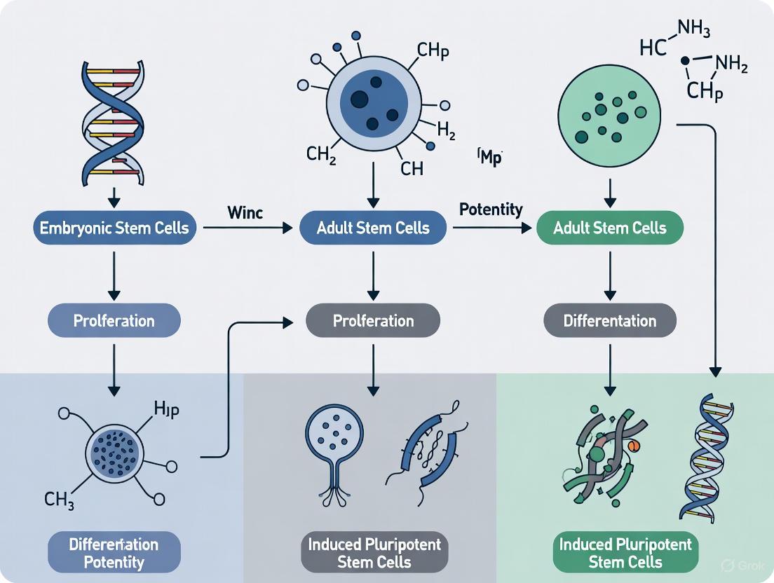
Defining Potency: Understanding the Core Characteristics of Stem Cell Types
Stem cell biology is fundamentally guided by the concept of cell potency, a hierarchical classification system that defines a cell's ability to differentiate into other cell types. This potency spectrum ranges from the unparalleled developmental potential of totipotent cells to the highly restricted fate of unipotent cells. For researchers and drug development professionals, understanding this hierarchy is not merely academic; it is crucial for selecting the appropriate stem cell type for specific applications, from disease modeling and drug screening to regenerative medicine and clinical therapy. The choice between using pluripotent versus multipotent stem cells, for instance, involves a critical trade-off between differentiation potential and clinical safety, a decision that directly impacts experimental design and therapeutic outcomes. This guide provides a systematic comparison of stem cell potency levels, supported by experimental data and protocols, to inform strategic decisions in research and development.
The Potency Spectrum: A Structured Hierarchy
Stem cells are classified based on their differentiation potential, or "potency," which describes the diversity of cell types they can generate. The hierarchy is as follows [1] [2] [3]:
- Totipotent Stem Cells: These represent the pinnacle of developmental potential. A single totipotent cell can give rise to all the cell types in an organism, including both embryonic and extra-embryonic tissues such as the placenta [1] [4]. The only indisputably totipotent cell is the zygote, formed upon the fertilization of an egg [2].
- Pluripotent Stem Cells: These cells can differentiate into cells derived from any of the three primary germ layers—ectoderm, mesoderm, and endoderm—but cannot form the extra-embryonic tissues required for fetal development [1] [2]. This category includes Embryonic Stem Cells (ESCs), derived from the inner cell mass of the blastocyst, and Induced Pluripotent Stem Cells (iPSCs), which are adult cells reprogrammed to an embryonic-like state [1].
- Multipotent Stem Cells: These adult stem cells are more limited, able to differentiate into multiple cell types, but only within a specific lineage or tissue [1] [2]. Examples include Mesenchymal Stem Cells (MSCs), which can form bone, cartilage, and fat cells, and Hematopoietic Stem Cells (HSCs), which generate all blood cell types [1].
- Oligopotent Stem Cells: These possess a further restricted potential, allowing them to differentiate into only a few closely related cell types. An example is the lymphoid or myeloid progenitor cells in the blood lineage [1].
- Unipotent Stem Cells: These are the most restricted, capable of producing only a single cell type. Their primary role is in the renewal and repair of their resident tissue. An example is muscle stem cells (satellite cells), which exclusively differentiate into muscle cells [1].
Table 1: The Hierarchy of Stem Cell Potency
| Potency Level | Defining Characteristic | Key Examples | Primary Sources |
|---|---|---|---|
| Totipotent | Can form a complete organism, including all embryonic and extra-embryonic tissues. | Zygote, early blastomere cells [1] [2]. | Early embryo (first few divisions post-fertilization). |
| Pluripotent | Can form all cells from the three embryonic germ layers (ectoderm, mesoderm, endoderm). | Embryonic Stem Cells (ESCs), Induced Pluripotent Stem Cells (iPSCs) [1] [5]. | Inner cell mass of the blastocyst; reprogrammed somatic cells. |
| Multipotent | Can form multiple cell types within a specific lineage. | Hematopoietic Stem Cells (HSCs), Mesenchymal Stem Cells (MSCs) [1]. | Adult tissues (bone marrow, adipose tissue). |
| Oligopotent | Can differentiate into a few, closely related cell types. | Myeloid or lymphoid progenitor cells [1]. | Arise from multipotent stem cells in specific tissues. |
| Unipotent | Can produce only one cell type. | Muscle satellite cells, progenitor cells in the epidermis [1]. | Resident in specific adult tissues for maintenance and repair. |
Experimental Assessment of Pluripotency
For human Pluripotent Stem Cells (hPSCs), including both ESCs and iPSCs, rigorous functional assays are required to confirm their pluripotent status beyond the mere expression of marker genes. The International Society for Stem Cell Research (ISSCR) provides clear guidelines for this characterization [5].
Key Methodologies for Demonstrating Pluripotency
- In Vitro Differentiation Assays: This is the primary method for demonstrating pluripotency. The protocol involves directing hPSCs to differentiate and then quantitatively measuring the induction of markers representative of all three germ layers. Evidence should include the generation of progenitors for definitive endoderm, mesoderm, and neuroectoderm. Marker analysis is performed via techniques such as flow cytometry, immunocytochemistry, or qRT-PCR, coupled with a clear downregulation of undifferentiated state markers like OCT4 [5].
- Teratoma Assay (Historical Context): While once a gold standard, the teratoma assay is no longer recommended by the ISSCR for routine assessment. This in vivo assay involved injecting hPSCs into an immunocompromised mouse and allowing the cells to form a teratoma—a benign tumor containing differentiated tissues from all three germ layers. Concerns over animal welfare and the development of robust in vitro alternatives have led to its deprioritization [5].
Standards for Reporting
The ISSCR emphasizes that markers of the undifferentiated state (e.g., OCT4, NANOG, SOX2) should not be called "pluripotency markers." Their expression indicates an undifferentiated state but does not, on its own, prove developmental potential, as "nullipotent" cells can also express them. Pluripotency must be defined by demonstrated differentiation capacity [5].
Signaling Pathways Governing Pluripotent States
Pluripotency is not a single static state but encompasses distinct developmental phases, primarily the naive and primed states, which are stabilized by different signaling pathways and transcriptional networks [2] [6].
Regulatory Network of Pluripotency
The core pluripotency network is centered around transcription factors OCT4, SOX2, and NANOG [6]. However, the stability of the naive versus primed states relies on two overlapping positive feedback modules:
- The Klf4/Esrrb/Nanog module stabilizes the naive state.
- The Oct4/Nanog module stabilizes the primed state [6].
The transition between these states and the eventual exit from pluripotency are critically regulated by the Erk/Gsk3 signaling module, which provides incoherent feedforward and negative feedback coupling within the network [6].
Diagram 1: Network regulating naive and primed pluripotency.
Directing Differentiation: A Transcription Factor Case Study
The mechanism by which multipotent stem cells commit to specific lineages often involves a complex interplay of transcription factors. Groundbreaking research in hematopoiesis has demonstrated that the timing and order of key transcription factor expression can direct lineage specification [7].
Experimental Protocol: Transcription Factor Timing
A seminal study used prospectively purified granulocyte/monocyte progenitors (GMPs) to investigate the development of eosinophils, basophils, and mast cells [7].
- Key Transcription Factors: The study focused on GATA-2 and CCAAT enhancer-binding protein α (C/EBPα).
- Methodology: GMPs were subjected to genetic manipulation to control the expression of GATA-2 and C/EBPα. This involved retroviral transduction to enforce the expression of these factors in different sequences.
- Findings: The order of factor expression was critically instructive.
- If GATA-2 was expressed first, it instructed C/EBPα-expressing GMPs to commit exclusively to the eosinophil lineage.
- If C/EBPα was suppressed at the GMP stage, GATA-2 expression induced basophil and/or mast cell lineage commitment.
- Remarkably, simply by switching the order of C/EBPα and GATA-2 transduction, even lymphoid-committed progenitors could be reprogrammed into these granulocytic lineages [7].
This experiment provides a powerful protocol for directing stem cell differentiation by sequentially controlling the expression of lineage-instructive transcription factors, rather than simply presenting them simultaneously.
Diagram 2: Transcription factor order dictates lineage fate.
The Scientist's Toolkit: Essential Reagents for Stem Cell Research
Working with stem cells across the potency hierarchy requires a specific set of reagents and tools to maintain, characterize, and differentiate them. The table below details key research solutions.
Table 2: Essential Reagents and Tools for Stem Cell Research
| Research Reagent/Tool | Primary Function | Application Examples |
|---|---|---|
| Leukemia Inhibitory Factor (LIF) | Activates JAK-STAT3 signaling pathway to maintain self-renewal and the naive pluripotent state in mouse ESCs [2] [6]. | Critical component in standard mouse ESC culture media. |
| Basic Fibroblast Growth Factor (bFGF/FGF2) | Supports the undifferentiated growth of human ESCs and primed pluripotent states; an absolute requirement for hESC culture [2]. | Key cytokine in human ESC and iPSC culture media. |
| Activin A | Activates Smad2/3 signaling; supports self-renewal of human ESCs in concert with bFGF [2]. | Used in defined culture systems for primed pluripotent stem cells. |
| Small Molecule Inhibitors | Pharmacologically inhibit key signaling pathways (e.g., MEK/Erk, Gsk3) to stabilize specific pluripotent states or direct differentiation [6]. | "2i" or "3i" culture systems to support naive pluripotency. |
| Quantitative PCR (qPCR) | Quantifies the expression levels of marker genes for the undifferentiated state (OCT4, NANOG) or specific germ layers (SOX17 - endoderm, BRACHYURY - mesoderm) [5]. | Standard molecular biology technique for characterizing stem cell status and differentiation efficiency. |
| Flow Cytometry | Quantitatively analyzes cell surface and intracellular markers at a single-cell level; essential for assessing population homogeneity and differentiation [5]. | Detection of undifferentiated state markers (e.g., SSEA-4, TRA-1-60) or lineage-specific markers (e.g., CD34 for hematopoietic progenitors). |
Comparative Analysis: Research and Clinical Implications
The choice of stem cell type is a strategic decision that balances differentiation potential against practical and safety considerations.
Pluripotent Stem Cells (PSCs: ESCs and iPSCs):
- Advantages: Unlimited self-renewal and broad differentiation potential across all three germ layers make them ideal for disease modeling, drug screening, and studying developmental mechanisms [1] [8]. iPSCs, in particular, offer the possibility of creating patient-specific cell lines for personalized medicine and avoiding immune rejection [1].
- Challenges: They carry a tangible risk of teratoma formation if undifferentiated cells remain after transplantation. Their use also requires complex and costly differentiation protocols. ESCs carry ethical controversies, though iPSCs have largely mitigated this issue [8] [4].
Multipotent Stem Cells (e.g., MSCs, HSCs):
- Advantages: Lower risk of tumorigenesis and immediate relevance to their tissue of origin. They are clinically accessible from adult tissues, umbilical cord blood, and bone marrow, making them the current workhorses of approved cell therapies (e.g., bone marrow transplants for HSCs) [1] [4].
- Challenges: Their limited differentiation range restricts their application to specific tissue types. They can also be difficult to expand in culture to the large quantities needed for some therapies [1].
Table 3: Comparison of Pluripotent and Multipotent Stem Cells for R&D
| Feature | Pluripotent Stem Cells (ESCs/iPSCs) | Multipotent Stem Cells (MSCs/HSCs) |
|---|---|---|
| Differentiation Potential | Broad (All 3 germ layers) | Restricted (Specific lineage) |
| Self-Renewal | Essentially unlimited in culture | Often limited in culture |
| Tumorigenic Risk | Higher (Risk of teratomas) | Lower |
| Key Research Applications | Disease modeling, drug discovery, developmental biology, generating rare cell types. | Tissue-specific repair, immunomodulation, hematopoiesis research. |
| Clinical Translation | Mostly in clinical trial phases (e.g., macular degeneration, Parkinson's). | Multiple approved therapies (e.g., bone marrow transplantation, graft-versus-host disease). |
| Ethical Considerations | Historically significant for ESCs; minimal for iPSCs. | Generally minimal. |
Embryonic stem cells (ESCs) represent a cornerstone of regenerative medicine and biological research, distinguished by their remarkable pluripotency—the ability to differentiate into any cell type in the adult body [9] [10]. Derived from the inner cell mass of blastocysts (early-stage embryos approximately five days post-fertilization), ESCs offer unprecedented opportunities for modeling human development, drug testing, and developing cell-replacement therapies for conditions ranging from Parkinson's disease to spinal cord injuries [9] [11] [10]. However, their derivation process, which involves the destruction of human embryos, has positioned them at the center of a complex and enduring ethical debate [9] [12]. This guide objectively examines the scientific properties of ESCs in comparison to alternative stem cell types, supported by experimental data, while framing the discussion within the broader ethical and regulatory landscape that researchers must navigate.
Pluripotency and Scientific Profile of ESCs
ESCs are pluripotent cells, a characteristic that places them at the top of the potency hierarchy alongside induced pluripotent stem cells (iPSCs). Pluripotency denotes the capacity to give rise to all derivatives of the three primary germ layers—ectoderm, endoderm, and mesoderm—but not to extra-embryonic tissues like the placenta [1] [10]. The primary source of human ESCs for research is spare embryos created during in vitro fertilization (IVF) treatments that are donated to research with informed consent, or from established ESC lines that are shared globally among laboratories [12].
Table 1: Hierarchy of Stem Cell Potency
| Potency Type | Definition | Example | Key Feature |
|---|---|---|---|
| Totipotent | Can develop into a complete organism, including extra-embryonic tissues. | Zygote (fertilized egg). | Highest potency; exists only in earliest embryonic stages. |
| Pluripotent | Can differentiate into any cell type from the three germ layers. | Embryonic Stem Cells (ESCs), Induced Pluripotent Stem Cells (iPSCs). | Foundation for entire body; cannot form a complete organism. |
| Multipotent | Can differentiate into a limited range of cell types within a specific lineage. | Hematopoietic Stem Cells (HSCs), Mesenchymal Stem Cells (MSCs). | Tissue-specific repair and maintenance. |
Comparative Analysis with Other Stem Cell Types
While ESCs were the first pluripotent cells isolated, the stem cell landscape now includes other types with varying capabilities and research applications.
Table 2: Comparative Profile of Key Stem Cell Types for Research
| Feature | Embryonic Stem Cells (ESCs) | Induced Pluripotent Stem Cells (iPSCs) | Mesenchymal Stem Cells (MSCs) |
|---|---|---|---|
| Source | Inner cell mass of a blastocyst [1] [10]. | Reprogrammed adult somatic cells (e.g., skin, blood) [1] [11]. | Various tissues (e.g., bone marrow, adipose, umbilical cord) [1] [13]. |
| Potency | Pluripotent [1]. | Pluripotent [1]. | Multipotent [1] [13]. |
| Key Research Applications | - Disease modeling- Developmental biology- Drug screening [10]. | - Patient-specific disease modeling- Personalized regenerative medicine- Drug discovery [9] [1]. | - Immunomodulation- Tissue engineering (bone, cartilage)- Anti-inflammatory therapy [1] [13]. |
| Major Advantages | - Gold standard for pluripotency- Extensive historical data. | - Avoids embryo destruction- Enables patient-specific studies. | - Lower ethical concerns- Immunomodulatory properties. |
| Major Challenges | - Ethical controversies- Risk of immune rejection in allogeneic transplants. | - Potential for tumorigenicity- Reprogramming efficiency and safety [11]. | - Heterogeneity between sources- Limited differentiation potential [13]. |
Recent proteomic studies reveal that while ESCs and iPSCs express a nearly identical set of proteins, they are not functionally interchangeable [14] [15]. Key differences include hiPSCs demonstrating increased total protein content, heightened mitochondrial metabolism, and elevated secretion of certain growth factors and extracellular matrix components compared to hESCs [14] [15]. This suggests that reprogramming may not fully reset the cellular profile to an embryonic state, with implications for their use in disease modeling and therapeutics.
The Ethical Landscape and Regulatory Framework
The ethical debate primarily revolves around the moral status of the human embryo [12]. This leads to a fundamental dilemma: balancing the duty to alleviate suffering through medical advances against the duty to respect the value of potential human life [12].
Key Viewpoints on the Moral Status of the Embryo
- Full Moral Status from Fertilization: This viewpoint holds that the embryo is a person from conception, granting it the full right to life. Destruction for research is therefore considered morally unacceptable [12].
- The 14-Day Cut-Off Point: A widely adopted regulatory benchmark in many countries, this position argues that special protection is warranted after 14 days because this is when the primitive streak (the precursor to the nervous system) appears and the embryo can no longer split into twins [12].
- Increasing Moral Status with Development: This perspective grants the embryo some respect from fertilization, but believes its moral status increases as it develops more human characteristics (e.g., after implantation, with nervous system development, or at viability) [12].
- No Moral Status: This view sees the pre-implantation embryo as a cluster of cells with no moral status different from other human tissue, as it lacks sentience, consciousness, and cannot develop without implantation [12].
Oversight and Guidelines
To navigate these ethical challenges, international standards have been established. The International Society for Stem Cell Research (ISSCR) provides comprehensive guidelines that are regularly updated. Key principles include [16]:
- Rigor, Oversight, and Transparency: All research must undergo independent scientific and ethical review.
- Specific Oversight for Sensitive Research: Research involving human embryos, stem cell-based embryo models (SCBEMs), and related activities requires specialized oversight.
- Clear Boundaries: The guidelines explicitly prohibit the transfer of human SCBEMs into a human or animal uterus, and their culture to the point of potential viability (ectogenesis) [16] [17].
Experimental Data and Methodologies
Key Experimental Workflow for Proteomic Comparison
A 2024 study provided a direct proteomic and functional comparison between hiPSCs and hESCs, offering quantitative data on their differences [14] [15]. The methodology below outlines a typical workflow for such a comparative analysis.
Workflow Title: Proteomic Comparison of hESCs and hiPSCs
Key Methodological Steps:
- Cell Culture: Multiple independent hESC and hiPSC lines are cultured under identical, defined conditions to minimize environmental variability [14].
- Pluripotency Confirmation: The expression of core pluripotency markers (e.g., Oct4, Sox2, Nanog) is verified via immunostaining or flow cytometry to ensure all lines are in a pluripotent state prior to analysis [14].
- Proteomic Sample Preparation: Proteins are extracted, digested into peptides, and labeled with isobaric Tandem Mass Tags (TMT). This allows for the multiplexing of samples, enabling simultaneous quantification of proteins from different cell lines in a single mass spectrometry run [14].
- Mass Spectrometry: Labeled peptides are analyzed using LC-MS/MS (Liquid Chromatography with Tandem Mass Spectrometry). The use of MS3 with Synchronous Precursor Selection (SPS) is critical for improving the accuracy of quantification by reducing reporter ion interference [14].
- Data Analysis - Proteomic Ruler: Instead of standard median normalization, the "proteomic ruler" method is applied. This approach uses the near-constant ratio of total protein to DNA in mammalian cells to estimate absolute protein copy numbers per cell, which is essential for detecting changes in total protein content and cell mass [14].
- Functional Validation: Proteomic findings are correlated with phenotypic assays. For example, increased abundance of glutamine transporters is validated by measuring glutamine uptake, and enhanced mitochondrial protein levels are correlated with high-resolution respirometry to measure mitochondrial potential [14] [15].
Quantitative Data from Comparative Studies
The aforementioned proteomic study yielded significant quantitative differences between hiPSCs and hESCs, summarized in the table below.
Table 3: Key Proteomic and Functional Differences (hiPSCs vs. hESCs)
| Parameter | Finding in hiPSCs vs. hESCs | Implication for Research/Therapeutics |
|---|---|---|
| Total Protein Content | >50% higher [14]. | Suggests fundamental differences in cell mass and metabolic activity; highlights importance of normalization methods in omics studies. |
| Differentially Abundant Proteins | 4,408 proteins significantly increased; 40 significantly decreased (FC>1.5) [14]. | Indicates widespread molecular differences beyond the nucleus. |
| Mitochondrial Metabolism | Increased abundance of mitochondrial metabolic proteins and enhanced mitochondrial membrane potential [14] [15]. | Suggests hiPSCs may have a higher metabolic rate, which could influence differentiation efficiency and cell fate. |
| Secretion Profile | Higher production of secreted proteins, including specific growth factors and immunomodulatory proteins [14] [15]. | May impact paracrine signaling in co-culture systems and carry potential tumorigenic risks that require careful evaluation. |
| Nutrient Transport & Storage | Increased glutamine transporters (correlated with higher uptake) and proteins for lipid synthesis (correlated with more lipid droplets) [14]. | Points to altered nutrient utilization, which is critical for optimizing culture media and bioprocessing. |
The Scientist's Toolkit: Essential Research Reagents
Table 4: Key Reagent Solutions for Pluripotent Stem Cell Research
| Research Reagent / Material | Function / Application |
|---|---|
| Tandem Mass Tags (TMT) | Isobaric chemical labels for multiplexed quantitative proteomics, allowing simultaneous comparison of multiple cell lines in one MS run [14]. |
| Pluripotency Markers (Oct4, Sox2, Nanog) | Antibodies for immunocytochemistry, flow cytometry, or Western Blot to confirm the undifferentiated, pluripotent state of ESCs and iPSCs [14] [10]. |
| Defined Culture Matrices (e.g., Vitronectin, Laminin-521) | Recombinant, xeno-free substrates for feeder-free culture of pluripotent stem cells, improving reproducibility and clinical compliance. |
| mTeSR1 or Similar Defined Media | Chemically defined, serum-free media formulations that support the maintenance of pluripotency without the need for feeder cells. |
| Y-27632 (ROCK inhibitor) | Small molecule inhibitor used to enhance the survival of pluripotent stem cells during passaging and cryopreservation by reducing apoptosis. |
| CRISPR-Cas9 Systems | Genome editing tools for creating precise genetic modifications in ESCs/iPSCs for gene function studies, disease modeling, and gene correction [10]. |
Embryonic Stem Cells remain the reference standard for pluripotency and an powerful tool for understanding human development and disease. Their extensive historical data and well-defined properties make them invaluable for specific research inquiries. However, the emergence of iPSCs offers a compelling, ethically less contentious alternative for patient-specific studies, despite not being functionally identical. The choice between ESCs, iPSCs, or adult stem cells like MSCs is not a matter of which is universally "best," but which is most appropriate for the specific scientific question and context. Researchers must weigh the superior pluripotency of ESCs against the ethical considerations and the patient-specific advantages of iPSCs, all while adhering to a robust and evolving framework of international guidelines designed to ensure scientific integrity and public trust [16] [17].
The development of induced pluripotent stem cell (iPSC) technology represents a paradigm shift in regenerative medicine and biomedical research. First established in 2006 by Takahashi and Yamanaka, iPSCs are generated by reprogramming adult somatic cells into an embryonic-like pluripotent state through the forced expression of specific transcription factors [18] [19]. This groundbreaking achievement demonstrated that cellular differentiation is not a unidirectional process, but rather can be reversed through epigenetic reprogramming [18]. Unlike embryonic stem cells (ESCs), iPSCs bypass ethical concerns associated with embryo destruction while retaining the fundamental capacity to differentiate into any cell type in the human body [9] [19]. For researchers and drug development professionals, iPSCs provide an unprecedented platform for disease modeling, drug screening, and developing patient-specific cell therapies [18] [20]. The unique combination of reprogrammable pluripotency and patient-specific origin positions iPSCs as a transformative technology with distinct advantages and limitations compared to other stem cell sources.
The selection of an appropriate stem cell source depends heavily on research objectives, with each cell type offering distinct advantages and limitations. The following comparison outlines key characteristics of major stem cell types relevant to research applications.
Table 1: Comparison of Key Stem Cell Types for Research Applications
| Stem Cell Type | Origin | Pluripotency | Ethical Concerns | Tumorigenic Risk | Patient-Specific | Key Research Applications |
|---|---|---|---|---|---|---|
| iPSCs | Reprogrammed somatic cells (e.g., skin, blood) | Pluripotent | Minimal | Moderate (teratoma formation) | Yes (autologous) | Disease modeling, drug screening, personalized cell therapy [18] [19] |
| Embryonic Stem Cells (ESCs) | Inner cell mass of blastocysts | Pluripotent | Significant (embryo destruction) | High (teratoma formation) | No (allogeneic) | Developmental biology, regenerative medicine [21] [9] |
| Mesenchymal Stem Cells (MSCs) | Adult tissues (bone marrow, adipose, umbilical cord) | Multipotent | Minimal | Low | Possible (requires matching) | Immunomodulation, tissue engineering, inflammation research [21] [22] |
| Natural Multipotent Stem Cells (nMS) | Various adult tissues including umbilical cord | Multipotent | Minimal | Reported as low | No (allogeneic) | Clinical transplantation for various intractable diseases [21] |
Table 2: Quantitative Comparison of Differentiation Potential and Technical Considerations
| Parameter | iPSCs | ESCs | MSCs | Natural Multipotent Stem Cells |
|---|---|---|---|---|
| Differentiation Potential | All three germ layers [20] | All three germ layers | Primarily mesodermal lineages [21] | Multiple lineages (varies by source) [21] |
| Reprogramming Time | 3-4 weeks [23] | N/A | N/A | N/A |
| Genetic Stability | Variable (epigenetic memory concerns) [20] | High | High | Reported as high [21] |
| Scalability | High (unlimited self-renewal) [18] | High (unlimited self-renewal) | Limited expansion capacity | Reported as highly scalable [21] |
| Immunogenicity | Low (if autologous) | High (allogeneic) | Low (immunoprivileged) [22] | Reported as low immunogenicity [21] |
Core Reprogramming Methodologies: From Yamanaka Factors to Clinical-Grade Protocols
Transcription Factor-Based Reprogramming
The original iPSC reprogramming method utilized retroviral transduction of four transcription factors—OCT4, SOX2, KLF4, and c-MYC (OSKM), collectively known as Yamanaka factors [18] [19]. Each factor plays a distinct role in resetting epigenetic memory: OCT4 and SOX2 are core pluripotency regulators that activate endogenous self-renewal networks; KLF4 promotes chromatin remodeling and suppresses somatic gene expression; while c-MYC enhances proliferation and global histone acetylation to facilitate epigenetic reprogramming [23]. An alternative combination (OCT4, SOX2, NANOG, LIN28) was subsequently developed by Thomson's group, with NANOG stabilizing pluripotency and LIN28 regulating metabolism and cell cycle progression [18]. The reprogramming process occurs in two primary phases: an early stochastic phase where somatic identity is silenced, followed by a deterministic phase where pluripotency networks are activated [18]. This transition involves extensive epigenetic remodeling, including DNA demethylation at pluripotency loci, histone modification, and chromatin restructuring [18].
Advanced Non-Integrating Methods
While revolutionary, initial viral vector methods raised safety concerns due to genomic integration and potential insertional mutagenesis [19]. Subsequently, non-integrating reprogramming methods have been developed for clinical translation:
- Sendai Virus Vectors: RNA-based viral vectors that remain episomal without nuclear integration, efficiently reprogramming somatic cells within 3-4 weeks before being diluted out through cell divisions [24] [19].
- Synthetic mRNA Reprogramming: Daily transfection of modified mRNAs encoding reprogramming factors achieves high efficiency without genomic integration, though it requires precise optimization to avoid immune activation [24].
- Episomal Plasmid Vectors: Non-viral DNA vectors that replicate extrachromosomally and are gradually lost during cell divisions, offering a cost-effective approach for clinical-grade iPSC generation [19].
- Small Molecule Reprogramming: Chemical cocktails that modulate signaling pathways and epigenetic modifiers can enhance reprogramming efficiency or in some cases replace transcription factors entirely [24] [18].
Figure 1: iPSC Reprogramming Workflow. This diagram illustrates the process of reprogramming somatic cells into induced pluripotent stem cells using different methodological approaches.
Signaling Pathways Governing Pluripotency and Differentiation
The maintenance of pluripotency in iPSCs is regulated by a complex network of signaling pathways that collectively suppress differentiation and promote self-renewal. The core pluripotency circuitry centers on the transcription factors OCT4, SOX2, and NANOG, which form an autoregulatory loop that activates their own expression while simultaneously inhibiting differentiation genes [23]. This transcriptional network interacts with multiple extrinsic signaling pathways:
- LIF/STAT3 Pathway: Activated by leukemia inhibitory factor (LIF), maintains pluripotency in mouse iPSCs through STAT3 activation [23].
- TGF-β/SMAD2/3 Signaling: Works in concert with OCT4/SOX2/NANOG to sustain self-renewal in human iPSCs [24].
- WNT/β-Catenin Pathway: Fine-tuned regulation is critical, as either inhibition or hyperactivation can promote differentiation [24].
- FGF Signaling: Basic fibroblast growth factor (FGF2) is essential for human iPSC self-renewal through MAPK/ERK pathway modulation [23].
- BMP Signaling: Context-dependent effects, with BMP4 promoting self-renewal in mouse iPSCs but differentiation in human iPSCs [24].
During differentiation, precise manipulation of these pathways directs lineage specification. For example, BMP4 combined with TGF-β inhibition induces mesoderm formation, while Wnt activation with nodal signaling promotes endodermal differentiation [24]. Neural ectoderm specification typically requires dual SMAD inhibition (noggin/SB431542) to suppress BMP and TGF-β signaling [25].
Figure 2: Signaling Pathways in Pluripotency. This diagram illustrates the major signaling pathways that maintain the pluripotent state in iPSCs and how their manipulation can drive differentiation.
The Scientist's Toolkit: Essential Reagents for iPSC Research
Table 3: Essential Research Reagents for iPSC Generation and Maintenance
| Reagent Category | Specific Examples | Function | Application Notes |
|---|---|---|---|
| Reprogramming Factors | OCT4, SOX2, KLF4, c-MYC (OSKM) [18] | Master transcription factors that reset epigenetic memory | Delivery via Sendai virus, mRNA, or episomal vectors [19] |
| Culture Media | mTeSR, Essential 8, StemFlex | Defined, xeno-free media for pluripotency maintenance | Contain FGF2 and TGF-β to support self-renewal [24] |
| Extracellular Matrices | Geltrex, Matrigel, Vitronectin | Synthetic or purified matrices for feeder-free culture | Provide adhesion signals and structural support [23] |
| Metabolic Regulators | L-ascorbic acid, Sodium pyruvate | Enhance reprogramming efficiency and genomic stability | Reduce oxidative stress during reprogramming [24] |
| Passaging Reagents | EDTA, ReLeSR, Accutase | Gentle dissociation methods for colony passaging | Maintain colony integrity while enabling expansion [21] |
| Characterization Antibodies | Anti-OCT4, Anti-SOX2, Anti-SSEA4, Anti-TRA-1-60 | Confirm pluripotency marker expression | Essential for quality control and validation [23] |
Experimental Workflows: From Somatic Cell to Differentiated Lineages
Protocol for mRNA-Based iPSC Generation
The following detailed protocol enables integration-free iPSC generation using synthetic modified mRNAs:
- Starter Cell Culture: Expand human dermal fibroblasts or peripheral blood mononuclear cells in appropriate medium. Ensure cells are at low passage and 70-80% confluent at initiation.
- mRNA Transfection: Transfect cells daily with 0.5-1μg of each modified mRNA (OCT4, SOX2, KLF4, c-MYC, LIN28) using lipid-based transfection reagent. Include B18R interferon inhibitor in medium to suppress innate immune response.
- Colony Monitoring: Observe morphological changes around day 7-10, with emergence of compact, ESC-like colonies with defined borders by day 18-25.
- Colony Picking: Mechanically pick individual colonies between days 21-28 and transfer to fresh matrix-coated plates.
- Expansion and Characterization: Expand clonal lines and validate pluripotency through immunocytochemistry (OCT4, NANOG, SSEA4, TRA-1-60), trilineage differentiation potential, and karyotype analysis.
This protocol typically achieves reprogramming efficiencies of 0.5-2.0% with proper optimization, significantly higher than early viral methods (0.01-0.1%) [24].
Directed Differentiation to Pancreatic β-Cells
For diabetes research and drug screening, iPSC differentiation into insulin-producing β-cells follows a stepwise protocol mimicking pancreatic development:
- Definitive Endoderm Induction (3 days): Culture iPSCs with 100ng/mL Activin A and 3μM CHIR99021 (Wnt activator) in low-glucose medium to specify definitive endoderm. Monitor for upregulation of SOX17 and FOXA2.
- Pancreatic Progenitor Specification (7 days): Transition to medium containing 50ng/mL FGF10, 0.25μM cyclopamine (hedgehog inhibitor), and 2μM retinoic acid to generate pancreatic progenitors expressing PDX1 and NKX6.1.
- Endocrine Progenitor Maturation (7 days): Further differentiate with 10μM ALK5 inhibitor II, 100nM gamma-secretase inhibitor XX, and 1μ T3 thyroid hormone to induce endocrine commitment.
- β-Cell Maturation (14+ days): Final maturation in medium containing 10μM T3, 10μ ALK5 inhibitor II, and 1mM N-acetyl cysteine to generate functional β-cells expressing MAFA, insulin, and displaying glucose-stimulated insulin secretion.
This 30+ day protocol generates polyhormonal cells that progressively mature into monohormonal insulin-positive cells with improved glucose responsiveness, though full functional maturation often requires additional weeks or in vivo implantation [26] [25].
Clinical Translation: Current Status and Safety Considerations
iPSC-based therapies have advanced to clinical trials for several conditions, with varying approaches to immune matching:
- Parkinson's Disease: Multiple trials using allogeneic (donor-derived) and autologous (patient-specific) iPSC-derived dopaminergic progenitors have demonstrated improved motor function without tumor formation in early-stage trials [19].
- Type 1 Diabetes: A 2024 clinical report described successful glycemic control restoration in a patient using iPSC-derived pancreatic islets from adipose-derived mesenchymal stromal cells, eliminating exogenous insulin requirements for one year post-transplantation [25].
- Ocular Diseases: iPSC-derived retinal pigment epithelial (RPE) cells have been transplanted in patients with age-related macular degeneration, showing vision stabilization with acceptable safety profiles [19]. Corneal epithelial sheets derived from iPSCs have restored vision in patients with limbal stem cell deficiency [25].
Despite these promising advances, significant challenges remain in clinical translation. Tumorigenicity risk persists due to potential residual undifferentiated cells or genetic abnormalities acquired during reprogramming [21] [19]. Immune responses against allogeneic iPSC-derived products necessitate immunosuppression or the development of hypoimmunogenic lines through genetic engineering [24]. Scalable manufacturing under Good Manufacturing Practice (GMP) conditions remains technically challenging and cost-prohibitive for widespread implementation [19]. Additionally, functional maturation of iPSC-derived cells often incomplete in vitro, with some lineages requiring extended timeframes or specific niche signals to achieve full functionality [25].
iPSC technology represents a transformative approach with distinct advantages for disease modeling, drug screening, and regenerative medicine. The capacity for patient-specific derivation enables creation of genetically matched disease models and autologous therapies, while pluripotent differentiation potential provides access to otherwise inaccessible human cell types. However, researchers must carefully consider technical challenges including reprogramming efficiency, functional maturation, and safety profiling when designing studies. For therapeutic applications, the choice between autologous (patient-specific) and allogeneic (donor-derived) approaches involves trade-offs between immune compatibility, manufacturing scalability, and cost-effectiveness. As reprogramming methods continue advancing toward non-integrating, xeno-free systems, and differentiation protocols achieve greater precision through pathway modulation and bioengineering, iPSCs are positioned to increasingly become indispensable tools for both basic research and clinical translation across diverse biomedical applications.
Adult stem cells, or somatic stem cells, are undifferentiated cells found throughout the body after development that serve as a repair system for damaged tissues. Unlike embryonic stem cells (ESCs), which are pluripotent and raise ethical concerns, adult stem cells are typically multipotent, meaning they can differentiate into a limited range of cell types related to their tissue of origin [27] [10] [28]. Their dynamic and adaptive therapeutic properties have led to their characterization as "living drugs," as they can sense environmental cues, home to injury sites, and integrate into tissues to exert longer-lasting effects compared to conventional medicines [28]. Among the various types, Mesenchymal Stem/Stromal Cells (MSCs) and Hematopoietic Stem Cells (HSCs) represent two of the most extensively researched and clinically applied adult stem cell populations, each with distinct biological properties, therapeutic mechanisms, and clinical applications [10] [29] [30].
HSCs are primarily responsible for the constant renewal of the blood and immune system. Residing in the bone marrow, they can differentiate into all types of blood cells, including erythrocytes, leukocytes, and platelets [29]. Their well-established role in bone marrow transplantation has made them a cornerstone in treating hematologic malignancies, such as leukemia and lymphoma, as well as other blood disorders [27] [10]. In contrast, MSCs are non-hematopoietic, multipotent stromal cells that can differentiate into mesodermal lineages like osteoblasts (bone cells), chondrocytes (cartilage cells), and adipocytes (fat cells) [30]. Originally identified in the bone marrow, MSCs have since been isolated from a variety of tissues, including adipose tissue, umbilical cord, dental pulp, and placental tissue [31] [30]. The therapeutic potential of MSCs extends beyond differentiation to include potent immunomodulatory functions and the release of bioactive molecules that promote tissue repair and reduce inflammation [32] [30]. This guide provides a detailed, data-driven comparison of these multipotent sources, focusing on their proliferation capacity, differentiation potential, and key experimental methodologies to inform research and drug development.
The following tables provide a structured comparison of MSCs from different tissue sources and a direct comparison between MSCs and HSCs, summarizing key characteristics, markers, and functional data.
Table 1: Comparative Profile of Mesenchymal Stem/Stromal Cells (MSCs) from Different Tissues
| Parameter | Bone Marrow-MSCs (BM-MSCs) | Adipose-Derived MSCs (AD-MSCs) | Umbilical Cord-MSCs (UC-MSCs) | Dental Pulp Stem Cells (DPSCs) |
|---|---|---|---|---|
| Key Source Tissue | Bone Marrow Aspirate | Lipoaspirate (Abdominal Fat) | Wharton's Jelly, Umbilical Cord | Dental Pulp (Coronal/Radicular) |
| Isolation Methods | Density Gradient Centrifugation, Plastic Adherence | Enzymatic Digestion (SVF), Mechanical Fragmentation (MF) [31] | Enzymatic Digestion, Explant Culture [30] | Mechanical Fragmentation, Explant Culture [31] |
| Common Surface Markers (Positive) | CD73, CD90, CD105 (≥95% positive) [30] | CD73, CD90, CD105 (≥95% positive) [30] | CD73, CD90, CD105 (≥95% positive) [30] | CD73, CD90, CD105 (≥95% positive) [31] |
| Common Surface Markers (Negative) | CD34, CD45, CD11b, CD19, HLA-DR (≤2% positive) [30] | CD34, CD45, CD11b, CD19, HLA-DR (≤2% positive) [30] | CD34, CD45, CD11b, CD19, HLA-DR (≤2% positive) [30] | CD34, CD45, CD11b, CD19, HLA-DR (≤2% positive) [31] |
| Proliferation & Morphology | Extensive study history, spindle-shaped [30] | Easier to harvest, high yield, spindle-shaped [30] | Enhanced proliferation, lower immunogenicity [30] | Higher proliferation rate, smaller size, Nestin-positive [31] |
| Distinct Differentiation Potential | Osteoblasts, Chondrocytes, Adipocytes [30] | Osteoblasts, Chondrocytes, Adipocytes [30] | Osteoblasts, Chondrocytes, Adipocytes [30] | Osteoblasts, Chondrocytes; Impaired Adipogenesis [31] |
| Key Secretome Factors | Cytokines, G-CSF, GM-CSF, IL-6 [30] | Specific sets of miRNAs (regulate cell cycle/proliferation) [31] | Anti-inflammatory cytokines, Growth Factors [32] | Specific sets of miRNAs (oxidative stress/apoptosis pathways) [31] |
| Reported Clinical Applications | Graft-vs-Host Disease (GVHD), Supporting Hematopoietic Recovery [33] [32] | Crohn's disease, Perianal Fistulas, Orthopedic Repair [32] [30] | Pediatric GVHD, Inflammatory Disorders [32] [30] | Dental and Craniofacial Regeneration [30] |
Table 2: Direct Comparison of Key Multipotent Stem Cells: MSCs vs. HSCs
| Parameter | Mesenchymal Stem/Stromal Cells (MSCs) | Hematopoietic Stem Cells (HSCs) |
|---|---|---|
| Primary Physiological Role | Tissue stroma support, immunomodulation, tissue repair [30] | Reconstitution of entire blood and immune system [29] |
| Main Tissue Sources | Bone Marrow, Adipose Tissue, Umbilical Cord, Dental Pulp [30] | Bone Marrow, Mobilized Peripheral Blood, Umbilical Cord Blood [29] |
| Defining Surface Markers | CD73+, CD90+, CD105+, CD34-, CD45- [30] | CD34+, CD45+, CD133+ [29] |
| Differentiation Potential (Lineages) | Mesodermal: Osteogenic, Chondrogenic, Adipogenic [30] | Hematopoietic: Erythroid, Myeloid, Lymphoid [29] |
| Therapeutic Mechanisms | Differentiation, potent paracrine signaling, immunomodulation, anti-apoptosis [32] [30] | Differentiation into functional blood cells, immune reconstitution [29] |
| Key Clinical Applications | GVHD, Crohn's disease, orthopedic repair, HSCT co-infusion [33] [32] | Leukemia, Lymphoma, Aplastic Anemia (Bone Marrow Transplantation) [29] |
| Ex Vivo Expansion/Culture | Relatively easy to expand in vitro; potency affected by culture system (2D vs. 3D) [34] | Possible but challenging; limited proliferation potential, variable outcomes [29] |
| Quantitative Performance Data | Neutrophil engraftment: ~13.96 days; Platelet engraftment: ~21.61 days (post-HSCT co-infusion) [33] | Culture time for RBCs: <21 days; Enucleation rate: 50% to 98% [29] |
Experimental Protocols for Key Assays
Standardized experimental protocols are crucial for the isolation, characterization, and functional assessment of adult stem cells. The following sections detail common methodologies used in research involving MSCs and HSCs.
Isolation and Culture of Adipose-Derived MSCs (AD-MSCs)
Two primary methods are employed for isolating AD-MSCs from lipoaspirate tissue [31].
- Enzymatic Digestion (Stromal Vascular Fraction - SVF): The washed lipoaspirate is subjected to overnight digestion with collagenase (e.g., Collagenase 1A) at 37°C. The digested material is then centrifuged (e.g., 10 minutes at 1200 g), and the resulting cell pellet, known as the stromal vascular fraction (SVF), is plated onto tissue culture dishes in a basic medium (e.g., αMEM) supplemented with 10% Fetal Bovine Serum (FBS). Upon reaching 80-90% confluence, the adherent cells (ADSCs-SVF) are detached using trypsin-EDTA and subcultured.
- Mechanical Fragmentation (Explant Method): The lipoaspirate is fragmented into small pieces and placed directly in a culture dish with a growth medium supplemented with 20% FBS. The culture is maintained without enzymatic treatment, allowing cells to migrate out from the tissue fragments over 1-2 weeks. The outgrowing, plastic-adherent cells (ADSCs-MF) are then passaged similarly upon confluence.
Isolation of Dental Pulp Stem Cells (DPSCs)
DPSCs are typically isolated using an explant method [31]. Sound teeth (e.g., third molars) are cut at the amelo-cement junction with a diamond disc, and the pulp tissue is gently removed. The pulp is separated into coronal and radicular portions, which are then fragmented into 1-2 mm³ pieces using a scalpel. After washing by centrifugation, these fragments are seeded onto tissue culture plates. Cells migrating from the explants form a monolayer over 2-4 weeks and are then passaged. Cells derived from the coronal and radicular compartments can be studied separately as Coronal Pulp Stromal Cells (CPSCs) and Radicular Pulp Stromal Cells (RPSCs) [31].
In Vitro Trilineage Differentiation Assay for MSCs
This assay is a defining criterion for confirming MSC identity, as per the International Society for Cellular Therapy (ISCT) [30]. The protocol involves culturing MSCs in specific induction media for 2-4 weeks [31].
- Osteogenic Differentiation: Cells are cultured in a medium supplemented with 50 µM ascorbic acid-2 phosphate, 10 mM β-glycerophosphate, and 0.1 µM dexamethasone. Differentiation is confirmed by the formation of mineralized nodules, which can be stained with Alizarin Red S.
- Chondrogenic Differentiation: A pellet culture system is often used, where cells are centrifuged to form a micromass. The pellet is cultured in a medium containing TGF-β (e.g., TGF-β3), ascorbate, and dexamethasone. Chondrogenesis is evidenced by the deposition of a cartilage-specific extracellular matrix, visible upon Safranin O or Alcian Blue staining.
- Adipogenic Differentiation: Cells are induced with a medium containing 0.5 mM isobutylmethylxanthine (IBMX), 1 µM dexamethasone, and 50 µM indomethacin. The formation of intracellular lipid vacuoles, detectable by Oil Red O staining, confirms successful adipogenesis. It is important to note that some MSC sources, like DPSCs, may have an impaired ability to undergo adipogenic differentiation [31].
In Vitro Generation of Red Blood Cells from HSCs
This protocol generates functional, enucleated red blood cells (RBCs) from HSCs for research and potential transfusion applications [29]. Isolated HSCs (from bone marrow, cord blood, or mobilized peripheral blood) are cultured in a specialized medium containing a combination of cytokines and growth factors, such as stem cell factor (SCF), erythropoietin (EPO), and interleukin-3 (IL-3). The culture is maintained under specific oxygen tension to mimic the physiological environment of erythropoiesis. The process involves the differentiation of HSCs through erythroid progenitor stages (erythroblasts) into mature, enucleated RBCs over approximately 21 days. Efficiency is evaluated by measuring the expansion factor (fold-increase in cell numbers) and the enucleation rate (percentage of cells lacking a nucleus), which, for HSCs, can range from 50% to over 98% in optimized systems [29].
Signaling Pathways and Experimental Workflows
The therapeutic actions of MSCs are mediated through complex signaling pathways and paracrine communication. The diagram below illustrates the key mechanistic pathways through which MSCs sense inflammatory signals and mount an immunomodulatory response, which is central to their therapeutic effect in inflammatory and autoimmune diseases.
Figure 1: MSC Immunomodulatory Pathway. This diagram shows how MSCs respond to inflammation and modulate immune cells.
The following diagram outlines a generalized experimental workflow for comparing the properties of MSCs derived from different tissue sources, from isolation through functional validation.
Figure 2: MSC Comparison Workflow. This diagram shows the key steps for comparing MSCs from different sources.
The Scientist's Toolkit: Key Research Reagent Solutions
The table below lists essential reagents, tools, and materials required for conducting experiments with adult stem cells, along with their critical functions in the research process.
Table 3: Essential Research Reagents and Tools for Adult Stem Cell Studies
| Reagent / Tool | Primary Function & Application |
|---|---|
| Fetal Bovine Serum (FBS) | Critical supplement for basal culture media (e.g., DMEM, αMEM) to provide essential growth factors, hormones, and nutrients for cell survival and proliferation [31]. |
| Collagenase (Type 1A, etc.) | Enzyme used for the enzymatic digestion of solid tissues (like adipose tissue) to release the stromal vascular fraction (SVF) containing AD-MSCs [31]. |
| Trypsin-EDTA | Proteolytic enzyme solution used to detach adherent cells (e.g., MSCs) from the surface of culture vessels for subculturing (passaging) and cell counting [31]. |
| Defined Cytokine Cocktails | Specific growth factors and cytokines (e.g., SCF, EPO, TGF-β, G-CSF) used to direct stem cell differentiation in vitro, such as inducing erythropoiesis from HSCs or trilineage differentiation from MSCs [31] [29]. |
| Flow Cytometry Antibodies | Fluorescently-labeled antibodies against specific cell surface markers (e.g., CD73, CD90, CD105, CD34, CD45) for the phenotypic identification and purity assessment of stem cell populations [30]. |
| 3D Culture Matrices (e.g., Hydrogels, Matrigel) | Biomimetic scaffolds used to create a more physiologically relevant three-dimensional (3D) environment for cell culture, which can enhance MSC potency, secretome production, and differentiation capacity compared to 2D plastic [34]. |
| Senescence-Associated β-Galactosidase (SA-β-Gal) Kit | A biochemical kit used to detect the activity of a specific β-galactosidase enzyme present in senescent (aged) cells, serving as a key marker for assessing cellular aging and culture health [34]. |
| ELISA / Multiplex Assay Kits | Tools for quantitatively measuring the concentration of specific proteins, cytokines, and growth factors secreted by cells into the conditioned medium (secretome analysis) [31]. |
Key Molecular Mechanisms Governing Self-Renewal and Differentiation
In stem cell biology, self-renewal and differentiation represent two fundamental, yet opposing, processes that are essential for development, tissue maintenance, and regeneration. Self-renewal refers to the capacity of a stem cell to divide and produce identical copies of itself, thereby maintaining the stem cell pool throughout an organism's life. Differentiation, in contrast, is the process by which a less specialized stem cell undergoes maturation to adopt a specific, specialized cell type with a distinct function, such as a neuron, cardiomyocyte, or adipocyte [8]. The precise balance between these two states is governed by a complex interplay of intrinsic molecular networks and extrinsic signals from the microenvironment, or "niche" [35] [36]. For researchers and drug development professionals, understanding these regulatory mechanisms is paramount for harnessing stem cells' potential in regenerative medicine, disease modeling, and therapeutic development. This guide objectively compares how different stem cell sources and their unique molecular wiring influence their proliferation and differentiation potential, providing a foundation for informed experimental design.
Core Molecular Regulators of Stem Cell Fate
The decision between self-renewal and differentiation is orchestrated by a core set of molecular regulators that integrate external cues and execute transcriptional and epigenetic programs.
The Cell Cycle and Pluripotency
A distinctive feature of many stem cells, particularly embryonic stem cells (ESCs), is their unique cell cycle structure, which is intrinsically linked to their pluripotent state. Unlike somatic cells, ESCs exhibit a dramatically shortened G1 phase and a prolonged S phase. This rapid cycling is not merely for swift proliferation; it helps maintain an open chromatin configuration that is permissive for the expression of pluripotency genes, thereby preventing the onset of differentiation signals that often are coordinated with specific cell cycle phases [35]. The naïve, formative, and primed states of pluripotency are tightly associated with specific cell cycle patterns, and this association exhibits species specificity [35].
Key Signaling Pathways
Several evolutionarily conserved signaling pathways act as central processors of external signals, directly influencing stem cell fate. The table below summarizes the primary functions of these key pathways.
Table 1: Key Signaling Pathways Governing Stem Cell Fate
| Pathway | Primary Role in Self-Renewal | Primary Role in Differentiation | Notable Ligands/Regulators |
|---|---|---|---|
| Wnt/β-catenin | Promotes self-renewal by stabilizing nuclear β-catenin and activating target genes like c-Myc and Cyclin D1 [36]. | Controls lineage specification; its inhibition is often required for differentiation to proceed [36]. | Wnt proteins, GSK-3β |
| TGF-β/BMP | TGF-β and Activin A support self-renewal in primed pluripotent stem cells [36]. | BMPs can induce differentiation; the pathway regulates lineage commitment (e.g., mesoderm, endoderm) [36]. | TGF-β, BMP, Nodal, Activin |
| Hedgehog (Hh) | Regulates proliferation and self-renewal in certain adult stem cell populations [36]. | Critical for embryonic patterning and differentiation of multiple tissue types [36]. | Sonic Hedgehog (Shh) |
| Notch | Maintains stem cell quiescence in niches like the hematopoietic system [36]. | Mediates cell-fate decisions through lateral inhibition [36]. | Delta, Jagged |
| FGF | Supports proliferation and self-renewal by activating MAPK/ERK and PI3K/Akt pathways [36]. | Drives differentiation towards specific lineages, particularly in neural and mesodermal contexts [36]. | FGF2 (bFGF) |
| Hippo/YAP | The transcriptional co-activator YAP promotes self-renewal and proliferation when localized to the nucleus [35] [37]. | Inactivation of YAP and activation of the Hippo kinase cascade can promote differentiation and restrict organ size [37]. | YAP, TAZ, LATS1/2 |
The following diagram illustrates the logical relationships between these core pathways and their primary outcomes in stem cell fate determination:
Figure 1: Core Signaling Network Logic. This diagram shows how external signals converge on key pathways to regulate the cell cycle, pluripotency factors, and epigenetic landscape, ultimately determining the balance between self-renewal and differentiation.
Epigenetic and Metabolic Control
Beyond transcription factors and signaling cascades, the stem cell state is heavily influenced by the epigenetic landscape. Histone modifications (e.g., methylation, acetylation) and DNA methylation dynamically repress or permit the expression of differentiation-related genes, allowing a stem cell to maintain its potential while preventing premature specialization [35]. This epigenetic state is coupled to a distinct metabolic profile. Stem cells predominantly rely on glycolysis rather than oxidative phosphorylation for energy production. This glycolytic mode supports rapid biosynthesis and helps maintain a low level of reactive oxygen species (ROS), which in turn influences the epigenetic machinery and supports the maintenance of pluripotency [35].
Comparative Analysis: Stem Cell Source Influences Molecular Mechanisms
The molecular mechanisms described above are not uniform across all stem cells. Their expression and activity can vary significantly depending on the cell source, which directly impacts proliferation and differentiation potential. A comparative study on mesenchymal stem cells (MSCs) from Hanwoo cattle provides a clear example of how the tissue of origin dictates cellular behavior [38].
Depot-Specific Differences in Adipose-Derived MSCs
This study directly compared MSCs isolated from perirenal adipose tissue (P-AMSCs) and subcutaneous adipose tissue (S-AMSCs) from the same animals. The results demonstrated stark differences in their molecular profiles and functional capacities [38].
Table 2: Comparative Analysis of Adipose-Derived MSC Sources
| Parameter | Perirenal AMSCs (P-AMSCs) | Subcutaneous AMSCs (S-AMSCs) |
|---|---|---|
| Surface Marker CD105 | High expression (26.3%) [38] | Low expression (1.2%) [38] |
| Proliferation Rate | Faster proliferation and shorter doubling times [38] | Slower proliferation [38] |
| Adipogenic Potential | Greater lipid accumulation; higher expression of PPARγ, FABP4, LPL, and FASN [38] | Lower lipid accumulation and adipogenic gene expression [38] |
| Osteogenic Potential | Stronger mineralization (91.8%); upregulation of COL1A1, RUNX2, DLX5 [38] | Weaker mineralization (60.5%) [38] |
| Chondrogenic Potential | Enhanced chondrogenesis with increased SOX9, COL2A1, and ACAN [38] | Reduced chondrogenic potential [38] |
Species-Specific and Tissue-Specific Organization
Further illustrating the impact of source, a comparative study of squamous epithelia revealed a human-specific organization of stemness and proliferation. Unlike other mammals, humans possess a quiescent basal stem cell layer that is physically separated from the parabasal transit-amplifying cells. This unique architecture, which decouples stemness from active proliferation, is proposed to be an evolutionary adaptation that enhances tissue longevity and suppresses tumorigenesis [39]. Additionally, human squamous epithelial stem cells express higher levels of DNA repair markers like MECP2 and XPC, pointing to enhanced cytoprotective mechanisms [39].
Experimental Data and Methodologies
To facilitate the replication and critical evaluation of comparative studies, this section outlines standard experimental protocols for assessing the molecular mechanisms of self-renewal and differentiation.
Standard Characterization Protocol for MSCs
The International Society for Cellular Therapy (ISCT) defines MSCs by a set of minimum criteria, which form the basis of their characterization [38] [30]:
- Adherence to Plastic: Cells must be adherent to plastic surfaces under standard culture conditions.
- Surface Marker Expression:
- Trilineage Differentiation Potential: The cells must be able to differentiate into osteoblasts, adipocytes, and chondrocytes in vitro [38] [30].
Detailed Trilineage Differentiation Assay
The following workflow, based on the Hanwoo cattle study, details a standard protocol for demonstrating multipotency [38]:
Figure 2: Trilineage Differentiation Workflow. A standard experimental protocol for validating MSC multipotency through directed differentiation and subsequent qualitative (staining) and quantitative (gene expression) analysis.
Methodology Details:
- Adipogenic Differentiation: Cells are treated with a cocktail including dexamethasone, isobutylmethylxanthine (IBMX), and indomethacin. Differentiation is quantified by Oil Red O staining of lipid droplets and the upregulation of genes like PPARγ, FABP4, and FASN [38].
- Osteogenic Differentiation: Cells are induced with media containing dexamethasone, ascorbic acid, and β-glycerophosphate. mineralization is assessed by Alizarin Red staining, and osteogenic commitment is confirmed by increased expression of RUNX2, COL1A1, and DLX5 [38].
- Chondrogenic Differentiation: Often performed in a pellet culture system with TGF-β supplementation. The formation of cartilage matrix is visualized by Alcian Blue staining, and chondrogenesis is validated by high expression of SOX9, COL2A1, and ACAN [38].
The Scientist's Toolkit: Key Research Reagents
Table 3: Essential Reagents for Stem Cell Fate Research
| Reagent / Tool | Primary Function | Application Example |
|---|---|---|
| Flow Cytometry Antibodies | Quantification of surface marker expression (CD73, CD90, CD105, CD34, CD45) for cell identification and purity assessment [38] [30]. | Standard immunophenotyping of MSCs according to ISCT criteria. |
| Trilineage Differentiation Kits | Provide optimized media formulations to direct stem cell differentiation into adipocytes, osteocytes, and chondrocytes [38]. | In vitro validation of MSC multipotency. |
| Small Molecule Pathway Modulators | Pharmacologically activate or inhibit key signaling pathways (e.g., CHIR99021 for Wnt activation, LDN-193189 for BMP inhibition) [36]. | Investigating the role of specific pathways in fate decisions; improving differentiation protocols. |
| qPCR Assays | Quantitative measurement of gene expression for pluripotency factors (OCT4, SOX2, NANOG) and lineage-specific markers [38]. | Molecular validation of stem cell state or differentiated cell type. |
| Cytochemical Stains (Oil Red O, Alizarin Red, Alcian Blue) | Histological detection of lipids, calcium, and glycosaminoglycans, respectively [38]. | Qualitative and semi-quantitative assessment of differentiation efficiency. |
The molecular mechanisms governing self-renewal and differentiation are not generic but are fine-tuned by the stem cell's source, species, and tissue context. The comparative data clearly shows that perirenal MSCs exhibit superior proliferation and broader differentiation potential compared to their subcutaneous counterparts, a finding with direct implications for selecting cell sources in tissue engineering and regenerative medicine [38]. Furthermore, the discovery of human-specific stem cell organization underscores the importance of choosing appropriate models for translational research [39].
For drug development and clinical applications, the strategic manipulation of these core pathways—through small molecules, genetic engineering, or modulation of the microenvironment—offers powerful avenues to enhance stem cell fitness, direct differentiation, and improve therapeutic outcomes [36] [30]. The emerging field of stem cell-derived extracellular vesicles (Stem-EVs) further expands this toolkit, offering a cell-free alternative that may mimic the paracrine, therapeutic effects of stem cells by carrying key regulatory molecules [37] [40]. As research progresses, a deeper, source-aware understanding of these molecular mechanisms will be crucial for developing safer and more effective stem cell-based therapies.
From Bench to Bedside: Differentiation Protocols and Clinical Applications
Stem cell differentiation potential varies significantly based on the cell source, influencing their applicability in regenerative medicine and drug development. This guide provides a data-driven comparison of stem cells from different sources and details a advanced protocol for directed differentiation, supplying researchers with the tools for informed experimental design.
Selecting the appropriate stem cell type is critical for research and therapeutic applications. The table below compares the biological characteristics of multipotent stem cells isolated from three different human sources: bone marrow, placental decidua basalis, and urine [41].
Table 1: Characteristics of Multipotent Stem Cells from Different Sources
| Feature | Bone Marrow-MSCs (BMSCs) | Placenta Decidua Basalis-MSCs (PDB-MSCs) | Urine-Derived Stem Cells (USCs) |
|---|---|---|---|
| Isolation Method | Invasive (hip replacement surgery) [41] | Non-invasive (post-birth placenta) [41] | Non-invasive (urine sample) [41] |
| Proliferation Capacity | Lower proliferation ability [41] | Superior proliferation ability [41] | Superior proliferation ability [41] |
| Colony-Forming Unit (CFU) Count | Lower CFU counts [41] | Highest CFU counts [41] | High CFU counts [41] |
| Key Ethical Considerations | - | Considered free of ethical concerns [41] | Considered ethically sound [41] |
| Osteogenic Differentiation | Superior capability [41] | Not specified | Limited capability [41] |
| Chondrogenic Differentiation | Superior capability [41] | Not specified | Limited capability [41] |
| Adipogenic Differentiation | Limited capability [41] | Not specified | Superior capability [41] |
| Endothelial Differentiation & Vascularization Potential | Limited capability [41] | Limited capability [41] | Superior capability [41] |
Quantitative data from proliferation and differentiation assays further highlights these differences. The following table summarizes experimental findings comparing the growth kinetics and lineage-specific potential of these stem cells [41].
Table 2: Quantitative Comparison of Proliferation and Differentiation Potential
| Parameter | Bone Marrow-MSCs (BMSCs) | Placenta Decidua Basalis-MSCs (PDB-MSCs) | Urine-Derived Stem Cells (USCs) |
|---|---|---|---|
| Proliferation (Optical Density at 490nm, Day 9) | Lowest value (~1.0) [41] | Intermediate value (~1.4) [41] | Highest value (~1.8) [41] |
| Colony-Forming Unit (CFU) Efficiency | Lowest [41] | Highest [41] | High [41] |
| Osteogenic Induction (Alkaline Phosphatase Activity) | Highest level [41] | Not specified | Lower level [41] |
| Adipogenic Induction (Lipid Droplet Formation) | Lower level [41] | Not specified | Highest level [41] |
Protocol: Chemically Defined Differentiation of hPSCs into Definitive Endoderm
Recent advances focus on guiding human pluripotent stem cells (hPSCs) toward specific lineages. The following workflow diagrams a 2025 protocol for efficient differentiation of hPSCs into definitive endoderm (DE) using a chemically defined, growth factor-free system, which offers a cost-effective and scalable platform for generating endodermal derivatives [42].
Detailed Experimental Methodology
This protocol ensures a highly efficient and reproducible process for generating definitive endoderm lineage cells [42].
- Resuscitation, Passaging, and Plating of hPSCs: Begin with thawing cryopreserved hPSCs and culturing them in a standard maintenance medium. Passage the cells using standard enzymatic or mechanical methods to maintain pluripotency and prevent spontaneous differentiation. Prior to induction, plate the hPSCs as single cells or small clusters at a pre-optimized density onto cultureware coated with a suitable substrate (e.g., Matrigel or Geltrex) to ensure uniform monolayer formation, which is critical for homogeneous differentiation.
- Definitive Endoderm Induction Culture: Replace the standard hPSC maintenance medium with the chemically defined, small-molecule-based induction medium. This specialized medium contains a precise combination of small molecules that modulate key signaling pathways (such as Wnt, Nodal, and PI3K) to direct cell fate toward the definitive endoderm lineage, eliminating the need for expensive recombinant growth factors. Culture the cells in this medium for a specified duration, typically 3-5 days, with daily medium changes.
- Immunostaining, Imaging, and Analysis: Following the induction period, fix the cells and perform immunocytochemistry to detect the presence of definitive endoderm-specific transcription factors, primarily SOX17 [42]. Use appropriate fluorescently-labeled secondary antibodies and nuclear counterstains (e.g., DAPI). Image the cells using a fluorescence or confocal microscope. Quantify the differentiation efficiency by calculating the percentage of SOX17-positive cells relative to the total number of nuclei (DAPI-positive) across multiple random fields of view. A successful differentiation should yield a high efficiency (e.g., >80%) of SOX17-positive cells.
The Scientist's Toolkit: Essential Research Reagents
Successful differentiation relies on a core set of validated reagents and materials. The following table lists essential items for executing the definitive endoderm protocol and related stem cell culture work [41] [42].
Table 3: Key Research Reagent Solutions for Stem Cell Differentiation
| Reagent/Material | Function & Application | Example in Protocol |
|---|---|---|
| Chemically Defined Induction Medium | Directs cell fate by modulating specific signaling pathways; ensures reproducibility and eliminates batch variability. | Growth factor-free, small-molecule-based medium for definitive endoderm induction [42]. |
| Extracellular Matrix (ECM) Substrate | Provides a physical scaffold and biochemical signals for cell attachment, survival, and organization. | Matrigel or Geltrex for coating cultureware before plating hPSCs [42]. |
| Small Molecule Inhibitors/Activators | Precisely controls key signaling pathways (e.g., Wnt, TGF-β) to steer differentiation. | Critical components in the defined medium that replace recombinant proteins [42]. |
| Characterization Antibodies | Identifies and quantifies specific cell types by detecting lineage-specific protein markers. | Anti-SOX17 antibody for confirming definitive endoderm identity via immunostaining [42]. |
| Cell Culture Medium (Basal) | Provides essential nutrients, vitamins, and salts for cell survival and growth. | DMEM-HG used for culturing BMSCs and PDB-MSCs [41]. |
| Fetal Bovine Serum (FBS) | Supplies a complex mixture of growth factors, hormones, and adhesion factors for cell growth. | 10% v/v FBS in the growth medium for BMSCs and PDB-MSCs [41]. |
Experimental Workflow for Lineage Specification
The differentiation process is governed by the precise manipulation of intrinsic and extrinsic signals. The following diagram maps the logical relationships and signaling pathways involved in directing a stem cell from its pluripotent state to a specialized lineage.
Choosing the right stem cell source and differentiation protocol is fundamental. Bone marrow-MSCs remain the gold standard for skeletal tissues, while non-invasive sources like USCs show superior potential for vascular and soft tissue applications. The advent of chemically defined differentiation protocols for hPSCs provides a robust, scalable, and ethically sound foundation for generating diverse cell types for modern research and drug development.
Stem cell-based therapies represent a paradigm shift in regenerative medicine, offering transformative, durable, and potentially curative outcomes for a diverse range of life-threatening conditions, injuries, degenerative diseases, and genetic disorders [43]. Unlike conventional medicines, which are typically derived from chemical or biological compounds and must be administered repeatedly, stem cells are considered "living drugs" as they are derived from living tissues and administered as viable, functional cells [44]. A single dose can have a profound and sustained impact by homing to injury sites, integrating into tissues, and actively contributing to the repair and regeneration of damaged body parts through various mechanisms, including differentiation, paracrine signaling, and immunomodulation [44]. This article provides a comprehensive overview of the current landscape of FDA-approved and late-stage stem cell therapies, framing the discussion within a broader thesis on comparing the proliferation and differentiation potential of different stem cell sources.
FDA-Approved Stem Cell Therapies: A Categorized Analysis
The U.S. Food and Drug Administration (FDA) maintains a selective list of approved cellular and gene therapy products, which can be broadly categorized by the type of stem cells they utilize and their therapeutic applications [45] [46] [43]. The following table summarizes the key FDA-approved stem cell-based therapies, highlighting their cellular origins and indications.
Table 1: FDA-Approved Stem Cell-Based Therapies (Selected Examples)
| Product Name (Generic Name) | Stem Cell Type / Therapy Category | Manufacturer | Indication(s) | Year Approved |
|---|---|---|---|---|
| OMISIRGE (omidubicel-onlv) [45] | Cord Blood-Derived Hematopoietic Progenitor Cells (HPCs) [46] | Gamida Cell Ltd. [45] | Accelerate neutrophil recovery in patients with hematologic malignancies undergoing umbilical cord blood transplantation [46] | 2023 [46] [43] |
| RYONCIL (remestemcel-L) [45] | Allogeneic Bone Marrow-Derived Mesenchymal Stem Cells (MSCs) [46] | Mesoblast, Inc. [45] | Pediatric steroid-refractory acute graft-versus-host disease (SR-aGVHD) [46] | 2024 [46] [43] |
| LYFGENIA (lovotibeglogene autotemcel) [45] | Autologous Hematopoietic Stem Cell (HSC) Gene Therapy [46] | bluebird bio, Inc. [45] | Sickle cell disease in patients aged 12 years and older with a history of vaso-occlusive events [46] | 2023 [46] [43] |
| HEMACORD (HPC, Cord Blood) [45] | Cord Blood-Derived Hematopoietic Progenitor Cells (HPCs) [43] | New York Blood Center [45] | Hematopoietic reconstitution for disorders affecting the blood and immune system [47] | 2011 [43] |
| PROVENGE (sipuleucel-T) [45] | Autologous Cellular Immunotherapy | Dendreon Corp. [45] | Asymptomatic or minimally symptomatic metastatic castrate-resistant prostate cancer [43] | 2010 [43] |
| KYMRIAH (tisagenlecleucel) [45] | Chimeric Antigen Receptor T-Cell (CAR-T) Therapy [43] | Novartis Pharmaceuticals Corporation [45] | Certain types of B-cell acute lymphoblastic leukemia (ALL) and large B-cell lymphoma [43] | 2017 [43] |
The approved therapies largely fall into several categories. Hematopoietic Stem Cell (HSC) Transplants, including numerous cord blood-derived products (e.g., HEMACORD, CLEVECORD), are the longest-standing and most routine stem cell treatments, used to reconstitute blood and immune systems in cancer and genetic disorders [47] [43]. More recently, gene-modified HSC therapies like LYFGENIA and CASGEVY have been approved for genetic blood diseases, representing a significant fusion of gene and cell therapy [45] [46]. Another major category is CAR-T cell therapies (e.g., KYMRIAH, YESCARTA), which are engineered autologous or allogeneic T cells for oncology, and Mesenchymal Stem Cell (MSC) therapies, with RYONCIL being the first FDA-approved MSC product for a severe inflammatory condition [46] [43]. Notably, the only stem cell-based treatment that is routinely reviewed and approved by the FDA for widespread use is hematopoietic stem cell transplantation; all other stem cell-based therapies for different conditions are still considered experimental [47].
Late-Stage Clinical Trial Pipeline and Emerging Trends
The late-stage clinical pipeline for stem cell therapies is rapidly expanding, fueled by expedited FDA designations like Regenerative Medicine Advanced Therapy (RMAT) and Fast Track [46]. These designations facilitate regulatory engagement and accelerate trial progress for promising therapies. The following table summarizes key investigational therapies in advanced clinical development.
Table 2: Select Late-Stage and Recently FDA-Authorized Stem Cell Therapies in Clinical Trials
| Therapy / Investigational Product | Stem Cell Type | Developer / Sponsor | Indication(s) | Trial Status / Key Designation |
|---|---|---|---|---|
| Orca-T [48] | Allogeneic, High-Precision T-cell Immunotherapy (includes HSCs) | Orca Bio [48] | Hematological malignancies (AML, ALL, MDS) [48] | BLA under FDA Priority Review; PDUFA date: April 6, 2026 [48] |
| Fertilo [46] | iPSC-Derived Ovarian Support Cells (OSCs) | Gameto [46] | In-vitro oocyte maturation [46] | FDA IND clearance for U.S. Phase III trial (First iPSC-based therapy in U.S. Phase III) [46] |
| OpCT-001 [46] | iPSC-Derived Therapy | BlueRock Therapeutics [46] | Retinal degeneration (e.g., retinitis pigmentosa) [46] | FDA IND clearance for Phase I/IIa trial [46] |
| FT819 [46] | Off-the-Shelf, iPSC-Derived CAR T-cell Therapy | Fate Therapeutics [46] | Systemic lupus erythematosus (SLE) including lupus nephritis [46] | FDA RMAT designation for Phase I trial [46] |
| CYP-001 (Cymerus iMSCs) [46] | iPSC-Derived Mesenchymal Stem Cells (iMSCs) | Not Specified | High-Risk Acute Graft-Versus-Host Disease (HR-aGvHD) [46] | Ongoing FDA-approved Clinical Trial (NCT05643638) [46] |
Several key trends are shaping the late-stage pipeline. There is a significant consolidation of pluripotent stem cell (PSC) trials—including both induced Pluripotent Stem Cells (iPSCs) and Embryonic Stem Cells (ESCs)—around three therapeutic anchors: the eye, the central nervous system (CNS), and oncology [46]. As of late 2024, a major review identified 115 global clinical trials involving 83 distinct PSC-derived products, with over 1,200 patients dosed and no class-wide safety concerns reported [46]. Furthermore, the emergence of iPSC-derived MSCs (iMSCs) is gaining momentum. iMSCs offer enhanced consistency, scalability, and a more reliable supply compared to primary MSCs sourced from bone marrow or adipose tissue, and are being investigated for conditions like osteoarthritis and GvHD [46]. Lastly, the regulatory environment is maturing, with an increasing number of products transitioning from Investigational New Drug (IND) authorization to Biologics License Application (BLA) submission, seeking full market approval [46] [48].
Experimental Protocols for Characterizing Stem Cell Therapies
The development and quality control of stem cell therapies rely on rigorous experimental protocols to characterize their critical quality attributes, including identity, potency, and purity.
In Vitro Trilineage Differentiation Assay for MSCs
This fundamental protocol confirms the multipotent differentiation capacity of Mesenchymal Stem Cells (MSCs) as defined by the International Society for Cellular Therapy (ISCT) [30].
- Objective: To demonstrate the capacity of MSCs to differentiate into osteocytes (bone), chondrocytes (cartilage), and adipocytes (fat) in vitro.
- Methodology:
- Cell Culture: MSCs are cultured under standard conditions until 70-80% confluent.
- Induction:
- Osteogenic Differentiation: Cells are cultured in medium supplemented with dexamethasone, ascorbate-2-phosphate, and β-glycerophosphate for 2-3 weeks. Differentiated osteoblasts are fixed and stained with Alizarin Red S to detect calcium deposits [30].
- Chondrogenic Differentiation: A pelleted micromass of cells is cultured in a specialized medium containing TGF-β3 for 3-4 weeks. The resulting pellet is embedded, sectioned, and stained with Alcian Blue or Safranin O to visualize sulfated proteoglycans, key components of cartilage matrix [1] [30].
- Adipogenic Differentiation: Cells are cultured in medium containing dexamethasone, isobutylmethylxanthine, and indomethacin for 1-3 weeks. Differentiated adipocytes are fixed and stained with Oil Red O to visualize intracellular lipid vacuoles [1] [30].
- Data Analysis: Stained cultures are qualitatively assessed microscopically for the presence of characteristic markers: mineralized nodules (osteogenesis), glycosaminoglycan-rich matrix (chondrogenesis), and lipid droplets (adipogenesis). Quantification can be achieved by dye elution and spectrophotometric measurement.
Flow Cytometry for Immunophenotyping
This protocol is essential for verifying the identity of stem cell products based on their surface marker profile.
- Objective: To confirm that a cell population expresses expected positive markers and lacks expression of negative markers, ensuring it meets defined criteria (e.g., ISCT criteria for MSCs) [30].
- Methodology:
- Cell Harvesting: Cells are detached from culture flasks using a non-enzymatic or mild enzymatic cell dissociation solution.
- Antibody Staining: A single-cell suspension is incubated with fluorochrome-conjugated antibodies against specific surface markers. For MSCs, this includes positive markers (CD73, CD90, CD105) and negative markers (CD34, CD45, CD11b, CD19, HLA-DR) [30].
- Data Acquisition and Analysis: Stained cells are analyzed using a flow cytometer. The data is processed to determine the percentage of cells expressing each marker. A population is typically defined as MSCs if ≥95% of cells express the positive markers and ≤2% express the negative markers [30].
In Vivo Animal Models for Assessing Therapeutic Efficacy
Preclinical in vivo models are critical for evaluating the therapeutic potential and safety of stem cell therapies before human trials.
- Objective: To assess the functional efficacy, biodistribution, and safety of a stem cell product in a living organism with a complex physiological system.
- Methodology:
- Disease Model Selection: An appropriate animal model (e.g., mouse, rat) of the human disease is selected. Examples include:
- Cell Administration: The stem cell product is administered via a clinically relevant route (e.g., intravenous injection for systemic delivery, intrathecal for CNS targets, or local implantation).
- Outcome Assessment: Animals are monitored over time for functional improvement using disease-specific behavioral tests (e.g., locomotor rating scales, cognitive tests). Post-mortem histological analysis of target tissues is performed to assess cell survival, integration, differentiation, and impact on pathology (e.g., reduced inflammation, tissue regeneration) [44].
Visualization of Stem Cell Therapy Workflow and Mechanisms
The journey from stem cell sourcing to clinical application and therapeutic action involves a complex, multi-stage process. The following diagram illustrates the generalized workflow for developing stem cell therapies.
Figure 1: Generalized Workflow for Stem Cell Therapy Development. This diagram outlines the key stages from cell sourcing through manufacturing to clinical application and the primary mechanisms of action in vivo.
The therapeutic effects of stem cells in the body are mediated through multiple key mechanisms, which can vary depending on the cell type and disease context, as shown in the diagram below.
Figure 2: Key Therapeutic Mechanisms of Action of Stem Cells. Administered stem cells can mediate repair through direct differentiation into needed cell types, secretion of bioactive factors that support healing, and modulation of the immune system to reduce damaging inflammation.
The Scientist's Toolkit: Essential Reagents for Stem Cell Research
The experimental protocols and manufacturing processes for stem cell therapies rely on a suite of essential reagents and materials. The following table details key components of the research toolkit.
Table 3: Essential Research Reagents and Materials for Stem Cell Therapy Development
| Reagent / Material | Function and Application in Stem Cell R&D |
|---|---|
| Cell Culture Media & Supplements (e.g., Basal media, Fetal Bovine Serum, Growth Factors, Cytokines) | Provides the essential nutrients, hormones, and growth factors to support the survival, proliferation, and maintenance of stem cells in vitro. Specific induction media are used to direct differentiation down specific lineages (e.g., osteogenic, adipogenic) [1] [30]. |
| Characterization Antibodies (e.g., anti-CD73, CD90, CD105, CD34, CD45) | Fluorochrome-conjugated antibodies used in flow cytometry to immunophenotype stem cell populations, confirming their identity and purity by detecting the presence or absence of specific surface markers [30]. |
| Differentiation Staining Kits (Alizarin Red S, Oil Red O, Alcian Blue) | Histochemical stains used to visually identify and quantify differentiated cell types in vitro: Alizarin Red for mineralized matrix (osteocytes), Oil Red O for lipid droplets (adipocytes), and Alcian Blue for proteoglycans (chondrocytes) [1] [30]. |
| Extracellular Matrices (e.g., Matrigel, Collagen, Laminin) | Provides a biomimetic scaffold that mimics the in vivo microenvironment, crucial for supporting cell attachment, proliferation, and organized differentiation, particularly for pluripotent stem cells and in tissue engineering applications [45] [49]. |
| Programmable Freezers / Cryopreservation Media | Enables the long-term storage of stem cell stocks and final products in liquid nitrogen. Controlled-rate freezing and cryoprotectants like DMSO are critical for maintaining cell viability and functionality during freezing and thawing processes [43]. |
The landscape of FDA-approved and late-stage stem cell therapies vividly illustrates the progress and future direction of the field. The current roster of approved products is dominated by hematopoietic stem cell transplants and CAR-T therapies, with a recent landmark approval for an MSC-based product, Ryoncil, signaling a broadening of therapeutic platforms [45] [46] [43]. The late-stage pipeline is increasingly defined by the transition to next-generation therapies, particularly those derived from pluripotent stem cells, which offer the potential for scalable, off-the-shelf treatments for a wide array of degenerative diseases [46]. The ongoing comparison of stem cell sources—from multipotent MSCs and HSCs to pluripotent iPSCs and ESCs—continues to be a central thesis in regenerative medicine, driving research that balances therapeutic potency, manufacturing scalability, and clinical safety. As the field evolves, the integration of advanced engineering and precise regulatory standards will be paramount in fully realizing the potential of these remarkable "living drugs" to transform patient care.
iPSCs in Disease Modeling and Personalized Drug Screening
The development of induced pluripotent stem cell (iPSC) technology represents a paradigm shift in biomedical research and regenerative medicine. First discovered in 2006 by Shinya Yamanaka's team, iPSCs are generated by reprogramming adult somatic cells into a pluripotent state using defined transcription factors, historically OCT4, SOX2, KLF4, and c-MYC (OSKM) [18]. This groundbreaking achievement demonstrated that mature, differentiated cells could be returned to an embryonic-like state, capable of generating tissues of all three germ layers [50]. The technology overcame major ethical concerns associated with human embryonic stem cells (hESCs) while providing an unlimited source of proliferating cells [50]. iPSCs have since evolved into an indispensable platform for disease modeling, drug screening, and therapeutic development, offering unprecedented opportunities for personalized medicine through patient-specific cell lines [19] [51].
Comparative Analysis: iPSCs Against Alternative Stem Cell Models
When evaluating stem cell sources for disease modeling and drug screening, researchers must consider the relative advantages and limitations of each system. The table below provides a comprehensive comparison of iPSCs against other established models.
Table 1: Comparison of Stem Cell Platforms for Disease Modeling and Drug Screening
| Disease Model | Advantages | Disadvantages | Best Applications |
|---|---|---|---|
| Induced Pluripotent Stem Cells (iPSCs) | Patient-specific cells; Pluripotent and unlimited supply; Ethically unbiased; Suitable for autologous therapy [50] [19] | Partially immature phenotype; Potential genetic instability; Tumorigenic risk if poorly differentiated; High production costs [50] [52] [19] | Patient-specific disease modeling; High-throughput drug screening; Personalized regenerative medicine |
| Embryonic Stem Cells (ESCs) | Gold standard pluripotency; Robust differentiation protocols; Extensive historical data [50] | Ethical controversies; Limited availability; Immunological rejection concerns [50] [19] | Basic developmental biology; Non-autologous cell therapies |
| Animal Models (Mouse, Rat) | In vivo context of whole organism; Standardized protocols; Established disease phenotypes [50] | Poor reflection of human disease physiology; Laborious and costly maintenance; Ethical concerns [50] | Preclinical validation of drug efficacy and toxicity; Systems-level biology |
| Classical Tissue Culture | Stable, standardized, and reproducible; Economical; High-throughput assays possible [50] | Limited physiological relevance; Often uses tumor-derived cell lines; No comprehensive disease modeling [50] | Initial drug screening; Mechanistic studies in cancer biology |
| Adult Stem Cells | Realistic model of human disease; Patient-specific cells possible; Ethically unbiased [50] | Very limited supply; Difficult isolation and expansion; Restricted differentiation potential [50] [53] | Hematopoietic disorders; Tissue-specific disease modeling |
The unique value proposition of iPSCs lies in their combination of patient specificity, unlimited expansion capacity, and developmental plasticity. Unlike animal models, which may not faithfully recapitulate human disease phenotypes, iPSC-derived tissues maintain the patient's exact genetic background [50]. For instance, mouse cardiomyocytes beat at 600 bpm with different ion channel distributions compared to humans, making iPSC-derived cardiomyocytes superior for modeling cardiac arrhythmias [50]. Furthermore, while classical cell culture relies on immortalized, often tumor-derived lines with genetic abnormalities, iPSCs provide genetically normal, physiologically relevant human cells [50].
Experimental Approaches: Methodologies for iPSC-Based Modeling and Screening
iPSC Generation and Reprogramming Methods
The initial step in iPSC-based research involves reprogramming somatic cells to pluripotency. Several methods have been developed, each with distinct efficiency and safety profiles.
Table 2: Comparison of iPSC Reprogramming Methods
| Reprogramming Method | Integration into Genome | Efficiency | Safety Profile | Key Applications |
|---|---|---|---|---|
| Retroviral/Lentiviral Vectors | Integrating | Low to Moderate | Low (oncogenic potential) [19] [51] | Foundational research |
| Sendai Virus Vectors | Non-integrating | High | High (viral vector) [19] | Clinical application development |
| Episomal Plasmids | Non-integrating | Low | High [50] [19] | Clinical-grade iPSC generation |
| Synthetic mRNA | Non-integrating | Moderate to High | High [50] [19] | Clinical-grade iPSC generation |
| Chemically Induced iPSCs (CiPSCs) | Non-integrating | Moderate | Highest (no genetic material) [54] | Future clinical applications |
The original reprogramming method used integrating retroviral vectors, raising safety concerns about potential tumorigenesis due to insertional mutagenesis and reactivation of oncogenes [19] [51]. This has prompted the development of non-integrating approaches, including Sendai virus vectors, episomal plasmids, synthetic mRNAs, and fully chemical reprogramming using small molecules [50] [19] [54]. These safer methods are now preferred for clinical translation.
The molecular mechanisms of reprogramming involve extensive transcriptional and epigenetic remodeling in two phases: an early phase where somatic identity is suppressed, and a late phase characterized by stabilization of the pluripotency network [51]. This process involves chromatin remodeling, DNA demethylation at pluripotency genes, and activation of signaling pathways such as BMP, Wnt, and TGF-β that facilitate the mesenchymal-to-epithelial transition (MET) critical for reprogramming success [51].
Figure 1: iPSC Reprogramming Workflow and Key Molecular Transitions. The process of reprogramming somatic cells to iPSCs occurs through distinct phases, beginning with factor transduction and progressing through stochastic early events to deterministic maturation of stable pluripotent colonies.
Disease Modeling Protocols
The general workflow for developing iPSC-based disease models involves: (1) somatic cell collection from patients and healthy controls, (2) reprogramming to iPSCs, (3) quality control and characterization, (4) differentiation into disease-relevant cell types, and (5) phenotypic analysis.
Cardiac Disease Modeling Example: For Long QT syndrome modeling, researchers generate iPSCs from patients with specific ion channel mutations (e.g., KCNQ1 for LQT1, KCNH2 for LQT2) [50]. These iPSCs are differentiated into cardiomyocytes using established protocols, typically involving Wnt signaling modulation through temporal application of CHIR99021 (GSK3β inhibitor) and IWP-2/Wnt-C59 (Wnt inhibitors) [19]. The resulting cardiomyocytes are analyzed using multi-electrode arrays or patch clamping to detect prolonged action potential duration and irregular electrophysiological activity characteristic of the disease [50].
Neurological Disease Modeling Example: For Parkinson's disease, iPSCs are generated from patients with mutations in genes like SNCA (A53T), LRRK2, or PARK2 [19] [51]. These are differentiated into dopaminergic neurons using dual SMAD inhibition (SB431542 and LDN193189) followed by patterning with SHH and FGF8b [19]. The neurons are assessed for disease phenotypes, including mitochondrial dysfunction, alpha-synuclein accumulation, and neurite retraction [51]. CRISPR-Cas9 gene correction creates isogenic controls where only the disease-causing mutation is altered, enabling confident attribution of phenotypes to the specific genetic defect [19] [51].
Drug Screening Applications
iPSC-based drug screening platforms leverage patient-specific cells for both efficacy testing and toxicity assessment. The general workflow includes: (1) establishment of disease-specific iPSC lines, (2) differentiation into target cell types, (3) platform adaptation (2D monolayers, 3D organoids, or engineered tissues), (4) compound library screening, and (5) multi-parameter outcome assessment.
High-Throughput Screening (HTS) Protocol: For cardiotoxicity screening, iPSC-derived cardiomyocytes are plated in 96- or 384-well plates and treated with compound libraries. Functional readouts include:
- Calcium imaging to assess calcium handling and arrhythmic events
- Impedance measurements to monitor contractility and beating patterns
- High-content imaging to evaluate structural changes and cell viability
The FDA Modernization Act 2.0 now permits these iPSC-based assays as alternatives to animal testing for drug applications, accelerating their adoption in pharmaceutical development [55].
Personalized Drug Screening Approach: For personalized oncology applications, iPSCs can be generated from cancer patients with specific genetic mutations [54]. These are differentiated into cell types relevant to the cancer origin (e.g., hepatocytes for liver cancer, bronchial epithelial cells for lung cancer) and used to screen panels of targeted therapies. This approach identifies the most effective compounds for an individual's genetic profile before treatment initiation [54].
Current Applications and Clinical Translation
iPSC technology has demonstrated remarkable versatility across numerous disease areas. The table below highlights key applications in disease modeling and drug screening.
Table 3: iPSC Applications in Disease Modeling and Drug Screening
| Disease Area | Specific Condition | iPSC-Derived Cell Type | Application Examples | Clinical Trial Phase |
|---|---|---|---|---|
| Cardiovascular | Long QT syndrome [50] | Cardiomyocytes | Drug safety screening; Mechanism studies [50] | Preclinical |
| Neurological | Parkinson's disease [19] [51] | Dopaminergic neurons | Cell therapy; Drug discovery [19] [51] | Phase I/II [19] [51] |
| Neurological | Spinal muscular atrophy [50] | Motor neurons | Drug testing [50] | Preclinical |
| Ophthalmological | Macular degeneration [55] | Retinal pigment epithelium | Cell therapy [55] | Phase I [55] |
| Hematological | Graft-versus-host disease [55] | Mesenchymal stem cells | Immunomodulation [55] | Phase II [55] |
| Metabolic | α-1-antitrypsin deficiency [50] | Hepatocytes | Disease modeling; Drug testing [50] | Preclinical |
Recent clinical advances highlight the accelerating translation of iPSC technology. In 2025, a Phase I/II trial reported that allogeneic iPSC-derived dopaminergic progenitors survived transplantation, produced dopamine, and did not form tumors in Parkinson's patients [19] [51]. Similarly, an autologous iPSC-derived dopamine neuron trial at Mass General Brigham is pioneering the use of a patient's own blood-derived iPSCs, eliminating the need for immune suppression [19] [51]. In the retinal field, Eyecyte-RPE, an iPSC-derived retinal pigment epithelium product, received IND approval in India in 2024 for geographic atrophy associated with age-related macular degeneration [19] [51].
Figure 2: iPSC Application Workflow in Disease Research and Therapy Development. iPSC technology enables parallel pathways for disease modeling, drug screening, and cell therapy development from the same patient-specific cellular resource.
Successful implementation of iPSC technology requires specialized reagents and tools. The following table details essential components for establishing iPSC-based disease modeling and drug screening platforms.
Table 4: Essential Research Reagents for iPSC-Based Disease Modeling and Drug Screening
| Reagent Category | Specific Examples | Function | Application Notes |
|---|---|---|---|
| Reprogramming Factors | OCT4, SOX2, KLF4, c-MYC (OSKM) [18]; OCT4, SOX2, NANOG, LIN28 [18] | Initiate epigenetic reprogramming | Non-integrating delivery methods preferred for clinical applications [19] |
| Reprogramming Enhancers | Valproic acid (HDAC inhibitor) [51]; CHIR99021 (GSK3β inhibitor) [51] | Improve reprogramming efficiency | Small molecules that modulate epigenetic and signaling pathways |
| Culture Matrices | Matrigel, Vitronectin, Laminin-521 | Provide structural support for pluripotent growth | Defined, xeno-free matrices essential for clinical-grade lines |
| Differentiation Inducers | CHIR99021 (Wnt activator) [19]; SB431542 (TGF-β inhibitor) [19] | Direct lineage-specific differentiation | Concentration and timing critical for patterning |
| Characterization Antibodies | Anti-OCT4, Anti-SOX2, Anti-NANOG (pluripotency); Anti-TRA-1-60, Anti-SSEA4 (surface markers) | Validate pluripotent state | Essential for quality control of iPSC lines |
| Gene Editing Tools | CRISPR-Cas9 systems [19] [51] | Create isogenic controls; Introduce disease mutations | Enables precise genetic modification for mechanistic studies |
Leading commercial providers of iPSC reagents and services include FUJIFILM Cellular Dynamics, Thermo Fisher Scientific, Evotec, REPROCELL, and Takara Bio [52] [55]. These companies offer standardized iPSC lines, differentiation kits, and custom services that can accelerate research workflow implementation.
Challenges and Future Directions
Despite considerable progress, several challenges remain in the widespread implementation of iPSC technology. Key limitations include:
- Genetic and epigenetic abnormalities that may arise during reprogramming [19] [51]
- Variable differentiation outcomes between cell lines [19] [51]
- Immature phenotypes of iPSC-derived cells that may not fully recapitulate adult tissue characteristics [50] [19]
- Tumorigenic risk from residual undifferentiated cells in therapeutic applications [19] [51]
- High production costs and complex manufacturing processes [52]
- Standardization hurdles in reprogramming methods and cell quality assessment [52]
Emerging technologies are addressing these limitations. CRISPR-Cas9 gene editing enables precise genetic correction in patient-derived iPSCs, creating isogenic controls that strengthen disease mechanism studies [19] [51]. Artificial intelligence and machine learning approaches are being applied to automate colony classification, predict differentiation outcomes, and enhance standardization in iPSC manufacturing [19] [51]. Organoid technology generates three-dimensional tissue-like structures that better mimic human physiology compared to two-dimensional cultures [18]. The global iPSC market reflects this progress, projected to grow from US$2.01 billion in 2024 to US$4.69 billion by 2033, driven by advancements in regenerative medicine, drug discovery, and disease modeling [52].
iPSC technology has fundamentally transformed approaches to disease modeling and drug screening by providing unprecedented access to patient-specific human cells. The capacity to generate virtually any cell type from individuals with specific genetic backgrounds enables researchers to model human diseases with high fidelity, screen drug candidates in physiologically relevant systems, and develop personalized therapeutic approaches. While challenges remain in standardization, maturation, and safety assessment, continuous technological advancements in gene editing, bioengineering, and artificial intelligence are accelerating the clinical translation of iPSC-based applications. As the field progresses, iPSCs are poised to play an increasingly central role in personalized medicine, drug development, and our fundamental understanding of human disease mechanisms.
Mesenchymal stem cells (MSCs) are multipotent adult stromal cells that have emerged as one of the most promising tools in regenerative medicine and immunomodulation. Originally identified in bone marrow, MSCs are defined by the International Society for Cellular Therapy (ISCT) as plastic-adherent cells expressing specific surface markers (CD73, CD90, CD105) while lacking hematopoietic markers, with the capacity to differentiate into osteoblasts, adipocytes, and chondrocytes [30]. Their therapeutic potential has progressively shifted from initial focus on their differentiation capacity to their powerful immunomodulatory properties and paracrine activities [32]. These unique characteristics position MSCs as attractive candidates for treating a spectrum of conditions, ranging from autoimmune diseases and inflammatory disorders to tissue injuries and graft-versus-host disease (GVHD) [56] [30].
The fundamental premise for comparing MSCs from different sources lies in the substantial heterogeneity in their biological properties and therapeutic efficacy based on their tissue of origin. While all MSCs share certain defining characteristics, their proliferation rates, differentiation potential, secretome profiles, and immunomodulatory strengths vary significantly [56] [57]. This comparative analysis examines the mechanisms underlying MSC functions and systematically evaluates how different tissue sources influence their therapeutic performance within the broader context of stem cell source comparison for proliferation and differentiation potential research.
Fundamental Mechanisms of MSC Action
MSCs exert their therapeutic effects through two primary mechanistic pathways: direct cell-to-cell contact with immune cells and paracrine secretion of bioactive molecules. These mechanisms enable MSCs to interact extensively with both innate and adaptive immune systems, creating a regenerative microenvironment conducive to tissue repair [56] [58].
Immunomodulation Through Cell-to-Cell Contact
Direct cellular contact allows MSCs to communicate with immune cells through surface molecule interactions that regulate immune responses. MSCs upregulate intercellular adhesion molecule-1 (ICAM-1) and vascular cell adhesion molecule-1 (VCAM-1), which are critical for T-cell activation and leukocyte recruitment to inflammation sites [56]. The Galectin-1 protein expressed on MSC surfaces plays a key role in modulating T-cell responses, with knockdown experiments demonstrating that its absence restores CD4+ and CD8+ T-cell proliferation [56]. Additionally, MSCs express programmed-death ligands (PD-L1 and PD-L2) that inhibit T-cell proliferation by arresting the cell cycle, while their Toll-like receptors (TLRs) 3 and 4 help restore efficient T-cell responses during infection [56].
Beyond T-cells, MSCs directly influence B-cells through contact-dependent mechanisms that increase survival of quiescent B-cells and facilitate their T-cell independent differentiation [56]. In the innate immune realm, MSC phagocytosis by monocytes induces phenotypic and functional changes that subsequently modulate adaptive immune cells, while direct contact with natural killer (NK) cells can either suppress or induce granule polarization depending on the context [56]. MSCs also prevent neutrophil apoptosis via ICAM-1 dependent mechanisms, further contributing to their tissue-protective effects [56].
Paracrine Signaling and Secretome Activity
The paracrine activity of MSCs constitutes a powerful mechanism whereby secreted factors mediate therapeutic effects without direct cellular engagement. MSCs release a diverse repertoire of multifunctional molecules including cytokines, growth factors, chemokines, and extracellular vesicles (EVs) that collectively modulate immune function and promote tissue repair [56] [32]. Key soluble factors include transforming growth factor-β1 (TGF-β1), prostaglandin E2 (PGE2), indoleamine-pyrrole 2,3-dioxygenase (IDO), nitric oxide, hepatocyte growth factor (HGF), and various interleukins [56].
These secreted factors act on multiple immune targets. MSCs inhibit T helper 17 cell (Th17) differentiation by inducing IL-10 and PGE2 production while suppressing IL-17, IL-22, and IFN-γ [56]. IDO secretion by MSCs induces regulatory T-cells (Tregs) responsible for kidney allograft tolerance, while MSC-derived exosomes can suppress peripheral blood mononuclear cell proliferation and enhance Treg function [56]. The MSC secretome also polarizes macrophages toward the anti-inflammatory M2 phenotype through PGE2 and TSG-6 secretion [58], and inhibits dendritic cell maturation by reducing IL-12 production and downregulating MHC and costimulatory molecules [58].
Table 1: Key Immunomodulatory Molecules Secreted by MSCs and Their Functions
| Molecule | Primary Source | Target Immune Cells | Biological Effect |
|---|---|---|---|
| PGE2 | MSCs from various sources | Macrophages, T-cells | Promotes M2 polarization; inhibits T-cell proliferation |
| IDO | Inflammatory cytokine-primed MSCs | T-cells | Depletes tryptophan; suppresses T-cell activation |
| TGF-β1 | MSCs from various sources | T-cells, B-cells | Induces Treg differentiation; inhibits B-cell proliferation |
| TSG-6 | MSCs in inflammatory environments | Macrophages | Binds to CD44; inhibits NF-κβ signaling |
| HLA-G5 | MSCs in inflammatory environments | NK cells, T-cells | Inhibits NK cell cytotoxicity and IFN-γ secretion |
| IL-10 | MSCs educated by immune cells | Macrophages, T-cells | Suppresses pro-inflammatory cytokine production |
Signaling Pathways in MSC-Mediated Immunomodulation
The immunomodulatory functions of MSCs are regulated through several key signaling pathways that respond to inflammatory cues. The Wnt/β-catenin pathway plays a critical role in MSC differentiation into intestinal epithelial cells and tissue regeneration [57]. The NF-κβ signaling pathway is activated through Toll-like receptors on MSCs in response to inflammatory stimuli like TNF-α and IFN-γ, leading to increased expression of immunomodulatory factors like COX2 and IDO [56] [58]. The Notch signaling pathway, particularly Notch1, interacts with FOXP3 to increase the percentage of CD4+CD25+FOXP3+ regulatory T-cells when MSCs coculture with CD4+ T-cells [56]. These pathways collectively enable MSCs to sense and respond to inflammatory environments with precision, amplifying immunosuppressive functions when needed while avoiding unnecessary immune suppression in homeostatic conditions.
Diagram 1: MSC immunomodulation signaling pathways in inflammatory environments.
MSCs can be isolated from multiple tissue sources, each with distinct advantages and limitations that influence their therapeutic potential. Understanding these differences is critical for selecting appropriate MSC sources for specific clinical applications.
Table 2: Comparative Characteristics of MSCs from Different Tissue Sources
| Source | Key Markers | Proliferation Capacity | Differentiation Potential | Immunomodulatory Strength | Clinical Advantages | Limitations |
|---|---|---|---|---|---|---|
| Bone Marrow (BM-MSCs) | CD73+, CD90+, CD105+, CD45- | Moderate; decreases with donor age | High osteogenic potential; stable trilineage capacity | Strong, particularly after inflammatory priming | Most extensively researched; reliable differentiation | Invasive collection; low yield; age-dependent quality |
| Adipose Tissue (AD-MSCs) | CD73+, CD90+, CD105+, CD31-, CD45- | High proliferative ability | Strong adipogenic differentiation; lower osteogenic potential | Potent; may exceed BM-MSCs in some applications | Easy access via liposuction; high cell yield | Donor obesity affects quality; lower osteogenic potential |
| Umbilical Cord (UC-MSCs) | CD73+, CD90+, CD105+, HLA-DR- | High expansion capacity | Enhanced chondrogenic potential | Strong with minimal allogeneic immune response | Low immunogenicity; no ethical concerns; young cell source | Must be collected at birth; limited donor availability |
| Dental Pulp (DP-MSCs) | CD73+, CD90+, CD105+, STRO-1+ | Moderate in younger cells | Neurogenic and odontogenic propensity | Moderate immunomodulatory capacity | High cell activity in young cells; neural repair potential | Limited tissue availability; difficult collection |
| Placenta (P-MSCs) | CD73+, CD90+, CD105+, HLA-G+ | Fast proliferation | Multilineage capacity with endothelial potential | Powerful; expresses immunotolerant HLA-G | Very low immunogenicity; abundant tissue source | Limited collection timing; variable quality |
The selection of MSC source significantly impacts experimental and therapeutic outcomes. BM-MSCs remain the gold standard for bone regeneration applications due to their strong osteogenic differentiation [30], while AD-MSCs offer practical advantages for autologous therapies requiring large cell numbers [57]. UC-MSCs exhibit superior proliferation capacity and minimal immunogenicity, making them ideal for allogeneic applications [56] [57]. Emerging sources like dental pulp and placental MSCs offer unique properties for specialized applications, though they present greater procurement challenges [57].
Experimental Approaches and Methodologies
Standardized experimental protocols are essential for comparative MSC research to ensure reproducible and meaningful results across studies. This section outlines key methodologies for evaluating MSC properties and functions.
Isolation and Culture Protocols
MSCs are typically isolated through two primary methods: explant culture and enzymatic digestion [59]. The explant culture technique involves mincing tissue into small fragments (1-2 mm³) and allowing cells to migrate from the tissue onto culture plastic. This method produces homogeneous populations with minimal manipulation but requires longer initial culture times [59]. Enzymatic digestion utilizes collagenase (typically 0.1-0.3% for adipose tissue) or collagenase/hyaluronidase mixtures (for bone marrow) to degrade extracellular matrix and liberate cells more rapidly. While faster, this approach creates proteolytic stress that can damage cell membranes and yields more heterogeneous populations with higher hematopoietic contamination [59].
For culture expansion, MSCs are maintained in minimal essential medium (such as α-MEM or DMEM) supplemented with 20% fetal bovine serum and basic fibroblast growth factor (1-5 ng/mL) at 37°C with 5% CO₂ [30]. Medium should be changed every 3-4 days, and cells should be passaged at 70-80% confluence using standard detachment reagents. Consistent culture conditions are critical for maintaining MSC properties across experimental comparisons.
Immunomodulatory Function Assays
T-cell Suppression Assays measure MSC immunomodulatory potency by co-culturing MSCs with activated peripheral blood mononuclear cells (PBMCs) or purified T-cells at varying ratios (typically 1:10 to 1:100 MSC:immune cell ratios) [56]. T-cell activation is induced by anti-CD3/CD28 antibodies or phytohemagglutinin (5 μg/mL). Proliferation is quantified after 3-5 days via ³H-thymidine incorporation or CFSE dilution measured by flow cytometry [56].
Macrophage Polarization Assays evaluate MSC effects on innate immunity by co-culturing MSCs with monocyte-derived macrophages (differentiated using 50 ng/mL GM-CSF for M1 or M-CSF for M2) in transwell systems or through conditioned media transfer [58]. Macrophage phenotype is assessed after 48-72 hours by flow cytometry analysis of CD86 (M1) and CD206 (M2) expression, along with ELISA measurement of TNF-α (M1) and IL-10 (M2) secretion [58].
IDO Activity Measurement quantifies the kynurenine/tryptophan ratio in MSC supernatants after 48-hour stimulation with IFN-γ (50 ng/mL). Tryptophan and kynurenine concentrations are measured spectrophotometrically or via HPLC, with IDO activity calculated as μmol kynurenine produced per mg protein [56].
Differentiation Potential Assessment
Osteogenic Differentiation is induced by culturing MSCs for 21-28 days in medium supplemented with 10 mM β-glycerophosphate, 50 μM ascorbic acid, and 100 nM dexamethasone [30]. Differentiation is quantified by Alizarin Red S staining of mineralized matrix and alkaline phosphatase activity measurement [30].
Adipogenic Differentiation requires 14-21 days culture in induction medium containing 0.5 mM 3-isobutyl-1-methylxanthine, 1 μM dexamethasone, 10 μM insulin, and 200 μM indomethacin [30]. Lipid accumulation is visualized by Oil Red O staining and quantified by spectrophotometric extraction [30].
Chondrogenic Differentiation utilizes pellet culture systems with 500,000 MSCs centrifuged in 15 mL polypropylene tubes cultured for 21 days in serum-free medium with 10 ng/mL TGF-β3, 100 nM dexamethasone, and 50 μg/mL ascorbic acid-2-phosphate [30]. Cartilage matrix production is assessed by Alcian blue or safranin O staining of glycosaminoglycans [30].
Diagram 2: Experimental workflow for comparative MSC analysis.
The Scientist's Toolkit: Essential Research Reagents
Table 3: Essential Research Reagents for MSC Immunomodulation Studies
| Reagent/Category | Specific Examples | Research Application | Key Function in MSC Studies |
|---|---|---|---|
| Cell Culture Media | α-MEM, DMEM, MSCGM | MSC expansion and maintenance | Provide optimized nutrient composition for MSC growth while preserving differentiation potential |
| Growth Supplements | Fetal Bovine Serum (FBS), Platelet Lysate | Culture medium supplementation | Supply essential growth factors and adhesion molecules for MSC proliferation |
| Differentiation Kits | Osteo-, Adipo-, Chondrogenic differentiation media | Multilineage differentiation assessment | Standardized reagent mixtures for inducing and quantifying MSC differentiation capacity |
| Flow Cytometry Antibodies | CD73, CD90, CD105, CD34, CD45, HLA-DR | Phenotypic characterization | Verify MSC identity per ISCT criteria and assess purity of isolated populations |
| Cytokines & Priming Agents | IFN-γ, TNF-α, IL-1β | MSC preconditioning | Enhance immunomodulatory potency through inflammatory priming; simulate inflammatory microenvironment |
| Cell Separation Kits | Ficoll-Paque, CD3+ T-cell isolation kits | Immune cell isolation for co-culture | Obtain pure populations of immune effector cells for functional assays |
| Cell Proliferation Assays | CFSE, ³H-thymidine, MTT | Functional assessment of immunomodulation | Quantify MSC-mediated suppression of immune cell proliferation |
| ELISA Kits | PGE2, IDO, TGF-β, IL-10 | Secretome analysis | Measure production of immunomodulatory factors in MSC supernatants |
| Extracellular Vesicle Isolation Kits | Exosome precipitation solutions, ultracentrifugation | Paracrine mechanism studies | Isolate and characterize MSC-derived vesicles for mechanistic studies |
| Gene Expression Analysis | IDO, COX2, TSG-6 primers | Molecular mechanism investigation | Quantify expression of immunomodulatory genes using RT-qPCR |
Clinical Translation and Regulatory Landscape
The clinical application of MSC-based therapies has progressed significantly, with over 2,000 registered clinical trials worldwide as of 2024 [32]. These trials span diverse therapeutic areas including graft-versus-host disease (GVHD), Crohn's disease, rheumatoid arthritis, myocardial infarction, stroke, and COVID-19 [32] [58]. The safety profile of MSC therapy has been well-established through extensive clinical testing, with minimal reports of significant infusion reactions or serious adverse events [32].
The regulatory landscape has evolved with recent landmark approvals. In December 2024, the FDA approved Ryoncil (remestemcel-L), an allogeneic bone marrow-derived MSC product, for pediatric steroid-refractory acute GVHD, marking the first MSC therapy approval in the United States [46]. This approval was based on clinical trials demonstrating response rates of approximately 70% in children with this life-threatening condition [46]. Other internationally approved MSC products include Alofisel (darvadstrocel) for complex perianal fistulas in Crohn's disease, approved in the European Union and Japan [32].
Despite these successes, demonstrating consistent efficacy remains challenging, with many clinical trials failing to meet their primary endpoints [32]. This variability is attributed to multiple factors including donor heterogeneity, differences in cell expansion protocols, passage number effects, and suboptimal delivery timing or route [32]. Current research focuses on addressing these challenges through cell engineering, preconditioning strategies, and optimized delivery protocols to enhance therapeutic consistency [60].
Future Directions and Emerging Technologies
The field of MSC research is rapidly evolving with several promising technological advancements. iPSC-derived MSCs (iMSCs) are gaining momentum as they offer enhanced consistency, scalability, and reduced donor variability compared to primary MSCs [46]. Clinical trials are ongoing for iMSCs in conditions such as high-risk acute GVHD (NCT05643638), leveraging their standardized manufacturing potential [46].
Genetic engineering approaches are being employed to enhance MSC potency and specificity. Modification techniques include overexpression of immunomodulatory genes (IDO, TSG-6, PD-L1) or homing receptors (CXCR4) to improve targeted migration and therapeutic efficacy [56] [60]. Similarly, preconditioning strategies using pro-inflammatory cytokines (IFN-γ, TNF-α) or small molecule compounds (such as ACY cocktail - A-83-01, CHIR99021, Y27632) significantly enhance MSC immunomodulatory functions [60].
The therapeutic application of MSC-derived extracellular vesicles (EVs) represents a promising cell-free alternative that maintains immunomodulatory capacity while reducing risks associated with whole cell transplantation [32] [57]. MSC-EVs contain proteins, lipids, and nucleic acids that can modulate recipient cell functions, with emerging evidence suggesting they may regulate epigenetic mechanisms such as ZBP1-associated H3K27 acetylation to attenuate intestinal epithelial apoptosis [57].
As the field advances, integration of tissue engineering approaches with MSC therapy holds potential for creating complex tissue constructs. Combining MSCs with biomaterial scaffolds that mimic native extracellular matrix can enhance cell survival, retention, and functional integration at injury sites, opening new avenues for regenerative medicine applications [27].
The convergence of stem cell biology with advanced manufacturing techniques like 3D bioprinting and organoid technology is revolutionizing regenerative medicine and drug development. The selection of an appropriate stem cell source is a critical determinant of success in these fields, influencing everything from differentiation fidelity and structural maturity to translational potential. This guide provides a comparative analysis of the major stem cell types—embryonic stem cells (ESCs), induced pluripotent stem cells (iPSCs), and mesenchymal stem cells (MSCs)—within the context of 3D bioprinting and organoid-based tissue engineering. By objectively evaluating their performance based on proliferation capacity, differentiation potential, and practical application data, this resource aims to equip researchers and drug development professionals with the evidence needed to select optimal cell sources for specific advanced applications.
The functional characteristics of stem cells vary significantly by source, impacting their suitability for different biofabrication applications. The following comparison outlines the core properties of each major stem cell type.
Table 1: Core Characteristics of Major Stem Cell Types for Advanced Applications
| Stem Cell Type | Key Sources | Proliferation Potential | Differentiation Potential | Primary Advantages | Major Challenges |
|---|---|---|---|---|---|
| Embryonic Stem Cells (ESCs) | Inner cell mass of blastocysts [1] [10] | Unlimited self-renewal in vitro [61] | Pluripotent: Can differentiate into any cell type from all three germ layers [1] [10] | Gold standard for pluripotency; extensive existing research [61] | Major ethical controversies; risk of immune rejection; teratoma formation [61] |
| Induced Pluripotent Stem Cells (iPSCs) | Reprogrammed somatic cells (e.g., skin fibroblasts) [62] [1] | Unlimited self-renewal, similar to ESCs [61] | Pluripotent: Capable of generating all somatic cell types [62] [61] | Patient-specific (autologous) models; avoids ethical issues of ESCs [62] [61] | Epigenetic instability; risk of tumorigenicity; lower reprogramming efficiency [62] [61] |
| Mesenchymal Stem Cells (MSCs) | Bone marrow, adipose tissue, umbilical cord [1] [13] | High, but more limited than pluripotent sources [13] | Multipotent: Primarily lineages of mesodermal origin (osteocytes, chondrocytes, adipocytes) [13] | Immunomodulatory properties; clinically well-characterized; fewer safety concerns [1] [13] | Variable potency based on donor age and tissue source [13] |
Performance in 3D Bioprinting and Organoid Engineering
When these cell types are deployed in advanced 3D culture systems, their performance diverges in key metrics critical for engineering functional tissues.
Structural Fidelity and Functional Maturation
ESC-Derived Constructs: ESCs provide a robust foundation for generating complex organoids due to their intrinsic developmental potential. However, their use in 3D bioprinting can be hampered by sensitivity to shear stress during the printing process, which can compromise viability and alter gene expression [63]. Furthermore, ESC-derived organoids often struggle with morphological and mechanical property control, which is a significant limitation for load-bearing tissues like bone and cartilage [64].
iPSC-Derived Constructs: Patient-specific iPSCs enable the creation of personalized bone and cartilage organoids for disease modeling and drug screening [62]. For instance, mineralized 3D constructs from human iPSCs have been successfully used for pathological analysis of hereditary osteogenesis imperfecta [62]. A key advantage of 3D bioprinting is its ability to arrange iPSCs into precise multilayered microstructures, enhancing the structural fidelity of the resulting organoids [62]. Nevertheless, challenges in standardization and scalability persist [62].
MSC-Derived Constructs: MSCs are workhorse cells in orthopedic bioprinting due to their well-characterized osteogenic and chondrogenic capacity [62] [63]. They demonstrate strong performance in extrusion-based bioprinting, with studies showing that printed structures containing MSCs can, over long-term culture, develop compressive moduli and biochemical content similar to native cartilage [63]. The therapeutic efficacy of MSC-based constructs is heavily influenced by their origin and microenvironment, which affect their paracrine secretions and differentiation potential [13].
Experimental Data and Quantitative Outcomes
The following table summarizes key experimental findings from studies utilizing different stem cell sources in 3D bioprinted and organoid models.
Table 2: Experimental Performance in 3D Bioprinting and Organoid Models
| Application / Model | Stem Cell Source | Key Experimental Outcomes | Reference |
|---|---|---|---|
| Tracheal Graft | iPSC-derived MSCs and chondrocytes | Supported regeneration of tracheal mucosa and cartilage in a rabbit model. | [63] |
| Cartilage Tissue Engineering | MSCs in Norbornene-modified Hyaluronic Acid bioink | Increase in compressive moduli and expression of native cartilage-like biochemical content after long-term culture. | [63] |
| Bone/Marrow Organoid | Cord blood-derived stem cells (UC-MSCs) | Successful construction of bone/marrow organoids in an in vivo model. | [62] |
| Osteoarthritis Model | Bone Marrow-derived MSCs (BM-MSCs) | Single intra-articular injection in patients significantly alleviated pain at 9 months and inhibited disease progression on MRI over 12 months. | [61] |
| Bone Organoid | Murine iPSCs | Self-organized into rudimentary bone/cartilage organoids via osteogenic and osteochondrogenic induction. | [62] |
Detailed Experimental Protocols for Key Applications
Protocol: 3D Bioprinting of a MSC-Laden Cartilage Construct
This protocol outlines the key steps for fabricating a cartilage-like tissue using extrusion-based bioprinting and MSCs, based on established methodologies [63] [65].
Pre-Bioprinting (Design and Bioink Preparation)
- Digital Model Creation: Generate a 3D model of the desired cartilage construct using computer-aided design (CAD) software or data from medical imaging (e.g., MRI) [65].
- Cell Expansion: Culture and expand human MSCs (e.g., from bone marrow or umbilical cord) in standard growth medium to achieve sufficient cell numbers [13].
- Bioink Formulation: Prepare a bioink by suspending MSCs (at a density of ~5-10 million cells/mL) within a hydrogel carrier. A common example is norbornene-modified hyaluronic acid (NorHA), which supports chondrogenesis [63]. Maintain the bioink in a sterile, temperature-controlled environment to preserve cell viability.
Bioprinting Process
- Printer Setup: Load the cell-laden bioink into a sterile cartridge of an extrusion bioprinter.
- Printing Parameters: Use a nozzle diameter of 200-400 µm. Set the pneumatic pressure or mechanical piston force to achieve a consistent flow rate, maintaining a printing speed of 5-10 mm/s to minimize shear stress on cells.
- Deposition: Deposit the bioink layer-by-layer according to the digital model into a sterile construct. The printing is typically performed in a controlled environment at 37°C if possible.
Post-Bioprinting (Maturation)
- Crosslinking: Immediately after printing, expose the construct to UV light (365 nm, 5-10 mW/cm² for 2-5 minutes) to crosslink the NorHA hydrogel and stabilize the structure [63].
- Culture: Transfer the crosslinked construct to a cell culture incubator (37°C, 5% CO₂). Submerge it in chondrogenic differentiation medium (typically containing TGF-β3, dexamethasone, and ascorbate-2-phosphate), which should be changed every 2-3 days.
- Maturation Duration: Culture the bioprinted construct for 4-6 weeks to allow for extracellular matrix deposition and functional maturation, assessed by an increase in compressive moduli and the expression of cartilage-specific markers (e.g., collagen type II, aggrecan) [63].
Protocol: Generating iPSC-Derived Bone Organoids
This protocol describes the generation of self-organizing bone organoids from induced pluripotent stem cells, ideal for disease modeling [62].
- iPSC Culture and Maintenance: Culture human iPSCs on a feeder-free substrate in defined pluripotent stem cell medium, passaging them as clumps to maintain their undifferentiated state.
- Induction of Osteo-Chondrogenic Progenitors: Dissociate iPSCs into a single-cell suspension and aggregate them in ultra-low attachment plates to form embryoid bodies. Over 5-7 days, transition the medium to a defined osteochondrogenic induction medium, which often includes BMP4, Wnt agonists, and ALK-5 inhibitors to direct differentiation toward mesodermal lineages [62] [61].
- 3D Organoid Culture and Maturation: After the progenitor stage, transfer the cell aggregates to a 3D support scaffold (e.g., a Matrigel droplet) or continue in suspension culture with agitation. Culture the developing organoids in a mineralization medium containing β-glycerophosphate and ascorbic acid for 3-5 weeks.
- Analysis: The resulting organoids can be analyzed for the presence of rudimentary bone and cartilage structures. Techniques include histological staining for mineralization (Alizarin Red) and cartilage (Alcian Blue), as well as gene expression analysis of markers like RUNX2 (osteogenesis) and SOX9 (chondrogenesis) [62].
Signaling Pathways and Workflow Visualization
The differentiation processes central to these protocols are governed by complex signaling pathways. The following diagram illustrates the key pathways involved in directing stem cells toward osteogenic (bone) and chondrogenic (cartilage) fates, which is crucial for engineering orthopedic tissues.
The overall workflow for creating 3D-bioprinted tissues and organoids integrates cell preparation, biofabrication, and maturation phases, as shown below.
The Scientist's Toolkit: Essential Research Reagents
Successful execution of these advanced protocols requires a suite of specialized reagents and materials. The following table details key components and their functions in 3D bioprinting and organoid research.
Table 3: Essential Research Reagents and Materials for Stem Cell Bioprinting and Organoid Culture
| Reagent/Material | Function/Application | Examples & Notes |
|---|---|---|
| Hydrogel Bioinks | Serves as a printable, ECM-mimetic scaffold to support cell viability and differentiation. | Hyaluronic Acid (NorHA) [63], Gelatin Methacryloyl (GelMA) [63], Alginate, Decellularized ECM (dECM) [65]. Must be biocompatible and have tunable rheological properties. |
| Growth Factors & Cytokines | Directs stem cell differentiation toward specific lineages. | TGF-β3 (chondrogenesis) [63], BMPs (osteogenesis) [62] [61], FGF, Wnt agonists. Critical for patterning and maturation in 3D. |
| Specialized Culture Media | Provides nutrients and biochemical cues for cell maintenance and differentiation. | Pluripotent Stem Cell Medium (for ESCs/iPSCs), Osteogenic Medium (with β-glycerophosphate, ascorbate), Chondrogenic Medium (with TGF-β, dexamethasone) [62] [63]. |
| Cell Culture Substrates | Provides a surface for 2D cell expansion and can be used as a support for 3D cultures. | Matrigel for pluripotent stem cells, Tissue Culture Plastic, Ultra-Low Attachment Plates for embryoid body/organoid formation. |
| Crosslinking Agents | Stabilizes the bioink post-printing to create a solid 3D structure. | UV Light for photopolymerizable inks (e.g., NorHA, GelMA) [63], Calcium Chloride (for ionic crosslinking of alginate). |
Navigating Challenges: Strategies for Enhancing Yield, Purity, and Safety
The efficacy of stem cell applications in regenerative medicine and drug development is fundamentally constrained by the ability to efficiently guide pluripotent or multipotent cells toward specific, functional lineages. While factors such as cell source and biochemical inducers are critical, the composition of the culture medium serves as the foundational biochemical environment that dictates cell fate. This guide objectively compares the performance of different medium formulations and their components, drawing on recent experimental data to illustrate their significant impact on the differentiation efficiency of stem cells. Framed within a broader thesis comparing stem cell sources, this analysis underscores that regardless of the originating cell line, the optimization of culture conditions is a paramount determinant of successful differentiation outcomes, influencing proliferation, self-renewal, and ultimate functional maturation.
Comparative Analysis of Medium Formulations and Their Efficacy
The choice between commercially available, defined media and serum-containing supplements can lead to markedly different results in differentiation experiments. The table below summarizes key findings from comparative studies.
Table 1: Impact of Basal Medium and Supplements on Stem Cell Culture and Differentiation
| Medium / Supplement | Cell Type | Key Effects and Performance | Reference |
|---|---|---|---|
| StemFit AK03 (Clinical-grade) | Human iPSCs | Maintains pluripotency for continuous passages; used as a base for testing pre-culture formulations. [66] | |
| Essential 8 (E8)-like Formulation | Human iPSCs | As a pre-culture medium, yielded 89-91% cardiac troponin T (cTnT) positivity after cardiomyocyte differentiation. [66] | |
| EB Formation Medium-like Formulation | Human iPSCs | As a pre-culture medium, achieved the highest cTnT positivity (95%) in differentiated cardiomyocytes. [66] | |
| DMEM + 10% hPL | Adipose-derived MSC (ASCs) | Promoted a high proliferation rate; cells met ISCT criteria for MSC phenotype. [67] | |
| αMEM + 10% hPL | Adipose-derived MSC (ASCs) | Resulted in the highest proliferation rate among tested media for ASC expansion. [67] | |
| DMEM + 20% FBS + bFGF | Adipose-derived MSC (ASCs) | Considered a "gold standard"; supported MSC growth but expressed CD146 antigen. [67] |
Beyond basal media, the strategic addition of specific growth factors and small molecules is a powerful tool for directing cell fate. Research has systematically quantified the effects of various supplements on the expansion and differentiation potential of Mesenchymal Stem Cells (MSCs).
Table 2: Effects of Specific Medium Supplements on MSC Expansion and Differentiation
| Supplement | Concentration | Key Effects on MSC Culture | Reference |
|---|---|---|---|
| Fibroblast Growth Factor-2 (FGF-2) | 10 ng/ml | Markedly increased total cell expansion (>1000-fold) but associated with loss of osteogenic/adipogenic potential. [68] | |
| Ascorbic Acid (AA) | 0.2 mM | Greatly enhanced total in vitro expansion capacity (>1000-fold increase in cell numbers). [68] | |
| Platelet-Derived Growth Factor-BB (PDGF-BB) | 10 ng/ml | Increased proliferation rate and number of cell doublings before senescence. [68] | |
| Epidermal Growth Factor (EGF) | 10 ng/ml | Enhanced proliferation and expansion capacity, though differentiation potentials were lost. [68] | |
| Y-27632 (ROCK inhibitor) | 10 μmol/L | Critical for enhancing single-cell survival in hiPSC suspension cultures and aggregate formation. [69] [70] |
Experimental Data and Protocols
Case Study: Pre-culture Medium for Cardiomyocyte Differentiation
A 2025 study directly investigated how the medium used to culture iPSCs before the initiation of differentiation (the "pre-culture medium") impacts the efficiency of cardiomyocyte generation [66].
- Objective: To evaluate the impact of pre-culture medium composition on the subsequent differentiation of human iPSCs into cardiomyocytes.
- Protocol:
- Cell Culture: Human iPSCs (CFiS-S01 line) were maintained on iMatrix-511-coated plates in StemFit AK03N medium [66].
- Pre-culture Variations: Seven variations of the StemFit medium were prepared, with compositions approximating different media, including E8 and EB formation medium [66].
- Differentiation Induction: iPSCs were pre-cultured in one of the seven media, followed by directed cardiac differentiation using a defined, serum-free induction medium [66].
- Outcome Measurement: Differentiation efficiency was quantified by the percentage of cells positive for cardiac troponin T (cTnT), a key cardiomyocyte marker. The expression of hormones ANP and BNP was also assessed to evaluate cardiac tissue maturation [66].
- Key Result: The pre-culture medium had a significant impact on cardiac differentiation efficiency. The formulation similar to EB formation medium (containing KnockOut Serum Replacement) yielded the highest cTnT positivity rate at 95%, outperforming the standard StemFit AK03 medium (84%) and E8-like media (89-91%) [66]. This underscores that medium composition, even prior to differentiation induction, can pre-condition cells and alter their differentiation competence.
Workflow: hiPSC Culture and Differentiation
The following diagram illustrates the general workflow for the mass culture and directed differentiation of hiPSCs, integrating key medium exchange and aggregate management steps critical for success.
Case Study: Large-Scale Expansion of hiPSCs
For industrial and clinical translation, scaling up hiPSC culture presents significant challenges, particularly in managing hydrodynamic forces that can damage cells and aggregates. A 2025 study developed a 10 L mass culture system that successfully maintained cell viability and proliferation [69].
- Objective: To design a scalable, reproducible 10 L suspension culture system for hiPSCs that minimizes cell damage while supporting high-yield expansion.
- Protocol:
- Scale-up Design: A procedure based on "cell manufacturability" was proposed, considering both cell-level (death, division) and aggregate-level (coalescence, collapse) behaviors [69].
- Intermittent Agitation with Plastic Fluid: To mitigate hydrodynamic stress, the system used a plastic fluid that exhibits solid-like behavior below a yield stress, preventing aggregate sedimentation during static periods and minimizing shear during agitation [69].
- Closed System and Medium Exchange: A single-use bioreactor and closed system were developed for aseptic processing. Medium exchange was achieved using a system designed to handle cell aggregates without causing clumping or damage [69].
- ROCK Inhibition: The addition of a ROCK inhibitor (Y-27632) was necessary at the 10 L scale to maintain aggregate structure and integrity [69].
- Key Result: The designed system enabled stable 10 L cultures, producing a final yield of (1.09 ± 0.02) × 10^10 cells while maintaining a specific growth rate comparable to small-scale controls and preserving the cells' undifferentiated state and trilineage differentiation potential [69]. This demonstrates that engineering solutions, combined with optimized medium composition (e.g., ROCK inhibitor), are critical for successful large-scale stem cell manufacturing.
The Scientist's Toolkit: Essential Reagents for Stem Cell Culture
The following table details key reagents and materials frequently used in stem cell culture and differentiation protocols, along with their primary functions.
Table 3: Key Research Reagent Solutions for Stem Cell Culture
| Reagent / Material | Function in Culture | Example Use Case |
|---|---|---|
| Human Platelet Lysate (hPL) | Xeno-free supplement for cell growth; provides a complex mix of growth factors, cytokines, and adhesion proteins. | Used as a serum replacement in MSC culture to promote high proliferation rates under GMP-compliant conditions. [67] |
| ROCK Inhibitor (Y-27632) | Enhances single-cell survival by inhibiting apoptosis following cell dissociation (e.g., passaging or inoculation). | Added to hiPSC suspension cultures to support initial aggregate formation and improve cell viability. [69] [70] |
| Small Molecule CHIR99021 | A GSK-3 inhibitor that activates the Wnt/β-catenin signaling pathway, directing cells toward mesodermal lineages. | Used in the initial stage of cardiomyocyte differentiation protocols from hPSCs. [70] |
| Small Molecule IWP2 | An inhibitor of Wnt production that suppresses Wnt signaling, crucial for the subsequent cardiac specification step. | Added after CHIR99021 treatment in cardiomyocyte differentiation to enhance cardiac progenitor formation. [70] |
| Growth Factor bFGF (FGF-2) | A potent mitogen that promotes self-renewal and proliferation of pluripotent stem cells and MSCs. | Supplemented in MSC culture media to significantly increase total cell expansion. [68] [67] |
| iMatrix-511 / Laminin-521 | A defined, xeno-free recombinant substrate based on laminin E8 fragments, supporting the attachment and growth of pluripotent stem cells. | Used as a coating material for the feeder-free culture of hiPSCs. [66] |
Signaling Pathways in Differentiation
The directed differentiation of stem cells often involves the sequential manipulation of key developmental signaling pathways. The diagram below outlines a canonical pathway involved in cardiomyocyte differentiation.
The capacity of human pluripotent stem cells (hPSCs), including both embryonic and induced pluripotent stem cells, for unlimited self-renewal and differentiation into any cell type makes them a leading candidate for revolutionary cell-based therapies [71]. These intrinsic properties, fundamental to their therapeutic promise, are paradoxically responsible for an equally fundamental tumorigenic potential [72]. Tumorigenicity presents a crucial clinical hurdle, manifesting primarily as two risks: the formation of benign teratomas from residual undifferentiated cells and the malignant transformation of differentiated PSCs [72]. As the field advances, with therapies for conditions like spinal cord injury and macular degeneration entering clinical trials, addressing these safety concerns is paramount to fulfilling the promise of regenerative medicine [71]. This guide objectively compares the tumorigenic risks associated with different PSC types and the evolving strategies to mitigate them, providing a framework for researchers and drug development professionals to evaluate safety protocols.
Comparative Analysis of PSC Tumorigenic Risk Factors
The tumorigenic potential of PSCs is not a singular entity but a multifaceted risk profile influenced by the cell source, reprogramming methodology, and culture history. The table below provides a comparative summary of these risk factors.
Table 1: Comparative Tumorigenic Risk Profiles of Pluripotent Stem Cell Types
| Stem Cell Type | Major Risk Factors | Primary Tumor Concerns | Key Molecular Drivers |
|---|---|---|---|
| Embryonic Stem Cells (ESCs) | • Residual undifferentiated cells in grafts• Genomic instability from prolonged in vitro culture [73]• Spontaneous differentiation into multiple germ layers | Teratoma (most common), Teratocarcinoma [72] [73] | Core pluripotency networks (Oct4, Nanog, Sox2) [72] |
| Induced Pluripotent Stem Cells (iPSCs) | • Genomic integration of reprogramming vectors• Reactivation of oncogenic transgenes (e.g., c-MYC)• Incomplete reprogramming [72]• Global hypomethylation resembling cancers [72] | Teratoma, Somatic tumors from insertional mutagenesis or transgene reactivation [72] | c-MYC network, Reactivated core pluripotency factors, Vector integration sites [72] |
Detailed Experimental Protocols for Assessing Tumorigenicity
Rigorous preclinical assessment is critical for evaluating the safety of PSC-derived products. The following protocols outline standard methodologies for quantifying and characterizing tumorigenic potential.
1In VivoTeratoma Formation Assay
The teratoma formation assay is the gold-standard bioassay for demonstrating pluripotency and, simultaneously, evaluating a significant safety risk.
- Objective: To confirm the presence of residual undifferentiated PSCs with tumor-initiating capacity and their ability to differentiate spontaneously into tissues of all three germ layers in vivo.
- Cell Preparation: The PSC population or differentiated progeny is harvested. Doses typically range from 1x10^6 to 1x10^7 cells per injection site, often administered in a Matrigel suspension to enhance engraftment.
- Animal Model: Immunodeficient mice (e.g., NOD/SCID, NSG) are used to prevent xenogeneic rejection. Common injection sites include the testis capsule, subcutaneous space, or kidney capsule, which provide a supportive niche for cell survival and growth.
- Monitoring and Endpoint: Animals are monitored for up to 6 months for palpable tumor formation. Tumor growth is measured periodically using calipers, and volume is calculated. The study endpoint is typically a predetermined tumor size or the end of the observation period.
- Histopathological Analysis: Formalin-fixed, paraffin-embedded tumors are sectioned and stained with Hematoxylin and Eosin (H&E). A qualified pathologist examines the sections for the presence of well-differentiated tissues derived from ectoderm (e.g., neural rosettes, pigmented epithelium), mesoderm (e.g., cartilage, bone, muscle), and endoderm (e.g., gut-like epithelial structures) [72]. This confirms the teratoma is derived from pluripotent cells.
Genomic Instability Screening Protocol
Prolonged culture can select for PSCs with genetic aberrations that confer a growth advantage, increasing tumorigenic aggressiveness [73]. Regular screening is essential.
- Objective: To detect chromosomal abnormalities and cancer-associated mutations that accumulate during PSC culture and reprogramming.
- Karyotype Analysis (G-banding):
- Method: Metaphase chromosomes are harvested, stained with Giemsa stain, and analyzed under a microscope.
- Outcome: Provides a genome-wide view of chromosomal number and large structural variations (e.g., trisomies, translocations) at a resolution of ~5-10 Mb. This is a routine quality control measure.
- Comparative Genomic Hybridization (CGH) or SNP Arrays:
- Method: Higher-resolution techniques that compare the test DNA against a normal reference genome.
- Outcome: Identifies submicroscopic copy number variations (CNVs) and regions of loss of heterozygosity (LOH) that are missed by karyotyping. Commonly reported aberrations in hPSCs include gains of chromosomes 12, 17, and 20.
- Next-Generation Sequencing (NGS):
- Method: Targeted sequencing panels, whole-exome, or whole-genome sequencing.
- Outcome: Detects single nucleotide variants (SNVs) and small indels in a wide range of genes. Specific attention is paid to mutations in tumor suppressor genes (e.g., TP53) and oncogenes (e.g., MYC family) known to be recurrent in hPSCs and human cancers [73].
Multiple strategic approaches have been developed to enhance the safety of PSC-based therapies, each with distinct strengths and weaknesses.
Table 2: Comparison of Strategies to Mitigate Tumorigenicity in PSC-Based Therapies
| Strategy | Mechanism of Action | Key Advantages | Notable Limitations |
|---|---|---|---|
| Vector-Free Reprogramming | Uses non-integrating methods: Sendai virus, mRNA, or episomal plasmids [72]. | Eliminates risk of insertional mutagenesis; vectors are diluted and lost over cell divisions. | Lower reprogramming efficiency for some methods; requires rigorous clearance validation. |
| Cell Sorting/Purification | Removes undifferentiated PSCs from final product using antibodies against surface markers (e.g., SSEA-4, Tra-1-60). | Directly targets the root cause of teratoma formation; can be highly specific. | Rare, residual undifferentiated cells may escape detection; potential cell loss and damage. |
| Inducible Suicide Genes | Introduces a "safety switch" (e.g., herpes simplex virus thymidine kinase) that makes transplanted cells susceptible to a prodrug (e.g., ganciclovir). | Offers a fail-safe mechanism to eliminate proliferating cells if a tumor forms. | Adds genetic modification complexity; potential immunogenicity; requires reliable in vivo activation. |
| Pharmacological Inhibition | Uses small molecules to selectively eliminate undifferentiated PSCs based on their unique dependence on specific signaling pathways. | No genetic modification needed; can be applied during manufacturing process. | Potential off-target toxicity on differentiated cells; efficacy and specificity require optimization. |
Visualization of Key Signaling Pathways and Safety Strategies
Shared Gene Networks in Pluripotency and Cancer
The molecular basis of tumorigenicity is rooted in the shared gene expression networks between PSCs and cancer cells. The diagram below illustrates these interconnected pathways.
Diagram 1: Shared pathways between pluripotency and cancer.
Integrated Safety Strategy Workflow
A comprehensive safety strategy integrates multiple approaches across the development pipeline, from cell line establishment to post-transplantation monitoring.
Diagram 2: Integrated safety workflow for PSC therapies.
The Scientist's Toolkit: Essential Reagents for Tumorigenicity Research
The following table details key reagents and their applications in safety research.
Table 3: Essential Research Reagents for Tumorigenicity Studies
| Research Reagent / Tool | Primary Function in Safety Research | Key Applications |
|---|---|---|
| Immunodeficient Mice (e.g., NSG) | Provides an in vivo model for assessing tumor-initiating potential without xenogeneic immune rejection. | Teratoma formation assays; tumorigenicity dose-finding studies [72]. |
| Flow Cytometry Antibodies (e.g., anti-SSEA-4, Tra-1-60) | Detection and quantification of residual undifferentiated PSCs based on cell surface markers. | Purging undifferentiated cells from final product; quality control during manufacturing. |
| Karyotyping & CGH/SNP Arrays | Tools for identifying chromosomal abnormalities and copy number variations acquired during culture. | Routine screening of PSC banks for genomic instability; safety profiling [73]. |
| Next-Generation Sequencing Panels | High-resolution detection of cancer-associated point mutations and small indels in PSCs. | Screening for mutations in genes like TP53; comprehensive genomic characterization [73]. |
| Matrigel | Basement membrane extract used as a vehicle for cell transplantation, enhancing engraftment efficiency. | Subcutaneous or intramuscular injection of cells for teratoma assays [72]. |
The transition of stem cell therapies from research laboratories to clinical applications represents one of the most promising yet challenging frontiers in regenerative medicine. While stem cells possess unprecedented potential to treat a wide range of debilitating diseases and injuries, their clinical translation is hampered by significant manufacturing and quality control hurdles that must be addressed to ensure consistent, safe, and efficacious products [10]. The inherent complexity of stem cell biology, coupled with their sensitivity to environmental conditions and variability in behavior, creates substantial bottlenecks in scalable production [74]. Unlike conventional pharmaceuticals, living stem cell products require sophisticated monitoring throughout the manufacturing process to maintain critical quality attributes (CQAs) that directly influence therapeutic efficacy and safety [74].
The journey from foundational research to clinical implementation involves navigating a complex terrain of biological challenges, technological limitations, and regulatory considerations. Manufacturing processes must be designed to maintain the delicate balance between stem cell expansion and the preservation of their unique properties, including self-renewal capacity, differentiation potential, and functional potency [10]. Furthermore, the field must address the persistent challenges of immunological rejection, tumorigenic risk, and precise manipulation of stem cell behavior for optimal therapeutic outcomes [10]. This review examines the current state of stem cell manufacturing, compares emerging quality control technologies, and provides a roadmap for overcoming the persistent hurdles in scaling up production for clinical use.
Critical Manufacturing Challenges in Stem Cell Production
Process Scalability and Reproducibility
The expansion of stem cells from laboratory scale to industrial production presents significant challenges in maintaining process consistency and product quality. Traditional two-dimensional culture systems, while adequate for research purposes, lack the scalability required for commercial and clinical applications [74]. The transition to three-dimensional bioreactor systems introduces additional complexities, including the need to manage turbulence, maintain homogeneity, and ensure reproducible cell yields [74]. Environmental parameters such as oxygen tension, pH stability, and nutrient gradients must be precisely controlled throughout the culture process to prevent undesirable changes in stem cell behavior and function [74].
The biological heterogeneity of stem cell populations further complicates manufacturing consistency. Even within purified phenotypic fractions, individual stem cells exhibit remarkable diversity in proliferation kinetics, differentiation potential, and functional behavior [75]. This inherent variability necessitates robust process controls and comprehensive monitoring strategies to ensure that the final product meets predetermined specifications for identity, purity, and potency [74]. Additionally, the donor-specific variations in stem cell characteristics introduce another layer of complexity that must be addressed through standardized manufacturing protocols and rigorous quality assurance measures [13].
Monitoring Challenges in Live Cell Products
Traditional quality control methods for stem cells rely heavily on endpoint assays that are destructive, labor-intensive, and provide only snapshots of product quality at specific time points [74]. Techniques such as flow cytometry, immunostaining, and manual microscopy offer limited temporal resolution and fail to capture the dynamic changes in cell populations during manufacturing [74]. This approach is particularly problematic for stem cell products, where real-time assessment of critical quality attributes is essential for making process adjustments and ensuring batch consistency [74].
The situation is further complicated by the fact that conventional monitoring methods often lack the sensitivity to detect subtle phenotypic changes that may预示 reduced therapeutic efficacy or increased safety concerns [74]. For example, early signs of genetic drift, spontaneous differentiation, or cellular senescence may go undetected until significant product quality issues have already occurred. There is a pressing need for non-invasive, real-time monitoring technologies that can track stem cell behavior and functionality throughout the manufacturing process without compromising product integrity or requiring destructive sampling [75].
Advanced Quality Control Technologies
AI-Driven Monitoring Systems
Artificial intelligence (AI) has emerged as a transformative technology for stem cell quality control, offering capabilities for real-time data analysis, predictive modeling, and automated feedback control [74]. By integrating heterogeneous data streams—including high-resolution imaging, environmental sensor data, and multi-omics profiles—AI systems can dynamically track critical quality attributes (CQAs), forecast culture trajectories, and proactively guide process interventions [74]. These advanced monitoring approaches represent a significant departure from traditional quality control methods and offer unprecedented insights into stem cell behavior during manufacturing.
Table 1: AI Applications in Stem Cell Quality Control
| Critical Quality Attribute (CQA) | AI Monitoring Strategy | Reported Accuracy/Performance |
|---|---|---|
| Cell morphology and viability | CNN-based image analysis [74] | >90% accuracy in predicting iPSC colony formation [74] |
| Differentiation potential | SVM classifiers for lineage classification [74] | 88% accuracy in forecasting differentiation outcomes [74] |
| Genetic stability | Multi-omics data fusion using deep learning [74] | Detection of latent instability trajectories [74] |
| Environmental conditions | Predictive modeling from IoT sensor data [74] | Prediction of oxygen saturation dips hours in advance [74] |
| Contamination risk | Anomaly detection via sensor data and random forest classifiers [74] | Early detection of microbial contamination [74] |
AI-driven approaches are particularly valuable for assessing stem cell potency and differentiation potential, which are among the most challenging CQAs to measure in real-time. For example, convolutional neural networks (CNNs) can analyze high-resolution images of stem cell cultures to identify morphological features associated with specific differentiation pathways or detect early signs of aberrant behavior [74]. Similarly, support vector machines (SVMs) have been employed to classify differentiation stages based on brightfield images, achieving over 90% sensitivity in distinguishing endocrine lineage commitment in pancreatic beta cell protocols [74]. These non-invasive assessment methods represent a significant advancement over traditional approaches that require destructive sampling or endpoint analysis.
Quantitative Imaging and Single-Cell Analysis
Recent advances in quantitative imaging technologies have enabled non-invasive, label-free monitoring of stem cell behavior at single-cell resolution. Quantitative phase imaging (QPI) techniques, in particular, have demonstrated remarkable utility in assessing the functional quality of stem cells without the need for fluorescent labels or other contrast agents that might compromise cellular function [75]. When combined with machine learning algorithms, QPI can extract detailed kinetic information from individual cells, providing insights into stem cell diversity and potency that were previously inaccessible [75].
In pioneering work using hematopoietic stem cells (HSCs), researchers integrated single-cell ex vivo expansion technology with QPI-driven machine learning to develop a prediction system for HSC diversity [75]. By analyzing cellular kinetics of individual HSCs, they discovered previously undetectable diversity that conventional snapshot analysis could not resolve [75]. The system successfully classified HSCs into distinct functional clusters based on parameters such as dry mass, sphericity, and division kinetics, revealing subpopulations with different proliferation capacities and differentiation potentials [75]. This approach represents a paradigm shift from static identification of stem cells to dynamic, time-resolved prediction of their functional quality based on past cellular behavior [75].
The following workflow illustrates the integrated approach of combining quantitative imaging with machine learning for stem cell quality prediction:
Comparative Analysis of Stem Cell Products
Performance Metrics Across Cell Types
Different stem cell types present distinct advantages and challenges in manufacturing and quality control. Direct comparative studies provide valuable insights into how cell origin influences production scalability, consistency, and therapeutic potential. In a comprehensive head-to-head comparison of various stem cell types for myocardial repair, cardiosphere-derived cells (CDCs) demonstrated superior performance across multiple in vitro and in vivo parameters [76]. The study compared human CDCs, bone marrow-derived mesenchymal stem cells (BM-MSCs), adipose tissue-derived mesenchymal stem cells (AD-MSCs), and bone marrow mononuclear cells (BM-MNCs) using standardized assays and animal models [76].
Table 2: Comparative Performance of Stem Cell Types in Cardiac Repair
| Cell Type | Myogenic Differentiation | Angiogenic Potential | Growth Factor Secretion | Functional Improvement | Engraftment Rate |
|---|---|---|---|---|---|
| CDCs | Highest | Highest | Balanced profile | Superior | Highest |
| BM-MSCs | Moderate | Moderate | Variable | Moderate | Moderate |
| AD-MSCs | Low | Moderate | Variable | Limited | Low |
| BM-MNCs | Minimal | Low | Limited | Minimal | Minimal |
CDCs exhibited the greatest myogenic differentiation potency, highest angiogenic potential, and relatively high production of various angiogenic and anti-apoptotic secreted factors [76]. In vivo, injection of CDCs into infarcted mouse hearts resulted in superior improvement of cardiac function, the highest cell engraftment and myogenic differentiation rates, and the least-abnormal heart morphology compared to other cell types [76]. Interestingly, the study also found that the c-kit+ subpopulation purified from CDCs produced lower levels of paracrine factors and provided inferior functional benefit compared to unsorted CDCs, highlighting the importance of cellular context and supporting cells in therapeutic efficacy [76].
Impact of Cell Source and Microenvironment
The manufacturing challenges and functional properties of stem cells are significantly influenced by their tissue origin and the microenvironments in which they are expanded. Mesenchymal stem cells (MSCs) from different sources exhibit variations in biological properties due to their diverse developmental origins, despite sharing basic morphological characteristics and surface marker expression [13]. Fetal-derived MSCs generally display greater plasticity and differentiation potential compared to adult-derived MSCs, though comparing their in vivo regenerative properties remains challenging [13].
The microenvironment during stem cell expansion plays a crucial role in determining their functional characteristics and therapeutic potential. Studies have shown that different culture conditions can affect the differentiation potential of MSCs obtained from various fetal and adult sources [13]. The secretory characteristics of MSCs, which are influenced by both cell origin and local microenvironment, significantly impact their differentiation capacities and overall therapeutic efficacy [13]. Elements such as growth factors, pharmaceutical compounds, microRNAs, 3D scaffolds, and mechanical stimulation all contribute to creating microenvironments that direct stem cell behavior and functionality [13].
Experimental Protocols for Quality Assessment
Quantitative Phase Imaging Workflow
The integration of quantitative phase imaging with machine learning represents a cutting-edge approach for non-invasive quality assessment of stem cells during manufacturing. The following protocol, adapted from published methodologies, outlines the key steps for implementing this technology [75]:
Single-Cell Culture Setup: Sort individual stem cells into 96-well U-bottom plates using fluorescence-activated cell sorting (FACS) or limited dilution. For hematopoietic stem cells, use the CD201+CD150+CD48−KSL phenotype for murine cells or Lin-CD34+CD38−CD45RA−CD90+CD201+ for human cells [75].
Time-Lapse QPI Acquisition: Culture cells under defined expansion conditions and acquire images at regular intervals (e.g., every 5-15 minutes) for extended periods (up to 96 hours) using a ptychographic QPI system. Maintain optimal environmental control (temperature, CO2, humidity) throughout imaging [75].
Kinetic Feature Extraction: Analyze QPI data to extract multiple kinetic parameters for each cell, including:
- Dry mass and dry mass distribution
- Cellular sphericity and elongation
- Division timing and symmetry
- Velocity and motility patterns
- Morphological changes during division [75]
Machine Learning Classification: Apply dimensionality reduction techniques (UMAP) to kinetic parameters and perform clustering analysis to identify distinct subpopulations with different functional properties [75].
Functional Validation: Correlate kinetic profiles with functional outcomes through in vitro differentiation assays or in vivo transplantation studies to validate prediction accuracy [75].
This protocol enables the identification of stem cell subpopulations with enhanced therapeutic potential and the early detection of undesirable characteristics that might compromise product quality.
Paracrine Factor Secretion Profiling
The therapeutic efficacy of many stem cell products depends largely on their paracrine activity rather than direct engraftment and differentiation. The following ELISA-based protocol provides a standardized method for quantifying secretory profiles, which can serve as critical potency markers [76]:
Cell Conditioning: Seed cells in 24-well culture plates at standardized densities (e.g., 1×10^5 cells/mL for most stem cells, 1×10^6 cells/mL for BM-MNCs) in serum-free media. Culture for 48-72 hours to allow accumulation of secreted factors [76].
Sample Collection: Collect conditioned media and centrifuge to remove cellular debris. Aliquot supernatant and store at -80°C until analysis to preserve factor stability [76].
Multiplex ELISA Analysis: Perform enzyme-linked immunosorbent assays for key therapeutic factors using commercial kits. Essential factors to quantify include:
- Angiogenic factors: VEGF, bFGF, angiopoietin-2
- Anti-apoptotic factors: HGF, IGF-1
- Homing factors: SDF-1
- Growth factors: PDGF [76]
Data Normalization and Analysis: Normalize concentration values to cell number and culture duration. Compare secretion profiles across different cell types or manufacturing batches to identify consistent patterns associated with therapeutic efficacy [76].
This quantitative assessment of paracrine factor secretion provides valuable data for quality control and potency determination, particularly for stem cell products that function primarily through indirect mechanisms.
The Scientist's Toolkit: Essential Research Reagents
Successful implementation of advanced manufacturing and quality control protocols requires access to specialized reagents and technologies. The following table summarizes essential tools for stem cell research and production:
Table 3: Essential Research Reagents for Stem Cell Manufacturing and QC
| Reagent Category | Specific Examples | Primary Function | Application Notes |
|---|---|---|---|
| Cell Separation | CD201, CD150, CD48, CD34, CD38, CD45RA, CD90, CD201 antibodies [75] | Isolation of pure stem cell populations | Critical for reducing starting heterogeneity in manufacturing |
| Culture Media | Specialty formulations for HSC expansion [75], Serum-free MSC media [13] | Support stem cell growth and maintenance | Formulation consistency is essential for batch-to-batch reproducibility |
| Quality Assessment Kits | ELISA kits for VEGF, HGF, IGF-1, bFGF, SDF-1 [76] | Quantification of paracrine factor secretion | Important potency markers for many stem cell products |
| Imaging Reagents | Label-free QPI systems [75], CNN-based image analysis software [74] | Non-invasive monitoring of cell behavior | Enables real-time quality assessment without destructive sampling |
| Differentiation Assays | Osteogenic, chondrogenic, adipogenic induction kits [13] | Validation of differentiation potential | Essential for demonstrating functional potency |
| Environmental Sensors | pH, dissolved oxygen, metabolite monitors [74] | Real-time culture monitoring | Enables adaptive process control during manufacturing |
The journey toward scalable, clinically-compliant stem cell manufacturing requires continued innovation in both biological understanding and technological capabilities. While significant challenges remain in areas such as process standardization, real-time quality monitoring, and potency assessment, emerging technologies like AI-driven analytics and quantitative imaging offer promising solutions to these persistent hurdles [74] [75]. The integration of these advanced tools into standardized manufacturing platforms will be essential for achieving the consistency and scalability required for widespread clinical application.
Future progress will depend on collaborative efforts between biologists, engineers, and data scientists to develop integrated systems that can dynamically monitor and control stem cell manufacturing processes. The implementation of predictive modeling, closed-loop feedback systems, and digital twin technologies will enable more robust and efficient production of high-quality stem cell therapies [74]. Additionally, continued comparative studies of different stem cell types and manufacturing approaches will provide valuable insights for optimizing both product selection and production methodology for specific clinical applications [76] [77]. Through these multidisciplinary efforts, the field can overcome current manufacturing hurdles and fully realize the transformative potential of stem cell therapies in clinical medicine.
Managing Immune Rejection in Allogeneic Transplantations
The successful clinical translation of allogeneic cell therapies is fundamentally constrained by the host immune system's capacity to recognize and reject transplanted cells as foreign. Unlike autologous therapies, which use a patient's own cells and thus circumvent these issues, allogeneic therapeutics derived from unrelated donors offer the significant advantage of "off-the-shelf" availability but require strategies to overcome immunological barriers [78] [79]. The host immune response presents a major obstacle to the long-term survival of cellular grafts, impacting the efficacy and durability of treatments for conditions ranging from intrauterine adhesions to type 1 diabetes [80] [81] [82]. Effectively managing this rejection is therefore a critical frontier in regenerative medicine, demanding a comprehensive understanding of both the innate and adaptive immune mechanisms involved. This guide objectively compares the current strategies—including immunosuppressive drugs, HLA matching, and advanced gene engineering approaches—based on experimental data and clinical outcomes, providing researchers and drug developers with a framework for selecting and optimizing protocols.
Immunological Mechanisms of Rejection
The rejection of allogeneic cell therapies is a coordinated process involving both the innate and adaptive arms of the immune system. Understanding these drivers is essential for developing effective countermeasures.
Innate Immune Recognition
The innate immune system provides the first line of defense against allogeneic cells, acting within hours to days post-transplantation. Key cellular players include natural killer (NK) cells, which target and eliminate cells that lack or express mismatched self-HLA class I molecules, a concept known as the "missing-self" hypothesis [81]. This reaction can be aggravated by specific culture conditions used during the generation of stem cell therapies, which may cause differentiated products to improperly express immune molecules [81]. The complement system, a cascade of soluble proteins, also contributes significantly to innate rejection. Activation of complement and coagulation cascades has been shown to limit the success of islet and hepatocyte transplants [81].
Adaptive Immune Rejection
The adaptive immune response is more specific and long-lasting, typically leading to potent and durable graft rejection. It is primarily triggered by the recognition of foreign major histocompatibility complex (MHC) molecules, known in humans as Human Leukocyte Antigens (HLAs), by recipient T cells [81]. Transplanted cells can activate adaptive immunity through three established pathways of allorecognition [81]:
- Direct Pathway: Recipient T cells directly recognize intact donor HLA-peptide complexes on the surface of the transplanted cells or donor antigen-presenting cells (APCs).
- Indirect Pathway: Recipient APCs phagocytose donor cells, process donor proteins into peptides, and present these allogeneic peptides on self-HLA molecules to recipient T cells.
- Semi-Direct Pathway: Recipient APCs acquire intact donor HLA-peptide complexes from donor cells and present them directly to recipient T cells.
For most regenerative cellular therapies, which are not expected to contain donor APCs, the indirect and semi-direct pathways are anticipated to dominate [81]. Activated CD4+ T cells provide help to CD8+ cytotoxic T cells, which directly kill donor cells, and to B cells, which produce allograft-specific antibodies, leading to both acute and chronic rejection [81].
The following diagram illustrates the coordinated interplay of these innate and adaptive immune mechanisms in the rejection of allogeneic cell transplants.
Comparative Analysis of Immune Rejection Management Strategies
Several strategies are being employed to mitigate immune rejection, each with distinct mechanisms, advantages, and limitations. The following table summarizes the experimental data and clinical evidence for the primary approaches.
Table 1: Comparison of Strategies for Managing Immune Rejection in Allogeneic Transplantations
| Strategy | Mechanism of Action | Key Experimental/Clinical Findings | Advantages | Limitations & Challenges |
|---|---|---|---|---|
| Pharmacologic Immunosuppression | Systemic inhibition of immune cell function using drugs. | - Used in Edmonton Protocol for islet transplantation; enables engraftment [82].- LANTIDRA allogeneic islet therapy requires concomitant immunosuppression [82]. | - Well-established drug protocols.- Broad-spectrum immune suppression. | - Increased susceptibility to infections, organ toxicity [79].- Does not prevent chronic rejection.- Requires lifelong use, reducing patient quality of life. |
| HLA Matching | Reducing immunogenicity by matching donor/recipient HLA alleles. | - Allogeneic hiPSC-RPE cells with homozygous HLA-A, -B, -DRB1 alleles greatly reduced in vitro and in vivo immune responses [81]. | - Leverages natural immune tolerance.- Can be combined with other strategies. | - Logistically challenging to find perfect matches.- Does not eliminate rejection risk due to minor antigen mismatches.- Not feasible for large-scale "off-the-shelf" products. |
| Gene Editing for Immune Evasion | Genetic modification of donor cells to evade immune detection. | - Generation of universal cells by knocking out HLA genes [78] [81].- Over-expression of PD-L1 in human islet-like cells resulted in long-term survival in diabetic mice [81].- Incorporation of NK inhibitory ligands (e.g., HLA-E, CD47) can ameliorate NK cell response [81]. | - Potential for creating "off-the-shelf" universal donor cells.- Targeted and precise intervention. | - HLA knockout can trigger NK cell "missing-self" killing [81].- Risk of tumorigenesis from precise gene editing.- Technical complexity and regulatory hurdles. |
| Use of Immunoprivileged Cell Types (e.g., MSCs) | Utilizing cells with inherent low immunogenicity and immunomodulatory properties. | - Mesenchymal stem cells (MSCs) are immune-privileged and can survive without acute rejection; may not require immunosuppression [79].- UC-MSCs noted for lower immunogenicity, suitable for allogeneic transplantation [30]. | - Inherent immunosuppressive capacity via paracrine factors and cell contact.- Reduced reliance on drugs/engineering. | - Therapeutic efficacy can be variable and donor-dependent [13].- Potential uncontrolled differentiation in vivo, e.g., into fibrotic cells [61]. |
| Encapsulation & Biomaterials | Physical barrier isolating transplanted cells from host immune system. | - Highlighted as a promising future direction for islet transplantation to protect grafts without immunosuppression [82]. | - Prevents direct contact between immune cells and graft.- Allows for nutrient/waste exchange. | - Limited long-term stability and biocompatibility.- Risk of hypoxia and nutrient limitation for encapsulated cells.- Fibrotic overgrowth of the device. |
Experimental Protocols for Assessing Immunogenicity
Robust preclinical assessment is critical for evaluating the success of any immune-modulation strategy. The following section details key methodologies cited in the literature.
In Vitro T Cell Activation Assay
This protocol assesses the potential of allogeneic cells to activate T cells via the direct pathway of allorecognition, a key mechanism of adaptive immune rejection [81].
Primary Objective: To quantify the activation and proliferation of allogeneic T cells in response to co-culture with candidate therapeutic cells.
Materials & Reagents:
- Peripheral Blood Mononuclear Cells (PBMCs): Isolated from healthy donor blood, serve as a source of responder T cells.
- Candidate Allogeneic Cells: The stem cell-derived therapeutic product (e.g., hiPSC-RPE, mesenchymal stem cells).
- Culture Medium: RPMI-1640 supplemented with L-glutamine, antibiotics, and fetal bovine serum.
- Mitomycin C or Gamma Irradiation: For cell cycle arrest and prevention of therapeutic cell overgrowth.
- CFSE (Carboxyfluorescein succinimidyl ester): A fluorescent cell staining dye for tracking T cell proliferation.
- Flow Cytometry Antibodies: Anti-CD3, anti-CD4, anti-CD8, and anti-CD69 (early activation marker) or anti-CD25 (mid-activation marker).
Procedure:
- Therapeutic Cell Preparation: Harvest and treat candidate allogeneic cells with Mitomycin C (e.g., 25 µg/mL for 30 minutes) or gamma irradiation. Wash cells thoroughly to remove the drug.
- Responder PBMC Isolation: Isolate PBMCs from donor blood using density gradient centrifugation (e.g., Ficoll-Paque). Label PBMCs with CFSE according to manufacturer's protocol to track proliferation.
- Co-culture Setup: Plate irradiated allogeneic cells as stimulators in a 96-well U-bottom plate. Add CFSE-labeled PBMCs as responders at a defined stimulator-to-responder ratio (e.g., 1:10). Include controls: PBMCs alone (negative control) and PBMCs with a potent mitogen like phytohemagglutinin (PHA, positive control).
- Incubation: Incubate co-cultures for 5-7 days at 37°C with 5% CO₂.
- Flow Cytometric Analysis: Harvest cells and stain with antibodies for T cell markers (CD3, CD4, CD8) and activation markers (CD25/CD69). Analyze using flow cytometry. T cell activation is indicated by an increase in the expression of activation markers and a decrease in CFSE fluorescence due to proliferation.
In Vivo Graft Survival Model
This protocol evaluates the survival and function of allogeneic cell transplants in an immunocompetent host, providing a comprehensive view of integrated immune responses.
Primary Objective: To measure the persistence, integration, and immune cell infiltration of an allogeneic cellular graft transplanted into a relevant animal model.
Materials & Reagents:
- Immunocompetent Animal Model: Mice or rats with a fully functional immune system. Humanized mouse models can be used for testing human cells.
- Candidate Allogeneic Cells: Luciferase-expressing therapeutic cells enable bioluminescent imaging for longitudinal tracking.
- Immunosuppressants: Such as Tacrolimus or Sirolimus, for positive control groups.
- Matrigel/Scaffold: To support cell retention at the transplant site.
- Fixation and Staining Reagents: Paraformaldehyde, antibodies for immunohistochemistry (anti-CD45, anti-CD3, anti-FoxP3, etc.).
Procedure:
- Cell Preparation: Harvest and prepare luciferase-expressing allogeneic cells in a suitable transplant medium, potentially mixed with Matrigel for support.
- Transplantation: Anesthetize the animal and transplant cells into a defined site (e.g., under the kidney capsule, intra-muscularly, or into the portal vein for islets). The contralateral side can be used for an autograft or syngeneic control.
- Longitudinal Monitoring:
- Bioluminescent Imaging (BLI): At regular intervals post-transplant, inject the animal with D-luciferin substrate and acquire images using an in vivo imaging system. A stable or increasing bioluminescent signal indicates graft survival, while a decreasing signal suggests rejection.
- Functional Assays: For metabolic or secretory grafts (e.g., islets), measure functional outputs like blood glucose levels or secreted hormone levels.
- Endpoint Analysis: At the study endpoint, explant the graft and surrounding tissue.
- Histological Analysis: Fix tissues, section, and stain with H&E to assess overall graft health and structure.
- Immunohistochemistry (IHC): Stain tissue sections with antibodies against immune cell markers (e.g., CD45 for leukocytes, CD3 for T cells). Quantify the degree of immune cell infiltration into the graft.
The workflow for a comprehensive immunogenicity assessment, from in vitro screening to in vivo validation, is summarized below.
The Scientist's Toolkit: Essential Research Reagents
Successfully investigating immune rejection requires a suite of specialized reagents and tools. The following table details key solutions for designing and executing experiments in this field.
Table 2: Key Research Reagent Solutions for Investigating Immune Rejection
| Research Reagent / Solution | Primary Function in Experimental Context |
|---|---|
| Flow Cytometry Antibody Panels | Phenotyping immune cells (T, B, NK cells) and analyzing activation markers (CD25, CD69, HLA-DR) and intracellular cytokines in co-culture assays [81]. |
| CRISPR/Cas9 Gene Editing Systems | Knocking out HLA genes or knock-in of immunomodulatory transgenes (e.g., PD-L1, CD47, HLA-E) in pluripotent stem cells to create immune-evasive cell lines [10] [81]. |
| Human Leukocyte Antigen (HLA) Typing Kits | Characterizing the HLA profile of donor and recipient cells to determine the degree of mismatch and select for homozygous HLA lines to reduce immunogenicity [81]. |
| Luciferase-Expressing Cell Lines | Engineering therapeutic cells to express luciferase enables non-invasive, longitudinal tracking of graft survival and rejection in vivo via bioluminescent imaging (BLI) [81]. |
| Recombinant Immunomodulatory Proteins (e.g., IFN-γ) | Pre-conditioning therapeutic cells in vitro to study the upregulation of HLA molecules (e.g., HLA-I/II) and immunomodulatory ligands (e.g., PD-L1) in response to inflammatory signals [81]. |
| Immunosuppressive Drugs (e.g., Tacrolimus) | Used as positive control compounds in in vivo models to demonstrate that graft loss is immune-mediated and can be mitigated by standard pharmacologic intervention [82]. |
Improving Cell Homing, Engraftment, and Functional Integration Post-Transplantation
Stem cell therapy stands as a transformative pillar of regenerative medicine, offering unprecedented potential for treating a wide spectrum of debilitating diseases and injuries. The fundamental promise of these therapies hinges on a critical biological journey: the successful migration of administered cells to target sites (homing), their stable incorporation into the host tissue (engraftment), and subsequent maturation into functional, integrated units (functional integration). These sequential processes are paramount for achieving durable therapeutic outcomes, whether the goal is rebuilding a damaged immune system, repairing cardiac tissue post-infarction, or restoring neurological function. However, the clinical efficacy of stem cell transplantation is often limited by significant bottlenecks at each of these stages. Poor homing efficiency, massive cell death post-infusion, and failure to integrate functionally with host circuitry remain formidable challenges. This guide provides a comparative analysis of how different stem cell sources—each with inherent biological properties—navigate these challenges, and details the experimental strategies being developed to enhance their therapeutic performance.
The choice of stem cell source significantly influences the strategy for improving transplantation outcomes. Different stem cell types possess varying capacities for homing, engraftment, and integration, shaped by their origin, potency, and molecular makeup.
Table 1: Comparative Overview of Major Stem Cell Sources and Their Transplantation Characteristics
| Stem Cell Type | Key Characteristics & Markers | Homing & Engraftment Cues | Therapeutic Primary Mechanisms | Associated Challenges Post-Transplantation |
|---|---|---|---|---|
| Hematopoietic (HSCs) [1] [83] | - Source: Bone Marrow, Cord Blood- Potency: Multipotent- Key Markers: CD34+ | - SDF-1/CXCR4 axis is a primary pathway.- Engraftment depends on bone marrow niche reconstitution. [83] | - Direct differentiation and reconstitution of entire blood and immune lineages. [44] [83] | - Graft-versus-host disease (GVHD).- Graft rejection.- Variable patient outcomes. [83] |
| Mesenchymal (MSCs) [1] [13] | - Source: Bone Marrow, Adipose, Umbilical Cord- Potency: Multipotent- Key Markers: CD105+, CD73+, CD90+ [13] | - Weaker innate homing; heavily reliant on inflammation (SDF-1, MCP-1).- Engraftment is often transient. [44] [13] | - Predominantly paracrine signaling (immunomodulation, trophic support).- Differentiation is more limited in vivo. [44] [13] | - Heterogeneity between sources and donors. [13]- Low viability in hostile injury microenvironments. [84] |
| Induced Pluripotent (iPSCs) [1] [61] | - Source: Reprogrammed Somatic Cells- Potency: Pluripotent- Key Markers: OCT4, SOX2, NANOG | - Transplanted as differentiated progenitors (e.g., dopaminergic neurons).- Homing is not typically relevant; precise delivery to target site is key. | - Cell replacement via differentiation into specific, functional target cells. [61] | - Risk of teratoma formation from residual undifferentiated cells.- Ethical concerns related to chimeric models and germline editing. [61] |
| Embryonic (ESCs) [1] [10] | - Source: Blastocyst Inner Cell Mass- Potency: Pluripotent- Key Markers: OCT4, SOX2, NANOG | - Similar to iPSCs, homing is not typically relevant; precise delivery to target site is key. | - Cell replacement via differentiation into any cell type in the body. [10] | - Ethical concerns surrounding embryo destruction. [10] [61]- Risk of immune rejection in allogeneic settings. |
Experimental Protocols for Tracking and Enhancing Transplantation Outcomes
To overcome the challenges outlined in Table 1, researchers have developed sophisticated experimental protocols to track the fate of transplanted cells and enhance their survival and function. The workflow below illustrates a generalized experimental pathway from cell preparation to in vivo assessment, integrating key enhancement strategies.
Diagram 1: Generalized experimental workflow for transplantation and tracking studies, covering key stages from cell preparation to functional analysis.
Detailed Methodologies for Key Experiments
1. In Vivo Cell Tracking Using Bioluminescence Imaging (BLI) [84] This protocol is critical for quantifying homing efficiency and short-to-medium term engraftment survival.
- Cell Preparation: Stem cells (e.g., MSCs) are genetically transduced to stably express a luciferase enzyme (e.g., Firefly Luciferase).
- Transplantation: Labeled cells are administered into animal models (e.g., murine models of liver fibrosis or myocardial infarction) via intravenous or local injection.
- Data Acquisition: At designated time points (e.g., 4, 24, 72 hours, 1 week post-transplant), animals are injected with the luciferin substrate. Bioluminescence signal is captured using an In Vivo Imaging System (IVIS). The total photon flux from regions of interest (e.g., the liver, heart) is quantified.
- Data Analysis: Signal intensity serves as a proxy for cell quantity and viability. A rapid signal decline indicates poor homing or early cell death, allowing researchers to evaluate the efficacy of enhancement strategies.
2. Enhancing MSC Potency with Nanoparticle Priming [84] This experiment details a strategy to improve MSC resilience and paracrine activity, directly addressing the challenge of low viability in hostile microenvironments.
- Synthesis of PZnONPs: Poly(ethylene glycol)-modified Zinc Oxide Nanoparticles (PZnONPs) are synthesized to ensure high dispersibility and lysosome-specific, pH-responsive degradation.
- Cell Labeling: Adipose-derived MSCs (ADSCs) are incubated with a non-toxic concentration of PZnONPs, creating "ZnBA" cells.
- In Vitro Validation: The intracellular release of Zn²⁺ and ROS is confirmed. Paracrine secretion of immunomodulatory factors (e.g., cytokines that promote M2 macrophage polarization) is quantified using ELISA.
- In Vivo Assessment: ZnBA cells are transplanted into disease models. Compared to untreated MSCs, ZnBA demonstrates enhanced homing to the injury site, improved survival, superior reduction of pro-inflammatory markers (e.g., TNF-α, IL-6), and promotion of functional recovery (e.g., reduced liver fibrosis, improved cardiac function).
3. Computational Modeling of HSC Engraftment Dynamics [83] This approach uses in silico models to optimize transplantation parameters without direct experimentation.
- Data Input: Patient-specific data is collected, including the number of HSCs per kg of recipient body weight, graft composition (e.g., progenitor cell dose), and donor genetic variants.
- Model Simulation: Mechanistic computational models simulate post-transplantation clonal dynamics and immune reconstitution over time.
- Outcome Prediction: The model identifies key leverage points, such as showing that transplanting fewer HSCs per kg might increase donor-derived clonal expansion, while the dose of progenitor cells has minimal impact. This guides optimal graft composition for improved engraftment success.
Signaling Pathways Governing Homing and Engraftment
The cellular journey post-transplantation is directed by a complex interplay of molecular signals. The diagram below maps the critical pathways involved in homing and engraftment, highlighting potential therapeutic targets for enhancement.
Diagram 2: Core signaling pathways in stem cell homing and engraftment, showing the sequence from initial migration signal to final tissue integration.
Pathway Synopses:
- SDF-1/CXCR4 Axis: This is the primary homing pathway for HSCs and a key pathway for MSCs. [83] Stromal cell-derived factor-1 (SDF-1/CXCL12) is upregulated at injury sites, creating a chemical gradient. Cells expressing the CXCR4 receptor migrate towards this higher concentration, directing them to the target tissue. [83]
- Adhesion and Integration: Following homing, cells undergo firm adhesion to the endothelial lining of blood vessels in the target tissue via interactions between adhesion molecules like VLA-4 on the stem cell and VCAM-1 on the endothelium. This is a critical step before extravasation and entry into the tissue parenchyma.
- Intracellular Survival Signaling: Binding of SDF-1 to CXCR4 activates pro-survival intracellular pathways, such as PI3K/Akt, which help protect the cell from apoptosis in the new, often stressful, microenvironment. This directly promotes medium-term engraftment success. [84]
- Microenvironmental Crosstalk: Successful engraftment is not a one-way process. It requires reciprocal interaction with the extracellular matrix (ECM) and resident cells. MSCs, for example, secrete paracrine factors that modulate the local immune response and promote a regenerative microenvironment, which in turn supports their own survival and function. [44] [13]
The Scientist's Toolkit: Essential Research Reagents and Solutions
Advancing the field requires a suite of reliable tools and reagents. The following table catalogues essential resources for conducting research in stem cell homing and engraftment.
Table 2: Key Research Reagent Solutions for Transplantation Studies
| Reagent/Material | Primary Function | Specific Application Examples |
|---|---|---|
| Zinc Oxide Nanoparticles (PZnONPs) [84] | Enhance stem cell paracrine function and survival post-transplantation. | Priming MSCs (creating ZnBA) for treating inflammatory liver injury; improves anti-inflammatory cytokine release and reduces fibrosis. [84] |
| CRISPR/Cas9 Systems [10] [61] | Gene editing to study gene function or enhance therapeutic properties. | Knocking out genes like ING5 to study its role in stem cell pool maintenance; creating gene-edited iPSC lines for therapeutic application. [61] |
| Fluorescent Cell Labels (e.g., GFP) [84] | Cell tracking and fate mapping in vitro and in vivo. | Genetically labeling HSCs to visualize their homing to the bone marrow niche in real-time using confocal microscopy. |
| Bioluminescence Substrates (e.g., D-Luciferin) [84] | In vivo tracking of cell location and viability. | Quantifying the homing efficiency and persistence of luciferase-expressing MSCs in a mouse model of stroke using IVIS imaging. |
| Cytokine Kits (ELISA/Luminex) [13] [84] | Quantify secretion of paracrine factors. | Measuring the levels of immunomodulatory factors (e.g., PGE2, IDO) in the supernatant of preconditioned MSCs. |
| Recombinant Growth Factors (e.g., SDF-1) [83] | Direct cell homing and differentiation in vitro and in vivo. | Pre-treating a cardiac infarct model with SDF-1 to enhance the subsequent recruitment of intravenously infused CXCR4+ stem cells. |
| 3D Scaffolds & Hydrogels [13] | Provide a protective, bioactive microenvironment for delivered cells. | Delivering iPSC-derived cardiomyocytes in a hydrogel matrix to the heart, improving retention and engraftment post-myocardial infarction. |
The journey of a stem cell from infusion to functional integration is a complex, multi-stage process that dictates the ultimate success of regenerative therapies. This comparison reveals that while "off-the-shelf" options like MSCs offer potent paracrine-mediated effects, their engraftment is often transient. In contrast, HSCs are engineered for permanent, functional engraftment but carry risks like GVHD. Pluripotent cells offer unparalleled flexibility but require precise control to mitigate tumorigenic risks. The future of the field lies in moving beyond a one-size-fits-all approach. Instead, researchers are leveraging a growing toolkit—including nanoparticle priming, biomaterial scaffolds, gene editing, and computational modeling—to actively engineer and select the right cell source with the right enhancements for a specific clinical application. By systematically addressing the bottlenecks of homing, engraftment, and integration, the vast therapeutic potential of stem cell transplantation can be fully realized.
Head-to-Head Analysis: Validating Function and Comparing Source Efficacy
The selection of an optimal cell source is a critical foundational step in regenerative medicine, drug screening, and cellular agriculture. The functional success of these applications hinges on two fundamental cellular properties: proliferation rate, which determines the ability to generate sufficient cell biomass, and lineage-specific differentiation efficiency, which defines the capacity to produce mature, functional target cells [10]. While mesenchymal stem cells (MSCs) are widely utilized for their multipotency, their biological properties are not uniform across different tissue sources [13]. This comparative guide provides a structured, data-driven analysis of proliferation and differentiation capabilities across diverse stem cell populations, offering researchers an evidence-based framework for selecting the most appropriate cell source for specific applications. By synthesizing quantitative experimental data from recent studies, this review aims to inform strategic decision-making for researchers, scientists, and drug development professionals working in both therapeutic and biotechnological domains.
Comparative Performance Data: Proliferation and Differentiation
Direct comparisons of stem cells from different anatomical sources and donors reveal significant variations in their growth capacity and ability to form specific tissues. The quantitative data below facilitate an objective, side-by-side comparison to inform cell source selection.
Table 1: Comparative Proliferation and Adipogenic Differentiation Potential
| Cell Type | Species/Tissue Origin | Proliferation Rate & Characteristics | Adipogenic Differentiation Efficiency | Key Markers Analyzed |
|---|---|---|---|---|
| Perirenal Adipose MSCs (P-AMSCs) | Hanwoo cattle | Faster proliferation, shorter doubling time, higher cell counts in MTS assay (p < 0.0001) [38] | Superior lipid accumulation; Higher expression of PPARγ, FABP4, LPL, FASN (p < 0.001); 10.95% differentiation via Oil Red O [38] | CD29, CD73, CD105, CD34, CD45 [38] |
| Subcutaneous Adipose MSCs (S-AMSCs) | Hanwoo cattle | Slower proliferation, longer doubling time [38] | Lower lipid accumulation; 7.26% differentiation via Oil Red O [38] | CD29, CD73 (high), CD105 (low: 1.2%), CD34, CD45 [38] |
| Adipose-Derived MSCs (ADMSCs) | Human subcutaneous tissue | Significantly lower proliferation compared to DPSCs and HDFa [85] | Robust adipogenic capacity; Early lipid droplet formation [85] | CD73, CD90, CD105 (positive); CD45 (negative) [85] |
| Dental Pulp Stem Cells (DPSCs) | Human dental pulp | High proliferation rate, similar to HDFa [85] | Similar adipogenic capacity after 21 days [85] | CD73, CD90, CD105 (positive); CD45 (negative) [85] |
| Human Dermal Fibroblasts (HDFa) | Human skin | High proliferation rate, similar to DPSCs [85] | Similar adipogenic capacity after 21 days [85] | CD73, CD90, CD105 (positive); CD45 (negative) [85] |
Table 2: Comparative Osteogenic and Chondrogenic Differentiation Potential
| Cell Type | Osteogenic Differentiation Efficiency | Chondrogenic Differentiation Efficiency | Key Markers Analyzed |
|---|---|---|---|
| Perirenal Adipose MSCs (P-AMSCs) | 91.8% mineralization (p < 0.0001); Upregulation of COL1A1, RUNX2, DLX5 (p < 0.001) [38] | Enhanced chondrogenesis; Increased SOX9, COL2A1, ACAN (p < 0.01) [38] | Osteo: COL1A1, RUNX2, DLX5Chondro: SOX9, COL2A1, ACAN [38] |
| Subcutaneous Adipose MSCs (S-AMSCs) | 60.5% mineralization (p < 0.0001) [38] | Lower chondrogenic potential [38] | Standard chondrogenic markers [38] |
| Adipose-Derived MSCs (ADMSCs) | Similar osteogenic capacity after 21 days; Exhibited earlier calcium deposit formation [85] | Not specified in study [85] | Osteogenic-related protein: FoxO1 [85] |
| Dental Pulp Stem Cells (DPSCs) | Similar osteogenic capacity after 21 days [85] | Not specified in study [85] | Osteogenic-related protein: FoxO1 [85] |
| Human Dermal Fibroblasts (HDFa) | Similar osteogenic capacity after 21 days [85] | Not specified in study [85] | Osteogenic-related protein: FoxO1 [85] |
Experimental Protocols for Comparison
To ensure the reliability and reproducibility of comparative studies, standardized methodologies are essential. The following sections detail common experimental protocols used to generate the data presented in this guide.
Protocol for Isolating and Culturing Adipose-Derived MSCs
The isolation of MSCs from adipose tissue involves a series of precise steps to ensure cell viability and purity, as exemplified by a study on Hanwoo cattle [38].
Protocol for Assessing Trilineage Differentiation Potential
The International Society for Cellular Therapy (ISCT) defines MSCs by their capacity to differentiate into adipocytes, osteoblasts, and chondrocytes in vitro [86]. The standard workflow for confirming this multipotency is outlined below.
- Adipogenic Differentiation: Cells are cultured in a commercial differentiation medium (e.g., StemPro Adipogenesis Differentiation Medium). Lipid accumulation is assessed after 21 days using Oil Red O staining, and expression of adipogenic markers like PPARγ and FABP4 is analyzed [38] [85].
- Osteogenic Differentiation: Cells are induced with a specialized medium (e.g., OsteoMAX-XF Differentiation Medium). Calcium deposition is visualized after 21 days using Alizarin Red staining, and osteogenic genes such as RUNX2 and COL1A1 are quantified [38] [85].
- Chondrogenic Differentiation: This often requires a 3D culture system, such as a pellet or micromass culture, induced with media containing TGF-β. The resulting cartilage matrix, rich in sulfated glycosaminoglycans, is stained with Alcian Blue, and markers like SOX9 and ACAN are evaluated [38].
Protocol for Proliferation Assays
- MTS Assay: This colorimetric method measures the metabolic activity of viable cells. Cells are seeded in multi-well plates, and at various time points (e.g., days 7, 14, 21), they are incubated with the MTS reagent. The resulting soluble formazan product, proportional to the number of living cells, is quantified by measuring absorbance at 490-570 nm [38] [85].
- Flow Cytometry for Surface Markers: To confirm MSC identity, isolated cells are stained with fluorescently labeled antibodies against characteristic markers. Positivity for CD73, CD90, and CD105, and negativity for hematopoietic markers like CD34 and CD45, are verified using flow cytometry analysis [38] [85].
The Scientist's Toolkit: Key Research Reagents
The following table catalogues essential reagents and their functions for conducting the experiments described in this comparison guide.
Table 3: Essential Research Reagents for MSC Proliferation and Differentiation Studies
| Reagent / Kit Name | Primary Function in Research | Experimental Context |
|---|---|---|
| Collagenase Type I | Tissue digestion for cell isolation; breaks down collagen to release stromal cells from adipose tissue [85]. | Initial isolation of ADMSCs from tissue samples [85]. |
| StemPro Adipogenesis Differentiation Medium | Induces differentiation of stem cells into adipocytes; contains specific cocktails of hormones and inducters [85]. | Standardized in vitro adipogenic differentiation [85]. |
| OsteoMAX-XF Differentiation Medium | Induces differentiation of stem cells into osteoblasts; typically contains dexamethasone, ascorbate, and β-glycerophosphate [85]. | Standardized in vitro osteogenic differentiation [85]. |
| Oil Red O Solution | Histochemical stain for detecting neutral lipids and lipid droplets in adipocytes [38] [85]. | Visualization and quantification of adipogenic differentiation. |
| AbsoluteIDQ p180 Kit | Targeted metabolomics kit for quantifying a wide range of metabolites (e.g., amino acids, lipids, sugars) for cell characterization [85]. | Metabolomic profiling of different MSC types to identify distinct biochemical fingerprints [85]. |
| MTT / MTS Reagent | Colorimetric assay for assessing cell proliferation, viability, and cytotoxicity via metabolic activity [38] [85]. | Quantifying proliferation rates of different cell types over time [38] [85]. |
| Antibodies (CD73, CD90, CD105, CD34, CD45) | Cell surface marker characterization via flow cytometry or immunofluorescence to confirm MSC phenotype [38] [86] [85]. | Essential for verifying the identity and purity of isolated MSCs according to ISCT criteria [38] [85]. |
This direct comparison elucidates that the anatomical and species origin of stem cells is a primary determinant of their functional performance. Key findings indicate that perirenal adipose-derived MSCs exhibit superior proliferative capacity and enhanced differentiation efficiency into adipogenic, osteogenic, and chondrogenic lineages compared to their subcutaneous counterparts [38]. Furthermore, among human MSCs, source-specific strengths exist, with ADMSCs showing early osteogenic marker expression and DPSCs demonstrating high proliferative and potential metabolic distinctiveness [85]. These findings underscore that there is no universal "best" stem cell source. The optimal choice is inherently dictated by the specific application, whether the priority is rapid biomass expansion, high-eidelity adipogenesis for cultured meat, or robust bone formation for regenerative therapy. Researchers must therefore align their cell source selection with their primary experimental or therapeutic objectives, leveraging the quantitative data and standardized protocols provided herein to make an evidence-based decision.
In stem cell research and regenerative medicine, the successful differentiation of pluripotent stem cells into target lineages represents only the first step toward therapeutic application. The ultimate litmus test lies in functional validation—a rigorous assessment of whether differentiated cells not only express correct markers but also achieve mature physiological function, integrate into existing tissue circuits, and exhibit long-term stability. This process is paramount for comparing the efficacy of cells derived from different stem cell sources, as the choice of starting material can significantly influence the functional maturity of the final cellular product. As the field advances toward clinical applications, standardized validation protocols become indispensable for ensuring that stem cell-derived tissues reliably recapitulate native physiology for accurate disease modeling, drug screening, and cell-based therapies.
The protracted maturation timeline of human pluripotent stem cell (hPSC)-derived neurons presents a particular challenge, often requiring months to years to achieve adult-like function in vitro, mirroring the slow pace of human brain development [87]. Similar maturation barriers exist across other lineages, including pancreatic β-cells, cardiomyocytes, and hepatocytes, creating a significant bottleneck in the clinical translation of stem cell technologies. This guide systematically compares current methodologies for assessing functional maturation across neuronal, renal, and other lineages, providing researchers with a framework for evaluating the physiological competence of differentiated cells derived from various stem cell sources.
Core Technologies for Functional Assessment
Electrophysiological Analysis
Multi-electrode arrays (MEAs) represent a cornerstone technology for network-level functional validation in neuronal populations. This non-invasive technique enables long-term monitoring of spontaneous and evoked electrical activity across cultured neuronal networks, providing quantitative metrics of functional maturation. In studies of human induced pluripotent stem cell (hiPSC)-derived cortical neurons, MEA recordings have documented the developmental transition from sporadic, uncoordinated firing to synchronized network bursting—a hallmark of mature neural circuits [88]. The technology's scalability makes it particularly valuable for comparing functional maturation across different stem cell differentiation protocols or for pharmacological screening.
Patch-clamp electrophysiology provides complementary single-cell resolution for characterizing intrinsic membrane properties and synaptic function. This gold-standard technique allows researchers to quantify action potential kinetics, threshold, and amplitude, along with postsynaptic currents mediated by various neurotransmitter receptors. Whole-cell patch-clamp recordings have validated the functional maturation of hiPSC-derived cortical neurons by demonstrating the development of tetrodotoxin-sensitive sodium currents, repetitive firing capabilities, and appropriate responses to glutamate and GABA receptor agonists and antagonists [88]. While more labor-intensive than MEA, patch-clamp offers unparalleled detail about the biophysical properties of individual cells.
Calcium Imaging and Dynamic Assays
Calcium fluorescence imaging serves as a vital functional readout across excitable and non-excitable cell types. Using chemical indicators (e.g., Fura-2, Fluo-4) or genetically encoded calcium indicators (GECIs), researchers can visualize intracellular calcium transients that underlie key physiological processes including neuronal firing, muscle contraction, and secretory cell function. This technique has been instrumental in documenting the maturation of stem cell-derived cardiomyocytes through the analysis of calcium handling properties and wave propagation, while in neuronal cultures, calcium imaging reveals the development of synchronized network oscillations [88].
Long-term potentiation (LTP) measurements provide a functional correlate for learning and memory in neuronal cultures. Using MEA systems, researchers can deliver high-frequency stimulation to induce activity-dependent synaptic plasticity, then monitor the persistence of enhanced synaptic strength over time. The successful induction of LTP that persists for at least one hour has been demonstrated in mature (day 40) hiPSC-derived cortical neuronal cultures, indicating the establishment of functional synaptic plasticity mechanisms [88]. This advanced functional capability appears only after sufficient maturation time and represents a critical validation milestone for disease modeling of neuropsychiatric and neurodegenerative disorders.
Molecular and Structural Validation
Immunocytochemical analysis of synaptic proteins provides structural correlates of functional maturation. The co-localization of pre-synaptic markers (e.g., synaptophysin) with post-synaptic components (e.g., PSD-95, GluR1, NMDAR1) demonstrates the anatomical foundation for neuronal communication [88]. Quantitative analysis of synapse density and distribution offers a structural metric that typically correlates with functional maturation measured electrophysiologically.
Single-cell RNA sequencing (scRNA-seq) has emerged as a powerful tool for validating differentiation fidelity and maturation at the transcriptional level. This technology enables comprehensive characterization of cellular heterogeneity within differentiated populations and comparison to native reference tissues. In kidney organoid differentiation protocols, scRNA-seq revealed that 10-20% of cells were non-renal lineages despite appropriate marker expression in the target population, highlighting the critical importance of comprehensive validation beyond a limited panel of markers [89]. Furthermore, transcriptional comparisons between hPSC-derived kidney cells and human fetal and adult kidney tissues have demonstrated that organoid cells consistently exhibit an immature phenotype, indicating incomplete functional maturation even after prolonged culture [89].
Table 1: Quantitative Functional Metrics for Neuronal Maturation
| Maturation Parameter | Immature Phenotype | Mature Phenotype | Measurement Technique |
|---|---|---|---|
| Network Activity | Sporadic, uncoordinated spikes | Synchronized bursting | MEA |
| Synaptic Function | Minimal post-synaptic currents | Robust AMPA/NMDA-mediated currents | Patch-clamp |
| Synaptic Plasticity | No persistent changes | LTP lasting >1 hour | MEA with patterned stimulation |
| Nuclear Morphology | Small, irregular nuclei | Large, round nuclei (130 μm²) | High-content imaging |
| IEG Induction | Minimal Fos/EGR1 response | >90% neurons responsive | Immunostaining |
| Receptor Expression | Mixed GABA/VGlut1 | Pure VGlut1 (cortical neurons) | ICC, RNA-seq |
Experimental Protocols for Functional Validation
Neuronal Maturation and Synaptic Function Assay
Protocol Overview: This comprehensive protocol assesses functional maturation in human pluripotent stem cell-derived cortical neurons through morphological, molecular, and electrophysiological readouts over a 40-day differentiation timeline [88].
Key Steps:
- Differentiation Initiation: Apply dual SMAD inhibition using LDN193189 (100nM) and SB431542 (10μM) for 10-14 days to induce neural precursor cells [88].
- Cortical Patterning: Add dorsalizing agents including Wnt inhibitors (DKK-1 at 100ng/mL) and SHH inhibitors (cyclopamine at 1μM) between days 10-20 to specify cortical fate [88].
- Maturation Phase: Culture cells in neuronal maturation medium containing BDNF (20ng/mL), GDNF (20ng/mL), cAMP (1mM), ascorbic acid (200μM), and laminin (1μg/mL) for 30-40 days to promote functional maturation [88].
- Functional Assessment:
- Day 21-40: Perform multi-electrode array recordings to monitor spontaneous activity and network synchronization.
- Day 30-40: Assess synaptic plasticity by delivering high-frequency stimulation (100Hz, 1s) to induce long-term potentiation.
- Day 40: Fix cells for immunocytochemical analysis of synaptic markers and neuronal subtype identity.
Validation Criteria: Mature cortical neurons should exhibit pure excitatory (VGlut1-positive) phenotype, synchronized network bursting on MEA, inducibility of LTP lasting >1 hour, and appropriate expression of cortical layer markers (CTIP2, TBR1) [88].
Figure 1: Experimental workflow for neuronal differentiation and functional validation
Small-Molecule Accelerated Maturation Protocol
Protocol Overview: Recent advances have identified small-molecule cocktails that significantly accelerate neuronal maturation, reducing the timeline from several months to just 3 weeks while achieving comparable functional endpoints [87].
Key Steps:
- Baseline Differentiation: Generate cortical neurons using standard protocols as described in section 3.1 through day 7.
- Small-Molecule Treatment: Apply the GENtoniK maturation cocktail from days 7-14:
- GSK2879552 (0.5μM): LSD1/KDM1A inhibitor for chromatin remodeling
- EPZ-5676 (5μM): DOT1L histone methyltransferase inhibitor
- NMDA (50μM): Glutamate receptor agonist for calcium signaling
- Bay K 8644 (2μM): L-type calcium channel agonist [87]
- Compound Withdrawal: Remove small-molecule cocktail and culture in standard maturation medium for an additional 7 days (days 14-21).
- Functional Assessment: Evaluate maturation parameters at day 21 using high-content imaging and electrophysiological approaches.
Validation Criteria: Treated neurons should exhibit significantly enhanced neurite outgrowth (>2500μm per neuron), enlarged rounded nuclei (approaching 130μm²), and robust immediate-early gene induction in response to depolarization (FOS/EGR1 in >90% of neurons) [87].
Table 2: Small-Molecule Maturation Cocktail Components
| Compound | Target | Function in Maturation | Concentration |
|---|---|---|---|
| GSK2879552 | LSD1/KDM1A | Histone demethylase, regulates chromatin accessibility | 0.5μM |
| EPZ-5676 | DOT1L | Histone H3K79 methyltransferase inhibitor | 5μM |
| NMDA | NMDA receptor | Glutamate receptor agonist, activates calcium signaling | 50μM |
| Bay K 8644 | LTCC | L-type calcium channel agonist, enhances Ca²⁺ influx | 2μM |
Kidney Organoid Functional Validation Protocol
Protocol Overview: This protocol validates functional maturation in hPSC-derived kidney organoids through structural, transcriptional, and functional assessments, with particular attention to off-target cell populations [89].
Key Steps:
- Organoid Differentiation: Generate kidney organoids using established protocols (e.g., Takasato or Freedman methods) over 18-25 days.
- Single-Cell Dissociation: Enzymatically dissociate organoids to single-cell suspension for transcriptomic analysis.
- scRNA-seq Library Preparation: Prepare libraries using droplet-based platforms (e.g., 10X Genomics) targeting 5,000-10,000 cells per organoid.
- Bioinformatic Analysis: Compare transcriptional profiles to human fetal and adult kidney reference datasets to assess maturation state and identity off-target populations.
- Functional Assessment of Tubular Function: Measure albumin uptake and processing using fluorescently-labeled albumin.
- Vascular Perfusion Capacity: Assess endothelial network formation and perfusion using dextran infusion assays.
Validation Criteria: High-quality organoids should contain >80% renal lineages (podocytes, proximal tubules, distal tubules, collecting duct), demonstrate appropriate albumin reabsorption, and exhibit transcriptional profiles that cluster with human fetal kidney rather than adult tissue [89].
Signaling Pathways Governing Functional Maturation
The molecular pathways controlling functional maturation represent promising targets for enhancing the physiological fidelity of stem cell-derived tissues. Recent research has identified several key signaling modules that can be manipulated to accelerate or enhance maturation across multiple lineages.
Chromatin Remodeling Pathways: Lysine-specific demethylase 1 (LSD1/KDM1A) and disruptor of telomerase-like 1 (DOT1L) have been identified as critical regulators of neuronal maturation through their control of histone methylation states [87]. Inhibition of these epigenetic regulators promotes maturation across multiple parameters including neurite outgrowth, nuclear morphology, and excitability. The combination of epigenetic modifiers with activators of calcium-dependent transcription appears particularly effective, suggesting synergistic interaction between chromatin remodeling and activity-dependent pathways.
Calcium-Dependent Signaling: Calcium influx through L-type calcium channels (LTCCs) and NMDA-type glutamate receptors activates downstream transcription factors including CREB and MEF2 that drive the expression of genes required for mature neuronal function [87]. The combination of LTCC agonist Bay K 8644 with NMDA receptor activation produces robust maturation effects that persist following compound withdrawal, indicating establishment of a stable mature state rather than transient activation.
WNT and SHH Patterning Pathways: In the differentiation of cortical interneurons, timed modulation of SHH signaling determines the specification of distinct ventral progenitor populations [90]. Early SHH activation suppresses forebrain markers and promotes floor plate fates, while later activation generates medial ganglionic eminence-like progenitors that give rise to specific interneuron subtypes. Similarly, WNT inhibition using tankyrase inhibitors (XAV939) enhances forebrain specification by antagonizing caudalizing signals [90].
Figure 2: Signaling pathways controlling functional maturation
The Scientist's Toolkit: Essential Reagents and Technologies
Table 3: Research Reagent Solutions for Functional Validation
| Reagent/Category | Specific Examples | Function in Validation |
|---|---|---|
| Small Molecule Inhibitors/Agonists | GSK2879552 (LSD1i), EPZ-5676 (DOT1Li), Bay K 8644 (LTCC agonist), Purmorphamine (SMO agonist), XAV939 (WNTi) | Accelerate maturation, direct lineage specification, modulate signaling pathways |
| Cytokines/Growth Factors | BDNF, GDNF, SHH, FGF8, BMP4, VEGF | Support cell survival, promote differentiation, enhance functional maturation |
| Electrophysiology Tools | Multi-electrode arrays, Patch-clamp systems, Picrotoxin (GABAAR antagonist), Tetrodotoxin (NaV blocker) | Assess electrical properties, synaptic function, network activity |
| Imaging/Markers | Synaptophysin, PSD-95, VGlut1, GAD67, CTIP2, MAP2, β-III-tubulin | Visualize structural maturation, confirm cell identity, quantify synapse density |
| scRNA-seq Platforms | 10X Genomics, Smart-seq2, Seq-Well | Comprehensive characterization, identify off-target populations, compare to reference |
| Maturation Assays | KCl depolarization, Albumin uptake, LTP induction, Calcium imaging | Test specific functional capabilities, quantify physiological responses |
The functional maturation capacity of differentiated cells varies significantly depending on the stem cell source, with important implications for research and therapeutic applications.
Pluripotent Stem Cells (ESCs and iPSCs): Both embryonic and induced pluripotent stem cells demonstrate extensive differentiation potential across all germ layers, but generate immature progeny that require extended maturation timelines [87]. ESCs face ethical constraints that limit their application, while iPSCs offer patient-specific modeling capabilities but may retain epigenetic memory of their somatic cell origin [61]. Functional validation is particularly critical for iPSC-derived lineages, as incomplete reprogramming or differentiation can result in aberrant function despite appropriate marker expression.
Adult Stem Cells (MSCs, HSCs): Mesenchymal stem cells from various sources (bone marrow, adipose tissue, perinatal tissues) demonstrate more restricted differentiation potential but often generate more functionally mature progeny within their lineage commitment [13]. However, MSC populations exhibit significant heterogeneity based on tissue source, with fetal-derived MSCs generally displaying greater plasticity and differentiation potential compared to adult sources [13]. The functional validation of MSC-derived tissues must account for this source-dependent variability.
Perinatal Derivatives: Tissues including placenta, umbilical cord, and amniotic membrane contain stem cell populations with intermediate properties between embryonic and adult stem cells [91]. These cells often exhibit robust expansion capacity and differentiation potential while avoiding ethical concerns associated with ESCs. However, rigorous functional validation remains essential, as the naïvity of these cells may result in unpredictable behavior in therapeutic contexts.
Functional validation represents the critical bridge between stem cell differentiation and meaningful research or clinical application. As the field progresses, standardized approaches to assessing maturation and physiological function will become increasingly important for comparing results across laboratories and evaluating the relative merits of different stem cell sources. The methodologies outlined in this guide—from electrophysiological assessment to single-cell transcriptomics—provide a framework for rigorous functional characterization that complements traditional marker-based analysis.
The emerging recognition that distinct stem cell sources produce differentially maturing progeny underscores the need for lineage-specific validation benchmarks. While small-molecule approaches to accelerate maturation show promise across multiple cell types [87], the field must remain vigilant against superficial maturation that does not recapitulate authentic physiology. Ultimately, the most informative validation approaches will be those that assess function across multiple complementary dimensions—molecular, structural, and physiological—to ensure that stem cell-derived tissues truly mirror their native counterparts.
Stem cell-derived exosomes have emerged as a primary mechanism behind the therapeutic effects of stem cell-based treatments for degenerative, autoimmune, and inflammatory diseases. This guide provides a direct comparison of exosomes derived from three prominent stem cell sources: Mesenchymal Stem Cells (MSCs), Embryonic Stem Cells (ESCs), and induced Pluripotent Stem Cells (iPSCs). It is structured to assist researchers in selecting the most appropriate exosome source based on objective performance data related to their molecular composition, functional properties, and therapeutic potential.
Exosomes are nanosized extracellular vesicles (EVs), typically 30–150 nm in diameter, that originate from the endosomal pathway. They function as vital messengers between cells, shuttling functional bioactive molecules such as proteins, lipids, and nucleic acids (including RNA and mRNA) that reflect the therapeutic characteristics of their parent cells [92]. The interest in stem cell-derived exosomes (SC-Exos) in regenerative medicine has surged due to their nanoparticle size for easy transport, excellent biocompatibility, and a favorable safety profile that presents a low risk of immune rejection and tumor formation compared to whole-cell therapies [92]. The therapeutic benefits of SC-Exos are largely attributed to their paracrine activity, which plays a predominant role in tissue development, homeostasis, and regeneration [92] [93]. This cell-free approach offers a promising alternative to traditional cell-based therapies.
Comparative Analysis of SC-Exos
Exosomes derived from MSCs, ESCs, and iPSCs differ significantly in their molecular composition, functional cargo, and consequent therapeutic applications. These differences are crucial for tailoring treatments to specific disease contexts. The table below summarizes the core characteristics of each exosome type.
Table 1: Core Characteristics of MSCs, ESCs, and iPSCs as Exosome Sources
| Feature | Mesenchymal Stem Cells (MSCs) | Embryonic Stem Cells (ESCs) | Induced Pluripotent Stem Cells (iPSCs) |
|---|---|---|---|
| Origin | Adult tissues (Bone Marrow, Adipose Tissue, Umbilical Cord) [92] [94] | Inner cell mass of a blastocyst [92] [10] | Reprogrammed adult somatic cells [92] [1] |
| Key Exosome Cargo | TGF-β, IL-10, VEGF; immunomodulatory and pro-angiogenic factors [92] | OCT4, SOX2, NANOG; pluripotency factors promoting proliferation [92] | Pluripotency factors (similar to ESCs); content can be patient-specific [92] |
| Therapeutic Strengths | Tissue repair, immunomodulation, angiogenesis, anti-fibrosis [92] [95] | Promoting cell proliferation, tissue regeneration [92] | Potential for autologous treatment, free from ethical concerns [92] |
| Primary Challenges | Heterogeneity based on tissue source and donor [95] | Ethical controversies and regulations [92] [10] | Potential for genomic instability; need for standardized reprogramming [10] [96] |
The functional properties of the exosomes are a direct reflection of their parent cells. MSC-derived exosomes are enriched with anti-inflammatory and pro-angiogenic molecules like TGF-β and VEGF, making them particularly attractive for immune modulation and tissue repair [92]. In contrast, exosomes from pluripotent sources (ESCs and iPSCs) carry common pluripotency factors such as OCT4, SOX2, and NANOG, which underpin their strong capacity to promote cell proliferation and tissue regeneration [92]. Furthermore, the diversity of MSCs is significantly shaped by their tissue source (e.g., bone marrow, adipose tissue, umbilical cord), which affects their cargo and function, whereas exosomes derived from pluripotent stem cells (PSCs) may demonstrate greater consistency as they originate from clonal populations [92].
Table 2: Quantitative Comparison of Key Performance Metrics
| Performance Metric | MSC-Exos | ESC-Exos | iPSC-Exos |
|---|---|---|---|
| Proliferation Rate Enhancement | Moderate to High [1] | High [92] | High (comparable to ESCs) [92] |
| Immunomodulatory Capacity | High (well-documented) [95] | Limited data | Moderate (efficient) [92] |
| Angiogenic Potential | High (VEGF-rich cargo) [92] [96] | Data insufficient | Promising (e.g., amelioration of skin aging) [97] |
| Risk of Tumorigenicity | Low [92] | Higher (ethical and safety concerns) [10] [96] | Low (in exosomes) [92] |
| Scalability for Production | High (easy to isolate and culture) [95] | Limited (ethical and regulatory constraints) [92] | High (unlimited expansion potential) [92] |
Experimental Protocols and Methodologies
To ensure the reliability and reproducibility of SC-Exos research, standardized protocols for their isolation and characterization are essential. The following section details common methodologies.
Standardized Exosome Isolation Workflow
The general workflow for obtaining and analyzing SC-Exos involves cell culture, exosome isolation, and downstream characterization. The following diagram illustrates the key steps from cell culture to functional analysis.
Diagram 1: General workflow for exosome isolation from stem cells.
Key Isolation Techniques
The most common methods for isolating and purifying exosomes include ultracentrifugation, size exclusion chromatography (SEC), and immunoaffinity capture [92].
- Ultracentrifugation: This is a widely used technique involving a series of centrifugation steps. It begins with low-speed spins (300–2,000 × g) to remove cells and debris, followed by a medium-speed step (10,000–20,000 × g) to pellet larger EVs. Finally, a high-speed ultracentrifugation (100,000 × g or higher) sediments the exosomes [92]. While this method is considered a gold standard, it may lead to exosome aggregation and co-precipitation of protein contaminants [92].
- Size Exclusion Chromatography (SEC): This technique separates exosomes based on their size by filtering fluids through a column packed with porous beads. Smaller exosome particles elute earlier than larger molecules and contaminants [92]. The main advantage of SEC is that it maintains exosome integrity and reduces protein contamination compared to ultracentrifugation, though it may not completely separate exosomes from co-eluting proteins and lipoproteins [92].
- Immunoaffinity Capture: This method uses specific antibodies (e.g., against CD9, CD63, CD81) bound to a solid phase to isolate exosomes with high specificity based on their surface markers [92]. While it allows for the isolation of highly pure subpopulations of exosomes, the trade-off is a lower yield due to the selective capture of only marker-positive vesicles [92].
For clinical translation, scalability becomes paramount. While the above methods are suitable for lab-scale production, Tangential Flow Filtration (TFF) is increasingly adopted in industrial settings, often in combination with SEC, for its scalability, higher purity, and ability to process large volumes [92].
Experimental Data on Therapeutic Efficacy
Quantitative data from key studies highlight the distinct therapeutic effects of different SC-Exos.
Table 3: Experimental Data from Key Functional Studies
| Exosome Source | Experimental Model | Key Outcome Measures | Results | Proposed Mechanism |
|---|---|---|---|---|
| iPSC-Exos [97] | Human Dermal Fibroblasts (HDFs) | Proliferation & Migration | Stimulated proliferation and migration of HDFs | Paracrine signaling via exosomal cargo |
| ESC-Exos [97] | Murine Myocardial Infarction | Cardiac Function | Augmented function in infarcted hearts | Promotion of endogenous repair mechanisms |
| Hypoxic MSC-Exos [94] | Rat Cardiac Ischemia-Reperfusion | Cardiomyocyte Survival | Improved cardiomyocyte survival | Enriched in miR-26a, activating Wnt signaling |
| MSC-Exos [92] | Ischemic Stroke | Post-Stroke Symptoms | Improved symptoms and outcomes | Delivery of therapeutic miRNAs to the brain |
Mechanisms of Action and Signaling Pathways
The therapeutic potential of SC-Exos is realized through their intricate molecular cargo, which orchestrates complex signaling pathways in recipient cells. The following diagram summarizes the key mechanisms and functional outcomes.
Diagram 2: Key mechanisms and functional outcomes of SC-Exos.
The molecular cargo delivered by exosomes activates specific downstream pathways:
- miRNA-Mediated Mechanisms: For example, exosomes from hypoxic MSCs were enriched in miR-26a, which activated Wnt signaling to promote cardiomyocyte survival in a rat model of cardiac ischemia-reperfusion [94].
- Protein Factor-Mediated Mechanisms: The MSC secretome contains immunomodulatory factors that regulate innate and adaptive immune responses, contributing to tissue repair [93]. Similarly, exosomes from pluripotent cells carry transcription factors that can alter the recipient cell's transcriptome to promote proliferation and self-renewal.
The Scientist's Toolkit: Research Reagent Solutions
To conduct research in this field, specific reagents and tools are essential for the culture, isolation, and characterization of stem cells and their exosomes.
Table 4: Essential Research Reagents and Tools for SC-Exos Research
| Reagent / Tool | Function | Application Note |
|---|---|---|
| Specific Antibodies (CD9, CD63, CD81) | Immunoaffinity capture and characterization of exosomes [92] | Critical for confirming exosome identity via flow cytometry or western blot. |
| Cytokines & Growth Factors | Priming MSCs to enhance secretome composition [94] | e.g., TNF-α preconditioning upregulates BMP2 in MSC exosomes, improving bone regeneration. |
| Size Exclusion Chromatography (SEC) Columns | Size-based isolation and purification of exosomes [92] | Preferred for maintaining exosome integrity and function; often used with TFF for scale-up. |
| 3D Culture Systems (e.g., Bioreactors) | Large-scale expansion of stem cells for exosome production [92] [94] | Shifts from static flask cultures to scalable systems like stirred-tank reactors. |
| Hypoxia Chambers | Preconditioning stem cells to modulate exosome cargo [94] | Hypoxia priming enriches exosomes with pro-regenerative factors like HGF and VEGF. |
The selection of an appropriate cell source is a fundamental consideration in regenerative medicine, directly impacting the therapeutic efficacy and commercial viability of cell-based treatments. Mesenchymal stromal/stem cells (MSCs) have emerged as powerful tools in regenerative medicine due to their multipotent differentiation potential, immunomodulatory properties, and paracrine signaling capabilities [98]. However, traditional primary MSCs derived from tissues such as bone marrow (BM-MSCs) and adipose tissue (AD-MSCs) present significant challenges in terms of heterogeneity and limited expansion capacity [99] [86]. The emergence of induced pluripotent stem cell-derived MSCs (iMSCs) offers a promising alternative, potentially addressing the critical limitations of their primary counterparts. This comprehensive analysis objectively compares the consistency and scalability of iPSC-derived versus primary MSCs through evaluation of current experimental data, providing researchers and drug development professionals with evidence-based insights for informed decision-making in therapeutic development.
Biological Properties and Marker Expression
Both primary MSCs and iMSCs must adhere to the minimal defining criteria established by the International Society for Cellular Therapy (ISCT), including plastic adherence, specific surface marker expression (CD73, CD90, CD105 ≥95%; CD45, CD34, CD14/CD11b, CD79α/CD19, HLA-DR ≤2%), and trilineage differentiation potential [86] [32]. However, significant differences exist in their biological properties and functional characteristics.
Table 1: Comparative Analysis of Biological Properties Between Primary MSCs and iMSCs
| Property | Primary MSCs | iMSCs | Experimental Support |
|---|---|---|---|
| Proliferative Capacity | Limited (20-40 population doublings); senescence at higher passages [99] | Enhanced; can be expanded over 40 generations while maintaining normal karyotype [100] | Growth curves, β-galactosidase staining, telomerase activity [100] |
| Donor Age Dependence | Significant inverse correlation between donor age and proliferative/differentiation potential [99] | Reduced age-related variability; "rejuvenated" phenotype regardless of donor age [100] [101] | Gene expression profiling, replicative lifespan analysis [100] |
| Surface Marker Expression | CD73, CD90, CD105 positive; but variable based on tissue source [32] | Consistent expression of standard markers; may exhibit unique profiles like elevated KDR and MSX2 [100] | Flow cytometry, immunophenotyping [100] [101] |
| Transcriptional Profile | Tissue-specific "HOX code" maintains memory of tissue origin [102] | Elevated HOX family gene expression; similarities to original tissue microenvironment [100] | RNA sequencing, microarray analysis [100] |
| Population Heterogeneity | High variability between donors and tissue sources [99] [100] | Superior homogeneity through standardized differentiation from clonal iPSCs [100] [101] | Single-cell RNA sequencing, subpopulation analysis [100] |
The functional differences between MSC sources extend beyond surface markers to their secretory profiles and therapeutic mechanisms. iMSCs demonstrate distinct paracrine signatures, with elevated expression of anti-inflammatory factors including NOS1, CD24, FOXP3, FOXP2, TGFBR1, and TGFB2 compared to primary umbilical cord MSCs (UC-MSCs), which show higher expression of pro-inflammatory factors like IL6, CXCL8, and IL1β [100]. This fundamental difference in secretome composition may significantly influence their mechanisms of action in therapeutic applications.
Functional Performance in Experimental Models
Proliferation and Senescence
Experimental data consistently demonstrates the superior proliferative capacity of iMSCs compared to primary MSCs. In direct comparative studies, iMSCs exhibited significantly higher proliferative activity and could be expanded over 40 generations while maintaining normal diploid karyotype, consistent gene expression, and surface antigen profiles [100]. This expanded replicative lifespan addresses a critical limitation of primary MSCs, which typically undergo replicative senescence after 20-40 population doublings, with the onset influenced by donor age and health status [99]. The enhanced proliferative capacity of iMSCs directly translates to improved manufacturing scalability, potentially enabling production of clinically relevant cell numbers from a single master cell bank.
Differentiation Potential
The differentiation capacity of MSCs varies significantly between sources and directly impacts their therapeutic utility:
Table 2: Comparative Differentiation Potential of MSC Sources
| Lineage | Primary MSCs | iMSCs | Experimental Evidence |
|---|---|---|---|
| Adipogenic | Variable depending on tissue source; generally robust in AD-MSCs [99] | Superior capacity compared to BM-MSCs in direct comparisons [100] | Oil Red O staining, adipogenic gene expression (PPARγ, FABP4) [100] |
| Osteogenic | Generally strong in BM-MSCs; tissue-dependent variation [86] | Comparable or enhanced potential, especially in specific subpopulations (e.g., NC-iMSCs) [100] | Alizarin Red staining, osteogenic gene expression (Runx2, Osterix) [100] |
| Chondrogenic | Variable; influenced by donor age and tissue source [99] | Enhanced potential following OCT4 overexpression [102] | Safranin O staining, chondrogenic gene expression (Sox9, Aggrecan) [102] [100] |
| Lineage Bias | Tissue-specific predisposition (e.g., BM-MSCs toward osteogenesis) [86] | Donor cell-dependent variation; shift toward pericytoid state reported [100] | Multilineage induction assays, gene expression profiling [100] |
Angiogenic Capacity
A critical functional difference emerged in a recent organ-on-a-chip study comparing the pro-angiogenic properties of iMSCs versus BM-MSCs. Fluorescence microscopy revealed that BM-MSCs robustly promoted the formation of long, interconnected endothelial vessels, while iMSCs barely stimulated neoangiogenesis [103]. Further investigation using transmission electron microscopy demonstrated that BM-MSCs closely associated with new vessels as perivascular cells, while iMSCs remained in proximity without forming functional connections. Bulk RNA sequencing confirmed decreased expression of pro-angiogenic genes in iMSCs co-cultures, providing a molecular explanation for their reduced vascularization potential [103]. This finding has significant implications for therapeutic applications where vascularization is critical for tissue repair.
Immunomodulatory Properties
The immunomodulatory capacity of MSCs represents one of their most valuable therapeutic attributes, yet significant functional differences exist between sources. In direct comparative studies, BM-MSCs demonstrated a significantly greater capacity to inhibit T-cell proliferation compared to iMSCs [100]. However, iMSCs exhibited stronger immunosuppressive abilities in other models, potentially attributable to their distinct cytokine secretion profiles [100]. The immunomodulatory function appears particularly enhanced in specific iMSC subpopulations, such as CD146⁺ iMSCs, which demonstrated superior ability to induce macrophage polarization toward the anti-inflammatory M2 phenotype in inflammatory bowel disease models [101]. This functional specialization highlights the potential for selecting specific subpopulations tailored to particular therapeutic applications.
Experimental Protocols for MSC Characterization
Standardized MSC Differentiation from iPSCs
Multiple protocols have been developed for the consistent differentiation of iPSCs into iMSCs. A representative, well-validated methodology involves:
Initial Induction: Replace iPSC culture medium with specialized induction medium (DMEM supplemented with 10% FBS, 10 ng/mL bFGF, 10 ng/mL PDGF-AB, and 10 ng/mL EGF) for 10-14 days [100]. Basic fibroblast growth factor (bFGF) is particularly crucial for facilitating mesodermal transformation and angiogenesis during the induction process.
Alternative Approach: Supplement basal medium with 5 ng/mL Activin A, 2 μM BIO, and 20 ng/mL BMP for 3 days, followed by culture with 10 ng/mL bFGF and 10 ng/mL EGF for an additional 10 days [100].
Validation: Successful differentiation must be confirmed through adherence to plastic, characteristic spindle-shaped morphology, expression of standard MSC surface markers (CD73, CD90, CD105), absence of hematopoietic markers, and demonstrated trilineage differentiation potential [86] [100].
Angiogenesis Assay Protocol
The reduced angiogenic capacity of iMSCs was demonstrated using a microfluidic organ-on-a-chip platform:
- Cell Loading: Co-load MSCs and endothelial cells in fibrin hydrogels at optimized ratios [103].
- Microfluidic Culture: Inject cell-hydrogel mixture into microfluidic channels and culture for ten days under controlled conditions [103].
- Visualization and Quantification: Analyze vessel formation using fluorescence microscopy to assess vessel length, interconnection, and complexity [103].
- Molecular Analysis: Perform bulk RNA sequencing on co-cultures to identify differential expression of pro-angiogenic genes [103].
- Ultrastructural Analysis: Use transmission electron microscopy to evaluate perivascular associations between MSCs and newly formed vessels [103].
Immunomodulation Assessment
The immunomodulatory capacity of MSC populations can be evaluated through:
- T-cell Proliferation Assay: Co-culture CFSE-labeled peripheral blood mononuclear cells (PBMCs) with MSCs in the presence of T-cell mitogens (e.g., anti-CD3/CD28 antibodies). Quantify T-cell proliferation suppression via flow cytometry analysis of CFSE dilution [100].
- Macrophage Polarization Assay: Co-culture MSCs with primary macrophages or macrophage cell lines (e.g., RAW264.7) under inflammatory conditions. Assess M1/M2 polarization ratios through flow cytometry analysis of surface markers (CD86 for M1, CD206 for M2) and cytokine secretion profiles (ELISA for IL-6, TNF-α, IL-10) [101].
- In Vivo Validation: Utilize dextran sulfate sodium (DSS)-induced colitis models to evaluate therapeutic effects on immune cell populations in colonic tissues and peripheral blood via flow cytometry [101].
Diagram 1: Experimental workflow for iMSC generation and characterization, highlighting key assessment parameters for comparing iMSCs with primary MSCs.
Signaling Pathways and Molecular Mechanisms
The functional differences between iMSCs and primary MSCs originate from distinct molecular signatures and signaling pathway activities. Transcriptomic analyses have revealed that iMSCs exhibit elevated expression of HOX family genes, suggesting potential similarities to MSCs in their original tissue microenvironment [100]. Additionally, iMSCs demonstrate a shift in cell fate toward a pericytoid state and enhanced secretion of paracrine cytokines and growth factors compared to their primary counterparts [100].
The reduced angiogenic capacity of iMSCs correlates with decreased expression of pro-angiogenic genes in co-culture systems, including those encoding vascular endothelial growth factor (VEGF), basic fibroblast growth factor (bFGF), and other regulators of vascular development [103]. In inflammatory conditions, CD146⁺ iMSCs exert therapeutic effects through suppression of IL-17 signaling pathway activity, downregulation of hub genes in this pathway, and modulation of macrophage polarization via the cGAS-STING axis [101].
Diagram 2: Key signaling pathways and molecular mechanisms differentiating iMSCs from primary MSCs, showing both enhanced (green) and reduced (red) functionalities.
The Scientist's Toolkit: Essential Research Reagents
Table 3: Essential Research Reagents for MSC Characterization and Differentiation
| Reagent/Category | Specific Examples | Research Application | Function |
|---|---|---|---|
| Reprogramming Factors | OCT4, SOX2, KLF4, c-MYC (OSKM) [19] | iPSC generation | Reprogram somatic cells to pluripotent state |
| iMSC Differentiation Factors | bFGF, PDGF-AB, EGF, Activin A, BMP [100] | iMSC induction from iPSCs | Direct differentiation toward mesenchymal lineage |
| Surface Marker Antibodies | CD73, CD90, CD105, CD45, CD34, CD14 [86] | MSC characterization | Confirm identity and purity of MSC populations |
| Trilineage Differentiation Kits | Osteogenic: Dexamethasone, β-glycerophosphate, ascorbate; Adipogenic: IBMX, indomethacin, insulin; Chondrogenic: TGF-β, ascorbate [86] | Multipotency validation | Demonstrate differentiation capacity per ISCT criteria |
| Senescence Assay Kits | β-galactosidase staining, telomerase activity assays [100] | Replicative lifespan assessment | Quantify cellular aging and proliferative capacity |
| Angiogenesis Assay Systems | Microfluidic organ-on-chip platforms, fibrin hydrogels [103] | Vascularization potential | Evaluate vessel formation and perivascular integration |
| Immunomodulation Assays | T-cell proliferation kits, macrophage polarization panels [100] [101] | Functional characterization | Quantify immune cell modulation capabilities |
The comprehensive comparison between iPSC-derived and primary MSCs reveals a nuanced landscape with distinct advantages and limitations for each cell source. iMSCs demonstrate superior consistency, scalability, and proliferative capacity, addressing critical manufacturing challenges associated with primary MSCs. The ability to generate iMSCs from clonal iPSC lines ensures higher homogeneity and reduced batch-to-batch variability, significant advantages for standardized therapeutic production [100] [101].
However, primary MSCs, particularly BM-MSCs, maintain functional advantages in specific therapeutic contexts, most notably in their robust pro-angiogenic capacity and perivascular integration potential [103]. The selection between these cell sources should therefore be guided by the specific therapeutic application, with iMSCs offering compelling advantages for applications requiring large-scale production and consistent product quality, while primary MSCs may be preferable for indications where vascular integration is paramount. As differentiation protocols continue to refine and our understanding of iMSC biology deepens, the strategic implementation of both cell sources will advance the field of regenerative medicine toward more effective and commercially viable therapies.
Regulatory Pathways and Industry Trends Shaping Stem Cell Source Selection
Stem cell research has significantly transformed regenerative medicine, offering unprecedented potential to address a wide range of debilitating diseases and injuries [10]. The selection of appropriate stem cell sources represents a critical decision point for researchers and drug development professionals, balancing differentiation potential, regulatory requirements, manufacturing considerations, and therapeutic applications. This complex landscape encompasses embryonic, adult, and induced pluripotent stem cells, each with distinct characteristics that make them suitable for specific research and clinical applications [1] [61].
The regulatory pathways for stem cell therapies have evolved significantly in recent years, with the FDA granting landmark approvals that signal maturation of the field [46]. Simultaneously, technological advancements in bioprocessing, characterization, and manufacturing are reshaping industry standards and capabilities. This guide provides a comprehensive comparison of stem cell sources through the dual lenses of regulatory frameworks and industry trends, offering researchers an evidence-based resource for strategic decision-making in therapeutic development.
Stem Cell Classification and Characteristics
Stem cells are classified according to their differentiation potential and origin, which collectively determine their research and clinical applications [1]. Understanding this fundamental hierarchy is essential for selecting appropriate sources for specific therapeutic targets.
Table 1: Stem Cell Classification by Differentiation Potential
| Classification | Differentiation Potential | Key Examples | Research Applications |
|---|---|---|---|
| Totipotent | Can develop into any cell type, including placental cells | Zygote (fertilized egg) | Early developmental studies |
| Pluripotent | Can become any cell type except those required for fetal development | Embryonic Stem Cells (ESCs), Induced Pluripotent Stem Cells (iPSCs) | Disease modeling, drug screening, regenerative medicine |
| Multipotent | Limited to developing into a specific range of cells within a lineage | Mesenchymal Stem Cells (MSCs), Hematopoietic Stem Cells (HSCs) | Tissue-specific regeneration, blood disorders, immunomodulation |
| Oligopotent | Can differentiate into a few related cell types | Lymphoid or myeloid stem cells | Specialized blood cell production |
| Unipotent | Produce only one cell type | Muscle stem cells | Tissue-specific maintenance and repair |
The classification of stem cells is not rigid but fluid, reflecting the dynamic nature of ongoing stem cell research [1]. Below is a visualization of the hierarchical relationship between these stem cell types and their differentiation pathways:
Pluripotent Stem Cells: ESCs and iPSCs
Embryonic Stem Cells (ESCs) are derived from the inner cell mass of blastocysts and represent the gold standard for pluripotency [1] [10]. Their unlimited self-renewal capacity and ability to differentiate into any cell type make them invaluable for developmental biology research and therapeutic exploration. However, their use raises ethical concerns regarding embryo destruction and faces regulatory restrictions in many countries [61]. Additionally, allogeneic transplantation carries risks of immune rejection, and their pluripotency presents tumorigenicity concerns if undifferentiated cells remain after transplantation [10].
Induced Pluripotent Stem Cells (iPSCs), discovered by Takahashi and Yamanaka in 2006, are genetically reprogrammed adult cells that mimic ESCs' pluripotent properties [1] [61]. They circumvent ethical concerns associated with ESCs and enable autologous transplantation, eliminating immune rejection risks. iPSCs also allow for the creation of disease-specific cell lines for modeling and drug screening. However, they face challenges including epigenetic instability, potential tumorigenicity from reprogramming factors, and inefficient differentiation protocols [61]. The reprogramming process itself can introduce genetic abnormalities that must be carefully characterized before clinical application.
Adult Stem Cells: MSCs and HSCs
Mesenchymal Stem Cells (MSCs) are multipotent cells found in various tissues including bone marrow, adipose tissue, and umbilical cord [1]. They possess robust immunomodulatory properties, making them attractive for treating inflammatory and autoimmune conditions [1] [27]. MSCs have demonstrated efficacy in clinical trials for graft-versus-host disease, osteoarthritis, and Crohn's disease, among others [1]. Their relative safety profile and ease of isolation have facilitated more rapid clinical translation compared to pluripotent stem cells. Limitations include restricted differentiation potential compared to pluripotent cells and variability based on tissue source and donor characteristics [61].
Hematopoietic Stem Cells (HSCs) are multipotent cells responsible for blood cell production, primarily residing in bone marrow [61]. They represent the most established stem cell therapy through hematopoietic stem cell transplantation (HSCT) for hematological malignancies and immunodeficiencies [61]. HSCs naturally differentiate into all blood cell lineages, making them ideal for blood disorder treatments. The main challenge is obtaining sufficient donor cells, with research focusing on in vitro expansion techniques [61]. Their application is largely restricted to hematopoietic lineages, limiting broader regenerative applications.
Regulatory Pathways for Stem Cell Therapies
The regulatory landscape for stem cell therapies has seen significant evolution, with the FDA establishing clear pathways for approval. Recent approvals signal maturation of the field and provide important precedents for developers.
Table 2: Recent FDA-Approved Stem Cell Therapies (2023-2025)
| Product Name | Approval Date | Cell Type | Indication | Key Significance |
|---|---|---|---|---|
| Omisirge (omidubicel-onlv) | April 17, 2023 | Cord Blood-Derived Hematopoietic Progenitor Cells | Hematologic malignancies undergoing cord blood transplantation | Accelerates neutrophil recovery, reduces infection risk |
| Lyfgenia (lovotibeglogene autotemcel) | December 8, 2023 | Autologous cell-based gene therapy | Sickle cell disease with history of vaso-occlusive events | One-time treatment modifying patient's own HSCs; 88% achieved resolution of vaso-occlusive events |
| Ryoncil (remestemcel-L) | December 18, 2024 | Allogeneic Bone Marrow-Derived MSCs | Pediatric steroid-refractory acute graft versus host disease (SR-aGVHD) | First MSC therapy approved; modulates immune response in life-threatening condition |
The regulatory pathway from research to approved therapy involves multiple stages with specific requirements. The diagram below outlines the key stages and major milestones in the FDA regulatory process for stem cell therapies:
Clinical Trial Landscape and Regulatory Designations
The pluripotent stem cell clinical trial landscape has expanded significantly, with 115 global clinical trials involving 83 distinct PSC-derived products identified as of December 2024 [46]. These trials primarily target indications in ophthalmology, neurology, and oncology, with over 1,200 patients dosed and no significant safety concerns reported to date [46]. This encouraging safety profile supports continued investment in PSC-based therapeutic development.
The FDA offers several expedited programs to facilitate development of promising therapies:
- RMAT (Regenerative Medicine Advanced Therapy) designation provides intensive FDA guidance and potential priority review [46]
- Fast Track designation facilitates more frequent communications with FDA [46]
- Orphan Drug designation provides incentives for rare disease treatments
Notably, the first iPSC-based therapy (Fertilo) received FDA IND clearance for Phase III trials in February 2025, representing a milestone for the field [46]. Additionally, several iPSC-derived therapies for Parkinson's disease, spinal cord injury, and ALS received FDA IND clearance in June 2025, demonstrating regulatory acceptance of increasingly complex PSC-based products [46].
Industry Trends and Market Analysis
The stem cell source market is experiencing rapid evolution, driven by technological advancements, strategic collaborations, and shifting therapeutic applications. The global stem cell source market was valued at $12.4 billion in 2024 and is projected to reach $34.2 billion by 2033, growing at a CAGR of 12.3% [104]. This growth reflects increasing investment in regenerative medicine and maturation of the therapeutic pipeline.
Table 3: Stem Cell Source Market Analysis and Trends
| Market Aspect | Current Status | Projected Trends | Regional Variations |
|---|---|---|---|
| Market Size & Growth | $12.4 billion (2024) | $34.2 billion (2033), CAGR: 12.3% [104] | Asia Pacific: Fastest growth; North America: Largest market share |
| Key Technologies | Automated separation systems, cell culture media | AI/ML integration, real-time monitoring, closed-system bioprocessing | North America: Leading in innovation; Europe: Strong regulatory frameworks |
| Therapeutic Applications | Hematologic malignancies, GVHD, clinical trials in ophthalmology, neurology | Expansion into autoimmune diseases, neurodegenerative disorders, tissue engineering | Global clinical trials consolidated around eye, CNS, and oncology [46] |
| Business Models | Therapy development, stem cell banking, research tools | Strategic collaborations, platform technologies, personalized medicine | Partnerships between biotech firms, device manufacturers, and research centers |
Key Technological Trends
Several technological trends are shaping stem cell source selection and manufacturing:
Automation and Technological Enhancements: The market is shifting toward advanced automated devices that enhance processing efficiency and cell yield. Modern systems feature automated viability assessment and density-gradient separation, which improve sterility, efficiency, and reproducibility [105]. The development of smart solutions that integrate artificial intelligence (AI), machine learning, and IoT enables real-time monitoring and data-driven decision-making [105].
iPSC-Derived MSCs (iMSCs) Gaining Momentum: iPSC-derived MSCs offer enhanced consistency and scalability compared to primary MSCs [46]. While not yet FDA-approved, iMSCs are gaining momentum in regenerative medicine trials targeting conditions such as osteoarthritis and tissue repair [46]. An ongoing FDA-approved clinical trial is investigating Cymerus iMSCs (CYP-001) for High-Risk Acute Graft-Versus-Host Disease [46].
Exosome-Based Therapeutics: Exosomes derived from stem cells contain paracrine-soluble factors that play important roles in tissue development, homeostasis, and regeneration [92]. This paracrine activity has been found to be a predominant mechanism by which stem cell-based therapies mediate their effects on degenerative, autoimmune and/or inflammatory diseases [92]. Exosomes offer advantages including low risk of immune rejection and tumor formation, making them attractive alternatives to cell-based therapies.
Shift Toward Personalized Medicine: Personalized medicine is increasingly influencing the market, with systems designed to deliver tailored treatments for individual patients [105]. Induced pluripotent stem cells (iPSCs), which can be reprogrammed from a patient's own adult cells, reduce the risk of immune rejection and are a key technology driving this trend [105].
Experimental Protocols and Methodologies
Standardized experimental protocols are essential for characterizing stem cell sources and validating their therapeutic potential. Below are key methodologies cited in recent literature for stem cell differentiation and characterization.
Hematopoietic Differentiation for RBC Generation
Research on stem cell-derived red blood cells (RBCs) provides valuable insights into hematopoietic differentiation protocols. A systematic review of 15 studies revealed key parameters for successful RBC generation [29]:
Culture Conditions:
- Cytokine Supplementation: Combination of SCF, EPO, IL-3, and hydrocortisone
- Oxygen Tension: Physiological hypoxia (3-5% O₂) enhances enucleation
- Stromal Co-culture: Feeder layers (e.g., OP9 cells) support erythroid maturation
- 3D Culture Systems: Scaffold-based systems improve yield and maturation
Performance Comparison by Cell Source:
- HSCs: Demonstrated most favorable outcomes with faster culture times (fewer than 21 days) and higher enucleation rates (50% to 98%)
- ESCs: Exhibited higher RBC yields but lower enucleation efficiency
- iPSCs: Showed the lowest enucleation rates, indicating challenges in maturation [29]
Exosome Isolation and Characterization
Exosomes derived from stem cells are increasingly investigated as cell-free therapeutic agents. Standard isolation and characterization protocols include:
Isolation Techniques:
- Ultracentrifugation: Series of centrifugation steps (300-2000g to remove cells/debris, 10,000-20,000g for larger EVs, 100,000g for exosomes) [92]
- Size Exclusion Chromatography (SEC): Size-based separation using porous beads; maintains exosome integrity better than ultracentrifugation [92]
- Immunoaffinity Capture: Specific antibodies against CD9, CD63, CD81; high specificity but lower yield [92]
- Tangential Flow Filtration (TFF) with SEC: Preferred for large-scale production; offers scalability, higher purity [92]
Characterization Methods:
- Nanoparticle Tracking Analysis: Size distribution and concentration
- Transmission Electron Microscopy: Morphological validation
- Western Blot: Surface markers (CD9, CD63, CD81)
- Proteomic Analysis: Cargo composition
The workflow below illustrates the relationship between different stem cell sources and their derivative products, including exosomes and differentiated cell types:
Research Reagent Solutions Toolkit
Selecting appropriate reagents and systems is critical for successful stem cell research and therapeutic development. The following table details essential tools and their applications.
Table 4: Essential Research Reagent Solutions for Stem Cell Research
| Product Category | Specific Examples | Function & Application | Key Manufacturers |
|---|---|---|---|
| Stem Cell Concentration Systems | Automated cell processors | Isolation, concentration, and processing of stem cells for clinical applications | Thermo Fisher Scientific, Terumo Corporation, Lonza Group AG, Miltenyi Biotec [105] |
| Reprogramming Systems | REPROCELL StemRNA Clinical iPSC Seed Clones | GMP-compliant iPSC generation; FDA Drug Master File (DMF) submitted in July 2025 [46] | REPROCELL |
| Cell Culture Media | Specialty media for ESC, iPSC, MSC culture | Maintenance of pluripotency or directed differentiation | Thermo Fisher Scientific, Lonza Group AG |
| Cell Separation Technologies | MACS Technology, apheresis systems | Isolation and purification of specific stem cell populations | Miltenyi Biotec, Terumo Corporation |
| Characterization Tools | Flow cytometry antibodies, PCR arrays | Pluripotency verification, differentiation monitoring | Multiple suppliers |
| Gene Editing Systems | CRISPR/Cas9 tools | Genetic modification of stem cells for research or therapeutic enhancement | Multiple suppliers |
| Bioprocessing Systems | Automated bioreactors, hollow-fiber systems | Large-scale stem cell expansion for clinical applications | Lonza Group AG, Miltenyi Biotec |
The global stem cell concentration system market specifically is projected to grow from USD 345.7 million in 2024 to USD 1032.4 million by 2035, at a CAGR of 10.46% [105], reflecting increasing investment in stem cell processing infrastructure.
Stem cell source selection requires careful consideration of multiple factors including therapeutic application, manufacturing capabilities, regulatory pathway, and commercial viability. ESCs remain valuable for disease modeling and fundamental research but face ethical and regulatory constraints. iPSCs offer unprecedented flexibility for personalized medicine and disease modeling but require careful attention to genomic stability and tumorigenic risk. MSCs provide robust immunomodulation with favorable safety profiles, while HSCs represent the most established therapeutic application with well-defined regulatory pathways.
The regulatory landscape continues to evolve, with recent FDA approvals creating important precedents for different stem cell types. Industry trends point toward increased automation, personalized approaches, and sophisticated characterization methods. As the field matures, strategic source selection will increasingly consider not only biological properties but also manufacturing scalability, regulatory precedents, and commercial viability.
Researchers and drug development professionals should monitor several emerging areas including the transition toward allogeneic off-the-shelf therapies, advances in gene editing to enhance therapeutic potential, continued development of exosome-based alternatives to cell therapy, and increasing regulatory clarity for complex stem cell products. By understanding both the current landscape and emerging trends, stakeholders can make informed decisions that maximize both scientific and translational success.
Conclusion
The comparative analysis of stem cell sources reveals a diversified toolkit for regenerative medicine, where the choice of cell type is dictated by a balance of potency, practicality, and safety. ESCs offer broad differentiation potential amid ethical considerations, while iPSCs provide a versatile, patient-specific platform that is rapidly advancing through clinical pipelines. MSCs continue to demonstrate immense value in immunomodulation and tissue repair. Future progress hinges on overcoming key challenges in manufacturing scalability, ensuring long-term safety, and refining differentiation protocols to generate fully functional, mature cell types. The integration of stem cell technology with gene editing, 3D bioprinting, and advanced biomaterials promises to unlock the next wave of transformative therapies, making this a critical and rapidly evolving field for biomedical researchers and clinicians.
References
- 1. Types of Stem Cells: Explained (2025) [https://www.dvcstem.com/post/different-types-of-stem-cells-and-their-functions]
- 2. Basics of Stem Cell Biology as Applied to the Brain - NCBI - NIH [https://www.ncbi.nlm.nih.gov/books/NBK435799/]
- 3. What are Stem Cells? | Ask a Scientist [https://www.thermofisher.com/blog/ask-a-scientist/what-are-stem-cells/]
- 4. What Is Cell Potency And Why Does It Matter In Therapy? [https://stemshealthregenerativemedicine.com/blog/what-is-cell-potency-and-why-does-it-matter-in-therapy/]
- 5. Section 2: Pluripotency and the Undifferentiated State [https://www.isscr.org/basic-research-standards/pluripotency]
- 6. Network Features and Dynamical Landscape of Naive and ... [https://pmc.ncbi.nlm.nih.gov/articles/PMC5773751/]
- 7. The order of expression of transcription factors directs ... [https://pmc.ncbi.nlm.nih.gov/articles/PMC1620021/]
- 8. Stem Cell Differentiation: Explained (2025) [https://www.dvcstem.com/post/stem-cell-differentiation]
- 9. Stem Cell Research Controversy: A Deep Dive (2025) [https://www.dvcstem.com/post/stem-cell-research-controversy]
- 10. recent developments and future prospects in stem-cell therapy [https://pmc.ncbi.nlm.nih.gov/articles/PMC11634165/]
- 11. Ethical and Regulatory Considerations Related to ... [https://pmc.ncbi.nlm.nih.gov/articles/PMC12398464/]
- 12. Ethical questions in embryonic stem cell research [https://www.eurogct.org/ethical-questions-embryonic-stem-cell-research]
- 13. Cell origin and microenvironment: The players of ... [https://www.sciencedirect.com/science/article/abs/pii/S0040816624004105]
- 14. Proteomic and functional comparison between human ... [https://elifesciences.org/reviewed-preprints/92025v1/pdf]
- 15. Proteomic and functional comparison between human ... [https://pubmed.ncbi.nlm.nih.gov/39540879/]
- 16. Guidelines — International Society for Stem Cell Research [https://www.isscr.org/guidelines]
- 17. New Guidance from ISSCR Ethics Committee Clarifies ... [https://www.isscr.org/isscr-news/new-guidance-from-isscr-ethics-committee-clarifies-scientific-justification-for-stem-cell-and-embryo-research]
- 18. Induced pluripotent stem cells (iPSCs) [https://www.nature.com/articles/s41392-024-01809-0]
- 19. Frontiers | Clinical translation of human iPSC technologies: advances, safety concerns, and future directions [https://www.frontiersin.org/journals/cell-and-developmental-biology/articles/10.3389/fcell.2025.1627149/full]
- 20. Application potential of induced pluripotent stem cells ... [https://www.spandidos-publications.com/10.3892/mmr.2025.13698]
- 21. Natural vs. Induced Pluripotent Stem Cell Therapies [https://esmed.org/natural-vs-induced-pluripotent-stem-cell-therapies/]
- 22. iPSC-, MSC- and iMSC-Derived Exosome Therapeutics [https://www.reprocell.com/blog/ipsc-msc-and-imsc-derived-exosome-therapeutics-the-cell-free-future-of-regenerative-medicine]
- 23. Frontiers | A comprehensive analysis of induced pluripotent stem cell (iPSC) production and applications [https://www.frontiersin.org/journals/cell-and-developmental-biology/articles/10.3389/fcell.2025.1593207/full]
- 24. Pluripotent Stem Cells: Recent Advances and Emerging ... [https://pmc.ncbi.nlm.nih.gov/articles/PMC12025069/]
- 25. A new kind of stem cell is revolutionizing regenerative ... [https://www.asbmb.org/asbmb-today/science/040125/stem-cells-revolutionize-regenerative-medicine]
- 26. Global research dynamics in the induced pluripotent stem ... [https://pmc.ncbi.nlm.nih.gov/articles/PMC12272591/]
- 27. Stem Cell Research: The Future of Regenerative Medicine ... [https://www.dvcstem.com/post/stem-cell-research]
- 28. Stem cells as biological drugs for incurable diseases [https://stemcellres.biomedcentral.com/articles/10.1186/s13287-025-04632-8]
- 29. Unraveling Common Stem Cell Sources and Key ... [https://bmrat.org/index.php/BMRAT/article/view/976]
- 30. Mesenchymal stem cells in treating human diseases [https://www.nature.com/articles/s41392-025-02313-9]
- 31. Comparative analysis of human Mesenchymal Stromal ... [https://pmc.ncbi.nlm.nih.gov/articles/PMC12616983/]
- 32. Mesenchymal stromal cell therapy: Progress to date and ... [https://www.sciencedirect.com/science/article/pii/S1525001625000930]
- 33. Mesenchymal stem cell infusion to accelerate ... [https://www.sciencedirect.com/science/article/abs/pii/S104084282500263X]
- 34. Comparison of 3D culture systems on mesenchymal stem cell ... [https://pubmed.ncbi.nlm.nih.gov/41077855/]
- 35. Coupling of stemness maintenance with cell cycle control ... [https://pubmed.ncbi.nlm.nih.gov/41133217/?utm_source=FeedFetcher&utm_medium=rss&utm_campaign=None&utm_content=1NUQW1ZjQGx_nb-pMgaoPrMklqK0auREP7aSv3aS0vtcmHckEh&fc=None&ff=20251031220401&v=2.18.0.post22+67771e2]
- 36. Pharmacological modulation of stem cells signaling pathway ... [https://stemcellres.biomedcentral.com/articles/10.1186/s13287-025-04438-8]
- 37. Cardiac repair and regeneration: cell therapy, in vivo ... [https://www.nature.com/articles/s12276-025-01549-3]
- 38. Comparative Analysis of the Proliferation and ... [https://www.sciencedirect.com/science/article/abs/pii/S2212429225019716]
- 39. A Comparative Study to Elucidate Differences in Stem Cell ... [https://pubmed.ncbi.nlm.nih.gov/40243939/]
- 40. Enhanced hematopoietic stem cell self-renewal and ... [https://www.sciencedirect.com/science/article/pii/S1525001625006495]
- 41. Comparison of the Proliferation and Differentiation ... [https://pmc.ncbi.nlm.nih.gov/articles/PMC6311712/]
- 42. Protocol for differentiation of human pluripotent stem cells ... [https://pubmed.ncbi.nlm.nih.gov/40512614/]
- 43. List of U.S. FDA Approved Cell and Gene Therapy ... [https://bioinformant.com/u-s-fda-approved-cell-and-gene-therapies/]
- 44. Stem cells as biological drugs for incurable diseases - PMC [https://pmc.ncbi.nlm.nih.gov/articles/PMC12505543/]
- 45. Approved Cellular and Gene Therapy Products [https://www.fda.gov/vaccines-blood-biologics/cellular-gene-therapy-products/approved-cellular-and-gene-therapy-products]
- 46. Current Landscape of FDA Stem Cell Approvals and Trials ... [https://www.reprocell.com/blog/current-landscape-of-fda-stem-cell-approvals-and-trials-2023-2025]
- 47. 1. What stem cell-based therapies are currently available? [https://www.hsci.harvard.edu/faq/stem-cell-therapies]
- 48. Orca Bio Announces FDA Acceptance and Priority Review ... [https://orcabio.com/orca-bio-announces-fda-acceptance-and-priority-review-of-the-biologics-license-application-bla-for-orca-t-to-treat-hematological-malignancies/]
- 49. Adipose derived stem cells – Sources, differentiation ... [https://www.sciencedirect.com/science/article/pii/S0753332225002306]
- 50. Induced Pluripotent Stem Cells as a Disease Modeling and ... [https://pmc.ncbi.nlm.nih.gov/articles/PMC3343213/]
- 51. Clinical translation of human iPSC technologies [https://pmc.ncbi.nlm.nih.gov/articles/PMC12498231/]
- 52. Induced Pluripotent Stem Cells (iPSCs) Market Prospects ... [https://www.businesswire.com/news/home/20251111238841/en/Induced-Pluripotent-Stem-Cells-iPSCs-Market-Prospects-2025-2033-Industry-Gains-Traction-with-Rising-Investments-and-Collaborations---ResearchAndMarkets.com]
- 53. Stem cells: a comprehensive review of origins and ... [https://orthopedicreviews.openmedicalpublishing.org/article/37498-stem-cells-a-comprehensive-review-of-origins-and-emerging-clinical-roles-in-medical-practice]
- 54. Exploring the promising potential of induced pluripotent ... [https://molecular-cancer.biomedcentral.com/articles/10.1186/s12943-023-01873-0]
- 55. Induced Pluripotent Stem Cell (iPSC) Industry Trends for ... [https://bioinformant.com/ipsc-industry-trends/]
- 56. Mesenchymal Stem Cell Immunomodulation: Mechanisms ... [https://pmc.ncbi.nlm.nih.gov/articles/PMC7751844/]
- 57. The therapeutic potential of mesenchymal stem cells in ... [https://pmc.ncbi.nlm.nih.gov/articles/PMC12281865/]
- 58. Immunomodulatory Mechanisms of Mesenchymal Stem ... [https://pmc.ncbi.nlm.nih.gov/articles/PMC9456387/]
- 59. MSCs vs. iPSCs: Potential in therapeutic applications - PMC [https://pmc.ncbi.nlm.nih.gov/articles/PMC9666898/]
- 60. Editorial: Immunomodulation of MSCs in tissue repairing ... [https://www.frontiersin.org/journals/immunology/articles/10.3389/fimmu.2023.1150106/full]
- 61. Frontier progress and translational challenges of ... [https://www.frontiersin.org/journals/genetics/articles/10.3389/fgene.2025.1583391/full]
- 62. 3D bioprinting bone/cartilage organoids - PubMed Central - NIH [https://pmc.ncbi.nlm.nih.gov/articles/PMC12451290/]
- 63. 3D bioprinting of cells, tissues and organs | Scientific Reports [https://www.nature.com/articles/s41598-020-70086-y]
- 64. Organoid-based tissue engineering for advanced ... [https://www.sciencedirect.com/science/article/pii/S2590006425006635]
- 65. 3D Bioprinting in Tissue Engineering: Advancements, ... [https://www.jscimedcentral.com/jounal-article-info/JSM-Regenerative-Medicine-and-Bioengineering/3D-Bioprinting-in-Tissue-Engineering:-Advancements,-Challenges,-and-Pathways-to-Clinical-Translation-12103]
- 66. Effect of iPS cell culture medium on the differentiation ... [https://www.nature.com/articles/s41598-025-13259-x]
- 67. The effect of culture media on large-scale expansion and ... [https://stemcellres.biomedcentral.com/articles/10.1186/s13287-019-1331-9]
- 68. Effects of Medium Supplements on Proliferation ... [https://pmc.ncbi.nlm.nih.gov/articles/PMC3659663/]
- 69. Frontiers | Development of 10 L mass culture system of human ... [https://www.frontiersin.org/journals/bioengineering-and-biotechnology/articles/10.3389/fbioe.2025.1664723/full]
- 70. A method for differentiating ... human induced pluripotent stem cells [https://pmc.ncbi.nlm.nih.gov/articles/PMC7595118/]
- 71. Tumorigenicity as a clinical hurdle for pluripotent stem cell therapies - PubMed [https://pubmed.ncbi.nlm.nih.gov/23921754/]
- 72. Tumorigenicity as a Clinical Hurdle for Pluripotent Stem Cell Therapies - PMC [https://pmc.ncbi.nlm.nih.gov/articles/PMC3967018/]
- 73. The Tumorigenic Potential of Human Pluripotent Stem Cells - PubMed [https://pubmed.ncbi.nlm.nih.gov/35679163/]
- 74. AI‐Driven Quality Monitoring and Control in Stem Cell Cultures [https://pmc.ncbi.nlm.nih.gov/articles/PMC12336434/]
- 75. Quantitative phase imaging with temporal kinetics predicts ... [https://www.nature.com/articles/s41467-025-61846-3]
- 76. Direct comparison of different stem cell types and ... [https://pmc.ncbi.nlm.nih.gov/articles/PMC3292778/]
- 77. A comprehensive review of clinical trials and progress in ... [https://www.sciencedirect.com/science/article/pii/S2352320425001944]
- 78. Progress and challenges in developing allogeneic cell therapies - PubMed [https://pubmed.ncbi.nlm.nih.gov/40185072/]
- 79. Autologous vs. Allogeneic Cell Therapies: Promises & ... [https://www.genedata.com/resources/learn/details/blog/autologous-allogeneic-cell-therapies]
- 80. Clinical Evaluation of Autologous and Allogeneic Stem Cell ... [https://pmc.ncbi.nlm.nih.gov/articles/PMC9289620/]
- 81. Immunological considerations and challenges for regenerative cellular therapies | Communications Biology [https://www.nature.com/articles/s42003-021-02237-4]
- 82. Frontiers | The future of islet transplantation beyond the BLA approval: challenges and opportunities [https://www.frontiersin.org/journals/transplantation/articles/10.3389/frtra.2025.1522409/full]
- 83. Editorial: Improving stem cell transplantation delivery using ... [https://www.frontiersin.org/journals/immunology/articles/10.3389/fimmu.2025.1579353/full]
- 84. Enhanced stem cell-mediated therapeutic immune ... [https://www.sciencedirect.com/science/article/abs/pii/S0142961225001516]
- 85. a Pilot Comparative Study | Stem Cell Reviews and Reports [https://link.springer.com/article/10.1007/s12015-025-10970-0]
- 86. Mesenchymal stem cells: opening a new chapter in the ... [https://pmc.ncbi.nlm.nih.gov/articles/PMC12465317/]
- 87. Combined small-molecule treatment accelerates ... [https://www.nature.com/articles/s41587-023-02031-z]
- 88. A functional hiPSC-cortical neuron differentiation and ... [https://pmc.ncbi.nlm.nih.gov/articles/PMC8758945/]
- 89. Comparative Analysis and Refinement of Human PSC ... [https://www.sciencedirect.com/science/article/pii/S1934590918304910]
- 90. Directed differentiation and functional maturation of cortical ... [https://pmc.ncbi.nlm.nih.gov/articles/PMC3681523/]
- 91. General consensus on multimodal functions and validation ... [https://www.frontiersin.org/journals/bioengineering-and-biotechnology/articles/10.3389/fbioe.2022.961987/full]
- 92. Stem cell derived exosome trilogy: an epic comparison of ... [https://stemcellres.biomedcentral.com/articles/10.1186/s13287-025-04440-0]
- 93. Mesenchymal stem cell secretome for regenerative medicine [https://www.sciencedirect.com/science/article/pii/S2090123224001814]
- 94. Therapeutic Properties of Mesenchymal Stromal/Stem Cells [https://pmc.ncbi.nlm.nih.gov/articles/PMC7828743/]
- 95. Mesenchymal Stem Cell-Based Therapies: Challenges ... [https://link.springer.com/article/10.1007/s12013-025-01895-z]
- 96. Utilization of Stem Cells in Medicine: A Narrative Review [https://www.mdpi.com/1422-0067/26/19/9659]
- 97. Exosomes Derived from Human Induced Pluripotent Stem ... [https://www.semanticscholar.org/paper/Exosomes-Derived-from-Human-Induced-Pluripotent-the-Oh-Lee/5e69dcf23770264a14c0163b2e1824207d802428]
- 98. From bench to bedside: translating mesenchymal stem cell ... [https://pmc.ncbi.nlm.nih.gov/articles/PMC12344367/]
- 99. Induced Mesenchymal Stem Cells: An Emerging Source for ... [https://pmc.ncbi.nlm.nih.gov/articles/PMC11943107/]
- 100. Induced pluripotent stem cell-derived mesenchymal stem cells [https://stemcellres.biomedcentral.com/articles/10.1186/s13287-024-03968-x]
- 101. Therapeutic evaluation of iPSC-derived CD146+ ... [https://stemcellres.biomedcentral.com/articles/10.1186/s13287-025-04695-7]
- 102. Understanding the molecular basis of mesenchymal stem ... [https://www.nature.com/articles/s41419-025-08094-x]
- 103. iPSC-derived mesenchymal stromal cells stimulate ... [https://www.sciencedirect.com/science/article/pii/S0024320524008889]
- 104. Stem Cell Source Market Opportunity Report 2025 [https://www.linkedin.com/pulse/stem-cell-source-market-opportunity-report-snwkc]
- 105. World's Top 40 Companies in Stem Cell Concentration ... [https://www.sphericalinsights.com/blogs/world-s-top-40-companies-in-stem-cell-concentration-system-2025-watchlist-statistical-report-2024-2035]