Stem Cell-Derived vs. Small Molecule Therapeutics: A Comparative Analysis of Efficacy, Applications, and Future Directions
This article provides a comprehensive comparison of stem cell-derived and small molecule therapeutics, two pillars of modern medicine.
Stem Cell-Derived vs. Small Molecule Therapeutics: A Comparative Analysis of Efficacy, Applications, and Future Directions
Abstract
This article provides a comprehensive comparison of stem cell-derived and small molecule therapeutics, two pillars of modern medicine. Tailored for researchers, scientists, and drug development professionals, it explores the foundational biology, distinct mechanisms of action, and therapeutic applications of each approach. It delves into the methodological challenges in development and manufacturing, including optimization strategies for safety and efficacy. The analysis extends to a direct, evidence-based comparison of their therapeutic profiles, supported by recent preclinical and clinical data. The objective is to offer a clear, nuanced understanding of the relative strengths, limitations, and optimal use cases for these powerful but distinct therapeutic modalities in regenerative medicine and targeted drug therapy.
Understanding the Core Principles: From Pluripotency to Targeted Inhibition
Stem cell-derived therapeutics represent a transformative approach in regenerative medicine and drug development, offering potential solutions for conditions ranging from degenerative diseases to traumatic injuries. These therapies leverage the unique properties of different stem cell types to repair damaged tissues, modulate immune responses, and provide novel platforms for drug screening. The three primary stem cell sources currently dominating therapeutic research are human Mesenchymal Stem Cells (hMSCs), human Embryonic Stem Cells (hESCs), and human induced Pluripotent Stem Cells (hiPSCs). Each possesses distinct biological characteristics, therapeutic mechanisms, and practical considerations that determine their suitability for specific clinical applications [1] [2]. Understanding these differences is crucial for researchers and drug development professionals seeking to harness stem cells for therapeutic purposes.
Unlike conventional small molecule drugs that typically target specific molecular pathways, stem cell-derived therapeutics often act through multiple mechanisms including direct cell replacement, paracrine signaling, immunomodulation, and trophic support. These complex mechanisms of action present both opportunities and challenges for their development and standardization [2] [3]. This guide provides a comprehensive comparison of hMSCs, hESCs, and hiPSCs, detailing their biological properties, therapeutic efficacy, experimental protocols, and applications in research and development.
Stem Cell Types: Characteristics and Therapeutic Mechanisms
Biological Properties and Key Distinctions
The therapeutic potential of each stem cell type is fundamentally determined by their origin, pluripotency, and biological characteristics. The following table compares the core properties of hMSCs, hESCs, and hiPSCs:
Table 1: Fundamental Characteristics of Therapeutic Stem Cell Types
| Characteristic | hMSCs | hESCs | hiPSCs |
|---|---|---|---|
| Origin | Adult tissues (bone marrow, adipose, umbilical cord) | Inner cell mass of blastocyst | Reprogrammed somatic cells |
| Pluripotency | Multipotent | Pluripotent | Pluripotent |
| Key Markers | CD73, CD90, CD105; lack CD34, CD45, HLA-DR | OCT4, SOX2, NANOG | OCT4, SOX2, NANOG, LIN28 |
| Self-Renewal | Limited | Unlimited | Unlimited |
| Ethical Concerns | Minimal | Significant due to embryo destruction | Minimal |
| Tumorigenic Risk | Low | Teratoma formation risk | Teratoma formation risk |
hMSCs are multipotent stromal cells isolated from various adult tissues including bone marrow, adipose tissue, and umbilical cord. They are defined by specific surface marker expression (CD73, CD90, CD105) and absence of hematopoietic markers (CD34, CD45, HLA-DR) [3]. Their therapeutic effects are primarily mediated through paracrine signaling and immunomodulation rather than direct differentiation and integration [1] [3]. In contrast, both hESCs and hiPSCs are pluripotent, capable of differentiating into all somatic cell types, making them valuable for disease modeling and cell replacement therapies [4] [5]. hESCs are derived from the inner cell mass of pre-implantation embryos, while hiPSCs are generated by reprogramming somatic cells through the introduction of specific transcription factors, most commonly the Yamanaka factors (OCT4, SOX2, KLF4, c-MYC) [5] [6].
Therapeutic Mechanisms of Action
The mechanistic pathways through which stem cell-derived therapeutics exert their effects vary significantly between cell types:
hMSCs primarily function through paracrine effects rather than direct engraftment and differentiation. They secrete bioactive molecules including growth factors, cytokines, and extracellular vesicles that modulate the local cellular environment, promote tissue repair, stimulate angiogenesis, and exert anti-inflammatory effects [3]. hMSCs also interact with various immune cells (T cells, B cells, dendritic cells, macrophages) through both direct cell-cell contact and release of immunoregulatory molecules, making them particularly valuable for treating autoimmune and inflammatory conditions [3].
hESCs and hiPSCs offer their therapeutic potential through direct differentiation into specialized cell types for tissue replacement. Additionally, they serve as powerful tools for disease modeling and drug screening applications [4] [5]. Recent research has also explored the use of exosomes derived from all three stem cell types, which contain molecular constituents (proteins, nucleic acids) of their cell of origin and can facilitate intercellular communication [1]. These exosomes demonstrate excellent therapeutic potential by shuttling various molecules between cells, mediating many of the paracrine effects previously attributed solely to the stem cells themselves.
Comparative Efficacy Analysis
Therapeutic Performance Across Applications
Direct comparative studies reveal significant differences in therapeutic efficacy between stem cell types based on application:
Table 2: Comparative Therapeutic Efficacy of Stem Cell Types
| Application Area | hMSCs Performance | hESC/hiPSC Performance | Key Findings |
|---|---|---|---|
| Cardiovascular Repair | Improved cardiac function post-MI; ADMSCs showed superior anti-apoptotic effects | iPSC-derived cardiomyocytes model heart diseases | In MI, ADMSCs outperformed UCMSCs in cardioprotection despite weaker angiogenesis [7] |
| Neurological Disorders | Limited differentiation into neural lineages | iPSCs successfully model Parkinson's, Alzheimer's, and screen neuroprotective drugs | iPSC-derived neural cells show promise for disease modeling and drug screening [2] [6] |
| Metabolic Diseases | Immunomodulation in diabetes | iPSC-derived beta cells enabled insulin independence for over a year in recent trials | iPSCs show strong potential for cell replacement therapies [2] |
| Hepatic Differentiation | Limited hepatic differentiation | Growth factor protocol produces more mature hepatocyte-like cells vs. small molecule approach | GF-derived HLCs better for metabolism, biotransformation, viral infection studies [8] |
Quantitative Functional Assessment
Recent studies provide quantitative data on the functional differences between stem cell types:
Molecular Composition Analysis: A comprehensive proteomic comparison between hiPSCs and hESCs revealed that while both cell types express a nearly identical set of proteins, hiPSCs consistently show higher abundance of cytoplasmic and mitochondrial proteins required to sustain high growth rates. Specifically, hiPSCs demonstrated >50% higher total protein content, increased levels of nutrient transporters and metabolic proteins, enhanced glutamine uptake, and elevated lipid droplet formation [9]. These differences correlate with functional phenotypes affecting growth and metabolism, which has implications for their therapeutic applications.
Tissue-Source Efficacy Differences: A 2025 study comparing umbilical cord-derived MSCs (UCMSCs) and adipose-derived MSCs (ADMSCs) for myocardial infarction treatment demonstrated that while UCMSCs exhibited greater pro-angiogenesis activity in vitro and in vivo, ADMSCs provided superior cardioprotective function in actual MI treatment, attributed to their stronger anti-apoptotic effects on residual cardiomyocytes [7]. This highlights that even within the same stem cell category, tissue source significantly influences therapeutic efficacy.
Experimental Protocols and Methodologies
Standardized Differentiation Protocols
Hepatic Differentiation from hiPSCs: A 2024 comparative study analyzed two primary protocols for generating hepatocyte-like cells from hiPSCs: the growth factor (GF) protocol and small molecule (SM) protocol [8]. The GF-based approach required only a single growth factor (HGF) beyond the endoderm stage, while the SM protocol utilized multiple chemical components. The research across fifteen different human iPSC lines demonstrated that HLCs derived from the GF protocol displayed mature hepatocyte morphological features including polygonal shape with well-defined refractile borders, granular cytoplasm with lipid droplets, and significantly elevated hepatocyte gene and protein expression (AFP, HNF4A, ALBUMIN) [8]. These cells exhibited proteomic and metabolic features more aligned with mature phenotype, making them better suited for studies of metabolism, biotransformation, and viral infection.
Cardiac Differentiation Protocol Efficiency: Studies comparing hiPSC and hESC differentiation to cardiovascular lineages have revealed variability in the efficiency and yield of functional cardiomyocytes. Some reports indicate a reduced and more variable yield of cardiovascular progeny from hiPSCs compared to hESCs, irrespective of the presence of reprogramming transgenes [4]. This variability presents challenges for standardizing cardiac cell therapies and highlights the need for cell line-specific optimization of differentiation protocols.
Experimental Workflow Visualization
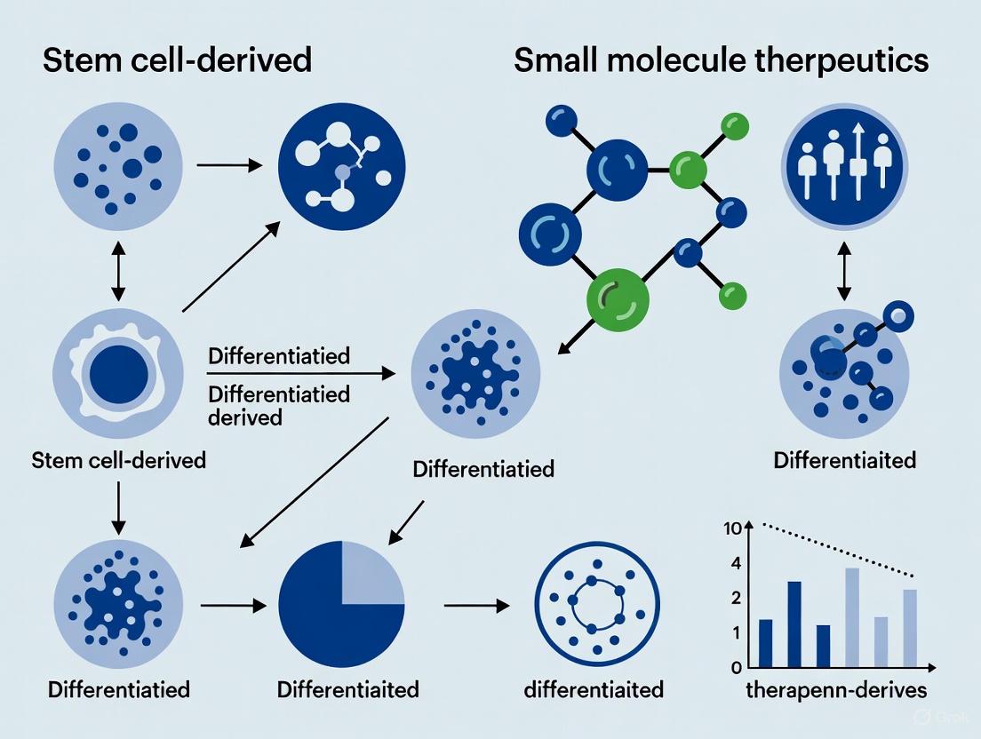
The following diagram illustrates the general workflow for deriving therapeutic cells from the three stem cell sources, highlighting key stages and comparative outputs:
Diagram 1: Stem Cell Therapeutic Derivation Workflow
Key Research Reagent Solutions
The following table outlines essential research reagents and their applications in stem cell therapeutic development:
Table 3: Essential Research Reagents for Stem Cell Therapeutic Development
| Reagent Category | Specific Examples | Research Application | Function |
|---|---|---|---|
| Reprogramming Factors | OCT4, SOX2, KLF4, c-MYC (Yamanaka factors) | hiPSC generation | Reprogram somatic cells to pluripotent state [5] |
| Pluripotency Media | mTeSR | hESC/hiPSC maintenance | Maintain pluripotent state in culture [8] |
| Differentiation Inducers | CHIR99021 (Wnt activator), HGF, Activin A | Directed differentiation | Guide lineage-specific differentiation [8] |
| Cell Separation | CD73, CD90, CD105 antibodies | MSC isolation and characterization | Isolate and characterize MSCs via surface markers [3] |
| Characterization Tools | ALBUMIN, AFP, HNF4A antibodies | Hepatocyte-like cell validation | Assess hepatic differentiation efficiency [8] |
Applications in Drug Development and Disease Modeling
Disease Modeling Applications
Stem cell-derived therapeutics have revolutionized disease modeling approaches, each offering distinct advantages:
hiPSC-based Disease Models: hiPSCs provide exceptional platforms for modeling human diseases, particularly genetic disorders. Patient-specific iPSCs enable the creation of in vitro models that accurately replicate clinical characteristics of conditions like long QT syndrome, hypertrophic cardiomyopathy, Parkinson's disease, and Alzheimer's disease [2] [6]. For example, iPSC-derived cardiomyocytes from long QT syndrome patients revealed that mutations in KCNQ1 cause potassium channel dysfunction, and these models have been used to identify potential therapeutic compounds like ML277 that restore ion channel function [6].
hESC-based Models: While ethically more complicated to obtain, hESCs provide a gold standard for pluripotency and remain valuable for establishing baseline differentiation protocols and understanding early human development [4]. Their use has been instrumental in developing initial protocols for differentiating functional cell types like dopaminergic neurons for Parkinson's disease and pancreatic beta cells for diabetes.
Drug Screening and Toxicity Testing
All three stem cell types contribute significantly to pharmaceutical development:
hMSC-based Screening: MSCs and their secretome are used to screen compounds for immunomodulatory and anti-inflammatory properties. Their response to potential drugs can predict efficacy for autoimmune conditions, graft-versus-host disease, and inflammatory disorders [3].
hiPSC/hESC-based Platforms: Pluripotent stem cells enable the generation of human-specific tissue models for more predictive toxicology and efficacy testing. iPSC-derived hepatocyte-like cells are used for metabolism and toxicity studies, while iPSC-derived cardiomyocytes are employed for cardiotoxicity screening [8] [6]. The availability of patient-specific iPSCs also enables screening of personalized treatment responses, moving toward precision medicine approaches in pharmaceutical development.
Challenges and Future Directions
Current Limitations and Safety Considerations
Each stem cell type presents distinct challenges for therapeutic application:
Tumorigenicity Risk: Pluripotent stem cells (both hESCs and hiPSCs) carry a risk of teratoma formation if undifferentiated cells remain in the final product [2] [10]. hiPSCs face additional concerns regarding genetic instability during reprogramming and prolonged culture. A 2024 review noted that Yamanaka himself has dedicated two decades of research to overcoming the issues of tumorigenicity, immunogenicity, and heterogeneity in iPSCs [10].
Immunogenicity: While hiPSCs were initially hoped to enable autologous transplantation without immune rejection, evidence suggests that even autologous iPSCs may elicit immune responses due to epigenetic abnormalities or reprogramming factors [2]. Allogeneic MSCs, though immunoprivileged, may still trigger immune responses in certain contexts.
Heterogeneity: Significant variability exists between different stem cell lines, influenced by donor age, tissue source, isolation methods, and culture conditions [7] [3]. This heterogeneity poses challenges for standardizing therapies and predicting clinical outcomes.
Emerging Trends and Technical Advancements
Several promising developments are addressing current limitations:
Manufacturing Scalability: Bioreactor systems such as stirred-tank reactors and hollow-fiber membranes are increasingly replacing traditional flask-based cultures to enable large-scale production of clinical-grade stem cells and their derivatives [1]. Combined purification approaches like tangential flow filtration with size exclusion chromatography are improving exosome isolation efficiency and scalability [1].
Gene Editing Integration: CRISPR-Cas9 gene editing is being combined with hiPSC technology to correct disease-causing mutations before cellular transplantation [2] [6]. This approach offers potential for treating genetic disorders while maintaining patient-specific genetic background.
Enhanced Differentiation Protocols: Continued refinement of differentiation protocols is improving the maturity and functionality of stem cell-derived tissues. Research demonstrates that growth factor-based approaches can yield more mature hepatocyte-like cells compared to small molecule methods [8], highlighting the importance of protocol optimization for specific applications.
As the field advances, the complementary strengths of hMSCs, hESCs, and hiPSCs will likely be leveraged for different therapeutic niches, with ongoing research addressing safety concerns and manufacturing challenges to fully realize the potential of stem cell-derived therapeutics.
Stem cells represent the foundational cornerstone of regenerative medicine and biomedical research, primarily due to two defining characteristics: self-renewal and differentiation capacity. Self-renewal refers to the ability of stem cells to undergo numerous cycles of cell division while maintaining their undifferentiated state. This process is crucial for preserving a stable pool of stem cells throughout an organism's life and can occur through either symmetric division (producing two identical stem cells) or asymmetric division (yielding one stem cell and one differentiated daughter cell) [11]. The second key property, differentiation capacity, or potency, describes the potential of a stem cell to develop into specialized cell types. The spectrum of potency ranges from totipotent cells, capable of forming an entire organism, to pluripotent cells (e.g., embryonic stem cells, ESCs; induced pluripotent stem cells, iPSCs), which can generate all three germ layers, and down to multipotent adult stem cells (e.g., hematopoietic stem cells, HSCs; mesenchymal stem cells, MSCs), which are limited to a narrower range of cell types within a specific lineage [11].
The strategic application of these properties is driving innovation across therapeutic domains, from generating human induced pluripotent stem cell-derived hepatocyte-like cells (iPSC-HLCs) for disease modeling to employing stem cell therapy for autoimmune conditions [8] [12]. Concurrently, small molecule drugs—organic compounds with low molecular weight (<900-1000 Da)—represent a powerful and versatile therapeutic modality. Their small size enables them to easily penetrate cell membranes, reach intracellular targets, and be administered orally, offering significant advantages in drug development [13] [14] [15]. This guide provides an objective, data-driven comparison of these two influential approaches in modern biomedical research.
Comparative Analysis: Stem Cell-Derived Therapeutics vs. Small Molecule Drugs
A direct comparison of stem cell-derived therapeutics and small molecule drugs reveals distinct mechanistic profiles, advantages, and challenges, as summarized in the table below.
Table 1: Comparative Profile: Stem Cell-Derived Therapeutics vs. Small Molecule Drugs
| Feature | Stem Cell-Derived Therapeutics | Small Molecule Drugs |
|---|---|---|
| Mechanism of Action | Cell replacement; secretion of paracrine factors (e.g., growth factors, exosomes) for immune modulation and tissue repair [12]. | Modulation of enzymatic activity, receptor signaling, or ion channel function (e.g., enzyme inhibitors, receptor agonists/antagonists) [13]. |
| Key Advantages | Potential for durable tissue repair and restoration of function; address complex, multifactorial disease pathways [12]. | Oral bioavailability; predictable in vivo behavior; established, scalable manufacturing; lower cost per treated patient [16] [13] [14]. |
| Primary Challenges | High cost and complexity of personalized therapy; risk of teratoma formation; potential for immune rejection; lack of long-term safety data [12] [17]. | Inability to reverse established tissue damage; potential for off-target effects and cumulative toxicity; complex manufacturing and regulatory hurdles [12] [15]. |
| Typical Administration Route | Intravenous infusion or local injection [12]. | Primarily oral (tablets, capsules); also injectable, topical [16] [14]. |
| Therapeutic Scope | Aim to treat the underlying pathophysiology via regeneration (e.g., autoimmune diseases, degenerative disorders) [12]. | Primarily manage disease symptoms and progression (e.g., chronic diseases like hypertension, cancer, infectious diseases) [16] [13]. |
Experimental Comparison in Hepatocyte Generation
A pivotal 2025 study directly compared the efficacy of two primary protocols—growth factors (GF) and small molecules (SM)—for generating human iPSC-derived hepatocyte-like cells (iPSC-HLCs) across fifteen different iPSC lines [8] [18]. The experimental findings highlight how the choice of differentiation agent profoundly influences the functional outcome of the resulting cells.
Experimental Protocols and Methodologies
The study employed two distinct, well-defined differentiation protocols:
- Growth Factor (GF) Protocol: This method mimics embryonic liver development by sequentially using specific protein growth factors. Beyond the initial definitive endoderm stage, the protocol utilizes a simplified approach requiring a single key growth factor: Hepatocyte Growth Factor (HGF) [8]. This factor is crucial for promoting hepatoblast maturation into hepatocyte-like cells.
- Small Molecule (SM) Protocol: This approach uses specific, low-molecular-weight chemical compounds to direct differentiation. The protocol involves a more complex cocktail of small molecules, including CHIR99021 (a GSK-3 inhibitor that activates Wnt signaling) and Dihexa (a hepatocyte growth factor mimetic), among others [8].
Researchers conducted a comprehensive analysis of the resulting HLCs, including morphological assessment, quantification of gene and protein expression (e.g., AFP, HNF4A, ALBUMIN), proteomic studies, and functional metabolic assays [8].
Key Experimental Findings and Data
The comparative analysis revealed striking differences in the phenotype and functionality of the HLCs generated by the two protocols.
Table 2: Experimental Outcomes of GF-derived vs. SM-derived Hepatocyte-like Cells
| Parameter | GF-Derived HLCs | SM-Derived HLCs |
|---|---|---|
| Morphology | Mature hepatocyte features: raised, polygonal shape with well-defined borders; granular cytoplasm containing lipid droplets/vacuoles; large central or multiple spherical nuclei [8] [18]. | Dedifferentiated, proliferative phenotype resembling liver tumor-derived cell lines [8] [18]. |
| Gene & Protein Expression | Significantly elevated levels of mature hepatocyte markers, including AFP, HNF4A, and ALBUMIN [8] [18]. | Expression profile less aligned with mature hepatocytes. |
| Proteomic & Metabolic Profile | More closely aligned with a mature, functional phenotype, making them better suited for studies of metabolism, biotransformation, and viral infection [8]. | Profile indicative of a less mature, more proliferative state. |
| Interpretation | The GF protocol produces cells with characteristics more synonymous with healthy primary human hepatocytes (PHHs) [8]. | The SM protocol yields cells that are more akin to immortalized hepatic tumor cell lines, which may have altered metabolic pathways [8]. |
Supporting Data from Clinical Trial Trends
The clinical translation of stem cell therapies is an active area of research, particularly for complex conditions like autoimmune diseases. A 2025 systematic review of global clinical trials (2006–2025) provides insight into current trends and reinforces the contextual challenges noted in Table 1 [12].
The analysis of 244 interventional trials revealed that the majority (83.6%) are in early to mid-stage development (Phase I-II), indicating a field that is still maturing toward widespread clinical application [12]. The most frequently studied diseases were Crohn's disease (n=85), systemic lupus erythematosus (n=36), and scleroderma (n=32) [12]. The primary therapeutic strategies employed were immune modulation, tissue repair via growth factors, and anti-infection/anti-proliferative effects, with academic institutions funding nearly half (49.2%) of all trials [12]. This data underscores both the significant promise of stem cell therapies and the ongoing nature of clinical validation.
Key Biological Pathways and Workflows
Stem Cell Fate Regulation
The fundamental processes of self-renewal and differentiation are tightly regulated by intrinsic signaling pathways and extrinsic metabolic cues. The following diagram illustrates the key regulators that maintain the delicate balance between these two states in pluripotent stem cells.
Small Molecule Drug Discovery Workflow
The development of a new small molecule drug is a lengthy, multi-stage process designed to identify and optimize a compound with a specific therapeutic effect. The workflow below outlines the major stages from initial discovery to market approval.
The Scientist's Toolkit: Essential Research Reagents
Successful research in stem cell biology and small molecule discovery relies on a suite of specialized reagents and tools. The following table details key solutions used in the featured experiments and broader field.
Table 3: Essential Research Reagents and Solutions
| Reagent / Solution | Function / Application |
|---|---|
| Hepatocyte Growth Factor (HGF) | A key protein growth factor used in the GF protocol to drive the maturation of hepatoblasts into hepatocyte-like cells [8]. |
| CHIR99021 | A small molecule inhibitor of GSK-3 that activates the Wnt signaling pathway, used in the SM protocol to direct stem cell fate [8]. |
| STEMdiff Definitive Endoderm Kit | A commercially available kit used to efficiently differentiate pluripotent stem cells into the definitive endoderm, the first germ layer required for liver development [8]. |
| Dimethyl Sulfoxide (DMSO) | A universal solvent for dissolving and storing many small molecule compounds used in research [8]. |
| Y-27632 (ROCK Inhibitor) | A small molecule that enhances the survival of stem cells after passaging or thawing, improving plating efficiency [8]. |
| Combinatorial Chemistry Libraries | Vast collections of structurally diverse small molecules used in High-Throughput Screening (HTS) to identify initial "hit" compounds with activity against a therapeutic target [13] [15]. |
| AI-Powered Discovery Platforms | Software and algorithms (e.g., deep generative models) used for de novo molecular design, virtual screening, and predicting the 3D structures of small molecules to accelerate lead identification [16] [13]. |
The comparative analysis of stem cell-derived therapeutics and small molecule drugs reveals two powerful, yet distinct, paradigms in modern biomedical research. The direct experimental comparison of differentiation protocols shows that growth factor-derived HLCs are superior for modeling mature hepatocyte function in applications like metabolism and toxicology studies [8]. In contrast, while small molecule-derived HLCs may offer a simpler logistical approach, they exhibit a less mature, more proliferative phenotype [8]. This underscores a critical principle: the choice between biological factors (like GFs) and chemical compounds (like SMs) is not merely one of convenience but can fundamentally determine the functional outcome of the resulting cells.
Both fields are being revolutionized by technological advancements. AI-driven drug discovery is accelerating the identification of novel small molecules [16] [14], while a growing understanding of mitochondrial metabolism and signaling pathways like Wnt is providing new knobs to fine-tune stem cell pluripotency and differentiation [11] [17]. The future of therapeutic development lies not in pitting these approaches against each other, but in strategically leveraging their complementary strengths—using small molecules to precisely control stem cell fate in vitro for subsequent cell-based therapies, or to create more predictive disease models for small molecule screening. This synergistic potential promises to advance both fields toward more effective and personalized medical treatments.
Small molecule drugs are low molecular weight organic compounds, typically under 900 daltons, that serve as foundational therapeutic agents in modern medicine [19]. These synthetically produced or naturally derived compounds account for a substantial proportion of approved pharmaceuticals, representing 69% of all new FDA-approved drugs in 2023 [19]. Their defining characteristic is the ability to precisely interact with specific biological targets—such as enzymes, receptors, or proteins—to modulate biochemical pathways and correct disease-associated dysfunctions [19].
The comparative therapeutic value of small molecules becomes particularly evident when evaluated against stem cell-derived therapies, especially in the context of diabetes treatment. While stem cell approaches aim to replace damaged or lost insulin-producing beta cells, small molecules offer a complementary strategy by inducing the differentiation of a patient's own stem cells into functional beta-like cells or by directly modulating metabolic pathways [20]. This review objectively examines the characteristics, synthesis, and therapeutic applications of small molecule drugs, with particular emphasis on their emerging role in regenerative medicine and their comparative efficacy against stem cell-based approaches.
Defining Characteristics of Small Molecule Drugs
Small molecule drugs possess distinct physicochemical and biological properties that determine their therapeutic application and manufacturing considerations. These characteristics directly influence their behavior in biological systems and their advantages over other therapeutic modalities.
Table 1: Key Characteristics of Small Molecule Drugs
| Characteristic | Description | Therapeutic Implications |
|---|---|---|
| Size & Molecular Weight | Typically 0.1-1 kDa (under 900 daltons) [19] | Enables penetration of cell membranes to reach intracellular targets [13] |
| Administration Route | Primarily oral (tablets, capsules); also injectable or inhalable [19] | Promotes patient compliance for chronic conditions; convenient self-administration [19] |
| Synthesis Method | Chemical synthesis from commercially available building blocks [21] | Scalable, cost-effective manufacturing; reproducible production [19] [13] |
| Stability | Generally stable at room temperature [19] | Simplified storage, distribution, and packaging logistics [19] |
| Immunogenicity | Low risk of triggering adverse immune responses [19] | Favorable safety profile with reduced immunogenic complications |
| Metabolism & Excretion | Hepatic metabolism with renal elimination [19] | Predictable pharmacokinetic profiles |
The structural simplicity and customizable nature of small molecules allow researchers to fine-tune their atomic composition to elicit specific therapeutic responses while minimizing unwanted side effects [13]. This flexibility to explore "chemical space" provides small molecules with a marked advantage over other therapeutic modalities, as their properties can be systematically optimized for particular purposes [13].
Mechanisms of Action: How Small Molecules Exert Therapeutic Effects
Small molecules employ several well-characterized mechanisms to modulate biological systems, with most targeting specific proteins involved in disease processes. Their low molecular weight enables them to easily penetrate cell membranes, accessing intracellular targets that may be inaccessible to larger biologic therapeutics [19] [13].
Primary Mechanisms
Enzyme Inhibition: Small molecules can block the activity of enzymes catalyzing biochemical reactions, interfering with disease processes. Statins represent this class, inhibiting HMG-CoA reductase involved in cholesterol production [13].
Receptor Modulation: These compounds interact with cell surface proteins as either agonists (activators) or antagonists (blockers). Albuterol, an agonist for beta-2 adrenergic receptors, opens airways in asthma treatment [13].
Ion Channel Regulation: Small molecules modulate proteins controlling ion flow across cell membranes, treating conditions like epilepsy by regulating neuronal excitability [13].
The interaction typically occurs through the "lock and key" mechanism, where small molecules bind to well-defined active sites on target proteins [13]. The strength and specificity of this interaction determines therapeutic efficacy, with more specific binding reducing the likelihood of off-target effects.
Figure 1: Small Molecule Drug Pathway and Mechanisms of Action
Chemical Synthesis of Small Molecules
The synthesis of small molecule drugs has traditionally relied on procedures highly customized for each target, presenting significant challenges for automated production [21]. Recent advances, however, have moved the field toward more standardized approaches that enable efficient, scalable synthesis of diverse molecular structures.
Building Block-Based Synthesis Strategy
A breakthrough in small molecule synthesis involves adopting a building block-based strategy inspired by nature's approach to constructing complex molecules [21]. This method utilizes bifunctional N-methyliminodiacetic acid (MIDA) boronates as fundamental building blocks, which can be iteratively assembled through sequential coupling and deprotection cycles [21].
The MIDA ligand plays a crucial role by attenuating boronic acid reactivity, preventing undesired oligomerization during synthesis [21]. This approach allows all required functional groups, oxidation states, and stereochemical elements to be pre-installed into building blocks, then faithfully translated into final products through mild, stereospecific coupling chemistry [21].
Automated Synthesis Platform
The development of a common synthesis strategy and purification protocol has enabled the creation of an automated synthesizer that iteratively assembles MIDA boronate building blocks [21]. This device comprises three modules that sequentially execute deprotection, coupling, and purification steps for each synthesis cycle [21].
A key innovation is the catch-and-release purification protocol, which exploits the unusual binary affinity of MIDA boronates for silica gel [21]. These compounds remain stationary when eluted with MeOH:Et₂O but move rapidly with THF, enabling a universal purification method applicable to all intermediates containing the MIDA boronate group [21].
Small Molecules in Stem Cell Research and Regenerative Medicine
Small molecules play an increasingly important role in stem cell biology, offering significant advantages for controlling stem cell fate, including maintenance, differentiation, and reprogramming. In the context of diabetes research, small molecules facilitate the generation of insulin-producing beta cells from various stem cell sources, presenting a promising therapeutic approach [20].
Advantages of Small Molecules in Stem Cell Research
Table 2: Benefits of Small Molecules in Stem Cell Applications
| Advantage | Explanation | Research Implication |
|---|---|---|
| Cost & Time Efficiency | Effects manifest within hours; reduce reprogramming time from weeks to days [22] | Accelerated experimental timelines; more efficient differentiation protocols |
| Synthetic Production | Chemically produced with high purity and low batch variation [22] | Consistent, reproducible results across experiments and laboratories |
| Cell Permeability | Ability to cross cell membranes and target intracellular pathways [22] | Access to intracellular targets; both in vitro and in vivo applications |
| Temporal Control | Rapid, reversible, dose-dependent effects [23] | Precise timing of interventions; fine-tuning of differentiation processes |
| Safety Profile | No genetic material integration; reduced tumorigenic risk [22] | Better suitability for clinical translation compared to viral vectors |
Experimental Comparison: Small Molecules vs. Growth Factors in Hepatocyte Differentiation
A comprehensive comparative analysis evaluated the efficacy of small molecule versus growth factor protocols for generating human induced pluripotent stem cell-derived hepatocyte-like cells (iPSC-HLCs) across fifteen different iPSC lines [8]. This study provides critical experimental data for objectively comparing these differentiation approaches.
Table 3: Experimental Comparison of Differentiation Protocols
| Parameter | Small Molecule Protocol | Growth Factor Protocol |
|---|---|---|
| Morphological Features | Dedifferentiated, proliferative phenotype resembling liver tumor-derived cell lines [8] | Mature hepatocyte morphology: polygonal shape with defined borders, granular cytoplasm with lipid droplets, multiple spherical nuclei [8] |
| Gene & Protein Expression | Reduced expression of mature hepatocyte markers [8] | Significantly elevated hepatocyte gene and protein expression (AFP, HNF4A, ALBUMIN) [8] |
| Proteomic & Metabolic Features | Altered metabolic pathways supporting proliferation [8] | Better alignment with mature hepatocyte phenotype; suitable for metabolism and biotransformation studies [8] |
| Protocol Complexity | Requires multiple components throughout differentiation process [8] | Simplified approach with single growth factor (HGF) beyond endoderm stage [8] |
| Recommended Applications | Studies requiring proliferative cell populations | Disease modeling, metabolic studies, viral infection research [8] |
Experimental Protocol: Differentiation of iPSCs to Hepatocyte-like Cells
Objective: To generate functional hepatocyte-like cells from human induced pluripotent stem cells using either small molecule or growth factor-based differentiation protocols.
Materials and Reagents:
- STEMdiff Definitive Endoderm Kit
- Hepatocyte Growth Factor (HGF)
- CHIR99021 (GSK-3 inhibitor)
- A-83-01 (TGF-β receptor inhibitor)
- Dimethyl sulfoxide (DMSO)
- RPMI/B27 medium
- KnockOut Serum Replacement
Methodology - Small Molecule Protocol [8]:
- Definitive Endoderm Induction: Use STEMdiff Definitive Endoderm Kit per manufacturer's instructions (3 days)
- Hepatoblast Specification: Culture cells in RPMI/B27 medium supplemented with 3μM CHIR99021, 0.5μM A-83-01, and additional small molecules per established protocols (5 days)
- Hepatocyte Maturation: Maintain cells in hepatocyte maturation medium containing continued small molecule supplementation (10-13 days)
Methodology - Growth Factor Protocol [8]:
- Definitive Endoderm Induction: Identical to small molecule protocol (3 days)
- Hepatoblast Specification: Transition to RPMI/B27 medium with 10ng/mL HGF (5 days)
- Hepatocyte Maturation: Continue culture with HGF supplementation (10-13 days)
Assessment Parameters:
- Morphological analysis by phase-contrast microscopy
- Relative quantification of hepatocyte-specific gene expression (AFP, HNF4A, ALB)
- Immunocytochemistry for protein expression (ALBUMIN, AAT)
- Functional assays: urea production, albumin secretion, glycogen storage
Figure 2: Experimental Workflow for iPSC Differentiation to Hepatocyte-like Cells
The Scientist's Toolkit: Essential Research Reagents
Successful investigation of small molecule therapeutics requires specific reagents and tools. The following table details essential materials for research in this field, particularly for studies comparing differentiation efficacy.
Table 4: Essential Research Reagents for Small Molecule Studies
| Reagent/Category | Specific Examples | Function/Application |
|---|---|---|
| GSK-3 Inhibitors | CHIR99021 [8] [23] | Promotes self-renewal and supports reprogramming; used in hepatocyte differentiation |
| TGF-β Inhibitors | A-83-01 [8] | Enhances reprogramming efficiency; facilitates definitive endoderm formation |
| MEK Inhibitors | PD0325901 [23] | Blocks differentiation pathways in stem cells; supports pluripotent state |
| HDAC Inhibitors | Valproic acid, Sodium butyrate [23] | Epigenetic modulators that promote reprogramming efficiency |
| Boron-Based Building Blocks | MIDA boronates [21] | Enables iterative synthesis of complex small molecules for research |
| Stem Cell Culture Supplements | B27, KnockOut Serum Replacement [8] | Supports maintenance and differentiation of pluripotent stem cells |
Small molecule drugs remain indispensable therapeutic tools characterized by precise target engagement, favorable pharmacokinetic properties, and manufacturing scalability. Their role continues to expand into regenerative medicine, where they facilitate stem cell differentiation for beta cell and hepatocyte generation [20] [8].
The comparative analysis of differentiation protocols reveals a nuanced picture: while small molecule approaches offer practical advantages in cost and convenience, growth factor methods may produce more mature hepatocyte-like cells for certain applications [8]. Similarly, in diabetes research, small molecules show promise for generating insulin-producing cells from mesenchymal stem cells, though current protocols typically yield "beta cell-like cells" rather than fully mature beta cells [20].
These findings suggest that the future of regenerative medicine may lie in integrated approaches that leverage the strengths of both small molecules and biological therapies. As small molecule discovery advances through artificial intelligence and automated synthesis platforms [19] [21], and as stem cell technologies mature, combined strategies may offer optimal therapeutic outcomes by targeting multiple disease pathways simultaneously while enabling the regeneration of functional tissues.
The pursuit of innovative therapeutics has given rise to two distinct yet complementary strategies: cell replacement therapy and targeted protein modulation. Cell replacement, often utilizing stem cell-derived products, aims to restore tissue function by introducing entirely new cellular units into damaged organs [24] [25]. In contrast, targeted protein modulation employs small molecule drugs to precisely alter the function of specific proteins within existing cells, thereby modifying disease pathways [23] [26]. Understanding their fundamental mechanisms is crucial for researchers and drug development professionals selecting the optimal therapeutic approach for specific disease contexts.
Cell-based therapies function as integrated living systems capable of sensing, decision-making, and executing complex responses within their microenvironment [24]. They represent not merely drugs but sophisticated biological devices that can integrate into host tissues. Small molecule therapeutics, typically under 900 daltons in molecular weight, offer a different set of advantages: oral bioavailability, precise temporal control, and the ability to target intracellular proteins [22] [16]. This guide provides a structured comparison of their mechanisms, experimental methodologies, and applications to inform therapeutic development decisions.
Fundamental Mechanisms of Action
Cell Replacement Therapies: Restorative Living Medicine
Cell replacement therapies operate through macro-level biological mechanisms focused on restoring lost function:
- Functional Tissue Integration: Administered cells engraft into damaged tissues and directly replace lost or dysfunctional cell populations. For example, in Type 1 diabetes, lab-made pancreatic beta cells can engraft and restore glucose-responsive insulin secretion, potentially offering a functional cure [25].
- Trophic Factor Secretion: Transplanted cells secrete growth factors, cytokines, and extracellular vesicles that modulate the local microenvironment. Mesenchymal stem cells (MSCs) release factors like TGF-β, PGE2, and regulatory miRNAs that suppress pathological immune responses while promoting tissue repair [12].
- Dynamic Regulation: Living cells provide autonomous regulation that small molecules cannot achieve. Engineered therapeutic cells can sense metabolic states and respond with precisely calibrated therapeutic output, essentially creating an endogenous drug factory that self-regulates based on physiological needs [24].
Targeted Protein Modulation: Precision Molecular Intervention
Small molecules function through precise molecular interactions with specific protein targets:
- Receptor Modulation: Small molecules can act as agonists, antagonists, or allosteric modulators of cell surface and intracellular receptors. For example, JNJ-799760 and JNJ-67869386 bind to the acidic pocket of acid-sensing ion channel 1 (ASIC1), stabilizing its closed state and preventing channel activation in response to protons [27].
- Enzyme Inhibition: Many small molecules function as enzyme inhibitors, blocking catalytic activity and downstream signaling. Kinase inhibitors represent a major class that targets dysregulated signaling pathways in cancer and inflammatory diseases [28].
- Epigenetic Modulation: Compounds like valproic acid and sodium butyrate inhibit histone deacetylases (HDACs), altering chromatin structure and gene expression patterns to influence cell fate decisions [23].
Table 1: Core Mechanistic Comparison Between Therapeutic Modalities
| Mechanistic Aspect | Cell Replacement Therapy | Targeted Protein Modulation |
|---|---|---|
| Primary Action | Restoration of functional cellular units | Modulation of specific protein function |
| Therapeutic Output | Dynamic, self-regulating | Fixed dose-response relationship |
| Target Engagement | Multiple simultaneous mechanisms | Single target specificity |
| Duration of Action | Long-term (months to years) | Transient (hours to days) |
| Spatial Control | Tissue-specific homing and integration | Dependent on pharmacokinetics and distribution |
| Manufacturing | Complex biological process | Synthetic chemical process |
Experimental Approaches and Methodologies
Establishing Cell Replacement Models
Cell therapy development requires specialized protocols focusing on cell sourcing, characterization, and functional validation:
- Cell Sourcing and Differentiation: Studies typically utilize patient-derived or allogeneic stem cells differentiated toward specific lineages. For neuronal disorders, protocols employ small molecule cocktails (including SB 431542, LDN 193189, XAV 939, PD 0325901, SU 5402, and DAPT) to differentiate induced pluripotent stem cells into functional cortical neurons within 16 days [22].
- In Vivo Transplantation Models: Preclinical testing involves transplanting therapeutic cells into disease-relevant animal models. In epilepsy trials, lab-made neurons engineered to suppress neural hyperexcitability were transplanted into patients' brains, demonstrating seizure reduction from daily to approximately weekly events [25].
- Engraftment and Functional Assessment: Researchers track cell survival, integration, and functional impact using imaging, electrophysiology, and behavioral tests. For spinal cord injury, studies assess functional recovery through standardized scales and imaging to confirm tissue repair [29].
Screening for Small Molecule Modulators
Small molecule therapeutic development employs distinct methodological approaches:
- High-Throughput Screening: Compound libraries are screened against purified target proteins or cellular assays. AI-driven platforms like those from Exscientia and Insilico Medicine use generative chemistry and machine learning to design novel compounds satisfying precise target product profiles [28].
- Structure-Activity Relationship (SAR) Studies: Systematic chemical modification identifies structural features critical for target engagement and efficacy. For ASIC1 inhibitors, crystallographic studies revealed binding at the acidic pocket, enabling structure-based optimization [27].
- Mechanistic Validation: Compounds advancing through screening undergo rigorous target validation. Techniques include crystallography (to visualize binding sites), mutagenesis (to confirm binding residues), and functional assays (to characterize effects on signaling pathways) [27].
Table 2: Key Research Reagents and Their Applications
| Reagent/Category | Function | Example Applications |
|---|---|---|
| CHIR99021 | GSK-3 inhibitor | Maintains pluripotency; enhances reprogramming [23] |
| PD0325901 | MEK inhibitor | Supports stem cell self-renewal; facilitates reprogramming [23] |
| Valproic Acid (VPA) | HDAC inhibitor | Promotes reprogramming efficiency; enables factor-reduced reprogramming [23] |
| Mesenchymal Stem Cells (MSCs) | Immunomodulation & tissue repair | Autoimmune diseases (Crohn's, SLE); secretes TGF-β, PGE2 [12] |
| Hematopoietic Stem Cells (HSCs) | Immune system reconstitution | Resets immune tolerance in autoimmune disorders [12] |
| JNJ-67869386 | ASIC1 allosteric inhibitor | Stabilizes closed channel state; impedes desensitization [27] |
Comparative Efficacy Across Disease Contexts
Neurological Disorders
- Cell Replacement Approaches: Clinical trials for spinal cord injury demonstrate that bone-marrow-derived and neural stem cells can promote functional recovery. In amyotrophic lateral sclerosis (ALS), mesenchymal and neural stem cells show potential to modify disease progression [29].
- Small Molecule Approaches: Neuroregenerative small molecules target innate repair pathways. Compounds like 16,16-dimethyl Prostaglandin E2 (dmPGE2) and neurotrophic factors can enhance endogenous stem cell activity and synaptic plasticity [26].
Autoimmune and Metabolic Diseases
- Cell Replacement Approaches: In Type 1 diabetes, Vertex Pharmaceuticals has demonstrated that transfusions of lab-made beta cells can enable insulin independence in some patients [25]. For autoimmune conditions like Crohn's disease and lupus, MSC-based therapies modulate immune responses through secretion of anti-inflammatory factors [12].
- Small Molecule Approaches: JAK-STAT pathway inhibitors and other immunomodulatory small molecules provide symptomatic control in autoimmune conditions but typically do not restore damaged tissue or establish immune tolerance [12].
Development Considerations and Challenges
Manufacturing and Scalability
- Cell Therapies: Face complex manufacturing challenges including cell expansion, quality control, and storage. Personalized approaches using autologous cells are particularly resource-intensive, with costs often exceeding traditional biologic therapies [12].
- Small Molecules: Benefit from established synthetic chemistry processes that are more easily scaled. AI-driven discovery platforms can accelerate this process, with companies like Exscientia reporting design cycles approximately 70% faster than industry standards [28].
Clinical Translation
- Cell Therapies: Require specialized delivery protocols and face challenges with engraftment, immune rejection, and long-term safety monitoring. While short-term safety data are generally favorable, long-term outcomes require further study [12].
- Small Molecules: Face traditional drug development hurdles including pharmacokinetic optimization, toxicity profiling, and target selectivity. However, their development pathway is more established with clearer regulatory precedents [16].
Visualizing Key Signaling Pathways and Workflows
Small Molecule Modulation of Ion Channel Gating
The following diagram illustrates the molecular mechanism by which small molecules allosterically modulate acid-sensing ion channel 1 (ASIC1), stabilizing its closed state and preventing proton-induced activation [27]:
Cell Therapy Development Workflow
This diagram outlines the key stages in developing stem cell-based therapies, from cell sourcing through functional validation:
Cell replacement and targeted protein modulation represent complementary rather than competing therapeutic strategies, each with distinct advantages for specific clinical contexts. Cell therapies excel in diseases characterized by irreversible cellular loss or requiring dynamic physiological regulation, offering potential functional cures for conditions like Type 1 diabetes and certain neurological disorders [24] [25]. Small molecule approaches provide superior temporal control, easier administration, and established manufacturing pathways, making them ideal for conditions where specific pathway modulation can achieve therapeutic effects without cellular replacement [22] [16].
Future therapeutic development will likely see increased convergence of these approaches, with small molecules potentially enhancing cell therapy outcomes by improving engraftment or modulating host immunity. Additionally, AI-driven discovery platforms are accelerating both fields, from designing novel small molecules to optimizing cell differentiation protocols [28]. The optimal choice between these mechanisms depends fundamentally on disease pathology, with cell replacement addressing structural deficits and small molecules targeting specific dysfunctional pathways within an otherwise intact cellular framework.
In the evolving landscape of therapeutic development, two distinct approaches have emerged as powerful modalities: stem cell-derived therapeutics and small molecule drugs. These approaches represent fundamentally different strategies for treating diseases, each with characteristic advantages and limitations. Stem cell-derived therapies, including growth factor-driven protocols, leverage the body's innate regenerative mechanisms to repair and replace damaged tissues, offering unprecedented potential for conditions with permanent tissue loss. In contrast, small molecule therapeutics utilize chemically synthesized compounds that target specific molecular pathways, providing superior bioavailability and established manufacturing pathways that often translate to better cost-effectiveness.
This comprehensive comparison guide examines the core strengths of each therapeutic class through the lens of regenerative potential, oral bioavailability, and cost-effectiveness—three critical considerations for researchers, pharmaceutical developers, and healthcare systems. We present experimental data, clinical outcomes, and market analyses to provide an evidence-based framework for therapeutic development decisions, highlighting that the optimal choice often depends on the specific clinical context, target tissues, and healthcare economics.
Regenerative Potential: Cellular Complexity vs. Targeted Simplicity
Experimental Evidence in Hepatic Differentiation
A direct comparative analysis of growth factor (GF) and small molecule (SM) protocols for generating human induced pluripotent stem cell-derived hepatocyte-like cells (iPSC-HLCs) reveals significant differences in morphological and functional outcomes. Researchers conducted this investigation across fifteen different human iPSC lines to ensure reproducibility and robust conclusions [8] [30].
Table 1: Experimental Outcomes of Hepatocyte-Like Cell Differentiation Protocols
| Parameter | Growth Factor Protocol | Small Molecule Protocol |
|---|---|---|
| Morphological Features | Raised, polygonal shape with well-defined refractile borders; granular cytoplasm with lipid droplets/vacuoles; multiple spherical nuclei or large central nucleus | Dedifferentiated, proliferative phenotype resembling liver tumor-derived cell lines |
| Gene & Protein Expression | Significantly elevated hepatocyte markers (AFP, HNF4A, ALBUMIN) | Reduced expression of mature hepatocyte markers |
| Proteomic & Metabolic Profile | Aligned with mature hepatocyte phenotype | Shifted toward proliferative metabolism (glycolysis, Krebs cycle alterations) |
| Recommended Applications | Studies of metabolism, biotransformation, and viral infection | Scenarios requiring expansion of progenitor populations |
The experimental methodology involved morphological assessment, relative quantification of gene expression, protein expression analysis, and comprehensive proteomic studies. The GF protocol utilized a simplified approach requiring only hepatocyte growth factor (HGF) beyond the endoderm stage, while the SM protocol involved multiple chemical components including CHIR99021 and other regulators [30]. The results demonstrated that GF-derived HLCs exhibited substantially better maturation and functionality, making them more suitable for modeling healthy adult hepatocytes and related metabolic applications [8].
Clinical Evidence in Periodontal Regeneration
In dental medicine, the regenerative potential of different biomaterials provides insightful clinical comparisons. An 18-month randomized clinical trial compared crosslinked hyaluronic acid (HA) with enamel matrix derivative (EMD) for periodontal regeneration in 53 patients with intrabony defects [31].
Table 2: Clinical Periodontal Regeneration Outcomes at 18 Months
| Clinical Parameter | Hyaluronic Acid (HA) Group | Enamel Matrix Derivative (EMD) Group |
|---|---|---|
| Clinical Attachment Level (CAL) Gain ≥4 mm | 48.1% of defect sites (13/27) | Not reported at this threshold |
| CAL Gain 2-3 mm at 6 months | Not reported | 53.8% of defect sites (14/26) |
| Probing Depth Reduction | Significant improvement (p<0.001) | Significant improvement (p<0.001) |
| Radiographic Bone Filling | Significant improvement (p<0.001) | Significant improvement (p<0.001) |
| Key Advantages | Cost-effectiveness, application ease, bioavailability | Early regenerative response |
The study demonstrated that while both materials significantly improved clinical and radiographic parameters, their regenerative profiles differed temporally. EMD promoted faster initial regeneration, while HA showed superior long-term outcomes for deeper defects, highlighting how regenerative potential can vary even within biomaterial approaches [31].
The surgical protocol involved minimally invasive techniques (MIST or M-MIST) with random assignment to test or control groups after defect debridement. Root surfaces in the EMD group were conditioned with 24% EDTA gel before EMD application, while the HA group received hyaluronic acid application. Clinical measurements (probing depth, clinical attachment level, recession, bleeding on probing) and radiographic assessments were conducted at baseline, 6, 12, and 18 months post-surgery [31].
Figure 1: Experimental workflow comparing differentiation protocols for hepatocyte-like cells from human induced pluripotent stem cells, showing divergent morphological and functional outcomes based on differentiation strategy [8] [30].
Oral Bioavailability and Administration Routes
Small Molecule Administration Advantages
Small molecule drugs demonstrate superior flexibility in administration routes, particularly their compatibility with oral delivery systems. The global pharmaceuticals market analysis reveals that conventional drugs (small molecules) continue to dominate with a 54.74% market share, largely due to their established oral administration profiles and predictable pharmacokinetics [32].
Table 3: Small Molecule Drug Administration Formats and Market Performance
| Administration Route/Formulation | Market Share (2024) | Growth Projection | Key Advantages |
|---|---|---|---|
| Oral Solid Dose (Tablets, Capsules) | 72% | Stable growth | Convenience, high patient compliance, affordability, safer administration |
| Parenteral/Injectables | Smaller segment | Fastest growing (2025-2034) | Bypasses absorption processes, avoids GI enzyme degradation and first-pass effect |
| Topical Formulations | Not specified | Not specified | Localized delivery, reduced systemic exposure |
The data indicates that oral solid dosage forms represent the most significant segment due to their convenience, high patient compliance, affordability, and safety profile [16]. This administration advantage translates directly to better treatment adherence and reduced healthcare utilization compared to delivery systems requiring clinical administration.
The molecular properties of small molecules (<900 Da) enable them to efficiently penetrate cell membranes and reach intracellular targets, a significant advantage for many therapeutic applications [16]. Their typically well-defined chemical structures contribute to predictable absorption and metabolism profiles, though this can vary based on specific chemical properties and formulation technologies.
Stem Cell Therapy Administration Limitations
In contrast, stem cell therapies currently face significant administration challenges that limit their delivery options. These regenerative treatments typically require site injections or intravenous infusion, necessitating clinical visits and medical supervision [33] [34]. For orthopedic conditions, stem cell injections target specific joints or damaged tissues, while systemic conditions require intravenous delivery [33].
The administration complexity for stem cell therapies contributes significantly to their cost structure and limits their scalability compared to conventional pharmaceuticals. Additionally, cell-based products face challenges related to stability, storage, and distribution that do not similarly impact small molecule drugs with established stabilization and formulation technologies.
Cost-Effectiveness Analysis: Manufacturing and Market Dynamics
Stem Cell Therapy Cost Structures
Stem cell therapy costs demonstrate extreme variability based on cell type, application, and geographic location. Current pricing data reveals a broad range from $5,000 to $50,000, with specific applications commanding different price points [33].
Table 4: Stem Cell Therapy Cost Analysis by Application and Type
| Therapy Type | Average Cost | Key Cost Factors | Insurance Coverage |
|---|---|---|---|
| Orthopedic Conditions (Knee osteoarthritis, tendonitis) | $5,000 - $8,000 | Lower cell dosage, localized application | Typically not covered |
| Systemic Conditions (Autoimmune, degenerative) | $15,000 - $30,000 | Higher cell counts, intravenous administration, regulatory compliance | Considered experimental, largely out-of-pocket |
| Umbilical Cord Tissue-derived | $15,000 - $45,000 | Cell sourcing, expansion, regulatory compliance | Not typically covered |
| Private Cord Blood Banking | $300 - $2,300 (initial) + annual fees | Collection, processing, storage | Not typically covered |
Cost factors influencing stem cell therapy pricing include the type of stem cells administered, cell quantity, quality control measures, laboratory location, and regulatory compliance requirements [33]. The high costs are further driven by complex manufacturing processes including isolation, expansion, and characterization of cells under Good Manufacturing Practice (GMP) conditions [33] [34].
Regulatory requirements significantly impact these costs, as agencies like the FDA mandate rigorous testing for new treatments. Maintaining GMP-compliant facilities for MSC production and conducting necessary clinical trials with extensive participant involvement add substantial expenses that are ultimately reflected in treatment pricing [33].
Small Molecule Drug Economics
The small molecule drug discovery market is projected to grow from USD 61.04 billion in 2025 to USD 110.23 billion by 2032, exhibiting a compound annual growth rate (CAGR) of 8.8% [35]. This robust market position is maintained despite rising competition from biologics, largely due to the cost advantages of small molecule therapeutics.
Small molecules offer significant economic benefits including scalable chemical synthesis, established manufacturing pathways, and comparatively lower cost per treated patient versus biologics and cell therapies [16]. The small molecules market encompasses patented/innovator brands, generics, OTC medicines, and contract development and manufacturing organizations (CDMOs), creating a diverse and competitive ecosystem [16].
Once patent protection expires, small molecule drugs face robust generic competition that can reduce costs by up to 80% compared to branded counterparts, dramatically improving accessibility [35]. This established generic manufacturing ecosystem represents a significant advantage for small molecules in healthcare systems focused on cost containment.
Comparative Cost-Effectiveness in Clinical Applications
A retrospective study comparing periodontal regeneration (PR) versus extraction and implant placement provides direct insight into cost-effectiveness considerations in regenerative medicine. The analysis found that both approaches achieved comparable long-term survival and success rates, but cost considerations strongly suggested personalized treatment decisions based on individual conditions [36].
The study revealed that the cost-effectiveness of implants depended significantly on initial tooth prognosis and furcation involvement, with a 60% reduction in incremental cost-effectiveness ratio (ICER) per additional year ($187) compared to teeth with good prognosis [36]. This highlights how patient-specific factors can dramatically influence the economic evaluation of regenerative versus replacement approaches.
The total complication rate in the implant group was 26.1%, largely due to peri-implantitis, compared to 9.1% in the PR group (OR = 3.54, p = 0.006), indicating that regenerative approaches may offer reduced complication profiles in appropriate cases [36].
Figure 2: Comparative analysis of cost structures between small molecule and stem cell therapeutics, highlighting key economic factors influencing development, manufacturing, and market positioning [33] [16] [35].
The Scientist's Toolkit: Essential Research Reagents
Successful research in both stem cell and small molecule therapeutics requires specialized reagents and materials. The following table outlines essential research solutions based on experimental protocols from the cited studies.
Table 5: Key Research Reagent Solutions for Therapeutic Development
| Research Reagent | Function/Application | Example Products/Categories |
|---|---|---|
| Hepatocyte Growth Factor (HGF) | Key growth factor for hepatocyte differentiation in GF protocols | R&D Systems 294-HGN-100 |
| Small Molecule Inducers (CHIR99021, Y-27632) | Wnt pathway activation; ROCK inhibition for cell survival | Stemgent CHIR99021; Tocris Y-27632 |
| Stem Cell Culture Media | Maintenance and differentiation of iPSCs | mTeSR (Stem Cell Technologies); RPMI/B27 medium |
| Definitive Endoderm Induction Kit | First step in hepatocyte differentiation | STEMdiff Definitive Endoderm Kit |
| Cell Characterization Antibodies | Assessment of hepatocyte markers via immunofluorescence | Antibodies against AFP, HNF4A, ALBUMIN (Santa Cruz Biotechnology) |
| Functional Assay Kits | Evaluation of hepatocyte functionality | Albumin ELISA Kit; Urea Assay Kit; Periodic Acid-Schiff Kit |
| Hyaluronic Acid Biomaterials | Periodontal regeneration applications | Hyadent BG (BioScience GmbH) |
| Enamel Matrix Derivative | Periodontal tissue regeneration | Emdogain (Institut Straumann AG) |
| EDTA Gel | Root surface conditioning for regeneration | PrefGel (Straumann) |
These research tools enable the implementation of the experimental protocols discussed throughout this comparison. The selection of appropriate reagents is critical for achieving consistent results in both stem cell differentiation studies and small molecule development workflows [30] [31].
The comparative analysis reveals a clear divergence in advantages between stem cell-derived therapeutics and small molecule drugs. Stem cell-based approaches, particularly those employing growth factor protocols, demonstrate superior regenerative potential for creating mature, functional tissues as evidenced by the hepatic differentiation studies and clinical periodontal regeneration outcomes. These approaches show particular promise for replacing damaged tissues and organs, offering solutions for conditions with limited treatment options.
Conversely, small molecule therapeutics maintain decisive advantages in oral bioavailability, administration flexibility, and cost-effectiveness throughout the treatment lifecycle. Their established manufacturing pathways, scalability, and compatibility with oral delivery systems position them as the dominant approach for most systemic conditions requiring chronic administration.
The future therapeutic landscape will likely see increased integration of both approaches, with small molecules potentially being used to enhance or modulate stem cell therapies in situ. Additionally, advances in manufacturing technologies, including automated cell culture systems and artificial intelligence-driven small molecule discovery, may alter the economic calculus for both therapeutic classes. Researchers and developers should consider the specific clinical context, target tissue, and healthcare economic factors when selecting between these approaches, as each possesses distinct advantages that may be decisive in different therapeutic scenarios.
From Bench to Bedside: Development, Manufacturing, and Clinical Applications
Stem cell research has revolutionized biomedical science, offering unprecedented opportunities for disease modeling, drug development, and regenerative medicine. Central to this field is the controlled manipulation of cell fate through specific workflows: reprogramming somatic cells into induced pluripotent stem cells (iPSCs), differentiating these pluripotent cells into specialized lineages, and rigorously characterizing the resulting cells. Understanding the efficacy of different methodological approaches—particularly the comparison between stem cell-derived therapeutics and small molecule-based strategies—is fundamental for advancing therapeutic applications. This guide provides an objective comparison of key protocols, experimental data, and methodological considerations essential for researchers navigating the complex landscape of stem cell technology.
Historical Context and Fundamental Principles
The foundation of modern stem cell reprogramming was established by Shinya Yamanaka's landmark 2006 discovery that somatic cells could be reprogrammed into induced pluripotent stem cells (iPSCs) using defined transcription factors (Oct4, Sox2, Klf4, and c-Myc, collectively known as OSKM) [5]. This built upon earlier work in somatic cell nuclear transfer by John Gurdon, demonstrating that cellular differentiation could be reversed through epigenetic reprogramming [5]. These discoveries revealed that while somatic cells maintain complete genetic information, phenotypic diversity is regulated through reversible epigenetic mechanisms rather than irreversible genetic changes [5].
The iPSC technology offers significant advantages over embryonic stem cells (ESCs) by avoiding ethical concerns associated with embryo destruction while providing a patient-specific cell source that minimizes risks of immune rejection [37]. iPSCs can be generated from readily accessible somatic cells like skin fibroblasts or blood cells and possess the capacity for unlimited self-renewal and differentiation into virtually any cell type, making them invaluable for regenerative medicine, disease modeling, and drug screening applications [5] [37].
Comparative Analysis: Stem Cell-Derived vs. Small Molecule-Based Hepatocyte Differentiation
A critical evaluation of differentiation efficacy was demonstrated in a 2025 study comparing growth factor (GF) and small molecule (SM) protocols for generating hepatocyte-like cells (HLCs) from human iPSCs across fifteen cell lines [8]. The findings revealed significant functional differences between the two approaches, summarized in the table below.
Table 1: Comparative Analysis of Growth Factor vs. Small Molecule Differentiation Protocols for Hepatocyte-Like Cell Generation
| Parameter | Growth Factor Protocol | Small Molecule Protocol |
|---|---|---|
| Morphology | Mature hepatocyte features: raised, polygonal shape with well-defined refractile borders, granular cytoplasm with lipid droplets/vacuoles, multiple spherical nuclei or large central nucleus [8] | Dedifferentiated, proliferative phenotype resembling liver tumor-derived cell lines [8] |
| Gene Expression | Significantly elevated mature hepatocyte markers: AFP, HNF4A, ALBUMIN [8] | Reduced expression of mature hepatocyte markers [8] |
| Proteomic Profile | Aligned with mature hepatocyte phenotype [8] | Resembled tumor-derived cell lines with alterations in energy metabolism pathways [8] |
| Functional Assessment | Superior for metabolism, biotransformation, and viral infection studies [8] | Less suitable for modeling normal hepatic function [8] |
| Protocol Complexity | Simplified approach requiring single growth factor (HGF) beyond endoderm stage [8] | Multiple components requiring precise timing and concentration [8] |
Experimental Protocol for Hepatocyte Differentiation
Growth Factor Protocol:
- Definitive Endoderm Induction: Use commercial kits (e.g., STEMdiff Definitive Endoderm Kit) [8]
- Hepatoblast Specification: Activate key developmental signaling pathways
- Hepatocyte Maturation: Treat with Hepatocyte Growth Factor (HGF) as primary differentiation driver [8]
Small Molecule Protocol:
- Definitive Endoderm Induction: Similar initial stage as GF protocol
- Hepatoblast Specification: Utilize small molecules including CHIR99021 (WNT pathway activator) [8]
- Hepatocyte Maturation: Employ combination of small molecules such as Dihexa [8]
Characterization Methods:
- Morphological Assessment: Light microscopy for hepatocyte-like morphology [8]
- Gene Expression Analysis: qRT-PCR for hepatocyte markers (AFP, HNF4A, ALBUMIN) [8]
- Protein Expression: Immunostaining and Western blot for hepatocyte-specific proteins [8]
- Functional Assays:
- Proteomic Analysis: Mass spectrometry for comprehensive protein profiling [8]
Neural Differentiation Pathways and Protocols
The differentiation of iPSCs into specific neural lineages requires precise recapitulation of developmental signaling pathways. The following diagram illustrates the key signaling pathways and morphogens that guide regional specification of neural progenitor cells (NPCs).
Diagram 1: Neural Differentiation Signaling Pathways. This diagram illustrates how morphogen gradients and signaling pathways direct the differentiation of human pluripotent stem cells into specific neural subtypes. Key signals include SHH (ventralizing), WNT/BMP (dorsalizing), and FGF/RA (anterior-posterior patterning).
Experimental Protocol for Neural Differentiation
Neural Commitment:
- Utilize dual SMAD inhibition (blocking TGF-β and BMP pathways) to promote neural induction [38]
- Generate neural rosette structures as intermediate neural progenitor stage [38]
Regional Patterning:
- Cortical Excitatory Neurons: Inhibit SMAD signaling pathway; further specification involves FEZF2–CTIP2 genetic pathway for deep-layer neurons or SATB2 for upper-layer neurons [38]
- Dopaminergic Neurons: Apply SHH (ventralizing) with FGF8/FGF2 (posteriorizing), followed by WNT activation to generate floor plate-derived progenitors expressing FOXA2 and LMX1α [38]
- Spinal Motor Neurons: Treat with SHH and retinoic acid (RA) for combined ventral and posterior patterning [38]
- Medial Ganglionic Eminence Interneurons: Activate SHH signaling in forebrain-primed NPCs [38]
Characterization Methods:
- Immunostaining: Cell type-specific markers (e.g., TUJ1 for neurons, GFAP for astrocytes, O4 for oligodendrocytes) [38]
- Flow Cytometry: Surface markers (e.g., FORSE1 for forebrain progenitors) [38]
- Electrophysiology: Patch clamping to verify functional neuronal properties [38]
- RNA Sequencing: Transcriptomic profiling to validate regional identity [38]
The Scientist's Toolkit: Essential Research Reagents
Table 2: Essential Research Reagents for Stem Cell Workflows
| Reagent Category | Specific Examples | Function and Application |
|---|---|---|
| Reprogramming Factors | OCT4, SOX2, KLF4, c-MYC (OSKM) or OCT4, SOX2, NANOG, LIN28 | Master transcription factors that induce pluripotency in somatic cells [5] |
| Pluripotency Media | TeSR series, mTeSR | Chemically defined media for maintaining hPSCs in undifferentiated state [39] |
| Passaging Reagents | Enzyme preparations (e.g., Accutase), enzyme-free reagents | Detach and dissociate hPSC colonies for routine maintenance while maintaining viability [39] |
| Extracellular Matrices | Corning Matrigel, Vitronectin XF | Substrates for feeder-free culture of hPSCs, providing essential adhesion signals [39] |
| Cryopreservation Media | Specific formulations for cell aggregates or single cells | Maintain high viability and maximize recovery after thawing [39] |
| Neural Induction Agents | SMAD inhibitors (SB431542, Noggin), SHH agonists/purmodamine | Direct neural commitment and regional patterning [38] |
| Hepatocyte Differentiation Factors | Hepatocyte Growth Factor (HGF), CHIR99021, Dihexa | Promote hepatic specification and maturation from definitive endoderm [8] |
| Characterization Tools | Flow cytometry antibodies, differentiation kits, PCR arrays | Assess pluripotency, differentiation efficiency, and functional maturity [39] |
Clinical Translation and Regulatory Landscape
The transition of stem cell technologies from research to clinical applications has achieved significant milestones. As of 2025, over 115 global clinical trials involving pluripotent stem cell-derived products have been documented, targeting indications in ophthalmology, neurology, and oncology, with more than 1,200 patients dosed and no class-wide safety concerns reported [40].
Recent FDA approvals highlight this progress:
- Ryoncil (remestemcel-L): First MSC therapy approved for pediatric steroid-refractory acute graft-versus-host disease (December 2024) [40]
- Lyfgenia (lovotibeglogene autotemcel): Autologous cell-based gene therapy for sickle cell disease (December 2023) [40]
- Omisirge (omidubicel-onlv): Cord blood-derived hematopoietic progenitor cells for hematologic malignancies (April 2023) [40]
The field is also witnessing the advancement of iPSC-based therapies through clinical trials:
- Fertilo: First iPSC-based therapy to receive FDA IND clearance for Phase III trials (February 2025) for supporting ex vivo oocyte maturation [40]
- OpCT-001: iPSC-derived therapy for retinal degeneration entering Phase I/IIa trials (September 2024) [40]
- FT819: Off-the-shelf, iPSC-derived CAR T-cell therapy for systemic lupus erythematosus with RMAT designation (April 2025) [40]
The comparative analysis of stem cell workflow methodologies reveals a complex landscape where specific application requirements should guide protocol selection. The direct comparison of growth factor versus small molecule approaches for hepatocyte differentiation demonstrates that GF-derived HLCs more closely resemble primary human hepatocytes functionally and molecularly, making them preferable for disease modeling and therapeutic applications [8]. Conversely, SM approaches may offer logistical advantages but produce cells with dedifferentiated characteristics less suitable for modeling normal physiology.
The optimal choice between stem cell-derived therapeutics and small molecule approaches depends on multiple factors, including target pathology, desired mechanism of action, manufacturing considerations, and regulatory pathway. As the field advances, emerging technologies such as organoid systems, gene editing tools like CRISPR-Cas9, and improved differentiation protocols continue to enhance the precision and efficacy of stem cell-based applications [37]. Researchers must carefully consider these comparative data when designing studies and developing therapeutic strategies, ensuring that selected methodologies align with specific research objectives and clinical goals.
The small molecule drug pipeline remains a cornerstone of pharmaceutical innovation, leveraging new technologies to accelerate the development of treatments for a wide range of diseases. Small molecule drugs—typically defined as chemically synthesized organic compounds with molecular weights under 1,000 daltons—continue to dominate FDA approvals, comprising 72% of novel drug approvals in early 2025 [14]. This persistent prominence stems from their significant advantages, including oral bioavailability, ease of manufacturing, and capacity to penetrate cell membranes to reach intracellular targets [41] [14].
The contemporary small molecule pipeline has been transformed by artificial intelligence (AI) and automation, compressing discovery timelines that traditionally required 3-5 years into potentially months while improving success rates [28] [41]. This review examines the current landscape of small molecule discovery, screening, and optimization, with particular emphasis on how these approaches compare to alternative therapeutic modalities within the context of efficacy research.
AI-Driven Platforms Revolutionizing Discovery
Leading AI Platforms and Methodologies
Artificial intelligence has progressed from experimental curiosity to clinical utility in small molecule discovery, with AI-designed therapeutics now advancing through human trials [28]. Multiple companies have established distinctive technological approaches that demonstrate the power of AI across the discovery pipeline.
Table 1: Leading AI-Driven Small Molecule Discovery Platforms
| Company/Platform | Core AI Methodology | Therapeutic Focus | Clinical Stage Candidates |
|---|---|---|---|
| Exscientia | Generative chemistry, automated design-make-test-learn cycles [28] | Oncology, immunology [28] | CDK7 inhibitor (GTAEXS-617) Phase I/II, LSD1 inhibitor (EXS-74539) Phase I [28] |
| Insilico Medicine | Generative adversarial networks (GANs), generative chemistry [28] [42] | Fibrosis, oncology [42] | INS018_055 (idiopathic pulmonary fibrosis) Phase IIa [28] [42] |
| Schrödinger | Physics-based simulations combined with machine learning [28] | Multiple disease areas [28] | TYK2 inhibitor (zasocitinib) Phase III [28] |
| Iktos | AI and robotics synthesis automation [42] | Inflammatory diseases, oncology, obesity [42] | MTHFD2 inhibitor (preclinical) [42] |
| Inductive Bio | Collaborative AI platform with multi-company data consortium [42] | Multiple therapeutic areas [42] | Compass platform (preclinical optimization) [42] |
These platforms employ diverse AI methodologies including generative chemistry, phenomics-first systems, integrated target-to-design pipelines, and physics-enhanced machine learning approaches [28]. The common objective across these systems is to accelerate the identification of promising drug candidates while optimizing their pharmacological properties.
AI Workflow Integration
The integration of AI into small molecule discovery follows a structured workflow that connects computational prediction with experimental validation. This integrated approach has demonstrated remarkable efficiency gains, with companies like Exscientia reporting ~70% faster design cycles requiring 10-fold fewer synthesized compounds than traditional industry norms [28].
Figure 1: AI-Driven Small Molecule Discovery Workflow. The integrated cycle connects computational design with experimental validation and continuous learning.
Screening Technologies and Experimental Approaches
Advanced Screening Methodologies
Modern screening approaches have evolved significantly from traditional high-throughput methods, incorporating AI and novel experimental designs to improve efficiency and predictive accuracy. Affinity selection mass spectrometry enables rapid testing of millions of compounds against disease targets, generating massive datasets that feed AI models for further analysis [14]. These advancements are particularly valuable for identifying small molecule modulators of complex biological pathways relevant to cancer immunotherapy and other targeted therapies [41].
Functional screening approaches are also gaining prominence. Companies like Light Horse Therapeutics employ a "function-first" methodology that uses precision genetic editing to identify chemically accessible functional domains within targets before screening for chemistry, essentially reversing traditional screening paradigms [42]. This approach addresses high-value, historically challenging oncology targets with potentially higher success rates.
Comparative Efficacy: Small Molecules vs. Growth Factor-Derived Therapeutics
A critical comparative analysis examined the differential outcomes of small molecule versus growth factor protocols for generating human induced pluripotent stem cell-derived hepatocyte-like cells (iPSC-HLCs) [8] [43]. This study provides valuable insights into how therapeutic modality influences cellular function and maturation.
Table 2: Small Molecule vs. Growth Factor Protocol Efficacy Comparison
| Parameter | Small Molecule Protocol | Growth Factor Protocol |
|---|---|---|
| Morphological Features | Dedifferentiated, proliferative phenotype resembling liver tumor-derived cell lines [8] | Mature hepatocyte morphology: polygonal shape with refractile borders, granular cytoplasm with lipid droplets/vacuoles [8] |
| Gene & Protein Expression | Reduced expression of mature hepatocyte markers [8] | Significantly elevated AFP, HNF4A, ALBUMIN expression [8] |
| Proteomic & Metabolic Profile | Altered metabolic pathways supporting proliferation [8] | Features aligned with mature hepatocyte phenotype [8] |
| Recommended Applications | Limited for metabolism and biotransformation studies [8] | Superior for metabolism, biotransformation, and viral infection studies [8] |
The experimental protocol for this comparison involved fifteen different human iPSC lines to ensure reproducibility across multiple cell sources [8]. The small molecule protocol utilized multiple chemical components including CHIR99021 (a GSK-3 inhibitor) and other defined small molecules, while the growth factor approach relied primarily on hepatocyte growth factor (HGF) beyond the endoderm stage [8]. The results demonstrated that despite the apparent logistical simplicity of small molecule protocols, growth factor-derived HLCs exhibited more physiologically relevant characteristics for modeling mature hepatocyte function.
Optimization Strategies and ADMET Profiling
Multi-Parameter Optimization
Lead optimization represents one of the most challenging phases in small molecule development, requiring careful balancing of potency, selectivity, and drug-like properties. AI platforms are particularly valuable in this domain, with companies like Inductive Bio developing collaborative systems that predict absorption, distribution, metabolism, excretion, and toxicity (ADMET) properties before synthesis [42]. This predictive capability allows researchers to focus only on molecules with the highest likelihood of success, potentially saving substantial time and resources.
The optimization process increasingly incorporates multi-omics data integration, combining genomics, transcriptomics, and proteomics to understand complex disease pathways and identify patient subgroups most likely to respond to specific small molecule therapies [41]. This precision medicine approach is especially relevant for cancer immunomodulation therapy, where small molecules can target intracellular immune regulators such as transforming growth factor beta (TGF-β) signaling intermediates or metabolic enzymes like arginase [41].
The Research Toolkit: Essential Reagents and Solutions
Modern small molecule discovery relies on specialized research reagents and platforms that enable precise manipulation and evaluation of candidate compounds.
Table 3: Essential Research Reagent Solutions for Small Molecule Discovery
| Research Tool | Function | Application in Discovery |
|---|---|---|
| STEMdiff Definitive Endoderm Kit [8] | Directs pluripotent stem cell differentiation toward definitive endoderm lineage | Cellular models for hepatotoxicity screening and metabolic studies |
| CHIR99021 [8] | Selective GSK-3 inhibitor that activates Wnt signaling pathway | Maintenance of pluripotency or directed differentiation in stem cell protocols |
| Hepatocyte Growth Factor (HGF) [8] | Induces hepatocyte proliferation and maturation during development | Growth factor protocol for hepatocyte-like cell generation |
| Y-27632 (ROCK inhibitor) [8] | Enhances survival of stem cells after passaging | Improves viability in cell-based screening assays |
| Affinity Selection Mass Spectrometry [14] | Rapidly identifies binding interactions between compounds and targets | High-throughput screening of compound libraries against protein targets |
Clinical Pipeline and Therapeutic Applications
Promising Late-Stage Candidates
The small molecule clinical pipeline remains robust across therapeutic areas, with notable advancements in oncology, neurology, and rare diseases. Several late-stage programs demonstrate how optimized small molecules are poised to reshape treatment paradigms.
In oncology, next-generation cereblon E3 ligase modulators (CELMoDs) like Bristol Myers Squibb's iberdomide and mezigdomide represent significant advances over previous immunomodulatory drugs, with potential to become cornerstone treatments for multiple myeloma [44]. Similarly, oral selective estrogen receptor degraders (SERDs) including Lilly's imlunestrant and AstraZeneca's camizestrant are advancing as promising hormone therapies for ER+/HER2- breast cancer, demonstrating 38% and 56% reductions in disease progression risk respectively in patients with ESR1 mutations [44].
The neurology pipeline also shows considerable small molecule innovation. In Alzheimer's disease, small molecule disease-targeted therapies comprise 43% of the development pipeline, reflecting continued investment in this challenging area [45]. The 2025 Alzheimer's disease drug development pipeline includes 182 trials testing 138 drugs, with small molecules playing a substantial role across multiple target mechanisms [45].
Market Landscape and Future Directions
The global small molecule drug discovery market is projected to grow from $61.04 billion in 2025 to $110.23 billion by 2032, exhibiting a compound annual growth rate (CAGR) of 8.8% [35]. This growth is fueled by several factors, including increasing pharmaceutical R&D expenditure, rising demand for targeted therapies, and advancements in AI-driven discovery platforms [35].
North America currently dominates the small molecule discovery market with approximately 40% share, though the Asia Pacific region is emerging as the fastest-growing market due to increasing government focus on generics production and growing research capabilities [16] [35]. By therapeutic area, oncology represents the largest segment (35.5% share), driven by the growing global cancer burden and continued research into targeted small molecule therapies [35].
Figure 2: Future Directions: Integrated Discovery Approach. Convergence of screening technologies, AI, and clinical insights accelerates therapeutic development.
The contemporary small molecule pipeline represents a dynamic landscape transformed by artificial intelligence, automated screening technologies, and sophisticated optimization approaches. While traditional small molecule discovery faced challenges of lengthy timelines and high failure rates, integrated AI platforms have demonstrated remarkable efficiency improvements, compressing discovery cycles and reducing synthetic requirements [28].
Comparative efficacy research continues to provide critical insights into the appropriate applications of small molecule therapies relative to alternative modalities. As evidenced by the differentiation study of hepatocyte-like cells, small molecule approaches may not always yield the most physiologically relevant outcomes compared to growth factor protocols, highlighting the importance of context-specific therapeutic selection [8]. Nevertheless, with ongoing advancements in target identification, lead optimization, and patient stratification, small molecules remain poised to address unmet medical needs across diverse therapeutic areas through increasingly precise and effective interventions.
The therapeutic landscape is witnessing a paradigm shift with the emergence of stem cell-based therapies, which function as "living drugs," contrasting sharply with conventional small molecule drugs [46]. While small molecules typically interact with a single target, such as a receptor or enzyme, to produce a transient pharmacological effect, stem cells are dynamic entities that engage with the diseased microenvironment through multiple sophisticated mechanisms [47] [46]. Their efficacy stems from complex behaviors including multi-mechanistic engagement, environmental responsiveness, and functional integration [46]. They can differentiate to replace lost cells, modulate the immune system, secrete reparative factors via paracrine signaling, and home to sites of injury, offering a comprehensive therapeutic approach particularly suited for complex, multifactorial diseases [47] [46]. This analysis compares the efficacy of these divergent therapeutic platforms across three key disease areas, supported by clinical trial data and experimental protocols.
Efficacy Comparison Tables: Stem Cell vs. Small Molecule Therapeutics
The tables below summarize the mechanisms of action, clinical development status, and key advantages of stem cell therapies compared to small molecule therapeutics in neurodegenerative, cardiovascular, and metabolic diseases.
Table 1: Neurodegenerative Diseases (Alzheimer's disease, Parkinson's disease, Amyotrophic Lateral Sclerosis, Huntington's disease)
| Aspect | Stem Cell-Derived Therapeutics | Small Molecule Therapeutics |
|---|---|---|
| Primary Mechanism | Cell replacement, neuroprotection, reduced neuroinflammation, trophic support [48] [49] | Symptom management via neurotransmitter modulation (e.g., cholinesterase inhibition, dopamine replacement) [48] |
| Clinical Status | Early phases (Phases 1 & 2 dominate); 3 Phase 3 trials identified (1 in ALS, 1 in HD) [48] | Standard of care; multiple FDA-approved drugs (e.g., Donepezil, Levodopa, Riluzole) [48] |
| Therapeutic Advantage | Potential for disease modification, addressing underlying pathology [48] [49] | Rapid symptom relief, well-established safety profiles [48] |
| Key Challenge | Cell survival, integration, tumorigenesis risk, delivering cells across BBB [48] | Inability to halt disease progression, side effects, poor BBB penetration for some [48] |
Table 2: Cardiovascular Diseases (Myocardial Infarction, Heart Failure, Atherosclerosis)
| Aspect | Stem Cell-Derived Therapeutics | Small Molecule Therapeutics |
|---|---|---|
| Primary Mechanism | Paracrine signaling (secretion of VEGF, HGF, exosomes), immunomodulation, angiogenesis, potential direct differentiation [50] [51] | Reduction of cardiac workload (Beta-blockers), afterload (ACE inhibitors), preload (Diuretics), thrombus prevention (Anticoagulants) [50] |
| Clinical Status | Multiple clinical trials; e.g., Phase 3 trials showing improved LVEF (3.8%) and reduced MACE [50] [51] | Standard of care; guidelines supported by extensive trial evidence [50] |
| Therapeutic Advantage | Potential for tissue regeneration and functional restoration, addressing the cause of damage [50] [46] | Effective symptom control, mortality reduction in heart failure, widespread availability [50] |
| Key Challenge | Low cell survival post-transplantation (<5-10%), functional heterogeneity, uncontrolled differentiation [50] | Inability to regenerate damaged myocardial or vascular tissue [50] |
Table 3: Metabolic Diseases (Diabetes)
| Aspect | Stem Cell-Derived Therapeutics | Small Molecule Therapeutics |
|---|---|---|
| Primary Mechanism | Differentiation into insulin-producing β-cells for replacement therapy [46] | Increasing insulin secretion (sulfonylureas), improving insulin sensitivity (metformin), mimicking incretins (GLP-1 agonists) [46] |
| Clinical Status | Early-stage clinical trials for encapsulated β-cells [46] | Standard of care; multiple classes of drugs available [46] |
| Therapeutic Advantage | Potential to restore endogenous insulin production, freeing patients from injections [46] | Effective glycemic control, oral administration for many drugs [46] |
| Key Challenge | Ensuring long-term survival and function of transplanted cells, immune rejection [46] | Requires lifelong administration, does not cure the disease, side effects (e.g., hypoglycemia) [46] |
Detailed Experimental Protocols for Key Studies
Protocol 1: Mesenchymal Stem Cell (MSC) Therapy for Heart Failure
This protocol is based on clinical trials demonstrating the safety and efficacy of intravenous MSC infusion for improving cardiac function in patients with heart failure and low ejection fraction [51].
- 1. Cell Source and Preparation: MSCs are isolated from human umbilical cord tissue (Wharton's Jelly) or bone marrow. Cells are culture-expanded under Good Manufacturing Practice (cGMP) conditions to achieve the required dose (e.g., 50-150 million cells). Quality control checks include viability (>95%), sterility, and surface marker expression (CD73+, CD90+, CD105+, CD34-, CD45-, HLA-DR-) [3] [51].
- 2. Patient Selection and Pre-treatment Assessment: Patients with ischemic heart failure and reduced left ventricular ejection fraction (LVEF < 40%) are enrolled. Key exclusion criteria include active malignancy and severe comorbid conditions. Pre-treatment assessment includes advanced imaging (cardiac MRI to measure LVEF and scar size), heart function tests, and blood work [51] [52].
- 3. Cell Delivery: The prepared MSCs are administered via intravenous infusion. The process is performed in a clinical setting with continuous monitoring of vital signs [51].
- 4. Post-treatment Monitoring and Efficacy Endpoints: Patients are followed for 6-12 months. Primary efficacy endpoints are measured at 6 months and include:
Protocol 2: Stem Cell-Derived Exosomes for Neurodegenerative Diseases
This protocol outlines the use of engineered exosomes, a promising cell-free alternative to whole stem cell transplantation, based on emerging preclinical and early clinical investigations [48].
- 1. Exosome Production and Engineering: Mesenchymal Stem Cells (MSCs) are cultured in exosome-depleted media. Exosomes are harvested from the conditioned media via ultracentrifugation or tangential flow filtration.
- Surface Modification: Exosomes are engineered with targeting ligands (e.g., RVG peptide) to enhance blood-brain barrier (BBB) crossing and neuronal targeting.
- Therapeutic Loading: Exosomes are loaded with therapeutic cargo (e.g., anti-inflammatory miRNAs, siRNA against mutant huntingtin, or neurotrophic factors) using electroporation or transfection agents [48].
- 2. In Vivo Model and Treatment: Transgenic mouse models of Alzheimer's disease (e.g., APP/PS1) or Parkinson's disease (e.g., α-synuclein overexpression) are used. Engineered exosomes are administered systemically (intravenous or intraperitoneal injection) once or repeatedly over several weeks [48].
- 3. Outcome Assessment:
- Behavioral Tests: Cognitive function (Morris Water Maze, Y-maze) and motor performance (Rotarod, Beam Walk) are assessed.
- Biochemical and Histological Analysis: Post-mortem brain tissue is analyzed for reductions in amyloid-beta plaques, phosphorylated tau, alpha-synuclein aggregates, and markers of neuroinflammation (e.g., GFAP, IBA-1). Immunostaining for synaptophysin and PSD-95 is used to quantify synaptic density [48].
- Biodistribution: Fluorescently labeled exosomes are tracked using in vivo imaging systems to confirm brain delivery [48].
Signaling Pathways and Molecular Mechanisms
Stem cell fate and their therapeutic actions are tightly regulated by a network of conserved signaling pathways. The diagram below illustrates the key pathways involved in self-renewal, differentiation, and paracrine-mediated repair, which are prime targets for pharmacological modulation to enhance therapeutic efficacy [47].
Stem Cell Signaling and Therapeutic Pathways
The Scientist's Toolkit: Essential Research Reagents
Table 4: Key Reagents for Stem Cell Therapy Research
| Reagent / Solution | Function in Research |
|---|---|
| CRISPR-Cas9 System | Gene-editing tool used to precisely modulate stem cell fate (e.g., knock out pluripotency genes), enhance therapeutic potential (e.g., CXCR4 overexpression for homing), or create disease models [50] [53]. |
| Superparamagnetic Iron Oxide Nanoparticles (SPIONs) | Used for labeling stem cells to enable non-invasive tracking of cell migration and engraftment in living animals via MRI [52]. |
| CD34+ Selection Kits | Immunomagnetic beads or columns for isolating pure populations of hematopoietic stem/progenitor cells from bone marrow or blood for transplantation [52]. |
| Smart Hydrogel Scaffolds | Biocompatible, 3D biomaterials that provide structural and biochemical support to improve stem cell survival, retention, and integration after transplantation into damaged tissues (e.g., heart, cartilage) [50]. |
| Recombinant Growth Factors (VEGF, FGF, BMP) | Proteins used in vitro to direct the differentiation of pluripotent stem cells (iPSCs/ESCs) into specific lineages like cardiomyocytes, neurons, or osteoblasts [50] [47]. |
| Sytox Green / AAD Viability Dyes | Fluorescent dyes that selectively stain dead cells, allowing for accurate quantification of cell viability before and after the transplantation process [51]. |
Stem cell therapies present a transformative approach to treating neurodegenerative, cardiovascular, and metabolic diseases by targeting disease modification and functional restoration, areas where small molecule therapeutics often fall short. While clinical success is increasingly documented, challenges such as low cell survival, product heterogeneity, and tumorigenic risk remain significant hurdles [50] [53] [46]. The future of the field lies in leveraging advanced technologies like SysBioAI for data analysis and patient stratification, CRISPR for precise cell engineering, and biomaterials to enhance delivery and engraftment [50] [53]. As research progresses, the synergy between stem cell-based "living drugs" and targeted small molecules may ultimately offer the most powerful strategy for tackling these complex and debilitating diseases.
The therapeutic landscape is broadly divided between two advanced classes of treatments: stem cell-derived therapeutics, which aim to repair or replace damaged tissues, and small molecule drugs, which modulate specific biological pathways. Stem cell therapies, including mesenchymal stem cells (MSCs) and induced pluripotent stem cell (iPSC)-derived cells, function as "living drugs" that can integrate into tissues and exert sustained effects through differentiation and paracrine signaling [46]. In contrast, small molecules are typically synthetic compounds with defined molecular weights that target specific proteins or pathways, offering advantages in oral bioavailability and tissue penetration [41]. This guide provides a comparative analysis of their performance across oncology, infectious diseases, and chronic conditions, supported by experimental data and methodologies.
Comparative Therapeutic Mechanisms
The fundamental mechanisms of stem cell-derived and small molecule therapeutics differ significantly, informing their respective applications and limitations. The table below summarizes their core characteristics.
| Feature | Stem Cell-Derived Therapeutics | Small Molecule Drugs |
|---|---|---|
| Nature | Living cells or cell-derived products (e.g., exosomes) [46] | Synthetic, low-molecular-weight chemical compounds [41] |
| Primary Mechanism | Cell replacement, immunomodulation, and paracrine signaling via bioactive molecules [46] [3] | Targeted modulation of specific proteins, enzymes, or signaling pathways [41] |
| Therapeutic Scope | Holistic tissue regeneration and immune reset; suited for degenerative diseases and tissue damage [46] [40] | Precise pathway inhibition or activation; suited for diseases with well-defined molecular targets [41] |
| Key Advantage | Potential for long-lasting, regenerative effects and tissue integration [46] | Oral bioavailability, standardized manufacturing, and superior tissue penetration [41] |
| Key Limitation | Complex logistics, risk of immune rejection, and tumorigenicity [46] [2] | Off-target effects and potential for drug resistance [41] |
Oncology
Stem Cell-Derived Therapeutics in Oncology
In oncology, stem cell-based approaches primarily focus on cell-based immunotherapies. Chimeric Antigen Receptor T-cell (CAR-T) therapy, which involves genetically engineering a patient's T-cells to target tumor antigens, is a prominent example. Furthermore, allogeneic, "off-the-shelf" therapies are emerging, such as FT536, a natural killer (NK) cell therapy derived from a master gene-edited iPSC line, currently in clinical trials for gynecologic cancers [40].
Key Experimental Data:
- Model: Clinical trials (e.g., NCT06342986) [40].
- Outcome: These therapies are designed to directly recognize and eliminate tumor cells, with ongoing trials assessing safety and efficacy metrics like tumor shrinkage and progression-free survival.
Small Molecules in Oncology
Small molecules are pivotal in cancer immunotherapy, targeting intracellular pathways that are inaccessible to larger biologics like antibodies. They are used to inhibit immune checkpoints (e.g., PD-L1), modulate the immunosuppressive tumor microenvironment (e.g., via IDO1 inhibition), or target metabolic enzymes [41].
Key Experimental Data:
- Target: PD-L1 [41].
- Compound: PIK-93, a small molecule that promotes PD-L1 ubiquitination and degradation [41].
- Outcome: In experimental models, PIK-93 enhanced T-cell activation and worked synergistically with anti-PD-L1 antibodies to improve antitumor responses [41].
Comparative Efficacy in Oncology
| Therapy | Mechanism of Action | Key Advantages | Reported Limitations |
|---|---|---|---|
| Stem Cell-Derived (e.g., CAR-T, iPSC-NK) | Direct cell-mediated cytotoxicity and engineered tumor targeting [40] | Potentially curative, capable of targeting solid and hematological malignancies | Complex and costly manufacturing; risk of cytokine Release Syndrome (CRS); limited efficacy in solid tumors to date [2] |
| Small Molecules (e.g., IDO1, PD-L1 inhibitors) | Pharmacological inhibition of key immunosuppressive or oncogenic pathways [41] | Oral administration, tunable chemistry, good tissue penetration, lower cost | Susceptibility to drug resistance mechanisms; potential for off-target toxicities [41] |
Infectious Diseases
While the search results provided limited direct data on infectious diseases, the therapeutic potential of both modalities can be inferred from their mechanisms.
Stem Cell-Derived Therapeutics
The application in infectious diseases is an emerging area. Mesenchymal stem cells (MSCs) and their derived exosomes are being investigated for their potent immunomodulatory and anti-inflammatory effects, which could be harnessed to mitigate the excessive immune activation (e.g., cytokine storm) seen in severe viral or bacterial infections [3] [54]. MSC-derived exosomes have been shown to modulate macrophage polarization and T-cell responses, potentially aiding in the restoration of immune homeostasis [54].
Small Molecules
Small molecules are the cornerstone of infectious disease treatment, comprising most antiviral, antibiotic, and antifungal agents. Their mechanism involves directly targeting pathogen-specific components (e.g., viral polymerases, bacterial cell wall synthesis enzymes) or host factors essential for pathogen replication.
Comparative Outlook for Infectious Diseases
| Therapy | Proposed/Anticipated Mechanism | Development Status |
|---|---|---|
| Stem Cell-Derived | Immunomodulation; mitigation of sepsis-related organ damage and hyperinflammation via paracrine signaling [3] [54] | Early preclinical research; potential adjunctive therapy [54] |
| Small Molecules | Direct pathogen killing or inhibition of replication | Well-established; first-line treatment for most infections |
Chronic Conditions
Chronic conditions, including degenerative diseases and organ failure, represent a primary focus for regenerative medicine.
Stem Cell-Derived Therapeutics
Clinical progress is most advanced in this area. Approved therapies and late-stage trials demonstrate the potential of stem cells to address the root cause of conditions by replacing lost cells or modulating disease pathology.
- Neurological Disorders: iPSC-derived dopaminergic neurons are being transplanted in clinical trials for Parkinson's disease to restore motor function [46] [40].
- Ophthalmology: iPSC-derived retinal pigment epithelial cells have shown promise in clinical trials for age-related macular degeneration, and therapies like OpCT-001 are in trials for retinitis pigmentosa [46] [40].
- Other Applications: Clinical trials are ongoing for spinal cord injury, ALS, and Duchenne muscular dystrophy using iPSC-derived neural progenitors and muscle progenitors [40].
Key Experimental Data: A 2025 study directly compared protocols for generating hepatocyte-like cells (HLCs) from iPSCs, a model for treating liver disease [8].
- Model: 15 different human iPSC lines differentiated using Growth Factor (GF) vs. Small Molecule (SM) protocols [8].
- Protocol: GF protocol used HGF; SM protocol used a combination of small molecules including CHIR99021 [8].
- Outcome: HLCs from the GF protocol exhibited mature hepatocyte morphology, significantly elevated expression of mature markers (ALBUMIN, HNF4A), and metabolic features more aligned with primary human hepatocytes. SM-derived HLCs showed a less mature, proliferative phenotype akin to liver tumor cells [8].
Small Molecules
For chronic conditions, small molecules primarily manage symptoms and slow progression. For example, in osteoarthritis, non-steroidal anti-inflammatory drugs (NSAIDs) manage pain and inflammation but do not regenerate cartilage.
Comparative Efficacy in Chronic Conditions
| Therapy | Therapeutic Approach | Key Evidence |
|---|---|---|
| Stem Cell-Derived | Cell replacement and tissue regeneration | Live births from iPSC-derived ovarian support cells (Fertilo); improved visual function in macular degeneration trials; maturation of iPSC-hepatocytes using GF protocol [8] [40] |
| Small Molecules | Symptom management and pathway modulation | Widespread clinical use for conditions like heart failure (beta-blockers) and diabetes (metformin) |
Experimental Protocols and Workflows
Protocol for Differentiating iPSCs to Hepatocyte-Like Cells
This protocol is adapted from the comparative study cited in the results [8].
- Definitive Endoderm Induction: Start with human iPSCs. Use a commercial kit (e.g., STEMdiff Definitive Endoderm Kit) or a cytokine-based medium (e.g., with Activin A) for 3-4 days to direct differentiation towards the endodermal lineage.
- Hepatoblast Specification: Replace the endoderm induction medium with a hepatoblast specification medium. The Growth Factor (GF) protocol uses Hepatocyte Growth Factor (HGF) as the primary component. The Small Molecule (SM) protocol uses a combination of molecules, typically including CHIR99021 (a GSK-3β inhibitor to activate Wnt signaling), amongst others.
- Hepatocyte Maturation: Culture the hepatoblasts in a maturation medium. This medium is often based on HepatoZYME-SFM or similar, supplemented with factors like dexamethasone, hydrocortisone, insulin-transferrin-selenium (ITS), and L-ascorbic acid.
- Analysis: Assess the resulting HLCs between days 18-21. Key assessments include:
- Gene Expression: qPCR for markers like ALBUMIN, AFP, HNF4A [8].
- Protein Expression: Immunocytochemistry for ALBUMIN and HNF4A, and ELISA for ALBUMIN secretion into the culture medium [8].
- Functional Assays: Periodic Acid-Schiff (PAS) staining for glycogen storage, and urea assay kits to measure urea production [8].
- Morphology: Microscopic evaluation for polygonal shape, lipid droplets, and binucleation [8].
AI-Driven Workflow for Small Molecule Discovery
This workflow highlights the modern approach to developing precision immunomodulators, as detailed in the search results [41].
- Target Identification: Use AI and multi-omics data (genomics, transcriptomics) to identify novel intracellular immune targets (e.g., IDO1, PD-L1 stabilizing factors).
- De Novo Molecular Design: Employ generative AI models (Variational Autoencoders - VAEs, Generative Adversarial Networks - GANs) to design novel chemical structures predicted to bind the target.
- Virtual Screening & Multi-Parameter Optimization: Use machine learning (Random Forests, Deep Neural Networks) to screen millions of virtual compounds for binding affinity and favorable ADMET (Absorption, Distribution, Metabolism, Excretion, Toxicity) properties.
- In Silico Validation: Perform molecular dynamics simulations to model drug-target interactions and stability.
- Synthesis & In Vitro/In Vivo Testing: The top-ranking candidate molecules are synthesized and evaluated in high-throughput biochemical and cellular assays, followed by testing in animal models of disease (e.g., murine cancer models).
Visualizing Therapeutic Mechanisms and Workflows
Stem Cell vs. Small Molecule Mechanisms
This diagram illustrates the fundamental mechanistic differences between the two therapeutic classes, leading to distinct clinical outcomes.
Small Molecule AI Discovery Workflow
This diagram summarizes the AI-driven, in-silico-first approach that is accelerating the development of precision small molecule drugs [41].
The Scientist's Toolkit: Key Research Reagents
The following table lists essential materials and their functions for research in this field, as derived from the experimental protocols discussed [8] [41].
| Research Reagent / Solution | Function in Experimental Protocol |
|---|---|
| Human iPSCs | The starting cellular material for deriving various therapeutic cell types, such as hepatocytes, neurons, and cardiomyocytes [8] [40]. |
| Definitive Endoderm Kit | A commercial kit used to efficiently differentiate pluripotent stem cells into the endodermal lineage, a precursor for liver and pancreatic cells [8]. |
| Hepatocyte Growth Factor (HGF) | A key growth factor used in protocols to specify and mature hepatocyte-like cells from progenitor stages [8]. |
| CHIR99021 | A small molecule GSK-3β inhibitor that activates Wnt signaling; used in differentiation protocols to direct cell fate [8]. |
| Dimethyl Sulfoxide (DMSO) | A universal solvent for reconstituting and storing small molecule compounds used in research and differentiation media [8]. |
| AI/ML Software Platforms | Tools for de novo molecular design, virtual screening, and ADMET prediction, crucial for modern small molecule discovery [41]. |
| ADMET Prediction Models | Machine learning models that predict a compound's Absorption, Distribution, Metabolism, Excretion, and Toxicity profile in silico [41]. |
The choice between stem cell-derived therapeutics and small molecules is not a matter of superiority but of strategic application. Stem cell-derived products show unparalleled potential for regenerating tissues and resetting pathological immune states in chronic and degenerative diseases, as evidenced by their progress in neurology and ophthalmology [8] [40]. Conversely, small molecule drugs remain the dominant modality for precise, pharmacologic intervention in oncology and infectious diseases, with AI-driven discovery poised to enhance their precision and development speed [41]. The future of therapeutics likely lies in combination approaches, where small molecules can create a conducive environment (e.g., by reducing inflammation or fibrosis) to enhance the engraftment and efficacy of subsequently administered stem cell therapies, ultimately providing more effective solutions for complex human diseases.
The choice between cell culture-based biomanufacturing and traditional chemical synthesis is pivotal in therapeutic development, each presenting a distinct profile of manufacturing complexities and capabilities. For researchers and drug development professionals, this decision influences not only the scalability and cost of production but also the fundamental biological activity and therapeutic potential of the final product. This guide provides an objective comparison of these platforms, focusing on their application in producing stem cell-derived therapeutics versus small molecule drugs. We examine critical parameters including process control, scalability, product purity, and environmental impact, supported by experimental data and detailed methodologies to inform strategic decision-making in therapeutic development pipelines.
Fundamental Process Comparison: Biomanufacturing vs. Chemical Synthesis
The core distinction between these platforms lies in their fundamental approach: cell culture harnesses living biological systems (microbial, mammalian, or stem cells) to produce target compounds, while chemical synthesis relies on controlled chemical reactions in non-living systems. Cell culture methods include microbial fermentation using engineered bacteria or yeast, mammalian cell culture for complex biologics, and stem cell culture for regenerative medicine applications. These processes are typically performed in bioreactors of varying complexity, from simple shake flasks to sophisticated single-use stirred-tank systems [55]. In contrast, chemical synthesis employs reactor vessels under specific conditions of temperature, pressure, and catalysis to facilitate molecular transformations through multi-step reactions [56].
Stem cell culture represents a particularly complex biomanufacturing modality, requiring precise differentiation protocols to direct pluripotent stem cells toward specific therapeutic cell types. These protocols utilize either growth factors (GF) or small molecule (SM) compounds to mimic developmental signaling pathways, resulting in functionally distinct products [8]. Comparatively, chemical synthesis offers more direct control over molecular structure but faces limitations in producing complex biomolecules with the required stereospecificity.
Table 1: Core Characteristics of Manufacturing Platforms
| Parameter | Cell Culture Biomanufacturing | Chemical Synthesis |
|---|---|---|
| Fundamental Basis | Utilizes living cells (microbial, mammalian, stem cells) as production factories | Employs chemical reagents and catalysts in controlled reaction conditions |
| Process Control | Requires control of biological parameters (pH, dissolved O₂, temperature, nutrient levels) | Requires control of physical/chemical parameters (temperature, pressure, reaction time) |
| Typical Reactor System | Bioreactor (stirred-tank, wave, fixed-bed) with monitoring and control systems | Chemical reactor vessel with temperature and pressure controls |
| Primary Applications | Complex biologics, vaccines, stem cell therapies, recombinant proteins | Small molecule drugs, synthetic intermediates, chemicals |
| Key Advantage | Capable of producing highly complex, chiral biomolecules | Excellent reproducibility and precise control over reaction pathways |
| Key Limitation | Genetic instability, contamination risks, complex downstream processing | Limited suitability for complex biomolecules, environmental concerns |
Manufacturing Complexities and Process Scalability
Cell Culture Scale-Up Challenges
Scaling cell culture processes presents unique biological and engineering challenges. Unlike chemical processes where reaction kinetics often scale predictably, biological systems exhibit complex, non-linear behaviors during scale-up. Key considerations include:
Oxygen Transfer and Mixing: As bioreactor volume increases, maintaining optimal dissolved oxygen levels becomes challenging. In stem cell cultures, which are particularly sensitive to shear stress, achieving adequate oxygenation without damaging cells requires careful impeller design and agitation speed optimization [55]. Computational fluid dynamics (CFD) has emerged as a crucial tool for modeling flow fields and ensuring consistent environmental conditions across scales [55].
Process Parameter Consistency: Maintaining consistent pH, temperature, and nutrient gradients becomes increasingly difficult in larger vessels. For stem cell cultures, minor deviations can significantly impact differentiation efficiency and final product characteristics [8]. The Quality by Design (QbD) framework provides a systematic approach for understanding the complex relationship between process parameters and product quality [55].
Cell Line Stability: Ensuring genetic stability and consistent phenotypic expression across scales is particularly challenging for stem cell therapies. Genetic drift or unwanted differentiation during expansion can compromise product quality and safety [57].
Chemical Synthesis Scale-Up Challenges
Chemical process scale-up faces distinct hurdles rooted in physical chemistry and engineering:
Heat Transfer and Reaction Kinetics: The surface-to-volume ratio decreases with increasing reactor size, significantly impacting heat transfer efficiency. Exothermic reactions that are easily controlled at bench scale can become hazardous at production scale if heat dissipation is inadequate [56]. Understanding reaction kinetics and thermal mass effects is crucial for safe scale-up.
Mass Transfer and Mixing: Reactions dependent on immiscible phase contact or gas-liquid interfaces often scale non-linearly due to changes in mixing efficiency. This can lead to altered reaction rates and by-product formation in production environments compared to laboratory conditions [56].
Environmental and Safety Considerations: Production-scale chemical synthesis requires comprehensive Process Hazards Analysis (PHA) to address risks including vessel pressure ratings, material compatibility, and emission controls [56]. Regulations such as the Toxic Substances Control Act (TSCA) may require additional notifications for large-scale production [56].
Table 2: Scale-Up Challenges and Mitigation Strategies
| Challenge | Cell Culture Biomanufacturing | Chemical Synthesis |
|---|---|---|
| Heat/Mass Transfer | Maintaining homogeneous conditions while minimizing shear stress; solved via CFD modeling and impeller optimization | Dissipating exothermic heat; addressed through jacketed reactors, internal coils, and controlled addition rates |
| Parameter Consistency | Gradients in pH, nutrients, dissolved O₂; monitored with in-line sensors and controlled through feeding strategies | Temperature variations affecting reaction kinetics; managed via robust temperature control systems |
| Process Control Strategy | Quality by Design (QbD) principles defining critical process parameters (CPPs) and critical quality attributes (CQAs) | Process Hazards Analysis (PHA) and management of change (MOC) protocols for safety and consistency |
| Environmental Impact | Relatively lower energy consumption but generates biological waste; requires sterilization and biohazard treatment | Higher energy consumption and chemical waste; requires emission controls and waste treatment systems |
| Equipment Considerations | Scalable bioreactor systems (stirred-tank, wave, fixed-bed) with sterile design requirements | Chemical reactors with corrosion-resistant materials, pressure ratings, and safety relief systems |
Emerging Hybrid and Advanced Platforms
Cell-free synthetic biology has emerged as a promising hybrid approach that addresses limitations of both traditional platforms. These systems utilize cellular machinery without intact cells, allowing precise control over biosynthetic pathways while bypassing challenges associated with maintaining cell viability [58] [59]. This platform is particularly valuable for pathway prototyping and producing toxic compounds that would inhibit cell-based systems [59]. Additionally, cell-free systems demonstrate potential for on-demand biomanufacturing in resource-limited settings due to their long-term stability and simplified requirements compared to traditional fermentation [60].
Experimental Data: Comparative Analysis of Stem Cell Differentiation Protocols
Experimental Methodology for Protocol Comparison
A direct comparison of manufacturing complexities is illustrated through a study comparing growth factor (GF) versus small molecule (SM) protocols for generating human induced pluripotent stem cell-derived hepatocyte-like cells (iPSC-HLCs) [8].
Cell Culture and Differentiation:
- Stem Cell Lines: 15 different human iPSC lines were used to ensure reproducibility across genetic backgrounds.
- Growth Factor Protocol: Cells were directed through definitive endoderm, hepatoblast, and hepatocyte maturation stages using specific growth factors, with hepatocyte growth factor (HGF) as the key component beyond the endoderm stage.
- Small Molecule Protocol: Utilized a combination of small molecules including CHIR99021 (a GSK-3β inhibitor), and other compounds such as Dihexa to direct differentiation along the hepatic lineage.
Assessment Methods:
- Morphological Analysis: Phase-contrast microscopy assessed cell morphology, including shape, nuclear characteristics, and cytoplasmic features.
- Gene Expression Profiling: Quantitative RT-PCR measured expression of hepatocyte-specific genes including AFP, HNF4A, and ALBUMIN.
- Protein Expression Analysis: Immunofluorescence staining and ELISA quantified hepatocyte-specific proteins including ALBUMIN and alpha-1 antitrypsin.
- Functional Assays: Urea production and glycogen storage were measured using specific assay kits.
- Proteomic Studies: Comprehensive proteomic profiling compared the global protein expression patterns between the two protocols.
Results and Comparative Performance
The study revealed significant differences in the resulting cells from each protocol [8]:
Table 3: Experimental Comparison of Growth Factor vs. Small Molecule Differentiation Protocols
| Parameter | Growth Factor Protocol | Small Molecule Protocol |
|---|---|---|
| Morphology | Mature hepatocyte features: raised, polygonal shape with well-defined refractile borders, granular cytoplasm with lipid droplets/vacuoles, multiple spherical nuclei or large central nucleus | Dedifferentiated, proliferative phenotype resembling liver tumor-derived cell lines |
| Gene Expression | Significantly elevated expression of mature hepatocyte markers (AFP, HNF4A, ALBUMIN) | Reduced expression of mature hepatocyte markers |
| Protein Expression | Higher levels of ALBUMIN and other hepatocyte-specific proteins | Lower expression of mature hepatocyte proteins |
| Proteomic Profile | More aligned with mature primary human hepatocytes | Similar to tumor-derived cell lines with alterations in metabolic pathways |
| Functional Capacity | Superior metabolic and biotransformation capabilities; more suitable for metabolism, biotransformation, and viral infection studies | Limited mature hepatocyte functionality |
| Protocol Complexity | Simplified with single GF component (HGF) beyond endoderm stage | More components required despite being initially perceived as simpler |
The experimental data demonstrates that the choice of manufacturing protocol significantly impacts the characteristics and functionality of the final cellular product. While the small molecule approach initially appeared logistically simpler, the growth factor protocol produced cells more closely resembling primary human hepatocytes, making them more suitable for applications requiring mature hepatocyte function [8].
Process Visualization and Workflows
Cell Culture Bioprocess Workflow
The following diagram illustrates the key stages in scaling up a cell culture bioprocess, highlighting critical decision points and challenges at each phase:
Chemical Synthesis Scale-Up Workflow
The following diagram outlines the systematic approach for scaling up a chemical synthesis process, emphasizing safety and characterization at each stage:
The Scientist's Toolkit: Essential Research Reagents and Materials
Successful process development and scale-up in both domains requires specific reagents, equipment, and analytical tools. The following table details key solutions and their applications:
Table 4: Essential Research Reagents and Materials for Manufacturing Process Development
| Reagent/Material | Application Context | Function/Purpose |
|---|---|---|
| DMEM/RPMI Media | Cell Culture | Base nutrient medium providing carbohydrates, amino acids, vitamins, and salts for cell growth [57] |
| Growth Factors (e.g., HGF) | Stem Cell Differentiation | Directs lineage-specific differentiation of stem cells through activation of developmental signaling pathways [8] |
| Small Molecules (e.g., CHIR99021) | Stem Cell Differentiation | Modulates signaling pathways (e.g., Wnt via GSK-3β inhibition) to control differentiation; potentially lower cost alternative to growth factors [8] |
| Palladium/Bismuth Catalysts | Chemical Synthesis | Facilitates selective oxidation reactions in chemical synthesis, such as lactose to lactobionic acid [61] |
| Cell Dissociation Agents (Trypsin/Accutase) | Cell Culture | Detaches adherent cells from culture surfaces for passaging or analysis; critical for maintaining cell viability and surface markers [57] |
| Microcarriers | Cell Culture Scale-Up | Provides surface area for adherent cell growth in suspension bioreactors, enabling high-density culture [55] |
| Computational Fluid Dynamics (CFD) | Bioprocess Scale-Up | Models fluid flow, mixing, and shear stress in bioreactors to predict performance during scale-up [55] |
| Process Hazards Analysis (PHA) | Chemical Synthesis Scale-Up | Systematic assessment of potential safety hazards in chemical processes prior to scale-up [56] |
The selection between cell culture biomanufacturing and chemical synthesis involves navigating a complex landscape of technical constraints, scalability considerations, and final product requirements. Cell culture systems, particularly those involving stem cells, excel at producing complex biologics and cellular therapies but face challenges in process control, scale-up predictability, and contamination control. Chemical synthesis offers superior process control and reproducibility for small molecules but encounters limitations with complex biomolecules and environmental impact.
The experimental comparison of differentiation protocols further reveals that even within a single manufacturing platform, methodological choices significantly impact product characteristics and functionality. As the field advances, hybrid approaches like cell-free synthetic biology and integrated biotic-abiotic systems may bridge the gap between these platforms, offering new paradigms for therapeutic manufacturing. For researchers and development professionals, understanding these manufacturing complexities is essential for strategic program planning and technology selection in the development of next-generation therapeutics.
Navigating Challenges: Safety, Efficacy, and Technical Hurdles
Stem cell therapies offer transformative potential for regenerative medicine but are accompanied by significant challenges that must be rigorously addressed before widespread clinical adoption. As the field progresses, understanding and mitigating the risks of tumorigenicity and immunogenicity becomes paramount for research and clinical translation. These biological risks exist within a complex framework of ethical considerations that continue to shape regulatory policies and research directions. This analysis examines these critical challenges within the broader context of therapeutic efficacy comparison between stem cell-derived and small molecule therapeutics, providing researchers with a comprehensive risk assessment framework. The evaluation covers current scientific understanding, experimental approaches for risk mitigation, and ethical dimensions that influence therapeutic development pathways. By synthesizing the latest research findings and clinical data, this review aims to equip drug development professionals with the critical information needed to navigate the complex landscape of stem cell-based therapeutic development.
Tumorigenicity of Stem Cells
Mechanisms and Risk Factors
Tumorigenicity represents one of the most significant safety concerns in stem cell therapeutics. The risk stems primarily from the shared biological properties between pluripotent stem cells (PSCs) and cancer cells. Research has demonstrated that both cell types exhibit unlimited self-renewal capacity, high telomerase activity, and a glycolysis-dominant metabolic pattern to support rapid proliferation [62]. The fundamental connection lies in their similar gene expression profiles; PSCs and cancer cells both express high levels of oncogenes and share parallel microRNA signatures and epigenetic status [62].
The tumorigenic potential is particularly associated with the core pluripotency factors used in reprogramming. As detailed in Table 1, the original Yamanaka factors (OSKM) include documented oncogenes, with c-Myc being particularly problematic. Studies have shown that chimaeras generated from conventional induced pluripotent stem cell (iPSC) clones frequently developed tumors within one year, with approximately 20% showing reactivation of c-Myc transgenes [62]. The pluripotency gene network active in both embryonic stem cells (ESCs) and iPSCs shares many expressed proteins with cancer cells, classified as oncofetal antigens that appear during embryonic development, disappear in adulthood, and reemerge during carcinogenesis [62].
Table 1: Tumorigenic Risk Profiles of Pluripotent Stem Cell Types
| Stem Cell Type | Key Tumorigenic Factors | Primary Risk Mechanisms | Reported Incidence in Models |
|---|---|---|---|
| Embryonic Stem Cells (ESCs) | Endogenous pluripotency network (OCT4, SOX2, NANOG) | Teratoma formation from residual undifferentiated cells; epigenetic instability | High teratoma incidence with undifferentiated cell contamination [63] |
| Induced Pluripotent Stem Cells (iPSCs) | Reprogramming factors (c-MYC, KLF4), insertional mutagenesis, epigenetic memory | Reactivation of oncogenic transgenes; copy number variations; single nucleotide variants | Up to 20% tumor incidence in chimaeric models with c-Myc reactivation [62] |
| Differentiated PSC Derivatives | Residual undifferentiated PSCs, immature progenitor populations | Teratoma formation from >1% residual PSCs; maturation arrest | Dose-dependent; significant risk with >1% undifferentiated cell contamination [64] |
Detection and Elimination Strategies
Robust strategies have been developed to address tumorigenicity risks, focusing on eliminating residual undifferentiated pluripotent stem cells from differentiated cell therapy products. Current approaches primarily target pluripotency-specific surface markers, intracellular enzymes, and differential culture requirements [64]. Techniques include fluorescence-activated cell sorting (FACS) and magnetic-activated cell sorting (MACS) using antibodies against cell surface markers like SSEA-5, CD90, and CD30, which are highly expressed in undifferentiated cells [64].
Metabolic selection strategies exploit the differential nutrient requirements between undifferentiated and differentiated cells. Pluripotent stem cells demonstrate heightened sensitivity to drugs targeting mitochondrial function, such as oligomycin A, enabling selective elimination while sparing differentiated progeny [64]. Additionally, introducing "suicide genes" under the control of pluripotency-specific promoters provides a safety switch to eliminate proliferating undifferentiated cells upon administration of a prodrug [62] [64].
Recent clinical data from over 115 global clinical trials involving 83 distinct pluripotent stem cell-derived products has shown encouraging safety results, with no class-wide safety concerns observed after more than 1,200 patients were dosed with over 10¹¹ cells [40]. This suggests that current elimination strategies are becoming increasingly effective, though long-term surveillance remains essential.
Figure 1: Tumorigenicity risk pathways in pluripotent stem cells and corresponding mitigation strategies. PSCs share multiple features with cancer cells, including oncogene expression and self-renewal capacity. Current mitigation approaches target these vulnerabilities through physical separation, metabolic selection, and genetic safety switches.
Immunogenicity of Stem Cell Therapies
Immune Recognition and Rejection Pathways
Stem cell immunogenicity presents a formidable barrier to clinical translation, particularly for allogeneic therapies. The immune response against stem cell-based products involves both innate and adaptive immunity, with major histocompatibility complex (MHC) mismatch being a primary trigger [63]. Human embryonic stem cells (hESCs) and their derivatives express HLA class I molecules and can upregulate HLA class II upon differentiation or interferon-γ exposure, making them vulnerable to T-cell-mediated rejection [63]. Even iPSCs, initially hoped to be immunoprivileged, can provoke immune responses through multiple mechanisms, including expression of immunogenic antigens and de novo mutations in mitochondrial DNA that generate neoepitopes [62] [63].
The method of derivation influences immunogenicity. Somatic cell nuclear transfer (SCNT)-derived ESCs show lower immunogenicity than conventionally derived ESCs due to better mitochondrial DNA matching [63]. Interestingly, recent clinical studies have reported absence of rejection in autologous iPSC transplantation for Parkinson's disease and macular degeneration without immunosuppression, suggesting context-dependent immunogenicity [62]. However, other studies have demonstrated MHC-mismatched iPSC-derived neurons undergoing rejection in non-human primates despite immunosuppression, highlighting the complexity of immune recognition [63].
Strategies for Immune Compatibility
Several innovative approaches have been developed to overcome immunogenicity challenges. HLA haplotype banking represents a practical strategy for matching donors and recipients; computational modeling suggests that 150 selected homozygous HLA types could match 90% of the UK population [63]. Japan has implemented this approach through the iPSC Stock Project, creating clinical-grade iPSC lines from HLA-homozygous donors [63].
Gene editing technologies, particularly CRISPR-Cas9, enable creation of "universal" donor cells by ablating HLA genes and introducing immunosuppressive molecules like CD47 [63] [65]. Additionally, cell encapsulation technologies physically shield cells from immune recognition while permitting nutrient exchange [63]. The combined efficacy of these approaches is summarized in Table 2.
Table 2: Immunogenicity Mitigation Strategies for Stem Cell Therapies
| Strategy | Mechanism of Action | Advantages | Limitations | Clinical Validation Stage |
|---|---|---|---|---|
| Autologous iPSCs | Patient-specific cells minimize immune recognition | Eliminates rejection risk; no immunosuppression needed | High cost; lengthy production time; variable quality | Phase I/II trials for Parkinson's, macular degeneration [62] |
| HLA-Matched Banking | Selection of HLA-homozygous lines for partial matching | Off-the-shelf availability; reduced immunosuppression | Limited ethnic diversity in banks; only partial matching | Clinical implementation in Japan (iPSC Stock Project) [63] |
| HLA Gene Editing | Knockout of HLA genes using CRISPR-Cas9 | Creates universal donor cells; complete elimination of HLA | Potential vulnerability to NK cell killing; off-target effects | Preclinical validation in humanized mouse models [63] |
| Local Immunosuppression | Site-specific delivery of immunosuppressive agents | Minimizes systemic side effects; targeted protection | Limited to implantable sites; potential local toxicity | Used in ongoing CNS trials [40] |
| Immunomodulatory Cells | Co-transplantation with MSCs or regulatory T-cells | Creates immune-privileged microenvironment; multifactorial suppression | Complex manufacturing; variable potency | In clinical trials for GvHD (Ryoncil approved) [40] |
Ethical Considerations in Stem Cell Research
Source-Dependent Ethical Challenges
The ethical landscape of stem cell research varies significantly based on cell source, with each presenting distinct considerations. Embryonic stem cells continue to raise fundamental questions about the moral status of human embryos, as their derivation typically involves embryo destruction [66] [65]. This remains contentious despite most hESCs being sourced from discarded IVF embryos that would otherwise be destroyed [37]. The ethical framework for hESC research typically applies the principles of beneficence (advancing medical treatments) against non-maleficence (avoiding embryo destruction) [65].
Induced pluripotent stem cells were initially celebrated for circumventing embryo destruction concerns, but they introduce other ethical considerations including donor consent for somatic cell sources, privacy of genetic information, and potential commercial exploitation [66] [65]. The reprogramming process itself raises safety concerns due to the use of integrating vectors and oncogenic factors [37].
Adult stem cells (including mesenchymal stem cells) generally pose fewer ethical challenges, though issues of informed consent, tissue ownership, and equitable access persist [65]. The commercial exploitation of unproven stem cell interventions represents an emerging ethical challenge, with clinics offering unregulated treatments that exploit patient vulnerability [67] [37].
Regulatory and Access Equity Considerations
Effective regulatory oversight is essential for ethical stem cell translation. The U.S. Food and Drug Administration (FDA) regulates stem cell products under a risk-based framework, with more than minimal manipulation triggering stricter requirements [65]. The FDA has established expedited pathways like the Regenerative Medicine Advanced Therapy (RMAT) designation to accelerate promising therapies while maintaining safety standards [40] [65].
Equitable access presents an increasing ethical concern as stem cell therapies advance. First-generation approved products like Omisirge (omidubicel) for hematologic malignancies and Ryoncil (remestemcel-L) for pediatric graft-versus-host disease carry substantial costs that may limit availability [40]. The principle of distributive justice requires addressing these disparities through innovative pricing models, insurance coverage expansion, and public-private partnerships [65].
The emergence of direct-to-consumer stem cell clinics highlights tensions between patient autonomy and provider beneficence. These clinics often market unproven interventions capitalizing on therapeutic misconception, where patients conflate research with established treatment [67]. Responsible translation requires transparent communication about evidence status, honest assessment of risks and benefits, and protection of vulnerable populations from exploitation [65] [37].
Comparative Analysis: Stem Cell vs. Small Molecule Therapeutics
Efficacy and Risk Profiles
The therapeutic landscape encompasses both stem cell-based and small molecule approaches, each with distinct risk-benefit profiles. Small molecule drugs offer advantages in standardized manufacturing, precise dosing, and established regulatory pathways, but often provide symptomatic relief rather than addressing disease causes [43]. Stem cell therapies offer potential for disease modification and tissue regeneration but face challenges in consistent manufacturing, precise delivery, and long-term safety [68] [37].
Recent comparative studies highlight these distinctions. In hepatocyte generation, researchers compared growth factor versus small molecule protocols across fifteen different human iPSC lines [43]. HLCs derived from the growth factor protocol displayed mature hepatocyte morphological features with significantly elevated hepatocyte gene and protein expression including AFP, HNF4A, and ALBUMIN, while HLCs from the small molecule protocol showed a dedifferentiated, proliferative phenotype more akin to liver tumor-derived cell lines [43]. This demonstrates how differentiation methodology significantly influences functional maturity and safety profiles.
Research and Development Considerations
The development pathways for stem cell versus small molecule therapeutics differ substantially in timeline, cost, and technical requirements. Stem cell programs require specialized manufacturing facilities, potency assays, and long-term safety monitoring for tumorigenicity [64] [40]. Small molecule development benefits from more predictable pharmacokinetics and established toxicology frameworks but faces challenges with target specificity and off-target effects [43].
Table 3 compares key development characteristics for both therapeutic modalities, highlighting distinct risk profiles and technical requirements.
Table 3: Comparative Analysis: Stem Cell vs. Small Molecule Therapeutics
| Development Characteristic | Stem Cell Therapeutics | Small Molecule Therapeutics |
|---|---|---|
| Therapeutic Mechanism | Cell replacement, trophic support, immunomodulation | Target protein binding and modulation |
| Manufacturing Complexity | High (aseptic processing, 3D culture, viability maintenance) | Moderate (chemical synthesis, purification) |
| Major Safety Concerns | Tumorigenicity, immunogenicity, ectopic tissue formation | Off-target toxicity, organ-specific damage |
| Pharmacokinetics | Complex (cell migration, engraftment, persistence) | Well-characterized (ADME profiling) |
| Dosing Strategy | Cell number, viability, potency; often single administration | Concentration-based; repeated administration |
| Regulatory Pathway | Complex (biological product, extensive safety monitoring) | Established (small molecule NDA) |
| Development Timeline | Extended (8-12 years including long-term safety data) | Standard (6-10 years) |
| Key Efficacy Metrics | Functional integration, tissue repair, durable effect | Target engagement, biomarker modulation, symptom relief |
Figure 2: Comparative development pathways for stem cell versus small molecule therapeutics. Stem cell programs require specialized manufacturing and extensive safety monitoring for tumorigenicity, while small molecule development follows more established chemical optimization and profiling routes. Both pathways converge at regulatory submission but require different evidence generation approaches.
The Scientist's Toolkit: Essential Research Reagents and Methodologies
Advancing stem cell therapies while managing risks requires specialized research tools and methodologies. This section details essential reagents, experimental systems, and analytical approaches for rigorous stem cell research and therapeutic development.
Table 4: Essential Research Reagents and Platforms for Stem Cell Research
| Research Tool Category | Specific Examples | Research Applications | Risk Mitigation Function |
|---|---|---|---|
| Reprogramming Systems | Sendai virus vectors, episomal plasmids, mRNA reprogramming | iPSC generation from somatic cells | Non-integrating methods reduce insertional mutagenesis risk [62] |
| Pluripotency Assays | Teratoma formation, immunocytochemistry (OCT4, NANOG), trilineage differentiation | Validation of pluripotent state | Ensures complete differentiation before transplantation [64] |
| Cell Sorting Technologies | FACS with SSEA-5, TRA-1-81, CD90; MACS with pluripotency markers | Removal of undifferentiated cells from differentiated products | Reduces tumorigenic risk in final cell product [64] |
| Genomic Safety Monitoring | Karyotyping, comparative genomic hybridization, whole genome sequencing | Detection of genetic abnormalities | Identifies oncogenic mutations and genomic instability [62] [63] |
| Tumorigenicity Assays | Soft agar colony formation, in vivo tumor formation assays | Assessment of tumor-forming potential | Quantifies residual risk in final cell product [64] |
| Immunogenicity Assessment | Mixed lymphocyte reaction, ELISpot, flow cytometry for HLA expression | Evaluation of immune recognition | Predicts transplant rejection risk [63] |
The experimental workflow for comprehensive risk assessment integrates multiple methodologies. For tumorigenicity evaluation, researchers employ a combination of in vitro assays measuring anchorage-independent growth in soft agar and in vivo studies monitoring teratoma formation in immunodeficient mice over extended periods (typically 16-20 weeks) [64]. The threshold for concern is typically >1% residual undifferentiated cells in the final product, though the exact risk is cell type-dependent [64].
For immunogenicity assessment, standardized mixed lymphocyte reaction assays measure T-cell activation against cell products, while ELISpot assays quantify interferon-γ secretion indicating immune recognition [63]. HLA expression profiling under inflammatory conditions (using interferon-γ stimulation) provides predictive data on potential immune responses post-transplantation [63].
Emerging technologies like single-cell RNA sequencing enable detection of heterogeneous cell populations within differentiated products, identifying residual undifferentiated cells or aberrant progenitor populations [63]. CRISPR-based screening platforms help identify genes that control tumorigenic potential or immunogenic presentation, enabling targeted engineering of safer cell products [63].
Stem cell therapies represent a promising but complex therapeutic modality with distinct risk profiles compared to small molecule approaches. The path to clinical translation requires meticulous attention to tumorigenicity, immunogenicity, and ethical considerations throughout the development process. Current evidence suggests that scientific advances are steadily addressing these challenges through improved differentiation protocols, genetic engineering approaches, and cell purification technologies. The encouraging safety data from initial clinical trials, with over 1,200 patients dosed without class-wide safety concerns, provides justification for continued cautious optimism [40].
For researchers and drug development professionals, the key considerations include selecting appropriate cell sources based on therapeutic goals, implementing robust safety monitoring throughout product development, and maintaining ethical rigor in clinical translation. As the field progresses, the convergence of stem cell biology with gene editing technologies and engineered materials promises to further enhance safety profiles while maintaining therapeutic potency. Through continued rigorous science and thoughtful attention to both biological risks and ethical dimensions, stem cell therapies are positioned to make increasingly significant contributions to the therapeutic landscape.
In the evolving field of therapeutic development, small molecules and stem cell-derived therapies represent two fundamentally distinct approaches. Small molecules, typically with molecular weights under 1,000 daltons, are chemically synthesized compounds designed to modulate specific biological targets [69]. Their historical dominance is evidenced by FDA approval statistics, with small molecules constituting 57% of novel drug approvals from 2012 to 2022, and 72% of approvals in the first half of 2025 [69]. Their advantages include oral bioavailability, ability to target intracellular proteins, straightforward manufacturing, and stability at room temperature [69]. In contrast, stem cell-derived therapies, including those utilizing mesenchymal stem cells (MSCs), induced pluripotent stem cells (iPSCs), and cardiac progenitor cells (CPCs), represent a regenerative strategy aimed at repairing or replacing damaged tissues [70]. Within cardiovascular medicine specifically, these therapies seek to stimulate angiogenesis, reduce inflammation, and improve cardiomyocyte survival through paracrine factor secretion and direct differentiation into cardiovascular cell types [70].
This guide objectively compares the performance of these modalities, focusing specifically on three critical limitations of small molecule therapeutics: drug resistance, off-target effects, and toxicity. By examining direct experimental evidence and current technological approaches to mitigate these challenges, we provide a framework for researchers to make informed decisions in therapeutic development.
Comparative Analysis of Key Limitations
Drug Resistance: Mechanisms and Experimental Evidence
Drug resistance remains a formidable challenge for small molecule therapeutics, particularly in antimicrobial and anticancer applications. Bacteria employ three primary mechanisms to evade antibiotics: modifying antibiotic targets, utilizing efflux pumps to remove antibiotics, and inactivating antibiotics through enzymatic modification [71]. Contemporary research focuses on small-molecule-based strategies to overcome resistance, including chemical adjuvants (β-lactamase inhibitors, efflux pump inhibitors, membrane permeabilizers), synergistic combination therapies, drug repurposing, and structural modifications of existing antibiotics [71].
The ciprofloxacin modification example in the table below demonstrates how structural alterations can combat resistance mechanisms. Structure-Activity Relationship (SAR) studies guide these modifications to enhance antibacterial activity against resistant strains.
Table 1: Experimental Evidence and Protocols for Studying Small Molecule Drug Resistance
| Resistance Mechanism | Experimental Model/Protocol | Key Findings | Reference |
|---|---|---|---|
| Target Modification & Efflux Pumps | In vitro antimicrobial susceptibility testing (MIC determination) with and without efflux pump inhibitors; SAR studies. | Structural modifications of ciprofloxacin can restore potency against resistant strains expressing efflux pumps or target modifications. [71] | |
| Context-Specific Secondary Target Engagement | DeepTarget computational analysis integrating drug viability with CRISPR-KO screens and omics data across 371 cancer cell lines. | Small molecules often engage secondary targets when primary targets are absent, revealing a malleable mechanism of action that varies by cellular context. [72] [73] | |
| Mutation-Specific Resistance | DeepTarget "mutant-specificity score" comparing Drug-KO Similarity (DKS) in wild-type vs. mutant cell lines. | Identifies whether a drug preferentially targets wild-type or mutant protein forms (e.g., EGFR T790M), crucial for predicting clinical efficacy in genetically defined populations. [72] |
Off-Target Effects: Prediction and Experimental Validation
Off-target effects occur when small molecules interact with proteins or pathways other than their intended primary target, leading to unintended biological consequences. A significant advancement in understanding and predicting these effects comes from the development of DeepTarget, a computational tool that integrates large-scale drug response profiles, genome-wide CRISPR knockout viability screens, and omics data (gene expression, mutation) from matched cancer cell lines [72] [73].
DeepTarget operates on the principle that CRISPR-Cas9 knockout of a drug's true target gene will phenocopy the drug's effect on cellular viability. The tool calculates a Drug-KO Similarity (DKS) score to identify these relationships [72]. In benchmark testing against eight gold-standard datasets of high-confidence drug-target pairs, DeepTarget significantly outperformed structure-based tools like RosettaFold All-Atom and Chai-1, achieving a mean AUC of 0.73 versus 0.58 and 0.53, respectively [72]. This superior performance highlights the importance of cellular context in predicting a drug's full mechanism of action, which purely structural methods often miss.
Table 2: Experimental Approaches for Profiling Off-Target Effects
| Methodology | Experimental Protocol Details | Key Outcome Measures | Case Study/Validation |
|---|---|---|---|
| DeepTarget Prediction | 1. Collect drug response, CRISPR-KO viability, and omics data for 371 cell lines.2. Compute DKS scores via linear regression correcting for confounders.3. Identify primary/secondary targets and mutation-specificity. [72] | - DKS Score (Pearson correlation).- Context-specific secondary target maps.- Mutant-specificity score. | Ibrutinib (BTK inhibitor): DeepTarget correctly predicted that its efficacy in BTK-negative solid tumors is mediated by the T790-mutated EGFR, a context-specific secondary target. [73] |
| Phenotypic Screening | Functional genomics or compound screens in disease-relevant models without predefined molecular targets. [74] | - Novel biological insights.- Identification of previously unknown targets. | Limitations include false positives/negatives and challenging target deconvolution, requiring mitigation strategies for optimal use. [74] |
The following diagram illustrates the integrated DeepTarget workflow for predicting primary and context-specific secondary targets, showcasing how it maps both on-target and off-target effects.
Toxicity: Mechanisms, Prediction, and Stem Cell Comparison
Toxicity is a leading cause of failure in small molecule drug development, with drug-induced liver injury (DILI) being a predominant concern [75]. Toxicity arises from specific cellular and molecular mechanisms, including mitochondrial toxicity, induction of endoplasmic reticulum (ER) stress, formation of reactive oxygen species (ROS), and direct cytotoxicity [75].
Modern approaches to toxicity prediction leverage artificial intelligence (AI) and high-content data. For instance, Axiom's AI models for clinical safety assessment are trained on a dataset of over 100,000 compounds tested in primary human liver cells and integrated with adverse event outcomes from clinical trials [75]. These models can predict DILI risk with higher accuracy and lower cost than traditional 3D spheroid models, identifying toxic substructures to guide safer molecule design [75].
In contrast, stem cell-derived therapies present a different toxicological profile. A direct comparison of differentiation protocols for generating hepatocyte-like cells (HLCs) from human iPSCs revealed striking differences. HLCs derived using a growth factor (GF) protocol displayed mature hepatocyte morphological features and significantly elevated expression of mature hepatocyte genes and proteins (AFP, HNF4A, ALBUMIN) [8]. Conversely, HLCs derived via a small molecule (SM) protocol exhibited a "dedifferentiated, proliferative phenotype" more akin to liver tumor-derived cell lines [8]. This finding is critical for the field of regenerative medicine, suggesting that GF-derived HLCs are better suited for studies of metabolism and biotransformation, where a mature, non-tumorigenic phenotype is essential for accurate safety and efficacy assessment [8].
Table 3: Comparative Toxicity and Phenotypic Profiling
| Therapeutic Modality | Toxicity/Maturity Assessment Method | Key Outcomes & Limitations |
|---|---|---|
| Small Molecules | - AI prediction from molecular structure.- High-content imaging in primary human liver cells (e.g., mitochondrial toxicity, ROS).- Integration with clinical adverse outcomes. [75] | - Mechanisms: Mitochondrial toxicity, ER stress, ROS, cytotoxicity.- Limitation: High attrition rates due to preclinical-to-clinical translation failures. |
| Stem Cell-Derived Hepatocytes (SM Protocol) | - Morphological assessment.- Relative gene/protein quantification (AFP, HNF4A, ALB).- Proteomic and metabolic studies. [8] | - Limitation: Generates HLCs with an immature, proliferative, tumor-like phenotype, unsuitable for predictive toxicology. [8] |
| Stem Cell-Derived Hepatocytes (GF Protocol) | - Morphological assessment.- Relative gene/protein quantification.- Proteomic and metabolic studies. [8] | - Advantage: Produces HLCs with mature hepatocyte features, aligned with primary human hepatocytes for metabolic and biotransformation studies. [8] |
The Scientist's Toolkit: Essential Research Reagents and Solutions
The following table details key reagents and computational tools referenced in the studies cited within this guide, providing a resource for researchers designing experiments in this domain.
Table 4: Key Research Reagents and Solutions
| Item/Category | Specific Examples | Function/Application | Experimental Context |
|---|---|---|---|
| CRISPR Screening Tools | Chronos-processed dependency scores | Accounts for sgRNA efficacy, screen quality, and copy number effects in genetic screens. [72] | Identifying genes whose knockout phenocopies drug treatment. |
| Stem Cell Differentiation Factors | Hepatocyte Growth Factor (HGF) | Key growth factor for promoting hepatic maturation in GF-based protocols. [8] | Generation of mature, functional hepatocyte-like cells (HLCs). |
| Small Molecule Inducers | CHIR99021 (GSK-3 inhibitor) | Small molecule used to direct stem cell differentiation. [8] | Generation of hepatocyte-like cells via SM protocol. |
| Computational Target Prediction | DeepTarget | Integrates drug and genetic screens to predict primary/secondary targets and mutation-specificity. [72] [73] | Uncovering context-specific mechanisms of action and off-target effects. |
| Toxicity Prediction Platforms | Axiom's AI/DILI Models | Predicts clinical DILI risk from molecular structure using biological and clinical data. [75] | Preclinical safety assessment and de-risking of small molecules. |
The comparative analysis underscores a fundamental distinction: small molecule therapeutics are plagued by context-independent limitations such as predictable resistance mechanisms, off-target interactions, and organ-level toxicity. Stem cell-derived therapies, while facing their own challenges in achieving consistent and mature phenotypes, offer a regenerative potential that small molecules lack. The choice between these modalities must be guided by the specific therapeutic objective. The future of therapeutic development lies not in the supremacy of one modality over the other, but in leveraging their complementary strengths. The integration of advanced computational tools like DeepTarget for target identification and AI-driven toxicity prediction models will be crucial for mitigating the inherent limitations of small molecules and accelerating the development of safer, more effective therapeutics.
The advancement of regenerative medicine hinges on the ability to efficiently generate mature, functional cell types from induced pluripotent stem cells (iPSCs). Two primary strategic approaches have emerged: growth factor (GF)-based differentiation and small molecule (SM)-based protocols. Growth factor protocols utilize naturally occurring signaling proteins to guide cells through developmental stages, while small molecule approaches employ synthetic compounds to manipulate key signaling pathways. Understanding the relative efficacy, advantages, and limitations of each method is critical for researchers selecting appropriate platforms for disease modeling, drug screening, and therapeutic development. This guide provides an objective, data-driven comparison of these methodologies, focusing on their application in deriving hepatocyte-like cells (HLCs), to inform protocol optimization in stem cell research.
Comparative Analysis: Growth Factor vs. Small Molecule Protocols
A direct comparative study of the two primary differentiation approaches across fifteen human iPSC lines revealed significant functional differences in the resulting hepatocyte-like cells (HLCs) [8]. The table below summarizes the key comparative findings.
Table 1: Comparative Analysis of Growth Factor and Small Molecule Differentiation Protocols
| Parameter | Growth Factor-Derived HLCs | Small Molecule-Derived HLCs |
|---|---|---|
| Morphological Features | Mature, polygonal shape with defined refractile borders, granular cytoplasm with lipid droplets/vacuoles, multiple spherical nuclei or a single large central nucleus [8]. | Dedifferentiated, proliferative phenotype resembling liver tumor-derived cell lines [8]. |
| Gene & Protein Expression | Significantly elevated expression of mature hepatocyte markers (AFP, HNF4A, ALBUMIN) [8]. | Expression profile less aligned with a mature phenotype [8]. |
| Proteomic & Metabolic Profile | More closely aligned with mature primary human hepatocytes [8]. | Aberrant metabolic pathways, akin to immortalized hepatic tumor cell lines [8]. |
| Recommended Applications | Studies of metabolism, biotransformation, and viral infection [8]. | Applications requiring a proliferative cell type; less suitable for mature metabolic studies. |
| Protocol Complexity & Cost | Logistically simpler with fewer components required beyond the endoderm stage [8]. | Often perceived as cheaper, but may require a larger number of components [8]. |
Detailed Experimental Protocols and Workflows
Optimized Protocol for Directed Differentiation of iPSCs to Liver Progenitor Cells (LPCs)
An optimized protocol for generating liver progenitors involves sequential differentiation mimicking embryonic liver development, applicable to both 2D and 3D culture systems [76]. The key stages are:
- Definitive Endoderm (DE) Differentiation: hiPSCs are harvested and seeded at a density of 100,000 cells per cm² on Matrigel-coated plates. Cells are cultured for 4 days in a basal medium (RPMI 1640, 1% B-27 supplement without Vitamin A, 1% Glutamax, 1% sodium pyruvate) supplemented with 100 ng/mL Activin A and 3 µM CHIR99021 for the first 24 hours, followed by 100 ng/mL Activin A and 10 ng/mL FGFβ for the subsequent three days, with daily medium changes [76].
- Anteroposterior Foregut Patterning: Following DE formation, cells are cultured in a basal medium supplemented with 50 ng/mL FGF10, 10 µM SB431542, and 10 µM retinoic acid [76].
- Liver Progenitor Cell (LPC) Specification: Cells are directed towards an LPC fate using a basal medium supplemented with 50 ng/mL FGF10 and 10 µM BMP4 [76].
- Generation of 3D Organoids: For 3D culture, harvested cells are combined with Matrigel to form droplets, which are then cultured using a commercial organoid kit (e.g., HepatiCult Organoid Kit) to promote differentiation into hepatocyte- and cholangiocyte-like cells [76].
Growth Factor Protocol for Hepatocyte-Like Cell Generation
The growth factor protocol for final HLC maturation is notably straightforward. After the initial stages of endoderm and hepatoblast specification, the protocol requires a single growth factor component (Hepatocyte Growth Factor, HGF) to drive maturation, resulting in cells with mature morphological, gene expression, and proteomic features [8].
Mesoderm Priming for Enhanced Cardiomyocyte Maturation
Beyond hepatic differentiation, maturation strategies are critical in other lineages. A mesoderm priming approach has been successfully used to enhance the maturation of iPSC-derived cardiomyocytes [77]. This involves:
- Brachyury Transfection: iPSCs are transfected with the mesoderm transcription factor Brachyury (T) prior to initiating standard cardiac differentiation [77].
- Standard Biphasic Wnt Modulation: The protocol follows a standard GiWi method, with initial Wnt activation using CHIR99021 followed by Wnt inhibition (e.g., with IWP-2) to specify cardiac lineage [77].
- Outcomes: This priming results in earlier and sustained upregulation of cardiac transcription factors (NKX2.5, GATA4, TBX20, MEF2C), enhanced sarcomere structure, increased mitochondrial respiration, and more mature calcium handling and action potential morphology [77].
Diagram 1: Stem Cell Differentiation Workflow
Signaling Pathways and Molecular Mechanisms
The differentiation protocols manipulate key evolutionary conserved developmental pathways. The growth factor approach directly activates these pathways using native proteins, while the small molecule method inhibits or activates the same pathways using synthetic chemicals.
- Wnt/β-catenin signaling is critical for initial mesoderm and definitive endoderm specification. Protocols often use a biphasic approach: initial activation (e.g., with CHIR99021, a GSK-3β inhibitor) followed by inhibition to drive cardiac or hepatic specification [77].
- TGF-β/Activin-Nodal signaling is essential for the induction of definitive endoderm. This is typically targeted using Activin A in the initial differentiation stage [76].
- Fibroblast Growth Factor (FGF) signaling is crucial for the patterning of the foregut endoderm and subsequent hepatic specification. Both GF and SM protocols utilize FGF10 for this stage [76] [8].
- Bone Morphogenetic Protein (BMP) signaling works in concert with FGF signaling to promote hepatoblast specification from the foregut endoderm. BMP4 is a key component in the LPC specification stage [76].
Diagram 2: Key Signaling Pathways
The Scientist's Toolkit: Essential Research Reagents
Successful differentiation relies on a core set of reagents. The table below details essential materials and their functions based on the cited protocols.
Table 2: Key Research Reagent Solutions for Stem Cell Differentiation
| Reagent Category | Specific Examples | Function in Protocol |
|---|---|---|
| Small Molecules | CHIR99021 [76] [8], SB431542 [76], Retinoic Acid [76], Y-27632 (ROCK inhibitor) [8], Dihexa [8] | Manipulate key signaling pathways (Wnt, TGF-β) to direct cell fate. ROCK inhibitor improves cell survival after passaging. |
| Growth Factors | Activin A [76], FGFβ [76], FGF10 [76], BMP4 [76], Hepatocyte Growth Factor (HGF) [8] | Mimic natural developmental signals to guide differentiation through specific lineage stages (endoderm, hepatic). |
| Basal Media & Supplements | RPMI 1640 [76], DMEM [8], B-27 Supplement (without Vitamin A) [76], GlutaMax [76], KnockOut Serum Replacement [8] | Provide nutritional base and essential survival factors for cells during differentiation process. |
| Cell Culture Surfaces | Matrigel [76] [8], Geltrex [8] | Provide a biologically relevant extracellular matrix to support cell attachment, growth, and polarization. |
| Characterization Antibodies | Anti-SSEA, -NANOG, -OCT-4 (pluripotency) [76], Anti-SOX17, -FOXA2 (endoderm) [76], Anti-ALBUMIN, -HNF4A (hepatocytes) [8] | Validate successful differentiation at each stage via immunofluorescence or flow cytometry. |
The field of regenerative medicine is increasingly divided between two powerful paradigms: stem cell-derived therapeutics and targeted small molecule drugs. While stem cell therapies offer the potential for tissue regeneration and replacement, small molecule drugs provide precise, pharmacologically controlled interventions. Recent advances in artificial intelligence (AI) are dramatically accelerating small molecule drug discovery, compressing development timelines from years to months while enabling complex polypharmacology approaches that were previously impossible [78] [79].
This comparison guide objectively evaluates the performance of both therapeutic strategies, with particular emphasis on how AI-driven approaches are overcoming traditional limitations in small molecule development. We present experimental data comparing differentiation protocols, analyze AI-accelerated discovery pipelines, and provide detailed methodologies for researchers navigating this rapidly evolving landscape. The integration of AI is not merely enhancing traditional discovery but fundamentally reshaping what's possible in therapeutic design, particularly for combination therapies targeting multiple pathways simultaneously [41] [80].
Comparative Analysis of Therapeutic Modalities
Small Molecule Drugs vs. Stem Cell-Derived Therapeutics
Table 1: Comparative analysis of small molecule drugs versus stem cell-derived therapeutics
| Parameter | Small Molecule Drugs | Stem Cell-Derived Therapeutics |
|---|---|---|
| Development Timeline | 2.5-4 years (traditional); 12-18 months (AI-accelerated) [81] [79] | 5+ years for clinical translation [2] |
| Manufacturing Considerations | Cost-effective chemical synthesis; 10-100x lower production costs vs. biologics [82] | Complex cell culture processes; requiring strict quality control [2] |
| Administration Route | Oral bioavailability preferred [41] | Typically invasive (e.g., intravenous, surgical implantation) [2] |
| Target Engagement | Precise target modulation; intracellular access [41] | Tissue regeneration; secretory effects; cell replacement [2] |
| Key Advantages | AI-driven design optimization; tissue penetration; polypharmacology potential [80] | Potential for functional tissue restoration; disease modeling [2] |
| Major Limitations | Off-target toxicity potential; limited efficacy in complex diseases [79] | Tumorigenicity risk; immune rejection; ethical concerns [2] |
AI-Driven Small Molecule Discovery Platforms and Outcomes
Table 2: Real-world outcomes of AI-driven small molecule discovery platforms
| AI Platform/Company | Therapeutic Focus | Key Outcomes | Development Stage |
|---|---|---|---|
| Insilico Medicine (Pharma.AI) [81] | Fibrosis, cardiometabolic diseases, oncology | 22 preclinical candidates nominated in 12-18 months each (vs. 2.5-4 years traditional); 60-200 molecules synthesized per program (vs. thousands traditionally) | Phase IIa completed for lead fibrosis candidate [81] [79] |
| Atomwise (AtomNet) [78] | Small molecule therapeutics | Structurally novel hits identified for 235 of 318 targets; comparable hit rates to high-throughput screening with significantly reduced resources | First development candidate nominated (TYK2 inhibitor) [78] |
| Exscientia [79] | Multiple disease areas | DSP-1181 entered clinical trials in <12 months; however, discontinued after Phase I | Phase I (discontinued) [79] |
| Mount Sinai AI Center [83] | Cancer, metabolic, neurodegenerative diseases | AI-driven exploration of chemical space "far beyond human capability" | Early discovery [83] |
Experimental Comparisons and Methodologies
Direct Comparison of Differentiation Protocols
A June 2025 study directly compared growth factor (GF) and small molecule (SM) protocols for generating human induced pluripotent stem cell-derived hepatocyte-like cells (iPSC-HLCs) across fifteen different iPSC lines [8]. This comprehensive analysis revealed significant functional differences with implications for therapeutic development.
Table 3: Experimental comparison of growth factor vs. small molecule differentiation protocols
| Assessment Parameter | Growth Factor Protocol | Small Molecule Protocol |
|---|---|---|
| Morphological Features | Mature hepatocyte morphology: raised, polygonal shape with defined borders, granular cytoplasm with lipid droplets/vacuoles, multiple spherical nuclei or large central nucleus [8] | Dedifferentiated, proliferative phenotype resembling liver tumor-derived cell lines [8] |
| Gene & Protein Expression | Significantly elevated mature hepatocyte markers: AFP, HNF4A, ALBUMIN [8] | Reduced expression of mature hepatocyte markers [8] |
| Functional Characteristics | Superior for metabolism, biotransformation, and viral infection studies; proteomic features aligned with mature phenotype [8] | Limited metabolic functionality; proteomic profile distinct from mature hepatocytes [8] |
| Protocol Complexity | Single GF component (HGF) beyond endoderm stage [8] | Multiple components required [8] |
| Therapeutic Relevance | Better suited for disease modeling requiring mature hepatocyte function [8] | Limited application for mature hepatocyte modeling [8] |
Detailed Experimental Protocol: Hepatocyte Differentiation
Methods and Materials for iPSC-HLC Differentiation Comparison [8]
Small Molecule Protocol:
- Definitive Endoderm Induction: STEMdiff Definitive Endoderm Kit
- Key Small Molecules: CHIR99021 (GSK-3β inhibitor), Y-27632 (ROCK inhibitor)
- Hepatoblast Differentiation: Combination of small molecule modulators
- Hepatocyte Maturation: Dihexa and additional small molecule compounds
- Culture Medium: RPMI/B27 medium with supplements
Growth Factor Protocol:
- Definitive Endoderm Induction: Same as SM protocol for baseline comparison
- Hepatoblast Differentiation: Growth factor-driven differentiation
- Hepatocyte Maturation: Hepatocyte Growth Factor (HGF) as primary driver
- Culture Medium: L-15 Medium with specific supplements including hydrocortisone-21-hemisuccinate, sodium-L-ascorbate, and dexamethasone
Assessment Methods:
- Morphological Analysis: Phase-contrast microscopy for characteristic hepatocyte features
- Gene Expression: Relative quantification via qRT-PCR for AFP, HNF4A, ALBUMIN
- Protein Expression: Immunostaining and Western blot for hepatocyte markers
- Functional Assays: Albumin ELISA, urea assay, Periodic Acid-Schiff (PAS) staining for glycogen storage
- Proteomic Studies: Comprehensive protein expression profiling
AI-Driven Discovery Workflows
AI-Powered Small Molecule Discovery Pipeline [41] [81] [79]
Target Identification:
- Multi-omics Integration: AI analysis of genomics, transcriptomics, proteomics
- Network Pharmacology: Identification of key nodes in disease networks
- CRISPR Functional Screens: Validation of novel therapeutic targets
Compound Design and Optimization:
- Generative AI: de novo molecular design using platforms like Insilico Medicine's Chemistry42
- Virtual Screening: AI-powered docking of billions of compounds using systems like AtomNet
- Multi-parameter Optimization: Simultaneous optimization of potency, selectivity, and ADMET properties
Experimental Validation:
- Focused Library Synthesis: 60-200 compounds (vs. thousands in traditional approaches)
- High-Content Screening: Cell-based assays for functional validation
- ADMET Profiling: AI-predicted properties confirmed through experimental testing
AI-Driven Small Molecule Discovery Workflow: This diagram illustrates the integrated AI-human workflow accelerating therapeutic development.
Key Signaling Pathways in Modern Therapeutics
AI-Targeted Pathways for Small Molecule Immunomodulation
Key Pathways for Small Molecule Immunomodulation: AI-driven discovery targets multiple immune signaling nodes simultaneously.
The Scientist's Toolkit: Essential Research Reagent Solutions
Table 4: Key research reagents for small molecule and stem cell therapeutic development
| Reagent/Category | Primary Function | Specific Examples | Application Context |
|---|---|---|---|
| AI/Computational Platforms | de novo molecule design, virtual screening | AtomNet (Atomwise), Pharma.AI (Insilico Medicine), Foundation Models (>200 published since 2022) [78] [84] | Small molecule discovery optimization |
| Stem Cell Differentiation Modulators | Direct pluripotent stem cell differentiation | CHIR99021 (GSK-3β inhibitor), Y-27632 (ROCK inhibitor), Hepatocyte Growth Factor (HGF) [8] | Hepatocyte generation from iPSCs |
| Specialized Cell Culture Media | Support specific cell types during differentiation | STEMdiff Definitive Endoderm Kit, mTesR, RPMI/B27, L-15 Medium [8] | Maintenance of pluripotency and directed differentiation |
| Analytical & Screening Tools | Functional assessment of differentiated cells/tissues | Albumin ELISA, urea assay kits, Periodic Acid-Schiff (PAS) Kit [8] | Hepatocyte functionality assessment |
| Multi-omics Integration Tools | Target identification, biomarker discovery | CODE-AE platform, causal AI analysis of biobanks [78] [41] | Patient stratification, target prioritization |
The comparative analysis presented herein demonstrates that AI-driven small molecule discovery and stem cell-derived therapeutics represent complementary rather than competing approaches. AI-accelerated small molecule programs offer unprecedented speed and precision for targeted interventions, particularly in complex diseases requiring multi-target approaches [80] [79]. Meanwhile, stem cell technologies provide unique capabilities for disease modeling and tissue regeneration, though challenges in maturation and functionality remain [8] [2].
The most promising future direction lies in the strategic integration of both paradigms: using stem cell-derived systems for disease modeling and toxicity screening while leveraging AI-driven small molecules for therapeutic intervention. This synergistic approach, coupled with emerging technologies in gene editing and tissue engineering, promises to accelerate the development of truly personalized, effective therapies for conditions that currently lack adequate treatment options. As AI capabilities continue to evolve, particularly with the emergence of foundation models and autonomous agentic systems, the distinction between biological and computational discovery continues to blur, opening new frontiers for therapeutic innovation [84] [79].
Standardization and Regulatory Hurdles for Cell-Based Products
The development of modern therapeutics is increasingly defined by two dominant yet divergent modalities: cell-based products and small molecule drugs. For researchers and drug development professionals, understanding the distinct regulatory pathways, standardization challenges, and efficacy evidence for each modality is crucial for strategic R&D planning. Cell-based therapies, including those derived from pluripotent stem cells (PSCs), represent a frontier in treating degenerative diseases and conditions requiring tissue repair or complex immune modulation [40] [85]. In contrast, small molecule drugs continue to be the workhorse of the pharmaceutical industry, benefiting from well-established chemical synthesis and regulatory precedents [86] [87]. This guide provides an objective, data-driven comparison of these two therapeutic classes, focusing on their regulatory hurdles, the evidence base from comparative studies, and the practical tools for their development.
Regulatory Frameworks and Standardization Challenges
The regulatory landscape for small molecules and cell-based products differs significantly in complexity, oversight bodies, and the maturity of international standards.
Small Molecule Therapeutics
Small molecule drugs are characterized by their low molecular weight (typically <900 Daltons) and well-defined chemical structures, which allows for straightforward synthesis, characterization, and generic replication [86].
- Regulatory Pathway: In the United States, they are approved via a New Drug Application (NDA) through the FDA's Center for Drug Evaluation and Research (CDER). Follow-on generic versions are approved through an Abbreviated New Drug Application (ANDA), which relies on demonstrating bioequivalence to the reference product [86].
- Standardization: Their simple, reproducible chemical nature allows for rigorous quality control and a high degree of standardization across manufacturing batches. This facilitates global harmonization under existing frameworks like the International Council for Harmonisation (ICH) [87].
Cell-Based Therapeutic Products
Cell-based therapies, such as human induced pluripotent stem cell-derived hepatocyte-like cells (iPSC-HLCs), are complex, living entities. This complexity introduces significant challenges in characterization, manufacturing consistency, and regulation [8] [88].
- Regulatory Pathway: These products are classified as biologics and are regulated by the FDA's Center for Biologics Evaluation and Research (CBER). Approval requires a Biologics License Application (BLA), which has more stringent requirements for manufacturing and characterization than an NDA [40] [89]. The manufacturing process itself is integral to the final product's identity [86].
- Standardization Challenges: A primary hurdle is the lack of international regulatory harmonization. Cell-based therapeutic products (CTPs) are classified and evaluated differently across the US, EU/UK, and Japan, reflecting the complex properties of living cells and unmet medical needs [88]. International consortia are actively working to standardize evaluation methods to facilitate global development [88]. Furthermore, the International Society for Stem Cell Research (ISSCR) provides guidelines that emphasize rigor, oversight, and transparency, which are not legally binding but inform regulatory development and professional practice [90].
The diagram below summarizes the core regulatory pathways for these two modalities in the US.
Regulatory Pathways for US Drug Approval
Table 1: Key Regulatory and Commercial Differentiators
| Characteristic | Small Molecules | Cell-Based Products / Biologics |
|---|---|---|
| Molecular Size | Low molecular weight (<900 Da) [86] | High molecular weight (can be >1 kDa) [87] |
| Manufacturing | Chemical synthesis [86] | Derived from living cells or organisms [86] |
| FDA Center | Center for Drug Evaluation and Research (CDER) [86] | Center for Biologics Evaluation and Research (CBER) [40] [89] |
| Approval Pathway | New Drug Application (NDA) [86] | Biologics License Application (BLA) [86] [40] |
| Follow-on Pathway | Abbreviated NDA (ANDA) for generics [86] | BPCIA pathway for biosimilars [86] |
| Interchangeability | Generics are automatically substitutable [87] | Biosimilars typically not interchangeable without prescriber authorization [87] |
| Market Exclusivity | 5-9 years [87] | 11-13 years [87] |
Efficacy and Performance: A Comparative Analysis
Objective comparison of therapeutic performance requires scrutiny of both clinical development trends and direct, head-to-head experimental studies.
Clinical Development and Approval Trends
Despite the rising profile of biologics, small molecules remain a dominant force in new drug approvals. From 2012 to 2022, they consistently accounted for approximately 57% of all novel FDA approvals, with a notable surge to 72% of approvals in mid-2025 [86]. Biologics, however, demonstrate a more favorable clinical development profile in some aspects. A comprehensive study found that biologics have significantly higher clinical trial success rates at every phase of development compared to small molecules. This lower attrition rate, combined with their higher median peak revenues ($1.1 billion vs. $0.5 billion for small molecules), makes them a strategically valuable, albeit complex, asset [86].
Direct Experimental Comparison: A Case Study in Hepatocyte Differentiation
A direct, head-to-head comparative study of the two primary protocols for generating human iPSC-derived hepatocyte-like cells (HLCs)—growth factor (GF) versus small molecule (SM) differentiation—provides critical, experimental evidence of their performance differences [8] [18].
- Experimental Protocol: The study compared 15 different human iPSC lines. Cells were directed through definitive endoderm, hepatoblast, and hepatocyte maturation stages using either a GF-based protocol (relying on HGF) or an SM-based protocol (using a cocktail of chemicals like CHIR99021 and Dihexa). The resulting HLCs were then subjected to:
- Morphological assessment via microscopy.
- Gene and protein expression analysis (e.g., AFP, HNF4A, ALBUMIN).
- Proteomic studies to evaluate metabolic and functional maturity [8].
- Key Findings: HLCs derived from the GF protocol displayed mature hepatocyte morphological features, including a polygonal shape with defined borders, granular cytoplasm, and lipid droplets. They also showed significantly elevated expression of key hepatocyte genes and proteins, with proteomic features aligned with a mature phenotype. In contrast, HLCs from the SM protocol showed a dedifferentiated, proliferative phenotype more akin to liver tumor-derived cell lines [8] [18].
- Conclusion: The study concluded that GF-derived HLCs are better suited for studies of metabolism, biotransformation, and viral infection, modeling healthy adult hepatocytes. SM-derived HLCs may be more applicable for proliferative or dedifferentiated disease models [8].
The workflow of this comparative experiment is outlined below.
HLC Differentiation Experiment Workflow
Table 2: Summary of Hepatocyte Differentiation Study Results [8] [18]
| Assessment Criteria | Growth Factor (GF)-Derived HLCs | Small Molecule (SM)-Derived HLCs |
|---|---|---|
| Morphology | Mature hepatocyte features: raised, polygonal shape, well-defined borders, granular cytoplasm with lipid droplets/vacuoles. | Dedifferentiated, proliferative phenotype. |
| Gene & Protein Expression | Significantly elevated levels of mature markers (AFP, HNF4A, ALBUMIN). | Expression profile less aligned with mature hepatocytes. |
| Proteomic & Metabolic Profile | Aligned with a mature hepatocyte phenotype. | Akin to liver tumor-derived cell lines. |
| Recommended Use | Studies of metabolism, biotransformation, and viral infection. | Research on proliferative or dedifferentiated states. |
The Scientist's Toolkit: Essential Reagents and Materials
Successful differentiation and analysis of cell-based products, as in the cited study, rely on a specific toolkit. The table below details key reagents and their functions based on the protocols described.
Table 3: Key Research Reagent Solutions for iPSC to Hepatocyte Differentiation
| Research Reagent | Function in Protocol |
|---|---|
| STEMdiff Definitive Endoderm Kit [8] | Directs initial differentiation of iPSCs into definitive endoderm, the first developmental germ layer towards liver cells. |
| Hepatocyte Growth Factor (HGF) [8] | A key protein signal in the GF protocol that promotes hepatoblast and hepatocyte maturation and growth. |
| CHIR99021 [8] | A small molecule inhibitor of GSK-3 that activates Wnt signaling, used in SM protocols to direct cell fate. |
| Y-27632 (ROCK inhibitor) [8] | A small molecule that increases survival of dissociated iPSCs and single cells, improving plating efficiency during differentiation steps. |
| Dihexa [8] | A small molecule agonist of the HGF receptor (c-Met), used in SM protocols to mimic the effect of HGF. |
| Geltrex [8] | A basement membrane matrix extract used to coat cultureware, providing a physiological substrate for cell attachment and growth. |
| B-27 & N-2 Supplements [8] | Chemically defined serum-free supplements providing essential hormones, lipids, and proteins for specialized cell culture. |
| Albumin & Urea Assay Kits [8] | Functional assay kits used to quantitatively measure hepatocyte-specific functions: protein synthesis and urea production. |
The therapeutic landscape is not a battleground for supremacy but an ecosystem where small molecules and cell-based products address medical needs with complementary strengths. Small molecules offer oral bioavailability, scalable manufacturing, and a clear regulatory path for generics, ensuring their continued dominance in treatment portfolios for a wide range of diseases [86] [87]. Cell-based products represent a paradigm shift, offering the potential to address currently untreatable conditions through tissue regeneration and complex cellular interventions, albeit with significant regulatory and standardization hurdles [40] [85] [88].
The choice between modalities must be driven by the biological target and clinical objective. As the direct comparison of HLC differentiation protocols shows, the choice of tool—GF-derived for mature metabolic function or SM-derived for proliferative phenotypes—must be matched to the research question [8]. For drug developers, this means that small molecules may offer a more straightforward and cost-effective path for well-defined intracellular targets, while cell-based therapies hold the key for diseases requiring structural repair or complex system modulation. The future of therapeutics lies in an integrated paradigm, leveraging the unique advantages of both modalities to provide synergistic solutions for improving human health [87].
Head-to-Head Analysis: Efficacy, Maturity, and Clinical Evidence
The development of robust in vitro human hepatocyte models represents a critical frontier in pharmaceutical development, disease modeling, and regenerative medicine. Primary human hepatocytes (PHHs) have long been considered the "gold standard" for in vitro studies due to their authentic physiological functionality, including elimination of toxins, production and secretion of plasma proteins and bile, and metabolic homeostasis of carbohydrates, amino acids, and lipids [91]. However, PHHs present significant limitations for widespread research application, including difficult expansion, rapid dedifferentiation in standard two-dimensional cultures, scarce availability, high cost, and considerable batch-to-batch variability [91] [8]. These constraints have stimulated the pursuit of alternative hepatocyte sources that can reliably replicate hepatic functions while offering greater accessibility and experimental consistency.
The emergence of stem cell technologies and cellular reprogramming techniques has produced two primary approaches for generating hepatocyte-like cells (HLCs): stem cell-derived differentiation (using growth factors and cytokines) and small molecule-mediated reprogramming and differentiation. While both strategies aim to generate cells with hepatocyte functionality, they differ fundamentally in their mechanistic approaches, efficiency, and resulting cellular phenotypes. Growth factor protocols typically mimic embryonic liver development through sequential exposure to specific proteins that activate natural developmental signaling pathways [8] [30]. In contrast, small molecule approaches utilize chemical compounds that target specific epigenetic processes, signaling pathways, and other cellular processes to manipulate cell fate, offering advantages in cost, shelf stability, and precise control over concentration and timing [92] [93].
This comprehensive analysis synthesizes evidence from direct comparative studies to evaluate the functional maturity, transcriptional profiles, metabolic competence, and therapeutic applicability of HLCs derived through these distinct approaches. By objectively examining the experimental data supporting each methodology, this guide aims to equip researchers with the evidence necessary to select appropriate hepatocyte models for specific research applications within drug development and disease modeling.
Performance Comparison of Hepatocyte Models
Transcriptomic and Functional Profiles Across HLC Types
A comprehensive transcriptomic comparison of hepatocyte model systems has revealed distinct performance characteristics across HLC types derived from different cellular sources and protocols. Research analyzing bulk RNA-seq data from multiple studies demonstrated that no current HLC model completely covers the entire hepatic transcriptome, highlighting the importance of selective model choice for specific research applications [91].
Table 1: Transcriptomic Similarity to Primary Human Hepatocytes Across HLC Models
| HLC Category | Specific Model | Cell of Origin | Transcriptomic Similarity to PHHs | Notable Functional Strengths | Key Limitations |
|---|---|---|---|---|---|
| Hepatocyte-derived HLCs | Xiang-Hep-HLC-D15 | Adult hepatocytes | Highest similarity | Broad hepatic function coverage | Limited expansion capacity |
| Fibroblast-derived HLCs | Xie-Fib-HLCs | Fibroblasts | High similarity | Multiple hepatic functions | Variable by protocol |
| Pluripotent stem cell-derived HLCs | Wang-PSC-HLCs | Extended PSCs | Moderate-high similarity | Mature morphological features | Inconsistent maturation |
| Pluripotent stem cell-derived HLCs | Standard PSC-HLCs | PSCs | Moderate similarity | Disease modeling capability | Immature metabolic profiles |
| Cholangiocyte-derived HLCs | Huch-Chol-HLCs | Intrahepatic cholangiocytes | Low similarity (closer to CBDs) | Slight improvement in specific functions | Retains cholangiocyte signature |
The functional characterization of these models reveals a complementary landscape of capabilities. Hep-HLCs generally demonstrate the highest performance across multiple hepatic functions, followed by select Fib-HLCs and PSC-HLCs. Importantly, functional capabilities evolve independently across models, with high performance in one area (e.g., albumin secretion) not necessarily predicting strong performance in others (e.g., CYP activity) [91]. This functional specialization underscores the importance of aligning model selection with specific research objectives.
Metabolic Competence Across Hepatocyte Models
Metabolic functionality represents a critical dimension for evaluating hepatocyte models, particularly for applications in metabolic disease modeling and drug safety assessment. A direct comparison of net glucose production, lipid composition, and metabolism across human hepatocyte models revealed profound functional differences [94].
Table 2: Metabolic Functional Comparison Across Hepatocyte Models
| Hepatocyte Model | Net Glucose Production | Lipidome Similarity to PHHs | Gluconeogenic Gene Induction (G6PC, PCK1) | Key Metabolic Applications |
|---|---|---|---|---|
| Primary human hepatocytes | Highest level | Reference standard | Strong increase | Gold standard for metabolic studies |
| Adult donor-derived liver organoids | Second highest | Closest similarity | Observed | Metabolic disease modeling |
| Stem cell-derived hepatocytes | Moderate | Moderate similarity | Observed | Genetic disease modeling |
| Upcyte-hepatocytes | Low | Considerable differences | Observed | Medium-throughput screening |
| HepG2 hepatoma cells | Lowest | Considerable differences | Observed | Basic hepatocyte functions |
When challenged to produce glucose, primary hepatocytes demonstrated the highest net output, followed by organoids, stem cell-derived hepatocytes, Upcyte-hepatocytes, and HepG2 cells respectively [94]. While all tested models showed induction of key gluconeogenic genes (G6PC and PCK1), the translation to functional glucose output varied significantly. Lipidomic analysis further distinguished the models, with organoids showing the closest similarity to primary hepatocytes in a common lipidome comprising 347 lipid species across 19 classes [94]. These metabolic competencies critically inform model selection for specific research applications.
Direct Comparison: Growth Factor vs. Small Molecule Differentiation Protocols
Experimental Outcomes from Protocol Comparisons
The two primary approaches for generating human induced pluripotent stem cell-derived hepatocyte-like cells (iPSC-HLCs) – growth factor (GF) and small molecule (SM) protocols – produce cells with notably different characteristics. A comprehensive comparative analysis across fifteen different human iPSC lines revealed significant differences in morphological, transcriptional, protein expression, and proteomic profiles [8] [30].
Table 3: Growth Factor vs. Small Molecule Protocol Comparison
| Evaluation Parameter | Growth Factor-Derived HLCs | Small Molecule-Derived HLCs | Implications for Research Use |
|---|---|---|---|
| Morphological features | Mature hepatocyte morphology: raised, polygonal shape with well-defined refractile borders, granular cytoplasm with lipid droplets/vacuoles, multiple spherical nuclei or large central nucleus | Dedifferentiated, proliferative phenotype resembling liver tumor-derived cell lines | GF-HLCs more suitable for morphological studies and transplantation |
| Gene and protein expression | Significantly elevated hepatocyte markers: AFP, HNF4A, ALBUMIN | Reduced mature hepatocyte marker expression | GF-HLCs better for disease modeling requiring mature phenotypes |
| Proteomic and metabolic features | Aligned with mature phenotype | Shifts toward tumor-like metabolic pathways | GF-HLCs superior for metabolism, biotransformation, viral infection studies |
| Functional capacity | Higher albumin secretion, urea synthesis, cytochrome P450 activity | Variable functional capacity, often reduced | GF-HLCs more predictive for drug metabolism and toxicity studies |
| Experimental complexity | Simplified approach requiring single GF (HGF) beyond endoderm stage | More components required despite initially appearing simpler | GF protocol offers logistical advantages for standardization |
The growth factor protocol produces HLCs with morphological features more closely resembling mature hepatocytes, while small molecule-derived HLCs often exhibit a dedifferentiated, proliferative phenotype more akin to liver tumor-derived cell lines [8] [30]. This fundamental difference in cellular identity has significant implications for research applications, particularly in studies requiring physiologically relevant hepatocyte function.
Protocol-Specific Methodologies
Growth Factor Protocol Methodology: The simplified GF approach requires minimal factors beyond the endoderm stage, primarily relying on hepatocyte growth factor (HGF) to drive hepatocyte maturation [8] [30]. This protocol typically follows a three-stage differentiation process mimicking embryonic liver development: definitive endoderm formation, hepatoblast specification, and hepatocyte maturation. The GF protocol leverages endogenous developmental signaling pathways through the sequential administration of specific proteins including Activin A, BMP-4, FGF, and HGF [8].
Small Molecule Protocol Methodology: SM protocols employ chemical compounds to manipulate cell fate through targeted inhibition or activation of specific signaling pathways. Typical components include CHIR99021 (a GSK-3 inhibitor that activates Wnt signaling), Repsox (a TGF-β pathway inhibitor), VPA (a histone deacetylase inhibitor), and various other epigenetic modulators [30] [92] [93]. These molecules collectively promote the epigenetic remodeling necessary for hepatic differentiation by modulating key pathways including Wnt, TGF-β, and histone modification processes.
Signaling Pathways and Experimental Workflows
Key Signaling Pathways in Hepatocyte Differentiation
The process of hepatocyte differentiation from pluripotent stem cells recapitulates embryonic liver development, engaging conserved molecular pathways that direct cell fate transitions. Both growth factor and small molecule protocols manipulate these core signaling cascades, though through different mechanistic approaches.
The differentiation process engages coordinated signaling events where growth factors and small molecules converge on core developmental pathways. Growth factors primarily activate receptors that trigger intracellular signaling cascades, while small molecules directly target intracellular pathway components and epigenetic modifiers [92] [93]. These pathways subsequently activate stage-specific transcription factors: FOXA1/2 for definitive endoderm commitment, GATA4/6 for hepatoblast specification, and HNF4A for hepatocyte maturation [95]. The successful generation of functional HLCs depends on the precise temporal activation and inhibition of these pathways throughout the differentiation process.
Experimental Workflow for Hepatocyte Model Comparison
The systematic evaluation of hepatocyte differentiation protocols follows a standardized workflow to enable direct comparison across models. This comprehensive assessment integrates morphological, transcriptional, functional, and metabolic analyses to fully characterize the resulting HLC populations.
The comparative workflow begins with the parallel differentiation of multiple pluripotent stem cell lines using both growth factor and small molecule protocols to account for line-to-line variability [8] [30]. The resulting HLC populations undergo comprehensive characterization through morphological assessment, transcriptomic analysis (typically RNA sequencing), and proteomic profiling [91] [8]. Functional validation includes quantification of essential hepatocyte functions such as albumin secretion, cytochrome P450 activity, urea synthesis, and glucose production [91] [94]. Metabolic competence is further evaluated through specialized analyses including lipidomics and assessment of gluconeogenic capability [94]. Finally, application-specific testing determines the suitability of each model for disease modeling, drug screening, or regenerative medicine applications [8] [96].
Research Reagent Solutions for Hepatocyte Differentiation Studies
The experimental comparison of hepatocyte differentiation protocols requires specific reagent systems designed to support distinct differentiation pathways and analytical endpoints. The following toolkit outlines essential materials and their applications in hepatocyte differentiation research.
Table 4: Essential Research Reagents for Hepatocyte Differentiation Studies
| Reagent Category | Specific Examples | Primary Function | Protocol Application |
|---|---|---|---|
| Growth Factors | HGF (hepatocyte growth factor), FGF (fibroblast growth factor), BMP-4 (bone morphogenetic protein-4) | Direct differentiation through developmental signaling pathways | Primarily used in GF protocols for hepatoblast specification and maturation |
| Small Molecules | CHIR99021 (GSK-3 inhibitor), Repsox (TGF-β inhibitor), VPA (valproic acid, HDAC inhibitor) | Modulate signaling pathways and epigenetic states to drive hepatic fate | Core components of SM protocols; sometimes used to enhance GF protocols |
| Basal Media | RPMI/B27 medium, KnockOut DMEM, L-15 Medium | Provide nutritional foundation for cell growth and differentiation | Used in both GF and SM protocols with specific supplements |
| Differentiation Kits | STEMdiff Definitive Endoderm Kit | Standardized system for initial endoderm differentiation | Foundation for both protocols; improves reproducibility |
| Serum Supplements | KnockOut Serum Replacement, Fetal Bovine Serum | Provide essential growth factors and adhesion factors | Concentration and timing varies by protocol |
| Hormones & Inducers | Hydrocortisone-21-hemisuccinate, Dexamethasone, Insulin-Transferrin-Selenium | Promote hepatocyte maturation and functional maintenance | Used in both protocols, particularly during maturation stages |
| Analysis Kits | Albumin ELISA Kit, Urea Assay Kit, Periodic Acid-Schiff Kit | Quantify hepatocyte-specific functions | Essential for functional comparison across protocols |
| Antibodies | Anti-HNF4A, Anti-ALBUMIN, Anti-AFP, Anti-FOXA2 | Detect hepatic proteins via immunostaining or flow cytometry | Critical for characterizing differentiation efficiency |
This reagent toolkit supports the implementation of both differentiation protocols and the subsequent functional characterization of resulting HLCs. Small molecules like CHIR99021 and Repsox function as potent signaling pathway modulators, activating Wnt signaling and inhibiting TGF-β signaling respectively to direct hepatic differentiation [30] [92]. Growth factors including HGF and FGF engage endogenous receptor systems to activate developmental signaling cascades [8]. Analytical reagents enable the quantification of protocol success through the assessment of hepatocyte-specific functions such as albumin secretion, urea synthesis, and glycogen storage [8] [94].
The direct comparative analysis of hepatocyte differentiation models reveals a complex landscape where no single approach universally surpasses others across all applications. Instead, the optimal model selection depends critically on research objectives, technical capabilities, and specific functional requirements.
Hepatocyte-derived HLCs demonstrate the closest transcriptional resemblance to primary human hepatocytes, making them particularly valuable for studies requiring high physiological relevance [91]. Growth factor-derived PSC-HLCs exhibit mature morphological features and metabolic characteristics better aligned with adult hepatocytes, recommending their use in metabolism studies, biotransformation assays, and viral infection research [8] [30]. In contrast, small molecule-derived HLCs, while offering advantages in cost and protocol control, frequently display dedifferentiated phenotypes more akin to liver tumor-derived cell lines, potentially limiting their application in physiological studies while offering utility in proliferation-focused research [8].
Emerging technologies including organoid culture systems, single-cell transcriptomics, and CRISPR-Cas9 gene editing continue to refine hepatocyte differentiation protocols and characterization methods [95] [37]. The ongoing development of standardized comparison platforms, such as the HLCompR web application, enables researchers to systematically evaluate new HLC protocols against established benchmarks [91]. As these tools evolve, they will further enhance our ability to precisely match hepatocyte model capabilities with specific research requirements across pharmaceutical development, disease modeling, and regenerative medicine.
In modern therapeutic development, a fundamental dichotomy exists between the durable, potentially permanent effects of stem cell-derived therapies and the transient, reversible nature of small molecule drugs. This distinction arises from their fundamentally different mechanisms of action: stem cell-based approaches aim to structurally remodel and regenerate tissues through cell replacement and paracrine signaling, while small molecules typically modulate specific biochemical pathways through targeted receptor interactions. Understanding this spectrum is crucial for researchers and drug development professionals selecting appropriate platforms for specific therapeutic applications, particularly in regenerative medicine where both paradigms are advancing rapidly [70] [97].
The therapeutic landscape is evolving with the emergence of hybrid approaches, including engineered extracellular vesicles that combine the targeting precision of small molecules with the complex biological functionality of cell-based therapies [54]. This analysis systematically compares these therapeutic modalities through the critical lenses of durability, mechanism, and experimental validation, providing a framework for strategic therapeutic development decisions across different disease contexts and clinical objectives.
Mechanistic Foundations: Cellular Replacement vs. Pathway Modulation
Stem Cell-Derived Therapeutics: Regeneration Through Cellular Engraftment and Paracrine Signaling
Stem cell-derived therapeutics, including mesenchymal stem cells (MSCs), induced pluripotent stem cells (iPSCs), and their secreted products, primarily exert their effects through two complementary mechanisms: direct cellular integration and potent paracrine activity. When administered, MSCs home to sites of injury where they can differentiate into tissue-specific cells, such as cardiomyocytes in cardiovascular applications, potentially leading to long-term structural integration and functional restoration [70] [97]. In cardiac regeneration, for instance, the primary goal is to replenish the approximately 1 billion cardiomyocytes lost during acute myocardial infarction, addressing the fundamental cellular deficit that drives heart failure progression [97].
Perhaps more significantly, stem cells continuously secrete paracrine factors that modulate the host tissue environment. These include extracellular vesicles (EVs) containing miRNAs, proteins, and growth factors that stimulate endogenous repair mechanisms, promote angiogenesis, reduce apoptosis, and modulate immune responses [54] [97]. This paracrine activity creates a supportive microenvironment that enhances the survival and function of both administered and resident cells. The persistence of administered cells, though sometimes limited, can initiate cascades of tissue remodeling that continue long after the initial intervention, contributing to the remarkable durability observed in successful applications [70].
Small Molecule Therapeutics: Precision Targeting with Transient Effects
Small molecule therapeutics operate through fundamentally different principles centered on molecular targeting and pathway modulation. These compounds are designed to interact with specific enzymes, receptors, or signaling proteins, temporarily altering their activity to achieve therapeutic effects. Unlike stem cell approaches, small molecules do not directly replace damaged cellular components but rather modify the biochemical environment to promote healing or slow degeneration [98] [99].
A key characteristic of small molecule drugs is their dose-dependent reversibility. Effects are typically maintained only while therapeutic concentrations persist at the target site, allowing for precise temporal control but requiring repeated administration for sustained benefits. For example, in stem cell differentiation protocols, small molecules like trichostatin A (TSA) and retinoic acid (RA) are used transiently to direct cell fate decisions, after which they are removed while the differentiated state persists [98]. This controlled, reversible manipulation makes small molecules particularly valuable for conditions requiring precise temporal regulation or where permanent modifications pose safety concerns.
Comparative Therapeutic Profiles
Table 1: Key Characteristics of Stem Cell-Derived vs. Small Molecule Therapeutics
| Parameter | Stem Cell-Derived Therapeutics | Small Molecule Drugs |
|---|---|---|
| Mechanism of Action | Cell replacement, paracrine signaling, tissue integration | Target protein binding, pathway modulation, enzyme inhibition |
| Typical Onset of Action | Delayed (days to weeks) | Rapid (hours to days) |
| Duration of Effect | Long-lasting to permanent (months to years) | Transient (hours to days) |
| Reversibility | Limited to non-reversible | Highly reversible |
| Therapeutic Scope | Tissue regeneration, structural repair | Pathway modulation, symptom management |
| Dosing Frequency | Single or infrequent administration | Repeated administration required |
| Manufacturing Complexity | High (cellular products) | Lower (chemical synthesis) |
| Key Advantages | Addressing underlying cellular deficiency, durable effects | Precise temporal control, standardized production |
Durability and Persistence Profiles
The temporal characteristics of therapeutic effects represent a critical distinction between these modalities. Stem cell-derived therapies demonstrate remarkable durability in successful applications, with evidence of continued benefit long after administration. In cardiovascular regeneration, for instance, MSC-derived extracellular vesicles continue to exert protective effects weeks after a single administration, reducing inflammation and apoptosis while promoting angiogenesis through persistent modulation of the tissue microenvironment [54] [97]. Clinical trials of MSC-based products like Ryoncil for graft-versus-host disease have demonstrated sustained responses following treatment courses, reflecting the enduring nature of the immunomodulatory reprogramming achieved [40].
In contrast, small molecule effects are intrinsically transient, maintained only while the compound remains at effective concentrations at its target site. This reversibility is exemplified in cancer therapeutics where combination approaches target interconnected pathways. For instance, the pairing of rapamycin (mTOR inhibition) with trametinib (MEK inhibition) produces additive lifespan extension in mouse models, but requires continuous administration to maintain effect [99]. Similarly, epigenetic modifiers like trichostatin A used in stem cell differentiation protocols produce transient histone modifications that direct cell fate decisions during the treatment window, but these effects diminish after withdrawal [98].
Reversibility and Safety Considerations
The reversibility of small molecule therapeutics offers distinct safety advantages in many clinical contexts. Dose-dependent effects and predictable pharmacokinetics allow for rapid intervention in case of adverse events through dose adjustment or treatment discontinuation. This profile is particularly valuable in chronic conditions requiring long-term management, where the risk-benefit balance may evolve over time. The ability to titrate effects precisely makes small molecules the preferred approach for many applications where permanent modifications would be undesirable [98] [99].
Stem cell-based approaches present more complex safety considerations due to their potential for persistent effects and limited reversibility. While this durability is therapeutically desirable, it necessitates rigorous safety assessment, including comprehensive tumorigenicity evaluation (particularly for pluripotent stem cell derivatives) and long-term monitoring for unexpected consequences of cellular integration or paracrine signaling. Advances in cell engineering, including suicide gene strategies, aim to mitigate these concerns by introducing controllability into cellular therapies [70] [40].
Experimental Approaches and Methodologies
Assessing Stem Cell Therapeutic Durability
Evaluating the long-term effects of stem cell-derived therapies requires specialized methodologies that track cellular fate and functional outcomes over extended periods. Critical experimental approaches include:
Cell Tracking and Engraftment Studies: Utilizing fluorescent labeling (e.g., GFP-transduced cells), radioactive tagging, or gender-mismatch approaches (Y-chromosome detection in female recipients) to quantify cell persistence, distribution, and differentiation over time in animal models. These studies typically assess timelines from weeks to months post-administration [97].
Functional Outcome Measures: In cardiovascular applications, serial echocardiography measures parameters such as left ventricular ejection fraction (LVEF), end-systolic volume, and fractional shortening at multiple timepoints (e.g., baseline, 2, 4, and 8 weeks post-treatment) to document sustained functional improvement [97].
Histological Integration Analysis: Post-sacrifice examination of tissue architecture, scar size reduction, vascular density, and immunohistochemical confirmation of donor cell integration using species-specific antibodies in xenograft models [70] [97].
EV Persistence and Biodistribution: Using lipophilic dye labeling (e.g., DiR) or radioactive tracing (e.g., 99mTc) followed by live imaging and subsequent tissue analysis to track extracellular vesicle distribution and clearance kinetics [54].
Quantifying Small Molecule Reversibility
Characterizing the transient effects of small molecule therapeutics focuses on pharmacokinetic and pharmacodynamic relationships:
Pharmacokinetic Profiling: Serial blood collection and LC-MS/MS analysis to determine plasma concentration-time curves, calculating key parameters including half-life (t1/2), Cmax, Tmax, and AUC, which directly correlate with effect duration [99].
Target Engagement Biomarkers: Assessment of proximal pharmacodynamic effects through techniques like phosphoprotein monitoring (Western blot, ELISA) to confirm pathway modulation (e.g., pS6RP for mTOR inhibitors) and its temporal relationship to drug exposure [98] [99].
Functional Reversibility Windows: In differentiation protocols, defined treatment periods (e.g., 48-72 hours with TSA or retinoic acid) followed by extended culture in compound-free media to assess persistence of induced phenotypes, demonstrating that transient exposure can produce lasting cellular changes without continuous presence [98].
Table 2: Experimental Models for Evaluating Therapeutic Persistence
| Model System | Stem Cell Therapy Applications | Small Molecule Applications |
|---|---|---|
| In Vitro Systems | Long-term coculture models (weeks); conditioned media transfer experiments | Washout studies; repeated dosing simulations |
| Rodent Models | Myocardial infarction; neural injury; graft-versus-host disease | Oncology models; metabolic disease; behavioral studies |
| Large Animal Models | Porcine myocardial infarction; primate Parkinson's disease | Toxicology and pharmacokinetic studies |
| Clinical Endpoints | Survival; functional improvement; biomarker normalization | Progression-free survival; symptom control; biomarker modulation |
Research Reagent Solutions Toolkit
Table 3: Essential Research Tools for Characterizing Therapeutic Profiles
| Reagent/Category | Specific Examples | Research Application |
|---|---|---|
| Cell Tracking | GFP-lentivirus; CM-Dil; BrdU/EdU | Cell fate mapping and persistence studies |
| EV Isolation | Size-exclusion chromatography; ultracentrifugation kits; PEG-based precipitation | Isolation of therapeutic vesicles from conditioned media |
| Differentiation Inducers | Trichostatin A; retinoic acid; sodium butyrate; vitamin C | Epigenetic priming and directed differentiation protocols |
| Pathway Inhibitors | Rapamycin (mTOR); trametinib (MEK); CHIR99021 (GSK-3) | Targeted pathway modulation and combination studies |
| Functional Assays | Seahorse XF Analyzer; transepithelial electrical resistance; contractility measurements | Metabolic profiling and functional outcome assessment |
| Molecular Analysis | Phospho-specific antibodies; Nanostring panels; single-cell RNA sequencing | Pathway activation mapping and transcriptional profiling |
The choice between stem cell-derived and small molecule therapeutics fundamentally reflects a strategic decision between durable structural restoration and controllable pathway modulation. Stem cell approaches offer the potential to address the underlying cellular deficiency in degenerative conditions, with evidence of long-lasting effects that continue beyond the initial intervention. Small molecule drugs provide precise temporal control and reversible effects, making them ideal for conditions requiring fine-tuned regulation or where safety concerns prioritize intervenability.
The evolving landscape of regenerative medicine suggests a future of complementary rather than competing approaches, with emerging technologies like engineered extracellular vesicles potentially bridging the divide by offering targeted, complex biological activity with improved controllability. As both fields advance, the strategic selection between these modalities will increasingly depend on a nuanced understanding of disease pathophysiology, therapeutic goals, and the fundamental trade-offs between durability and reversibility in achieving optimal patient outcomes.
The fields of stem cell therapy and small molecule drugs represent two distinct yet increasingly convergent approaches in modern medicine. Stem cell therapeutics aim to repair or replace damaged tissues and organs, leveraging the unique regenerative capacity of living cells. In contrast, small molecule drugs are chemically synthesized compounds that typically modulate specific biological targets through well-defined mechanisms [16]. The clinical trial landscape for both modalities has evolved significantly from 2023 to 2025, with notable regulatory approvals and expanding late-stage pipelines across diverse therapeutic areas.
This guide provides an objective comparison of these platforms, focusing on their respective clinical trial maturity, efficacy evidence, and technical requirements. For researchers and drug development professionals, understanding the comparative advantages and limitations of each approach is crucial for strategic therapeutic development planning.
Approved Therapies: Current Market Landscape
FDA-Approved Stem Cell Therapies (2023-2025)
Recent years have seen critical regulatory milestones for stem cell therapies, particularly with the first FDA approvals of mesenchymal stem cell (MSC) and pluripotent stem cell (PSC)-derived products [40].
Table 1: Recently FDA-Approved Stem Cell Therapies (2023-2025)
| Therapy Name | Approval Date | Cell Type | Indication | Key Clinical Outcome |
|---|---|---|---|---|
| Omisirge (omidubicel-onlv) | April 17, 2023 | Cord blood-derived hematopoietic progenitor cells | Hematologic malignancies undergoing cord blood transplantation | Accelerates neutrophil recovery and reduces infection risk after myeloablative conditioning [40] |
| Lyfgenia (lovotibeglogene autotemcel) | December 8, 2023 | Autologous cell-based gene therapy | Sickle cell disease with history of vaso-occlusive events | 88% of patients achieved complete resolution of vaso-occlusive events between 6-18 months post-treatment [40] |
| Ryoncil (remestemcel-L) | December 18, 2024 | Allogeneic bone marrow-derived MSCs | Pediatric steroid-refractory acute graft versus host disease (SR-aGVHD) | First MSC therapy approved for this life-threatening condition; modulates immune response and mitigates inflammation [40] |
The stem cell therapy market continues to expand, with the global market projected to reach $928.6 million by 2031 [52]. Clinical success rates vary substantially by condition, with hematopoietic stem cell transplantation for blood cancers achieving 60-70% success rates, while joint repair and autoimmune conditions demonstrate approximately 80% positive outcomes [100].
FDA-Approved Small Molecule Drugs (2024-2025)
Small molecule drugs continue to dominate FDA approvals, representing 62% (27 of 50) of novel drugs approved in 2024 and 72% (18 of 25) of approvals in 2025 to date [14]. Their therapeutic advantages include oral bioavailability, cost-effective manufacturing, and broad tissue penetration, including the ability to cross the blood-brain barrier [14].
Table 2: Notable Recent FDA-Approved Small Molecule Drugs (2024-2025)
| Therapy Name | Approval Date | Therapeutic Area | Indication | Key Feature |
|---|---|---|---|---|
| Brensocatib (Brinsupri) | 2024 | Respiratory | Non-cystic fibrosis bronchiectasis | First oral treatment for this condition [14] |
| Zongertinib (Hernexeos) | 2024 | Oncology | Non-squamous non-small cell lung cancers | Oral kinase inhibitor [14] |
| Dordaviprone (Modeyso) | 2024 | Oncology | Diffuse midline glioma tumors | Novel anti-cancer medication [14] |
| Sebetralstat (Ekterly) | 2024 | Genetic | Hereditary angioedema attacks | First oral therapy for hereditary angioedema attacks [14] |
The small molecule drug market remains substantial, with emerging trends including increased AI integration in drug discovery and a focus on targeted protein degraders and molecular glues [14].
Late-Stage Pipeline: Phase III and Notable Phase II Trials
Stem Cell Therapy Pipeline
The pluripotent stem cell (PSC) clinical trial landscape has consolidated around three primary areas: the eye, the central nervous system (CNS), and oncology [40]. As of December 2024, a major review identified 115 global clinical trials involving 83 distinct PSC-derived products, with over 1,200 patients dosed and no class-wide safety concerns observed [40].
Table 3: Notable Late-Stage Stem Cell Therapy Clinical Trials
| Therapy Name | Development Phase | Cell Type | Indication | Key Details |
|---|---|---|---|---|
| Fertilo | Phase III (FDA IND cleared Feb 2025) | iPSC-derived ovarian support cells (OSCs) | In vitro oocyte maturation | First iPSC-based therapy to enter U.S. Phase III trials; uses REPROCELL's StemRNA Clinical Seed iPSCs [40] |
| OpCT-001 | Phase I/IIa (FDA IND cleared Sept 2024) | iPSC-derived therapy | Retinal degeneration (retinitis pigmentosa and cone-rod dystrophy) | First iPSC-based cell therapy clinically tested for primary photoreceptor diseases [40] |
| FT819 | Phase I (FDA RMAT designation April 2025) | iPSC-derived CAR T-cell therapy | Systemic lupus erythematosus (SLE) including lupus nephritis | Off-the-shelf, allogeneic CAR T-cell therapy [40] |
| CYP-001 (Cymerus iMSCs) | Ongoing FDA-approved trial | iPSC-derived MSCs (iMSCs) | High-Risk Acute Graft-Versus-Host Disease | In combination with corticosteroids (NCT05643638) [40] |
iPSC-derived MSCs (iMSCs) are gaining momentum in regenerative medicine trials, offering enhanced consistency and scalability compared to primary MSCs, though not yet FDA-approved [40].
Small Molecule Drug Pipeline
The small molecule pipeline remains robust across therapeutic areas, with oncology continuing to be a dominant focus (approximately 30% market share in 2024) [16]. The Alzheimer's disease (AD) drug development pipeline alone hosts 182 trials and 138 novel drugs in 2025, with small molecule disease-targeted therapies (DTTs) comprising 43% of the pipeline [45].
Significant industry movements in 2025 include Eli Lily and Superluminal Medicines' $1.3 billion collaboration to develop small-molecule therapeutics targeting G protein-coupled receptors (GPCR) for cardiometabolic diseases and obesity, and Merck Group's commitment to spend up to $2 billion on small-molecule RNA-targeting drugs in a deal with Skyhawk Therapeutics [14].
Artificial intelligence is increasingly transforming small-molecule development, with AI-designed molecules like DSP-1181 (a serotonin receptor agonist) entering clinical trials in less than a year—an unprecedented milestone in the industry [41].
Direct Comparative Analysis: Stem Cell vs. Small Molecule Platforms
Efficacy and Clinical Success Metrics
Stem cell therapies demonstrate variable success rates depending on application:
- Blood cancer treatments: 60-70% success rates for stem cell transplants [100]
- Joint repair and autoimmune conditions: ~80% success rates [100]
- Hematopoietic stem cell transplants: 79% survival rate three years post-treatment [52]
- Bone marrow transplants: 92% survival rate at three-year follow-up [52]
Small molecule drugs face different efficacy challenges, with only approximately 10% of investigational drugs successfully passing through Phase III clinical trials to reach the market [2]. However, they benefit from more established development pathways and potentially broader patient populations.
Manufacturing and Regulatory Considerations
Stem cell therapies face complex manufacturing challenges, including the need for standardized GMP-compliant processes, rigorous donor screening, and comprehensive quality control measures [40]. The regulatory pathway continues to evolve, with the FDA requiring Investigational New Drug (IND) approval and progression through structured Phase I-III trials, sometimes supported by expedited designations like RMAT (Regenerative Medicine Advanced Therapy) and Fast Track [40].
Small molecule drug manufacturing involves specialized expertise in formulation, process optimization, late-phase development, and commercial supply capabilities, with regulatory compliance (particularly Chemistry, Manufacturing, and Controls) representing a major hurdle [16]. The current regulatory framework provides biologics with longer exemption periods from Medicare price negotiation (11 years vs. 7 years for small molecules), though this may change with recent executive actions [14].
Experimental Protocols: Direct Comparison in Hepatocyte Differentiation
Comparative Experimental Design
A 2025 study directly compared growth factor and small molecule protocols for generating human induced pluripotent stem cell-derived hepatocyte-like cells (iPSC-HLCs) across fifteen different human iPSC lines [8] [18]. This comprehensive analysis included morphological assessment, gene expression quantification, protein expression analysis, and proteomic studies.
Key Experimental Outcomes
The comparative analysis revealed significant differences in the resulting hepatocyte-like cells:
Growth Factor-Derived HLCs:
- Displayed mature hepatocyte morphological features including raised, polygonal shape with well-defined refractile borders
- Contained granular cytoplasm with lipid droplets and/or vacuoles with multiple spherical nuclei or large centrally located nucleus
- Showed significantly elevated hepatocyte gene and protein expression including AFP, HNF4A, and ALBUMIN
- Demonstrated proteomic and metabolic features more aligned with mature phenotype
- Better suited for studies of metabolism, biotransformation, and viral infection [8] [18]
Small Molecule-Derived HLCs:
- Showed a dedifferentiated, proliferative phenotype more akin to liver tumor-derived cell lines [8] [18]
The Scientist's Toolkit: Essential Research Reagents
Table 4: Essential Research Reagents for Stem Cell and Small Molecule Research
| Reagent Category | Specific Examples | Function/Application | Platform Relevance |
|---|---|---|---|
| Cell Culture Media | mTesR, RPMI/B27, KnockOut DMEM | Maintenance and differentiation of pluripotent stem cells | Stem Cell Research [8] |
| Differentiation Factors | STEMdiff Definitive Endoderm Kit, Hepatocyte Growth Factor (HGF) | Directed differentiation of stem cells toward specific lineages | Stem Cell Research [8] |
| Small Molecules | CHIR99021 (GSK-3 inhibitor), Y-27632 (ROCK inhibitor), Dihexa | Modulation of signaling pathways to control cell fate | Primarily Stem Cell Research [8] |
| Characterization Antibodies | Anti-OCT4, Anti-SOX2, Anti-SOX17, Anti-SSEA, Anti-TRA | Confirmation of pluripotency and differentiation stages through immunostaining | Stem Cell Research [8] |
| Analytical Kits | Human Serum ALBUMIN ELISA, Urea Assay, Periodic Acid-Schiff (PAS) Kit | Functional assessment of hepatocyte-like cells and other differentiated cell types | Stem Cell Research [8] |
| AI/Computational Tools | Deep generative models (VAEs, GANs), reinforcement learning algorithms | de novo molecular design, virtual screening, ADMET prediction | Small Molecule Research [41] |
The clinical trial landscape for both stem cell and small molecule therapeutics continues to evolve rapidly, with each platform offering distinct advantages. Stem cell therapies show remarkable progress in regenerative applications, with the first FDA approvals for MSC-based products and an expanding pipeline of iPSC-derived therapies entering late-stage trials. Small molecule drugs maintain their dominance in traditional pharmaceutical development, with AI-driven approaches accelerating discovery timelines and enabling precision targeting of complex pathways.
For researchers and drug development professionals, the choice between platforms depends heavily on the therapeutic objective: stem cell approaches offer potential for tissue regeneration and replacement, while small molecules provide systemic modulation of specific targets with established manufacturing scalability. As both fields advance, convergent approaches combining cell therapies with small molecule adjuvants or utilizing small molecules to enhance stem cell differentiation may offer the next frontier in therapeutic development.
Within drug development, the economic and accessibility profiles of novel therapeutic modalities are as critical as their efficacy. Stem cell-derived therapies and small molecule drugs represent two fundamentally different approaches, with distinct implications for cost of therapy and treatment burden. Stem cell therapies, a cornerstone of regenerative medicine, offer the potential to address the root causes of degenerative diseases but face significant manufacturing and regulatory hurdles [2]. In contrast, small molecule drugs—the traditional mainstay of pharmaceutical interventions—benefit from established, scalable production processes but may only manage symptoms rather than provide cures [101]. This guide objectively compares these platforms by synthesizing current cost data, detailing essential experimental methodologies for their evaluation, and visualizing key workflow differences to inform research and development decisions.
Comparative Economic and Treatment Burden Analysis
The total cost of therapy and treatment burden for patients and healthcare systems are influenced by direct drug costs, administration logistics, and long-term care requirements. The tables below summarize key comparative metrics for stem cell-derived and small molecule therapeutics.
Table 1: Direct Cost and Manufacturing Comparison
| Parameter | Stem Cell-Derived Therapeutics | Small Molecule Drugs |
|---|---|---|
| Average Therapy Cost | $5,000 - $50,000+; highly variable by condition [33] [102] | Varies widely; generics cost a few dollars per pack [101] |
| Typical Orthopedic Cost | $5,000 - $10,000 (e.g., knee osteoarthritis) [33] [103] | Lower cost; often available as generic formulations |
| Typical Chronic Disease Cost | $20,000 - $50,000 (e.g., autoimmune, neurodegenerative) [33] [102] | Cost varies; chronic use can lead to high cumulative costs |
| Insurance Coverage | Generally not covered; considered experimental [33] [103] | Widely covered by insurance; established formularies |
| Primary Manufacturing Cost Drivers | Donor screening, cell culture expansion, rigorous release testing, complex logistics [104] | Chemical synthesis, scalable manufacturing, simpler quality control [101] |
| Production Cost per Pack | Information missing | Approximately $5 [101] |
Table 2: Treatment Burden and Market Dynamics
| Parameter | Stem Cell-Derived Therapeutics | Small Molecule Drugs |
|---|---|---|
| Administration Route | Often invasive (e.g., injections, infusions, surgery) [40] | Primarily oral (pills/tablets) [101] |
| Dosing Frequency | Often single or few doses; potential for long-term effect [40] | Frequently chronic, daily dosing [101] |
| Manufacturing Scale | Complex scale-up; batch consistency challenges [105] [104] | Highly scalable, standardized processes [101] |
| Storage & Logistics | Often requires cryopreservation, cold chain logistics [106] | Generally stable at room temperature; simpler logistics [101] |
| Competitive Landscape | Few approved products; limited market competition [40] | Mature market; high competition, especially from generics [101] |
| Patent/Exclusivity Protection | Dense "patent thickets"; median 14 patents per product [101] | Less dense protection; median 3 patents per product [101] |
Experimental Protocols for Economic and Workflow Analysis
Robust comparison of therapeutic platforms requires standardized experimental protocols to quantify key parameters. The following methodologies are essential for generating comparable data on manufacturing efficiency and treatment impact.
Protocol for Manufacturing Cost Analysis
This protocol outlines a standardized method for calculating and comparing the production costs of autologous versus allogeneic stem cell therapies, based on an established model [104].
- Objective: To quantitatively determine and compare the cost-per-dose for manufacturing autologous and allogeneic stem cell therapies under standardized conditions.
- Materials:
- Cost data for growth factors and cell culture media.
- Capital and operational cost data for a GMP-compliant cleanroom facility with automated cell culturing capabilities.
- Quotations for donor screening and product release testing services.
- Method Steps:
- Define Standard Unit: Establish a base unit for comparison (e.g., 100 million Mesenchymal Stem Cells (MSCs) per dose) [104].
- Model Allogeneic Process:
- Screen 10 donors to establish one master cell bank.
- Culture-expand cells from the bank over a defined period (e.g., 3 weeks).
- Perform release testing on each final product batch (assume 100 doses/batch).
- Calculate total cost and divide by the number of doses produced annually (e.g., 2,500) to determine cost-per-dose.
- Model Autologous Process:
- Perform donor screening and cell harvest for each individual patient.
- Culture-expand each patient's cells separately.
- Perform release testing on each individual patient's final product (1 dose/batch).
- Calculate the total cost for each dose.
- Cost Calculation: Include all capital (facility setup) and operational (materials, labor, testing) expenses. The key cost differential arises from the number of donor screenings and release tests performed per dose [104].
- Expected Outcome: The model calculates that manufacturing one dose of allogeneic therapy costs between $1,490 and $1,830, while one dose of autologous therapy costs between $3,630 and $4,890, highlighting the significant economic advantage of the allogeneic, "off-the-shelf" model at scale [104].
Protocol for Analyzing Small Molecule vs. Biologic Revenue Profiles
This methodology describes a retrospective analysis of revenue data to inform understanding of the commercial landscape and incentive structures for different drug modalities [107].
- Objective: To compare the cumulative revenues generated by small molecule drugs versus biologics (including cell therapies) over 9 and 13 years post-FDA approval.
- Materials:
- FDA approval records for drugs from 2012-2022.
- Annual U.S. and global sales revenue data (net of rebates) from a commercial data provider (e.g., Evaluate Pharma).
- Medicare Drug Spending Dashboard data to apply Inflation Reduction Act (IRA) eligibility criteria.
- Method Steps:
- Cohort Identification: Identify drugs approved from 2012-2022 that meet the IRA's eligibility criteria (e.g., >$200 million in annual Medicare spending).
- Data Extraction: For each product, extract annual U.S. and global sales revenue data for the first 13 years post-approval.
- Revenue Calculation: Calculate the cumulative economic value of revenues over 9-year and 13-year periods using an appropriate discount rate (e.g., 10.5%) to account for the cost of capital.
- Statistical Analysis: Compare median revenues for biologics and small-molecule drugs using statistical tests like the two-tailed Mann-Whitney U test.
- Expected Outcome: The analysis of 153 products demonstrates that biologics generate substantially higher median cumulative revenues than small molecule drugs—$4.9 billion more over 9 years and $7.9 billion more over 13 years—highlighting a significant commercial disparity that can influence R&D investment decisions [107].
Visualizing Workflow and Cost Structures
The following diagrams illustrate the core workflows and inherent cost structures for autologous and allogeneic stem cell therapies, highlighting the sources of their economic differences.
Stem Cell Therapy Manufacturing Workflows
Therapeutic Modality Decision Pathway
The Scientist's Toolkit: Research Reagent Solutions
Research and development in this field rely on specialized reagents and tools. The following table details key materials essential for conducting experiments related to stem cell therapy and small molecule drug development.
Table 3: Essential Research Reagents and Materials
| Reagent/Material | Function in Research & Development |
|---|---|
| StemRNA Clinical Seed iPSCs | Standardized, GMP-compliant induced pluripotent stem cell seeds used as a starting source for deriving consistent, scalable therapeutic cell products. A Drug Master File (DMF) submission to the FDA supports regulatory compliance [40]. |
| CRISPR-Cas9 / Base Editing Systems | Gene editing tools used to enhance the safety and efficacy of stem cell therapies. Base editors offer an improved safety profile over traditional CRISPR for introducing precise single nucleotide changes [2] [105]. |
| Lentiviral Vectors | A common gene transfer technology used in the development of cell and gene therapies, including CAR-T products and in vivo therapies for rare diseases [105]. |
| Mesenchymal Stem Cells (MSCs) | A widely researched adult stem cell type with immunomodulatory capabilities. Sourced from bone marrow, adipose tissue, or umbilical cord tissue for regenerative applications [33] [2]. |
| cGMP Growth Media & Cytokines | Critical, high-quality components for the ex vivo expansion of stem cells under Good Manufacturing Practice (cGMP) conditions to ensure product safety, viability, and consistency [104]. |
| Hydrogel Encapsulation Systems | Biomaterial technologies used for the controlled release of biologics and small molecules, enhancing site-specific efficacy and reducing systemic toxicities by holding therapeutics at the desired tissue site [105]. |
In the field of regenerative medicine and therapeutic development, two powerful paradigms have emerged for creating functional cell types: differentiation using biological growth factors (GFs) and manipulation using synthetic small molecules. The choice between these tools is not merely a matter of protocol preference but has profound implications for the resulting cell phenotype, functionality, and suitability for specific applications. This guide provides an objective, evidence-based comparison of these approaches, drawing on recent research to help scientists select the optimal method for their specific indication.
Small molecules are defined as low molecular weight compounds that can diffuse into cells to modulate specific signaling pathways and biological processes [108]. They offer advantages including cost-effectiveness, batch-to-batch consistency, and precise temporal control over differentiation [109]. In contrast, growth factors are naturally occurring proteins that signal through specific cell surface receptors to direct cellular processes, closely mimicking developmental signaling [8].
Recent comparative studies reveal that these approaches can yield dramatically different cellular outcomes, necessitating careful selection based on the intended research or therapeutic application.
Head-to-Head Comparison: Hepatocyte Differentiation
A comprehensive 2025 study directly compared growth factor and small molecule protocols for differentiating human induced pluripotent stem cells (iPSCs) into hepatocyte-like cells (HLCs) across fifteen different cell lines, providing robust comparative data [8] [18].
Table 1: Comparative Analysis of GF vs. SM-derived Hepatocyte-like Cells
| Parameter | Growth Factor-Derived HLCs | Small Molecule-Derived HLCs |
|---|---|---|
| Morphology | Raised, polygonal shape with well-defined refractile borders; granular cytoplasm with lipid droplets/vacuoles; multiple spherical nuclei or large central nucleus [8] | Dedifferentiated, proliferative phenotype [8] |
| Gene/Protein Expression | Significantly elevated mature hepatocyte markers (AFP, HNF4A, ALBUMIN) [8] | Reduced maturity markers compared to GF approach [8] |
| Proteomic & Metabolic Features | Aligned with mature hepatocyte phenotype [8] | Resembled liver tumor-derived cell lines [8] |
| Recommended Applications | Studies of metabolism, biotransformation, viral infection [8] | Applications where proliferation is prioritized |
| Differentiation Process | Mimics embryonic liver developmental stages: endoderm → hepatoblast → hepatocyte maturation [8] | Uses chemical compounds to manipulate signaling pathways [8] |
Experimental Protocols and Methodologies
Growth Factor Protocol for Hepatocyte Differentiation
The GF protocol follows a sequential approach that mimics in vivo embryonic liver development [8]:
- Definitive Endoderm Induction: Uses commercially available kits (e.g., STEMdiff Definitive Endoderm Kit) [8]
- Hepatoblast Specification: Employs specific growth factors to initiate hepatic commitment
- Hepatocyte Maturation: Utilizes Hepatocyte Growth Factor (HGF) as a single GF component beyond the endoderm stage to promote functional maturation [8]
This protocol requires fewer components than SM approaches during later stages, with HGF serving as the primary maturation factor [8].
Small Molecule Protocol for Hepatocyte Differentiation
The SM protocol employs chemical compounds to direct differentiation:
- Key Components: Typically includes CHIR99021 (a GSK3 inhibitor), Dimethyl sulfoxide (DMSO), and other pathway-specific modulators [8]
- Mechanism: Targets specific signaling pathways (Wnt, TGF-β, etc.), epigenetic processes, and other cellular processes to manipulate cell fate [23] [93]
- Administration: Compounds are added in specific sequences and concentrations throughout the differentiation timeline
While potentially cheaper and logistically simpler in concept, the SM protocol often requires a larger number of components than the simplified GF approach [8].
Differentiation Workflow
The following diagram illustrates the general workflow for differentiating stem cells into target cells using both growth factor and small molecule approaches:
Performance Across Applications
Cardiac Differentiation
Small molecules have demonstrated significant efficacy in cardiac differentiation applications. A systematic bioinformatics approach identified several compounds that enhance cardiomyocyte differentiation, with Famotidine increasing the percentage of Myh6-positive cells from 33% to 56% and significantly enhancing expression of cardiac markers Nkx2.5 and Tnnt2 at the protein level [110].
Table 2: Small Molecule Efficacy in Cardiac Differentiation
| Small Molecule | Effect on Cardiac Progenitor Markers | Effect on Mature Cardiomyocyte Markers |
|---|---|---|
| Famotidine | Increased Gata4 (1.93-fold), Nkx2-5 (3.5-fold), Mef2c (2.66-fold) [110] | Increased Myh6-positive cells from 33% to 56% [110] |
| Butyrate | Increased Gata4 (1.49-fold), Mef2c (1.96-fold) [110] | Enhanced Actc1 expression (9.3-fold) [110] |
| Bethanechol | Increased Gata4 (1.65-fold) [110] | Enhanced Actc1 expression (5.8-fold) [110] |
| Prilocaine | Increased Gata4 (1.78-fold) [110] | Enhanced Actc1 expression (4.1-fold) [110] |
Pancreatic Beta Cell Differentiation
In pancreatic beta cell differentiation from mesenchymal stem cells (MSCs), small molecules face particular challenges. Most protocols generate "beta cell-like cells" rather than mature, fully functional beta cells, regardless of the small molecules used [108]. The plasticity of the starting MSC population significantly influences outcomes, with perinatal tissue-derived MSCs showing superior differentiation potential compared to adult sources [108].
Definitive Endoderm Differentiation
For definitive endoderm formation - a critical first step in generating many endodermal lineages including hepatocytes and pancreatic cells - small molecules can potentially replace expensive growth factors. One study demonstrated that a newly identified compound could induce endoderm formation in the absence of growth factors, achieving the same differentiation efficiency with a 90% cost reduction [111].
Signaling Pathways and Mechanisms
The differential outcomes observed between GF and SM approaches stem from their distinct mechanisms of action at the molecular level. The following diagram illustrates key signaling pathways targeted by small molecules during stem cell differentiation:
Small molecules target specific nodes within cellular signaling networks:
- Epigenetic Modulators: Compounds like Valproic acid (HDAC inhibitor) and BIX-01294 (G9a HMT inhibitor) alter the epigenetic landscape to facilitate reprogramming and differentiation [23] [93]
- Kinase Inhibitors: CHIR99021 (GSK3 inhibitor) and PD0325901 (MEK inhibitor) modulate key signaling pathways to direct cell fate decisions [23]
- Pathway-Specific Modulators: Inhibitors of mTOR, PI3K, and JNK pathways have been identified as regulators of definitive endoderm formation [111]
In contrast, growth factors signal through natural receptor-mediated pathways, potentially leading to more balanced activation of downstream effectors and more physiologically relevant outcomes in certain contexts [8].
Essential Research Reagents
Table 3: Key Research Reagent Solutions for Stem Cell Differentiation
| Reagent Category | Specific Examples | Function in Differentiation |
|---|---|---|
| Small Molecules | CHIR99021, VPA, BIX-01294, PD0325901, Y-27632 [8] [23] | Modulate signaling pathways, epigenetic state, and cell fate [23] [93] |
| Growth Factors | HGF, FGF, BMP [8] | Direct lineage specification through receptor-mediated signaling [8] |
| Basal Media | RPMI/B27, Knock Out DMEM, L-15 Medium [8] | Provide nutritional support for cell growth and differentiation |
| Supplements | B27, GlutaMax, Insulin-Transferrin-Selenium, KnockOut Serum Replacement [8] | Supply essential nutrients, hormones, and factors for specialized culture conditions |
| Extracellular Matrices | Geltrex, Matrigel [8] | Provide structural support and biochemical signals for proper cell differentiation and function |
| Detection Assays | ELISA Kits, Periodic Acid-Schiff Kit, Urea assay kit [8] | Assess functional maturation of differentiated cells |
The evidence clearly demonstrates that the choice between growth factors and small molecules for stem cell differentiation is highly indication-dependent. Growth factor-derived HLCs exhibit more mature morphological features, gene expression profiles, and metabolic characteristics, making them better suited for disease modeling, metabolism studies, biotransformation research, and viral infection studies [8]. Small molecule-derived cells, while potentially more cost-effective and scalable, may exhibit less mature characteristics in some lineages, though they show excellent results in others such as cardiac differentiation [8] [110].
The decision framework should consider:
- Target Application: Mature functionality requirement vs. proliferative capacity
- Budget Constraints: Small molecules can reduce costs by up to 90% in some protocols [111]
- Scalability Needs: Small molecules often offer advantages for large-scale production
- Regulatory Considerations: Small molecules may offer more consistent batch-to-batch profiles [109]
As the field advances, integrated approaches that combine the precision of growth factors with the practicality of small molecules may offer the optimal path forward for specific indications. Researchers are encouraged to carefully evaluate their specific functional requirements when selecting a differentiation strategy, as the tool should be matched to the indication for maximal scientific and therapeutic impact.
Conclusion
The comparative analysis reveals that stem cell-derived and small molecule therapeutics are not competing but complementary strategies, each with a distinct and powerful profile. Stem cells offer unprecedented potential for regenerating damaged tissues and treating degenerative diseases, while small molecules excel in targeting specific pathways with precision and convenience. The choice between them hinges on the disease pathology—whether it requires cell replacement or pathway modulation. Future progress will be driven by converging technologies: gene-editing tools like CRISPR to enhance stem cell safety and efficacy, and AI to accelerate small molecule discovery. The most promising frontier lies in combination therapies that leverage the strengths of both, such as using small molecules to direct stem cell fate or creating antibody-drug conjugates. For researchers and clinicians, a nuanced understanding of both modalities is essential for navigating the evolving landscape of next-generation therapeutics and delivering on the promise of personalized regenerative medicine.
References
- 1. Stem cell derived exosome trilogy: an epic comparison of ... [https://stemcellres.biomedcentral.com/articles/10.1186/s13287-025-04440-0]
- 2. Stem cell therapy: a revolutionary cure or a pandora's box - PMC [https://pmc.ncbi.nlm.nih.gov/articles/PMC12096755/]
- 3. Mesenchymal stem cells in treating human diseases [https://www.nature.com/articles/s41392-025-02313-9]
- 4. Comparison of Human Induced Pluripotent and Embryonic ... [https://pmc.ncbi.nlm.nih.gov/articles/PMC3070108/]
- 5. Induced pluripotent stem cells (iPSCs) [https://www.nature.com/articles/s41392-024-01809-0]
- 6. odyssey of induced pluripotent stem cells in precision cell ... [https://pmc.ncbi.nlm.nih.gov/articles/PMC11487032/]
- 7. The therapeutic efficacy comparison of MSCs derived ... [https://pmc.ncbi.nlm.nih.gov/articles/PMC12077008/]
- 8. Comparative analysis of small molecule and growth factor ... [https://www.frontiersin.org/journals/cell-and-developmental-biology/articles/10.3389/fcell.2025.1594340/full]
- 9. Proteomic and functional comparison between human ... [https://elifesciences.org/reviewed-preprints/92025v2]
- 10. Natural vs. Induced Pluripotent Stem Cell Therapies [https://esmed.org/natural-vs-induced-pluripotent-stem-cell-therapies/]
- 11. Stem Cell Self-Renewal - an overview [https://www.sciencedirect.com/topics/biochemistry-genetics-and-molecular-biology/stem-cell-self-renewal]
- 12. Frontiers | Global clinical trials on stem therapy for autoimmune... cell [https://www.frontiersin.org/journals/immunology/articles/10.3389/fimmu.2025.1616231/full]
- 13. Introduction to small molecule drug discovery and ... [https://www.frontiersin.org/journals/drug-discovery/articles/10.3389/fddsv.2023.1314077/full]
- 14. Are small-molecule drugs having their big moment? [https://www.definitivehc.com/blog/small-molecule-drug-rise]
- 15. Paving the way for small-molecule drug discovery - PMC [https://pmc.ncbi.nlm.nih.gov/articles/PMC8014367/]
- 16. Small Molecules Market 2025 Trends, Forecast and Shares [https://www.towardshealthcare.com/insights/small-molecules-market-sizing]
- 17. Empowering self-renewal and differentiation: the role of ... [https://pmc.ncbi.nlm.nih.gov/articles/PMC3006229/]
- 18. Comparative analysis of small molecule and growth factor ... [https://pubmed.ncbi.nlm.nih.gov/PMC12240953]
- 19. What are Small Molecule Drugs? [https://www.patheon.com/us/en/insights-resources/blog/what-are-small-molecule-drugs.html]
- 20. An overview on small molecule-induced differentiation of ... [https://pmc.ncbi.nlm.nih.gov/articles/PMC6757413/]
- 21. Synthesis of many different types of organic small ... [https://pmc.ncbi.nlm.nih.gov/articles/PMC4687482/]
- 22. Five Reasons to use Small Molecules in Stem Cell Research [https://www.tocris.com/resources/tocris-blog/five-reasons-use-small-molecules-stem-cell-research]
- 23. the chemical manipulation of stem cell fate and somatic ... - PMC [https://pmc.ncbi.nlm.nih.gov/articles/PMC4067267/]
- 24. Cell-Based Therapeutics: The Next Pillar of Medicine - PMC [https://pmc.ncbi.nlm.nih.gov/articles/PMC3772767/]
- 25. - Stem that work: 10 Breakthrough Technologies cell therapies 2025 [https://www.technologyreview.com/2025/01/03/1108944/stem-cell-therapies-epilepsy-type-1-diabetes-breakthrough-technologies-2025/]
- 26. Recent advances in small molecule stimulation of ... [https://www.sciencedirect.com/science/article/pii/S0960894X22000774]
- 27. Molecular mechanism and structural basis of small ... [https://www.nature.com/articles/s42003-021-01678-1]
- 28. Leading AI-Driven Drug Discovery Platforms [https://www.sciencedirect.com/science/article/abs/pii/S0031699725075118]
- 29. Clinical Efficacy of Stem in Neurotraumatic and... Cell Therapy [https://link.springer.com/article/10.1007/s13770-025-00757-2]
- 30. Comparative analysis of small molecule and growth factor ... [https://pmc.ncbi.nlm.nih.gov/articles/PMC12240953/]
- 31. an 18-month follow-up randomized clinical trial [https://link.springer.com/article/10.1007/s00784-025-06278-5]
- 32. Pharmaceuticals Market | Global Market Analysis Report [https://www.factmr.com/report/pharmaceuticals-market]
- 33. Stem Cell Therapy Cost (2025 Update) [https://www.dvcstem.com/post/stem-cell-therapy-cost-2020]
- 34. Exploring the Price of Stem Cell Therapy [https://cnsorthopedics.com/exploring-the-price-of-stem-cell-therapy/]
- 35. Small Molecule Drug Discovery Market Forecast, 2025-2032 [https://www.coherentmarketinsights.com/market-insight/small-molecule-drug-discovery-market-5214]
- 36. Cost-effectiveness and long-term outcomes of periodontal ... [https://pubmed.ncbi.nlm.nih.gov/41159906/]
- 37. Stem cell therapy: a revolutionary cure or a pandora's box [https://stemcellres.biomedcentral.com/articles/10.1186/s13287-025-04334-1]
- 38. Evaluating cell reprogramming, differentiation and conversion ... [https://pmc.ncbi.nlm.nih.gov/articles/PMC6276815/]
- 39. What Should You Use for Each Step of Your hPSC Workflow? [https://www.stemcell.com/hpsc-workflow]
- 40. Current Landscape of FDA Stem Cell Approvals and Trials ... [https://www.reprocell.com/blog/current-landscape-of-fda-stem-cell-approvals-and-trials-2023-2025]
- 41. Integrating artificial intelligence into small molecule ... [https://www.nature.com/articles/s44386-025-00029-y]
- 42. 9 small molecule drug discovery companies to look out ... [https://www.labiotech.eu/best-biotech/small-molecule-drug-discovery-companies/]
- 43. Comparative analysis of small molecule and growth factor ... [https://pubmed.ncbi.nlm.nih.gov/40641603/]
- 44. Pipeline Report 2025: Late-stage programs set to reshape ... [https://www.mmm-online.com/features/pipeline-report-2025-late-stage-programs-set-to-reshape-treatment/]
- 45. Alzheimer's disease drug development pipeline: 2025 - PMC [https://pmc.ncbi.nlm.nih.gov/articles/PMC12131090/]
- 46. Stem cells as biological drugs for incurable diseases - PMC [https://pmc.ncbi.nlm.nih.gov/articles/PMC12505543/]
- 47. Pharmacological modulation of stem cells signaling pathway ... [https://pmc.ncbi.nlm.nih.gov/articles/PMC12210473/]
- 48. Stem Cell Clinical Trials and Promise of Engineered Exosomes [https://pmc.ncbi.nlm.nih.gov/articles/PMC12409071/]
- 49. Stem cells therapy in neurodegenerative and ... [https://www.sciencedirect.com/science/article/pii/S1043661825003858]
- 50. Stem cell therapy for myocardial infarction and ... [https://pmc.ncbi.nlm.nih.gov/articles/PMC12512856/]
- 51. Stem Cell Therapy for Heart Failure (2025) [https://www.dvcstem.com/post/stem-cells-reverse-heart-disease]
- 52. Stem Cell Therapy Success Rates Hit 78%: New Research ... [https://globalrph.com/2025/03/stem-cell-therapy-success-rates-hit-78-new-research-reveals-breakthrough-results/]
- 53. Artificial intelligence and systems biology analysis in stem cell ... [https://pmc.ncbi.nlm.nih.gov/articles/PMC12476622/]
- 54. Extracellular vesicle-based drug overview [https://www.nature.com/articles/s41392-025-02312-w]
- 55. Technologies and challenges for scaling up bioprocesses ... [https://www.sciencedirect.com/science/article/abs/pii/S0924224425001761]
- 56. How to Scale Up a New Synthesis Reaction [https://www.labmanager.com/how-to-scale-up-a-new-synthesis-reaction-28996]
- 57. A Beginner's Guide to Cell Culture: Practical Advice for ... [https://pmc.ncbi.nlm.nih.gov/articles/PMC10000895/]
- 58. Cell-free synthetic biology for natural product biosynthesis and ... [https://pubs.rsc.org/en/content/articlehtml/2025/cs/d4cs01198h]
- 59. Cell-Free Synthesis: Expediting Biomanufacturing of Chemical ... [https://pmc.ncbi.nlm.nih.gov/articles/PMC11054211/]
- 60. Applications, challenges, and needs for employing ... [https://www.nature.com/articles/s41467-021-21740-0]
- 61. Chemical Versus Biological Approaches to the Synthesis of ... [https://pmc.ncbi.nlm.nih.gov/articles/PMC12388456/]
- 62. Tumorigenic and Immunogenic Properties of Induced ... [https://pmc.ncbi.nlm.nih.gov/articles/PMC7494249/]
- 63. Stem Cell Research & Therapy [https://link.springer.com/article/10.1186/s13287-025-04826-0]
- 64. Elimination of tumorigenic pluripotent stem cells from their ... [https://www.sciencedirect.com/science/article/pii/S221367112500147X]
- 65. Ethical and Regulatory Considerations Related to ... [https://pmc.ncbi.nlm.nih.gov/articles/PMC12398464/]
- 66. Stem Cell Research Controversy: A Deep Dive (2025) [https://www.dvcstem.com/post/stem-cell-research-controversy]
- 67. Are Stem Cell Therapies Safe to Try? [https://www.nytimes.com/2025/06/05/well/stem-cell-therapy-health.html]
- 68. Stem Cell Therapy: Overview, Benefits & Risks (2025) [https://www.dvcstem.com/post/stem-cell-therapy]
- 69. A New Chapter in Pharma's Old Story: The Resurgence of ... [https://www.drugpatentwatch.com/blog/a-new-chapter-in-pharmas-old-story-the-resurgence-of-the-small-molecule/?srsltid=AfmBOooy1Xkev5R2bM-x-17psDV9WPODpTISQAG0hWYNHH53rMm4NCPx]
- 70. Current status and new horizons in stem cell therapy in ... [https://pmc.ncbi.nlm.nih.gov/articles/PMC12406451/]
- 71. Small-molecule strategies to combat antibiotic resistance [https://pubs.rsc.org/en/content/articlelanding/2025/ra/d5ra04047g]
- 72. DeepTarget predicts anti-cancer mechanisms of action ... [https://www.nature.com/articles/s41698-025-01111-4]
- 73. New Computational Tool Shows Strong Accuracy in ... [https://ascopost.com/news/november-2025/new-computational-tool-shows-strong-accuracy-in-predicting-cancer-drug-targets]
- 74. The limitations of small molecule and genetic screening in ... [https://pubmed.ncbi.nlm.nih.gov/41232535/]
- 75. Axiom's AI Models for Clinical Safety Assessment [https://insidescientific.com/webinar/towards-eliminating-small-molecule-toxicity-axioms-ai-models-for-clinical-safety-assessment/]
- 76. The Optimization of a Protocol for the Directed ... [https://pmc.ncbi.nlm.nih.gov/articles/PMC12189164/]
- 77. Maturation of human induced pluripotent stem cell-derived ... [https://www.nature.com/articles/s41598-025-97676-y]
- 78. 12 AI companies you should know about in drug discovery 2025 [https://www.labiotech.eu/best-biotech/ai-drug-discovery-companies/]
- 79. Artificial Intelligence in - Small : A Critical... Molecule Drug Discovery [https://pubmed.ncbi.nlm.nih.gov/41011141/]
- 80. AI-Driven Polypharmacology in Small-Molecule Drug ... [https://pubmed.ncbi.nlm.nih.gov/40725243/]
- 81. BIO Europe 2025 | Pushing the frontiers of generative AI for ... [https://insilico.com/tpost/89a86bpg91-bio-europe-2025-pushing-the-frontiers-of]
- 82. Small Molecule Drug Discovery Market Size, Growth & ... [https://www.mordorintelligence.com/industry-reports/small-molecule-drug-discovery-market]
- 83. Mount Sinai Launches AI Center Small Molecule Drug Discovery [https://www.tradingview.com/news/reuters.com,2025-04-02:newsml_GNX77jB9v:0-mount-sinai-launches-ai-small-molecule-drug-discovery-center/]
- 84. Foundation models in drug discovery: phenomenal growth ... [https://www.sciencedirect.com/science/article/pii/S1359644625002314]
- 85. Cell-based therapy technology classifications and ... [https://pmc.ncbi.nlm.nih.gov/articles/PMC4634004/]
- 86. A New Chapter in Pharma's Old Story: The Resurgence of ... [https://www.drugpatentwatch.com/blog/a-new-chapter-in-pharmas-old-story-the-resurgence-of-the-small-molecule/?srsltid=AfmBOoqiRJRC95MmqvBXqKnmGD335qVAyrVG0envCss7KQED_wIbWvKS]
- 87. Regulatory approvals of therapeutic biologics: continuing ... [https://www.sciencedirect.com/science/article/abs/pii/S1359644625002260]
- 88. Country-specific regulation and international ... [https://www.sciencedirect.com/science/article/pii/S2213671123001819]
- 89. How are Stem Cells Regulated [https://www.aabb.org/blood-biotherapies/biotherapies/facts-about-cellular-therapies/how-are-stem-cells-regulated]
- 90. Guidelines — International Society for Stem Cell Research [https://www.isscr.org/guidelines]
- 91. A comprehensive transcriptomic comparison of hepatocyte ... [https://www.nature.com/articles/s42003-022-04046-9]
- 92. Small-molecule-mediated reprogramming: a silver lining ... [https://www.nature.com/articles/s12276-020-0383-3]
- 93. Role of small molecules as drug candidates for ... [https://www.sciencedirect.com/science/article/abs/pii/S0010482524007467]
- 94. A comparison between different human hepatocyte models ... [https://www.sciencedirect.com/science/article/pii/S0014482724000995]
- 95. Comparative analysis of cell lineage differentiation during ... [https://pmc.ncbi.nlm.nih.gov/articles/PMC7784864/]
- 96. Small Molecule Screening in Human Induced Pluripotent ... [https://pmc.ncbi.nlm.nih.gov/articles/PMC3931017/]
- 97. Cardiac repair and regeneration: cell therapy, in vivo ... [https://www.nature.com/articles/s12276-025-01549-3]
- 98. Small Molecule Treatments Improve Differentiation ... [https://www.frontiersin.org/journals/bioengineering-and-biotechnology/articles/10.3389/fbioe.2021.623886/full]
- 99. Two FDA-Approved Drugs Additively Extend Lifespan [https://www.nad.com/news/latest-research-two-fda-approved-drugs-additively-extend-lifespan]
- 100. Stem Cell Success Rate: Evaluated (2025) [https://www.dvcstem.com/post/stem-cell-success-rate]
- 101. Small Molecules: Unsung Heroes of Pharma [https://www.drugpatentwatch.com/blog/small-molecules-unsung-heroes-of-pharma/?srsltid=AfmBOop7ujSJjoLpG1rX0BjJ02-bXhJjOO3DwvEp2Hbv8Bkb8iyr94bI]
- 102. How Much Does Stem Cell Therapy Cost in 2025 [https://www.startstemcells.com/stem-cell-therapy-cost.html]
- 103. Stem cell therapy cost goes up & what to consider looking ahead [https://ipscell.com/2025/10/stem-cell-therapy-cost-goes-up-what-to-consider-looking-ahead/]
- 104. Allogeneic Versus Autologous Stem-Cell Therapy [https://www.biopharminternational.com/view/allogeneic-versus-autologous-stem-cell-therapy]
- 105. Cell and gene therapy: Industry predictions for 2025 [https://www.ddw-online.com/cell-and-gene-therapy-industry-predictions-for-2025-33600-202502/]
- 106. 5 Reasons Biologics Remain Expensive [https://medicalaffairsspecialist.org/blog/5-reasons-biologics-remain-expensive]
- 107. Revenue Differences Between Top-Selling Small-Molecule ... [https://pmc.ncbi.nlm.nih.gov/articles/PMC12534848/]
- 108. An overview on small molecule-induced differentiation of ... [https://stemcellres.biomedcentral.com/articles/10.1186/s13287-019-1396-5]
- 109. Small Molecules Overview [https://www.stemcell.com/products/product-types/small-molecules-overview.html]
- 110. Systematic selection of small molecules to promote ... [https://pmc.ncbi.nlm.nih.gov/articles/PMC4979408/]
- 111. In silico discovery of small molecules for efficient stem cell ... [https://www.sciencedirect.com/science/article/pii/S2213671123000115]